Nitric oxide regulates the repair of injured skeletal muscle

Summary. Skeletal (striated) muscle is one of the fourbasic tissue types, together with the epithelium,connective and nervous tissues. Lungs, on the otherhand, develop from the foregut and among various celltypes contain smooth, but not skeletal muscle. Therefore,during earlier stages of development, it is unlikely thatskeletal muscle and lung depend on each other.However, during the later stages of development,respiratory muscle, primarily the diaphragm and theintercostal muscles, execute so called fetal breathing-likemovements (FBMs), that are essential for lung growthand cell differentiation. In fact, the absence of FBMsresults in pulmonary hypoplasia, the most commoncause of death in the first week of human neonatal life.Most knowledge on this topic arises from in vivoexperiments on larger animals and from various in vitroexperiments. In the current era of mouse mutagenesisand functional genomics, it was our goal to develop amouse model for pulmonary hypoplasia. We employedvarious genetically engineered mice lacking differentgroups of respiratory muscles or lacking all the skeletalmuscle and established the criteria for pulmonaryhypoplasia in mice, and therefore established a mousemodel for this disease. We followed up this discoverywith systematic subtractive microarray analysisapproach and revealed novel functions in lungdevelopment and disease for several molecules. Webelieve that our approach combines elements of both invivo and in vitro approaches and allows us to study thefunction of a series of molecules in the context of lungdevelopment and disease and, simultaneously, in thecontext of lung’s dependence on skeletal muscle-executed FBMs.
Key words: Skeletal muscle, Lung, Mouse,Development, Epigenetics.
Introduction
During the last third of gestation, in humans and inmice, intermittent fetal breathing-like movements(FBMs) generate pressure changes in the developinglung. These prenatal pressure changes are believed to besimilar to the pressure changes caused by postnatalbreathing (N.B., unlike regular breathing, the FBMbursts are separated by a resting period). Mountingevidence suggests that FBMs are important for normallung growth and functional maturation, because absenceof FBMs results in pulmonary hypoplasia (Kitterman,1996; Inanlou et al., 2005), which is the leading cause ofdeath, in the neonate (Liggins, 1984). FBMs seem toproduce strain-induced fine tuning of lung growth andalveolar epithelial differentiation because an increasedvolume and pressure of the fluid in the developing lungis shifted back and forth between different parts of thelung as a consequence of the intermittent distensioncaused by the FBMs (Wirtz and Dobbs, 2000; Inanlou etal., 2005).
Physical forces sometimes referred to as stretch ordistension are in fact more precisely describable as avariety of different mechanical forces, such as: stress(force per unit surface area), strain (lengthening of astructure), shear stress (force of fluid flow on cellsurface), spring force (returns the spring to its originallength), surface tension (differences in intracellularadhesion and cytoskeletal contractility), and pre-stress(isometric tension that balances intracellular andextracellular tensional pulling), etc. (Wirtz and Dobbs,2000; Mammoto and Ingber, 2010). These differenttypes of mechanical forces generated inside and betweencells, tissues and organs are as essential as genes andchemical signals for the control of embryonic
Review
Role of skeletal muscle in lung developmentMark Baguma-Nibasheka1, Dijana Gugic2,3, Mirna Saraga-Babic2 and Boris Kablar11Department of Anatomy and Neurobiology, Dalhousie University Faculty of Medicine, Halifax, Canada, 2Department of Histology andEmbryology, University of Split School of Medicine, Split, Croatia and 3Clinical Department of Pathology, Forensic Medicine andCytology, University of Split School of Medicine, Split, Croatia
Histol Histopathol (2012) 27: 817-826
Offprint requests to: Dr. Boris Kablar, Department of Anatomy andNeurobiology, Dalhousie Universityh Faculty of Medicine, 5850 CollegeStreet, PO Box 15000, Halifax, NS, Canada B3H 4R2. e-mail:[email protected]
http://www.hh.um.esHistology andHistopathology
Cellular and Molecular Biology
development, neoplastic transformations, and stem cellpotency. This realization calls for synergistic action ofbiologists, physicists, engineers and other scientists todefine an interdisciplinary approach leading to thediscovery of links between mechanical, chemical andother factors that operate to form a functional livingbody.
Various methods have been developed to study therole of physical forces in lung growth and alveolarepithelial differentiation. For example, surgically-induced tracheal ligation, diaphragmatic hernia, spinalcord or phrenic nerve transection were performed tocause the lack of different types of physical stimulationsto the developing lung (Wirtz and Dobbs, 2000; Inanlouet al., 2005). In summary, these different surgically-induced approaches resulted in at least somewhatdifferent and sometimes even opposing effects on thelung. Generally speaking, under-distension (as it occursin diaphragmatic hernia, lung liquid drainage, abolitionof FBMs) caused the lung to be too small and it favoredtype II cell phenotype (at the expense of the type I cellphenotype), while over-distension (as it occurs intracheal ligation) favored type I cell phenotype (at theexpense of the type II cell phenotype) (Wirtz and Dobbs,2000; Inanlou et al., 2005). This is relevant information,considering that during lung developmentundifferentiated cuboidal alveolar epithelial cells furtherdifferentiate into type II cells, and subsequently some oftype II cells differentiate into type I cells (Liu and Post,2000).
Lung tissue consists of a number of cell types andshows a great degree of cellular and special complexity.It is therefore difficult to ascribe the effects ofmechanical cues to individual components in vivo. Forthat reason, various in vitro approaches have also beendeveloped to investigate the role of mechanical forces inlung development (e.g., primary cultures, cell lines, 2Dand 3D cultures with single or mixed cell type), resultingin diversity and discrepancy, but also essentiallyagreeing that mechanical cues affect lung cell cycle anddifferentiation (Liu and Post, 2000). It seems however,that the major issue is in finding a way to differentiatebetween the intermittent versus continuous static andcyclic stretch. It has been found that intermittent stretchstimulates cell proliferation and extra-cellular matrixproduction, without causing cell injury, while continuousstretch increases cytokine production and cell injury (Liuand Post, 2000). Therefore, the type of physical forcewill likely employ different transduction pathways andmolecules to translate the mechanical stimuli to themeaningful information at the cellular level.
FBMs and lung volume are interdependent and haveso far been studied within the same in vivo experimentalapproach, mainly by employing the spinal cordtransection procedure. This approach eliminates theFBMs, but it also reduces the amount of the fetal lungliquid (Harding et al., 1993), making it difficult todiscriminate between the effects of the static (andcontinuous) stretch of the liquid and the cyclic (and
intermittent) stretch from FBMs. In an attempt to approach in vivo (but non-
surgically) at least some aspects of the complexrelationship that operates to connect the mechanical,molecular and other factors to form a body, wedeveloped a two-step process based on mousemutagenesis, anatomical sciences and microarrayanalysis. We consider this a genetically-inducedapproach, as opposed to the previously performedsurgically-induced approaches, mentioned before. Morespecifically, our interest was to understand the nature ofinteractions between the skeletal (or striated) muscle, theexecutor of FBMs, and therefore the provider of aparticular kind of mechanical force, and the developingmouse lung (N.B., as mentioned before, there are variouskinds of physical forces that act upon lung in vivo andtherefore various types of mechanical forces, and theircombinations, were previously studied in different invivo approaches). In other words, the elimination ofFBMs without eliminating the innervation by spinal cordtransaction may provide a “cleaner” model for studyingthe role of cyclic (intermittent) stretch from FBMs inlung development, because this approach affects less thestatic (and continuous) stretch by the liquid, as it leavesthe innervation intact (Inanlou and Kablar, 2005b). Wewere particularly interested in the lung growth and itsfunctional maturation, as viewed by studying the twotypes of alveolar epithelial cells, the one that producesthe surfactant (type II cells) and the one that is the partof the blood-air barrier for gas exchange (type I cells).The result of interactions between the muscle and thelung is not predictable from the intrinsic development ofeither the muscle or the lung, but could be understoodonly by studying the connection (i.e., relationship)between the two (Kablar, 2011). The involvement ofskeletal muscle in the shaping of developing cells,tissues and organs is an important example ofWaddingtonian epigenetics, i.e., the study of the sum ofgenetic and non-genetic factors that control geneexpression and produce phenotypic complexity.
The current review will explain the steps weundertook to date and represents the continuation of ourwork previously reviewed (Inanlou et al., 2005). Webelieve that the approach currently reviewed combinessome interesting abilities of both in vivo and in vitroapproaches, making it possible to suggest particularmolecular players that bridge the gap between themechanical cues produced by the muscle and the effectthat they have on lung growth and cell differentiation.The intention of this review is to provide principal pointsof our discoveries, so that the readers can easily followthe steps we undertook, in case they would like to applyour approach to their own research topic.Step I: mechanical relationship between the skeletalmuscle and the developing lung
The first attempt that we undertook towardselucidation of the complex relationship between the
818Skeletal muscle and the lung

skeletal muscle and the lung was to examine lungdevelopment in the complete absence of all skeletalmusculature (in Myf5:MyoD null embryos and fetuses),as well as in the absence of particular groups ofrespiratory muscles, such as the diaphragm and theintercostal muscles (Inanlou and Kablar, 2003, 2005a,b).We established that the lung in the absence of the muscle(and therefore mechanical forces from the FBMs) washypoplastic, and that type I and II pneumocytes failed tofully differentiate (Inanlou and Kablar, 2005b). Indeed,even though it is possible that the muscle also has aparacrine role in this process (e.g., as the source ofcertain circulatory proteins), it is unlikely that a muscle-specific protein or a group of proteins, as opposed to justmechanical forces, had an impact on the lung phenotypefound in muscleless fetuses (Inanlou et al., 2006).Furthermore, Myf5 and MyoD are not expressed orcontained in the lung (Inanlou and Kablar, 2003, 2005a).Details of our findings can be found in individual paperscited in the current review, and also in the review articleand book chapter previously published (Inanlou et al.,2005; Kablar, 2011). Here, we can summarize that in theanalyzed Myf5:MyoD null embryos and fetuses, the lungweight was significantly decreased due to reduced cellproliferation and increased cell death in the lung tissue.Histopathologically, the hypoplastic lung was arrested atthe canalicular stage. Thyroid transcription factor (TTF)-1 lost its normal proximal-to-distal distribution gradient.In addition to the failure in growth, the lungs alsoexhibited failures in cell differentiation. Specifically,type II pneumocytes, responsible for the synthesis ofsurfactant, failed to assemble (i.e., did not utilizeglycogen adequately), store (i.e., had irregular lamellarbodies) and secrete (i.e., had irregular myelin figures)the surfactant. At the same time, type I pneumocytes,responsible for gas exchange, failed to differentiate froma cuboidal cell type into the squamous cell type (i.e.,failed to flatten) in order to become a part of the blood-air barrier (Inanlou and Kablar, 2005b). Taken together,we concluded that the growth of the lung, and thedifferentiation of the alveolar epithelium (type I and IIpneumocytes), depended on the mechanical stimuli fromthe respiratory musculature. Indeed, recent in vitro studyalso concludes that mechanical stretch promotes, viagrowth factors, fetal type II cell differentiation (Wang etal., 2009). Together, we are getting closer to anunderstanding of how lung cells sense and convertmechanical signals into biochemical responses essentialfor lung development. Step II: molecular relationship between the skeletalmuscle and the developing lung
The next step of our analysis was to identifymolecular players that may be involved in thismechanical relationship between the muscle and thelung. The specific differentiation failure of type I and IIpneumocytes, found in Myf5:MyoD null fetuses,prompted us to perform the so called systematic
subtractive microarray analysis approach (SSMAA) toreveal a profile of genes involved in type I and IIpneumocyte differentiation. In other words, wehypothesized that the difference in gene expressionpatterns between the control and the mutant lung wouldbe related to the described differentiation failures of thealveolar epithelium. Indeed, our Affymetrix Gene ChipcDNA microarray analysis revealed 9 up-regulated and54 down-regulated genes (Baguma-Nibasheka et al.,2007). Out of the 54 down-regulated genes, the literatureand databases search detected 24 viable and fertileknockout mice that did not show an abnormal lungphenotype as single knockouts (N.B., generation ofdouble-knockouts would be useful for further elucidationof the role of these 24 molecules in lung developmentand disease). Furthermore, two knockout mice died tooearly during development to be useful for studiesrelevant to lung organogenesis (N.B., generation ofconditional mutants would be useful for furtherelucidation of the role of these two molecules in lungdevelopment and disease). Finally, our analysis revealedfour molecules whose knockouts die at birth due torespiratory failure: T-cell receptor ß, variable 13 (Tcrb-V13), connective tissue growth factor (Ctgf), special AT-rich sequence binding protein 1 (Satb1), andmyeloblastosis oncogene (Myb).
Tcrb-V13 (also known as LKLF) nulls had beenanalyzed before our microarrays were performed, byanother group. Consistent with our microarray data,pulmonary hypoplasia was revealed in the mousechimeras, with apparently normal pneumocytes (Kuo etal., 1997).
Ctgf null lungs revealed all the criteria for mousepulmonary hypoplasia and specifically a failure of typeII pneumocytes to properly assemble and store thesurfactant (Baguma-Nibasheka and Kablar, 2008).
Satb1 null lungs (Alvarez et al., 2000; kindlyprovided by Dr. Terumi Kohwi-Shigematsu and Dr.Masaru Miyano, Lawrence Berkeley NationalLaboratory, Berkeley, CA, USA, at postnatal day, P1)revealed several criteria for mouse pulmonaryhypoplasia (e.g., 67% reduction of lung weight,disturbance of the TTF-1 gradient) and, specifically, theratio between type II and I pneumocytes was strikinglyaffected. While controls contained 66±11% of type IIcells and 34±10% of type I cells within the pneumocytepopulation, the number of type II cells was significantlyincreased (85±10%) and the number of type I cells wassignificantly decreased (15±9%) in the Satb1-/- lungs(p<0.05) (Fig. 1).
Myb null lungs (Sumner et al., 2000; kindlyprovided by Dr. Jonathan Frampton, BirminghamUniversity Medical School, Birmingham, UK, atembryonic day, E15.5) revealed several criteria formouse pulmonary hypoplasia (e.g., 63% reduction oflung weight, decrease in the cell proliferation index, PI,as revealed by PCNA). The average PI in Myb-/- lungswas decreased to 43±5% in the epithelium and 34±4% inthe mesenchyme as compared to 75±5% and 65±4%,
819Skeletal muscle and the lung

respectively, in the controls (p<0.05) (Fig. 2).In addition, we examined the fetal lungs of mice
lacking some genes which microarray analysis hasshown to be down-regulated in our amyogenic fetuses,but whose knockout does not appear to affect viability(Baguma-Nibasheka et al., 2007), such as: Rag1(recombination activating gene 1) null lungs(Mombaerts et al., 1992; kindly provided by Dr. ClausNerlov, EMBL, Monterotondo, Rome, IT, at E18.5) and
Rock2 (Rho-associated kinase 2) null lungs (Pelosi et al.,2007; kindly provided by Dr. Nadia Rosenthal and Dr.Michele Pelosi, EMBL, Monterotondo, Rome, IT, atE18.5), that showed a normal lung phenotype (Fig. 2).
In conclusion, we provided here several examples ofSSMAA that accomplished its purpose by revealingseveral molecular players involved in the mechanicalrelationship between the muscle and the lung. A numberof molecules from our microarray list are in the pipeline
820Skeletal muscle and the lung
Fig. 1. Satb1-/- lung has disturbed TTF-1 distribution pattern and decreased number of type I pneumocytes at P1. A, B. Satb1-/- (B) neonatal lung is67% reduced in weight, but histologically appears similar to the control (A). C, D. The TTF-1 (Santa Cruz rabbit polyclonal against TTF-1, 4 µg/ml)gradient is reversed in Satb1-/- lungs, with almost all the columnar epithelial cells of the proximal conductive ducts (C) still staining strongly for TTF-1(as in pulmonary hypoplasia). Unlike pulmonary hypoplasia (and unlike the controls), TTF-1 is absent in the distal ducts (D). The asterisks in D areplaced inside the alveolar space and near the nucleus of unstained (TTF-1-negative) alveolar epithelial cells. E, F. (TEM, Transmission ElectronMicrograph). Type II cell (E) is cuboidal, containing normal lamellar bodies (lb) in its cytoplasm. Type I cell (F) contains a few organelles and has anexquisitely thin cytoplasm (c) and a flat nucleus. Morphometry, performed on these cells in four randomly selected grid squares covering an area of0.03 mm2 for each embryo, as previously described (Baguma-Nibasheka and Kablar, 2008), revealed a misbalance in type II vs. type I cells. Theasterisk in E and F indicates the alveolar space. A, B, x 400; C, D, x 630; E, F, x 12,000

821Skeletal muscle and the lung
Fig. 2. Myb-/- lung is hypoplastic, while the lung of Rag1-/- and Rock2-/- fetuses is normal. A, B. Myb-/- E15.5 lung (B) is 63% reduced in weight, withproliferation index (PI) decreased in both the epithelial and the mesenchymal cell compartments, in comparison to the control (A). In fact, it is clearlyvisible that some regions of the Myb-/- lung epithelium (asterisks in the ducts, in B) are completely PCNA-negative, as revealed byimmunohistochemistry (Dako mouse monoclonal against PCNA, 1 µg/ml) on paraffin sections at E15.5. C, D. Rag1-/- lungs (D) show normalhistopathological features in comparison to the control littermates (C) and have no alterations in the PI, as revealed by PCNA immunohistochemistry onparaffin sections at E18.5. E, F. Rock2-/- lungs (F) show normal histological features like the control littermates (E) and have no alterations in the PI, asrevealed by PCNA immunohistochemistry on paraffin sections at E18.5. x 400
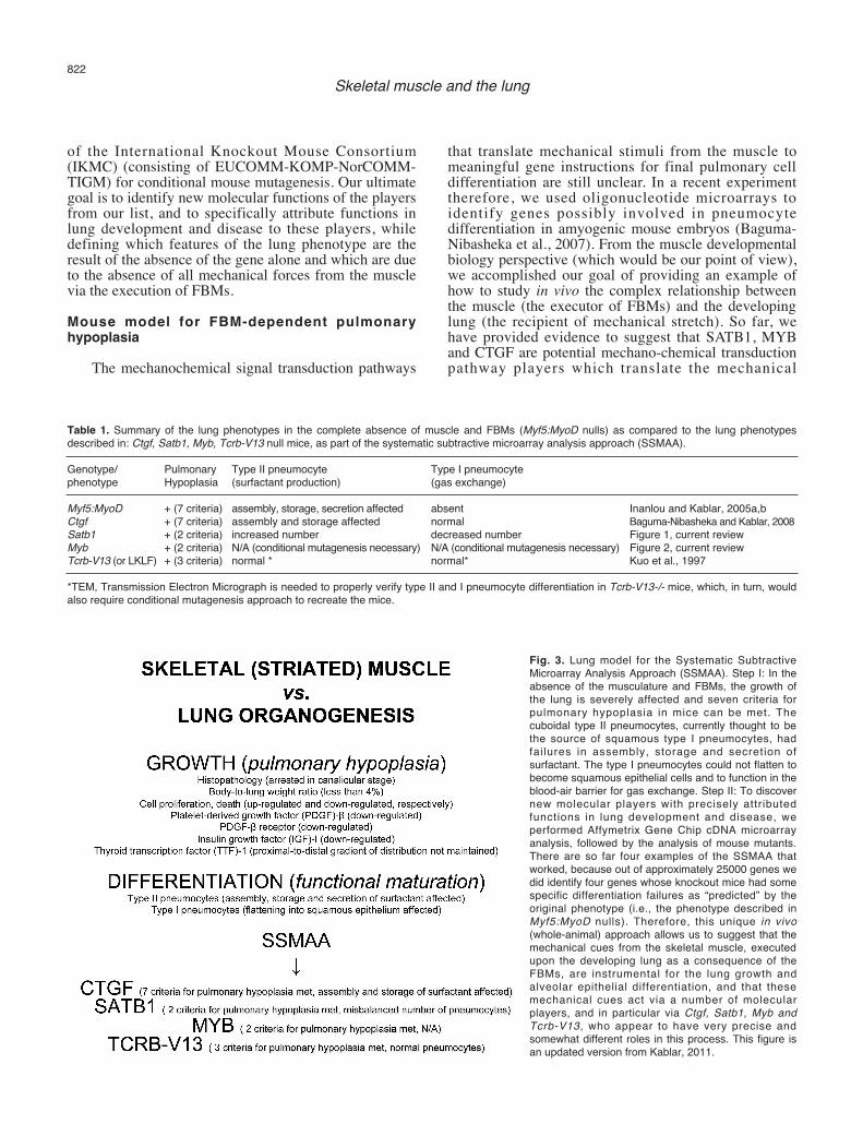
of the International Knockout Mouse Consortium(IKMC) (consisting of EUCOMM-KOMP-NorCOMM-TIGM) for conditional mouse mutagenesis. Our ultimategoal is to identify new molecular functions of the playersfrom our list, and to specifically attribute functions inlung development and disease to these players, whiledefining which features of the lung phenotype are theresult of the absence of the gene alone and which are dueto the absence of all mechanical forces from the musclevia the execution of FBMs.Mouse model for FBM-dependent pulmonaryhypoplasia
The mechanochemical signal transduction pathways
that translate mechanical stimuli from the muscle tomeaningful gene instructions for final pulmonary celldifferentiation are still unclear. In a recent experimenttherefore, we used oligonucleotide microarrays toidentify genes possibly involved in pneumocytedifferentiation in amyogenic mouse embryos (Baguma-Nibasheka et al., 2007). From the muscle developmentalbiology perspective (which would be our point of view),we accomplished our goal of providing an example ofhow to study in vivo the complex relationship betweenthe muscle (the executor of FBMs) and the developinglung (the recipient of mechanical stretch). So far, wehave provided evidence to suggest that SATB1, MYBand CTGF are potential mechano-chemical transductionpathway players which translate the mechanical
822Skeletal muscle and the lung
Fig. 3. Lung model for the Systematic SubtractiveMicroarray Analysis Approach (SSMAA). Step I: In theabsence of the musculature and FBMs, the growth ofthe lung is severely affected and seven criteria forpulmonary hypoplasia in mice can be met. Thecuboidal type II pneumocytes, currently thought to bethe source of squamous type I pneumocytes, hadfailures in assembly, storage and secretion ofsurfactant. The type I pneumocytes could not flatten tobecome squamous epithelial cells and to function in theblood-air barrier for gas exchange. Step II: To discovernew molecular players with precisely attributedfunctions in lung development and disease, weperformed Affymetrix Gene Chip cDNA microarrayanalysis, followed by the analysis of mouse mutants.There are so far four examples of the SSMAA thatworked, because out of approximately 25000 genes wedid identify four genes whose knockout mice had somespecific differentiation failures as “predicted” by theoriginal phenotype (i.e., the phenotype described inMyf5:MyoD nulls). Therefore, this unique in vivo(whole-animal) approach allows us to suggest that themechanical cues from the skeletal muscle, executedupon the developing lung as a consequence of theFBMs, are instrumental for the lung growth andalveolar epithelial differentiation, and that thesemechanical cues act via a number of molecularplayers, and in particular via Ctgf, Satb1, Myb andTcrb-V13, who appear to have very precise andsomewhat different roles in this process. This figure isan updated version from Kablar, 2011.
Table 1. Summary of the lung phenotypes in the complete absence of muscle and FBMs (Myf5:MyoD nulls) as compared to the lung phenotypesdescribed in: Ctgf, Satb1, Myb, Tcrb-V13 null mice, as part of the systematic subtractive microarray analysis approach (SSMAA).
Genotype/ Pulmonary Type II pneumocyte Type I pneumocytephenotype Hypoplasia (surfactant production) (gas exchange)
Myf5:MyoD + (7 criteria) assembly, storage, secretion affected absent Inanlou and Kablar, 2005a,bCtgf + (7 criteria) assembly and storage affected normal Baguma-Nibasheka and Kablar, 2008Satb1 + (2 criteria) increased number decreased number Figure 1, current reviewMyb + (2 criteria) N/A (conditional mutagenesis necessary) N/A (conditional mutagenesis necessary) Figure 2, current reviewTcrb-V13 (or LKLF) + (3 criteria) normal * normal* Kuo et al., 1997
*TEM, Transmission Electron Micrograph is needed to properly verify type II and I pneumocyte differentiation in Tcrb-V13-/- mice, which, in turn, wouldalso require conditional mutagenesis approach to recreate the mice.

823
Fig. 4. Distribution of SATB1, MYB and CTGF in human lung development corresponds to their function in mice. A, B. During the 40th week, SATB1(Santa Cruz goat polyclonal against SATB1, 40 µg/ml) is contained in the alveolar epithelial cells of the normal lung (A, asterisks) and in a very lownumber of only mesenchymal cells of the hypoplastic lung (B). C, D. At the same age, MYB (Santa Cruz rabbit polyclonal against MYB, 40 µg/ml) isvisible in both the epithelial and the mesenchymal cells of the normal lung (C), while in the hypoplastic lung (D) MYB is present in only a small numberof mesenchymal cells. E, F. Similarly, CTGF (Santa Cruz goat polyclonal against CTGF, 40 µg/ml) is present in both the epithelial and themesenchymal cells of the normal lung (E) and essentially abolished in the hypoplastic lung (F). Controls for immunostaining were the negative resultswhen SATB1, MYB and CTGF antibodies were used on the appropriate mouse mutants. x 400.

information from the muscle into the meaningful cellsignal in the alveolar epithelium. Additional evidence forthis statement could be provided by an in vitroexperiment in which stress is applied to either a singlefetal alveolar epithelial cell or a sheet of cells, whilevarious molecules, and in particular SATB1, aremonitored to see in what way type II-to-I pneumocytetransition depends on this particular mechanical cue andits transducing molecules.
Meanwhile, we propose here a mouse model forFBM-dependent lung hypoplasia employing SSMAA(Fig. 3). In the absence of the respiratory musculatureand FBMs, the growth of the lung is severely affected,showing seven criteria for pulmonary hypoplasia in mice(Inanlou and Kablar, 2005a,b). Additionally, veryspecific differentiation defects of the alveolar epitheliumwere found in the hypoplastic lung. For example, thecuboidal type II pneumocytes, currently thought to bethe source of type I pneumocytes, had failures inassembly, storage and secretion of surfactant, while thetype I pneumocytes could not flatten to function in theblood-air barrier for gas exchange. To discover newmolecular players with precisely attributed functions inlung development (i.e., differentiation of the type I andII alveolar epithelial cells) and disease (i.e., pulmonaryhypoplasia), we performed Affymetrix Gene Chip cDNAmicroarray analysis, followed by the analysis of mousemutants. In fact, Ctgf, Satb1, Myb and Tcrb-V13 are fourexamples of successful SSMAA, because, out ofapproximately 25000 genes in the Affymetrix GeneChip, we (and others, for Tcrb-V13) identified four geneswhose knockout mice had specific differentiationfailures “predicted” by the original mouse phenotype(i.e., the phenotype described in Myf5:MyoD nulls).Moreover, defining which features of the phenotypewere the result of the absence of the individual genes(e.g., Ctgf, Satb1, Myb, Tcrb-V13) and which were dueto the general absence of mechanical forces, as seen inthe muscleless Myf5:MyoD nulls, was another advantageof this approach. An illustration of this approach’sadvantage is shown in Table 1.
Further, we believe that an approach analogous tothis one would be successful for the analysis of themolecular basis of the interface between various othertissues and the skeletal muscle. In fact, by employingthis approach, we have continued to analyze thedevelopment of various tissues and organs of the fetal orembryonic body that were affected by the absence of theskeletal musculature, such as the retina (Baguma-Nibasheka and Kablar, 2009a,b), crista ampullaris of theinner ear (Rot and Kablar, 2010), the palate (Rot-Nikcevic et al., 2006), temporomandibular joint (Rot-Nikcevic et al., 2007) and motor neurons innervatinglimb and back musculature (reviewed by Kablar, 2011). Preliminary human studies and future directions
The data discovered in mice using mouse
mutagenesis and the battery of phenotypic analysisapproaches will eventually deliver a precise map offunctions of various molecules. However, the function ofvarious molecules discovered in mice as models ofhuman diseases will have to be examined employinghuman material, adult and embryonic, normal anddiseased. So far, there are several large scale projectsthat are trying to provide information on the distributionpattern of various molecules employing human material.For example, The Human Protein Atlas, http://www.proteinatlas.org, Uppsala University, containsinformation about the distribution pattern of a number ofmolecules in normal adult human tissues and also inneoplastic tissues. Unfortunately, this Atlas does not yetcontain information on protein distribution pattern inembryonic, fetal, neonatal normal and diseased material.As a collaborative effort of our two laboratories (inCanada and Croatia), our intention is to increase theamount of information on molecules’ distributionpatterns, employing human embryonic, fetal andneonatal normal and diseased archival materials.
For the purposes of this theme, on the role of FBMsin lung development, we would like to stress that FBMsin normal human fetuses are an ultra-sound measure ofthe fetal wellbeing. There are several human conditionsthat may impair FBMs, such as: various causes of fetalhypoxemia and hypoglycemia, maternal alcohol,narcotics, sedatives consumption and smoking, in uteroinfections, etc (Harding, 1997). It is impossible to obtaina “clean” experimental system in humans, since animpairment or absence of FBMs often comes with otherfactors associated with pulmonary hypoplasia, such aspremature rupture of fetal membranes andoligohydramnios (Harding, 1997). However, variousdeformations of the human rib cage may be comparableto the mouse studies on the role of FBMs in our model.
For example, as a further follow up to ourmicroarrays (Baguma-Nibasheka et al., 2007), thedistribution pattern of the three molecules (SATB1,MYB and CTGF) with the newly discovered functions inlung development and disease in mice (Figs. 1 and 2 inthis review; Baguma-Nibasheka and Kablar, 2008), wasstudied employing human neonatal normal and diseasedarchival material (i.e., pulmonary hypoplasia caused bymechanical impairments of lung development). Weintended to verify if any of the three players have adistribution pattern in humans consistent with their rolein mice. In the human adult lung, MYB (strongpresence) and CTGF (moderate levels) are present in thealveolar epithelium, while SATB1 is not (The HumanProtein Atlas, http://www.proteinatlas.org, UppsalaUniversity). Here, in the human neonatal lungs(gestational age 40 weeks; archival material from theUniversity of Split Medical School, Croatia), we showthat during normal lung development, all threemolecules were present in the epithelial lining, whileMYB and CTGF were also visible in the mesenchyme(approximately 6% of the cells contained MYB or
824Skeletal muscle and the lung

CTGF, and many epithelial cells contained SATB1) (Fig.4). The hypoplastic lung was considerably smaller thanthe control, with a weight of 8g (left lung; lung-to-bodyweight ratio was 0.0081) before fixation, versus thenormal 25g. Histopathologically, the hypoplastic lungappeared to be arrested in the saccular to early alveolarstage. Alveoli were of a very small diameter and partlyatelectatic (unexpanded). There was evidence ofinterstitial hemorrhage and tissue edema, with a singleinflammatory focus (possibly aspiration pneumonia). Inthe hypoplastic lung, none of the three molecules weredetectable in the epithelium, and the presence of thethree molecules was restricted to the mesenchyme,where the number of cells containing the proteins wasextremely reduced (less than 0.3% of the mesenchymalcells contained any of the three molecules) (Fig. 4).Together, these data indicate that SATB1, MYB andCTGF, as in mice, may also have a role in human lungdevelopment and in the pathogenesis of pulmonaryhypoplasia. Similar preliminary data are available forfetal human lung (Gugic, Saraga-Babic and Kablar,unpublished data).Acknowledgements. Our grateful appreciation goes to Prof. IvanaKuzmic-Prusac (MD, PhD), from the University of Split School ofMedicine, for her pathological descriptions of the human archivaltissues. We are also grateful to Mary Ann Trevors, Asja Miletic andHeather Angka for their expert technical assistance. We thank Dr. DavidGaskin (Dalhousie University, Department of Pathology) for his help asa Fetal Pathologist. This work was funded by an operating grant fromthe National Science and Engineering Research Council of Canada(NSERC) (Grant Number 238726-01), and the infrastructure grants fromthe Canada Foundation for Innovation (CFI) and the Dalhousie MedicalResearch Foundation (DMRF) to BK. This work has also beensupported by the Ministry of Science, Education and Sports of theRepublic of Croatia (Grant Number 021-2160528-0507) to MSB.
References
Alvarez J.D., Yasui D.H., Niida H., Joh T., Loh D.Y. and Kohwi-Shigematsu T. (2000). The MAR-binding protein SATB1orchestrates temporal and spatial expression of multiple genesduring T-cell development. Genes Dev. 14, 521-535.
Baguma-Nibasheka M. and Kablar B. (2008). Pulmonary hypoplasia inthe connective tissue growth factor (Ctgf) null mouse. Dev. Dyn.237, 485-493.
Baguma-Nibasheka M. and Kablar B. (2009a). Abnormal retinaldevelopment in the Btrc null mouse. Dev. Dyn. 238, 2680-2687.
Baguma-Nibasheka M. and Kablar B. (2009b). Altered retinal celldifferentiation in the AP-3 delta mutant (Mocha) mouse. Int. J. Dev.Neurosci. 27, 701-708.
Baguma-Nibasheka M., Angka H.E., Inanlou M.R. and Kablar B. (2007).Microarray analysis of Myf5-/-:MyoD-/- hypoplastic mouse lungsreveals a profile of genes involved in pneumocyte differentiation.Histol. Histopathol. 22, 483-495.
Harding R. (1997). Fetal pulmonary development: the role of respiratorymovements. Equine Vet. J. Suppl. 24, 32-39.
Harding R., Hooper S.B. and Han V.K. (1993). Abolition of fetalbreathing movements by spinal cord transection leads to reductionsin fetal lung liquid volume, lung growth, and IGF-II gene expression.Pediatr. Res. 34, 148-153.
Inanlou M.R. and Kablar B. (2003). Abnormal development of thediaphragm in mdx:MyoD-/- 9th embryos leads to pulmonaryhypoplasia. Int. J. Dev. Biol. 47, 363-371.
Inanlou M.R. and Kablar B. (2005a). Abnormal development of theintercostal muscles and the rib cage in Myf5-/- embryos leads topulmonary hypoplasia. Dev. Dyn. 232, 43-54.
Inanlou M.R. and Kablar B. (2005b). Contractile activity of skeletalmusculature involved in breathing is essential for normal lung celldifferentiation, as revealed in Myf5-/-:MyoD-/- embryos. Dev. Dyn.233, 772-782.
Inanlou M.R., Baguma-Nibasheka M. and Kablar B. (2005). The role offetal breathing-like movements in lung organogenesis. Histol.Histopathol. 20, 1261-1266.
Inanlou M.R., Baguma-Nibasheka M., Keating M.-M. and Kablar B.(2006). Neurotrophins, airway smooth muscle and the fetalbreathing-like movements. Histol. Histopathol. 21, 931-940.
Kablar B. (2011). Role of skeletal muscle in the epigenetic shaping oforgans, tissues and cell fate choices. In: Epigenetics: Linkinggenotype and phenotype in development and evolution.Hallgrímsson B. and Hall B.K. (eds). University of California Press.San Francisco. pp 254-266.
Kitterman J.A. (1996). The effects of mechanical forces on fetal lunggrowth. Clin. Perinatol. 23, 727-740.
Kuo C.T., Veselits M.L., Barton K.P., Lu M.M., Clendenin C. and LeidenJ.M. (1997). The LKLF transcription factor is required for normaltunica media formation and blood vessel stabilization during murineembryogenesis. Genes Dev. 11, 2996-3006.
Liggins C.C. (1984). Growth of the fetal lung. J. Dev. Physiol. 6, 237.Liu M. and Post M. (2000). Mechanochemical signal transduction in the
fetal lung. J. Appl. Physiol. 89, 2078-2084.Mammoto T. and Ingber D.E. (2010). Mechanical control of tissue and
organ development. Development 137, 1407-1420.Mombaerts P., Iacomini J., Johnson R.S., Herrup K., Tonegawa S. and
Papaioannou V.E. (1992). RAG-1-deficient mice have no mature Band T lymphocytes. Cell 68, 869-877.
Pelosi M., Marampon F., Zani B.M., Prudente S., Perlas E., Caputo V.,Cianetti L., Berno V., Narumiya S., Kang S.W., Musarò A. andRosenthal N. (2007). ROCK2 and its alternatively spliced isoformROCK2m positively control the maturation of the myogenic program.Mol. Cell Biol. 27, 6163-6176.
Rot I. and Kablar B. (2010). The influence of acoustic and static stimulion development of inner ear sensory epithelia. Int. J. Dev. Neurosci.28, 309-315.
Rot-Nikcevic I., Reddy T., Downing K.J., Belliveau A.C., HallgrímssonB., Hall B.K. and Kablar B. (2006). Myf5-/-:MyoD-/- amyogenicfetuses reveal importance of early contraction and static loading byskeletal muscle in mouse skeletogenesis. Dev. Genes Evol. 216, 1-9.
Rot-Nikcevic I., Downing K.J., Hall B.K. and Kablar B. (2007).Development of the mouse mandibles and clavicles in the absenceof skeletal myogenesis. Histol. Histopathol. 22, 51-60.
Sumner R., Crawford A., Mucenski M. and Frampton J. (2000). Initiationof adult myelopoiesis can occur in the absence of c-Myb whereassubsequent development is strictly dependent on the transcriptionfactor. Oncogene 19, 3335-3342.
825Skeletal muscle and the lung

Wang Y., Maciejewski B.S., Soto-Reyes D., Lee H.S., Warburton D. andSanchez-Esteban J. (2009). Mechanical stretch promotes fetal typeII epithelial cell differentiation via shedding of HB-EGF and TGF-alpha. J. Physiol. 587, 1739-1753.
Wirtz H.R. and Dobbs L.G. (2000). The effects of mechanical forces onlung functions. Resp. Physiol. 119, 1-17.
Accepted January 25, 2012
826Skeletal muscle and the lung
Copyright © 2022 FDOKUMEN