Skeletal muscle differentiation of human iPSCs meets ... - Nature
30
REVIEW ARTICLE OPEN Skeletal muscle differentiation of human iPSCs meets bioengineering strategies: perspectives and challenges Federica Iberite 1,2 ✉ , Emanuele Gruppioni 3 and Leonardo Ricotti 1,2 Although skeletal muscle repairs itself following small injuries, genetic diseases or severe damages may hamper its ability to do so. Induced pluripotent stem cells (iPSCs) can generate myogenic progenitors, but their use in combination with bioengineering strategies to modulate their phenotype has not been sufficiently investigated. This review highlights the potential of this combination aimed at pushing the boundaries of skeletal muscle tissue engineering. First, the overall organization and the key steps in the myogenic process occurring in vivo are described. Second, transgenic and non-transgenic approaches for the myogenic induction of human iPSCs are compared. Third, technologies to provide cells with biophysical stimuli, biomaterial cues, and biofabrication strategies are discussed in terms of recreating a biomimetic environment and thus helping to engineer a myogenic phenotype. The embryonic development process and the pro-myogenic role of the muscle-resident cell populations in co-cultures are also described, highlighting the possible clinical applications of iPSCs in the skeletal muscle tissue engineering field. npj Regenerative Medicine (2022)7:23 ; https://doi.org/10.1038/s41536-022-00216-9 INTRODUCTION Skeletal muscles enable voluntary movements and, consequently, a series of dynamic interactions between individuals and their surrounding environment. In vivo, skeletal muscles can self- regenerate: after traumas or other tissue damage, resident muscle stem cells named muscle satellite cells (MuSCs), are activated. MuSCs are located between the cell membrane and the basal lamina of myofibers, and their activation leads to cell proliferation and eventually exit at G1 phase. MuSCs then fuse to form terminally differentiated multinucleated myofibers, thereby restor- ing the pool of highly specialized cells in the tissue 1 . However, the skeletal muscle’s ability to self-repair may be impaired due to aging, genetic diseases 2 , or injuries with volumetric muscle loss (VML) 3 . In such cases, having healthy patient-specific muscle grafts developed in vitro and ready to be implanted in the impaired area would be highly desirable to restore tissue functionality. Fully- functional muscle grafts could also be exploited in lab-on-a-chip platforms for testing the efficacy, toxicity, and possible side effects of drugs, thereby significantly reducing (ideally eliminating) the need for animal sacrifices 4 . To obtain such muscle grafts in vitro, appropriate myogenic precursors in a three-dimensional (3D) construct need to be engineered, pushing their differentiation to match the morpho- logical and functional features of the native human muscle tissue. This is thus the objective of skeletal muscle tissue engineering, which aims to harness the knowledge derived from studying embryogenesis processes, and partly reproducing them in vitro. Induced pluripotent stem cells (iPSCs) were generated for the first time in 2006 by Shinya Yamanaka 5 and marked a crucial milestone in the field of biomedical sciences. These cells exhibit both transcriptional and epigenetic signatures similar to those of embryonic stem cells (ESCs), and thus they open up exciting scenarios for tissue engineering. Using human iPSCs, it is theoretically possible to create tissues or organs with patient- derived cells, thus eliminating immunogenicity issues. Furthermore, patient-specific tissues/organs-on-a-chip can be created, on which drugs can be tested in a customized way. The most effective and safest drug could be first tested on the custom patient-reflective chip, before administering it to the subject. Consequently, iPSCs also have an enormous potential in the field of skeletal muscle tissue engineering. Several biochemical protocols for the myogenic induction of iPSCs have been proposed. Some recent reviews analyze and compare the different approaches pursued 6–10 . However, in almost all of them, the focus is only on the biochemical stimuli affecting stem cell fate. Two main approaches have been described: (1) transgenic approaches by the overexpression of master myogenic transcrip- tion factors (e.g., MYOD1, PAX3/7), and 2) directed differentiation with stepwise induction using small molecules and growth/ differentiation factors. These two approaches focus on creating myogenic progenitors in vitro (from both healthy and unhealthy donors), which are often subsequently differentiated into myo- tubes. For clinical applications of these cells, in vitro differentiation of muscle progenitor terminal is performed to evaluate their myogenicity. This potential is then validated with xenotransplan- tation and the evaluation of the progenitors’ ability to create new myotubes and repopulate the stem cell niche (see Tables 1 and 2) 11–20 . Being able to repopulate the stem cell niche is key to ensuring long-term homeostasis in future tissue regeneration requirements. When the differentiation protocols are used to generate in vitro platforms for disease modeling and drug development, the in vitro induction of muscle progenitor differentiation is performed to create cultures of 2D myotubes, or three-dimensional artificial muscles with disease-specific hallmarks 21–24 . In some cases, their ability to depolarize or level of contractility is also tested 13,15,22,25,26 , to further validate their physiological relevance. Such information is useful also when using healthy cells for more advanced biomedical applications, such as the fabrication of iPSC-derived contractile biohybrid actuators 27 . 1 The BioRobotics Institute, Scuola Superiore Sant’Anna, 56127 Pisa (PI), Italy. 2 Department of Excellence in Robotics & AI, Scuola Superiore Sant’Anna, 56127 Pisa (PI), Italy. 3 Centro Protesi INAIL, Istituto Nazionale per l’Assicurazione contro gli Infortuni sul Lavoro, 40054 Vigorso di Budrio (BO), Italy. ✉ email: [email protected] www.nature.com/npjregenmed Published in partnership with the Australian Regenerative Medicine Institute 1234567890():,;
-
Upload
khangminh22 -
Category
Documents
-
view
4 -
download
0
Transcript of Skeletal muscle differentiation of human iPSCs meets ... - Nature

REVIEW ARTICLE OPEN
Skeletal muscle differentiation of human iPSCs meetsbioengineering strategies: perspectives and challengesFederica Iberite 1,2✉, Emanuele Gruppioni 3 and Leonardo Ricotti1,2
Although skeletal muscle repairs itself following small injuries, genetic diseases or severe damages may hamper its ability to do so.Induced pluripotent stem cells (iPSCs) can generate myogenic progenitors, but their use in combination with bioengineeringstrategies to modulate their phenotype has not been sufficiently investigated. This review highlights the potential of thiscombination aimed at pushing the boundaries of skeletal muscle tissue engineering. First, the overall organization and the keysteps in the myogenic process occurring in vivo are described. Second, transgenic and non-transgenic approaches for themyogenic induction of human iPSCs are compared. Third, technologies to provide cells with biophysical stimuli, biomaterial cues,and biofabrication strategies are discussed in terms of recreating a biomimetic environment and thus helping to engineer amyogenic phenotype. The embryonic development process and the pro-myogenic role of the muscle-resident cell populations inco-cultures are also described, highlighting the possible clinical applications of iPSCs in the skeletal muscle tissue engineering field.
npj Regenerative Medicine (2022) 7:23 ; https://doi.org/10.1038/s41536-022-00216-9
INTRODUCTIONSkeletal muscles enable voluntary movements and, consequently,a series of dynamic interactions between individuals and theirsurrounding environment. In vivo, skeletal muscles can self-regenerate: after traumas or other tissue damage, resident musclestem cells named muscle satellite cells (MuSCs), are activated.MuSCs are located between the cell membrane and the basallamina of myofibers, and their activation leads to cell proliferationand eventually exit at G1 phase. MuSCs then fuse to formterminally differentiated multinucleated myofibers, thereby restor-ing the pool of highly specialized cells in the tissue1.However, the skeletal muscle’s ability to self-repair may be
impaired due to aging, genetic diseases2, or injuries withvolumetric muscle loss (VML)3.In such cases, having healthy patient-specific muscle grafts
developed in vitro and ready to be implanted in the impaired areawould be highly desirable to restore tissue functionality. Fully-functional muscle grafts could also be exploited in lab-on-a-chipplatforms for testing the efficacy, toxicity, and possible side effectsof drugs, thereby significantly reducing (ideally eliminating) theneed for animal sacrifices4.To obtain such muscle grafts in vitro, appropriate myogenic
precursors in a three-dimensional (3D) construct need to beengineered, pushing their differentiation to match the morpho-logical and functional features of the native human muscle tissue.This is thus the objective of skeletal muscle tissue engineering,which aims to harness the knowledge derived from studyingembryogenesis processes, and partly reproducing them in vitro.Induced pluripotent stem cells (iPSCs) were generated for the
first time in 2006 by Shinya Yamanaka5 and marked a crucialmilestone in the field of biomedical sciences. These cells exhibitboth transcriptional and epigenetic signatures similar to those ofembryonic stem cells (ESCs), and thus they open up excitingscenarios for tissue engineering. Using human iPSCs, it istheoretically possible to create tissues or organs with patient-derived cells, thus eliminating immunogenicity issues.
Furthermore, patient-specific tissues/organs-on-a-chip can becreated, on which drugs can be tested in a customized way. Themost effective and safest drug could be first tested on the custompatient-reflective chip, before administering it to the subject.Consequently, iPSCs also have an enormous potential in the fieldof skeletal muscle tissue engineering.Several biochemical protocols for the myogenic induction of
iPSCs have been proposed. Some recent reviews analyze andcompare the different approaches pursued6–10. However, inalmost all of them, the focus is only on the biochemical stimuliaffecting stem cell fate.Two main approaches have been described: (1) transgenic
approaches by the overexpression of master myogenic transcrip-tion factors (e.g., MYOD1, PAX3/7), and 2) directed differentiationwith stepwise induction using small molecules and growth/differentiation factors. These two approaches focus on creatingmyogenic progenitors in vitro (from both healthy and unhealthydonors), which are often subsequently differentiated into myo-tubes. For clinical applications of these cells, in vitro differentiationof muscle progenitor terminal is performed to evaluate theirmyogenicity. This potential is then validated with xenotransplan-tation and the evaluation of the progenitors’ ability to create newmyotubes and repopulate the stem cell niche (see Tables 1 and2)11–20. Being able to repopulate the stem cell niche is key toensuring long-term homeostasis in future tissue regenerationrequirements.When the differentiation protocols are used to generate in vitro
platforms for disease modeling and drug development, the in vitroinduction of muscle progenitor differentiation is performed tocreate cultures of 2D myotubes, or three-dimensional artificialmuscles with disease-specific hallmarks21–24. In some cases, theirability to depolarize or level of contractility is also tested13,15,22,25,26,to further validate their physiological relevance. Such informationis useful also when using healthy cells for more advancedbiomedical applications, such as the fabrication of iPSC-derivedcontractile biohybrid actuators27.
1The BioRobotics Institute, Scuola Superiore Sant’Anna, 56127 Pisa (PI), Italy. 2Department of Excellence in Robotics & AI, Scuola Superiore Sant’Anna, 56127 Pisa (PI), Italy. 3CentroProtesi INAIL, Istituto Nazionale per l’Assicurazione contro gli Infortuni sul Lavoro, 40054 Vigorso di Budrio (BO), Italy. ✉email: [email protected]
www.nature.com/npjregenmed
Published in partnership with the Australian Regenerative Medicine Institute
1234567890():,;

Table1.
Tran
sgen
icap
proaches
forhuman
inducedpluripotentstem
cells
skeletal
muscle
differen
tiation.T
hebiblio
graphic
research
was
perform
eduntilDecem
ber
2020
usingPu
bMed
and
SCOPU
Sdatab
ases.T
hesearch
queriesusedfortitles
and/orab
stractsstartingfrom
2006
(yearofthefirstreportoniPSC
s)were[(inducedpluripotentstem
cells)AND(skeletalm
uscle)];[(skeletal
muscle
cell)
AND
(differen
tiation)AND
(inducedpluripotentstem
cells)];[(in
ducedpluripotentstem
cells)AND
(myo
gen
esis)OR(m
yogen
icdifferen
tiation)].R
eview
articles,b
ookch
apters,a
nd
conference
abstracts/pap
erswerenotincluded
.
Ref.
iPSC
origin
andlin
esCulture
conditions
inproliferation
Tran
sgen
ean
doverexp
ressionsystem
Myo
gen
icprogen
itor
derivationa
Term
inal
differen
tiationa
Culture
type
Myo
gen
iccell
selection
Functional
tests
Invivo
engraftmen
t
Darab
iet
al.,
2012
11
Fibroblasts
(IPRN13
.13,
IPRN14
.57)
Matrigel
®co
ating
withmTeSR
1PA
X7(dox-inducible
lentiviralsystem
)IM
DM,15
%FB
S,10
%HS,
1%ch
ickem
bryo
extract,50
µg/m
LAsA
c,4.5mM
MTG
(11days);
samemed
ium
asbefore
withdox
0.75
μg/m
L(4
days);
sorted
forPA
X7+
(GFP
+);samemed
ium
asbefore
withdox
0.75
μg/m
Lan
dhuman
FGF2
(5ng/m
L)(2
wee
ks)
Marke
rsGE:
T,MYO
D1
PE:M
RFs,C
D29
/44,
CD56
/63/10
5,CDH15
,ITGA7
logDMEM
,5%
HS
(7days)
Marke
rsGE:
DMD,M
YHPE
:MYH,M
YOG
EBsan
dthen
adheren
tProgen
itor
purification(EBs):
FACSforiPAX7+
(GFP
+)
n/a
Celltype:
myo
gen
icprogen
itors
iPAX7+
Endpoint:2months
Outco
me:
successful
engraftmen
twithfibers
human
DMD+
and
LMNA.Increase
intetanic,a
bsolute,a
nd
specificforce,
butno
influen
ceonfatigue
tests.Notumor
form
ationafter
46wee
ks(in
jected
inTA
musclesofNSG
mice)
Tedesco
etal.,
2012
12
Fibroblast
and
myo
blasts
iMEFswithKO
DMEM
;25
%KO
SR,
2mM
L-glut,1mM
Napyr,1
00IU
mL
pen
,100
mg/m
Lstrep,1
%NEA
A,0
.2mM
2-ME,
10ng/
mlhuman
FGF2
MYO
D1(tam
oxifen-
inducible
lentiviralsystem
)Gen
erationof
mesan
gioblast-likecells
(HIDEM
s):Matrigel
®
coatingwithα-MEM
,pen
(100
IU/m
L),strep
(100
mg/m
L),10
%FB
S,1%
NEA
A,0
.2%
2-ME
(14days);M
egaC
ell
DMEM
(7days)
Marke
rsGE:
n/a
PE:C
D44
,CD13
,CD14
6,PD
GFR
A,MYO
D1
4OH-tam
oxifenor
stan
dardtamoxifen
(5days)
Marke
rsGE:
MYO
D1,
MYO
G,
SGCA
PE:M
YH,M
YOD1
Adheren
tProgen
itor
purification:FACS
forSS
EA1-
n/a
Celltype:
HIDEM
sfrom
LGMD2D
iPSC
sGFP
+En
dpoint:1month
Outco
me:
5–7%
cell
survival,SG
CA+fibers
arepresentan
dDPC
was
reco
nstituted(fibers
SGCB+/SGCG+).
Increase
intetanic
force
exvivo
.Rep
opulationof
alkalin
ephosphatase+
pericytepool.(injected
inTA
musclesof
Sgca
null /scid/
beigemice)
Tanakaet
al.,
2013
13
Fibroblasts(201
B7,
253G
1,25
3G4)
Collagen
Ior
Matrigel
®co
ating
withprimateES
med
ium
with4ng/
mLFG
F2
MYO
D1(dox-inducible
PiggyB
actran
sposon-Tet-
ON
system
)
Collagen
IorMatrigel
®
coating;primateES
med
ium,10
µM
ROCK
inhibitor(1
day),ad
ded
1µg/m
Ldox(1
day);α-
MEM
,5%
KOSR
,50mU/
Lpen
/50mg/L
strep,
and10
0mM
2-ME
(5days)
Marke
rsGE:CK
M,M
YOD1,MEF2C
PE:n
/a
DMEM
with5%
HS,
50mU/L
pen
,50mg/
Lstrep,10ng/m
LIGF-
1,2mM
L-glutan
d10
0mM
2-ME(7
days)
Marke
rsGE:
MYO
G,D
MD,
microarray
PE:D
MD,
MYH,A
CTA
1
Adheren
tFA
CSforiM
YOD1
+(m
Cherry+)
ContractionuponES
tat
day
14(100
V,3ms,
1Hz)
Celltype:
myo
gen
icprogen
itors
(day
6of
differen
tiation)
Endpoint:28
days
Outco
me:
cells
displayedfusion
potential
andthe
expressionofhuman
DMDan
dhuman
SYNE1
(injected
inTA
muscles
ofNOD/Scid-DMD
mice)
Abujarour
etal.,20
1424
FTc01-C1an
dFT
c01-C2
Matrigel
®co
ating
withDMEM
/F12
,20
%KO
SR,1
%NEA
A,2
mM
L-glut,
100mM
2-ME,
10ng/m
LFG
F2
MYO
D1(dox-inducible
lentiviralsystem
)DMEM
,10
%FB
S,1µg/m
Ldox(4
days)
Marke
rsGE:
n/a
PE:n
/a
logDMEM
,5%
HS
(3days)
Marke
rsGE:
n/a
PE:M
YOD1,
MYH,M
YOG
Adheren
tn/a
n/a
n/a
Quattrocelli
etal.,20
1514
Fibroblastsan
dmesan
gioblasts
iMEFswithDMEM
/F1
2with20
%KO
SR,1
%pen
/strep,1
%L-glut,
1%NEA
A,0
.2%
2-
PAX3
/7(transien
toverexp
ressionby
pSP
ORT6
.1plasm
id)
DMEM
,2%
HS,
1%ITS,
100ng/m
Lnoggin,
1mM
TGF-βinhibitor
(10days)
Marke
rs
n/a
Adheren
tPD
GFR
A/B
+CD44
+ofthe
EBsbefore
differen
tiation
n/a
Celltype:
iPAX3/7cells
PDGFR
A/B+CD44
+En
dpoint:2months
Outco
me:
muscle
fiber
regen
erationwithSG
CB
F. Iberite et al.
2
npj Regenerative Medicine (2022) 23 Published in partnership with the Australian Regenerative Medicine Institute
1234567890():,;

Table
1continue
d
Ref.
iPSC
origin
andlin
esCulture
conditions
inproliferation
Tran
sgen
ean
doverexp
ressionsystem
Myo
gen
icprogen
itor
derivationa
Term
inal
differen
tiationa
Culture
type
Myo
gen
iccell
selection
Functional
tests
Invivo
engraftmen
t
ME,
and
5ng/m
LFG
F2GE:
T,MEO
X1,T
BX5,
PAX3
/7,K
DR
PE:M
YH2
expressionin
Sgcb
null /
Rag
2null /
γcnullmicean
dDMD
expressionin
dystrophic
mouse
Shojiet
al.,
2015
25
Fibroblasts
iMEFswithprimate
ESmed
ium
with
4ng/m
LFG
F2
MYO
D1(dox-inducible
PiggyB
actran
sposon-Tet-
ON
system
)
Matrigel
®orco
llagen
Ico
ating,2
0%KO
SRreplacemen
tmed
ia,
100μg/m
Lneo
mycin
sulphate(1
day);20
%KO
SRreplacemen
tmed
ia,1μg
/mLdox
(1day);5%
KOSR
/α-
MEM
med
ia,d
ox,
2-ME
(5days)
Marke
rsGE:
MYO
D1,
MYO
G,
DMD,C
KM,T
PM2
PE:n
/a
DMEM
,2%
HS
(7days)
Markers
GE:
DMD,T
PM2
PE:A
CTA
1,CKM,
MYH,u
ltrastructure
Adheren
tMyo
gen
icprogen
itors:
FACSforiM
YOD1
+(m
Cherry+)
DetectionofCa2
+
influxuponES
t(12V,
50ms,0.2Hz)
(day
9ofdifferen
tiation)
n/a
Lenzi
etal.,
2016
26
Fibroblasts
hES
C-qualified
Matrigel
®co
ating
withNutristem-XF
MYO
D1(dox-inducible
PiggyB
actran
sposon-Tet-
ON
system
)
DMEM
/F12
with
GlutaMAXTM,2
0%KO
SR,1
XNEA
A,
100U/m
Lpen
,100
μg/
mLstrep,0
,1mM
2-ME,
(5days);200
ng/m
Ldox
(2days)
Marke
rsGE:
TPE
:n/a
Skeletal
Muscle
Cell
Differen
tiation
Med
ium
(Promocell),
100U/m
Lpen
,10
0μg
/mLstrep,
200ng/m
Ldox
(5days)
Marke
rsGE:
DMD,M
YOD1,
MYO
G,M
YH,M
EF2C
PE:M
YH2,
MYO
G
Adheren
tn/a
Patch-clump
reco
rdingsofACh-
evokedcu
rren
ts,a
nd
intracellularCa2
+
releasewithACh
stim
ulations
n/a
Uch
imura
etal.,20
1751
414C
2,40
9B2
Matrigel
®co
ating
withStem
Fit
AK02
N,0.5%
pen
/strep
MYO
D1(tetracycline-
inducible
system
)Stem
FitAK02
N,0.5%
pen
/strep
(1day);
primateES
Cellmed
ia,
0.5%
pen
/strep
(1day);
samemed
ium
asbefore,1
μg/m
Ldox(1-
2days);α
-MEM
,0.5%
pen
/strep
,5%
KSR
,20
0μM
2-ME,
1μg
/mL
dox(6–7days)
Marke
rsGE:MYO
D1,MYO
G,M
YHPE
:MYH2,
MYO
G
DMEM
,0.5%
pen
/strep,2
mM
L-glut,
200μM
2-ME,
5%HS,
10ng/m
LIGF-1,
5μM
SB43
1542
(2-3
days);
DMEM
,0.5%
pen
/strep,2
mM
L-glut,
200μM
2-ME,
2%HS,
10ng/m
LIGF-1,
5μM
SB43
1542
(7days)
Marke
rsGE:
n/a
PE:M
YH2
Adheren
tn/a
n/a
n/a
Sato
etal.,
2016
204
n/d
iMEFswithDMEM
/F1
2,20
%KO
SR,1
%GlutaMAXTM,0.01%
2-ME,
NEA
A,FG
F2
MYO
D1co
nstitutive
expression(le
ntiviral
system
withEEF1
A1
promoter)
Collagen
Ico
ating,α
-MEM
,5%
KOSR
(7–10
days)
Marke
rsGE:
PAX7
/3,M
YOD1,
PE:n
/a
Marke
rsGE:
MYH
2,Ach
receptor
PE:M
YH1,
ultrastructure
Adheren
tn/a
n/a
n/a
Rao
etal.,
2018
15
H9,
TRiPS,
GM25
2564
6,an
dFu
cci
Matrigel
®co
ating
withE8
med
ium
PAX7
(dox-inducible
lentiviralsystem
)Matrigel
®co
ating,w
ith
E6med
ia,10
µM
CHIR99
021(2
days);E
6med
ia,1µg/m
Ldox
(18days)
Marke
rsGE:
MYH
sPE
:MYO
D1,
PAX3,
MYF5
,MYO
G,
sarcomeric
α-actinin,PAX7
logDMEM
,10
%FB
S,Fetuin
(500
X),hEG
F(100
0X),DE(100
0X),
pen
(100
unit/m
L),
strep(50µg/m
L)until
80%
confluen
ce(for
2Dcu
lture)or4days
(for3D
culture);
logDMEM
,N-2
Supplemen
t(100
X),
pen
G(100
unit/m
L)(~
2wee
ks)
Adheren
tvs
3DMyo
gen
icprogen
itors:
FACSforiPAX7+
(GFP
+)
EStat
20%
stretch
(40V/cm,10
ms):
twitch
forceper
cross-
sectional
area
of0.8
mN/m
m2
Ca2
+tran
sien
tsreco
rdingafter1,
2,4wee
ks(in
vitro)
uponES
t(10ms
pulse,
3V/m
m),an
dfrom
muscle
explants
Celltype:
progen
itors
(day
14of
differen
tiation)
Endpoint:15
days
Outco
me:
thebundles
werevascularizedan
dremained
functional
afterexplant(in
jected
dorsally
orin
TAmuscle
ofNSG
ornudemice)
F. Iberite et al.
3
Published in partnership with the Australian Regenerative Medicine Institute npj Regenerative Medicine (2022) 23

Table
1continue
d
Ref.
iPSC
origin
andlin
esCulture
conditions
inproliferation
Tran
sgen
ean
doverexp
ressionsystem
Myo
gen
icprogen
itor
derivationa
Term
inal
differen
tiationa
Culture
type
Myo
gen
iccell
selection
Functional
tests
Invivo
engraftmen
t
Marke
rsGE:
n/a
PE:sarco
meric
α-actinin,AChreceptor,
MYH,C
a2+
han
dlin
ggen
es
7-15
dayspost-
implantation
Selvaraj
etal.,
2019
46
Fibroblasts(PLZ
,TC
-113
3,MNP-12
0,MNP-11
9)
Matrigel
®co
ating
withmTeSR
1PA
X7(dox-inducible
lentiviralsystem
)IM
DM
basal
med
ium,
15%
FBS,
10%
HS,
1%pen
/strep
,1%
GlutaMAXTM,1
%KO
SR,
50µg/m
LAsA
c,4.5mM
MTG
(altogether
defi
ned
“myo
gen
icmed
ium”),1
0µM
CHIR99
021(2
days);
myo
gen
icmed
ium,
200nM
LDN19
3189
,10
µM
(SB43
1542
(1day);myo
gen
icmed
ium,1µg/m
Ldox
(3days);g
elatin
coating,m
yogen
icmed
ium,1µg/m
Ldox,
5ng/m
LFG
F2(4
days)
Marke
rsGE:MEO
X1,TCF
15,PAX3
,FO
XC2
PE:n
/a
KODMEM
,20%
KOSR
,1%
NEA
A,1
%GlutaMAXTM,1%
pen
/strep,1
0µM
SB43
1542
,10
µM
DAPT,1
0µM
DE,
10µM
PD03
2590
1,10
µM
Forsko
lin(5
days)
Marke
rsGE:
MYO
G,M
YOD1,
MYH
2,MYH
3,MYH
8,MYH
7PE
:MYH8,
TTN,D
ES
EBsthen
adheren
tMyo
gen
icprogen
itors:
FACSforiPAX7+
(GFP
+)
3Dco
nstruct
with
bovinefibrinogen
,thrombin,andgrowth
factorreduced
Matrigel
®3D
.ESt
at20
%stretch(10ms,
0.5Hz,or20
Hz):
twitch
forceof
0.4mN.
n/a
Gen
ean
dprotein
symbolsarein
capital
letters.Gen
esymbolsareitalicized
.a Culture
cond
ition
san
dmarkers,2-M
E2-mercaptoethan
ol,ACh
Acetylcholin
e,ACT
A1Actin
alpha1,
skeletal
muscle,A
sAcAscorbicacid,B
SABovineserum
albumin,C
DH15
M-cad
herin,C
HIR99021GSK
3βinhibitor,
CKM
Creatinekinasemuscle
isoform
,DAPT
Notchinhibitor,DEDexam
ethasone,
DES
Desmin,DMD
Dystrophin,do
xDoxycycline,
DPC
Dystrophin-associated
protein
complex,
EBsEm
bryoid
bodies,
EEF1A1
Eukaryotictran
slationelongationfactor1alpha1,
ESEm
bryonic
stem
,EStElectrical
stim
ulation,FA
CSFluorescen
ce-activated
cellsorting,FBSFetalbovineserum,FG
F2Fibroblast
growth
factor2,
GEGen
eexpression,GFP
Green
fluorescen
tprotein,higD
MEM
Highgluco
seDMEM
,HSHorseserum,IGF-1Insulin
e-likegrowth
factor-1,
IMDM
Isco
ve’sModified
Dulbecco’sMed
ium,iMEF
Irradiatedmouse
embryonic
fibroblasts,iMYO
D1InducibleMYO
D1,iPAX7
InduciblePA
X7,ITGA7α7
-integrin,ITS
Insulin
-transferrin-selen
ium,KDRKinaseinsertdomainreceptor,KO
DMEM
KnockoutDMEM
,KOSR
Knockoutserum
replacemen
t,LD
N193189
BMPtypeIinhibitor,L-glut
L-glutamine,
LGMD2D
Limb-girdle
musculardystrophy2D
,LIFLeukemia
inhibitory
factor,LM
NALamin
A/C,logD
MEM
Low
gluco
seDMEM
,LY294002
Phosphoinositide
3-kinaseinhibitor,MEF2C
Myo
cyte
enhan
cerfactor2C
, MEO
X1Mesen
chym
ehomeo
box1,MRF
Muscle
regulatory
factor,MSG
N1Mesogen
in1,mTeSR
cGMPfeed
er-freemaintenan
cemed
ium
forhuman
ESCsan
diPSC
s,MTG
Monothioglycerol,MYH
Myo
sinheavy
chain,M
YOD1myo
blast
determinationprotein
1,MYO
GMyo
gen
in,n
/anotap
plicab
le,N
apyrSo
dium
pyruvate,n/d
Notdefi
ned
,NEA
ANon-essen
tialam
inoacid,
NSG
NOD
scid
gam
ma,
PAXPa
ired
box,
PD0325901MEK
/ERKpathway
inhibitor,PD
GFRA/B
Platelet-derived
growth
factorreceptoralpha/beta,
PEProtein
expression,penPe
nicillin,PSM
Presomitic
mesoderm,
SB431542
TGF-βinhibitor,SG
CA/B/G
α/β/γ-sarcoglycan,SSEA
1Stag
e-specificem
bryonic
antigen
1,strepStreptomycin,S
YNE1
Spectrin,T
Brachyury,TA
tibialis
anterio
r,TPM2Trop
omyosin2,
TTNtitin.
F. Iberite et al.
4
npj Regenerative Medicine (2022) 23 Published in partnership with the Australian Regenerative Medicine Institute

The main muscular diseases treated in clinical trials are geneticconditions such as muscular dystrophies and laminopathies, ortissue degeneration and VML due to injured or aged muscle. Someof the most studied muscular dystrophies are the Duchenne andBecker types, caused by mutations in the dystrophin (DMD) gene,which leads to a lack or a dysfunction of the related protein.Disease modeling with iPSCs can help treat these diseases, thoughthere are challenges such as the high degree of clinicalheterogeneity of the dystrophies28.Nevertheless, few papers have highlighted the importance of
biophysical stimuli for skeletal muscle differentiation7,29,30. In fact,during their embryonic development, cells are greatly influencedby their surroundings, which consist of complex interactionsbetween biochemical and mechanical stimuli, distributed in spaceand time31–34.To achieve a differentiated and functional skeletal muscle tissue
starting from iPSCs, a controlled promyogenic environment thusneeds to be created. This environment is not only ensured by thebiochemical components, but also by a complex set of stimuli,resembling the ones available in an in vivo microenvironment.This review is organized as follows. Section “Skeletal muscle
tissue embryonic development and architecture” provides anoverview of skeletal muscle development and differentiation,along with a description of the macroscopic and microscopicstructure of the skeletal muscle in vivo as well as the various celltypes. Section “Methods to induce skeletal muscle differentiationin iPSCs” details the two main methods for iPSC myogenicinduction along with the main state of the art protocols and theirin vivo application (Table 1 and Table 2).Section “Biophysical stimulations for iPSC skeletal muscle
differentiation” describes the biophysical stimuli that have beenapplied or could be applied to iPSCs to pursue skeletal muscletissue engineering. Section “Challenges in the clinical translationof iPSC-derived skeletal muscle”, the challenges for iPSC transla-tion in clinical settings are described. Finally, the section “Co-culture of skeletal muscle cells with muscle-resident phenotypes”focuses on multicellular cultures and the combinations of skeletalmuscle cells with different muscle-resident cell types.
SKELETAL MUSCLE TISSUE EMBRYONIC DEVELOPMENT ANDARCHITECTURESkeletal muscle development during embryogenesisMyofibers mainly derive from the mesoderm, which is one of thethree germ layers created between the ectoderm and theendoderm during gastrulation, preceding the neural tube forma-tion. The mesoderm undergoes a process of specification on themediolateral axis, thanks to the action of Noggin and bonemorphogenetic protein (BMP) signaling on the same axis. Thisthen leads to the formation of the paraxial mesoderm, theintermediate mesoderm, and the lateral plate mesoderm (Fig. 1a).Skeletal muscle cell development is a multistep process
characterized by complex morphogen signaling, influences fromthe neural tube and notochord, and regulation of specific muscle-related genes. These processes are detailed in previous reviewarticles35–37 and are shown in Fig. 1b, c.Below is a summary of the key steps encountered by the
differentiating cells, steps which are also recapitulated duringin vitro iPSC differentiation.Myogenic precursors of the axial and limb muscles originate
from the segmented region of the paraxial mesoderm progenitorsexpressing the early mesoderm marker brachyury (T). Thesegments are called somites, which are transitory epithelialclusters of multipotent stem cells, located bilaterally to the neuraltube. The different regions of the paraxial mesoderm aredetermined by gradients of Wnt signaling factors, fibroblastgrowth factor (FGF2), and retinoic acid, whose key target genes
include mesogenin 1 (MSGN1) and T-box transcription factor 6(TBX6) which are both presomitic mesoderm markers. Cells of thedorsal somatic region, the dermomyotome, then start expressingtwo paired-box transcription factors, PAX3 and PAX7, under theactivation of Wnt signaling37.Myogenesis is then divided into three stages: (1) primary
myogenesis (with the formation of a scaffold of primary musclefibers from embryonic progenitors); (2) fetal secondary myogen-esis from PAX7+ cells (with the formation of MuSCs, and ofsecondary muscle fibers, fusing with each other and with primaryfibers); and (3) adult-type myogenesis (muscle adaptation toapplied stimuli and regeneration)38. Signals coming from thedorsal region of the neural tube (WNT1 and delta-like canonicalNotch ligand 1, DLL1), specifically from the neural crest cells,activate the expression of muscle-specific transcription factors (i.e.the muscle regulatory factors), such as myogenic factor 5 (MYF5),myoblast determination protein 1 (MYOD1), myogenin (MYOG),and myogenic factor 6 (MYF6, also known as muscle regulatoryfactor 4, MRF4). MYOD1 and MYF5 are markers of terminalspecification of the muscle lineage39, while MYOG controls theterminal differentiation of the myoblasts fusing with each otherand forming multinucleated myotubes.These primary myofibers, derived from dermomyotomal PAX3+
progenitors, start to express slow, embryonic, and perinatalmyosin heavy chain (MYH) isoforms (MYH7, MYH3, MYH8respectively) and myosin light chains35. In the secondarymyogenesis, the central dermomyotome loses its epithelialfeatures. PAX3+ cells then migrate towards the myotome, startexpressing PAX7, and fuse together, as well as with the primarymyofiber scaffold. They express fast MYH isoforms, such as MYH2(MyHC-2A), MYH1 (MyHC-2X/D), MYH4 (MyHC-2B) (Fig. 1c).Besides skeletal muscle cells, there are other cells that are key to
muscle development, and which help achieve a mature musclephenotype. Some of these cells have origins and timeframessimilar to the skeletal muscle cells. The sclerotome is derived fromthe ventromedial somites under the myotome, with the cellsundergoing an epithelial-mesenchymal transition and migratingventrally. The sclerotome has three main progenitor zones: (1) thesyndetome, which is located dorsally and that generates tendons;(2) the internal and lateral regions that form the joints, bones, andcartilage in the spine and the rib cage; and (3) the ventralposterior somites, endothelial precursor cells that form the dorsalaorta, the first intraembryonic blood vessel40.
Cell and tissue organizationSkeletal muscle tissue is mainly composed of elongated multi-nucleated myofibers, which are specialized skeletal muscle cells.However, several other cell populations are present throughoutthe tissue, and are essential for muscle development andfunctioning: progenitor cells, cells from the connective tissues,cells of the vascular network, adipogenic cells, immune cells, andmotor neurons. This section describes how these cells in adultmuscle tissue are organized, while Section “Co-culture of skeletalmuscle cells with muscle-resident phenotypes” gives an overviewof their embryonic development in relation to muscle cells, andreports the results of co-culture experiments.Myofibers are composed of packed myofibrils filling the whole
sarcoplasm, i.e. the myofiber cytoplasm, which is enclosed in themyofiber membrane, called the sarcolemma. Myofibrils run alongthe length of the myofiber and have a modular architecture: thesarcomeres are repeated longitudinally and intercalated bystructures called Z-disks.The sarcomere is the contractile unit, and by analyzing its
ultrastructure, the filaments of myosin sliding on the actinfilaments are observable, shortening the sarcomere and drawingnearer the two Z-disks. The sarcomere is composed of two halvesof the I-band at the two extremities, the band that surrounds the
F. Iberite et al.
5
Published in partnership with the Australian Regenerative Medicine Institute npj Regenerative Medicine (2022) 23

Table2.
Non-transgen
icap
proaches
forhuman
inducedpluripotentstem
cells
skeletal
muscle
differen
tiation.T
hebiblio
graphicresearch
was
perform
eduntilD
ecem
ber
2020
usingPu
bMed
and
SCOPU
Sdatab
ases.T
hesearch
queriesusedfortitles
and/orab
stractsstartingfrom
2006
(yearofthefirstreportoniPSC
s)were[(inducedpluripotentstem
cells)AND(skeletalm
uscle)];[(skeletal
muscle
cell)
AND
(differen
tiation)AND
(inducedpluripotentstem
cells)];[(in
ducedpluripotentstem
cells)AND
(myo
gen
esis)OR(m
yogen
icdifferen
tiation)].R
eview
articles,b
ookch
apters,a
nd
conference
abstracts/pap
erswerenotincluded
.
Ref.
iPSC
origin
andlin
esCulture
conditionsin
proliferation
Myo
gen
icprogen
itor
derivationa
Term
inal
differen
tiationa
Culture
type
Myo
gen
iccell
selection
Functional
tests
Invivo
engraftmen
t
Awayaet
al.,
2012
16
Fibroblasts
(01B
6,20
1B7,
253G
1,25
3G4)
iMEFswithDMEM
/F12
,20
%KO
SR,1
%NEA
A,
5mM
NAOH,10
0µM
2-ME,
2mM
L-glut,5ng/
mLFG
F2
0.1%
gelatin
coating,
DMEM
,ITS-X,N
EAA,
GlutaMAXTM,1
00µM
2-ME(14days)
Marke
rsGE:
PAX3
,MYF5
PE:n
/a
higDMEM
,10
%FC
S,5%
HS,
NEA
A,10
0µM
2-ME
(98days)
Marke
rsGE:
PAX3
/7,M
YF5,
MYO
D1,
MYO
G,D
ES,M
YH2
PE:M
YH
EBs
n/a
n/a
Celltype:
myo
gen
icprogen
itors
(day
49of
differen
tiation)
Endpoint:4,
12,a
nd24
wee
ksOutco
me:
detectionofhuman
LMNAin
thenuclei,an
dev
iden
ceofcellintegration
withthepresentmuscle
fibers.
PAX7+
cells
within
thelamina
rara
(injected
incardiotoxin-
treatedTA
musclesofNOD/Shi-
scid/IL-2R
γnullmice)
Saku
raiet
al.,
2012
131
Fibroblasts
(201
B7,
253G
4)n/a
Collagen
Ico
ating,α
-MEM
,5%
KOSR
,0.1mM
2-ME(6
days);FACS
purification;SF
-O3,
5mM
LiCl,10
ng/m
LIGF-1,
10ng/m
LHGF,
10ng/m
LFG
F2(3
days)
Marke
rsGE:
T,PD
GFRA,T
BX6,
MESP2,K
DR
PE:n
/a
SF-O
3,5mM
LiCl,10
ng/m
LIGF-1,
5mM
SB43
1542
(4days);S
F-O3,
10ng/m
LIGF-1,
5mM
SB-431
542,
10ng/m
LHGF(7
days)
Marke
rsGE:
n/a
PE:M
YH
Adheren
tMyo
gen
icprogen
itors:FA
CSfor
PDGFR
A+/KDR‐
n/a
n/a
Hosoyama
etal.,20
1463
Fibroblasts(IM
R90
)Matrigel
®co
ating
withmTeSR
1EZ
spherein
Stem
line
med
ium,10
0ng/
mLFG
F2,
100ng/m
LEG
F,5ng/
mLhep
arin
sulfa
te(42–
84days).
Marke
rsGE:
PAX3
,PAX7
,low
MYF5,
low
MYO
D1,
low
MYO
GPE
:PAX3,
MYO
D1,
MYO
G,M
YH
Poly-L-lysine/laminin
or
Matrigel
®co
ating,D
MEM
,2%
B27
or2%
HS
Marke
rsGE:
n/a
PE:M
YOD1,
MYO
G,M
YH
EZspheres
n/a
n/a
n/a
Chal
etal.,
2016
62
hiPS1
1a,
NCRM1,
NCRM5
Matrigel
®co
ating
withmTeSR
1Matrigel
®co
ating,
DMEM
/F12
,20
IU/m
Lpen
,0.02mg/m
Lstrep
(2%),1%
NEA
A,1
%ITS,
3μM
CHIR99
021,
0.5μM
LDN19
3189
(3days);sam
emed
ium
asbefore
with20
ng/
mLFG
F2(3
days);
DMEM
/F12
,15
%KO
SR,
1%NEA
A,2%
pen
/strep,0
.1mM
2-ME,
10ng/m
LHGF,2ng/m
LIGF-1,
20ng/m
LFG
F2,
0.5μM
LDN19
3189
(2days)
DMEM
/F12
,15
%KO
SR,1%
NEA
A,2%
pen
/strep
,0.1mM
2-ME,
2ng/m
LIGF-1
(4days);sam
emed
ium
asbefore
with10
ng/m
LHGF
(18days);D
MEM
/F12
,1%
ITS,
2%pen
/strep
,1%
N-2
Supplemen
t,1%
L-glut
Marke
rsGE:
n/a
PE:M
YOD1,
MYO
G,PAX7,
MYH2,
TTN,D
MD
Adheren
tn/a
n/a
n/a
F. Iberite et al.
6
npj Regenerative Medicine (2022) 23 Published in partnership with the Australian Regenerative Medicine Institute

Table
2continue
d
Ref.
iPSC
origin
andlin
esCulture
conditionsin
proliferation
Myo
gen
icprogen
itor
derivationa
Term
inal
differen
tiationa
Culture
type
Myo
gen
iccell
selection
Functional
tests
Invivo
engraftmen
t
Marke
rsGE:
n/a
PE:T
BX6,
PAX3
Iovinoet
al.,
2016
61
Fibroblasts
Matrigel
®co
ating
withmTeSR
1ST
EMDiff
Apel
med
ium
(STE
MCELL
Tech
nologies),10
ng/
mLFG
F2,0
.5μM
BIO,
20M
forsko
lin(7
days;
FGF2
,BIO,a
nd
forsko
linad
ded
atdays
1,3,
and5)
Marke
rsGE:
PAX7
,MYF5,
MYO
D1
PE:n
/a
Matrigel
®co
ating,D
MEM
,2%
HS(29days)
Marke
rsGE:
MYO
G,M
YH2
PE:n
/a
EBsan
dthen
adheren
tn/a
n/a
n/a
Wuet
al.,
2016
205
DF1
9-9,
WiCell
Matrigel
®co
atingwithE8
essential
med
ium
IMDM,10
%HS,
20%
FBS,
pen
/strep
,3μM
CHIR99
021(4
days);
samemed
ium
asbefore
without
CHIR99
021,
10ng/m
LFG
F2(8
days)
Marke
rsGE:
MYF5
PE:n
/a
DMEM
,2%
HS(5
days)
Marke
rsGE:
n/a
PE:M
YH2
EBsan
dthen
adheren
tMyo
gen
icprogen
itors:FA
CSfor
MYF5
+(GFP
+)
n/a
n/a
Sheltonet
al.,
2016
58
n/d
Matrigel
®co
atingwith
E8med
ium
Matrigel
®co
ating,E
6med
ium,10
µM
CHIR99
021(2
days);E
6med
ium
(10days)
Marke
rsGE:
T,MSG
N1,
TBX6
,PA
X3,M
EOX1
PE:T
Stem
Pro-34med
ium,
0.45
mM
MTG
,5µg/m
Lgen
t,2mM
L-glut,10
.7µg/
mLtran
sferrin,5ng/m
LFG
F2(10days);E
6med
ia(15days);D
MEM
/F12
,1%
ITS,
1%N-2
Supplemen
t,0.01
%gen
t(15days)
Marke
rsGE:
MYO
D1,
MYO
GPE
:PAX7,
MYH2
Adheren
tn/a
n/a
n/a
Swartz
etal.,
2016
60
Fibroblasts
Vitronectinco
atingwith
TeSR
-E8med
ium
IMDM/F12
,5mg/m
LBSA
,100
Xlip
ids,15
µg/
mLtran
sferrin,45
0µM
MTG
,7µg/m
Linsulin
,20
ng/m
LFG
F2,1
0µM
LY29
4002
,10
ng/mL
BMP4
,10
µM
CHIR99
021(36hours);
samemed
ium
asbefore
withoutBMP4
andCHIR99
021
(5.5
days);M
B-1,15
%FB
S(5
days)
Marke
rsGE:
T,PA
X3,T
BX6
PE:PAX3
DMEM
,2%
HS(10days);
DMEM
/F12
,1%
N-2
supplemen
t,1%
ITS
(7–10
days)
Marke
rsGE:
MYO
G,M
YOD1,
MYH
,PA
X3PE
:PAX7,
MYO
G,low
TUBB3,
MYH2,
DES
,TT
N
Adheren
tn/a
Spontaneo
us
contractionat
day
34(0.1–0.3
contractions/s)
n/a
Maffioletti
etal.,20
1821
NCRM1,
NCRM5,
A13
777
iMEFswithKO
DMEM
;25
%KO
SR,2
mM
L-glut,
1mM
Napyr,1
00IU
mL
pen
,100
mg/m
Lstrep,1
%NEA
A,0
.2mM
2-ME,
10ng/m
lFG
F212
Collagen
Ico
atingwith
SKM-01,
5%HS,
3µM
CHIR99
021,
2µM
Alk5
Inhibitor,10
ng/m
LEG
F,10
µg/m
Linsulin
,0.4µg/m
LDE,
200µM
AsA
c(10days);SKM-02,
5%HS,
10µg/m
Linsulin
,10
µg/m
LEG
F,
SKM-03,
10µg/m
Linsulin
,20
µg/m
Lonco
statin,50
nM
necrosulfo
nam
ide,
200µM
AsA
c(7
days)
Marke
rsGE:
n/a
PE:M
YH
3Dn/a
n/a
n/a
F. Iberite et al.
7
Published in partnership with the Australian Regenerative Medicine Institute npj Regenerative Medicine (2022) 23

Table
2continue
d
Ref.
iPSC
origin
andlin
esCulture
conditionsin
proliferation
Myo
gen
icprogen
itor
derivationa
Term
inal
differen
tiationa
Culture
type
Myo
gen
iccell
selection
Functional
tests
Invivo
engraftmen
t
20ng/m
LHGF,10
ng/
mLPD
GF,20
ng/m
LFG
F2,2
0µg/m
Lonco
statin,10
ng/m
LIGF-1,
2mM
SB43
1542
,20
0µM
AsA
c(8
days)
Marke
rsGE:
n/a
PE:PAX7,
DES
Sakai-
Takemura
etal.,20
1817
253G
4,20
1B7,
409B
2,45
4E2
iMatrix-51
1co
ating
withmTE
SR1
iMatrixco
ating,D
MEM
/F1
2,1%
ITS,
3μM
CHIR99
021,
0.5μM
LDN19
3189
(3days);
samemed
ium
asbefore
with20
ng/m
LFG
F2(3
days);DMEM
/F1
2,10
ng/m
LHGF,
2ng/m
LIGF-1,
20ng/
mLFG
F2,0
.5μM
LDN-
1931
89(2
days);
DMEM
/F12
,15
%KO
SR,
2ng/m
LIGF-1(4
days).
InductionofEZ
sphere
culture
inStem
line,
100ng/m
LFG
F2,
100ng/m
LEG
F,5µg/
mLhep
arin
sodium
salt
forat
differen
ttime
points
(6–10
wee
ks)63
Marke
rsGE:
T,TBX6
,PAX3
,PAX7
PE:M
YOD,PAX7,MYO
G
Collagen
coating,D
MEM
,10
%FB
S(4
wee
ks);DMEM
,10
%FB
S,10
μMSB
4315
42,
and/or10
μMDAPT
Marke
rsGE:
MRF4,
MYH
2,MSTN,
MYH
8,PA
X7PE
:MYH2,
MYO
G,PAX7
Combinationof
adheren
t2D
culture
andEZ
spheres
Myo
gen
icprogen
itors:FA
CSfor
CD57
−/CD10
8−/
CD27
1+/ERBB3+
n/a
Celltype:
CD57
−/CD10
8−/
CD27
1+/ERBB3+
Endpoint:1month
Outco
me:
myo
tubes
were
foundpositive
forLM
NA,
SYNE1
,DMD
(injected
inTA
musclesofNSG
-mdx4
Cvmice,
daily
injectionofSB
4315
42to
enhan
cecelldifferen
tiationfor
fourdays)
Vander
Wal
etal.,20
1818
Fibroblasts(8
healthy
donors)
iMEFswithDMEM
/F12
,20
%KO
SR,1
%NEA
A,1
%pen
/strep
/L-glut,2mM
2-ME,
20ng/m
LFG
F2
DMEM
/F12
,1%
ITS,
1%pen
/strep
/L-glut,
3.5µM
CHIR99
021
(5days);sam
emed
ium
asbefore
with20
ng/
mLFG
F2(14days)
Marke
rsGE:
n/a
PE:PAX7
DMEM
/F12
,1%
ITS,
1%pen
/strep/L-glut(16days)
Marke
rsGE:
RNAseq
PE:M
YH,TT
N,α
-ACTN
Adheren
tMyo
gen
icprogen
itors:FA
CSfor
c-MET
+/H
NK-1-
Spontaneo
us
contraction
Celltype:
myo
gen
icprogen
itors
Endpoint:1month
Outco
me:
cells
werefound
positive
forLM
NA,SYNE1
,DMD
andco
ntributedto
new
fiber
form
ation.Asm
allsubsetof
LMNA+cells
wereperivascu
lar
andPA
X7+
(injected
ininjured
TAmuscle
ofNSG
mice)
AlTanoury
etal.2
0206
7NCRM1withVe
nus
reporter
cassette
inPA
X7
Matrigel
®co
ating
withmTeSR
1Matrigel
®co
ating,
DMEM
/F12
,20
IU/m
Lpen
,0.02mg/m
Lstrep
(2%),1%
NEA
A,1
%ITS,
3μM
CHIR99
021,
0.5μM
LDN19
3189
(3days);sam
emed
ium
asbefore
with20
ng/
mLFG
F2(3
days);
DMEM
/F12
,15
%KO
SR,
1%NEA
A,2%
pen
/strep,0
.1mM
2-ME,
10ng/m
LHGF,2ng/m
LIGF-1,
20ng/m
LFG
F2,
0.5μM
LDN19
3189
(2days);D
MEM
/F12
,15
%KO
SR,1
%NEA
A,
2%pen
/strep
,0.1mM
2-ME,
2ng/m
LIGF-1
(4days);sam
emed
ium
Skeletal
muscle
growth
med
ium
(SkG
M-2,Lo
nza),
10µM
ROCKinhibitor
(1day);SkGM-2
(1-2
days);
DMEM
/F12
,2%
KSR
,1µM
Chiron,0.2%
pen
/strep
,1×
ITS(1-2
wee
ks)
Marke
rsGE:
n/a
PE:PAX7,
MYH2
Adheren
tMyo
gen
icprogen
itors
(dissociated
after
21daysof
differen
tiation):FA
CS
forPA
X7V
enus
n/a
Celltype:
PAX7V
enusmyo
gen
icprogen
itors
(day
21of
differen
tiation)
Endpoint:6–
8wee
ksOutco
me:
restorationofDMD
expression,co
ntributionto
new
fiber
form
ation(hLM
NA
+/hSY
NE1
+)an
drepopulation
ofstem
cellco
mpartm
ent
(hLM
NA+/PAX7+
)(in
jected
inTA
muscle
ofRag
2−/−
γc−/−
orNOD;R
ag1−/−
;Dmdmdx-5Cvmice)
F. Iberite et al.
8
npj Regenerative Medicine (2022) 23 Published in partnership with the Australian Regenerative Medicine Institute

Table
2continue
d
Ref.
iPSC
origin
andlin
esCulture
conditionsin
proliferation
Myo
gen
icprogen
itor
derivationa
Term
inal
differen
tiationa
Culture
type
Myo
gen
iccell
selection
Functional
tests
Invivo
engraftmen
t
asbefore
with10
ng/
mLHGF(9
days);
DMEM
/F12
,1%
ITS,
2%pen
/strep
,1%
N-2
Supplemen
t,1%
L-glut
Marke
rsGE:
n/a
PE:T
BX6,
PAX3,
MYO
D1,
MYO
G,PAX7,
MYH2,
TTN,D
MD
Baciet
al.,
2020
19
Pericytesan
dfibroblasts
Geltrex
matrixwith
E8med
ium
Geltrex
matrix,
E6med
ium,1
%ITS,
10µM
CHIR99
021(2
days);E
6med
ium,1%
ITS,
5mM
LiCl,10
ngFG
F2,1
0ng
IGF-1,
50µg/m
LEV
sfrom
MT(4
days);cell
splitting;co
llagen
Ico
ating,E
6med
ium,
1%ITS,
10ngFG
F2,
10ngIGF-1,
50µg/m
LEV
sfrom
MT(10days)
Marke
rsGE:
MSG
N1,
PAX3
,PAX7
,MYO
D1,
MYO
GPE
:n/a
E6med
ium,1%
ITS(10days)
Marke
rsGE:
MYO
G,M
YH8,
CKM,M
YHPE
:MYH2,
NCAM1,
MYO
D1,
MYO
G,M
YH2
Adheren
tn/a
n/a
Celltype:
pericyte-iPSC
-derived
muscularcells
(day
25ofdifferen
tiation)
Endpoint:20
days
Outco
me:
sufficien
ten
graftmen
tan
dintegration
into
regen
eratingmuscle
fibers
reve
aled
bythelabelingof
human
-derived
cells
by
immunofluorescen
ceag
ainst
human
-specificlamin
A/C
antibody(in
jected
inTA
muscle
ofSC
ID-Beige/SG
CAnull
LGMD2D
mice)
Heet
al.,
2020
20
Peripheral
blood
mononuclearcells
Matrigel
®co
ating
withmTeSR
1ST
EMDiff
Apel
med
ium
(STE
MCELL
Tech
nologies),10
ng/
mLFG
F2,0
.5mM
BIO,
20mM
forsko
lin(7
days)
Marke
rsGE:
n/a
PE:PAX7
Matrigel
®co
ating,D
MEM
,2%
HS(26days)
Marke
rsGE:
n/a
PE:PAX7,
MYF5
,MYH4,
DES
,DMD
EBsan
dthen
adheren
tn/a
n/a
Celltype:
EGFP
hiPSC
-derived
myo
gen
icprogen
itors
(day
14ofdifferen
tiation)
Endpoint:4,
8,an
d12
wee
ksOutco
me:
restorationofDMD
expression,co
ntributionto
new
fiber
form
ationan
drepopulationofstem
cell
compartm
ent(systemic
injectionorin
TAmuscle
of
mdxmice)
Gen
ean
dprotein
symbolsarein
capital
letters.Gen
esymbolsareitalicized
.a Culture
cond
ition
san
dmarkers,2
-ME2-mercaptoethan
ol,AsAcAscorbic
acid,B
IO6-bromoindirubin-3′-o
xime,
GSK
3βinhibitor,BM
PBonemorphogen
etic
protein,B
SABovineserum
albumin,C
HIR99021GSK
3βinhibitor,CK
MCreatinekinasemuscle
isoform
,DAPT
Notchinhibitor,DEDexam
ethasone,
DMDDystrophin,EBs
Embryoid
bodies,EG
FEp
idermal
growth
factor,EVsfrom
MTExtracellularvesicles
released
from
differen
tiated
myo
tubes,FA
CSFluorescen
ce-activated
cellsorting,F
BSFetalbovineserum,F
CSFetalcalfserum,FG
F2Fibroblast
growth
factor2,
GEGen
eexpression,gent
Gen
tamicin,GFP
Green
fluorescen
tprotein,HGFHep
atocyte
growth
factor,higD
MEM
Highgluco
seDMEM
,HSHorseserum,IGF-1Insulin
e-likegrowth
factor-1,
IMDM
Isco
ve’sModified
Dulbecco’sMed
ium,iMEF
Irradiated
mouse
embryonic
fibroblasts,ITSInsulin
-transferrin-selen
ium,KDRKinaseinsertdomainreceptor,KO
DMEM
KnockoutDMEM
,KOSR
knockoutserum
replacemen
t,LD
N193189
BMPtypeIinhibitor,L-glut
L-glutamine,LG
MD2D
limb-
girdle
musculardystrophy2D
,LMNALamin
A/C,p
latelet-derived
growth
factor,LY294002
Phosphoinositide3-kinaseinhibitor,MEO
X1Mesen
chym
ehomeo
box1,
MESP2
Mesoderm
posteriorbHLH
tran
scription
factor2,
MSG
N1Mesogen
in1,
mTeSR
cGMP
feed
er-freemaintenan
cemed
ium
forhuman
ESCsan
diPSC
s,MTG
Monothioglycerol,MYF5Myo
gen
icfactor5,
MYH
Myo
sin
heavy
chain,MYO
D1Myo
blast
determinationprotein
1,MYO
GMyo
gen
in,n
/aNotap
plicab
le,n
/dNotdefi
ned
,NEA
ANon-essen
tial
aminoacid,N
SGNODscid
gam
ma,
PAXPa
ired
box,
PDGFPlatelet-derived
growth
factor,PD
GFRA/B
Platelet-
derived
growth
factorreceptoralpha/beta,
PEProtein
expression,penPe
nicillin,SB431542
TGF-βinhibitor,SF-O3serum-freecu
lture
med
ium,SG
CAα-sarcoglycan,strepStreptomycin,TBrach
yury,TA
Tibialis
anterior,TBX6
T-boxtran
scriptionfactor6,
TTNTitin,TU
BB3Tu
bulin
beta3.
F. Iberite et al.
9
Published in partnership with the Australian Regenerative Medicine Institute npj Regenerative Medicine (2022) 23

Z-disk and is composed of actin filaments without myosin, and acentral A-band, containing both actin and myosin filaments. Titin(TTN), actinin (ACTN), and desmin (DES), which are three of theproteins in the Z-disk, help to associate the myofiber cytoskeletonwith the dystrophin-associated protein complex (DPC) and theα7β1 integrin (Fig. 2a). The DPC and the integrin are sarcoplasmicmolecular complexes connecting the sarcomere with the extra-cellular matrix (ECM), specifically the basement membrane.The basement membrane is part of the second most abundant
tissue in the SM from a volumetric viewpoint, which is theconnective tissue. Muscle connective tissues are important formuscle structural integrity and contractile force transmission, butthey are also key in regulating muscle development. Theintramuscular connective tissue is composed of continuousnetwork structures, represented by the endomysium, the perimy-sium, and the epimysium. The musculoskeletal system also
comprises other connective tissues, such as bones, cartilage,tendons, ligaments, and the adipose tissue41.With regard to the components of the intramuscular connective
tissue, the endomysium is wrapped around a single muscle cell,the perimysium surrounds bundles of muscle cells, while theepimysium is located around the whole muscle (Fig. 2b). Theintramuscular connective tissue is composed of dispersed cells inan ECM of proteoglycans rich in fibrillar protein such as collagenand elastin. Parallel bundles of type I collagen confer tensilestrength and rigidity to all three layers; type III collagen conferselasticity to endo- and perimysium; type IV collagen, with itshelical structure, can be found in all three layers, but it isconcentrated mostly in the basement membrane.Knowledge of their specific architecture, protein, and cellular
composition is impaired by a lack of standardized and systematicapproaches in the analysis protocols42.
Fig. 1 Skeletal muscle development. a Scheme of the mesoderm patterning along the mediolateral axis by gradients of specific signalingmolecules, as Noggin and BMP. D dorsal, L lateral, M medial, V ventral, R rostral, C caudal. b Color-coded scheme of the differentiating somitesand the surrounding structures during gastrulation and neurulation. Signaling molecules are indicated in green if acting as pro-differentiativeactors, in red if they inhibit the differentiation process; dashed lines show paths of cell migration. c Representation of the differentiationprocess of skeletal muscle cells of the axial and limb muscles, starting from the paraxial mesoderm (PM) progenitors. Marker genes are shownin the bottom boxes, while the main signaling molecules are indicated in green if acting as pro-differentiative actors, in red if they inhibit thedifferentiation process. PSM presomitic mesoderm, SM skeletal muscle. Schemes adapted and modified from35,206.
F. Iberite et al.
10
npj Regenerative Medicine (2022) 23 Published in partnership with the Australian Regenerative Medicine Institute

The endomysium (0.2–1.0 µm thick) is a mesh of quasi-randomly orientated collagen fibers. It interacts with thesarcolemma through the 50 nm-thick basement membrane,mainly composed of type IV collagen and laminin, which in turninteracts with the two abovementioned sarcolemmal structures:the DPC and α7β1 integrin43. The perimysium of the differentmuscles varies in thickness, with a small amount of elastin next tocollagen bundles laying at ±55° to the muscle fiber direction atrest41. Lastly, in the epimysium, collagen bundles are orientedsimilarly to the perimysium or are parallel to the muscle long axis,depending on the muscle type.On the other hand, tendons attach muscles to bones, thanks to
a continuum with the intramuscular connective tissue. They arecomposed of an ECM mainly made of crosslinked type I collagenfibrils (which can endure strong tensile forces), and tenocytes, afibroblast subtype.The skeletal muscle tissue has a high metabolism and therefore
needs continuous nutrition, which is enabled by a thick network ofcapillaries wrapping every individual muscle fiber. Pericytes,together with endothelial cells CD31+ and the basal lamina, formthe walls of the smallest division of the vascular system, i.e. themicro-vessels. Pericytes are present in skeletal muscle tissue with aratio of approximately 1:10 with respect to endothelial cells44.They affect the migration, proliferation, and contractility of thecapillary endothelial cells.Skeletal muscle voluntary contraction is controlled by the motor
neurons, which interact with the muscle cells at the neuromus-cular junction. The motor neurons are divided into upper andlower. The upper motor neurons have the cell body in the cerebralcortex, while the lower are located in the spinal cord and the
brainstem. Lower motor neurons are in direct contact with thecontrolled muscles, and are further subdivided into other groupsaccording to the innervated target. The lower neurons includesomatic motor neurons which specifically innervate skeletalmuscles45. The lower spinal motor neurons have been studiedthe most, and are the longest cells in the body.
METHODS TO INDUCE SKELETAL MUSCLE DIFFERENTIATIONIN IPSCSTransgenic approaches for iPSC-derived skeletal muscle cellsA successful approach for the myogenic differentiation of iPSCs isbased on the transient overexpression (e.g., with mRNAs, non-integrative vectors) or stable genome integration (e.g., withintegrative vectors) of muscle-related transcription factors suchas MYOD1, PAX3, and PAX7. Some of the most relevant protocolsare described in Table 1.Different systems can be used to stably integrate a specific
cDNA sequence in the iPSC genome for gene overexpression, suchas the PiggyBac transposon system. The insertion of a doxycycline-responsive element in the transposon vector allows gene over-expression to be controlled by antibiotic addition in the culturemedium. The stable integration and subsequent expression of theexogenous cDNA sequence can be tracked at the beginning of thedifferentiation protocol. Proliferating iPSCs can be enriched bymanual clone selection or by fluorescence-activated cell sorting(FACS) for the successful transgenic expression of MYOD1 or PAX3/7 fused to a fluorescent reporter gene, such as green fluorescentprotein (GFP) construct11,15,46 or mCherry13,25.
bPerimysium
Tendon
Epimysium
Skeletal muscle
Endomysium
Fascicle Muscle fiber Myofibril
Perimysium
Tendon
Epimysium
Endomysium
Fascicle Muscle fiber Myofibril
Collagen VI
Fibronectin
Collagen I, III, V, IX, XI (fibrillar)
Decorin
Laminin
Titin
DystrophinTitin
Troponin I
Troponin T
Troponin C
Myofibril
Integrin
Interstizial Matrix
Sarcoplasm
Z-disk
Actinin
VinculinActin
PerlecanBiglycan
Sarcoglycan
Dystrobrevin
Syndecan
Collagen IV
Basal Lamina
a
Fig. 2 Skeletal muscle microenvironment and architecture. a The skeletal muscle cell contractile unit (the sarcomere, at the bottom) and itsinterface with the extracellular matrix. Image reproduced and adapted with permission from43. b Organization of the muscle tissue and theintramuscular connective tissue. Image reproduced and adapted with permission from42.
F. Iberite et al.
11
Published in partnership with the Australian Regenerative Medicine Institute npj Regenerative Medicine (2022) 23

Only a few protocols follow these enrichment procedures at thisstage of differentiation, which may put mechanical stress on thedifferentiating cells (e.g., FACS).The use of muscle-related transcription factor overexpression
started with the initial demonstration that fibroblasts can beconverted into muscle cells by using 5-azacytidine, an aspecificdemethylating agent, which also targets the MYOD1 locus47.MYOD1 is a master regulator for myogenic specification, andMYOD1 expression is crucial for myogenic induction. MYOD1 alsoplays a role in myogenic commitment in non-muscle cells48 andESCs49. Regarding iPSCs, the coexpression of MYOD1 andSMARCD3 (BAF60C), a chromatin remodeler, is needed. Albiniet al. demonstrated that the absence of SMARCD3 in proliferatingiPSCs impairs the activation of myogenic genes mediated byMYOD150. MYOD1-reprogrammed iPSCs using the PiggyBacsystem resulted in 70-90% of myogenic cells after five days ofdifferentiation13. MYOD1 overexpression has also led to theestablishment of in vitro systems for high-throughput drugscreening51.Another strategy for direct myogenic induction consists of
overexpressing transcription factors that precede MYOD1 expres-sion during embryonic development, namely PAX3 and PAX7.PAX7-reprogrammed iPSCs can generate myogenic progenitorsCD29+/CD44+/CD56+ when cultured in the form of embryoidbodies (EBs). EBs are three-dimensional cell aggregates, success-fully used for 2D or 3D tissue modeling, with a size of a fewhundred micrometers. They mimic early human embryogenesis byrecapitulating the three embryonic germ layers. In one study,myogenic precursors derived from PAX7-reprogrammed EBs wereengrafted for two months into a dystrophic mouse muscle, andrestored dystrophin (DMD) expression and improved the muscle-generated force11. EB formation is strongly influenced by variousparameters (e.g. culture medium conditions, cell number), whichcan lead to non-specific differentiation or core necrosis. Therefore,to standardize and scale up the procedure, various methods havebeen developed, such as the use of bioreactors or non-adhesivemicrowells52,53.Protocols based on transgenic approaches are characterized by
an initial differentiation phase towards a mesodermal phenotype.This phase is followed by the consequent induction of thetranscription factor overexpression by introducing the specificantibiotic in a nutrient-rich medium, enriched with between 2 and20% serum. These initial phases are followed by terminaldifferentiation in the presence of a medium with a low serumconcentration (2–5%), in the presence of insulin stimulation (N-2 supplement or insulin-like growth factor-1, IGF-1). The differ-entiation efficiency is high and provides terminally differentiatedmyotubes MYH+/TTN+/DES+ in 10–15 days.In some cases, MYOD1 overexpression from the beginning of
the differentiation protocol means that early embryonic differ-entiation can be bypassed, thus starting the myogenic inductionfrom myoblast-like progenitors13,24,25,51. This means that the cellscannot be used to study early myogenesis processes. It is also notentirely clear how well the reprogrammed cell populationphenotypically and genotypically represents a mature skeletalmuscle tissue, since these cells do not follow the very definedtransitions through all the myogenic developmental stages.A few studies have coupled the development of a differentia-
tion protocol and the functional evaluation of muscle fibercontractility or depolarization ability upon biomimetic stimuli,such as electrical13,25 or chemical stimulation by acetylcholine26.Rao et al. reported a functional 3D muscle bundle in vitro, thusbypassing EB formation15. They induced PAX3 expression in iPSCsand generated differentiated 3D fascicles in two weeks. Thesefascicles produced a force (~0.8 mN/mm2) similar to primarymyobundles, whose functionality was maintained even after thetwo-week engraftment. Rao also reported a short in vivoobservation, which revealed host vascularization of the construct.
However, the functional analyses of the bundle provided keyinformation on the potential of the construct.From a therapeutic viewpoint, the induction of PAX3/7 in iPSCs
can generate muscle progenitor cells that populate the stem cellniche when implanted in vivo, and then repair injured muscles54.Furthermore, the implantation of PAX7-induced myogenic
progenitors led to an increase in the tetanic, absolute, andspecific muscle force in NSG mice11. A follow-up study showedthat starting from PAX7-induced myogenic progenitors andenriching for ICAM1+/integrin α9β1+/SDC2+, a considerableregenerative capacity can be obtained in vivo. In fact, 10 monthspost-transplantation, the triple-positive cells replenished thesatellite cell pool and generated new fibers, and no teratomaformation was observed55.MYOD1-reprogrammed iPSCs cannot replenish the muscle stem
cell niche, as they do not express PAX3 or PAX7 and thus do notshow the regenerative potential of adult stem cells54. Conversely,using cells that are slightly different from classic PAX7+ muscleprogenitors, MYOD1-reprogrammed iPSC-derived mesangioblast-like cell transplantation in Sgcanull/scid/beige mice fused with thehost fibers, reconstitute the DPC and repopulate the regenerativepool of the alkaline phosphatase+ pericytes12. However, too fewstudies have assessed the potential of myogenic progenitorsgenerated with transgenic approaches to repopulate the stemcell niche.Despite successful long-term studies on mouse models, the
random integration of the overexpressed gene due to the use ofintegrative vectors may limit the translation of this technology tothe clinic. In fact, the insertion of exogenous DNA in a randomlocus in the genome may cause genomic instability, thusinterfering with cellular processes. Alternative transientapproaches would be worth investigating such as non-integrative vectors, mRNA transfection, or transduction ofrecombinant proteins, even though they lead to a less efficientdifferentiation (~40%)56.
Non-transgenic approaches for iPSC-derived skeletal musclecellsDirected differentiation of iPSCs just by using defined cultureconditions aim to recapitulate in vitro the multi-step differentia-tion process of the in vivo development. This is a spatio-temporalcontrolled concert of molecular and cellular processes (see Section“Skeletal muscle tissue embryonic development and architec-ture”). The most relevant protocols are described in Table 2, whichexploit a sequential addition to the culture medium of differentmorphogens, growth, and differentiation factors, responsible forcell proliferation, migration, and differentiation in vivo.Preliminary attempts to engineer skeletal muscle in vitro used
laborious protocols that started with EB formation but ended inlow-efficiency myogenesis16,57. Consequently, protocols on 2Dcultures were developed. Initial steps regarded paraxial mesoderminduction and the formation of presomitic mesoderm progenitors(MSGN1+/TBX6+/PDGFRA+) by Wnt activation, using GSK3βinhibitors such as CHIR9902115,17,21,58–60 or 6-bromoindirubin-3′-oxime61. The simultaneous inhibition of BMP signaling is crucial asit controls the mediolateral identity of the paraxial mesoderm,with inhibitors such as LDN19318917,62 or SB43154217,21. The cellsstart expressing PAX3, an anterior presomitic mesoderm marker,and they can be used to generate a great number of myotubes59.Later, during myotome formation, Wnt signaling is still critical fordermomyotome specification and PAX3/7 expression. Otherfactors can then be added to the differentiating myoblasts, suchas FGF2, which acts on dermomyotome progenitors (PAX+) andpromotes myoblast proliferation; hepatocyte growth factor (HGF),which supports myoblast migration and the expression of MYF5;and insulin, which activates Wnt signaling and thus promotesearly myogenesis35.
F. Iberite et al.
12
npj Regenerative Medicine (2022) 23 Published in partnership with the Australian Regenerative Medicine Institute

As a final step, serum-free or low-serum media (frequently withN-2 supplement)62 promote terminal differentiation and sponta-neous activity of skeletal myotubes60,63.Regarding additional factors during terminal differentiation,
IGF-1 promotes cell fusion and terminal myogenesis in MYOD+myocytes, the first postmitotic cells. The addition of dexametha-sone, a synthetic member of the glucocorticoid class, also plays arole in the terminal differentiation by inducing the synthesis ofsarcolemmal and structural proteins, thereby enhancing sarco-meric organization15,21. Baci et al. proposed an intriguingapproach: they used extracellular vesicles derived from C2C12differentiated myotubes in synergy with CHIR99021 to deriveMYH2+ myotubes19. The combination of CHIR99021 and extra-cellular vesicles, containing various myogenic factors, resulted in amore differentiated population compared to CHIR99021 alone, ashighlighted by a greater expression of the myogenic markersMYH2 and the creatine kinase muscle isoform.Non-transgenic protocols usually lead to a heterogeneous cell
population that is, in some cases, enriched by FACS for variousmyogenic progenitor markers, thus trying to level out thepopulation phenotype. Cell population enrichment may also helpto remove non-differentiated cells, whose presence leads to non-fully differentiated in vitro culture and teratoma formation in vivo.The progenitor population is enriched by positively selecting thecells for markers such as VCAM1 (CD106, SM/C2.6), CD34, NCAM1(CD56), CXCR4 (CD184), and others that enrich the population forcertain myogenic phenotypes with different efficiencies (exten-sively reviewed by Tey et al.64). Of the various markers, ERBB3, areceptor tyrosine-protein kinase, is a much better surface markerfor myogenic selection from PAX7+ muscle progenitors in adirected differentiation17,65 than the frequently used combinationof CD56 and CD82. ERBB3+ cells have a similar engraftmentefficiency to fetal myogenic progenitors in mdx-NSG mice, andcan restore approximately 10% of the myofibers65. However,ERBB3 is also enriched in Schwann cell progenitors, thusadditional markers for myogenic population purification probablyneed to be identified66.Although considerable effort has been made to identify cell
markers that can be used to enrich the cell population ofmyogenic progenitors, there is still no unique marker orcombination of markers that can be used to identify iPSC-derived myogenic progenitors. Given the importance of reducingthe heterogeneity of the population in these experiments, severalresearch groups are currently comparing the expression of surfacemarkers between different differentiation protocols, with con-troversial results on marker expression in myogenic progenitors,maybe also due to the use of different cell lines and differentculture conditions64,66.Compared to protocols based on a transgenic approach,
directed differentiation protocols are longer (mature myotubesTTN+/DES+ emerge after at least 25-50 days) since the cellpopulation is guided through several differentiation stages,mirroring the developmental ones (Fig. 1b, c). Protocols can lastup to 10 weeks if they combine 2D culture with EBs17,63,sometimes leading to a population with a low fusion index andlevel of maturity. Nevertheless, several protocols have successfullyobtained muscle progenitors in vitro, which can differentiate intocontractile myotubes TTN+/DES+, and show regenerative capa-city also in vivo.CD57−/CD108−/CD271+/ERBB3+ cells generate new muscle
fibers after implantation, but need a daily injection of TGF-βinhibitor to enhance and sustain cell differentiation, whichotherwise can be poor17. In vivo studies show that myogenicprogenitors generate new muscle fibers even up to six monthspost-implant16,67. They can also restore DMD production in NSG-mdx4Cv mice18,20,67, and repopulate the stem cell compart-ment16,18,20,67. Nevertheless, to achieve a high number of PAX7+muscle progenitors for use in clinical applications, the problem of
expanding them remains, since PAX7+ cell proliferation potentialand stemness are impaired when subcultured in vitro68.Differentiating iPSCs could also be used in clinical trials and
studied at different developmental points. Furthermore, due tothe absence of any gene insertion, iPSCs differentiated by directedmyogenic induction have shown stability for up to four monthsin vitro, with many MYH+ cells surrounded by PAX7+ progeni-tors60. These protocols can generate a great number of myogenicprogenitors, which can also be subcultured and subsequentlydifferentiated19,58,62.Moving beyond classical in vitro protocols, a new trend is
potentially opening up a new frontier in the genesis of muscleprogenitors69. Chan et al. isolated ITGA7+/VCAM1+ myogenicprogenitors from iPSC-derived teratomas generated in NSG-mdx4Cv mice, demonstrating their ability to pervasively regenerateDMD+ fibers after one month. The cells generate PAX7+progenitors that differentiate into MYH+ myotubes when excisedand differentiated in vitro, and no signs of teratomas were seenafter 12 months post-implant.
BIOPHYSICAL STIMULATIONS FOR IPSC SKELETAL MUSCLEDIFFERENTIATIONElectromagnetic stimulationDuring in vivo muscle development, motor neuron precursorselectrically stimulate the cells they are connected to, thuspromoting myogenesis. This takes place in mature skeletalmuscles, whose healthy maintenance depends, among otherfactors, on innervation which ensures the differentiation of MuSCsand the conversion of MYH into the fast isoforms. Electricalstimulation is thus a possible biophysical tool for skeletal muscletissue engineering.Exogenous electrical stimuli typically affect the behavior of
voltage-gated ion channels on the cell membrane. This results inthe migration of a series of ions such as Ca2+ from their respectivestores to the intracellular environment, eventually leading to acontraction of the sarcomeres, but it also activates intracellularpathways (e.g., MAPK/ERK or PI3K/AKT/mTOR) that ultimatelyaffect the expression of certain genes and proteins.Electric fields can promote myogenesis in C2C12 murine
myoblast models and human primary muscle progenitor cells.The typical stimulation protocols (intermittent or continuous) arebased on cells positioned between two or more flat electrodes,using frequencies ranging from 1 to 20 Hz, intensities rangingfrom 2 to 40 V/cm, and pulse widths from 2 to 20ms. The maineffect of such stimuli is a higher expression of DMD and fast MYHisoforms, and a more efficient organization of the sarcomericunits70–73.The beneficial effects of electrical stimulation on the cardiac
differentiation of human iPSCs are well known, thanks to theactivation of Ca2+/PKC/ERK pathways and the expression ofseveral genes leading to improved maturation of myocardialtissues74–76. In these studies, the electrical stimulus typicallyconsisted of a brief (acute) stimulation (~5 min per day) at afrequency of 1–5 Hz, an intensity of 0.2–5 V/cm, and 1mspulse width.With regard to human iPSC-derived skeletal muscle cells, Rao
et al. obtained interesting results via transient overexpression ofPAX7 in paraxial mesoderm cells differentiated from humaniPSCs15. Rao stimulated the constructs with a 40 V/cm, 10 ms longelectrical pulse at 5, 10, 20, and 40 Hz using a pair of platinumelectrodes. However, such stimulation patterns were only appliedat the endpoint to assess the tissue twitch force and thus toevaluate its maturation level. There are currently no extensivestudies on the effects of acute/chronic electrical stimulation onthe differentiation of iPSCs into skeletal muscle tissue. Morestudies are needed to verify the precise effects of electrical
F. Iberite et al.
13
Published in partnership with the Australian Regenerative Medicine Institute npj Regenerative Medicine (2022) 23

stimulation (and of its tunable parameters) in inducing specificphenotypes, for this cell type. Mild electrical stimulation appearsto strongly influence ESCs to assume a neuronal fate. Thisinfluence peaked when 10 V were applied, but was less evidentwhen lower or higher voltages were used77. Although iPSCs andESCs show similar pluripotency, directly transferring the findingson one cell type to another is not straightforward. Systematicstudies are thus needed on iPSCs to clarify whether electricalstimulation boosts the expression of mesodermal or ectodermalmarkers.Similar considerations apply to magnetic stimulation. The exact
mechanism of how magnetic fields trigger muscle restoration ordifferentiation has been not been deeply studied in vitro and themajority of these studies focus on rehabilitation procedures inmice and humans73. However, magnetic fields from 80 mT to 10 Tcan boost myofiber differentiation and alignment. Like electricalstimulation, magnetic stimuli have never been used to boost iPSCdifferentiation in the skeletal muscle phenotype.Although electromagnetic stimulation deserves to be explored
more in-depth the electromagnetic stimuli currently used areoften variable and the set-ups to generate them are notstandardized. Proper control of the energy dose delivered isneeded, as well as a systematic screening of different experi-mental conditions in order to identify the most appropriate onesto tune cell behavior. Moreover, not all biological laboratorieshave a direct access to such technologies. Finally, when usingthese systems, undesired phenomena may occur such as waterelectrolysis, ion release, electrode degradation, and electrogenicdamage to cells78. However, some of these effects can be reduced,for example by using alternated currents and pulsed stimulationand by protecting electrodes with functional coatings79.
Mechanical stimulationSkeletal muscle is very responsive to mechanical stimuli, asdemonstrated for instance by the growth of muscle cell volumedue to myofibrillar hypertrophy following periodic weightlifting.On the other hand, a long period of immobility may end up inmuscle atrophy, and one of the reasons is the lack of mechanicalstimulation. Mechanical stimuli are therefore essential to maintainthe adult skeletal muscle structure, but they are also a vitalstimulus during muscle development and regeneration80.Mechanical stimulation in vitro has thus been exploited as abiomimetic input to enhance the maturation and contractility ofengineered muscles.Regarding the general response of cells to mechanical
stimulation, at the microscopic level, cells adhere to a certainsubstrate via adhesion molecules (e.g., dystroglycan, integrins),which link the cytoskeleton to the external environment. The cellmembrane has several mechanosensors, among which integrinreceptors and mechanosensitive channels such as stretch-activated ion channels (SACs), which are all responsible forphysical force transduction. Mechanical force transduction mainlypasses through changes in protein conformation due to theinteraction with a specific ligand or due to microenvironmentperturbation, with the subsequent activation of intracellular signaltransduction pathways affecting the cell behavior.The activation of integrin receptors and subsequent clustering
in focal adhesions (the major tension sensors), recruits severalsignal transducers, e.g. cytoskeletal proteins, kinases, and phos-phorylases. SACs, instead, are influenced by the change inmembrane tension which increases the probability to openchannels, thus leading to an ion influx81.The role of mechanical stimulation in myogenesis has been
explained by three main hypotheses (Fig. 3a)82,83. First, themechanically-triggered signaling may directly activate the tran-scription of myogenic transcription factors such as MYOD1, due tothe direct stimulation of the nuclear membrane84. Second, the
activation of mechanosensitive channels such as SACs can lead toa Ca2+ influx, which coordinates the expression of terminaldifferentiation markers downstream of MYOG expression via theactivation of Ca2+-dependent signaling85. Third, mechanical cuesmay activate the neuronal nitric oxide synthase pathway, whichcan amplify the strain stimulation by nitric oxide release86 andprevent muscular atrophy by AKT serine/threonine kinase 1 (AKT1/PKB) phosphorylation87.These pathways (isolated or in synergy) activate different
intracellular responses, leading to the improvement of myoblastfusion, myofiber and sarcomere organization88, cell alignmentalong the principal axis of strain89, an increase in cross-striation ofthe muscle cells, and support for terminal differentiation byenhancing the switch from embryonic to adult MYH isoforms90.The main mechanical stimuli that can be exploited for skeletal
muscle tissue engineering are tensile strain, ultrasound, andaltered gravity. These stimuli are discussed in the followingsubsections.
Tensile strain. A strain stimulus has several parameters, such asstrain frequency, amplitude, duration, and resting period. Uniaxialtensile strain has a more powerful myogenic effect compared tobiaxial tensile strain, probably due to its biomimetic action, whilestatic and cyclic strain may have different effects by interferingwith distinct pathways83.The contribution of mechanical strain to myogenic progenitors/
myoblasts differentiation has been reviewed by Wang et al.91. Thestrain regimes vary but positive effects have been reported on themyogenic outcome following different stimulation33,83,92. Heheret al.89 applied 6 h/day of 10%-static strain starting from thebeginning of the C2C12 differentiation protocol, recapitulating thenative growth of the musculoskeletal apparatus93, and thusenhancing the myogenic outcome by improving the expressionof myogenic differentiation markers (Fig. 3b).The role of cyclic strain in late differentiation stages (on formed
myotubes), driving cell hypertrophy and maturation94 (Fig. 3c) hasbeen extensively investigated. These two stretching regimenshave been combined in a biomimetic protocol. In some studies,the constructs initially underwent a static pre-strain, which is ahallmark of embryonic development, followed by cyclic stretching,typical of the postnatal physical activity94,95.A 10–15% strain amplitude seems to foster myogenic differ-
entiation83,91,96,97 and a stimulation frequency of 0.5 Hz inducescell alignment97. However, there have been inconsistent resultsshowing that mechanical strain may also impede myoblastwithdrawal from the cell cycle. These contradictory conclusionsremain to be elucidated and may perhaps be due to the cellsource, differentiation status, culture conditions, and lack ofstandardized protocols91. One key issue is the difficulty inmeasuring the real entity of the transmission efficiency of thetensile stimulus from the 2D or 3D substrate directly to the cells.This factor depends on the cells’ interaction efficiency with thesubstrate, and the parameters of the stimulation protocol maylikely be specific to the cell type used and the biomaterial chosen.To ensure that the displacement reported at the macroscopic
level is the same microstrain experienced by the cells, polystyrenemicrospheres (500 nm diameter) mixed in the matrix couldpossibly be tracked98. Nevertheless, these differences need to bemore deeply characterized to ensure greater reproducibility of theexperiments.Despite many studies on the effect of strain-based mechanical
stimuli on different myogenic cells, iPSC-derived skeletal musclecells have not been investigated. Some research groups havestudied the anti-pluripotency effect on undifferentiated iPSCs(down-regulating pluripotency markers after 12 h of cyclicstrain99), and another group focused on the effects on tenogenicdifferentiation100. However, most reports on iPSC mechanicalstimulation regard cardiomyocyte differentiation protocols101.
F. Iberite et al.
14
npj Regenerative Medicine (2022) 23 Published in partnership with the Australian Regenerative Medicine Institute

e
a b
c d
Optimal LIPUS frequency for C2C12 differentiation
Optimal LIPUS intensity for C2C12 differentation
F. Iberite et al.
15
Published in partnership with the Australian Regenerative Medicine Institute npj Regenerative Medicine (2022) 23
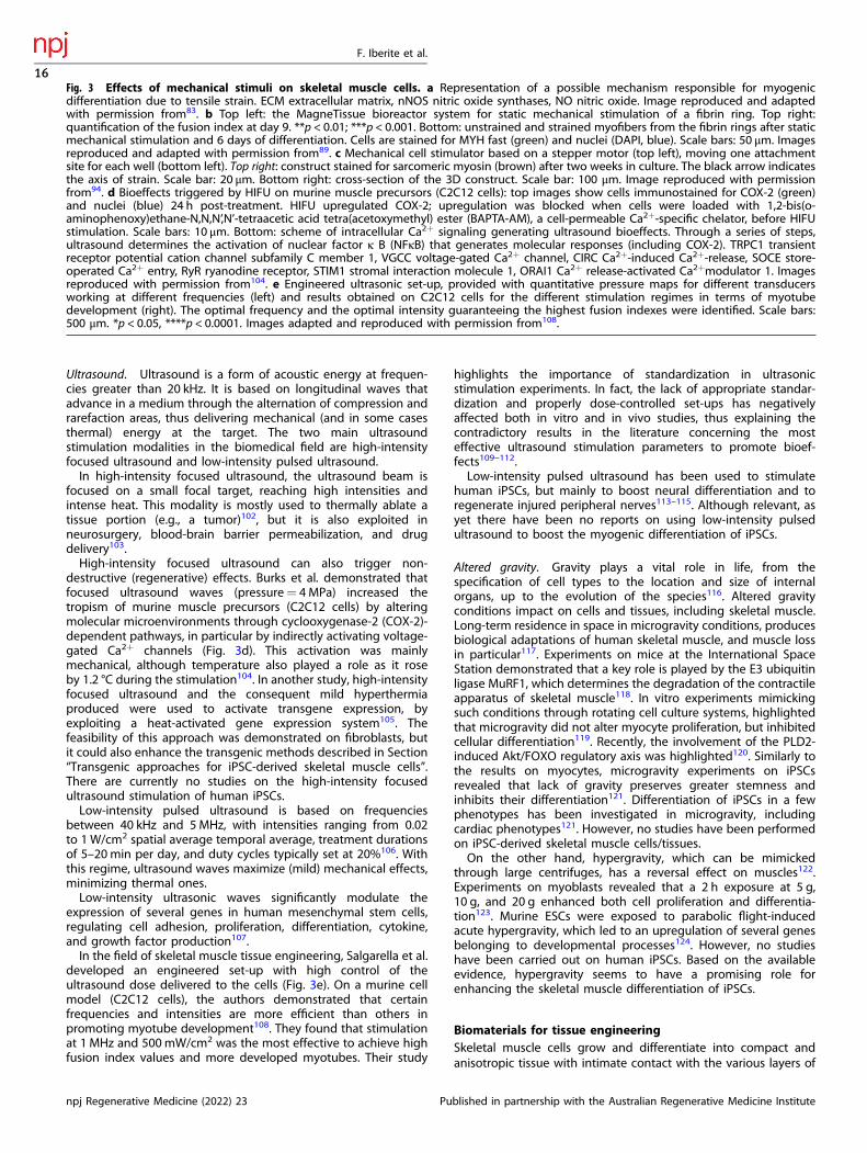
Ultrasound. Ultrasound is a form of acoustic energy at frequen-cies greater than 20 kHz. It is based on longitudinal waves thatadvance in a medium through the alternation of compression andrarefaction areas, thus delivering mechanical (and in some casesthermal) energy at the target. The two main ultrasoundstimulation modalities in the biomedical field are high-intensityfocused ultrasound and low-intensity pulsed ultrasound.In high-intensity focused ultrasound, the ultrasound beam is
focused on a small focal target, reaching high intensities andintense heat. This modality is mostly used to thermally ablate atissue portion (e.g., a tumor)102, but it is also exploited inneurosurgery, blood-brain barrier permeabilization, and drugdelivery103.High-intensity focused ultrasound can also trigger non-
destructive (regenerative) effects. Burks et al. demonstrated thatfocused ultrasound waves (pressure= 4 MPa) increased thetropism of murine muscle precursors (C2C12 cells) by alteringmolecular microenvironments through cyclooxygenase-2 (COX-2)-dependent pathways, in particular by indirectly activating voltage-gated Ca2+ channels (Fig. 3d). This activation was mainlymechanical, although temperature also played a role as it roseby 1.2 °C during the stimulation104. In another study, high-intensityfocused ultrasound and the consequent mild hyperthermiaproduced were used to activate transgene expression, byexploiting a heat-activated gene expression system105. Thefeasibility of this approach was demonstrated on fibroblasts, butit could also enhance the transgenic methods described in Section“Transgenic approaches for iPSC-derived skeletal muscle cells”.There are currently no studies on the high-intensity focusedultrasound stimulation of human iPSCs.Low-intensity pulsed ultrasound is based on frequencies
between 40 kHz and 5MHz, with intensities ranging from 0.02to 1W/cm2 spatial average temporal average, treatment durationsof 5–20min per day, and duty cycles typically set at 20%106. Withthis regime, ultrasound waves maximize (mild) mechanical effects,minimizing thermal ones.Low-intensity ultrasonic waves significantly modulate the
expression of several genes in human mesenchymal stem cells,regulating cell adhesion, proliferation, differentiation, cytokine,and growth factor production107.In the field of skeletal muscle tissue engineering, Salgarella et al.
developed an engineered set-up with high control of theultrasound dose delivered to the cells (Fig. 3e). On a murine cellmodel (C2C12 cells), the authors demonstrated that certainfrequencies and intensities are more efficient than others inpromoting myotube development108. They found that stimulationat 1 MHz and 500mW/cm2 was the most effective to achieve highfusion index values and more developed myotubes. Their study
highlights the importance of standardization in ultrasonicstimulation experiments. In fact, the lack of appropriate standar-dization and properly dose-controlled set-ups has negativelyaffected both in vitro and in vivo studies, thus explaining thecontradictory results in the literature concerning the mosteffective ultrasound stimulation parameters to promote bioef-fects109–112.Low-intensity pulsed ultrasound has been used to stimulate
human iPSCs, but mainly to boost neural differentiation and toregenerate injured peripheral nerves113–115. Although relevant, asyet there have been no reports on using low-intensity pulsedultrasound to boost the myogenic differentiation of iPSCs.
Altered gravity. Gravity plays a vital role in life, from thespecification of cell types to the location and size of internalorgans, up to the evolution of the species116. Altered gravityconditions impact on cells and tissues, including skeletal muscle.Long-term residence in space in microgravity conditions, producesbiological adaptations of human skeletal muscle, and muscle lossin particular117. Experiments on mice at the International SpaceStation demonstrated that a key role is played by the E3 ubiquitinligase MuRF1, which determines the degradation of the contractileapparatus of skeletal muscle118. In vitro experiments mimickingsuch conditions through rotating cell culture systems, highlightedthat microgravity did not alter myocyte proliferation, but inhibitedcellular differentiation119. Recently, the involvement of the PLD2-induced Akt/FOXO regulatory axis was highlighted120. Similarly tothe results on myocytes, microgravity experiments on iPSCsrevealed that lack of gravity preserves greater stemness andinhibits their differentiation121. Differentiation of iPSCs in a fewphenotypes has been investigated in microgravity, includingcardiac phenotypes121. However, no studies have been performedon iPSC-derived skeletal muscle cells/tissues.On the other hand, hypergravity, which can be mimicked
through large centrifuges, has a reversal effect on muscles122.Experiments on myoblasts revealed that a 2 h exposure at 5 g,10 g, and 20 g enhanced both cell proliferation and differentia-tion123. Murine ESCs were exposed to parabolic flight-inducedacute hypergravity, which led to an upregulation of several genesbelonging to developmental processes124. However, no studieshave been carried out on human iPSCs. Based on the availableevidence, hypergravity seems to have a promising role forenhancing the skeletal muscle differentiation of iPSCs.
Biomaterials for tissue engineeringSkeletal muscle cells grow and differentiate into compact andanisotropic tissue with intimate contact with the various layers of
Fig. 3 Effects of mechanical stimuli on skeletal muscle cells. a Representation of a possible mechanism responsible for myogenicdifferentiation due to tensile strain. ECM extracellular matrix, nNOS nitric oxide synthases, NO nitric oxide. Image reproduced and adaptedwith permission from83. b Top left: the MagneTissue bioreactor system for static mechanical stimulation of a fibrin ring. Top right:quantification of the fusion index at day 9. **p < 0.01; ***p < 0.001. Bottom: unstrained and strained myofibers from the fibrin rings after staticmechanical stimulation and 6 days of differentiation. Cells are stained for MYH fast (green) and nuclei (DAPI, blue). Scale bars: 50 μm. Imagesreproduced and adapted with permission from89. c Mechanical cell stimulator based on a stepper motor (top left), moving one attachmentsite for each well (bottom left). Top right: construct stained for sarcomeric myosin (brown) after two weeks in culture. The black arrow indicatesthe axis of strain. Scale bar: 20 μm. Bottom right: cross-section of the 3D construct. Scale bar: 100 μm. Image reproduced with permissionfrom94. d Bioeffects triggered by HIFU on murine muscle precursors (C2C12 cells): top images show cells immunostained for COX-2 (green)and nuclei (blue) 24 h post-treatment. HIFU upregulated COX-2; upregulation was blocked when cells were loaded with 1,2-bis(o-aminophenoxy)ethane-N,N,N’,N’-tetraacetic acid tetra(acetoxymethyl) ester (BAPTA-AM), a cell-permeable Ca2+-specific chelator, before HIFUstimulation. Scale bars: 10 μm. Bottom: scheme of intracellular Ca2+ signaling generating ultrasound bioeffects. Through a series of steps,ultrasound determines the activation of nuclear factor κ B (NFκB) that generates molecular responses (including COX-2). TRPC1 transientreceptor potential cation channel subfamily C member 1, VGCC voltage-gated Ca2+ channel, CIRC Ca2+-induced Ca2+-release, SOCE store-operated Ca2+ entry, RyR ryanodine receptor, STIM1 stromal interaction molecule 1, ORAI1 Ca2+ release-activated Ca2+modulator 1. Imagesreproduced with permission from104. e Engineered ultrasonic set-up, provided with quantitative pressure maps for different transducersworking at different frequencies (left) and results obtained on C2C12 cells for the different stimulation regimes in terms of myotubedevelopment (right). The optimal frequency and the optimal intensity guaranteeing the highest fusion indexes were identified. Scale bars:500 μm. *p < 0.05, ****p < 0.0001. Images adapted and reproduced with permission from108.
F. Iberite et al.
16
npj Regenerative Medicine (2022) 23 Published in partnership with the Australian Regenerative Medicine Institute

the extracellular matrix (ECM) (see Section “Skeletal muscle tissueembryonic development and architecture”). The chemical inter-actions between the differentiating myoblasts and the ECM aretrivial during developmental processes and in the differentiatedtissue, where myotube force transmission is highly dependent onthese interactions. Biomaterials that mimic ECM features arecrucial for controlling iPSC fate and iPSC-derived skeletal musclecell functionality.Biomaterials for skeletal muscle tissue engineering are mainly
divided into two groups: (1) natural materials e.g. collagen, fibrin,alginate, Matrigel®, hyaluronic acid, gelatin, silk fibroin, chitosan,decellularized tissue ECM; and (2) synthetic materials (e.g. poly(glycolic acid), poly(lactic‐co‐glycolic acid), poly(vinyl alcohol), poly(glycerol sebacate)33,125. Natural biomaterials have inherentbiocompatibility as well as several biochemical cues fostering celladhesion and differentiation. Synthetic biomaterials must befunctionalized with cues mimicking the biological environment,
but their mechanical and structural features can be easily tuned toachieve the desired characteristics.Choosing the right material with specific tunable features is
therefore crucial when designing a supportive myogenic environ-ment (Fig. 4a).Biomaterial stiffness is key in the interaction between scaffolds
and cells. Cells can “feel” the environmental stiffness by pullingagainst the matrix through focal adhesions126 (Fig. 4b, c). Theforce exerted to deform the matrix influences the response ofthese mechanotransducers, generating different intracellularsignals thus influencing cell fate. The difference between thestiffness of the cells (a few kPa) and the material used for cellculture, such as glass or polystyrene (a few GPa), can impairmyotube contractions and typically results in myotube detach-ment, thus shortening culture duration. Stem cell lineagespecification is also strongly influenced by environmentalstiffness34. Regarding skeletal muscle tissue, MuSCs respond toenvironmental stiffness by showing a greater self-renewal ability
a b
c
INTRINSIC EXTRINSIC
Viscoelasticity
Stiffness
Functional groups
Degradability Dimensionality
Porosity
Topography
Roughness
Biomaterial meshwork
Cell Soluble factor Functional group
Degradative enzyme
Soft matrix Stiff matrix
Adh
esio
nsC
ytos
kele
ton
Diff
eren
tiaito
nd
Fig. 4 Biomaterial features and their effects on skeletal muscle cells. a Scheme of biomaterial properties relevant for cell/tissueengineering, divided into intrinsic and extrinsic ones. b Scheme of the intracellular biochemical cascades triggered by the stiffness of theextracellular environment. Images reproduced with permission from126. c Top: stress/strain curves for different soft tissues (skin, muscle, andbrain) from which the slope E can be extracted, representing the Young’s elastic modulus. Dashed lines represent (i) polylactic acid; (ii) artery-derived acellularized matrix; (iii) Matrigel®. Bottom: influence of soft and stiff matrix on actin cytoskeleton assembly, cell spreading, andmyotube differentiation. Images reproduced with permission from126. d Left: immunofluorescence staining of iPSC-derived myotubes at twoweeks of differentiation on different substrates. Right: evaluation of the fusion index and percentage of striated myotubes in the differentconditions. (N= 10 fields). *p < .05 versus unpatterned rigid. #p < .05 versus unpatterned soft and micropatterned rigid, and **p < .05 versusunpatterned soft. Images adapted and reproduced with permission from30.
F. Iberite et al.
17
Published in partnership with the Australian Regenerative Medicine Institute npj Regenerative Medicine (2022) 23

in vitro and in vivo if cultured on soft hydrogels (~12 kPa),compared to more rigid substrates (106 kPa)127.The effects of different substrate stiffnesses (3.5-141 kPa) on the
differentiation of MYOD1-reprogrammed iPSCs during earlymyogenesis have been studied and no influence was found inthe skeletal muscle differentiation of iPSCs into iPSC-derivedmyogenic progenitors concerning this range of stiffness128.However, studies on the terminal differentiation of iPSC-derived
progenitors have shown that a soft patterned substrate (85-μmwidth lanes of Matrigel® patterned on 15-kPa-soft polydimethylsi-loxane) can promote later stages of differentiation, such asmyoblast fusion and the formation of striated myotubes (Fig.4d)30.In fact, another crucial factor for skeletal muscle tissue
development, is the scaffold anisotropy. Myofibers have a stronglyanisotropic organization that maximizes their force transmission(Fig. 2). The anisotropic topography of the substrate stronglyinfluences cell differentiation by addressing skeletal muscle cellsin a preferential direction and also driving muscle differentia-tion129. iPSC-derived skeletal muscle cells from healthy donors, asopposed to cells derived from dystrophic patients, align nearlyperpendicular to anisotropic nano-grooves. Alteration of the DPCimpairs the cell capacity to self-align and form a densely packedSM bundle130.iPSC culture methods in growth conditions typically entail
irradiated mouse embryonic fibroblasts (iMEFs) as a feeder layer,and therefore the use of this co-culture for skeletal muscle tissueengineering seemed quite difficult at the beginning. Theintroduction of feeder-free culture methods has created moredefined culture conditions by using recombinant proteins (e.g.,vitronectin, laminin) or complex hydrogels (e.g. Matrigel®), thuseasing the process of keeping the stem cells in an undifferentiatedstate. These coatings are needed for long-term cultures of iPSCs,and they are sometimes specifically optimized for defined growthmedia to support iPSC growth and to maintain pluripotency.The most common coating in iPSC myogenic induction
protocols is Matrigel® (see protocols in Table 1 and Table 2), withsome protocols using type I collagen13,21,131, gelatin16,59, oriMEFs14. Matrigel® is a natural matrix extracted from Englebreth‐Holm‐Swarm sarcoma in mice. It mainly consists of a mixture oflaminin, type IV collagen, entactin, and several growth factors (e.g.,FGF2, EGF, IGF-1, TGFβ, PDGF, NGF)132. Thanks to its composition,Matrigel® creates a supportive and rich environment for iPSCs, butthe variability of the concentration of the ingredients can causereproducibility problems and tumorigenic response if implanted.In fact, Hughes et al. demonstrated only a ~53% batch‐to‐batchsimilarity132. Nevertheless, also in the creation of iPSC-derived 3Dskeletal muscle constructs, Matrigel® has been extensively used toform hydrogels, with the addition of fibrinogen, hyaluronic acid,gelatin, or fibrin23,46,133,134.Fibrin is one of the ideal candidates for natural biomaterials and
scaffolding proteins. Fibrin is a protein derived from the action ofthe protease thrombin on fibrinogen, and it has a stronginteraction with the myotube integrins (integrins α7 and α5), thusoptimizing the efficiency of force transmission33. It is a develop-mental matrix and compared to other frequently used proteinssuch as collagen (an adult matrix), it is less stiff and morecompliant to muscle cell contraction135.However, transformed cell lines such as C2C12 display a natural
fibrinolytic activity as a result of the high levels of plasminogenproduction. To solve this issue, some anti-proteolytic agents havebeen used such as aprotinin, genipin135, and aminocaproic acid136.Maffioletti et al.21 resuspended dystrophic iPSCs and healthycontrols in a composite of fibrin gel and Matrigel®, and showedhow they can be differentiated towards a multilineage isogenicculture system with endothelial cells, pericytes, myofibers, andmotor neurons spreading from neurospheres placed above the 3Dconstruct. A similar hydrogel composition has also been used to
embed differentiating myogenic progenitors, forming alignedmyotubes exhibiting electrical responsivity15.Given the hierarchical and modular structure of SMT, 3D
bioprinting is becoming increasingly common as it can createmacroscopic constructs in a layer-by-layer fashion, by depositingcells and biomaterials simultaneously with a high resolution. Thefeatures of the biomaterial used are crucial, and there is anincreasing need for smart bioinks supportive for myogenicdifferentiation and effective for printing126.A milestone in this field is the work of Kang et al.137. In 2016
developed a bioprinting approach based on an integrated tissue-organ printing (ITOP) system that can generate a 3D freeformshape (Fig. 5a left). The authors fabricated a 15mm× 5mm×1mm skeletal muscle construct using a bioink composed offibrinogen, gelatin, hyaluronic acid, and glycerol embeddingC2C12 myoblasts and Pluronic F-127 as a sacrificial bioink. Afterprinting, a thrombin solution was used to crosslink the fibrinogenin the cell-laden bioink thereby stabilizing it, while the sacrificialbioink was washed out, to obtain void spaces thus enhancing thediffusion of oxygen and nutrients in the internal regions of theconstruct. In vivo experiments were also performed by implantingsubcutaneously (ectopically) on rat structures differentiated forseven days. The dissected distal end of the proximal stump of thecommon peroneal nerve was embedded within the construct topromote integration. The results highlighted a well-organizedmuscle fiber structure (Fig. 5a right), the presence of acetylcholinereceptor, nerve integration, and vascularization throughout themuscle constructs indicated by endothelial cell marker expression.More recently, other bioprinting approaches have been used, for
example custom extrusion-based systems, microfluidic printing,inkjet printing, and fused deposition modeling with surface coating.Various bioinks were tested, such as collagen, alginate, polyethyleneoxide, polyethylene glycol, silk, and methacrylated gelatin138,139.The ITOP technique and a gelatin fibrinogen-based bioink were
proposed again in 2018 by Kim et al.133., who fabricated abioengineered SMT based on human primary muscle progenitorcells harvested from biopsies of human gracilis muscles andexploiting the ITOP system (Fig. 5b top). They obtained amacroscopic construct (15 × 15 × 15mm3) with integrated voidmicrochannels (~200 µm wide) between the cell-laden patterns,facilitating the diffusion of oxygen and nutrients. The cells wereladen in a bioink composed of fibrinogen, gelatin, hyaluronic acid,and glycerol (similarly to Kang et al.), while the sacrificial bioink(used to generate the void microchannels) was only composed ofgelatin, hyaluronic acid, and glycerol. Their construct inducedmyofiber and contractile protein formation in vitro, with respect tothe bulk (non-printed) hydrogel (Fig. 5b bottom left). In vivoresults on rats with muscle defects also showed that bioprintedconstructs performed much better (Fig. 5b bottom right).Bioinks can also be based on decellularized ECM. Choi et al.
proposed a skeletal muscle and a vascular decellularized ECMbioink, using the porcine tibialis anterior muscle and thedescending aorta, respectively140. The first bioink was laden withhuman skeletal myoblasts, and the second one with endothelialcells (HUVEC). These cell-laden bioinks were printed in differentconfigurations, and mixed and organized into a coaxial structure(Fig. 5c top). Choi’s results showed the formation of myotubesin vitro, with the presence of an endothelial network throughoutthe construct in the coaxially printed structure (Fig. 5c bottomleft). In vivo, well-organized de novo muscle fibers were found inthe coaxial printing, whereas severe scar tissue deposition wasobserved in the mixed group (Fig. 5c bottom right). These findingshighlight the importance of cell co-culture for achieving a goodmuscle maturation, and that cell spatial organization duringfabrication is a key aspect.Human iPSCs were bioprinted for the first time by Jodat et al.141,
who used a bioink based on gelatin and methacrylated gelatin(which was photocrosslinked by UV light), laden with iPSC-derived
F. Iberite et al.
18
npj Regenerative Medicine (2022) 23 Published in partnership with the Australian Regenerative Medicine Institute

muscle progenitor cells. The authors also proposed a non-conventional bioprinting approach, exploiting a pre-gelledmethacrylated gelatin supporting matrix as a block, within whichthe cell-laden bioink was printed by producing vertical lines(instead of the usual horizontal lines). The authors suggested that
the role of this methacrylated gelatin supporting matrix iscomparable to the hierarchical ECM structure of the nativeendomysium, a connective tissue that physically supports denselybound aligned myofibers. Jodat used a thermo-reversible gelatinbioink as the sacrificial one to create a perfusable construct, and
Tissue-derived bioink
3D cell printing of large muscle constrct
CD31 / MHC / DAPI
Printing Printing (Mixed) Coaxial Printing
Printing (Mixed) Coaxial printingMHC / HuNu / DAPI
Week 4
Week 8
b
c
a
F. Iberite et al.
19
Published in partnership with the Australian Regenerative Medicine Institute npj Regenerative Medicine (2022) 23

the 3D pre-vascularized tissue construct was successfullyimplanted in a volumetric muscle loss-injured animal model. Theresults were promising and highlight that Jodat’s approach couldbe used for VML repair.
CHALLENGES IN THE CLINICAL TRANSLATION OF IPSC-DERIVED SKELETAL MUSCLEIn a clinical setting, iPSCs have many advantages over other stemcells, such as MuSCs, ESCs, or mesenchymal stem cells. AlthoughMuSCs in vivo are capable of extensive self-renewal for muscleregeneration in the case of injury or tissue degeneration, only theinjection of freshly isolated MuSCs enables robust engraftmentand in situ self-renewal68. Clinical trials require a considerablenumber of cells, and MuSC in vitro expansion deeply impairs theirregeneration capacity68. The problem with ESCs is that they areisolated from the inner mass of a blastocyst with difficult andinefficient protocols, and their use in clinics raises various ethicalissues.On the other hand, mesenchymal stem cells have a high
potential as they tend not to stimulate an immunogenic response.However, the invasive process needed for their isolation and themyogenic potential of less than 40% means that much progress isneeded before they can be used in a clinical setting142.iPSCs can be extensively expanded in vitro, leading to a high
number of cells to be transplanted or differentiated potentiallytowards any phenotype. No external donors are needed sinceisogenic iPSCs can be generated from different sources, such asfibroblasts from a non-invasive cutaneous biopsy or T cells fromperipheral blood. Furthermore, gene-editing techniques such asthe CRISPR/Cas9 system can be used to correct molecular defects.There are however limitations in using iPSCs for clinical trials.
The iPSC genesis and the reprogramming processes of somaticcells are characterized by epigenetic remodeling and alterations inthe chromatin structure. These alterations and changes maymodify the iPSC phenotype, while in other cases the residualepigenetic memory from the somatic donor cell source mayreduce the pluripotency of the generated cell line, leading to abiased differentiation potential143.Several reviews have highlighted that the reprogramming
process can be very inefficient (with a yield of less than 1%144),and the cost of generating a patient-specific clinical-grade iPSCcell line is high, around U.S. $800,000145. Reprogramming methodsrange from using integrative viruses, such as retroviruses andlentiviruses, to non-integrative technologies with adenoviruses,PiggyBac transposons, Sendai viruses, episomal vectors, orrecombinant proteins. These non-integrative technologies avoidpossible insertional mutagenesis, though some genome altera-tions can be inherited from the somatic donor cells. Genomicalterations can cause aberrant phenotypes after implantation withpossible teratoma development, and although preclinical studieshave not noted signs of teratoma formation, this concern remains.Isogenic iPSCs are widely known to be immune-privileged, and
one study on the immunotolerance of undifferentiated iPSCs andiPSCs derivates demonstrated that only iPSC differentiation led toa tolerogenic immune response146. Other studies showed no
lymphocyte or macrophage infiltration after transplantation ofiPSC-derived dopamine neurons into primate brains over twoyears, without any immunosuppression147. Allogenic iPSCs withdifferent human leukocyte antigens could be exploited in auniversal transplantation technology148.Due to the high variability of iPSC lines (e.g., batch-to-batch,
clone-to-clone), the huge cost, and possible immune rejection,iPSC banking services have been established in the last ten years.They aim to standardize iPSC culture and handling, according togood manufacturing practices and quality standards145. Althoughthese services can reduce the variability at the beginning of theiPSC genesis and handling procedure, subsequent steps can alsobe taken, for example, (clone selection, differentiation protocols,reagent lots, and experimental conditions. Other standardizedcontrols must therefore be introduced in the following phases,including the absence of mycoplasma contamination, checking fornormal karyotypes, no chromosomal aberrations, and no presenceof reprogramming transgenes or vectors149.Because of the obstacles encountered in the development of
iPSC-based therapies, iPSCs have not been used much in clinics.Deinsberger et al. performed a systematic worldwide analysis ofclinical trials involving PSCs, divided into interventional trials, withcell transplants in patients, and observational ones, regarding thegeneration of patient-specific cell lines used for in vitro testing150.Out of a total of 131 clinical trials, 77% were observational andonly 23% were interventional. Clinical trials involving iPSCs aremainly observational (only 27% of the interventional studiesinvolve iPSCs), meaning that a solid basis for the clinicaltranslation is under development, but many challenges remain.With regard to skeletal muscle tissue engineering, there have
been no interventional clinical trials using iPSC-derived musclecells. To date, patient-derived iPSCs have mainly been used suchas in vitro tools to model muscular diseases, to study thepathological molecular mechanisms, and for drug testing beforein vivo translation151.The use of gene-editing technologies can be valuable by
providing isogenic healthy control cell lines. Some preclinicalstudies have implanted iPSC-derived myogenic progenitor inanimal disease models, as described in Table 1 and Table 2. Thereare no effective treatments for these genetic conditions, withclinical trials aimed at correcting the molecular defect in order torestore protein expression by gene replacement strategies withRNA-based (conventional or based on the CRISPR/Cas9 system) orcell-based approaches. Interestingly, a study on the transplanta-tion of iPSC-derived mesangioblast-like cells expressing the α-sarcoglycan (SGCA) gene into limb-girdle muscular dystrophy 2Dmice (Sgcanull) succeeded in generating SGCA+ muscle fibers12.The autotransplantation of genetically-corrected iPSCs is promis-ing, however the main issues are the poor survival and migrationof the iPSC-derived progenitor cells. The tissue atrophy anddegeneration caused by the abovementioned muscular disordersare associated with an inflammatory environment and the loss oftissue integrity, which considerably impair cell viability andmigration152.On the other hand, for conditions such as VML, with the need to
substitute most of the muscle, the clinical approach must focus on
Fig. 5 3D bioprinting for skeletal muscle tissue engineering. a Left: the ITOP system and its major units, and illustration of the targeted 3Darchitecture. Right: staining for myosin heavy chain after 7 days of differentiation (top) and image of the construct and desmin staining afterin vivo implantation (bottom). Images adapted from137. b Construct based on a cell-laden bioink made of gelatin, fibrinogen, hyaluronic acid,and glycerol. MPCs muscle progenitor cells. Top: fabrication procedure based on an ITOP system. Bottom left: in vitro results of bioprinted andnon-bioprinted (bulk) system. MHC myosin heavy chain. Bottom right: in vivo results after implantation in rat muscle defect models. Imagesadapted from133. c Top: construct based on decellularized extracellular matrix-derived bioinks, laden with muscle cells and endothelial cells,organized in different patterns. Bottom left: results of in vitro differentiation, in terms of expression and organization of endothelial markerCD31 and of myosin heavy chain. Bottom right: results of the in vivo experiments. Scale bar: 100 µm. HuNu: human nuclei. Images adaptedfrom140.
F. Iberite et al.
20
npj Regenerative Medicine (2022) 23 Published in partnership with the Australian Regenerative Medicine Institute

the fabrication of meso/macroscopic 3D constructs, recapitulatingthe skeletal muscle architecture complexity and the representa-tion of the resident cell populations. This is necessary both for theprodifferentiative effect that these populations have on skeletalmuscle cells (e.g. fibroblasts, motor neurons), and the need tofabricate terminally differentiated functional tissue.As described in Section “Skeletal muscle tissue embryonic
development and architecture”, several cell populations are foundin skeletal muscle tissue, including muscle-resident populationssuch as cells from the vasculature, fibroblasts from the connectivetissue wrapping the muscle, tenocytes, adipocytes, fibro-adipogenic progenitors, and motor neurons. In vitro fabricationof a construct recapitulating a functional skeletal muscle tissuetherefore involves several steps. Most of the muscle-resident cellsare in the G0/G1 state, highlighting the low turnover of theskeletal muscle tissue153. Knowing the development path and themorphogenetic signaling of the individual phenotypes is crucial tobetter engineer the co-culture of different cell populations, thus tolead a concert of all the different differentiation timings,morphogens, and growth factors.Future evolutions of 3D bioprinting will open up interesting
scenarios in this domain. Fabrication techniques are evolving toproduce macro-structures that are as packed as possible, stableover time, and with a micrometric resolution. Recent interestingexamples of new bioprinting approaches include continuouschaotic bioprinting, which led to the fabrication of hierarchically-structured engineered muscle-like constructs in a continuous andsimple fashion154, fibrillation/leaching of poly(vinyl alcohol)(included in the bioink) after the printing process, to create auniaxially aligned micro-topographical structure in the printedconstruct, thus promoting self-alignment of muscle cells155 andelectric-field assisted bioprinting for aligning a cell-laden bioink156
or to promote myogenesis157.There are several interesting innovations regarding bioinks, and
newly formulated molecules and polymers may be an excitingroute. However, traditional polymeric formulations are alsopromising if they are supplied with active nanomaterials thatcan turn these formulations into multifunctional ones112,158. Theseevolutions, together with the use of iPSCs in the printing process,will advance the field considerably.The combination of chemical, topographical, and stiffness-
related cues on the one hand (ensured by appropriate biomater-ials and ad hoc fabrication technologies) and biophysical stimuli(provided through mechanical, electromagnetic, and other typesof energy transfer to cells), has barely been explored in the state ofthe art for iPSCs. However, all these approaches have the potentialto recapitulate part of the intricated series of dynamic inputs thatthe cells receive during embryogenesis.Several questions still need to be answered: (1) What is the
optimal modality and energy dose for each stimulus, to drive theexpression of a certain phenotype, at a given time-point? (2) Whenand how should different stimuli be combined to enhance thiseffect, and which ones should be used? (3) What is the weight ofeach stimulus used for this purpose, depending on the desiredtarget tissue type, and how does this weight vary over time?Answering these questions would considerably advance the field
of skeletal muscle tissue engineering, as well as other regenerativemedicine domains. To pursue this objective, systematic experimentsshould be designed, exploring the “sequence space” of the variouspossible combinations of the above-mentioned stimuli over timethereby creating a clear map of the cell response. However relyingonly on experiments, this is an enormous task. Mathematicalmodeling and advanced data processing techniques (e.g. artificialintelligence, and in particular deep learning) may make adifference159–162, by reducing the complexity of this task andpredicting cell response patterns more reliably.In addition to bioengineering platforms to enhance iPSC
differentiation, it is also worth considering platforms that monitor
the performance of these constructs. For example, a contractileforce screening system can be created using custom multiwellplates with microcantilevers163.Active prostheses control is an alternative field of skeletal
muscle tissue engineering. Such control is generally achieved bysuperficial electromyographic sensors placed in the socket of theprosthetic device and in contact with the residual limb. Thecontrol of a single degree of freedom per time is carried out byapplying a simple threshold or a proportional amplitude methodon superficial electromyographic sensor signals recorded fromantagonistic muscles (e.g., wrist flexor and wrist extensor). In thecase of prosthetic devices endowed with several degrees offreedom, but still with two control signals, switching amongdegrees of freedom is normally achieved by co-contraction, as infinite state machines. This serial operation is slow and unnatural; inaddition, it requires considerable training and cognitive effort inthe execution of the task164.Multi-fingered prosthetic hand control techniques based on
machine learning and targeted muscled re-innervation165 areinteresting alternatives but they are still constrained by a fewindependent control signals.The regenerative peripheral nerve interface technique re-
innervates a muscle portion removed from another body region,preventing the need for denervation166. However, regenerativeperipheral nerve interfaces clearly entail, albeit limited, furtherharm to the patient.Consequently, an engineered in vitro muscle, as well as
multifunctional afferent and efferent artificial structures, are ofgreat technical and scientific interest to overcome the drawbacksand to maximize the performance of the afore-mentionedprosthetic control techniques. Such an improvement in thecontrol would promote greater functionality of the prostheticdevice, with clear clinical benefits for the amputees. Human iPSCshold great promise for this application, at the frontier betweenrobotics and bioengineering.
Co-culture of skeletal muscle cells with muscle-residentphenotypesVascular cells. The cellular components of the vascular network(endothelial cells, vascular smooth muscle cells, and pericytes)share a different embryonic origin depending on the localizationof the anatomical location of the vasculature167,168. Endothelialcells, which line the vessel lumen, start the process.A combination of BMP, FGF2, and WNT signals activate the
transcription factor ETV2 in the lateral plate mesoderm leading tothe formation of endothelial cell precursors, the hemangioblasts,from the splanchnic mesoderm (Fig. 6a).Subsequently, the expression of FLT1 (VEGFR-1), KDR (VEGFR-2/
Flk-1), and the receptors for the vascular endothelial growthfactors A (VEGF-A) on the hemangioblast membrane start thedifferentiation and proliferation into angioblasts. Under VEGF-Aguidance, cells begin sprouting new vessels and eventuallyundergo arteriovenous specification168. In the subsequent angio-genesis, vessel maturation occurs, with the recruitment of muralcells (pericytes and vascular smooth muscle cells) and theexpansion of the vessel network. Through the tyrosine-kinasetransmembrane receptors transcribed from the TEK gene (TIE),angiopoietins mediate the interaction between endothelial cellsand pericytes, which are recruited by the endothelial cells169.More than other cell types, vascular smooth muscle cells have
multiple embryonic origins, as reviewed by Majesky170. However,the majority of smooth vascular muscle cells generally have amesodermal origin, specifically from somites of the ventralposterior sclerotome170,171.Although pericytes express canonical markers of mesenchymal
stem cells such as CD29, CD44, CD73, CD90, CD105172, and arenegative for CD31, PAX7, and MYOD1, they are very
F. Iberite et al.
21
Published in partnership with the Australian Regenerative Medicine Institute npj Regenerative Medicine (2022) 23

heterogeneous in terms of marker expression origin, andmorphology44,173. The chondroitin sulfate proteoglycan 4 (Cspg4or Ng2) has been used as a pericyte marker in murine modelsduring vascular morphogenesis174. Pericytes can differentiatetowards adipogenic, chondrogenic, osteogenic, and myogeniclineages, however the mechanisms regulating pericyte myogenicdifferentiation are still mostly unknown. There are manyhypothesesregarding their embryological origin, and the literaturesuggests that they most probably share the same lineages asvascular smooth muscle cells, depending on the organ studied44.The capillaries found inside the skeletal muscle tissue are
exclusively composed of endothelial cells and pericytes, and a lackof vascularization limits the size of the engineered tissue becauseof the limited diffusion of nutrients and oxygen. One of theoptions is therefore to pursue scaffold vascularization throughin vivo angiogenesis, by implanting a 3D scaffold and developingtechniques to stimulate host vessel ingrowth. This can beachieved by adding VEGF in the scaffold or by anastomosingthe construct to the host vasculature175, although the infiltratingvessels often remain limited to the construct periphery. Anotheroption is the pre-vascularization of the scaffold in vitro throughvasculogenesis, thus entailing a co-culture of myoblast andendothelial cells. Rosa et al. were able to generate functionalarterial and venous-like endothelial cells from human iPSCs176. Theco-culture option involves the fabrication of an a priori perfusableconstruct, thus easing vascularization and survival upon implanta-tion. Skeletal muscle and endothelial cell co-culture systems canbe based on 3D microporous scaffolds recreating the skeletalmuscle tissue architecture, or 3D bioprinted constructs embed-ding vessel-like structures. Gholobova et al.177 established a co-culture of a mixed population of primary muscle cells (with 8%fibroblasts) and HUVEC in a compact 3D fibrin gel. Seven days of
differentiation led to an advanced vascular network, withouthampering myoblast fusion and differentiation, thanks also to theproangiogenic activity of the fibrin gel (Fig. 6b). However, theprimary muscle cells did not show a high level of myogenicdifferentiation, with the myotubes not showing the canonicalsarcomere striated ultrastructure, thus leaving margins for furtherimprovements. Nevertheless, the presence of endothelial cellsalone may not be sufficient to create a spread vascular network.Pericytes during HUVEC vasculogenesis have in fact beendemonstrated to foster endothelial cell branching and the totallength of the vessels compared to HUVECs alone19 (Fig. 6c).
Fibroblasts. The connective tissue cellular components are thefibroblasts. Different methods have been proposed to identifythem, e.g., by staining collagen, the intermediate filamentvimentin, or through localization in the interstitial ECM. Thesefeatures cannot be defined as fibroblast-specific markers orfeatures since, for example, the myocytes themselves can secreteECM proteins178. The great heterogeneity of fibroblasts highlydepends on their activity and their localization. Being tightlyassociated with the skeletal muscle tissue, fibroblasts share with itmost of the embryonic sites of origin. In terms of skeletal muscledevelopment, appropriately coordinated development of muscleand connective tissue lineages (also including tendons, which willbe described later) is required for the formation of themusculoskeletal system179. There are various possible fibroblastorigins, from differentiation from hematopoietic stem cells,pericytes, monocyte subpopulations, to epithelial-mesenchymaltransition from epithelial cells180.Intramuscular connective tissue fibroblasts throughout the
body express the platelet-derived growth factor receptor alpha(PDGFRA), the transcription factor TCF4 (TCF7L2), and the T-box
a
b c
Hemangioblast
T, KDR, ETV2, CD31, CD34
BMP4FGF2Wnt
KDR, CDH5, TEK, CD31
Angioblast
VEGFs
Arterial endothelial cell
Venous endothelial cell
EFNB2, CDH5, CD31
EPHB4, FLT4, CDH5, CD31
Notch
COUP-TFII
PM progenitor
T
PM segmentation Primary vessel maturationProgenitor specification
Fig. 6 Vasculogenesis, endothelial cell development, and co-culture with skeletal muscle cells: influence on myogenesis. a Representationof the formation of primary vessels during vasculogenesis, with endothelial cell differentiation starting from the paraxial mesoderm (PM)progenitor. Marker genes are shown in the bottom boxes, while the main signaling molecules are indicated in green if acting as pro-differentiative actors, in red if they inhibit the differentiation process. b Fluorescence confocal images of a co-culture of HUVECs (50%, ingreen) and muscle cells (50%, in red) in a fibrin matrix (left image). Focus on the endothelial network formation of HUVEC alone (right image).Scale bars: 50 μm. Images adapted and reproduced with permission from177. c Fluorescence image of HUVECs (von Willebrand factor,magenta) in co-culture with primary pericytes (GFP) showing the formation of a network. Nuclei were identified by DAPI staining (blue). Thegraphs show the quantification of the tubular structures, in terms of total segment length, total mesh area, and total branching length ofHUVECs without (grey columns) or with pericytes (black columns). Images adapted and reproduced with permission from19.
F. Iberite et al.
22
npj Regenerative Medicine (2022) 23 Published in partnership with the Australian Regenerative Medicine Institute

transcription factor 5 (TBX5), while a limb subpopulation expressesthe odd-skipped related transcription factor 1 (OSR1)181. Due tothe overlapping of some molecular markers and similar rolesduring muscle homeostasis, fibroblasts from intramuscular con-nective tissue and bipotent fibro-adipogenic progenitors may beidentified as the same cell population, as recently suggested bySefton and Kardon182. Fibro-adipogenic progenitors (PDGFRA+/ATAXIN1+/CD34+) are mesenchymal cells, developmentallydistinct from myogenic progenitors, and highly abundant in theadult muscle153. They are a source of fibroblasts and adipocytesduring muscle injury scarring and fibrosis. In the quiescent state,they are localized near the blood vessels outside the capillarybasement membrane183. Fibro-adipogenic progenitors maintainthe MuSC pool184, and secrete pro-myogenic factors (IL-6, WNT,IGF-1), increasing the terminal differentiation of myogenicprogenitors185. The role of fibroblasts in myogenesis has beenlargely neglected, mainly focusing on their actions during fibrosisand muscle regeneration. Recent studies have instead highlightedthat fibroblasts influence muscle development, morphogenesis,and localization, as reviewed in182,186. As the muscle develops, theintramuscular connective tissue surrounds the epaxial and hypaxialmuscles, with the intramuscular connective tissue fibroblastsintercalated amongst the myogenic cells in the myotome. Thanksto the easier accessibility in avian and murine models, a greatnumber of studies have been carried out on the role ofintramuscular connective tissue fibroblasts on the developmentof limb muscles. Somitic PAX3+ myogenic precursor cells migratetowards the limb bud, and fibroblasts regulate muscular patterningby the chemoattractive action of CXCL12 chemokine on CXCR4+muscle progenitors, and the chemorepulsive role of EPHRINA5 onEPHA4+ muscle progenitors186. TCF4 gain- and loss-of-functionexperiments have been performed on murine models, and amuscle mispatterning occurred186.Over the years, different protocols have been developed to
isolate fibroblast-free MuSCs, aimed at obtaining a pure myogenicculture in which fibroblast proliferation would not take over on thelow-proliferative MuSCs. Other groups on the other hand, exploitedthis intrinsic co-culture and demonstrated the possibility ofexploiting fibroblasts for the fabrication of 3D rolled skeletalmuscle constructs, such as the pioneering work of Dennis et al.187.The role as a support in the skeletal muscle cell auto-assemblyobserved during embryonic development has been confirmed.In a culture of mouse embryonic fibroblasts and MuSCs at a ratio
of 1:1, fibroblasts were distributed throughout the tight structure
without impairing the SM contractile forces (Fig. 7a) and onlyfibroblasts and MuSC co-culture showed the formation of a rolledcylindrical structure compared to a monoculture of MuSCs (Fig. 7b).Fibroblasts also contributed to a MuSC higher survival rate and theformation of more hypertrophic myotubes (Fig. 7c)188, highlightingthe stimulatory effect of fibroblasts on myogenic differentiationand myoblast fusion189. In addition, a triculture of MuSCs, HUVEC,and human skeletal muscle fibroblasts evidenced their angiogeniceffect by secreting high levels of HGF and promoting VEGFproduction in MuSCs190.Due to the regulatory role of fibroblasts both in muscle
regeneration and in fostering myogenic differentiation, it is clearthat fibroblasts are necessary to construct a functional 3D muscleconstruct in vitro. However, to date no attempts on the co-cultureof fibroblasts and iPSC-derived skeletal muscle cells have beenperformed.
Tenocytes. Tenocytes are other cellular components of themuscular connective tissue. These are interstitial cells that arealso found in adult skeletal muscle, expressing tendon markerssuch as tenomodulin (TNMD), thrombospondin 4 (THBS4), earlygrowth response 1 (EGR1), collagen type I alpha 1 chain (COL1A1),and scleraxis (SCX), a transcription factor essential for tendondifferentiation during development153,191 (Fig. 8a). Tenocytesshare the same developmental pathway as myogenic cells untilthe paraxial mesoderm induction192. They derive from PAX1+mesenchymal cells of the sclerotome, generated by the combinedaction of notochord-derived paracrine factors, especially sonichedgehog (SHH) (Fig. 8b).At this point, a clear division of the cell fate from myogenic cells
is marked by the expression of muscle regulatory factor inhibitorsin the tenocyte progenitors153,193. The subsequent induction byFGF, directly secreted by the myotome, and transforming growthfactor-beta (TGFβ), leads to the development of the tenocyteprogenitors SCX+.The connection between the future myofibers and tenocytes
starts here, with the association of tenocytes and myogeniccells179.Connective tissue plays a role in muscle patterning regulation n
Drosophila melanogaster, where tendons are involved in musclepatterning and the formation of the myotendinous junction194.However, the influence of tendons on muscle tissue developmentis less clear in vertebrates. In Scxmutant mice, the forelimb muscleis impaired, underlying tendon implications only in the late events
Fig. 7 Fibroblast co-culture with skeletal muscle cells: influence on myogenesis. a Fluorescence images of enhanced-GFP-expressingmouse embryonic fibroblasts (MEFs) evenly distributed in a co-culture with primary mouse myoblasts (PMM), stained for fast myosin heavychain (red) and nuclei (blue). b 2D myotube monocultures degenerated after 18 days in culture (left), while the presence of fibroblasts in co-culture drastically enhanced their stability (right). c Left: MEF/PMM co-culture led to the assembly into a 3D fibrin construct with consequentfibrin degradation. MEF monoculture assembled in a 3D construct (middle), but PMM monoculture without MEFs did not show any 3Dautoassembled construct or fibrin degradation (right). Images adapted and reproduced with permission from188.
F. Iberite et al.
23
Published in partnership with the Australian Regenerative Medicine Institute npj Regenerative Medicine (2022) 23

of muscle morphogenesis195.Tenocytes also play a role in skeletal muscle development
through chemical and mechanical signaling between the twotissues in morphogenesis and differentiation. The developmentalpaths of tendons and skeletal muscle tissue are very closely linked,with the mechanical forces generated by the developing muscleinducing a modification in the ECM composition at the interfacebetween the two tissues under maturation (Fig. 8b). Thesechanges are driven by the action of different mechanoresponsivereceptors exposed on the cells, including integrins, integratingoutside-in and inside-out signaling. ECM remodeling and changes in mechanical properties induce tenocyte terminal prodiffer-entiative signals (mechanisms reviewed in ref. 196). The strongphysical interaction of skeletal muscle cells and tenocytes leads tothe formation of the specialized myotendinous junction, whosemechanical integrity and structural characteristic is still difficult toreproduce in vitro. Initial studies have tried to recreate theinterface between the two tissues using neonatal or adult
explanted tendons in co-culture with primary rat myoblasts197.The interface can withstand a tangent modulus of 37.2 kPa ± 10.3kPa before breaking, but was still poorly organized compared tothe highly interdigitated natural structure.Without exploiting explanted tissue, which can be very difficult
to translate in a clinical setting, Laternser et al. used 3D bioprintingfor the specific spatial localization of tenocytes and SM cells198.They succeeded in the fabrication process using two bioinks (agelatine methacryloyl-polyethylenglycol dimeth-acrylate-basedink and a pure gelatine methacryloyl based) around two rigidanchor posts, and in the differentiation of the two cell types in co-culture. However, the poor organization of the interface was tooweak to withstand the developing tension of the skeletal muscletissue (Fig. 8c).
Motor neurons. Motor neurons derive from a different germlayer, namely the ectodermal region. After ectodermal invagina-tion and the formation of the neural tube and the external
Late tenocyte
Early tenocyte
SCX, TNMD, COL1A1,
MKX, EGR1/2
SCX, TNMD, COL1A1, MKX
Tenocyte progenitor
Mesenchymal stem cell
SCX, TNMD, COL1A1,
THBS4, TNC
SHH
PAX1
FGFTGFβ
PM progenitor
PSM cell
T MSGN1TBX6
FGF2Wnts
PM segmentation Progenitor specification Tenocyte maturation
a
b c
Fig. 8 Tenocyte development and co-culture with skeletal muscle cells: influence on myogenesis. a Representation of the differentiationprocess of tenocytes starting from the paraxial mesoderm (PM) progenitors. Marker genes are shown in the bottom boxes, while the mainsignaling molecules are indicated in green if acting as pro-differentiative actors, in red if they inhibit the differentiation process. PSMpresomitic mesoderm. b Scheme of the myotendinous junction formation. Image adapted and reproduced with permission from196. c Top left:3D printed co-culture of myoblasts (red) and tenocytes (green) just after printing. Scale bar: 2 mm. Top middle: co-culture differentiated forseven days and stained for myosin heavy chain (green) and nuclei (red). Arrows indicate striated and multinucleated myofibers. Top right:focus on the tenocytes in the co-culture stained for type I collagen. Scale bar: 50 µm. Bottom: gene analysis expression of muscle and tendonmonoculture in proliferation medium (PM= gray bars) or differentiation medium (DM= black bars). Relative expression is shown as mean ±standard error of the mean (SEM). Images adapted and reproduced with permission from198.
F. Iberite et al.
24
npj Regenerative Medicine (2022) 23 Published in partnership with the Australian Regenerative Medicine Institute

ectoderm, the dorsoventral patterning of the neural tube beginsthanks to the action of WNT; TGFβ family proteins, and retinoicacid from surrounding regions (Fig. 1b).The ventrodorsal gradient of SHH is crucial: the cells exposed to
the highest levels of SHH are the motor neuron progenitors,expressing NK6 homeobox 1 (NKX6.1), the oligodendrocytetranscription factor 2 (OLIG2), and PAX6. The differentiationprogram starts at this point, stimulated by retinoic acid signalingand OLIG2. OLIG2 together with one of its targets, neurogenin 2(NEUROG2), contribute to promoting motor neuron fate, byinducing the expression of motor neuron and pancreas homeobox1 (MNX1 or HB9) (Fig. 9a).MNX1, which is a specific marker for postmitotic spinal motor
neurons, activates its own expression, making the motor neuronsindependent from SHH and retinoic acid signaling. There areseveral motor neuron subtypes, which differ in terms of the fibersthey innervate, and they follow different specific molecularpatterning, as extensively reviewed by Stifani45.Besides being an essential interface for the control of skeletal
muscle contraction, motor neurons are a key to terminal myogenicdifferentiation. In vivo, myotube responsiveness to motor neuronstimulation grows during tissue maturation by increasing theexpression of the acetylcholine receptor. The expression of thesetransmembrane proteins also occurs in vitro, but only motorneurons in co-culture lead to an improved myofiber maturation35.Some studies have aimed to establish a co-culture with iPSC-
Ventral spinal cord cell
Neuralization
SOX1 PAX6
Patterning
Motor neuron progenitor
PAX6, NKX6.1/6.2, OLIG2, NEUROG2
SHHRA
Motor neuron maturation
Late general motor neuron
ISL1/2, MNX1, CHAT
Early motor neuron
ISL1/2, MNX1
a
b
c
Fig. 9 Motor neuron development and co-culture with skeletal muscle cells: influence on myogenesis. a Representation of the formationof motor neurons. Marker genes are shown in the bottom boxes, while the main signaling molecules are indicated in green if acting as pro-differentiative factors. b Left: representative phase-contrast images of a human iPSC-derived moto-neurosphere at different time points afterplating. Small neurites were outgrowing from the moto-neurosphere. In the inserts are shown contacts between neurites and myotubes.Middle left: maturation of acetylcholine receptor clusters and neuromuscular junction formation. Co-culture between human iPSC-derivedmotor neurons with CD34-enrichment derived myotubes. α-Bungarotoxin (α-BT) labeling after 21 days indicates the formation of a matureneuromuscular junction. Middle right: Bassoon labeling after 21 days indicates presynaptic terminals along the axons (top figure). At the endplate region, a close apposition between presynaptic and postsynaptic markers could be detected, as shown by Bassoon and α-BT stainings(bottom figure). Scale bar: 20 μm. Right: electrophysiological properties of myotubes in culture after differentiation and representative tracesof current-clamp measurements and the generation of an action potential following acetylcholine treatment. MP resting membrane potential,AP action potential. Images reproduced with permission from202. c Left: scheme of a bioprinted construct with the cell-laden bioink, theacellular sacrificing bioink, and the supporting polycaprolactone pillar. Right: histological examination of skeletal muscle regenerationthrough the 3D bioprinted constructs at 4 and 8 weeks after implantation. Dashed lines: defected area; MTS: Masson’s trichome staining; H&E:hematoxylin and eosin. Images reproduced with permission from134.
F. Iberite et al.
25
Published in partnership with the Australian Regenerative Medicine Institute npj Regenerative Medicine (2022) 23

derived motor neurons and muscle cells,, with differentapproaches for the challenging formation of neuromuscularjunctions199–201. Demestre et al. differentiated skeletal musclecells and motor neurons from isogenic iPSCs in two parallelprotocols. They used EB-based methods and enriched the skeletalmuscle population for CD34202. After the skeletal muscle cellsshowed contractile properties and responsiveness to acetylcho-line, iPSC-derived lower motor neurons were seeded on top ofiPSC-derived skeletal muscle cells for 3–4 weeks. Matureneuromuscular junctions were identified at later stages of theco-culture (Fig. 9b).One protocol with a co-differentiation in a co-culture was
developed and pursued for a period of approximately 30 days203.Among other differentiation factors, the use of LDN193189, a BMPpathway inhibitor, induced both muscle and neural differentiation.Interestingly, the temporary presence of DAPT, a Notch pathwayinhibitor inducing postmitotic motor neuron differentiation,caused the early onset of myotube contractile properties (fromtwo months post differentiation to 19-20 days from the onset ofdifferentiation).The integration of motor neurons in skeletal muscle cell cultures
has not only been performed in 2D dishes, but also in a 3Dbioprinted millimeter-scale construct134 (Fig. 9c). The constructimplantation in a model of extensive muscle defect injurydemonstrated its potential for the functional and structuralrestoration of the damaged muscle. A full restoration of muscleforce was observed after eight weeks compared to the shamcontrol, and the skeletal muscle cells aided tissue regeneration bydifferentiating and forming an organized architecture.Moving towards a multilineage with four different phenotypes,
Maffioletti and coworkers created an iPSC-derived 3D artificialskeletal muscle construct using isogenic iPSCs. They increased thehistological complexity of the skeletal muscle construct by adding30% of iPSC-derived vascular cells (endothelial cells and pericytes)and fibrin neurospheres with neural progenitor cells on top of the3D culture. This first attempt at integrating such a variety ofdifferent phenotypes derived from iPSCs, further confirmed thatthe maturation of a functional artificial skeletal muscle tissuewould strongly benefit from other cell lineages.
CONCLUSIONSSignificant results are possible in skeletal muscle tissue engineeringthanks to the use of iPSCs, however various challenges still need tobe addressed. Although biochemical differentiation protocols (bothtransgenic and non-transgenic) have become more efficient, morerobust protocols are needed to reduce the output variability whenstarting with different cell lines. In this view, Van der Wal et al.developed and optimized a differentiation protocol on iPSCs from15 different donors and performed over 50 individual differentiationexperiments, highlighting the importance of protocol reproduci-bility18. Comparing the differentiation efficiency between protocolsis still a major problem, mainly due to differences in the reportingmethods. High levels of differentiation, for example, can be obtainedfrom the pooled percentage of cells expressing PAX7 or MYOD1,thus preventing a comparison with protocols that do not follow thisprocedure. Several groups are therefore debating how to establishstandards for the evaluation of muscle cell maturation andconsequent protocol differentiation efficiency8,64.Future progress in iPSC technology will exploit knowledge from
different fields, from embryology to material science andmechatronics for biophysical stimulation.This review has provided an overview of the engineering tools
that can be applied in vitro (in synergy with biochemicalprotocols) for the fabrication and biophysical stimulation offunctional muscle tissues. Such tools include electromagneticstimulation systems, platforms for providing different kinds ofmechanical stimuli, biomaterials, and microfabrication techniques.
The importance of co-cultures has also been highlighted. This360-degree approach may widen the scope for future applicationsof iPSC technology in a clinical setting. The challenges aheadinclude the reprogramming processes leading to a biaseddifferentiation potential, the high variability of the iPSC lines, thehard-to-modify pathological genetic conditions, and difficulties inrecapitulating the whole skeletal muscle architecture complexityand a realistic representation of the resident cell populations.Once these issues have been solved, skeletal muscle tissue
engineering may become a viable solution for volumetric muscleloss, but also for other intriguing applications, such as thedevelopment of patient-specific muscle-on-a-chip platforms fordrug screening and the implementation of novel strategies for thecontrol of active prostheses.
Note added to proofGene and protein names and symbols follow 1) for human: HUGOGene Nomenclature Committee (European Bioinformatics Insti-tute), available at http://www.genenames.org; 2) for mouse:Mouse Genome Informatics Web Site, available at http://www.informatics.jax.org/.
DATA AVAILABILITYThe authors declare that the data supporting the findings of this study are availablewithin the paper.
Received: 22 April 2021; Accepted: 1 March 2022;
REFERENCES1. Fu, X., Wang, H. & Hu, P. Stem cell activation in skeletal muscle regeneration.
Cell. Mol. Life Sci. 72, 1663–1677 (2015).2. Nowak, K. J. & Davies, K. E. Duchenne muscular dystrophy and dystrophin:
pathogenesis and opportunities for treatment. EMBO Rep. 5, 872–876 (2004).3. Grogan, B. F. & Hsu, M. A. J. J. R. Volumetric Muscle Loss. J. Am. Acad. Orthop.
Surg. 19, 35–37 (2011).4. Osaki, T., Uzel, S. G. M. & Kamm, R. D. On-chip 3D neuromuscular model for drug
screening and precision medicine in neuromuscular disease. Nat. Protoc. 15,421–449 (2020).
5. Takahashi, K. & Yamanaka, S. Induction of pluripotent stem. Cells MouseEmbryonic Adult Fibroblast Cult. Defined Factors Cell 126, 663–676 (2006).
6. Sato, T. Induction of skeletal muscle progenitors and stem cells from humaninduced pluripotent stem cells. J. Neuromuscul. Dis. 7, 395–405 (2020).
7. del Carmen Ortuño-Costela, M. et al. iPSCs: a powerful tool for skeletal muscletissue engineering. J. Cell. Mol. Med. 23, 3784–3794 (2019).
8. Jiwlawat, N., Lynch, E., Jeffrey, J., Van Dyke, J. M. & Suzuki, M. Current progressand challenges for skeletal muscle differentiation from human pluripotent stemcells using transgene-free approaches. Stem Cells Int 2018, 1–18 (2018).
9. Kodaka, Y., Rabu, G. & Asakura, A. Skeletal muscle cell induction from pluripotentstem cells. Stem Cells Int 2017, 1–16 (2017).
10. Khodabukus, A., Prabhu, N., Wang, J. & Bursac, N. In vitro tissue-engineeredskeletal muscle models for studying muscle physiology and disease. Adv.Healthc. Mater. 7, e1701498 (2018).
11. Darabi, R. et al. Human ES- and iPS-derived myogenic progenitors restoreDYSTROPHIN and improve contractility upon transplantation in dystrophic mice.Cell Stem Cell 10, 610–619 (2012).
12. Tedesco, F. S. et al. Transplantation of genetically corrected human iPSC-derivedprogenitors in mice with limb-girdle muscular dystrophy. Sci. Transl. Med. 4,140ra89 (2012).
13. Tanaka, A. et al. Efficient and reproducible myogenic differentiation from human iPScells: prospects for modeling miyoshi myopathy in vitro. PLoS One 8, e61540 (2013).
14. Quattrocelli, M. et al. Mesodermal iPSC-derived progenitor cells functionallyregenerate cardiac and skeletal muscle. J. Clin. Invest 125, 4463–4482 (2015).
15. Rao, L., Qian, Y., Khodabukus, A., Ribar, T. & Bursac, N. Engineering humanpluripotent stem cells into a functional skeletal muscle tissue. Nat. Commun. 9,126 (2018).
16. Awaya, T. et al. Selective development of myogenic mesenchymal cells fromhuman embryonic and induced pluripotent stem cells. PLoS ONE 7, e51638 (2012).
F. Iberite et al.
26
npj Regenerative Medicine (2022) 23 Published in partnership with the Australian Regenerative Medicine Institute
17. Sakai-Takemura, F. et al. Premyogenic progenitors derived from human plur-ipotent stem cells expand in floating culture and differentiate into transplan-table myogenic progenitors. Sci. Rep. 8, 6555 (2018).
18. van der Wal, E. et al. Large-scale expansion of human iPSC-derived skeletalmuscle cells for disease modeling and cell-based therapeutic strategies. Stemcell Rep. 10, 1975–1990 (2018).
19. Baci, D. et al. Extracellular vesicles from skeletal muscle cells efficiently promotemyogenesis in induced pluripotent stem cells. Cells 9, 1527 (2020).
20. He, R. et al. Engraftment of human induced pluripotent stem cell-derivedmyogenic progenitors restores dystrophin in mice with duchenne musculardystrophy. Biol. Res. 53, 22 (2020).
21. Maffioletti, S. M. et al. Three-dimensional human iPSC-derived artificial skeletalmuscles model muscular dystrophies and enable multilineage tissue engi-neering. Cell Rep. 23, 899–908 (2018).
22. Selvaraj, S. & Perlingeiro, R. C. R. Induced pluripotent stem cells for neuromus-cular diseases: potential for disease modeling, drug screening, and regenerativemedicine. 1–3, 471–481 (2019).
23. Madden, L., Juhas, M., Kraus, W. E., Truskey, G. A. & Bursac, N. Bioengineeredhuman myobundles mimic clinical responses of skeletal muscle to drugs. Elife2015, e04885 (2015).
24. Abujarour, R. et al. Myogenic differentiation of muscular dystrophy-specificinduced pluripotent stem cells for use in drug discovery. Stem Cells Transl. Med3, 149–160 (2014).
25. Shoji, E. et al. Early pathogenesis of Duchenne muscular dystrophy modelled inpatient-derived human induced pluripotent stem cells. Sci. Rep. 5, 1−13 (2015).
26. Lenzi, J. et al. Differentiation of control and ALS mutant human iPSCs intofunctional skeletal muscle cells, a tool for the study of neuromuscolar diseases.Stem Cell Res 17, 140–147 (2016).
27. Ricotti, L. et al. Biohybrid actuators for robotics: a review of devices actuated byliving cells. Sci. Robot. 2, eaaq0495 (2017).
28. Piga, D. et al. Human induced pluripotent stem cell models for the study andtreatment of Duchenne and Becker muscular dystrophies. Ther. Adv. Neurol.Disord. 12, 1–28 (2019).
29. Nogami, K., Blanc, M., Takemura, F., Takeda, S. & Miyagoe-Suzuki, Y. MakingSkeletal Muscle from Human Pluripotent Stem Cells. Muscle Cell and Tissue -Current Status of Research Field (2018).
30. Jiwlawat, N. et al. Micropatterned substrates with physiological stiffness promotecell maturation and Pompe disease phenotype in human induced pluripotentstem cell-derived skeletal myocytes. Biotechnol. Bioeng. 116, 2377–2392 (2019).
31. Watt, F. M. & Hogan, B. L. M. Out of eden: Stem cells and their niches. Science287, 1427–1430 (2000).
32. Ricotti, L. & Menciassi, A. Engineering stem cells for future medicine. IEEE Trans.Biomed. Eng. 60, 727–734 (2013).
33. Maleiner, B. et al. The importance of biophysical and biochemical stimuli indynamic skeletal muscle models. Front. Physiol. 9, 1130 (2018).
34. Engler, A. J., Sen, S., Sweeney, H. L. & Discher, D. E. Matrix elasticity directs stemcell lineage specification. Cell 126, 677–689 (2006).
35. Chal, J. & Pourquié, O. Making muscle: skeletal myogenesis in vivo and in vitro.Development 144, 2104–2122 (2017).
36. Buckingham, M. Skeletal muscle formation in vertebrates. Curr. Opin. Genet. Dev.11, 440–448 (2001).
37. Bentzinger, C. F., Wang, Y. X. & Rudnicki, M. A. Building muscle: molecularregulation of myogenesis. Cold Spring Harb. Perspect. Biol. 4, a008342 (2012).
38. Sakuma, K. The Plasticity of Skeletal Muscle: From Molecular Mechanism to ClinicalApplications. (Springer Singapore, 2017).
39. Pownall, M. E., Gustafsson, M. K. & Emerson, C. P. J. Myogenic Regulatory Factorsand the Specification of Muscle Progenitors in Vertebrate Embryos. Annu. Rev.Cell Dev. Biol. 18, 747–783 (2003).
40. Sato, Y. Dorsal aorta formation: separate origins, lateral-to-medial migration, andremodeling. Dev. Growth Differ. 55, 113–129 (2013).
41. Purslow, P. P. The structure and role of intramuscular connective tissue inmuscle function. Front. Physiol. 11, 495 (2020).
42. Gillies, A. R. & Lieber, R. L. Structure and function of the skeletal muscle extra-cellular matrix. Muscle Nerve 44, 318–331 (2011).
43. Csapo, R., Gumpenberger, M. & Wessner, B. Skeletal muscle extracellular matrix –what do we know about its composition, regulation, and physiological roles? anarrative review. Front. Physiol. 11, 253 (2020).
44. Armulik, A., Genové, G. & Betsholtz, C. Pericytes: developmental, physiological,and pathological perspectives, problems, and promises. Developmental Cell 21,193–215 (2011).
45. Stifani, N. Motor neurons and the generation of spinal motor neuron diversity.Front. Cell. Neurosci. 8, 293 (2014).
46. Selvaraj, S. et al. Screening identifies small molecules that enhance thematuration of human pluripotent stem cell-derived myotubes. Elife 8, (2019).
47. Constantinides, P. G., Jones, P. A. & Gevers, W. Functional striated muscle cellsfrom non-myoblast precursors following 5-azacytidine treatment. Nature 267,364–366 (1977).
48. Davis, R. L., Weintraub, H. & Lassar, A. B. Expression of a single transfected cDNAconverts fibroblasts to myoblasts. Cell 51, 987–1000 (1987).
49. Nabeshima, Y. et al. Myogenin gene disruption results in perinatal lethalitybecause of severe muscle defect. Nature 364, 532–535 (1993).
50. Albini, S. et al. Epigenetic reprogramming of human embryonic stem cells intoskeletal muscle cells and generation of contractile myospheres. Cell Rep. 3,661–670 (2013).
51. Uchimura, T., Otomo, J., Sato, M. & Sakurai, H. A human iPS cell myogenicdifferentiation system permitting high-throughput drug screening. Stem Cell Res25, 98–106 (2017).
52. Pettinato, G., Wen, X. & Zhang, N. Formation of well-defined embryoid bodiesfrom dissociated human induced pluripotent stem cells using microfabricatedcell-repellent microwell arrays. Sci. Rep. 4, 1–11 (2014). 2014 41.
53. Rungarunlert, S., Techakumphu, M., Pirity, M. K. & Dinnyes, A. Embryoid bodyformation from embryonic and induced pluripotent stem cells: Benefits ofbioreactors. World J. Stem Cells 1, 11 (2009).
54. Sato, T. et al. Core transcription factors promote induction of PAX3-positiveskeletal muscle stem cells. Stem cell Rep. 13, 352–365 (2019).
55. Magli, A. et al. PAX7 Targets, CD54, Integrin α9β1 and SDC2, Allow Isolation ofHuman ES/iPS Cell-Derived Myogenic Progenitors. Cell Rep. 19, 2867 (2017).
56. Warren, L. et al. Highly efficient reprogramming to pluripotency and directeddifferentiation of human cells with synthetic modified mRNA. Cell Stem Cell 7,618–630 (2010).
57. Mizuno, Y. et al. Generation of skeletal muscle stem/progenitor cells frommurine induced pluripotent stem cells. FASEB J. Publ. Fed. Am. Soc. Exp. Biol. 24,2245–2253 (2010).
58. Shelton, M., Kocharyan, A., Liu, J., Skerjanc, I. S. & Stanford, W. L. Robust gen-eration and expansion of skeletal muscle progenitors and myocytes fromhuman pluripotent stem cells. Methods 101, 73–84 (2016).
59. Chal, J. et al. Recapitulating early development of mouse musculoskeletal pre-cursors of the paraxial mesoderm in vitro. Development 145, dev157339 (2018).
60. Swartz, E. W. et al. A novel protocol for directed differentiation of C9orf72-associated human induced pluripotent stem cells into contractile skeletalmyotubes. Stem Cells Transl. Med 5, 1461–1472 (2016).
61. Iovino, S., Burkart, A. M., Warren, L., Patti, M. E. & Kahn, C. R. Myotubes derivedfrom human-induced pluripotent stem cells mirror in vivo insulin resistance.Proc. Natl Acad. Sci. U. S. A. 113, 1889–1894 (2016).
62. Chal, J. et al. Generation of human muscle fibers and satellite-like cells fromhuman pluripotent stem cells in vitro. Nat. Protoc. 11, 1833–1850 (2016).
63. Hosoyama, T., McGivern, J. V., Van Dyke, J. M., Ebert, A. D. & Suzuki, M. Derivationof myogenic progenitors directly from human pluripotent stem cells using asphere-based culture. Stem Cells Transl. Med 3, 564–574 (2014).
64. Tey, S.-R., Robertson, S., Lynch, E. & Suzuki, M. Coding cell identity of humanskeletal muscle progenitor cells using cell surface markers: current status andremaining challenges for characterization and isolation. Front. Cell Dev. Biol. 0,284 (2019).
65. Hicks, M. R. et al. ERBB3 and NGFR mark a distinct skeletal muscle progenitor cellin human development and hPSCs. Nat. Cell Biol. 20, 46–57 (2018).
66. Nalbandian, M. et al. Characterization of hiPSC-derived muscle progenitorsreveals distinctive markers for myogenic cell purification toward cell therapy.Stem cell Rep. 16, 883–898 (2021).
67. Al Tanoury, Z. et al. Differentiation of the human PAX7-positive myogenicprecursors/satellite cell lineage in vitro. Dev 147, dev187344 (2020).
68. Rocheteau, P., Gayraud-Morel, B., Siegl-Cachedenier, I., Blasco, M. A. & Tajbakhsh,S. A subpopulation of adult skeletal muscle stem cells retains all template DNAstrands after cell division. Cell 148, 112–125 (2012).
69. SS, C. et al. Skeletal muscle stem cells from PSC-derived teratomas have func-tional regenerative capacity. Cell Stem Cell 23, 74–85.e6 (2018).
70. Iberite, F. et al. Combined effects of electrical stimulation and protein coatingson myotube formation in a soft porous scaffold. Ann. Biomed. Eng. 48, 734–746(2020).
71. Khodabukus, A. et al. Electrical stimulation increases hypertrophy and metabolicflux in tissue-engineered human skeletal muscle. Biomaterials 198, 259–269 (2019).
72. Hasebe, A. et al. Biohybrid actuators based on skeletal muscle-poweredmicrogrooved ultrathin films consisting of poly(styrene- block-butadiene- block-styrene). ACS Biomater. Sci. Eng. 5, 5734–5743 (2019).
73. Mueller, C. et al. Effects of External Stimulators on Engineered Skeletal MuscleTissue Maturation. Adv. Mater. Interfaces 8, 2001167 (2020).
74. Ma, R. et al. Electrical stimulation enhances cardiac differentiation of humaninduced pluripotent stem cells for myocardial infarction therapy. Antioxid. RedoxSignal 28, 371–384 (2018).
F. Iberite et al.
27
Published in partnership with the Australian Regenerative Medicine Institute npj Regenerative Medicine (2022) 23

75. Ruan, J. L. et al. Mechanical stress conditioning and electrical stimulation pro-mote contractility and force maturation of induced pluripotent stem cell-derived human cardiac tissue. Circulation 134, 1557–1567 (2016).
76. Hernández, D. et al. Electrical stimulation promotes cardiac differentiation ofhuman induced pluripotent stem cells. Stem Cells Int. 2016, 1718041 (2016).
77. Yamada, M. et al. Electrical stimulation modulates fate determination of differ-entiating embryonic stem cells. Stem Cells 25, 562–570 (2007).
78. Merrill, D. R., Bikson, M. & Jefferys, J. G. R. Electrical stimulation of excitabletissue: design of efficacious and safe protocols. J. Neurosci. Methods 141,171–198 (2005).
79. Zheng, X. S., Tan, C., Castagnola, E. & Cui, X. T. Electrode materials for chronicelectrical microstimulation. Adv. Healthc. Mater. 10, 2100119 (2021).
80. Stewart, D. M. The role of tension in muscle growth. in Regulation of Organ andTissue Growth. 77–100 (New York: Associated Press, 1972).
81. Orr, A. W., Helmke, B. P., Blackman, B. R. & Schwartz, M. A. Mechanisms ofmechanotransduction. Developmental Cell 10, 11–20 (2006).
82. Burkholder, T. J. Mechanotransduction in skeletal muscle. Front. Biosci. 12,174–191 (2007).
83. Somers, S. M., Spector, A. A., DiGirolamo, D. J. & Grayson, W. L. Biophysicalstimulation for engineering functional skeletal muscle. Tissue Eng. Part B Rev. 23,362–372 (2017).
84. Thomas, C. H., Collier, J. H., Sfeir, C. S. & Healy, K. E. Engineering gene expressionand protein synthesis by modulation of nuclear shape. Proc. Natl Acad. Sci. U. S.A. 99, 1972–1977 (2002).
85. Formigli, L. et al. Cytoskeleton/stretch-activated ion channel interaction reg-ulates myogenic differentiation of skeletal myoblasts. J. Cell. Physiol. 211,296–306 (2007).
86. Tidball, J. G. et al. Mechanical loading regulates NOS expression and activity indeveloping and adult skeletal muscle. Am. J. Physiol. -Cell Physiol. 275,C260–C266 (1998).
87. Bodine, S. C. et al. Akt/mTOR pathway is a crucial regulator of skeletal musclehypertrophy and can prevent muscle atrophy in vivo. Nat. Cell Biol. 3,1014–1019 (2001).
88. De Deyne, P. G. Formation of sarcomeres in developing myotubes: Role ofmechanical stretch and contractile activation. Am. J. Physiol. - Cell Physiol. 279,C1801–C1811 (2000).
89. Heher, P. et al. A novel bioreactor for the generation of highly aligned 3Dskeletal muscle-like constructs through orientation of fibrin via application ofstatic strain. Acta Biomater. 24, 251–265 (2015).
90. Juhas, M. & Bursac, N. Roles of adherent myogenic cells and dynamic culture inengineered muscle function and maintenance of satellite cells. Biomaterials 35,9438–9446 (2014).
91. Wang, Y. et al. Multiple effects of mechanical stretch on myogenic progenitorcells. Stem Cells Dev. 29, 336–352 (2020).
92. Bursac, N., Juhas, M. & Rando, T. A. Synergizing engineering and biology to treatand model skeletal muscle injury and disease. Annu. Rev. Biomed. Eng. 17,217–242 (2015).
93. Vandenburgh, H. H. & Karlisch, P. Longitudinal growth of skeletal myotubesin vitro in a new horizontal mechanical cell stimulator. Vitr. Cell. Dev. Biol. 25,607–616 (1989).
94. Powell, C. A., Smiley, B. L., Mills, J. & Vandenburgh, H. H. Mechanical stimulationimproves tissue-engineered human skeletal muscle. Am. J. Physiol. Physiol. 283,C1557–C1565 (2002).
95. Candiani, G. et al. Cyclic mechanical stimulation favors myosin heavy chainaccumulation in engineered skeletal muscle constructs. J. Appl. Biomater. Bio-mech. 8, 68–75 (2010).
96. Moon, D. G., Christ, G., Stitzel, J. D., Atala, A. & Yoo, J. J. Cyclic mechanicalpreconditioning improves engineered muscle contraction. Tissue Eng. - Part A14, 473–482 (2008).
97. Pennisi, C. P., Olesen, C. G., De Zee, M., Rasmussen, J. & Zachar, V. Uniaxial cyclicstrain drives assembly and differentiation of skeletal myocytes. Tissue Eng. - PartA 17, 2543–2550 (2011).
98. Liao, I.-C., Liu, J. B., Bursac, N. & Leong, K. W. Effect of electromechanical sti-mulation on the maturation of myotubes on aligned electrospun fibers. Cell.Mol. Bioeng. 1, 133 (2008).
99. Teramura, T. et al. Mechanical stimulation of cyclic tensile strain inducesreduction of pluripotent related gene expressions via activation of Rho/ROCK and subsequent decreasing of AKT phosphorylation in humaninduced pluripotent stem cells. Biochem. Biophys. Res. Commun. 417,836–841 (2012).
100. Czaplewski, S. K., Tsai, T. L., Duenwald-Kuehl, S. E., Vanderby, R. & Li, W. J.Tenogenic differentiation of human induced pluripotent stem cell-derivedmesenchymal stem cells dictated by properties of braided submicron fibrousscaffolds. Biomaterials 35, 6907–6917 (2014).
101. Geuss, L. R. & Suggs, L. J. Making cardiomyocytes: How mechanical stimulationcan influence differentiation of pluripotent stem cells. Biotechnol. Prog. 29,1089–1096 (2013).
102. Kok, H. P. et al. Heating technology for malignant tumors: a review. Int. J.Hyperth. 37, 711–741 (2020).
103. Elhelf, I. A. S. et al. High intensity focused ultrasound: the fundamentals, clinicalapplications and research trends. Diagnostic Interventional Imaging 99, 349–359(2018).
104. Burks, S. R., Lorsung, R. M., Nagle, M. E., Tu, T. W. & Frank, J. A. Focused ultrasoundactivates voltage-gated calcium channels through depolarizing TRPC1 sodiumcurrents in kidney and skeletal muscle. Theranostics 9, 5517–5531 (2019).
105. Moncion, A. et al. Spatiotemporally-controlled transgene expression inhydroxyapatite-fibrin composite scaffolds using high intensity focused ultra-sound. Biomaterials 194, 14–24 (2019).
106. Jiang, X. et al. A review of low-intensity pulsed ultrasound for therapeuticapplications. IEEE Trans. Biomed. Eng. 66, 2704–2718 (2019).
107. Liu, D. D., Ullah, M., Concepcion, W., Dahl, J. J. & Thakor, A. S. The role ofultrasound in enhancing mesenchymal stromal cell-based therapies. Stem CellsTransl. Med 9, 850–866 (2020).
108. Salgarella, A. R. et al. Optimal ultrasound exposure conditions for maximizingC2C12 muscle cell proliferation and differentiation. Ultrasound Med. Biol. 43,1452–1465 (2017).
109. Fontana, F. et al. Highly controlled and usable system for Low-Intensity PulsedUltrasound Stimulation of Cells. in Annual International Conference of the IEEEEngineering in Medicine and Biology Society. IEEE Engineering in Medicine andBiology Society 2513–2516 (Institute of Electrical and Electronics Engineers(IEEE), 2019).
110. Leskinen, J. J. & Hynynen, K. Study of factors affecting the magnitude and nature ofultrasound exposure with in vitro set-ups. Ultrasound Med. Biol. 38, 777–794 (2012).
111. Shaw, A. & Ter Haar, G. R. Telling it like it is. J. Ther. Ultrasound 1, 1–2 (2013).112. Cafarelli, A. et al. Piezoelectric nanomaterials activated by ultrasound: the pathway
from discovery to future clinical adoption. ACS Nano 15, 11066–11086 (2021).113. Lv, Y., Zhao, P., Chen, G., Sha, Y. & Yang, L. Effects of low-intensity pulsed
ultrasound on cell viability, proliferation and neural differentiation of inducedpluripotent stem cells-derived neural crest stem cells. Biotechnol. Lett. 35,2201–2212 (2013).
114. Xia, B., Zou, Y., Xu, Z. & Lv, Y. Gene expression profiling analysis of the effects oflow-intensity pulsed ultrasound on induced pluripotent stem cell-derived neuralcrest stem cells. Biotechnol. Appl. Biochem. 64, 927–937 (2017).
115. Xia, B. et al. Low‐intensity pulsed ultrasound combination with induced plur-ipotent stem cells‐derived neural crest stem cells and growth differentiationfactor 5 promotes sciatic nerve regeneration and functional recovery. J. TissueEng. Regen. Med. 13, 625–636 (2019).
116. Morey-Holton, E. R. The impact of gravity on life. In Evolution on Planet Earth: TheImpact of the Physical Environment 143–159 (Elsevier Ltd, 2003).
117. Clément, G. The Musculo-Skeletal System in Space. In Fundamentals of SpaceMedicine 181–216 (Springer Science & Business Media, 2011).
118. Cadena, S. M. et al. Skeletal muscle in MuRF1 null mice is not spared in low-gravity conditions, indicating atrophy proceeds by unique mechanisms inspace. Sci. Rep. 9, (2019).
119. Slentz, D. H., Truskey, G. A. & Kraus, W. E. Effects of chronic exposure to simu-lated microgravity on skeletal muscle cell proliferation and differentiation. Vitr.Cell. Dev. Biol. - Anim. 37, 148–156 (2001).
120. Zhou, J. et al. Real microgravity condition promoted regeneration capacity ofinduced pluripotent stem cells during the TZ‐1 space mission. Cell Prolif. 52,e12574 (2019).
121. Wnorowski, A. et al. Effects of spaceflight on human induced pluripotent stem cell-derived cardiomyocyte structure and function. Stem cell Rep. 13, 960–969 (2019).
122. Tominari, T. et al. Hypergravity and microgravity exhibited reversal effects onthe bone and muscle mass in mice. Sci. Rep. 9, 1–10 (2019).
123. Ciofani, G. et al. Hypergravity effects on myoblast proliferation and differ-entiation. J. Biosci. Bioeng. 113, 258–261 (2012).
124. Acharya, A. et al. Modulation of differentiation processes in murine embryonicstem cells exposed to parabolic flight-induced acute hypergravity and micro-gravity. Stem Cells Dev. 27, 838–847 (2018).
125. Dunn, A. et al. Biomaterial and stem cell‐based strategies for skeletal muscleregeneration. J. Orthop. Res. 37, 1246–1262 (2019).
126. Discher, D. E., Janmey, P. & Wang, Y.-L. Tissue cells feel and respond to thestiffness of their substrate. Sci. (80-.) 310, 1139–1143 (2005).
127. Gilbert, P. M. et al. Substrate elasticity regulates skeletal muscle stem cell self-renewal in culture. Science 329, 1078–1081 (2010).
128. Iberite, F., Salerno, M., Canale, C., Rosa, A. & Ricotti, L. Influence of substrate stiffnesson human induced pluripotent stem cells: preliminary results. In Annual Interna-tional Conference of the IEEE Engineering in Medicine and Biology Society. IEEE
F. Iberite et al.
28
npj Regenerative Medicine (2022) 23 Published in partnership with the Australian Regenerative Medicine Institute

Engineering in Medicine and Biology Society 1039–1043 (Institute of Electrical andElectronics Engineers (IEEE), 2019).
129. Ricotti, L. et al. Proliferation and skeletal myotube formation capability of C2C12and H9c2 cells on isotropic and anisotropic electrospun nanofibrous PHBscaffolds. Biomed. Mater. 7, 035010 (2012).
130. Xu, B. et al. Nanotopography-responsive myotube alignment and orientation asa sensitive phenotypic biomarker for Duchenne Muscular Dystrophy. Bioma-terials 183, 54–66 (2018).
131. Sakurai, H. et al. In vitro modeling of paraxial mesodermal progenitors derivedfrom induced pluripotent stem cells. PLoS ONE 7, e47078 (2012).
132. Hughes, C. S., Postovit, L. M. & Lajoie, G. A. Matrigel: a complex protein mixturerequired for optimal growth of cell culture. Proteomics 10, 1886–1890 (2010).
133. Kim, J. H. et al. 3D bioprinted human skeletal muscle constructs for musclefunction restoration. Sci. Rep. 8, 12307 (2018).
134. Kim, J. H. et al. Neural cell integration into 3D bioprinted skeletal muscle con-structs accelerates restoration of muscle function. Nat. Commun. 11, 1–12 (2020).
135. Khodabukus, A. & Baar, K. Regulating fibrinolysis to engineer skeletal musclefrom the C2C12 cell line. Tissue Eng. - Part C. Methods 15, 501–511 (2009).
136. Cvetkovic, C. et al. Investigating the life expectancy and proteolytic degradationof engineered skeletal muscle biological machines. Sci. Rep. 7, 1–13 (2017).
137. Kang, H. W. et al. A 3D bioprinting system to produce human-scale tissueconstructs with structural integrity. Nat. Biotechnol. 34, 312–319 (2016).
138. Zhuang, P., An, J., Chua, C. K. & Tan, L. P. Bioprinting of 3D in vitro skeletalmuscle models: A review. Mater. Des. 193, 108794 (2020).
139. Agrawal, A., Raut, A., Bagde, A., Fulzele, P. & Quazi Syed, Z. A mini review on 3-Dbioprinting in skeletal muscle tissue engineering. Mater. Today Proc. (2021).
140. Choi, Y. J. et al. A 3D cell printed muscle construct with tissue-derived bioinkfor the treatment of volumetric muscle loss. Biomaterials 206, 160–169(2019).
141. Jodat, Y. A. et al. hiPSC-derived 3D Bioprinted Skeletal Muscle Tissue ImplantsRegenerate Skeletal Muscle Following Volumetric Muscle Loss. Res. Sq. Preprint,(2021).
142. Pantelic, M. N. & Larkin, L. M. Stem cells for skeletal muscle tissue engineering.Tissue Eng. - Part B Rev. 24, 373–391 (2018).
143. Kim, K. et al. Epigenetic memory in induced pluripotent stem cells. Nature 467,285–290 (2010).
144. Malik, N. & Rao, M. S. A review of the methods for human iPSC derivation. inMethods in Molecular Biology 997, 23–33 (NIH Public Access, 2013).
145. Huang, C.-Y. et al. Human iPSC banking: barriers and opportunities. J. Biomed.Sci. 26, 1–14 (2019).
146. De Almeida, P. E. et al. Transplanted terminally differentiated induced plur-ipotent stem cells are accepted by immune mechanisms similar to self-tolerance. Nat. Commun. 5, 3903 (2014).
147. Hallett, P. J. et al. Successful function of autologous iPSC-derived dopamineneurons following transplantation in a non-human primate model of Parkin-son’s disease. Cell Stem Cell 16, 269–274 (2015).
148. Alvarez-Palomo, B. et al. Adapting cord blood collection and banking standardoperating procedures for hla-homozygous induced pluripotent stem cells pro-duction and banking for clinical application. J. Clin. Med. 8, 476 (2019).
149. Doss, M. X. & Sachinidis, A. Current challenges of iPSC-based disease modelingand therapeutic implications. Cells 8, 403 (2019).
150. Deinsberger, J., Reisinger, D. & Weber, B. Global trends in clinical trials involvingpluripotent stem cells: a systematic multi-database analysis. npj Regen. Med. 5,1–13 (2020).
151. Sanjurjo-Rodríguez, C. et al. Versatility of induced pluripotent stem cells (iPSCs)for improving the knowledge on musculoskeletal diseases. Int. J. Mol. Sci. 21,1–28 (2020).
152. Han, W. M., Jang, Y. C. & García, A. J. Engineered matrices for skeletal musclesatellite cell engraftment and function. Matrix Biol. 60–61, 96–109 (2017).
153. Giordani, L. et al. High-dimensional single-cell cartography reveals novel skeletalmuscle-resident cell populations. Mol. Cell 74, 609–621.e6 (2019).
154. Bolívar-Monsalve, E. J. et al. Continuous chaotic bioprinting of skeletal muscle-like constructs. Bioprinting 21, e00125 (2021).
155. Lee, H. et al. Self-aligned myofibers in 3D bioprinted extracellular matrix-basedconstruct accelerate skeletal muscle function restoration. Appl. Phys. Rev. 8,021405 (2021).
156. Yang, G. H., Kim, W., Kim, J. & Kim, G. A skeleton muscle model using GelMA-based cell-aligned bioink processed with an electric-field assisted 3D/4D bio-printing. Theranostics 11, 48 (2021).
157. Kim, W. et al. A bioprinting process supplemented with in situ electrical sti-mulation directly induces significant myotube formation and myogenesis. Adv.Funct. Mater. 31, 2105170 (2021).
158. Bakht, S. M. et al. Engineering next-generation bioinks with nanoparticles:moving from reinforcement fillers to multifunctional nanoelements. J. Mater.Chem. B 9, 5025–5038 (2021).
159. Waters, S. L., Schumacher, L. J. & El Haj, A. J. Regenerative medicine meetsmathematical modelling: developing symbiotic relationships. npj Regen. Med 6,1–8 (2021). 2021 61.
160. Zhu, Y. et al. Deep learning-based predictive identification of neural stem celldifferentiation. Nat. Commun. 12, 1–13 (2021). 2021 121.
161. Waisman, A. et al. Deep learning neural networks highly predict very early onsetof pluripotent stem cell differentiation. Stem Cell Rep. 12, 845–859 (2019).
162. del Sol, A. & Jung, S. The importance of computational modeling in stem cellresearch. Trends Biotechnol. 39, 126–136 (2021).
163. Thavandiran, N. et al. Functional arrays of human pluripotent stem cell-derivedcardiac microtissues. Sci. Rep. 10, 1–13 (2020).
164. Ortiz-Catalan, M., Håkansson, B. & Brånemark, R. Real-time and simultaneouscontrol of artificial limbs based on pattern recognition algorithms. IEEE Trans.NEURAL Syst. Rehabil. Eng. 22, (2014).
165. Parker, P., Englehart, K. & Hudgins, B. Myoelectric signal processing for control ofpowered limb prostheses. J. Electromyogr. Kinesiol. 16, 541–548 (2006).
166. Kung, T. A. et al. Regenerative peripheral nerve interface viability and signal trans-duction with an implanted electrode. Plast. Reconstr. Surg. 133, 1380–1394 (2014).
167. Speck, N., Peeters, M. & Dzierzak, E. Development of the Vertebrate Hemato-poietic System. in Mouse Development 191–210 (Elsevier, 2002).
168. Coultas, L., Chawengsaksophak, K. & Rossant, J. Endothelial cells and VEGF invascular development. Nature 438, 937–945 (2005).
169. De Val, S. Key transcriptional regulators of early vascular development. Arter-iosclerosis, Thrombosis, Vasc. Biol. 31, 1469–1475 (2011).
170. Majesky, M. W. Developmental basis of vascular smooth muscle diversity.Arterioscler. Thromb. Vasc. Biol. 27, 1248–1258 (2007).
171. Sinha, S., Iyer, D. & Granata, A. Embryonic origins of human vascular smoothmuscle cells: Implications for in vitro modeling and clinical application. Cell. Mol.Life Sci. 71, 2271–2288 (2014).
172. Murray, I. R. et al. Skeletal and cardiac muscle pericytes: functions and ther-apeutic potential. Pharmacol. Therapeutics 171, 65–74 (2017).
173. Armulik, A., Abramsson, A. & Betsholtz, C. Endothelial/pericyte interactions.Circulation Res 97, 512–523 (2005).
174. Ozerdem, U., Grako, K. A., Dahlin-Huppe, K., Monosov, E. & Stallcup, W. B. NG2proteoglycan is expressed exclusively by mural cells during vascular morpho-genesis. Dev. Dyn. 222, 218–227 (2001).
175. Bitto, F. F. et al. Myogenic differentiation of mesenchymal stem cells in a newlydeveloped neurotised AV-loop model. Biomed Res. Int. 2013, 935046 (2013).
176. Rosa, S. et al. Functional characterization of ipsC-derived arterial-and venous-likeendothelial cells. Sci. Rep. 9, 1–15 (2019).
177. Gholobova, D. et al. Endothelial network formation within human tissue-engineered skeletal muscle. Tissue Eng. Part A 21, 2548–2558 (2015).
178. Beach, R. L., Rao, J. S. & Festoff, B. W. Extracellular-matrix synthesis by skeletalmuscle in culture. Major secreted collagenous protein of clonal myoblasts.Biochem. J. 225, 619–627 (1985).
179. Brent, A. E., Braun, T. & Tabin, C. J. Genetic analysis of interactions between thesomitic muscle, cartilage and tendon cell lineages during mouse development.Development 132, 515–528 (2005).
180. Chang, Y., Li, H. & Guo, Z. Mesenchymal stem cell-like properties in fibroblasts.Cell. Physiol. Biochem. 34, 703–714 (2014).
181. Chapman, M. A., Meza, R. & Lieber, R. L. Skeletal muscle fibroblasts in health anddisease. Differentiation 92, 108–115 (2016).
182. Sefton, E. M. & Kardon, G. Connecting muscle development, birth defects, andevolution: An essential role for muscle connective tissue. in Current Topics inDevelopmental Biology 132, 137–176 (Academic Press Inc., 2019).
183. Biferali, B., Proietti, D., Mozzetta, C. & Madaro, L. Fibro–adipogenic progenitorscross-talk in skeletal muscle: the social network. Front. Physiol. 10, 1074(2019).
184. Wosczyna, M. N. et al. Mesenchymal stromal cells are required for regenerationand homeostatic maintenance of skeletal muscle. Cell Rep. 27, 2029–2035.e5(2019).
185. Joe, A. W. B. et al. Muscle injury activates resident fibro/adipogenic progenitorsthat facilitate myogenesis. Nat. Cell Biol. 12, 153–163 (2010).
186. Nassari, S., Duprez, D. & Fournier-Thibault, C. Non-myogenic contribution tomuscle development and homeostasis: The role of connective tissues. Front. CellDevelopmental Biol. 5, 22 (2017).
187. Dennis, R. G. & Kosnik II, P. E. Excitability and isometric contractile properties ofmammalian skeletal muscle constructs engineered in vitro. In Vitro Cellular &Developmental Biology - Animal 36, 327–335 (2000).
188. Li, M., Dickinson, C. E., Finkelstein, E. B., Neville, C. M. & Sundback, C. A. The roleof fibroblasts in self-assembled skeletal muscle. Tissue Eng. - Part A 17,2641–2650 (2011).
189. Mackey, A. L., Magnan, M., Chazaud, B. & Kjaer, M. Human skeletal musclefibroblasts stimulate in vitro myogenesis and in vivo muscle regeneration. J.Physiol. 595, 5115–5127 (2017).
F. Iberite et al.
29
Published in partnership with the Australian Regenerative Medicine Institute npj Regenerative Medicine (2022) 23

190. Thummarati, P. & Kino-oka, M. Effect of co-culturing fibroblasts in human ske-letal muscle cell sheet on angiogenic cytokine balance and angiogenesis. Front.Bioeng. Biotechnol. 8, 1121 (2020).
191. Murchison, N. D. et al. Regulation of tendon differentiation by scleraxis distin-guishes force-transmitting tendons from muscle-anchoring tendons. Develop-ment 134, 2697–2708 (2007).
192. Brent, A. E. & Tabin, C. J. Developmental regulation of somite derivatives:muscle, cartilage and tendon. Curr. Opin. Genet. Dev. 12, 548–557 (2002).
193. Chen, C. M. A., Kraut, N., Groudine, M. & Weintraub, H. I-mf, a novel myogenicrepressor, interacts with members of the MyoD family. Cell 86, 731–741(1996).
194. Wayburn, B. & Volk, T. LRT, a tendon-specific leucine-rich repeat protein, pro-motes muscle-tendon targeting through its interaction with Robo. Development136, 3607–3615 (2009).
195. Huang, A. H. et al. Repositioning forelimb superficialis muscles: tendon attach-ment and muscle activity enable active relocation of functional myofibers. Dev.Cell 26, 544–551 (2013).
196. Valdivia, M., Vega-Macaya, F. & Olguín, P. Mechanical control of myotendinousjunction formation and tendon differentiation during development. Front. cellDev. Biol. 5, 26 (2017).
197. Larkin, L. M., Calve, S., Kostrominova, T. Y. & Arruda, E. M. Structure and func-tional evaluation of tendon-skeletal muscle constructs engineered in vitro. Tis-sue Eng. 12, 3149–3158 (2006).
198. Laternser, S. et al. A novel microplate 3D bioprinting platform for the engi-neering of muscle and tendon tissues. SLAS Technol. 23, 599–613 (2018).
199. Laine, J. et al. Development of the excitation-contraction coupling machineryand its relation to myofibrillogenesis in human iPSC-derived skeletal myocytes.Skelet. Muscle 8, 1 (2018).
200. Osaki, T., Uzel, S. G. M. M. M. & Kamm, R. D. Microphysiological 3D model ofamyotrophic lateral sclerosis (ALS) from human iPS-derived muscle cells andoptogenetic motor neurons. Sci. Adv. 4, eaat5847 (2018).
201. Lin, C.-Y. et al. iPSC-derived functional human neuromuscular junctionsmodel the pathophysiology of neuromuscular diseases. JCI insight 4,e124299 (2019).
202. Demestre, M. et al. Formation and characterisation of neuromuscular junctionsbetween hiPSC derived motoneurons and myotubes. Stem Cell Res. 15, 328–336(2015).
203. Mazaleyrat, K. et al. Multilineage differentiation for formation of innervatedskeletal muscle fibers from healthy and diseased human pluripotent stem cells.Cells 9, 1531 (2020).
204. Sato, Y. et al. TFEB overexpression promotes glycogen clearance of Pompe dis-ease iPSC-derived skeletal muscle. Mol. Ther. Methods Clin. Dev. 3, 16054 (2016).
205. Wu, J., Hunt, S. D., Xue, H., Liu, Y. & Darabi, R. Generation and Characterization ofa MYF5 Reporter Human iPS Cell Line Using CRISPR/Cas9 Mediated HomologousRecombination. 6, (2016).
206. Gilbert, S. & Barresi, M. J. F. Developmental Biology, Eleventh Edition. (SinauerAssociates, Inc., 2016).
ACKNOWLEDGEMENTSThis work received both financial and technical support from Centro Protesi INAIL(Istituto Nazionale per l’Assicurazione contro gli Infortuni sul Lavoro), in the framework ofthe project MIO-PRO (Engineered patient-specific muscles for the restoration ofmyoelectric channels and prosthesis control, grant number: PR19-CR-P1).
AUTHOR CONTRIBUTIONSF.I. prepared the overall structure of the manuscript, performed the literature searchand analysis, and contributed to drafting all manuscript sections; L.R. performed theliterature search and analysis and drafted the part on biophysical stimulations. E.G.performed the literature search and analysis and drafted the part on prosthesescontrol and translation-related aspects. All authors critically revised the work. Allauthors have read and agreed with the submission of the manuscript and haveagreed to be personally accountable for the author’s own contributions.
COMPETING INTERESTSThe authors declare no competing interests.
ADDITIONAL INFORMATIONCorrespondence and requests for materials should be addressed to Federica Iberite.
Reprints and permission information is available at http://www.nature.com/reprints
Publisher’s note Springer Nature remains neutral with regard to jurisdictional claimsin published maps and institutional affiliations.
Open Access This article is licensed under a Creative CommonsAttribution 4.0 International License, which permits use, sharing,
adaptation, distribution and reproduction in anymedium or format, as long as you giveappropriate credit to the original author(s) and the source, provide a link to the CreativeCommons license, and indicate if changes were made. The images or other third partymaterial in this article are included in the article’s Creative Commons license, unlessindicated otherwise in a credit line to the material. If material is not included in thearticle’s Creative Commons license and your intended use is not permitted by statutoryregulation or exceeds the permitted use, you will need to obtain permission directlyfrom the copyright holder. To view a copy of this license, visit http://creativecommons.org/licenses/by/4.0/.
© The Author(s) 2022
F. Iberite et al.
30
npj Regenerative Medicine (2022) 23 Published in partnership with the Australian Regenerative Medicine Institute