Troponin T nuclear localization and its role in aging skeletal muscle
18
Troponin T nuclear localization and its role in aging skeletal muscle Tan Zhang & Alexander Birbrair & Zhong-Min Wang & Jackson Taylor & María Laura Messi & Osvaldo Delbono Received: 18 August 2011 /Accepted: 8 December 2011 # American Aging Association 2011 Abstract Troponin T (TnT) is known to mediate the interaction between Tn complex and tropomyosin (Tm), which is essential for calcium-activated striated muscle contraction. This regulatory function takes place in the myoplasm, where TnT binds Tm. However, recent findings of troponin I and Tm nuclear translocation in Drosophila and mammalian cells imply other roles for the Tn–Tm complex. We hypothesized that TnT plays a nonclassical role through nuclear translocation. Immunoblotting with different anti- bodies targeting the NH2- or COOH-terminal region uncovered a pool of fast skeletal muscle TnT3 localized in the nuclear fraction of mouse skeletal muscle as either an intact or fragmented protein. Construction of TnT3– DsRed fusion proteins led to the further observation that TnT3 fragments are closely related to nucleolus and RNA polymerase activity, suggesting a role for TnT3 in regulating transcription. Functionally, overex- pression of TnT3 fragments produced significant defects in nuclear shape and caused high levels of apo- ptosis. Interestingly, nuclear TnT3 and its fragments were highly regulated by aging, thus creating a possible link between the deleterious effects of TnT3 and sarco- penia. We propose that changes in nuclear TnT3 and its fragments cause the number of myonuclei to decrease with age, contributing to muscle damage and wasting. Keywords Troponin T . Nuclear localization . Skeletal muscle . Aging . Apoptosis . Nucleolus . RNA polymerase Abbreviations TnT3 Troponin T3 Tm Tropomyosin ActD Actinomycin D Pol Polymerase PML Promyelocytic leukemia Introduction The thin filament regulatory proteins troponin (Tn) and tropomyosin (Tm) are essential for contraction of striated muscle (skeletal and cardiac), which is regulated by the concentration of intracellular calcium (Tobacman 1996; Gordon et al. 2000; Szczesna and Potter 2002). The Tn complex is composed of three AGE DOI 10.1007/s11357-011-9368-4 Electronic supplementary material The online version of this article (doi:10.1007/s11357-011-9368-4) contains supplementary material, which is available to authorized users. T. Zhang : A. Birbrair : Z.-M. Wang : J. Taylor : M. L. Messi : O. Delbono Department of Internal Medicine-Gerontology and Geriatric Medicine, Wake Forest School of Medicine, 1 Medical Center Blvd., Winston-Salem, NC 27157, USA A. Birbrair : J. Taylor : O. Delbono (*) Neuroscience Program, Wake Forest School of Medicine, 1 Medical Center Blvd., Winston-Salem, NC 27157, USA e-mail: [email protected]
Transcript of Troponin T nuclear localization and its role in aging skeletal muscle

Troponin T nuclear localization and its role in aging skeletalmuscle
Tan Zhang & Alexander Birbrair &
Zhong-Min Wang & Jackson Taylor &
María Laura Messi & Osvaldo Delbono
Received: 18 August 2011 /Accepted: 8 December 2011# American Aging Association 2011
Abstract Troponin T (TnT) is known to mediate theinteraction between Tn complex and tropomyosin(Tm), which is essential for calcium-activated striatedmuscle contraction. This regulatory function takesplace in the myoplasm, where TnT binds Tm.However, recent findings of troponin I and Tm nucleartranslocation in Drosophila and mammalian cells implyother roles for the Tn–Tm complex. We hypothesizedthat TnT plays a nonclassical role through nucleartranslocation. Immunoblotting with different anti-bodies targeting the NH2- or COOH-terminal regionuncovered a pool of fast skeletal muscle TnT3 localizedin the nuclear fraction of mouse skeletal muscle as eitheran intact or fragmented protein. Construction of TnT3–DsRed fusion proteins led to the further observation thatTnT3 fragments are closely related to nucleolus and
RNA polymerase activity, suggesting a role for TnT3in regulating transcription. Functionally, overex-pression of TnT3 fragments produced significantdefects in nuclear shape and caused high levels of apo-ptosis. Interestingly, nuclear TnT3 and its fragmentswere highly regulated by aging, thus creating a possiblelink between the deleterious effects of TnT3 and sarco-penia. We propose that changes in nuclear TnT3 and itsfragments cause the number of myonuclei to decreasewith age, contributing to muscle damage and wasting.
Keywords Troponin T. Nuclear localization . Skeletalmuscle . Aging . Apoptosis . Nucleolus . RNApolymerase
AbbreviationsTnT3 Troponin T3Tm TropomyosinActD Actinomycin DPol PolymerasePML Promyelocytic leukemia
Introduction
The thin filament regulatory proteins troponin (Tn)and tropomyosin (Tm) are essential for contractionof striated muscle (skeletal and cardiac), which isregulated by the concentration of intracellular calcium(Tobacman 1996; Gordon et al. 2000; Szczesna andPotter 2002). The Tn complex is composed of three
AGEDOI 10.1007/s11357-011-9368-4
Electronic supplementary material The online version of thisarticle (doi:10.1007/s11357-011-9368-4) containssupplementary material, which is available to authorized users.
T. Zhang :A. Birbrair : Z.-M. Wang : J. Taylor :M. L. Messi :O. DelbonoDepartment of Internal Medicine-Gerontology and GeriatricMedicine, Wake Forest School of Medicine,1 Medical Center Blvd.,Winston-Salem, NC 27157, USA
A. Birbrair : J. Taylor :O. Delbono (*)Neuroscience Program, Wake Forest School of Medicine,1 Medical Center Blvd.,Winston-Salem, NC 27157, USAe-mail: [email protected]
subunits: calcium-binding troponin C (TnC), inhibitorytroponin I (TnI), and Tm-binding troponin T(TnT). Muscle fiber depolarization leads to sarco-plasmic reticulum calcium release and contraction(Jimenez-Moreno et al. 2008, 2010). Calciumbinds to TnC, causing conformational changes inthe Tn complex, exposing binding sites for myosinon the actin filaments, and initiating interactionbetween myosin and actin, with consequent musclecontraction (Geeves and Holmes 1999).
Vertebrates have various TnT isoforms: cardiacTnT1, slow skeletal muscle TnT2, and fast skeletalmuscle TnT3, with variable sizes (233–305 aminoacids) and charges resulting from multiple regulatorymechanisms, including alternative RNA splicing andposttranslational modifications. The TnT NH2-terminal region is hypervariable in contrast to theCOOH-terminal and middle regions, which are highlyconserved across species (Jin et al. 2008). Functionally,the NH2-terminal region of TnT does not bind anyknown myofilament proteins and can be selectivelyremoved during cardiac ischemia reperfusion throughcalpain cleavage (Zhang et al. 2006). In contrast, theCOOH-terminal and middle regions of TnT carry thebinding sites for TnI, TnC, and Tm (Perry 1998)and play a central role, activating actomyosin evenwhen the NH2-terminal variable region is deleted(Pan et al. 1991). Therefore, the COOH-terminaland middle regions are the core TnT domain,making TnT the key mediator of the Tn complexself-organization and interaction with the musclethin filament (Tobacman 1996; Perry 1998).
Still, evolutionarily and developmentally, the hyper-variable N-terminus of TnT seems to be required for thefine functional regulation of the core domain of eachTnT isoform. The NH2-terminal structure modulatesTnT conformation and middle and C-terminal regioninteraction with other thin filament proteins (Ogut andJin 1996; Wang and Jin 1998; Biesiadecki et al. 2007;Jin and Root 2000). Deletion of the N-terminal variableregion of cardiac TnT increases heart efficiency (Zhanget al. 2006; Feng et al. 2008). TnT N-terminal variationalters thin filament Ca2+ sensitivity and force production(Reiser et al. 1992; Gomes et al. 2002; Biesiadecki andJin 2002; Chandra et al. 1999), and the negative chargesof the N-terminal region determine the overall TnTcharge (Wang and Jin 1997), while the TnT N-terminalacidic residues provide binding capacity for Ca2+
(Zhang et al. 2004).
Age-related loss of muscle mass, muscle function,and muscle quality, termed sarcopenia (Zinna andYarasheski 2003), is characterized by muscle weak-ness (Degens and Alway 2003, 2006; Onambele et al.2006; Morse et al. 2004, 2005; Klitgaard et al. 1990;Larsson 1978; Larsson and Ansved 1995), reducedmaximal shortening velocity, and slow contractionand relaxation (Klitgaard et al. 1990; Larsson 1978;Larsson and Ansved 1995; Narici and Maganaris2006) with a consequent decrease in force-generatingcapacity (Barbieri et al. 2003; Brooks and Faulkner1991; Runge et al. 2004; Gonzalez and Delbono 2001;Gonzalez et al. 2000; for a review see, Delbono 2011).Sedentary lifestyle and reduced levels and/or respon-siveness to trophic hormones and factors contribute tomuscle atrophy (Thomas 2010). In addition, age-related decrease in muscle mass has been associatedwith myonuclei loss (Marzetti et al. 2010; Buford et al.2010), fewer stem cells, and diminished regenerativecapacity (Snijders et al. 2009; Carlson and Conboy2007; Day et al. 2010; Shefer et al. 2010). Loss infiber type II (Larsson 1978; Tomlinson et al. 1973) andfewer satellite cells (Verdijk et al. 2007) also contrib-ute to the predominant atrophy of fast-twitch fiberswith aging. A rigorous analysis of the involvement ofTn isoforms in muscle fiber atrophy is needed.
Whether Tn plays another role than the classicallydescribed regulation of striated muscle contraction isunknown. Here, for the first time, we show that fastskeletal muscle TnT3 and its fragments translocateinto the skeletal myofiber nuclei in vivo. We furthercharacterized their subnuclear localization, proved thatthey are closely related to the nucleolus and RNApolymerase activity, and examined changes in full-length TnT3 and TnT COOH-terminal fragmentmyonuclei expression with aging. Finally, we examinedthe cytotoxicity of TnT3 fragments. We propose thatnuclear TnT3 and its fragments cause myonucleardecrease in aging muscle and mediate muscle damageand disease. TnT3 may prove an effective therapeutictarget to improve muscle quality in health and disease.
Methods
Cell culture and transfection
The mouse skeletal muscle cell line C2C12 was cul-tured as described previously (Zhang et al. 2009).
AGE

Only undifferentiated C2C12 myoblasts, which do notexhibit endogenous myofilaments, were used for tran-sient exogenous protein expression. Briefly, undiffer-entiated C2C12 myoblasts were plated on tissueculture dishes or glass coverslips coated with0.5% gelatin in growth medium consisting ofDulbecco’s modified Eagle medium (1 g/l glucose),containing 10% FBS (Atlanta Biologicals, Atlanta,GA, USA) and 2 mM Glutamax (Invitrogen,Carlsbad, CA, USA). The mouse NIH3T3 fibro-blasts were cultured in the same medium used forC2C12. Lipofactamine 2000 (Invitrogen) was usedfor cell transfection.
TnT3 cDNA construct and primers
The full-length cDNA of TnT3 was amplified by PCRusing a TnT3 cDNA fragment subcloned in thepGADT7 vector as a template (obtained from a YeastTwo Hybrid assay from our lab, unpublished data) andinto the NH2-terminal region of pDsRed2-N1 vector(Clontech, Mountain View, CA, USA) betweenHindIII and SacII restriction enzyme digestionsites. The other three TnT3 fragments encodingcDNA were further cloned by PCR using the samestrategy. Primer sequences are listed in Fig. S1.Sequencing confirmed all constructs (DNA sequencinglaboratory, Wake Forest University School of Medicine[WFUSM]).
Transcription inhibition
Three hours before fixation, C2C12 cells transfectedwith different TnT3/DsRed constructs were treatedwith 0.05 or 20 μg/ml actinomycin D (ActD)(Sigma-Aldrich, St. Louis, MO, USA) to inhibitRNA polymerase I (Pol I) or Pol I and polymerase II(Pol II), respectively (Dousset et al. 2000; Pinol-Romaand Dreyfuss 1992) or treated with 25 μg/ml 5,6-dichlorobenimidazole ribose (DRB) (Sigma-Aldrich) toinhibit RNA Pol II specifically (van Koningsbruggen etal. 2004).
RNA extraction, reverse transcription, and qPCR
Total RNA was extracted from young and old mouseflexor digitorum brevis (FDB) muscles using Trizolreagent (Invitrogen) following the user manual. Geneexpression was analyzed by quantitative real-time
PCR (qPCR) using Stratagene Mx3000 (Stratagene,La Jolla, CA, USA). qPCR master mix and TaqManprimer/probes for TnT3 and GAPDH genes were pur-chased from Applied Biosystems (Foster City, CA,USA). To determine TnT3 or GAPDH tissue expres-sion levels, 10 ng of total RNAwas added to the PCRreaction tube with a mixture of qPCR master mix,Taqman primer/probes, superscript III reverse transcrip-tase (Invitrogen), and RNAse inhibitor (Promega,Fitchburg, WI, USA) in a total reaction volume of25μl. The PCR parameters were 48°C, 45min×1 cycle;95°C, 10 min×1 cycle; 95°C, 15 s, and 60°C, 1 min×40cycles.
Animal and tissue lysates
Tibialis anterior (TA) muscles were dissected from 3- to28-month-old FVB (Friend Virus B, our colony) mice,which have been used as a model of aging skeletalmuscle in our laboratory (Renganathan et al. 1998;Payne et al. 2004). Animals were housed at WFUSMand killed by cervical dislocation. Handling and proce-dures were approved by the WFUSM Animal Care andUse Committee.
To extract cytosolic and nuclear protein fractionsfrom the mouse TA, we used a procedure reportedbefore (Siu et al. 2006) as shown in Fig. S2. In brief,50 mg of muscle was ground in liquid nitrogen andhomogenized on ice in 1 ml lysis buffer (10 mMNaCl, 1.5 mM MgCl2, 20 mM HEPES, pH 7.4, 20%glycerol, 0.1% Triton X-100, 1 mM dithiothreitol) sup-plemented with a protease inhibitor cocktail (Roche,Indianapolis, IN, USA). Then the samples were filteredthrough a 40-μm cell strainer (BD Biosciences,Bedford, MA, USA) to remove tissue debris and centri-fuged (Heraeus’ Biofuge Fresco, Thermo FisherScientific, Pittsburgh, PA, USA) at 5,000 rpm for5 min at 4°C to pellet the nuclei and cell debris.Supernatants were collected and centrifuged three moretimes at 6,000 rpm for 5 min at 4°C to remove residualnuclei. The final collected supernatants were stored asnuclei-free total cytosolic protein fractions. Theremaining pellet was resuspended in 360 μl oflysis buffer in the presence of 49.8 μl of 5 MNaCl and protease inhibitor cocktail and rotatedfor 2 h at 4°C to release soluble nuclear protein.Following a spin at 13,000 rpm for 15 min at 4°C,the supernatants were collected and stored ascytosolic-free nuclear protein fractions. The final
AGE

pellet, mainly containing the myofibrillar proteinsand insoluble nuclear matrix, was resuspended inLaemmli SDS-PAGE sample buffer containing 2%SDS. Protein concentration was determined by Bio-RadDC assay (Hercules, CA, USA). For immunoblots,10 μg protein from each preparation was used.
To prepare whole tissue lysates, muscles werehomogenized in Laemmli SDS-PAGE sample buffercontaining 2% SDS and heated at 95°C for5 min. Insoluble material was removed by centri-fugation, and the soluble supernatants saved aswhole cell lysis.
Muscle electroporation and single intact fiberpreparation
Two mouse strains, C57B16 and FVB, were used forintramuscular plasmid injection and electroporationaccording to DiFranco et al. (2006). Briefly, FDBmuscles were injected with 5 μl of 2 mg/ml hyaluron-idase and injected 1 h later with 20 μg DsRed-conjugated TnT3 cDNAs or control DsRed cDNA.Ten minutes later, two sterile, gold-plated acupunctureneedles were placed under the skin on adjacent sidesof the muscle. Twenty 100 V/cm, 20-ms square-wavepulses of 1-Hz frequency were applied to the musclefor 1 s each using a Grass stimulator (Grass S48; W.Warwick, RI, USA).
The technique for single intact fiber dissection fol-lowed the procedures previously described (Gonzalezet al. 2000; Lannergren and Westerblad 1987; Birbrairet al. 2011). After isolation, suspended fibers werepelleted for 20 min and fixed with 4% paraformalde-hyde (15 min at room temperature), then stainedwith Hoechst 33342 and mounted for microscopicanalysis.
Microscopy and image analysis
Cells cultured on coverslips were fixed with 4% para-formaldehyde (15 min at room temperature), and thecell membrane was permeabilized for 5 min at roomtemperature with 0.5% Triton X-100 in PBS buffer afterfixation. After three washes with PBS, cells were incu-bated for 1 h at room temperature in blocking buffer(PBS with 10% normal goat serum [Sigma]) and labeledat room temperature with primary and secondary anti-bodies for 2 and 1 h, respectively. Cells were counter-stained with Hoechst 33342 (Invitrogen) and mounted
in DAKO fluorescent mounting medium (DAKO,Carpinteria, CA, USA).
Widefield immunofluorescence images were takenon an inverted motorized fluorescent microscope(Olympus, IX81, Tokyo, Japan), and an Orca-R2Hamamatsu CCD camera (Hamamatsu, Japan) wasused to acquire images. Camera driver and imageacquisition were controlled with a MetaMorphImaging System (Olympus). Digital image fileswere transferred to Photoshop 7.0 to assemblemontages. Images are representative examples fromthree independent experiments.
Annexin V and 7-AAD staining and flowcytometry
The C2C12 or NIH3T3 cells were transfected withdifferent constructs (TnFL/DsRed, TnNT/DsRed,TnM/DsRed, TnCT/DsRed, and DsRed) (Fig. S1),cultured for 48 h, and stained with FITC Annexin Vand 7-amino act inomycin D (7-AAD) (BDBioscience, San Jose, CA, USA), following the man-ufacturer’s protocol. Briefly, the cells were harvestedby trypsinization, washed twice with PBS, andstained with 2.5 μg/ml FITC Annexin V and 7-AADin 1× binding buffer (10 mMHEPES (pH 7.4), 140 mMNaCl, 2.5 mM CaCl2) at room temperature for 15 min.This method allows us to discriminate viable(AnnexinV−/7-AAD−), apoptotic (Annexin V+/7-AAD−), and necrotic (Annexin V+/7-AAD+; AnnexinV−/7-AAD+) cells. Flow cytometry analysis wascarried out using a FACSCalibur flow cytometer(FACScan; Becton Dickinson ImmunocytometrySystems, San Jose, CA, USA). The samples wereacquired immediately after staining, and the valuein the dot plot indicates the percent of each gatedregion that fell into the quadrant. DsRed fluores-cence was detected with the FL2 channel, AnnexinV FITC fluorescence with the FL1 channel, and 7-AADfluorescence with the FL3 channel. Background fluo-rescence of (1) unstained nontransfected cells, (2) non-transfected cells stained for FITC Annexin Vor 7-AAD,and (3) transfected DsRed-positive cells was used to setup gates, photomultiplier tube voltages, and amperagegain for each channel. The gate was set up to quantifyonly the transfected DsRed-positive cells for 7-AADand Annexin V staining. Data were collected on at least100,000 freshly stained cells and analyzed usingCellQuest software (BD).
AGE

Electrophoresis and immunoblotting
SDS-PAGE was conducted using a 4.5% stacking gelwith a 10% resolving gel in a Mini-Protean gel system(BioRad Laboratories, Hemel-Hemptstead, Herts, UK)as described (Taylor et al. 2009). Gels were transferredto PVDF membranes (Amersham Health, LittleChalfont, Buckinghamshire, UK) and held overnightat 4°C. Blots were blocked in 5% nonfat dry milk with0.1% Tween in TBS for primary antibody incubation.Horseradish peroxidase-conjugated secondary anti-bodies were used at a 1:5,000 dilution at room tem-perature for 1 h. Band intensity was measured usingthe Kodak Gel Doc imaging system (CarestreamHealth, Inc, Rochester, NY, USA). Peroxidase activitywas revealed with the Amersham ECL plus westernblot detection reagents (GE Healthcare, Piscataway,NJ, USA). Data are representative of three indepen-dent experiments.
Reagents and antibodies
Rabbit anti-TnT3 polyclonal antibody was purchasedfrom Aviva Systems Biology (San Diego, CA, USA),mouse anti-TnT3 monoclonal antibody from NOVUSBiologicals (Littleton, CO, USA), rabbit anti-histoneH3 from Cell Signaling Technology (Beverly, MA,USA), mouse anti-myosin heavy chain (MHC) MF20from Developmental Studies Hybridoma Bank(University of Iowa), mouse anti-α-tubulin fromSigma, mouse anti-actin from Millipore (Billerica,MA, USA), mouse anti-fibrillarin from Abcam(Cambridge, MA, USA), mouse anti-PRA194 (C-1)monoclonal antibody from Santa Cruz (Santa Cruz,CA, USA), and mouse anti-RNA polymerase II 8GW16monoclonal antibody from Covance (Emeryville, CA,USA).
Alexa 488- or 568-conjugated anti-mouse oranti-rabbit IgG was purchased from Invitrogen.NA931V goat anti-mouse (Amersham Health) wasused as a secondary antibody for immunoblotting.
Statistical analysis
All graphs and statistical analyses were made withPrism 5.0a (GraphPad Software, Inc., La Jolla, CA,USA). Data are expressed as means±D. The unpairedStudent’s t test was used to compare experimentalgroups.
Results
TnT3 is located in the myofiber nucleus andcytoplasm
Western blot analysis of total (W), nuclear (N), myo-fibrillar (F), and cytoplasmic (C) extracts of untrans-fected mouse TA muscles showed that endogenousTnT3 localizes mainly in the myofibrillar pool.However, a small fraction localized in the nucleus(Fig. 1a). To verify the purity of the fractions, welooked for α-tubulin as a cytoplasmic marker andMHC as a myofibrillar marker. We detected α-tubulinpredominantly in the total and cytoplasmic fractions andMHC only in the total and myofibrillar fractions, withthe strongest bands in the myofibrillar pool.Histone H3 was consistently in both the total andnuclear fractions but not the cytoplasmic pool. H3was also detected in the myofibrillar pool, indicat-ing that a fraction of the nuclear proteins was notcompletely released (Fig. 1a).
Subcellular localization of TnT3/DsRed fusionconstructs in mouse C2C12 myoblasts
To investigate the subnuclear localization of TnT3 andits fragments and to determine the domain that con-tains the nuclear localization signal, we conjugatedDsRed with intact TnT3 or each of its three fragments,representing the NH2-terminal variable region, themiddle, conserved region, and the COOH-terminalconserved region (Figs. 1b and S1). All constructswere overexpressed in the mouse C2C12 myoblasts,and their distribution pattern was compared with con-trol DsRed protein (Fig. 1c). Surprisingly, both theTnFL/DsRed and the TnCT/DsRed showed sharp,punctate nuclear localization, while TnCT/DsRed alsoshowed diffuse distribution in the nucleoplasm. Incontrast, both TnNT/DsRed and TnM/DsRed, similarto control DsRed protein, showed diffuse localizationthroughout the cytoplasm and nucleus. Note that theTnM/DsRed construct may have been distributedalong the stress fibers in the cytosolic area. Similarsubcellular localization patterns for all of these con-structs were observed in NIH3T3 and COS7 cell lines(data not shown). GFP NH2-terminal-tagged, full-length TnT3 exhibited similar subcellular localizationin C2C12 cells (Fig. S3). These data indicate thatTnT3 is mainly localized in the nucleus when
AGE

overexpressed in cells in the absence of myofilaments,and the nuclear localization signal resides in the TnT3COOH-terminal region.
Nuclear TnT3 associates with dense nucleolarstructures
To further examine the subnuclear localization ofTnT3, we performed immunofluorescence staining us-ing an antibody against the nucleolar protein fibril-larin, a dense nucleolar marker. TnCT/DsRed co-localized with fibrillarin in large puncta, while TnFL/DsRed was surrounded mainly with fibrillarin “caps”(Fig. 2a). TnNT/DsRed, TnM, and control DsRed didnot have any effect on the fibrillarin staining pattern(data not shown). The NH2-terminal region of TnT3seems to affect TnCT’s subnuclear localization or itsrelationship with fibrillarin.
Since fibrillarin-depleted cells showed abnormalnuclear morphology (Amin et al. 2007), we usedreported parameters to analyze the effects of TnT
constructs on nuclear morphology and found thatTnFL/DsRed and TnCT/DsRed, which both showclose spatial association with fibrillarin, had the stron-gest effects on changes from normal (round or ellipsewith smooth edge) to abnormal (indented, curved withuneven edge) when transiently overexpressed inC2C12 cells (Fig. 2b).
TnT3 is associated with RNA polymerase I and IIand their related transcription activity
The nucleoli are the major nuclear structures whereRNA polymerase I synthesizes ribosomal RNA (Sirriet al. 2008). We further stained C2C12 cells trans-fected with TnT3 constructs with either RNA Pol Iantibody (PRA194(C-1)) or RNA Pol II antibody(8WG16). Pol I staining co-localized with bothTnFL/DsRed and TnCT/DsRed in the nucleolar area,but interestingly, only TnFL/DsRed recruited Pol II,which normally localizes in the nucleoplasm, to thenucleolar area. As a control, TnNT/DsRed did not
Fig. 1 Troponin T3 is localized in the muscle nucleus. Wholecell lysis protein extract (W), cytosolic (C), myofibrillar (F), andnuclear (N) pools were prepared from the TA muscle. a A 35-kDa band showed that full-length TnT3 (TnFL) was most abun-dant in the myofibrillar pool and the whole cell lysate. A smallportion was present in the nuclear pool. Histone H3 was used asa nuclear marker and detected in the nuclear pool and the wholecell lysis and the myofibrillar pool. Consistently, tubulin wasmainly detected in the whole cell lysis and the cytosolic pool. b
Schematic drawings of different TnT3 cDNA constructs withDsRed conjugation to the C-terminus. c Immunofluorescenceimages of various TnT3/DsRed proteins transiently expressed inthe C2C12 cells. TnNT/DsRed, TnM/DsRed, and controlDsRed are distributed throughout the cell. A distribution alongthe stress fibers was also noticed for TnM/DsRed. In contrast,TnCT/DsRed and TnFL/DsRed are mainly found in the nucleusand showed a punctate distribution pattern. Scale bar, 20 μm
AGE

Fig. 2 Transiently overexpressed TnFL/DsRed or TnCT/DsRedco-localized with fibrillarin and affected C2C12 nuclearmorphology. TnT3/DsRed constructs were transientlyexpressed in C2C12 in growth medium for 2 days. Cellswere then fixed, immunostained with fibrillarin antibody(nucleolar marker), Hoechst 33342, and examined by im-munofluorescence microscopy. a Both TnFL/DsRed andTnCT/DsRed localized in the nucleolar area, yet TnFL/DsRed gathered fibrillarin around it and formed the nucle-olar “cap” (arrows). TnCT/DsRed both co-localized withfibrillarin puncta and exhibited a diffuse nucleoplasmicdistribution (arrow heads). In contrast, TnNT/DsRedshowed no obvious effect on fibrillarin distribution (arrow
heads). Data shown are representative of two independentexperiments. b TnFL/DsRed and TnCT/DsRed were theonly two constructs found in the nucleus, which changedfrom smooth and round or elliptic to irregular. In contrast,TnNT/DsRed showed the least effect on nuclear morpholo-gy, like control DsRed (arrows indicating nuclei from redfluorescence-positive cells). Scale bars, 25 μm. The graphquantifies the nuclei with irregular morphology for TnFL/DsRed-, TnCT/DsRed-, TnNT/DsRed-, and DsRed-transfected cells. The number of irregular nuclei is greaterin TnFL/DsRed- and TnCT/DsRed- than TnNT/DsRed- andcontrol DsRed-transfected cells (***P≤0.001, n03; **P<0.01, n03)
AGE
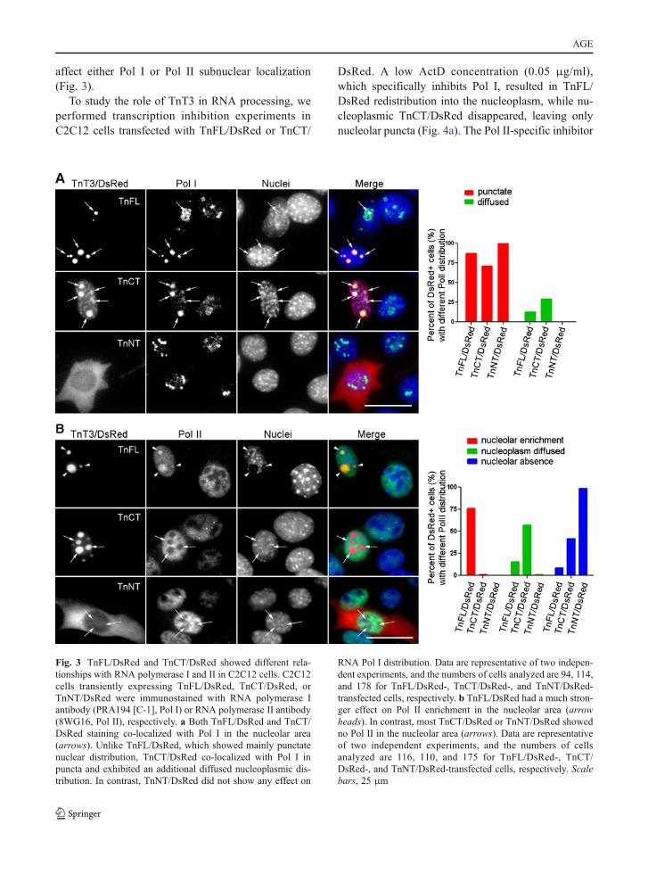
affect either Pol I or Pol II subnuclear localization(Fig. 3).
To study the role of TnT3 in RNA processing, weperformed transcription inhibition experiments inC2C12 cells transfected with TnFL/DsRed or TnCT/
DsRed. A low ActD concentration (0.05 μg/ml),which specifically inhibits Pol I, resulted in TnFL/DsRed redistribution into the nucleoplasm, while nu-cleoplasmic TnCT/DsRed disappeared, leaving onlynucleolar puncta (Fig. 4a). The Pol II-specific inhibitor
Fig. 3 TnFL/DsRed and TnCT/DsRed showed different rela-tionships with RNA polymerase I and II in C2C12 cells. C2C12cells transiently expressing TnFL/DsRed, TnCT/DsRed, orTnNT/DsRed were immunostained with RNA polymerase Iantibody (PRA194 [C-1], Pol I) or RNA polymerase II antibody(8WG16, Pol II), respectively. a Both TnFL/DsRed and TnCT/DsRed staining co-localized with Pol I in the nucleolar area(arrows). Unlike TnFL/DsRed, which showed mainly punctatenuclear distribution, TnCT/DsRed co-localized with Pol I inpuncta and exhibited an additional diffused nucleoplasmic dis-tribution. In contrast, TnNT/DsRed did not show any effect on
RNA Pol I distribution. Data are representative of two indepen-dent experiments, and the numbers of cells analyzed are 94, 114,and 178 for TnFL/DsRed-, TnCT/DsRed-, and TnNT/DsRed-transfected cells, respectively. b TnFL/DsRed had a much stron-ger effect on Pol II enrichment in the nucleolar area (arrowheads). In contrast, most TnCT/DsRed or TnNT/DsRed showedno Pol II in the nucleolar area (arrows). Data are representativeof two independent experiments, and the numbers of cellsanalyzed are 116, 110, and 175 for TnFL/DsRed-, TnCT/DsRed-, and TnNT/DsRed-transfected cells, respectively. Scalebars, 25 μm
AGE

DRB (25 μg/ml) showed effects mainly on TnCT/DsRed redistribution (Fig. 4b). When both Pol I andPol II were inhibited with a higher ActD concentration(20 μg/ml), fibrillarin puncta disappeared almost com-pletely and redistributed into the nucleoplasm. As aresult, both TnFL/DsRed and TnCT/DsRed were alsoredistributed into the nucleoplasm (Fig. 4c). This
transcription inhibition experiment and Pol I and/orPol II co-localization with TnT3 constructs suggest arole for TnT3 in RNA biogenesis.
Subcellular localization of TnT3/DsRed fusionproteins in mouse FDB fibers
To examine whether TnT3/DsRed fusion constructsshow subcellular distribution patterns similar to thoseobserved in the C2C12 cells in skeletal muscle fiber invivo, we performed in vivo electroporation experimentsin mouse FDB muscle. As early as 3 days after electro-poration, we observed expression of all constructs.When dissected, whole FDB muscles were observed atlow magnification (×4 objective), the TnNT/DsRed andcontrol DsRed showed a similar diffuse distributionthroughout the muscle, while the TnFL/DsRed, TnM/DsRed, and TnCT/DsRed showed mainly a punctatedistribution in addition to some large patches and diffusebackground. The TnFL/DsRed and particularly theTnCT/DsRed construct showed much finer puncta,reflecting possible nuclear localization (Fig. 5a).
We then dissected single FDB muscle fibers toobserve this subnuclear localization in more detail.Isolated fibers were stained with Hoechst 33342 to labelDNA, which allowed us to follow the nuclear localiza-tion of various TnT3/DsRed constructs (Fig. 5b). Athigh magnification (×40 objective), TnFL/DsRed andTnCT/DsRed localized in some myonuclei. Notably,TnFL/DsRed localized mainly between the strongestDNA-stained puncta (heterochromatin), while TnCT/DsRed mainly co-localized with the DNA stain. Also,TnFL/DsRed showed a striated pattern throughout thefiber, where fluorescence was dimmer than in the nucle-us. In contrast, TnCT/DsRed localized only in the myo-nuclei (Figs. 5b and S4).
TnNT/DsRed was also expressed throughout thefiber, including the myonuclei, yet the myonuclearDNA staining pattern was not affected. TnM/DsRedmainly localized in the cytoplasm and showed a punctatestriation pattern, reflecting its binding to the myofiberthin filaments. Like TnNT/DsRed, control DsRedexpressionwas distributed throughout the fiber (Fig. S5).
Endogenous TnT3 and its fragments changeexpression pattern with aging
To examine age-related changes in endogenous TnT3expression and nuclear localization, we collected the
Fig. 4 Transcription inhibition with actinomycin D (ActD) orDRB in TnFL/DsRed or TnCT/DsRed transiently transfectedC2C12 cells. a Incubating transfected cells in 0.05 μg/ml ActDfor 3 h did not seem to affect fibrillarin distribution, yet TnFL/DsRed was dispersed into the nucleoplasm. In contrast, TnCT/DsRed remained punctate and co-localized with some fibrillarinpuncta. Notably, the weak diffusive distribution of TnCT/DsRedin the nucleoplasm disappeared. b When only RNA Pol II wasinhibited with 25 μg/ml DRB for 3 h, fibrillarin adopted a largepunctate pattern in both TnFL/DsRed- and TnCT/DsRed-transfected cells. In contrast, TnCT/DsRed showed a diffusenucleoplasmic distribution while TnFL/DsRed remained mainlypunctate. c RNA Pol I and Pol II inhibition by 20 μg/ml ActD for3 h resulted in diffuse fibrillarin dispersion into the nucleoplasm.Consistently, both TnFL/DsRed and TnCT/DsRed showed mainlya diffuse nuclear distribution pattern. Data are representative oftwo independent experiments, and at least 50 cells were analyzedper group. Scale bars, 25 μm
AGE

whole cell lysis as well as the nuclear fractions fromyoung (3-month) and old (26–28-month) FVB mouseTA muscle for immunoblotting. We found slightlydecreased TnFL expression in the old compared tothe young group (Fig. 6a, b). Consistently, TnT3mRNA also decreased slightly with aging, as mea-sured by quantitative real-time PCR (Fig. 6c).Whole cell lysis and nuclear fractions from youngand old mice were further analyzed by immuno-bloting with TnT3, α-tubulin, and pan-actin antibodies.As expected, α-tubulin was found mainly in the wholecell lysis pool, while actin was in both fractions.Interestingly, besides full-length TnT3 (TnFL,
35 kDa), two smaller bands of 18 and 13 kDa weredetected in the nuclear fractions. These bands werespecific to NH2-terminal and the COOH-terminalepitope-targeted antibodies, respectively, and thuslikely represent endogenous NH2-terminal andCOOH-terminal fragments of full-length TnT3(Fig. 6d, e).
We compared the age groups for the relativeamount (normalized to actin) of TnNT, TnCT, andTnFL in the nuclear extracts. While TnNT did notdiffer, TnCT was 50% greater while TnFL was dra-matically decreased in the old group (P<0.05, n03)(Fig. 6d, f). The nuclear ratio between TnNT or TnCT
Fig. 5 Subcellular localization of TnT3/DsRed proteins inmouse FDB fibers. TnT3/DsRed constructs and control plas-mids were electroporated in vivo into the mouse FDB muscle. aExpression pattern of all constructs in whole isolated muscle.Control DsRed and TnNT/DsRed were expressed evenlythroughout the muscle. In contrast, TnFL/DsRed, TnM/DsRed,and TnCT/DsRed showed a punctate distribution. b Highermagnification imaging analysis of individual myofibers showedthat both TnFL/DsRed and TnCT/DsRed localized in somemyonuclei. Additionally, TnFL/DsRed showed weak striatedpattern. Notably, these two constructs localized at different
subnuclear domains as revealed by their different co-localization pattern with Hoechst 33342 DNA staining. TnNT/DsRed also expressed in the myonuclei, but the myonuclearDNA staining pattern was not affected. TnM/DsRed mainlylocalized in the cytoplasm as small puncta, while most of itwas highly ordered and reflects binding to myofibrils. Arrowspoint to nuclei in red fluorescence-positive areas. Scale bars,800 μm (a), 50 μm (b). Fibers are representative of at least 200fibers in each group from 2 to 13 experiments (DsRed, n03;TnFL/DsRed, n05; TnNT/DsRed, n06; TnM/DsRed, n02;TnCT/DsRed, n013)
AGE
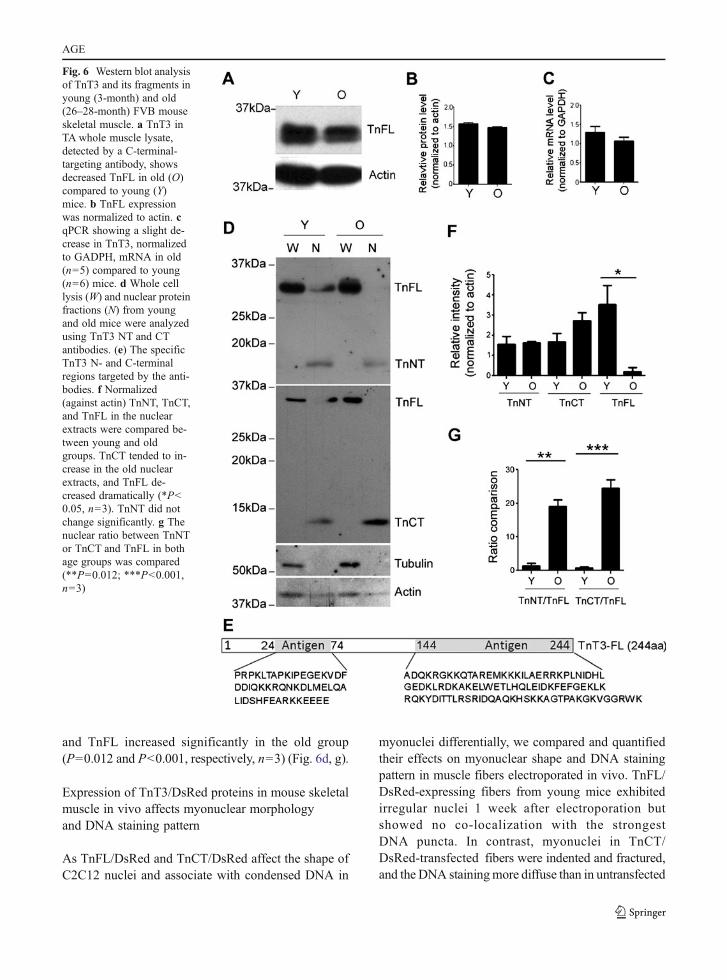
and TnFL increased significantly in the old group(P00.012 and P<0.001, respectively, n03) (Fig. 6d, g).
Expression of TnT3/DsRed proteins in mouse skeletalmuscle in vivo affects myonuclear morphologyand DNA staining pattern
As TnFL/DsRed and TnCT/DsRed affect the shape ofC2C12 nuclei and associate with condensed DNA in
myonuclei differentially, we compared and quantifiedtheir effects on myonuclear shape and DNA stainingpattern in muscle fibers electroporated in vivo. TnFL/DsRed-expressing fibers from young mice exhibitedirregular nuclei 1 week after electroporation butshowed no co-localization with the strongestDNA puncta. In contrast, myonuclei in TnCT/DsRed-transfected fibers were indented and fractured,and the DNA stainingmore diffuse than in untransfected
Fig. 6 Western blot analysisof TnT3 and its fragments inyoung (3-month) and old(26–28-month) FVB mouseskeletal muscle. a TnT3 inTA whole muscle lysate,detected by a C-terminal-targeting antibody, showsdecreased TnFL in old (O)compared to young (Y)mice. b TnFL expressionwas normalized to actin. cqPCR showing a slight de-crease in TnT3, normalizedto GADPH, mRNA in old(n05) compared to young(n06) mice. d Whole celllysis (W) and nuclear proteinfractions (N) from youngand old mice were analyzedusing TnT3 NT and CTantibodies. (e) The specificTnT3 N- and C-terminalregions targeted by the anti-bodies. f Normalized(against actin) TnNT, TnCT,and TnFL in the nuclearextracts were compared be-tween young and oldgroups. TnCT tended to in-crease in the old nuclearextracts, and TnFL de-creased dramatically (*P<0.05, n03). TnNT did notchange significantly. g Thenuclear ratio between TnNTor TnCT and TnFL in bothage groups was compared(**P00.012; ***P<0.001,n03)
AGE

neighboring myonuclei. As a control, TnNT/DsRedaffected neither myonuclear shape nor the DNAstaining pattern (Fig. 7a). Based on our findingthat the endogenous nuclear fraction of TnFL decreaseddramatically with aging, while TnCT increased, wecompared the effect of TnCT/DsRed on the myonucleifrom young and old mice. TnCT/DsRed-transfectedfibers from old mice showed elongated, broken myonu-clei, with a general loss of condensed DNA staining(heterochromatin) (Fig. 7b). The effect of TnCT/DsRed on nuclei shape and DNA pattern was greaterin old fibers than young fibers (Fig. 7c).
Overexpressing TnT3/DsRed fusion proteins resultsin apoptosis
To verify whether TnT3 constructs had toxiceffects, we performed Annexin V and 7-AADstaining to detect early and late apoptotic cells(Gil et al. 2011). The flow cytometry data
(Fig. 8) demonstrate that TnNT/DsRed did notincrease early or late apoptosis in the DsRed-positive cells compared to DsRed-transfected con-trol cells. However, early and late apoptosis increaseddramatically in cells expressing TnFL/DsRed or TnCT/DsRed. TnM/DsRed also showed a weaker toxiceffect than TnFL/DsRed or TnCT/DsRed cells.The strong cell toxicity of both TnFL/DsRed andTnCT/DsRed was also confirmed using the sameflow cytometry assay to test the NIH3T3 fibroblastcultures (Fig. S6).
Discussion
TnT is well known to mediate the interactionbetween the Tn complex and Tm, which is essen-tial for the contraction of calcium-activated striatedmuscle (Perry 1998). This regulatory functiontakes place in the myoplasm, where TnT binds
Fig. 7 Transient expression of TnT3/DsRed proteins in mouseskeletal muscle in vivo affects myonuclear phenotype. a TnFL,TnCT, and TnNT nuclear expression in young FDB musclefibers. Nuclei staining pattern was examined at two planes.Arrows indicate nuclei DNA staining in a clear, sharp punctatepattern; arrow heads indicate a blurred DNA staining pattern inthe TnCT/DsRed-positive nucleus. b TnCT/DsRed expression
in FDB muscle fibers from old FVB mice. Under the brightfield(BF) view, the TnCT/DsRed-positive nucleus showed abnormalshape and DNA stain (arrow heads) compared to neighboringnormal untransfected nuclei (arrows). c Effects of TnCT/DsRedon nuclear shape and DNA staining pattern in young (Y, 271nuclei) and old (O, 156 nuclei) muscle fibers from two experi-ments. Scale bars, 25 μm (a), 100 μm (b)
AGE

Tm. Recent studies support alternative roles forTnI. During Drosophila development, TnI andTm were found in the nucleus and to regulatechromosomal integrity (Sahota et al. 2009).Additionally, fast skeletal muscle TnI may alsolocalize in the mammalian cell nucleus and workas a co-activator of the nuclear receptor-estrogenreceptor-related alpha (Li et al. 2008).
Here, we report the novel finding that TnT—specifically, fast skeletal muscle TnT3—localizes
in the nucleus of myofibers as an intact or fragmentedprotein. The relationship with nucleolar structures andRNA polymerases implies a role for TnT3 in RNAtranscription regulation. Their cytotoxic effects, theirrelative abundance in young and old skeletal musclemyonuclei, and differences between intact TnT3 andthe COOH-terminal fragment in subnuclear localizationall support the idea that TnT3 plays a surprising andcritical role in aging skeletal muscle and justify furtherstudies.
Fig. 8 TnCT/DsRed and TnFL/DsRed overexpression inducesapoptosis in C2C12 cells. Cells were analyzed with a FACSCa-libur flow cytometer 48 h posttransfection. Data were collectedon at least 100,000 freshly stained cells. Representative analysesof 7-AAD and Annexin V staining (b) followed pregating onDsRed (a). b Results were plotted as fluorescence intensity ofAnnexin V as a function of fluorescence intensity of 7-AAD.The numbers in each square represent the percentage of
Annexin V−/7-AAD−(viable cells, left bottom corner); AnnexinV+/7-AAD−(early apoptotic cells, right bottom corner);Annexin V+/7-AAD+(late apoptotic cells, right top corner);and Annexin V−/7-AAD+(broken cells, left top corner) in theDsRed-positive cell population. Data shown are representativeof two independent experiments. c The percent of total apoptoticcells was obtained by adding early (Annexin V+/7-AAD−) andlate apoptotic cells (Annexin V+/7-AAD+)
AGE
Evidence for TnT3 nuclear localization
TnFL/DsRed showed a striated distribution in thesarcoplasm and/or the nucleus of mouse FDB myofib-ers. The competition between expressed and endoge-nous TnT3 may explain the weak striated pattern. Inaddition, several lines of evidence support nuclearlocalization of TnT3. First, in the western blot assayof muscle nuclear protein fractions, two specific anti-bodies, targeting either the TnT3 NH2- or COOH-terminal epitope, detected endogenous TnT3. Theseantibodies detected two TnT3 fragments with differentmolecular weights that supposedly contain the domaincarrying either the NH2-terminal or COOH-terminalepitope, respectively. Second, overexpressing full-length TnT3 or fragments with COOH-terminallytagged DsRed showed distinct subcellular localiza-tion. Not only did the overexpressed TnT3 localizein the nuclei of undifferentiated C2C12 myoblasts orother non-muscle cells (NIH3T3 fibroblasts) but alsoin myonuclei in vivo. Similar subcellular localizationobserved with GFP NH2-terminally tagged to TnT3 inC2C12 cells (Fig. S3) deters the possibility that TnT3nuclear localization is an artifact resulting from theexpression of the fusion protein or the tag itself. Inaddition, truncation analysis indicates that the nuclearlocalization signal resides in the COOH-terminal re-gion. Third, fibrillarin immunofluorescence stainingshowed slight differences between the subnuclear lo-calization of full-length TnT3 or its COOH-terminalfragment and that of the nucleolus, consistent withreports that the NH2-terminal region may affect theCOOH-terminal region’s function (Wei and Jin 2011)or protein conformation. The fusion TnT3 protein hadmuch higher level in the nucleus than that incor-porated into the myofibrils; therefore, the structuralmanipulation might have altered the functional behaviorof TnT3. This observation may implicate that misfoldedor damaged TnT3 would have increased tendency totranslocate to the nucleus. Although we have not testedthe hypothesis that the COOH-terminal regionmeditates TnT3 nuclear translocation through inter-action with TnI and Tm, they have been reportedin the nucleus (Li et al. 2008; Sahota et al. 2009).Conversely, TnT may regulate TnI and Tm nucleartranslocation. Considering the high degree of con-servation in this region across species (Jin et al.2008), future work should determine the nuclearlocalization signal for other TnT isoforms.
TnT3 subnuclear localization and myonuclearapoptosis and/or dysfunction
Effects on cell apoptosis vary among the TnT3 regionswhen expressed in C2C12 myoblasts or NIH3T3fibroblasts, consistent with a recent finding from theJin group on cytotoxicity of the non-myofilament-associated TnT fragments, including the NH2-terminal variable region from human cardiac TnTand the highly conserved middle and COOH-terminal regions from the mouse slow TnT (Jeong etal. 2009). We generated a TnT3 fragment with amolecular weight similar to that detected by theTnT3 COOH-terminal epitope antibody by subcloningthe last 85 amino acids of the COOH-terminal region.Although our COOH-terminal region is 25 amino acidsshorter and the middle region 13 amino acids shorterthan that studied by the Jin group, sequence comparisonindicated that the middle and COOH-terminal regionswe chose are highly conserved among TnT isoforms,possibly explaining the similar toxicity of these TnTconstructs. Our findings confirm that middle andCOOH-terminal regions are not only conserved in thepeptide sequence but also in their function (apoptosis).Note that the DsRed conjugation enabled us to analyzeTnT3 subnuclear localization and to elucidate themechanism involved in cytotoxicity, which is prob-ably mediated by nuclear translocation and nucle-oli targeting.
The nucleolus is a large nuclear domain with acontrasted structure directly involved in ribosome bio-synthesis (Sirri et al. 2008). Its function is tightlylinked to cell growth, proliferation, and other impor-tant cell events, including cell cycle regulation, senes-cence, and stress responses (Visintin and Amon 2000;Guarente 1997; Kennedy et al. 1997; Sherr and Weber2000; Olson 2004). Changes in nucleolar organizationare widely thought to represent diagnostic markers fordifferent pathologies (Zimber et al. 2004). A recentproteomic study identified hundreds of human nucle-olar proteins (Andersen et al. 2005), among them,fibrillarin, which is known to play an important rolein pre-rRNA processing during ribosomal biogenesis.Knocking down fibrillarin in mammalian cells hasbeen reported to lead to abnormal nuclear morphologyand inhibition of cell proliferation (Amin et al. 2007).TnT3/DsRed or TnCT/DsRed transfection alters fibril-larin location, which may lead to abnormal nuclearshape and, consequently, cell toxicity. Nuclear
AGE
morphology has been shown to influence fundamentalaspects of nuclear function, including chromatinremodeling and gene transcription (Dalby et al.2007; Itano et al. 2003; Lammerding et al. 2004;Thomas et al. 2002). Changes in myonuclear morphol-ogy, including shrinkage, fracture, and fragmentation,indicate cell death. Our flow cytometry results inC2C12 myoblasts, and NIH3T3 fibroblasts furthersupport this link between apoptosis and altered nuclearmorphology. Further investigation of TnT3’s down-stream partners will clarify the mechanism that acti-vates the cell death pathway.
TnT3 nuclear imbalance and muscle aging
Myonuclear apoptosis and structural changes relatedto impaired RNA processing occur during muscleaging as reported for sarcopenia (Malatesta et al.2009, 2010; Alway and Siu 2008; Dirks andLeeuwenburgh 2002). Although apoptotic nuclei havebeen documented in skeletal myofibers, the precisesignaling mechanisms leading to nuclear loss are notfully understood. Sarcopenia-related apoptosis hasbeen shown to be regulated differently in fast- andslow-twitch muscles; type II fast muscle is more affectedand exhibits fewer satellite cells (Rice and Blough 2006;Verdijk et al. 2007). Our findings on TnT3 nuclearlocalization and cytotoxicity provide new insight intothis preferential type II fast fiber atrophy/loss withaging. TnT3 is a muscle-specific fast isoform, andits expression has been related to the fast myosinisoforms at the single fiber level (Brotto et al.2006). Our findings that nuclear TnT3 COOH-terminal fragment expression increases with agingand that this fragment has several negative effectsto the cell offer a plausible link to sarcopenia.
We have found that the nuclear full-length TnT3decreases while its fragments increase with aging, thusincreasing the ratio between TnT3 fragments, in par-ticular the COOH-terminal fragments. We hypothesizethat this imbalance plays a physiological role in mus-cle nuclei. Muscle electroporation was relatively inef-ficient in inducing myonuclear expression, socalculating the number of lost nuclei and whethermuscle force was affected was difficult. The differen-tial consequences of transfecting full-length TnT3 orthe COOH-terminal region construct may beexplained by their relationship with the nucleolusand different nucleolar proteins, including fibrillarin
and RNA polymerases I and II. RNA Pol I subunitsare enriched in the nucleolar FC region, and RNA PolII activity is largely absent from this area and localizesin the nucleoplasm (Hernandez-Verdun 2006; Boisvertet al. 2007; Sirri et al. 2008). Our study found TnT3/DsRed mainly surrounded by the endogenous fibril-larin (similar to Pol I), which formed the “nucleolarcap” structure (Fox and Lamond 2010; Shav-Tal et al.2005) and recruited endogenous Pol II to the nucleolararea. In contrast, the TnCT/DsRed mainly co-localizedwith fibrillarin and Pol I, but not Pol II, in the nucle-olar area, with some remaining in the nucleoplasm (asPol I does), where the Pol II is mainly distributed.Transcription inhibition with ActD or DRB demon-strated that TnT3 and the COOH-terminal fragmentsare associated with RNA transcription activities (vanKoningsbruggen et al. 2004) and interact with Pol Iand Pol II differentially. These differences in subnu-clear localization are also reflected by the DNA stain-ing pattern: TnT3/DsRed clearly localizes between thecondensed heterochromatin DNA stain, while TnCT/DsRed showed less sharp staining (Figs. 5b and 7a).Theoretically, therefore, the fragments function differ-ently, considering that most subnuclear bodies, includ-ing Cajal and PML bodies and nuclear speckles, residein the interchromatin space (Lamond and Spector2003). With the reported modifying effects of theTnT NH2-terminal region on middle and COOH-terminal region functions (Wei and Jin 2011), furtheridentification of nuclear partners that may bind to full-length TnT3 or the COOH-terminal region will eluci-date their functional differences.
In conclusion, fast skeletal muscle TnT3 is localizedin the nucleus and is found predominantly at full lengthin the young, while its fragments predominate in the old,which may lead to aging-related myonuclear death ordysfunction. In addition to its potential clinical use as adiagnostic marker of muscle damage, TnT may be atherapeutic target to prevent muscle aging and damage.In either case, blocking TnT fragmentation and/ornuclear translocation will be considered in futurework focusing on the regulatory mechanism ofTnT3 fragmentation and the role of the proteases—calpains and caspases, both reported to cleave differentTnTs (Dargelos et al. 2007, 2008; Wei and Jin2011). Since the COOH-terminal region is highlyconserved among isoforms and across species, fur-ther work should also compare the cardiac andslow muscle isoforms.
AGE

Acknowledgments This study was supported by grants fromthe National Institutes of Health/National Institute on Aging(AG13934, AG033385, AG15820, and FIRCA-BB TW008091)and the Muscular Dystrophy Association (MDA #33149) toOsvaldo Delbono and the Wake Forest Claude D. Pepper OlderAmericans Independence Center (P30-AG21332).
References
Alway SE, Siu PM (2008) Nuclear apoptosis contributes tosarcopenia. Exerc Sport Sci Rev 36(2):51–57
Amin MA, Matsunaga S, Ma N, Takata H, Yokoyama M,Uchiyama S, Fukui K (2007) Fibrillarin, a nucleolar pro-tein, is required for normal nuclear morphology and cellu-lar growth in HeLa cells. Biochem Biophys Res Commun360(2):320–326
Andersen JS, Lam YW, Leung AK, Ong SE, Lyon CE, LamondAI, Mann M (2005) Nucleolar proteome dynamics. Nature433(7021):77–83
Barbieri M, Ferrucci L, Ragno E, Corsi A, Bandinelli S, BonafeM, Olivieri F, Giovagnetti S, Franceschi C, Guralnik JM,Paolisso G (2003) Chronic inflammation and the effect ofIGF-I on muscle strength and power in older persons. Am JPhysiol Endocrinol Metab 284(3):E481–E487
Biesiadecki BJ, Jin JP (2002) Exon skipping in cardiac troponinT of turkeys with inherited dilated cardiomyopathy. J BiolChem 277(21):18459–18468
Biesiadecki BJ, Chong SM, Nosek TM, Jin JP (2007) TroponinT core structure and the regulatory NH2-terminal variableregion. Biochemistry 46(5):1368–1379
Birbrair A, Wang ZM, Messi ML, Enikolopov GN, Delbono O(2011) Nestin-GFP transgene reveals neural precursor cellsin adult skeletal muscle. PLoS One 6(2):e16816
Boisvert FM, van Koningsbruggen S, Navascues J, Lamond AI(2007) The multifunctional nucleolus. Nat Rev Mol CellBiol 8(7):574–585
Brooks SV, Faulkner JA (1991) Maximum and sustained powerof extensor digitorum longus muscles from young, adult,and old mice. J Gerontol 46(1):B28–B33
Brotto MA, Biesiadecki BJ, Brotto LS, Nosek TM, Jin JP (2006)Coupled expression of troponin T and troponin I isoforms insingle skeletal muscle fibers correlates with contractility. AmJ Physiol Cell Physiol 290(2):C567–C576
Buford TW, Anton SD, Judge AR, Marzetti E, Wohlgemuth SE,Carter CS, Leeuwenburgh C, Pahor M, Manini TM (2010)Models of accelerated sarcopenia: critical pieces for solv-ing the puzzle of age-related muscle atrophy. Ageing ResRev 9(4):369–383
Carlson ME, Conboy IM (2007) Loss of stem cell regenerativecapacity within aged niches. Aging Cell 6(3):371–382
Chandra M, Montgomery DE, Kim JJ, Solaro RJ (1999)The N-terminal region of troponin T is essential for themaximal activation of rat cardiac myofilaments. J MolCell Cardiol 31(4):867–880
Dalby MJ, Gadegaard N, Herzyk P, Sutherland D, Agheli H,Wilkinson CD, Curtis AS (2007) Nanomechanotransductionand interphase nuclear organization influence on genomiccontrol. J Cell Biochem 102(5):1234–1244
Dargelos E, Brule C, Combaret L, Hadj-Sassi A, Dulong S,Poussard S, Cottin P (2007) Involvement of the calcium-dependent proteolytic system in skeletal muscle aging. ExpGerontol 42(11):1088–1098
Dargelos E, Poussard S, Brule C, Daury L, Cottin P(2008) Calcium-dependent proteolytic system andmuscle dysfunctions: a possible role of calpains insarcopenia. Biochimie 90(2):359–368
Day K, Shefer G, Shearer A, Yablonka-Reuveni Z (2010) Thedepletion of skeletal muscle satellite cells with age isconcomitant with reduced capacity of single progenitorsto produce reserve progeny. Dev Biol 340(2):330–343
Degens H, Alway SE (2003) Skeletal muscle function andhypertrophy are diminished in old age. Muscle Nerve 27(3):339–347
Degens H, Alway SE (2006) Control of muscle size duringdisuse, disease, and aging. Int J Sports Med 27(2):94–99
Delbono O (2011) Expression and regulation of excitation–contraction coupling proteins in aging skeletal muscle.Curr Aging Sci (in press)
DiFranco M, Neco P, Capote J, Meera P, Vergara JL (2006)Quantitative evaluation of mammalian skeletal muscle as aheterologous protein expression system. Protein Expr Purif47(1):281–288
Dirks A, Leeuwenburgh C (2002) Apoptosis in skeletal musclewith aging. Am J Physiol Regul Integr Comp Physiol 282(2):R519–R527
Dousset T, Wang C, Verheggen C, Chen D, Hernandez-VerdunD, Huang S (2000) Initiation of nucleolar assembly isindependent of RNA polymerase I transcription. Mol BiolCell 11(8):2705–2717
Feng HZ, Biesiadecki BJ, Yu ZB, Hossain MM, Jin JP (2008)Restricted N-terminal truncation of cardiac troponin T: anovel mechanism for functional adaptation to energeticcrisis. J Physiol 586(14):3537–3550
Fox AH, Lamond AI (2010) Paraspeckles. Cold Spring HarbPerspect Biol 2(7):a000687
Geeves MA, Holmes KC (1999) Structural mechanism of mus-cle contraction. Annu Rev Biochem 68:687–728
Gil C, Falques A, Sarro E, Cubi R, Blasi J, Aguilera J,Itarte E (2011) Protein kinase CK2 associates to lipidrafts and its pharmacological inhibition enhances neu-rotransmitter release. FEBS Lett 585(2):414–420
Gomes AV, Guzman G, Zhao J, Potter JD (2002) Cardiactroponin T isoforms affect the Ca2+ sensitivity andinhibition of force development. Insights into the roleof troponin T isoforms in the heart. J Biol Chem 277(38):35341–35349
Gonzalez E, Delbono O (2001) Age-dependent fatigue in singleintact fast- and slow fibers from mouse EDL and soleusskeletal muscles. Mech Ageing Dev 122(10):1019–1032
Gonzalez E, Messi ML, Delbono O (2000) The specific force ofsingle intact extensor digitorum longus and soleus mousemuscle fibers declines with aging. J Membr Biol 178(3):175–183
Gordon AM, Homsher E, Regnier M (2000) Regulation ofcontraction in striated muscle. Physiol Rev 80(2):853–924
Guarente L (1997) Link between aging and the nucleolus. GenesDev 11(19):2449–2455
Hernandez-Verdun D (2006) Nucleolus: from structure todynamics. Histochem Cell Biol 125(1–2):127–137
AGE
Itano N, Okamoto S, Zhang D, Lipton SA, Ruoslahti E (2003)Cell spreading controls endoplasmic and nuclear calcium: aphysical gene regulation pathway from the cell surface tothe nucleus. Proc Natl Acad Sci USA 100(9):5181–5186
Jeong EM, Wang X, Xu K, Hossain MM, Jin JP (2009)Nonmyofilament-associated troponin T fragments induceapoptosis. Am J Physiol Heart Circ Physiol 297(1):H283–H292
Jimenez-Moreno R, Wang ZM, Gerring RC, Delbono O (2008)Sarcoplasmic reticulum Ca2+ release declines in musclefibers from aging mice. Biophys J 94(8):3178–3188
Jimenez-Moreno R, Wang ZM, Messi ML, Delbono O (2010)Sarcoplasmic reticulum Ca2+ depletion in adult skeletalmuscle fibres measured with the biosensor D1ER.Pflugers Arch 459(5):725–735
Jin JP, Root DD (2000) Modulation of troponin T molecularconformation and flexibility by metal ion binding to theNH2-terminal variable region. Biochemistry 39(38):11702–11713
Jin JP, Zhang Z, Bautista JA (2008) Isoform diversity, regula-tion, and functional adaptation of troponin and calponin.Crit Rev Eukaryot Gene Expr 18(2):93–124
Kennedy BK, Gotta M, Sinclair DA, Mills K, McNabb DS,Murthy M, Pak SM, Laroche T, Gasser SM, Guarente L(1997) Redistribution of silencing proteins from telomeresto the nucleolus is associated with extension of life span inS. cerevisiae. Cell 89(3):381–391
Klitgaard H, Mantoni M, Schiaffino S, Ausoni S, Gorza L,Laurent-Winter C, Schnohr P, Saltin B (1990) Function,morphology and protein expression of ageing skeletal mus-cle: a cross-sectional study of elderly men with differenttraining backgrounds. Acta Physiol Scand 140(1):41–54
Lammerding J, Schulze PC, Takahashi T, Kozlov S, Sullivan T,Kamm RD, Stewart CL, Lee RT (2004) Lamin A/C defi-ciency causes defective nuclear mechanics and mechano-transduction. J Clin Invest 113(3):370–378
Lamond AI, Spector DL (2003) Nuclear speckles: a model fornuclear organelles. Nat Rev Mol Cell Biol 4(8):605–612
Lannergren J, Westerblad H (1987) The temperature depen-dence of isometric contractions of single, intact fibresdissected from a mouse foot muscle. J Physiol 390:285–293
Larsson L (1978) Morphological and functional characteristicsof the ageing skeletal muscle in man. A cross-sectionalstudy. Acta Physiol Scand Suppl 457:1–36
Larsson L, Ansved T (1995) Effects of ageing on the motor unit.Prog Neurobiol 45(5):397–458
Li Y, Chen B, Chen J, Lou G, Chen S, Zhou D (2008) Fastskeletal muscle troponin I is a co-activator of estrogenreceptor-related receptor alpha. Biochem Biophys ResCommun 369(4):1034–1040
Malatesta M, Perdoni F, Muller S, Zancanaro C, Pellicciari C(2009) Nuclei of aged myofibres undergo structural andfunctional changes suggesting impairment in RNA pro-cessing. Eur J Histochem 53(2):97–106
Malatesta M, Perdoni F, Muller S, Pellicciari C, Zancanaro C(2010) Pre-mRNA processing is partially impaired in satellitecell nuclei from aged muscles. J Biomed Biotechnol2010:410405
Marzetti E, Privitera G, Simili V, Wohlgemuth SE, Aulisa L,Pahor M, Leeuwenburgh C (2010) Multiple pathways to
the same end: mechanisms of myonuclear apoptosis insarcopenia of aging. ScientificWorldJournal 10:340–349
Morse CI, Thom JM, Davis MG, Fox KR, Birch KM, NariciMV (2004) Reduced plantarflexor specific torque in theelderly is associated with a lower activation capacity. Eur JAppl Physiol 92(1–2):219–226
Morse CI, Thom JM, Reeves ND, Birch KM, Narici MV (2005)In vivo physiological cross-sectional area and specificforce are reduced in the gastrocnemius of elderly men. JAppl Physiol 99(3):1050–1055
Narici MV, Maganaris CN (2006) Adaptability of elderly humanmuscles and tendons to increased loading. J Anat 208(4):433–443
Ogut O, Jin JP (1996) Expression, zinc-affinity purification, andcharacterization of a novel metal-binding cluster in tropo-nin T: metal-stabilized alpha-helical structure and effects ofthe NH2-terminal variable region on the conformation ofintact troponin T and its association with tropomyosin.Biochemistry 35(51):16581–16590
Olson MO (2004) Sensing cellular stress: another new functionfor the nucleolus? Sci STKE 2004 (224):pe10
Onambele GL, Narici MV, Maganaris CN (2006) Calf muscle-tendon properties and postural balance in old age. J ApplPhysiol 100(6):2048–2056
Pan BS, Gordon AM, Potter JD (1991) Deletion of the first 45NH2-terminal residues of rabbit skeletal troponin Tstrengthens binding of troponin to immobilized tropomyo-sin. J Biol Chem 266(19):12432–12438
Payne AM, Zheng Z, Gonzalez E, Wang ZM,Messi ML, DelbonoO (2004) External Ca(2+)-dependent excitation–contractioncoupling in a population of ageing mouse skeletal musclefibres. J Physiol 560(Pt 1):137–155
Perry SV (1998) Troponin T: genetics, properties and function. JMuscle Res Cell Motil 19(6):575–602
Pinol-Roma S, Dreyfuss G (1992) Shuttling of pre-mRNA bindingproteins between nucleus and cytoplasm. Nature 355(6362):730–732
Reiser PJ, Greaser ML, Moss RL (1992) Developmentalchanges in troponin T isoform expression and tensionproduction in chicken single skeletal muscle fibres. JPhysiol 449:573–588
RenganathanM, Messi ML, Delbono O (1998) Overexpression ofIGF-1 exclusively in skeletal muscle prevents age-relateddecline in the number of dihydropyridine receptors. J BiolChem 273(44):28845–28851
Rice KM, Blough ER (2006) Sarcopenia-related apoptosis isregulated differently in fast- and slow-twitch muscles of theaging F344/N×BN rat model. Mech Ageing Dev 127(8):670–679
Runge M, Rittweger J, Russo CR, Schiessl H, Felsenberg D(2004) Is muscle power output a key factor in the age-related decline in physical performance? A comparison ofmuscle cross section, chair-rising test and jumping power.Clin Physiol Funct Imaging 24(6):335–340
Sahota VK, Grau BF, Mansilla A, Ferrus A (2009) Troponin Iand tropomyosin regulate chromosomal stability and cellpolarity. J Cell Sci 122(Pt 15):2623–2631
Shav-Tal Y, Blechman J, Darzacq X, Montagna C, Dye BT,Patton JG, Singer RH, Zipori D (2005) Dynamic sortingof nuclear components into distinct nucleolar caps duringtranscriptional inhibition. Mol Biol Cell 16(5):2395–2413
AGE

Shefer G, Rauner G, Yablonka-Reuveni Z, Benayahu D (2010)Reduced satellite cell numbers and myogenic capacity inaging can be alleviated by endurance exercise. PLoS One 5(10):e13307
Sherr CJ, Weber JD (2000) The ARF/p53 pathway. Curr OpinGenet Dev 10(1):94–99
Sirri V, Urcuqui-Inchima S, Roussel P, Hernandez-Verdun D(2008) Nucleolus: the fascinating nuclear body. HistochemCell Biol 129(1):13–31
Siu PM, Pistilli EE, Murlasits Z, Alway SE (2006) Hindlimbunloading increases muscle content of cytosolic but notnuclear Id2 and p53 proteins in young adult and aged rats.J Appl Physiol 100(3):907–916
Snijders T, Verdijk LB, van Loon LJ (2009) The impact ofsarcopenia and exercise training on skeletal muscle satellitecells. Ageing Res Rev 8(4):328–338
Szczesna D, Potter JD (2002) The role of troponin in the Ca(2+)-regulation of skeletal muscle contraction. Results Probl CellDiffer 36:171–190
Taylor JR, Zheng Z,Wang ZM, Payne AM,MessiML, Delbono O(2009) Increased CaVbeta1A expression with aging contrib-utes to skeletal muscle weakness. Aging Cell 8(5):584–594
Thomas CH, Collier JH, Sfeir CS, Healy KE (2002)Engineering gene expression and protein synthesis bymodulation of nuclear shape. Proc Natl Acad Sci USA 99(4):1972–1977
Thomas DR (2010) Sarcopenia. Clin Geriatr Med 26(2):331–346Tobacman LS (1996) Thin filament-mediated regulation of cardiac
contraction. Annu Rev Physiol 58:447–481Tomlinson BE, Irving D, Rebeiz JJ (1973) Total numbers of
limb motor neurones in the human lumbosacral cord and ananalysis of the accuracy of various sampling procedures. JNeurol Sci 20(3):313–327
van Koningsbruggen S, Dirks RW, Mommaas AM, OnderwaterJJ, Deidda G, Padberg GW, Frants RR, van der Maarel SM
(2004) FRG1P is localised in the nucleolus, Cajal bodies,and speckles. J Med Genet 41(4):e46
Verdijk LB, Koopman R, Schaart G, Meijer K, Savelberg HH,van Loon LJ (2007) Satellite cell content is specificallyreduced in type II skeletal muscle fibers in the elderly. AmJ Physiol Endocrinol Metab 292(1):E151–E157
Visintin R, Amon A (2000) The nucleolus: the magician’s hatfor cell cycle tricks. Curr Opin Cell Biol 12(6):752
Wang J, Jin JP (1997) Primary structure and developmentalacidic to basic transition of 13 alternatively spliced mousefast skeletal muscle troponin T isoforms. Gene 193(1):105–114
Wang J, Jin JP (1998) Conformational modulation of troponin Tby configuration of the NH2-terminal variable region andfunctional effects. Biochemistry 37(41):14519–14528
Wei B, Jin JP (2011) Troponin T isoforms and posttranscrip-tional modifications: evolution, regulation and function.Arch Biochem Biophys 505(2):144–154
Zhang T, Zaal KJ, Sheridan J, Mehta A, Gundersen GG, RalstonE (2009) Microtubule plus-end binding protein EB1 isnecessary for muscle cell differentiation, elongation andfusion. J Cell Sci 122(Pt 9):1401–1409
Zhang Z, Jin JP, Root DD (2004) Binding of calcium ions to anavian flight muscle troponin T. Biochemistry 43(9):2645–2655
Zhang Z, Biesiadecki BJ, Jin JP (2006) Selective deletion of theNH2-terminal variable region of cardiac troponin T inischemia reperfusion by myofibril-associated mu-calpaincleavage. Biochemistry 45(38):11681–11694
Zimber A, Nguyen QD, Gespach C (2004) Nuclear bodies andcompartments: functional roles and cellular signalling inhealth and disease. Cell Signal 16(10):1085–1104
Zinna EM, Yarasheski KE (2003) Exercise treatment to coun-teract protein wasting of chronic diseases. Curr Opin ClinNutr Metab Care 6(1):87–93
AGE