Evaluation of the pituitary\p=n-\gonadalresponse to GnRH, and adrenal status, in the leopard...
10
Evaluation of the pituitary\p=n-\gonadalresponse to GnRH, and adrenal status, in the leopard (Panthera pardus japonensis) and tiger (Panthera tigris) J. L. Brown, K. L. Goodrowe, L. G. Simmons, D. L. Armstrong and D. E. Wildt Departments of * Obstetrics and Gynecology, and fPhysiology, Uniformed Services University of the Health Sciences, Bethesda, MD 20814; %National Zoological Park, Smithsonian Institution, Washington, DC 20008; and%Henry Doorly Zoo, Omaha, E 68107, U.S.A. Summary. Frequent blood samples were collected to study hormonal responses to GnRH in male and female leopards and tigers. Animals were anaesthetized with ketamine\p=n-\HCl and blood samples were collected every 5 min for 15 min before and 160 min after i.v. administration of GnRH (1 \g=m\g/kgbody weight) or saline. No differences in serum cortisol concentrations were observed between sexes within species, but mean cortisol was 2-fold greater in leopards than tigers. GnRH induced a rapid rise in LH in all animals (18\m=.\3\m=+-\ 0\m=.\9min to peak). Net LH peak height above pretreatment levels was 3-fold greater in males than conspecific females and was also greater in tigers than leopards. Serum FSH increased after GnRH, although the magnitude of response was less than that observed for LH. Basal LH and FSH and GnRH-stimulated FSH con- centrations were not influenced by sex or species. Serum testosterone increased within 30\p=n-\40min after GnRH in 3/3 leopard and 1/3 tiger males. Basal testosterone was 3-fold greater in tiger than leopard males. LH pulses (1\p=n-\2pulses/3 h) were detected in 60% of saline-treated animals, suggesting pulsatile gonadotrophin secretion; however, in males concomitant testosterone pulses were not observed. These results indicate that there are marked sex and species differences in basal and GnRH-stimulated hormonal responses between felids of the genus Panthera which may be related to differences in adrenal activity. Keywords: GnRH, leopard, tiger, LH, cortisol, testosterone Introduction Basic endocrine characteristics have been studied in a number of species of wild felids including the lion (Panthera leo), cheetah (Acinonyx jubatus), tiger (Panthera tigris), clouded leopard (Neofelis nebulosa), leopard (Panthera pardus) and puma (Felis concolor) (Schmidt et al, 1979; Wildt et al, 1984a, 1986a, 1987a (review); Seal et al, 1985). These investigations confirmed that taxonomically related felids exhibit species-specific endocrine patterns. Circulating cortisol concentrations are greater in North Chinese (P. p. japonensis) and clouded leopards than in tigers or pumas, and the concentrations in tigers and pumas are greater than those in cheetahs or domestic cats (Carter et al, 1984; Wildt et al, 1987a, b). Serum testosterone concentrations are similar among male tigers, pumas and clouded leopards but are lower in cheetahs (Wildt et al, 1984a, b, 1987a, b). Serum LH concentrations appear less species-dependent, but can vary considerably within individual animals presumably due to episodic release (Wildt et al, 1984a, b, 1987a; Goodrowe et al, 1985). The infrequent blood-sampling protocols used in these earlier studies, however, precluded identification of pulsatile hormone secretion.
-
Upload
independent -
Category
Documents
-
view
1 -
download
0
Transcript of Evaluation of the pituitary\p=n-\gonadalresponse to GnRH, and adrenal status, in the leopard...
Evaluation of the pituitary\p=n-\gonadalresponse to GnRH,and adrenal status, in the leopard (Panthera pardus
japonensis) and tiger (Panthera tigris)J. L. Brown, K. L. Goodrowe, L. G. Simmons, D. L. Armstrong and
D. E. WildtDepartments of * Obstetrics and Gynecology, and fPhysiology, Uniformed Services University of the
Health Sciences, Bethesda, MD 20814; %National Zoological Park, Smithsonian Institution,Washington, DC 20008; and%Henry Doorly Zoo, Omaha, E 68107, U.S.A.
Summary. Frequent blood samples were collected to study hormonal responses to GnRHin male and female leopards and tigers. Animals were anaesthetized with ketamine\p=n-\HCland blood samples were collected every 5 min for 15 min before and 160 min after i.v.administration of GnRH (1 \g=m\g/kgbody weight) or saline. No differences in serum
cortisol concentrations were observed between sexes within species, but mean cortisolwas 2-fold greater in leopards than tigers. GnRH induced a rapid rise in LH in allanimals (18\m=.\3\m=+-\0\m=.\9min to peak). Net LH peak height above pretreatment levels was
3-fold greater in males than conspecific females and was also greater in tigers thanleopards. Serum FSH increased after GnRH, although the magnitude of response was
less than that observed for LH. Basal LH and FSH and GnRH-stimulated FSH con-
centrations were not influenced by sex or species. Serum testosterone increased within30\p=n-\40min after GnRH in 3/3 leopard and 1/3 tiger males. Basal testosterone was 3-foldgreater in tiger than leopard males. LH pulses (1\p=n-\2pulses/3 h) were detected in 60% ofsaline-treated animals, suggesting pulsatile gonadotrophin secretion; however, in malesconcomitant testosterone pulses were not observed. These results indicate that there aremarked sex and species differences in basal and GnRH-stimulated hormonal responsesbetween felids of the genus Panthera which may be related to differences in adrenalactivity.Keywords: GnRH, leopard, tiger, LH, cortisol, testosterone
Introduction
Basic endocrine characteristics have been studied in a number of species of wild felids including thelion (Panthera leo), cheetah (Acinonyx jubatus), tiger (Panthera tigris), clouded leopard (Neofelisnebulosa), leopard (Panthera pardus) and puma (Felis concolor) (Schmidt et al, 1979; Wildt et al,1984a, 1986a, 1987a (review); Seal et al, 1985). These investigations confirmed that taxonomicallyrelated felids exhibit species-specific endocrine patterns. Circulating cortisol concentrations are
greater in North Chinese (P. p. japonensis) and clouded leopards than in tigers or pumas, and theconcentrations in tigers and pumas are greater than those in cheetahs or domestic cats (Carter et
al, 1984; Wildt et al, 1987a, b). Serum testosterone concentrations are similar among male tigers,pumas and clouded leopards but are lower in cheetahs (Wildt et al, 1984a, b, 1987a, b). Serum LHconcentrations appear less species-dependent, but can vary considerably within individual animalspresumably due to episodic release (Wildt et al, 1984a, b, 1987a; Goodrowe et al, 1985). Theinfrequent blood-sampling protocols used in these earlier studies, however, precluded identificationof pulsatile hormone secretion.
Administration of GnRH has been useful for evaluating pituitary-gonadal function in manymammalian species, including the domestic cat (Chakraborty et al, 1979; Goodrowe et al, 1985),as well as for studying the mechanisms and regulation of fertility (Sandow, 1982). To date, theeffects of GnRH on wild felids have been limited to the cheetah (Wildt et al, 1984a) and cloudedleopard (Wildt et al, 1986a). As propagation of captive animals continues to play a role in preserv¬ing rare species, it becomes increasingly important to establish a data base of hormonal norms forunderstanding the basic biology and evaluating the reproductive potential of these animals.
In this study the hormonal responses ofmale and female Chinese leopards and tigers to exogenousGnRH were compared while simultaneously assessing species variability in adrenal activity. Afrequent blood sampling protocol was used to monitor closely exogenous GnRH-stimulatedhormonal responses and enhance the probability of detecting pulsatile hormonal patterns.
Materials and Methods
Animals and treatments. Three male and three female adult North Chinese leopards (Panthera pardus japonensis) andtigers (P. tigris) were maintained in indoor-outdoor enclosures at the Henry Doorly Zoo (Omaha, NE, U.S.A.). Meanages were 110 + 0-7 years for leopards and 8-8 + 1-1 years for tigers. Body weights averaged 56 + 2 and 33 + 2 kgfor male and female leopards and 184 + 9 and 102 + 5 kg for male and female tigers, respectively. Sampling was
conducted in April, corresponding to mid- to late-breeding season for both species (Seager & Demorest, 1978; Seal etal, 1985).
Blood samples were collected from animals anaesthetized with ketamine-HCl (13-17 mg/kg i.m.) delivered via a
darting rifle. An attempt was made to minimize disturbance of the animal before darting. Generally, the cage room
was entered and the ketamine-HCl injection was successfully administered within 2-5 min. A light surgical planeof anaesthesia was maintained for 3—3-5 h with supplemental ketamine-HCl injections (100-300 mg i.V.). Animalswere immobilized and sampled twice, 48 h apart (bleeding Periods I and II), so that hormonal responses to saline(0T4M-NaCl, pH6-9) and GnRH (Gonadorelin; Abbott Labs, Chicago, IL, U.S.A.) could be evaluated in eachanimal. Because tigers often experience brief ( 15-90 sec) episodic convulsions under ketamine-HCl anaesthesia (Wildtet al., 1987a) a low dose of diazepam (Valium: Hoffman LaRoche, Inc., Nutley, NJ, U.S.A.; 0-5-2-5 mg i.v.) wasadministered after the first seizure to minimize their intensity. All tigers convulsed during Period 1(1-5 seizures/tiger);however, only one female experienced a single seizure during Period II. Leopards did not convulse and received no
diazepam. Treatments (GnRH or saline) were assigned at random and stratified by species and sex between bleedingPeriods I and II. Animals were weighed immediately before Period J. Indwelling catheters with obturators (Becton-Dickinson, Rutherford, NJ, U.S.A.) were placed into a saphenous or lateral coccygeal vein and blood sampling was
begun 15-35 min after the initial injection of ketamine-HCl. Blood samples (3-4 ml) were collected and placed on iceat 5-min intervals for 15 min before and 160 min after i.v. administration of GnRH (1 µg/kg body weight) or salinevehicle. Blood samples were centrifuged and the sera stored at
—
20°C until subsequent hormonal analyses.Radioimmunoassays. A heterologous double-antibody radioimmunoassay validated for the domestic cat
(Chakraborty et al, 1979) was used to measure serum LH. Rabbit anti-bovine antiserum was provided by Dr J. J.Reeves (JJR No. 5), highly purified ovine LH (LER-1056-C2) used as iodinated tracer and canine LH (LER-1685-1)used as standard were provided by Dr L. E. Reichert, Jr. Recovery was determined by adding increasing amounts ofcanine LH to 100 µ pooled serum from each species. Recovery estimates after subtracting endogenous serum LHfrom the assay of 0-2, 0-4, 0-8, 1-6 and 3-2 ng LH were 0-26, 0-47, 0-91, 1-65 and 308 ng (y = 0-94* + 011; r = 0-99; < 0001) for leopard serum and 0-23, 0-42, 0-82, 1-64 and 2-82 ng (y = 0-87x+0-l 1; r = 0-99; < 0001) for tigerserum, respectively. Inhibition curves for serum pools were parallel to the canine standard. Sensitivity was 0-5 ng/tube.Inter- and intra-assay coefficients of variation were 7-5 and 5-9%, respectively.
A double-antibody radioimmunoassay previously developed for measuring bovine FSH (Acosta et al, 1983) was
validated for use with leopard and tiger serum. Rabbit anti-ovine FSH was provided by Dr J. A. Dias (JAD-LER178), highly purified ovine FSH (LER-1976-A2) used as the iodinated tracer was provided by Dr L. E. Reichert, Jrand ovine FSH (NIH-FSH-S8) served as the standard. Cross-reactivity was <3% for 200 ng LH (NIH-LH-S18),growth hormone (NIH-GH-S11), prolactin (NIH-PRL-S12) and GnRH. Upon addition of 5, 10, 20 and 40 ng ovineFSH to 100 µ of pooled serum from each species, and after subtracting endogenous hormone, 5-2, 10-9, 22-5and 41-5 ng (y = 103.x + 0-64; r = 0-99; < 0001) and 4-6, 90, 21-6 and 440 ng (y = 114.x + 1-59; r = 0-99; < 0001) were recovered for leopard and tiger serum, respectively. Inhibition curves of NIH-FSH-S8 and the serum
pools tested were parallel. Sensitivity was 1-5 ng/tube and inter- and intra-assay coefficients of variation were 9-5 and7-2% respectively.
Testosterone was measured in male felid sera using a double-antibody radioimmunoassay 125I kit (RadioassaySystems Laboratory, Carson, CA) as described previously by Howard et al (1983). Sensitivity was 001 ng/tube.Inter- and intra-assay coefficients of variation were 5-5 and 4-6%, respectively.
Cortisol was measured using a 125I radioimmunoassay kit (New England Nuclear, N. Billerica, MA) (Carter el al,1984). Because of high cortisol concentrations, leopard serum was diluted 1:1 with Gel-PBS buffer (001 M-P04,
O-14 M-NaCl, O-1 % gelatin, pH 7-2) before analysis. Sensitivity was 0-2 ng/tube. Inter- and intra-assay coefficients ofvariation were 5-8 and 5-8%, respectively.
Serum oestradiol-17ß (Korenman et al, 1974) was quantified in a single serum pool from each saline-treated female.Recovery of [3H]oestradiol-17ß from pooled samples after extraction with diethyl ether was >90%. Sensitivity was3 pg/tube. Oestradiol-17ß concentrations were quantified in a single assay and the intra-assay coefficient of variationwas 5-6%.
Data analysis. Data were analysed using a split-plot analysis of variance for repeated measures in a factorialarrangement with treatment (GnRH or saline), sex and species as main effects. Gonadotrophin responses to GnRHand testosterone responses to LH were evaluated as net peak height (greatest post-treatment value minus mean pre¬treatment level) and as net area under the response curves, using analysis of variance. Net area, expressed as
mm2/120 min (LH and FSH) or 160 min (testosterone), was determined using a planimeter with a 3% coefficient ofvariation. A paired / test was used to determine differences in cortisol levels for individual animals between bleedingPeriods I and II. Data are presented as means + s.e.m.
Basal serum LH and FSH concentrations were determined by an iterative process similar to that used by Melson etal (1986) in which high values were excluded if they exceeded the mean plus two standard deviations (s.d.) of theremaining values. Fluctuations in serum hormones were determined to be pulses if: (1) the amplitude was at least 2 s.d.greater than mean basal levels, (2) the peak occurred within two samples of a previous nadir value and, (3) the peakwas followed by at least three successive values that were declining or represented basal levels. Pulse amplitude was
defined as the highest point associated with the peak minus the mean basal concentration.In males, the increase in serum testosterone after GnRH was considered significant if the rise was greater than 2
s.d. above pretreatment levels.
Results
Serum LH
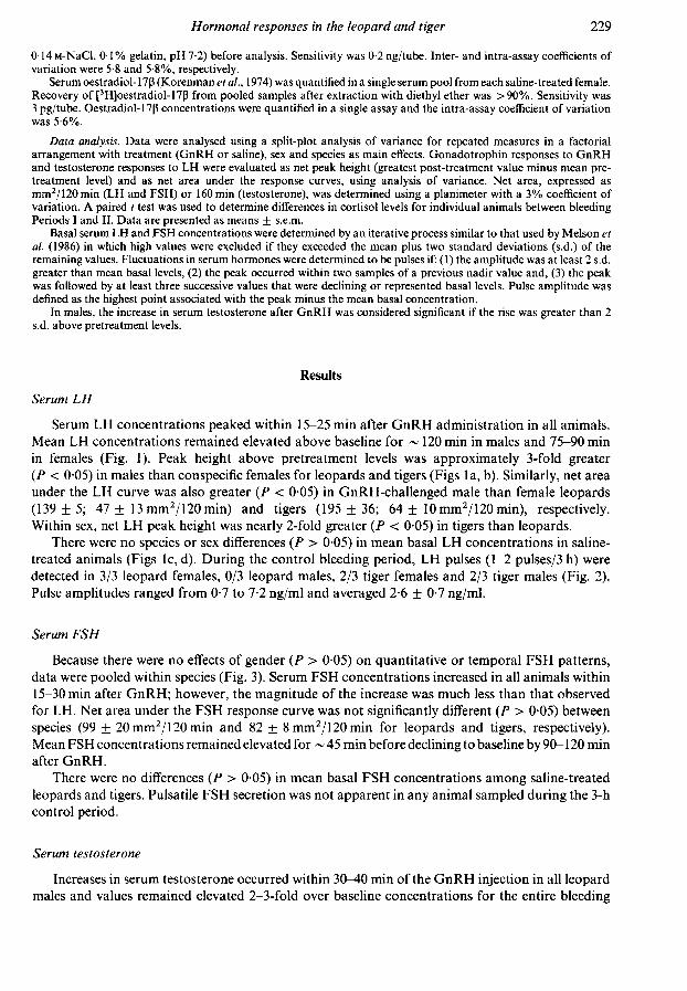
Serum LH concentrations peaked within 15-25 min after GnRH administration in all animals.Mean LH concentrations remained elevated above baseline for ~ 120 min in males and 75-90 minin females (Fig. 1). Peak height above pretreatment levels was approximately 3-fold greater(P < 005) in males than conspecific females for leopards and tigers (Figs la, b). Similarly, net areaunder the LH curve was also greater (P < 005) in GnRH-challenged male than female leopards(139 ± 5; 47 + 13 mm2/120 min) and tigers (195 + 36; 64 + 10 mm2/120 min), respectively.Within sex, net LH peak height was nearly 2-fold greater (P < 0-05) in tigers than leopards.
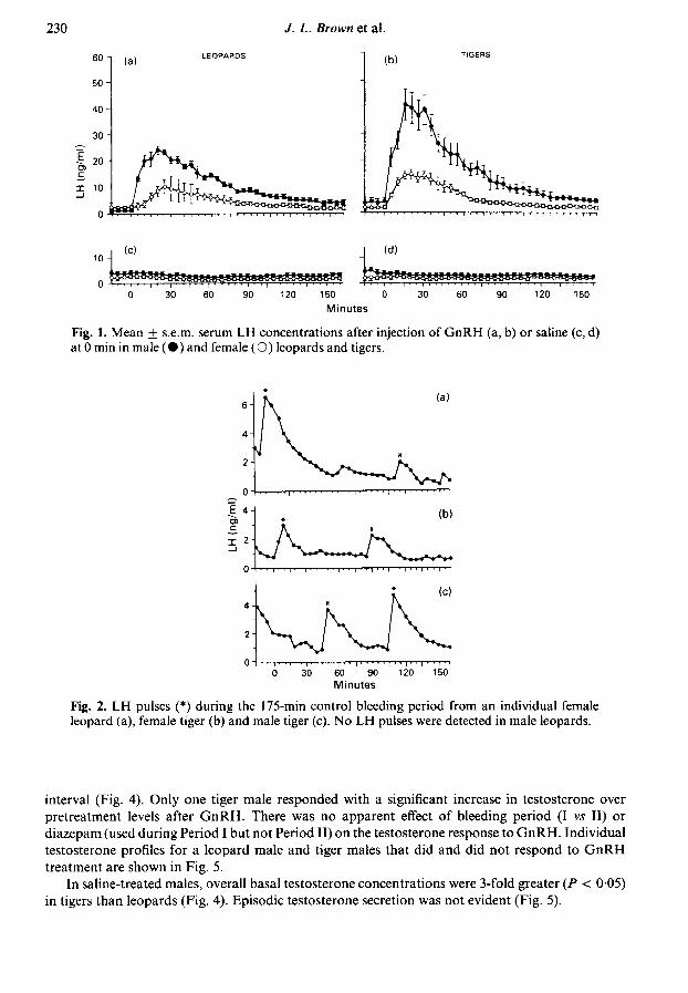
There were no species or sex differences (P > 005) in mean basal LH concentrations in saline-treated animals (Figs lc, d). During the control bleeding period, LH pulses (1-2 pulses/3 h) weredetected in 3/3 leopard females, 0/3 leopard males, 2/3 tiger females and 2/3 tiger males (Fig. 2).Pulse amplitudes ranged from 0-7 to 7-2 ng/ml and averaged 2-6 + 0-7 ng/ml.
Serum FSH
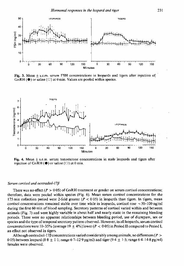
Because there were no effects of gender (P > 005) on quantitative or temporal FSH patterns,data were pooled within species (Fig. 3). Serum FSH concentrations increased in all animals within15-30 min after GnRH; however, the magnitude of the increase was much less than that observedfor LH. Net area under the FSH response curve was not significantly different (P > 005) betweenspecies (99 + 20mm2/120min and 82 + 8mm2/120min for leopards and tigers, respectively).Mean FSH concentrations remained elevated for ~ 45 min before declining to baseline by 90-120 minafter GnRH.
There were no differences (P > 005) in mean basal FSH concentrations among saline-treatedleopards and tigers. Pulsatile FSH secretion was not apparent in any animal sampled during the 3-hcontrol period.
Serum testosterone
Increases in serum testosterone occurred within 30-40 min of the GnRH injection in all leopardmales and values remained elevated 2-3-fold over baseline concentrations for the entire bleeding
I ' I ' I ' 'I ' ' I ' '1.'30 60 90 120 150 0
Minutes
Fig. 1. Mean + s.e.m. serum LH concentrations after injection of GnRH (a, b) or saline (c, d)at 0 min in male (·) and female (O) leopards and tigers.
30 60 90 120 150Minutes
Fig. 2. LH pulses (*) during the 175-min control bleeding period from an individual femaleleopard (a), female tiger (b) and male tiger (c). No LH pulses were detected in male leopards.
interval (Fig. 4). Only one tiger male responded with a significant increase in testosterone over
pretreatment levels after GnRH. There was no apparent effect of bleeding period (I vs II) or
diazepam (used during Period I but not Period II) on the testosterone response to GnRH. Individualtestosterone profiles for a leopard male and tiger males that did and did not respond to GnRHtreatment are shown in Fig. 5.
In saline-treated males, overall basal testosterone concentrations were 3-fold greater (P < 005)in tigers than leopards (Fig. 4). Episodic testosterone secretion was not evident (Fig. 5).
150 0Minutes
Fig. 3. Mean ± s.e.m. serum FSH concentrations in leopards and tigers after injection ofGnRH (·) or saline (O) at 0 min. Values are pooled within species.
150 0Minutes
Fig. 4. Mean + s.e.m. serum testosterone concentrations in male leopards and tigers afterinjection of GnRH (· ) or saline ( O ) at 0 min.
Serum cortisol and oestradiol-17ßThere was no effect (P > 005) of GnRH treatment or gender on serum cortisol concentrations;
therefore, data were pooled within species (Fig. 6). Mean serum cortisol concentrations for the175 min collection period were 2-fold greater (P < 0-05) in leopards than tigers. In tigers, mean
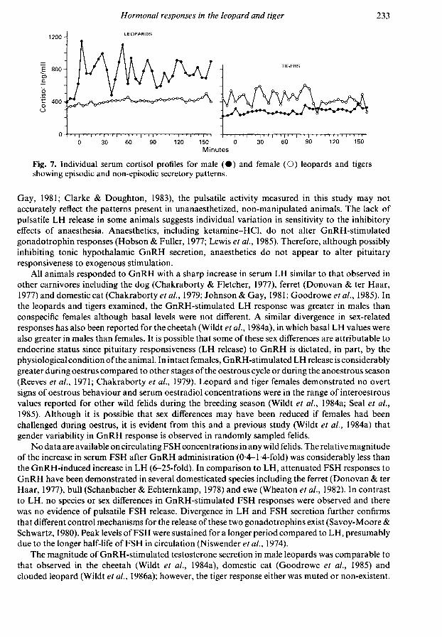
cortisol concentrations remained stable over time while in leopards, cortisol rose ~ 50-100 ng/mlduring the first 60 min of blood sampling. Secretory patterns of cortisol varied within and betweenanimals (Fig. 7) and were highly variable in about half and nearly static in the remaining bleedingperiods. There were no apparent relationships between bleeding period, use of diazepam, sex or
species and the type of temporal secretory pattern observed. However, in all leopards, serum cortisolconcentrations were 10-35% (average 19 + 4%) lower (P < 0-05) in Period II compared to Period I,an effect not observed in tigers.
Although oestradiol-17ß concentrations varied considerably among animals, no differences (P >
005) between leopard (8-8 + IT; range 6-7-12-9 pg/ml) and tiger (9-4 + 1-5; range 6-4-14-8 pg/ml)females were observed.
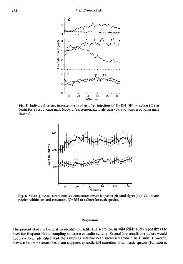
Fig. 5. Individual serum testosterone profiles after injection of GnRH (·) or saline (O) at0 min for a responding male leopard (a), responding male tiger (b), and non-responding maletiger (c).
60 90Minutes
Fig. 6. Mean + s.e.m. serum cortisol concentrations in leopards (·) and tigers (O). Values are
pooled within sex and treatment (GnRH or saline) for each species.
Discussion
The present study is the first to identify pulsatile LH secretion in wild felids and emphasizes theneed for frequent blood sampling to assess episodic activity. Several low-amplitude pulses wouldnot have been identified had the sampling interval been increased from 5 to 10 min. However,because ketamine anaesthesia can suppress episodic LH secretion in domestic species (Johnson &
1200-
E 800-
LEOPARDS
tOO
400
"I" I1'120 150
' I ' ' I ' ' 1 ' ' ' I ' ' "60 90 120 150
Minutes
Fig. 7. Individual serum cortisol profiles for male (·) and female (O) leopards and tigersshowing episodic and non-episodic secretory patterns.
Gay, 1981; Clarke & Doughton, 1983), the pulsatile activity measured in this study may notaccurately reflect the patterns present in unanaesthetized, non-manipulated animals. The lack ofpulsatile LH release in some animals suggests individual variation in sensitivity to the inhibitoryeffects of anaesthesia. Anaesthetics, including ketamine-HCl, do not alter GnRH-stimulatedgonadotrophin responses (Hobson & Fuller, 1977; Lewis et al, 1985). Therefore, although possiblyinhibiting tonic hypothalamic GnRH secretion, anaesthetics do not appear to alter pituitaryresponsiveness to exogenous stimulation.
All animals responded to GnRH with a sharp increase in serum LH similar to that observed inother carnivores including the dog (Chakraborty & Fletcher, 1977), ferret (Donovan & ter Haar,1977) and domestic cat (Chakraborty et al, 1979; Johnson & Gay, 1981; Goodrowe et al, 1985). Inthe leopards and tigers examined, the GnRH-stimulated LH response was greater in males thanconspecific females although basal levels were not different. A similar divergence in sex-relatedresponses has also been reported for the cheetah (Wildt et al, 1984a), in which basal LH values werealso greater in males than females. It is possible that some of these sex differences are attributable toendocrine status since pituitary responsiveness (LH release) to GnRH is dictated, in part, by thephysiological condition of the animal. In intact females, GnRH-stimulated LH release is considerablygreater during oestrus compared to other stages of the oestrous cycle or during the anoestrous season
(Reeves et al, 1971; Chakraborty et al, 1979). Leopard and tiger females demonstrated no overtsigns of oestrous behaviour and serum oestradiol concentrations were in the range of interoestrousvalues reported for other wild felids during the breeding season (Wildt et al, 1984a; Seal et al,1985). Although it is possible that sex differences may have been reduced if females had beenchallenged during oestrus, it is evident from this and a previous study (Wildt et al, 1984a) thatgender variability in GnRH response is observed in randomly sampled felids.
No data are available on circulating FSH concentrations in any wild felids. The relative magnitudeof the increase in serum FSH after GnRH administration (0-4-1-4-fold) was considerably less thanthe GnRH-induced increase in LH (6-25-fold). In comparison to LH, attenuated FSH responses toGnRH have been demonstrated in several domesticated species including the ferret (Donovan & terHaar, 1977), bull (Schanbacher & Echternkamp, 1978) and ewe (Wheaton et al, 1982). In contrastto LH, no species or sex differences in GnRH-stimulated FSH responses were observed and therewas no evidence of pulsatile FSH release. Divergence in LH and FSH secretion further confirmsthat different control mechanisms for the release of these two gonadotrophins exist (Savoy-Moore &Schwartz, 1980). Peak levels ofFSH were sustained for a longer period compared to LH, presumablydue to the longer half-life of FSH in circulation (Niswender et al, 1974).
The magnitude of GnRH-stimulated testosterone secretion in male leopards was comparable tothat observed in the cheetah (Wildt et al, 1984a), domestic cat (Goodrowe et al, 1985) andclouded leopard (Wildt et al, 1986a); however, the tiger response either was muted or non-existent.
Inexplicably, this decreased testicular response in tigers occurred in the face of greater overallGnRH-induced LH responses.
Basal testosterone concentrations in leopards were about 30% those in tigers, and theseconcentrations, in turn, were much lower than values previously reported for the domestic cat,clouded leopard and puma (Goodrowe et al, 1985; Wildt et al, 1986a, 1987a). The lower basaltestosterone concentrations of North Chinese leopards cannot be explained by inadequate gonado¬trophin stimulation since LH concentrations were similar to those observed in the tiger, as well as
values previously reported for other felids (Wildt et al, 1984a, b, 1987a; Goodrowe et al, 1985).Taken together, these data serve to identify marked functional differences in the hypothalamo-
pituitary-gonadal axis of the North Chinese leopard and tiger. This striking variability betweentwo taxonomically related species may be related, in part, to (1) differences in the biological activityof LH (Beitins et al, 1981); (2) altered steroidogenic response of Leydig cells to LH (Purvis &Hansson, 1978; Catt et al, 1979); or (3) depressions in genetic diversity, since increased geneticmonomorphism has been associated with compromised basal testosterone secretion in cheetahsand lions (O'Brien et al, 1985; Wildt et al, 1984b, 1986b).
The extreme individual variability in temporal cortisol profiles was inexplicable. The literaturehas not addressed the question of cortisol release patterns in anaesthetized mammals. In leopards,the consistently lower overall cortisol concentrations detected during the second bleeding periodsuggests rapid adrenal adaptation to repeated manipulation, similar to that observed in domesticatedspecies (Riegle, 1973; Amario et al, 1984). In contrast, no differences in serum cortisol concentrationsbetween bleeding periods in tigers were observed, suggesting that the adrenal response to handlingand/or anaesthetic Stressors differs amongst species.
The relationship between increased adrenal activity and reproductive function is complex and notwell understood. Administration of ACTH, cortisol or dexamethasone has been shown to attenuateGnRH-stimulated LH release in the cow and ram (Matteri & Moberg, 1982; Matteri et al, 1984).Glucocorticoids have been shown to reduce bioactive and immunoactive LH concentrations(Chantaraprateep & Thibier, 1978; Mann et al, 1982; Sapolsky, 1985), suppress basal and LH-stimulated testosterone secretion (Thibier & Rolland, 1976; Doerr & Pirke, 1976) and directlyinhibit testicular function by reducing the concentrations of Leydig cell LH receptors (Bambino &Hsueh, 1981). However, in other studies, serum testosterone and/or LH are either increased or
unchanged depending upon the Stressor (Pollard et al, 1980; Liptrap & Raeside, 1983; Lescoatet al, 1984). The cortisol concentrations measured in the North Chinese leopard are 2- to 15-foldgreater than values reported for other felids (Carter et al, 1984; Wildt et al, 1984b, 1987a, b). It istherefore possible that the hyperadrenal activity of the North Chinese leopard may be related to thecomparatively depressed pituitary and testicular responsiveness observed in this study.
We thank Dr Jerry Reeves for the gift of LH antiserum; Dr Leo Reichert, Jr and Dr Jim Dias forthe FSH antiserum; Dr Leo Reichert, Jr for the hormones for iodination and canine LH standardpreparation; NIDDK for the FSH standard preparation; Dr Lyndsay Phillips and the animalkeepers and personnel of the Henry Doorly Zoo for assistance in handling the animals and collect¬ing blood samples; Dr Steven Monfort for technical assistance; and J. Z. Koeser for manuscriptpreparation. This study was supported, in part, by Friends of the National Zoo and the Center forNew Opportunities in Animal Health Sciences. K.L.G. was supported, in part, by a grant from theWomen's Committee of the Smithsonian Associates, Washington, D.C.
References
Acosta, ., Tarnavsky, G.K., Platt, T.E., Haniermk, D.L.,Brown, J.L., Schoenemann, H.M. c& Reeves, J.J.( 1983) Nursing enhances the negative effect ofestrogenon LH release in the cow. J. Anim. Sci. 57,1530-1535.
Amano, ., Castellanos, J.M. & Baiaseli, J. (1984) Effectof acute and chronic psychogenic stress on cortico-adrenal and pituitary-thyroid hormones in male rats.Horm. Res. 6, 142-145.
Bambino, T.H. & Hsueh, A.J.W. (1981) Direct inhibitoryeffect of glucocorticoids upon testicular luteinizinghormone receptor and steroidogenesis in vivo and invitro. Endocrinology 108, 2142-2148.
Beitins, I.Z., Axelrod, L., Ostrea, I., Little, R. &Badger, I.M. (1981) Hypogonadism in males with an
immunologically active, biologically inactive luteiniz¬ing hormone: characterization of the abnormalhormone. J. clin. Endocr. Metab. 152, 1143-1149.
Carter, K.K., Chakraborty, P.K., Bush, M. & Wildt, D.E.(1984) Effects of electroejaculation and ketamine-HCl on serum cortisol, progesterone and testosteronein the male cat. /. Androl. 5, 431-437.
Cart, K.J., Baukai, A.J., Davies, T.F. & Dufau, M.L.(1979) Luteinizing hormone-releasing hormone-induced regulation of gonadotropin and prolactinreceptors in the rat testis. Endocrinology 104, 17-25.
Chakraborty, P.K. & Fletcher, W.S. (1977) Responsive¬ness of anestrous Labrador bitches to GnRH. Proc.Soc. exp. Biol. Med. 154, 125-126.
Chakraborty, P.K., Wildt, D.E. & Seager, S.W.J. (1979)Serum luteinizing hormone and ovulatory responseto luteinizing hormone-releasing hormone in theestrous and anestrous domestic cat. Lab. Anim. Sci29,338-344.
Chantaraprateep, P. & Thibier, M. (1978) Effects ofdexamethasone on the responses of luteinizing hor¬mone and testosterone by two injections of luteinizinghormone releasing hormone in young post-pubertalbulls. J. Endocr. 11, 389-395.
Clarke, I.J. & Doughton, B.W. (1983) Effect of variousanaesthetics on resting plasma concentrations ofluteinizing hormone, follicle-stimulating hormoneand prolactin in ovariectomized ewes. J. Endocr. 98,78-89.
Doerr, P. & Pirke, K.M. (1976) Cortisol-inducedsuppression of plasma testosterone in normal adultmales. J. clin. Endocr. Metab. 43, 622-629.
Donovan, B.T. & ter Haar, M.B. (1977) Effects of lutein¬izing hormone releasing hormone on plasma follicle-stimulating hormone and luteinizing hormone levelsin the ferret. J. Endocr. 73, 37-52.
Goodrowe, K.G., Chakraborty, P.K. & Wildt, D.E. (1985)Pituitary and gonadal response to exogenous LH-releasing hormone in the male domestic cat. J.Endocr. 105, 175-181.
Hobson. W. & Fuller, G.B. (1977) LH-RH-inducedgonadotropin release in chimpanzees. Biol. Reprod.17, 294-297.
Howard, J.G., Wildt, D.E., Chakraborty, P.K. & Bush, M.(1983) Reproductive traits including seasonal obser¬vations on semen quality and serum hormone con¬
centrations in the Dorcas gazelle. Theriogenology 20,221-234.
Johnson, L.M. & Gay, V.L. (1981) Luteinizing hormonein the cat. I. Tonic secretion. Endocrinology 109,240-246.
Korenman, S.G., Stevens, R.H., Carpenter, L.A., Robb,M., Niswender, G.D. & Sherman, B.M. (1974) Estra¬diol radioimmunoassay without chromatography:Procedure validation and normal values. J. clin.Endocr. Metab. 38, 718-720.
Lescoat, G., Lescoat, D. & Gamier, D. (1984) Dynamicchanges in plasma luteinizing hormone and testo¬sterone after stress in the male rat. Influences of
adrenalectomy. Can. J. Physiol. Pharm. 62, 1231-1233.
Lewis, G.S., Miller, K.F. & Bolt, D.J. (1985) Effects ofsodium pentobarbital on release ofgonadotropins inewes immediately after ovariectomy and after treat¬ment with gonadotropin-releasing hormone. Dom.Anim. Endocr. 2, 77-84.
Liptrap, R.M. & Raeside, J.I. (1983) Effect of cortisol on
the response to gonadotrophin releasing hormone inthe boar. J. Endocr. 97, 75-81.
Mann, D., Jackson, G. & Blank, M. (1982) Influence ofadrenocorticotropin and adrenalectomy on gonado¬tropin secretion in immature rats. Neuroendocrinology34, 20-25.
Matteri, R.L. & Moberg, G.P. (1982) Effect of cortisolor adrenocorticotrophin on release of luteinizinghormone induced by luteinizing hormone releasinghormone in the dairy heifer. J. Endocr. 92, 141-146.
Matteri, R.L., Watson, J.G. & Moberg, G.P. (1984)Stress or acute adrenocorticotrophin treatmentsuppresses LHRH-induced LH release in the ram. J.Reprod. Fert. 12, 385-393.
Melson, B.E., Brown, J.L., Schoenemann, H.M.,Tarnavsky, G.K. & Reeves, J.J. (1986) Elevation ofserum testosterone during chronic LHRH agonisttreatment in the bull. J. Anim. Sci. 62, 199-207.
Niswender, G.D., Nett, T.M. & Akbar, A.M. (1974) Thehormones of reproduction. In Reproduction in FarmAnimals, pp. 57-81. Ed. E. S. E. Hafez. Lea &Febiger, Philadelphia.
O'Brien, S.J., Roelke, M.E., Marker, L., Newman, .,Winkler, C.W., Meltzer, D., CoUy, L., Everman, J.,Bush, M. & Wildt, D.E. (1985) Genetic basis forspecies vulnerability in the cheetah. Science, N.Y.227, 1428-1434.
Pollard, I., Bassett. J.R. & Joss, J.M.P. (1980) Plasmatestosterone levels and 3ß-hydroxysteroid dehydro¬genase activity in the testis of the rat followingprolonged exposure to stress. J. Reprod. Feri. 59,101-106.
Purvis, K. & Hansson, V. (1978) Hormonal regulation ofLeydig cell function. Molec. cell. Endocr. 12,123-138.
Reeves, J.J., Arimura, A. & Schally, A.V. (1971) Pituitaryresponsiveness to purified luteinizing hormone-releasing hormone (LH-RH) at various stages of theestrous cycle in sheep. /. Anim. Sci. 32, 123-126.
Riegle, G.D. (1973) Chronic stress effects on adreno-corticol responsiveness in young and aged rats.
Neuroendocrinology 11, 1-10.Sandow, J. (1982) Gonadotropic and antigonadotropic
actions of LH-RH analogues. In NeuroendocrinePerspectives, Vol. 1. pp. 339-395. Eds E. E. Müller &R. M. MacLeod. Elsevier Biomedicai Press,Amsterdam.
Sapolsky, R.M. (1985) Stress-induced suppression oftesticular function in the wild baboon: role of gluco¬corticoids. Endocrinology 116, 2273-2278.
Savoy-Moore, R.T. & Schwartz, N.B. (1980) Differentialcontrol of FSH and LH secretion. In ReproductivePhysiology III (Int. Rev. Physiol.), Vol. 22, pp.203-248. Ed. R. O. Greep. University Park Press,Baltimore.
Schanbacher, B.D. & Echternkamp, S.E. (1978) Testicularsteroid secretion in response to GnRH-mediated LHand FSH secretion in bulls. J. Anim. Sci. 47, 514-520.
Schmidt, A.M., Nadal, L.A., Schmidt, M.J. & Beamer,N.B. (1979) Serum concentrations of estradiol andprogesterone during the estrous cycle and early preg¬nancy in the lion (Panthera leo). J. Reprod. Fert. 57,267-272.
Seager, S.W.J. & Demorest, C.N. (1978) Reproductionof captive wild carnivores. In Zoo and Wild AnimalMedicine, pp. 668-673. Ed. M. E. Fowler. W. B.Saunders Company, Philadelphia.
Seal, U.S., Plotka, E.D., Smith, J.D., Wright, F.H.,Reindl, N.J., Taylor, R.S. & Seal, M.F. (1985)Immunoreactive luteinizing hormone, estradiol, pro¬gesterone, testosterone, and androstenedione levelsduring the breeding season and anestrus in Siberiantigers. Biol. Reprod. 32, 361-368.
Thibier, M. & Rolland, D. (1976) The effect of dexa¬methasone (DXM) on circulating testosterone (T)and luteinizing hormone (LH) in young postpubertalbulls. Theriogenology 5, 53-60.
Wheaton, J.E., Recabarren, S.E. & MuUett, M.A. (1982)GnRH-FSH and LH dose-response relationships inanestrous sheep and effects of estradiol-17ß andprogesterone pretreatment. J. Anim. Sci. 55, 384-390.
Wildt, D.E., Chakraborty, P.K., Meltzer, D. & Bush, M.(1984a) Pituitary and gonadal response to LH releas¬ing hormone administration in the female and malecheetah. J. Endocr. 101, 51-56.
Wildt, D.E., Meltzer, D., Chakraborty, P.K. & Bush, M.(1984b) Adrenal-testicular-pituitary relationships inthe cheetah subjected to anesthesia/electroejaculation.Biol. Reprod. 30,665-672.
Wildt, D.E., Howard, J.G., Chakraborty, P.K. & Bush, M.(1986a) Reproductive physiology of the cloudedleopard. II. A circannual analysis ofadrenal-pituitary-testicular relationships during electroejaculation orafter an adrenocorticotropin hormone challenge. Biol.Reprod. 34,949-959.
Wildt, D.E., O'Brien, S.J., Packer, C, Brown, J.L. &Bush, M. (1986b) Reproductive and genetic conse¬quences of founding an isolated population of EastAfrican lions. Biol. Reprod. 34, (Suppl. 1), 203, Abstr.
Wildt, D.E., Phillips, L.G., Simmons, L.G., Goodrowe,K.G., Howard, J.G., Brown, J.L. & Bush, M. (1987a)Seminal-endocrine characteristics of the tiger and thepotential for artificial breeding. In Tigers of theWorld: The Biology, Biopolitics, Management andConservation of an Endangered Species, pp. 255-279.Eds R. L. Tilson & U. S. Seal. Noyes Publications,Park Ridge.
Wildt, D.E., O'Brien, S.J., Howard, J.G., Caro, T.M.,Roelke, M.E., Brown, J.L. & Bush, M. (1987b)Similarity in ejaculate-endocrine characteristics incaptive versus free-ranging cheetahs of two subspecies.Biol. Reprod. 36, 351-360.
Received 5 May 1987


![[Ovulation induction by pulsatile GnRH therapy in 2014: literature review and synthesis of current practice]](https://static.fdokumen.com/doc/165x107/6333c99c28cb31ef600d6b7b/ovulation-induction-by-pulsatile-gnrh-therapy-in-2014-literature-review-and-synthesis.jpg)