Emerging Therapeutic Landscape of Peripheral T-Cell ... - MDPI
21
cancers Review Emerging Therapeutic Landscape of Peripheral T-Cell Lymphomas Based on Advances in Biology: Current Status and Future Directions Maliha Khan *, Felipe Samaniego, Fredrick B. Hagemeister and Swaminathan P. Iyer * Citation: Khan, M.; Samaniego, F.; Hagemeister, F.B.; Iyer, S.P. Emerging Therapeutic Landscape of Peripheral T-Cell Lymphomas Based on Advances in Biology: Current Status and Future Directions. Cancers 2021, 13, 5627. https://doi.org/ 10.3390/cancers13225627 Academic Editor: Joseph R. Bertino Received: 24 May 2021 Accepted: 27 August 2021 Published: 10 November 2021 Publisher’s Note: MDPI stays neutral with regard to jurisdictional claims in published maps and institutional affil- iations. Copyright: © 2021 by the authors. Licensee MDPI, Basel, Switzerland. This article is an open access article distributed under the terms and conditions of the Creative Commons Attribution (CC BY) license (https:// creativecommons.org/licenses/by/ 4.0/). Department of Lymphoma/Myeloma, University of Texas, MD Anderson Cancer Center, Houston, TX 77030, USA; [email protected] (F.S.); [email protected] (F.B.H.) * Correspondence: [email protected] (M.K.); [email protected] (S.P.I.); Tel.: +1-713-792-5242 (S.P.I.) Simple Summary: Peripheral T-cell lymphoma is a rare but aggressive tumor. Due to its rarity, the disease has not been completely understood. In our review, we look at this lymphoma at the molecular level based on available literature. We highlight the mechanism behind the progression and resistance of this tumor. In doing so, we bring forth possible mechanism that could be exploited through novel chemotherapy drugs. In addition, we also look at the current available drugs used in treating this disease, as well as highlight other new drugs, describing their potential in treating this lymphoma. We comprehensively have collected and present the available biology behind peripheral T-cell lymphoma and discuss the available treatment options. Abstract: T-cell lymphomas are a relatively rare group of malignancies with a diverse range of pathologic features and clinical behaviors. Recent molecular studies have revealed a wide array of different mechanisms that drive the development of these malignancies and may be associated with resistance to therapies. Although widely accepted chemotherapeutic agents and combinations, including stem cell transplantation, obtain responses as initial therapy for these diseases, most patients will develop a relapse, and the median survival is only 5 years. Most patients with relapsed disease succumb within 2 to 3 years. Since 2006, the USFDA has approved five medications for treatment of these diseases, and only anti-CD30-therapy has made a change in these statistics. Clearly, newer agents are needed for treatment of these disorders, and investigators have proposed studies that evaluate agents that target these malignancies and the microenvironment depending upon the molecular mechanisms thought to underlie their pathogenesis. In this review, we discuss the currently known molecular mechanisms driving the development and persistence of these cancers and discuss novel targets for therapy of these diseases and agents that may improve outcomes for these patients. Keywords: PTCL; non-Hodgkin’s lymphoma; novel therapies; targeted therapies; recent advances; current treatment 1. Introduction Peripheral T-cell lymphomas (PTCLs) are a diverse set of aggressive T-cell lymphomas that arise from mature T cells [1]. Most PTCLs are associated with a poor prognosis [2]. PTCLs constitute 15–20% of aggressive non-Hodgkin’s lymphomas (NHLs) and 5–10% of all NHLs [3]. PTCL has a diverse morphology, and definitive markers of PTCL subtypes are scarce, making the diagnosis and classification of PTCL complex [4]. The 2016 World Health Organization classification system describes 27 different types of PTCL [5]. Most PTCLs are treated similarly due to the lack of specific targeted therapies for different PTCL subtypes. This, in turn, is due to an inadequate understanding of the pathobiology of these tumors. However, recent advances in whole-genome sequencing Cancers 2021, 13, 5627. https://doi.org/10.3390/cancers13225627 https://www.mdpi.com/journal/cancers
-
Upload
khangminh22 -
Category
Documents
-
view
3 -
download
0
Transcript of Emerging Therapeutic Landscape of Peripheral T-Cell ... - MDPI

cancers
Review
Emerging Therapeutic Landscape of Peripheral T-CellLymphomas Based on Advances in Biology: Current Status andFuture Directions
Maliha Khan *, Felipe Samaniego, Fredrick B. Hagemeister and Swaminathan P. Iyer *
�����������������
Citation: Khan, M.; Samaniego, F.;
Hagemeister, F.B.; Iyer, S.P. Emerging
Therapeutic Landscape of Peripheral
T-Cell Lymphomas Based on
Advances in Biology: Current Status
and Future Directions. Cancers 2021,
13, 5627. https://doi.org/
10.3390/cancers13225627
Academic Editor: Joseph R. Bertino
Received: 24 May 2021
Accepted: 27 August 2021
Published: 10 November 2021
Publisher’s Note: MDPI stays neutral
with regard to jurisdictional claims in
published maps and institutional affil-
iations.
Copyright: © 2021 by the authors.
Licensee MDPI, Basel, Switzerland.
This article is an open access article
distributed under the terms and
conditions of the Creative Commons
Attribution (CC BY) license (https://
creativecommons.org/licenses/by/
4.0/).
Department of Lymphoma/Myeloma, University of Texas, MD Anderson Cancer Center,Houston, TX 77030, USA; [email protected] (F.S.); [email protected] (F.B.H.)* Correspondence: [email protected] (M.K.); [email protected] (S.P.I.);
Tel.: +1-713-792-5242 (S.P.I.)
Simple Summary: Peripheral T-cell lymphoma is a rare but aggressive tumor. Due to its rarity,the disease has not been completely understood. In our review, we look at this lymphoma at themolecular level based on available literature. We highlight the mechanism behind the progressionand resistance of this tumor. In doing so, we bring forth possible mechanism that could be exploitedthrough novel chemotherapy drugs. In addition, we also look at the current available drugs used intreating this disease, as well as highlight other new drugs, describing their potential in treating thislymphoma. We comprehensively have collected and present the available biology behind peripheralT-cell lymphoma and discuss the available treatment options.
Abstract: T-cell lymphomas are a relatively rare group of malignancies with a diverse range ofpathologic features and clinical behaviors. Recent molecular studies have revealed a wide arrayof different mechanisms that drive the development of these malignancies and may be associatedwith resistance to therapies. Although widely accepted chemotherapeutic agents and combinations,including stem cell transplantation, obtain responses as initial therapy for these diseases, mostpatients will develop a relapse, and the median survival is only 5 years. Most patients with relapseddisease succumb within 2 to 3 years. Since 2006, the USFDA has approved five medications fortreatment of these diseases, and only anti-CD30-therapy has made a change in these statistics. Clearly,newer agents are needed for treatment of these disorders, and investigators have proposed studiesthat evaluate agents that target these malignancies and the microenvironment depending uponthe molecular mechanisms thought to underlie their pathogenesis. In this review, we discuss thecurrently known molecular mechanisms driving the development and persistence of these cancersand discuss novel targets for therapy of these diseases and agents that may improve outcomes forthese patients.
Keywords: PTCL; non-Hodgkin’s lymphoma; novel therapies; targeted therapies; recent advances;current treatment
1. Introduction
Peripheral T-cell lymphomas (PTCLs) are a diverse set of aggressive T-cell lymphomasthat arise from mature T cells [1]. Most PTCLs are associated with a poor prognosis [2].PTCLs constitute 15–20% of aggressive non-Hodgkin’s lymphomas (NHLs) and 5–10% ofall NHLs [3]. PTCL has a diverse morphology, and definitive markers of PTCL subtypesare scarce, making the diagnosis and classification of PTCL complex [4]. The 2016 WorldHealth Organization classification system describes 27 different types of PTCL [5].
Most PTCLs are treated similarly due to the lack of specific targeted therapies fordifferent PTCL subtypes. This, in turn, is due to an inadequate understanding of thepathobiology of these tumors. However, recent advances in whole-genome sequencing
Cancers 2021, 13, 5627. https://doi.org/10.3390/cancers13225627 https://www.mdpi.com/journal/cancers

Cancers 2021, 13, 5627 2 of 21
and gene expression profiling (GEP) have enabled us to better elucidate the pathogenesisof PTCL and differentiate among its various subtypes [6]. Recent studies have uncoveredindividual molecular signatures that may not only provide prognostic information aboutthe disease but may also provide potential therapeutic targets. Multiple promising therapiesare being tested and based on evidence from clinical trials; these therapies are beingincorporated into the currently recommended treatment regimens.
In this review, we highlight the underlying pathobiology of PTCL, an improvedunderstanding of which would potentially uncover novel therapeutic targets. We alsodescribe the current treatment standards for PTCL and focus on novel targeted therapiesthat have demonstrated efficacy in recent clinical trials.
2. Origins of PTCL
Understanding the origin and development of T-cell lymphoma requires the knowl-edge of normal T-cell development and function. Mature T cells can be divided into twomain subtypes based on their type of T-cell receptor (TCR) expression. Most T cells areαβ T cells, which express the αβ TCR chains, whereas fewer than 2% of T cells are γδ Tcells, which express the γδ TCR chains [7]. T cells are further differentiated into subtypesthrough a process controlled by transcriptional regulators and cytokines, which exhibitssignificant plasticity under environmental pressure. The interaction of a normal T cellwith an antigen-presenting cell (APC) forms a T-cell APC immunological synapse thatemploys multiple signaling pathways to induce T-cell activation [8]. Most notable amongthese pathways are the JAK-STAT, PI3 K-AKT, and MAPK signal transduction cascades [9].These pathways are boosted by the activity of costimulatory molecules such as CD28 [7].Epigenetic mechanisms also play a role in the immune response, increasing its complex-ity [9]. Dysfunction in these pathways could be at the core of lymphomagenesis, and hence,these pathways are being investigated with genomic and molecular approaches.
PTCLs can acquire unique phenotypes owing to mutations that modulate the tran-scription or the cytokine-driven plasticity of T cells and other tumor microenvironmentsignals. This modulation may lead to features that are dissimilar to those of the tumor’scell of origin [10]. Hence, researchers have not yet established the precise cell of origin fordifferent types of PTCL. However, global GEP, which has been used to define some typesof B-cell lymphomas, has also been used to define some types of PTCLs [11]. For example,GEP has been used to define angioimmunoblastic T-cell lymphoma (AITL), a PTCL thathas a GEP similar to that of T-follicular helper (Tfh) cells, which provides clues regardingits cell of origin [12]. Genetic signatures have been used to identify subgroups of PTCL-nototherwise specified (PTCL-NOS), a group of unclassified PTCLs [13]. The expression ofmultiple transcripts that match the genetic signature of normal natural killer (NK) cells hasalso been identified in some PTCLs [14].
3. PTCL Subtypes and Their Pathogenesis
This section highlights the biological underpinnings that characterize different PTCLsubtypes. Common pathogenic mechanisms associated with PTCL subtypes are listed inTable 1. Some of these mechanisms are also highlighted in Figure 1.
3.1. AITLs and PTCLs with the Tfh Phenotype
AITLs and PTCLs with the Tfh phenotype are grouped together, because they shareTfh cells as their putative cell of origin and gene expression patterns [5]. For a PTCL to bedesignated as having the Tfh phenotype, its cells must express at least two antigens relatedto Tfh, which include PD1, BCL6, CD10, CXCL13, ICOS, and CXCR5. GEP studies haverevealed that certain oncogenic pathways are enriched in AITL, including the NF-κB, IL6,and TGF-β signaling pathways [7,15]. Cytogenetic studies have demonstrated that AITLfrequently also harbors trisomy 5, which is often accompanied by trisomy 21. Furthermore,small focal gene deletions can dysregulate the PI3 K-AKT-mTOR pathway [4,7].
Cancers 2021, 13, 5627 3 of 21
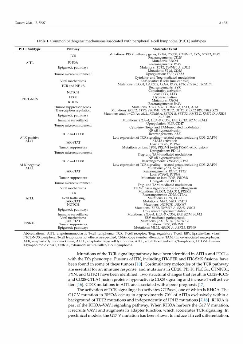
Table 1. Common pathogenic mechanisms associated with peripheral T-cell lymphoma (PTCL) subtypes.
PTCL Subtype Pathway Molecular Event
AITL
TCR Mutations: PI3 K pathway genes, CD28, PLCG1, CTNNB1, FYN, GTF21, VAV1Rearrangements: CD28
RHOA Mutations: RHOARearrangements: VAV1
Epigenetic pathways Mutations: TET2, DNMT3 A, IDH2
Tumor microenvironmentMutations: B2 M, CD28
Upregulation: FLIP, PD-L1Cytokine- and Treg-mediated modulation
Viral mechanisms EBV-positive B cells (unclear role)
PTCL-NOS
TCR and NF-κB Mutations: PLCG1, CARD11, CD28, VAV1, FYN, PTPRC, TNFAIP3Rearrangements: SYK
NOTCH Constitutive activationLoss: TCF1, LEF1
PI3 K HyperactivationRHOA Mutations: RHOA
Rearrangements: VAV1Tumor suppressor genes Mutations: TP53, TP63, CDKN2 A, FAT1, ATMTranscription regulation Mutations: IKZF2, ETV6, PRDM1, YTHDF2, DDX3 X, IRF2 BP2, TBL1 XR1
Epigenetic pathways Mutations and/or CNAs: MLL, KDM6 A, SETD1 B, SETD2, KMT2 C, KMT2 D, ARID1A, EP300
Immune surveillance Mutations: HLA-A, HLA-B, CD58, FAS, CIITA, B2 M, PD-L1
Tumor microenvironment Upregulation: FLIP, CD47Cytokine-, Treg-, and TAM-mediated modulation
ALK-positiveALCL
TCR and CD30NF-κB hyperactivationRearrangements: ALK
Low expression of TCR signaling—related genes, including CD3, ZAP70JAK-STAT STAT3 activation
Loss: PTPN2, PTPN6Tumor suppressors Mutations or loss: TP53, PRDM1 (with TRAF1-ALK fusion)
Tumor microenvironment Upregulation: PD-L1Treg- and TAM-mediated modulation
ALK-negativeALCL
TCR and CD30NF-κB hyperactivation
Rearrangements: DUSP22, TP63Low expression of TCR signaling—related genes, including CD3, ZAP70
JAK-STATMutations: JAK1, STAT3
Rearrangements: ROS1, TYK2Loss: PTPN2, PTPN6
Tumor suppressors Mutations or loss: TP53, PRDM1
Tumor microenvironment Upregulation: PD-L1Treg- and TAM-mediated modulation
ATLL
Viral mechanisms HTLV-1 has a significant role in pathogenesisTCR Mutations: PLCG1, CARD11, PRKCB
Rearrangements: CD28, CTLA4T-cell trafficking Mutations: CCR4, CCR7
JAK-STAT Mutations: JAK1, JAK3, STAT3NOTCH Mutations: NOTCH1, FBXW7
Epigenetic pathways Mutations: TET2, DNMT3 A, EZH2, PRC2CpG island hypermethylation
Immune surveillance Mutations: HLA-A, HLA-B, CD58, FAS, B2 M, PD-L1
ENKTL
Viral mechanisms EBV-mediated pathogenesisJAK-STAT Mutations: JAK3, STAT3, STAT5 B
Tumor suppressors Mutations: TP53, PRDM1Epigenetic pathways Mutations: MLL2, ARID1 A, ASXL3, EP300
Abbreviations: AITL, angioimmunoblastic T-cell lymphoma; TCR, T-cell receptor; Treg, regulatory T-cell; EBV, Epstein-Barr virus;PTCL-NOS, peripheral T-cell lymphoma not otherwise specified; CNAs, copy number alterations; TAM, tumor-associated macrophages;ALK, anaplastic lymphoma kinase; ALCL, anaplastic large cell lymphoma; ATLL, adult T-cell leukemia/lymphoma; HTLV-1, humanT-lymphotropic virus 1; ENKTL, extranodal natural killer/T-cell lymphoma.
Mutations of the TCR signaling pathway have been identified in AITLs and PTCLswith the Tfh phenotype. Fusions of ITK, including ITK-FER and ITK-SYK fusions, havebeen found in some of these tumors [10]. Costimulatory molecules of the TCR pathwayare essential for an immune response, and mutations in CD28, PI3 K, PLCG1, CTNNB1,FYN, and GTF2 I have been identified. Two structural changes that result in CD28-ICOSand CD28-CTLA4 fusion proteins hyperactivate CD28 signaling and increase T-cell activa-tion [16]. CD28 mutations in AITL are associated with a poor prognosis [17].
The activation of TCR signaling also activates GTPases, one of which is RHOA. TheG17 V mutation in RHOA occurs in approximately 70% of AITLs exclusively within abackground of TET2 mutations and independently of IDH2 mutations [7,18]. RHOA ispart of the RHOA-VAV1 signaling pathway. When RHOA harbors the G17 V mutation,it recruits VAV1 and augments its adapter function, which accelerates TCR signaling. Inpreclinical models, the G17 V mutation has been shown to induce Tfh cell differentiation,

Cancers 2021, 13, 5627 4 of 21
upregulate ICOS expression, and increase PI3 K and MAPK signaling. An isolated VAV1mutation and a VAV1-STAP2 translocation have also been identified in AITL [17,19].
Cancers 2021, 13, x FOR PEER REVIEW 4 of 22
Figure 1. Mechanisms of peripheral T-cell lymphoma development and agents used for targeted
therapies. Multiple mechanisms are responsible for the development and proliferation of different
subtypes of PTCL, including signaling pathway deregulation, epigenetic dysregulation, tumor mi-
croenvironment signals, and virus-mediated oncogenesis. Not all of these mechanisms are involved
in the pathogenesis of each PTCL subtype. Some potential therapeutic targets and agents targeting
them are also shown.
3.1. AITLs and PTCLs with the Tfh Phenotype
AITLs and PTCLs with the Tfh phenotype are grouped together, because they share
Tfh cells as their putative cell of origin and gene expression patterns [5]. For a PTCL to be
designated as having the Tfh phenotype, its cells must express at least two antigens re-
lated to Tfh, which include PD1, BCL6, CD10, CXCL13, ICOS, and CXCR5. GEP studies
have revealed that certain oncogenic pathways are enriched in AITL, including the NF-
κB, IL6, and TGF-β signaling pathways [7,15]. Cytogenetic studies have demonstrated that
AITL frequently also harbors trisomy 5, which is often accompanied by trisomy 21. Fur-
thermore, small focal gene deletions can dysregulate the PI3 K-AKT-mTOR pathway [4,7].
Mutations of the TCR signaling pathway have been identified in AITLs and PTCLs
with the Tfh phenotype. Fusions of ITK, including ITK-FER and ITK-SYK fusions, have
been found in some of these tumors [10]. Costimulatory molecules of the TCR pathway
are essential for an immune response, and mutations in CD28, PI3 K, PLCG1, CTNNB1,
FYN, and GTF2 I have been identified. Two structural changes that result in CD28-ICOS
and CD28-CTLA4 fusion proteins hyperactivate CD28 signaling and increase T-cell acti-
vation [16]. CD28 mutations in AITL are associated with a poor prognosis [17].
The activation of TCR signaling also activates GTPases, one of which is RHOA. The
G17 V mutation in RHOA occurs in approximately 70% of AITLs exclusively within a
background of TET2 mutations and independently of IDH2 mutations [7,18]. RHOA is
part of the RHOA-VAV1 signaling pathway. When RHOA harbors the G17 V mutation, it
recruits VAV1 and augments its adapter function, which accelerates TCR signaling. In
preclinical models, the G17 V mutation has been shown to induce Tfh cell differentiation,
Figure 1. Mechanisms of peripheral T-cell lymphoma development and agents used for targetedtherapies. Multiple mechanisms are responsible for the development and proliferation of differentsubtypes of PTCL, including signaling pathway deregulation, epigenetic dysregulation, tumormicroenvironment signals, and virus-mediated oncogenesis. Not all of these mechanisms are involvedin the pathogenesis of each PTCL subtype. Some potential therapeutic targets and agents targetingthem are also shown.
AITLs and PTCLs with the Tfh phenotype have a high degree of epigenetic dysreg-ulation, where the mutations TET2, IDH2, and DNMT3A have been detected in thesesubtypes. TET2—inactivating mutations occur in up to 85% of AITLs [20]. TET2 muta-tions cause DNA hypermethylation, affect other proteins such as histone deacetylase 1(HDAC1) and HDAC2, and measurably decrease 5-hydroxymethylcytosine, as detectedby immunohistochemistry. They are also correlated with the Tfh phenotype and poorclinical outcomes [7,20]. Mutations in DNMT3A, which is responsible for de novo DNAmethylation, result in hypermethylation. The loss of both DNMT3A and TET2 drives thelymphoid transformation of Tfh cells in mouse models. Similar alterations of TET2 andDNMT3A have been found in AITL and myeloid neoplasm precursor cells, which suggeststhat these alterations are premalignant and, paired with additional defects, cause malignanttransformations [10,21]. In addition, approximately 30% of AITLs bear IDH2 mutations atthe R172 position, producing elevated levels of 2-hydroxyglutarate, which may act as anoncometabolite by inhibiting DNA hydroxylases and histone lysine-demethylases of theTET family [22]. IDH2 mutations are associated with AITL and are significantly related toTfh signatures. TET2 and IDH R172 mutations often occur simultaneously in AITL, whichsuggests that they have a synergistic effect that promotes oncogenesis [23,24].
The tumor microenvironment may also play a significant role in T-cell lymphomagen-esis. AITL frequently has B2M mutations, which protect tumor cells from cytotoxic T-cellrelated damage. As discussed above, the mutation and dysregulation of the costimulatoryprotein CD28 play a role in AITL oncogenesis. The upregulation of FLIP, an antiapoptotic
Cancers 2021, 13, 5627 5 of 21
regulatory protein, has also been documented in AITL. These mutations decrease thetumor cell immunogenicity and also provide survival and proliferation benefits [17,25,26].AITLs and their related Tfh-like entities are known to overexpress cytokines that modifyantitumor responses (e.g., interleukin (IL)-17, IL-10, IL-6, and IL-2) and angiogenesis (IL-8).Incompetent regulatory T cells (Tregs), which are functionally impaired and indirectly favortumor growth, dampen the antitumor immune response in AITL [27]. The upregulation ofPD-L1, a checkpoint protein, has also been identified in AITLs and other PTCLs. Intratumorendothelial cells, which produce excessive VEGF (which, in turn, promotes tumor cellgrowth), could explain the high vascular density in AITL [17,28].
Epstein–Barr virus (EBV)-infected B cells have been found in AITL; however, their rolein its pathogenesis is not clear. Owing to the immunosuppressive environment in AITL,EBV-infected B cells might proliferate into diffuse large B-cell lymphomas. Interestinglyhowever, EBV-negative diffuse large B-cell lymphomas have also been reported in patientswith Tfh-cell lymphomas [10,29].
3.2. PTCL-NOS
GEP studies have subdivided PTCL-NOS into two main subgroups. One subgrouphas upregulation of the transcription factor GATA3 and its downstream targets (e.g., CCR4,CXCR7, IK, and IL18 RA). This subgroup is associated with a poor prognosis, has ahigh proliferation signature, and lacks a strong tumor microenvironment signature. TheGATA3 subgroup is associated with a greater genomic complexity, as evidenced by thepresence of mutations of tumor suppressor genes in the PTEN-PI3K and CDKN2AB-TP3pathways and augmented activity of MYC and STAT3. The other subgroup has a highexpression of TBX21 and EOMES and their target genes (e.g., CXCR3, IL2RB, CCL3, andIFN-γ). The TBX21 subgroup is generally associated with more favorable outcomes anddisplays enrichment of the NF-κB pathway. A small subset of the TBX21 subgroup hasa frequent CD8 expression and a cytotoxic signature; this subset is associated with poorclinical outcomes. GATA3 regulates the differentiation of T-helper type 2 cells, and TBX21regulates the differentiation of T-helper type 1 cells and cytotoxic T cells. Furthermore, thetwo subtypes have significantly different copy number aberrations, indicating that theyeach have a different cell of origin [4,7,10,13].
One distinct molecular subtype of PTCL-NOS that lacks the Tfh phenotype primarilyharbors TP53 and CDKN2A mutations. These alterations are inversely related to the Tfhphenotype in PTCL-NOS. Copy number profiling has identified extensive chromosomalabnormalities in this subtype, which has frequent alterations in genes related to immunesurveillance. In addition, the subtype demonstrates frequent somatic mutations in tran-scription factors (IKZF2, ETV6, and PRDM1); transcriptional corepressors (IRF2BP2 andTBL1XR1); and RNA-binding proteins (YTHDF2 and DDX3X) [30].
Mutations and copy number variations in components of the TCR and NF-κB path-ways, including in PLCG1, CARD11, CD28, VAV1, FYN, PTPRC, and TNFAIP3, have beendetected in PTCL-NOS [10]. SYK, which is downstream of the TCR, is constitutively ex-pressed in some cases of PTCL-NOS. The translocation t (5; 9) (q33; q22) results in ITK-SYKfusion, which results in SYK overexpression [31]. Constitutive NOTCH signaling, whichcould be attributed to genomic defects or a microenvironment-mediated mechanism, occursin more than half of PTCL-NOS cases [32]. In mouse models, the tumorigenic activity ofNOTCH1 involves the loss of TCF-1 and subsequent upregulation LEF-1 [33]. Interestingly,TCF-1 and LEF-1 losses have been detected primarily in PTCLs with the T-helper type 2phenotype. Inactivating RHOA mutations have been noted in PTCL-NOS [17]. In addition,PTCL-NOS is known to carry VAV1 alterations in which VAV1 is translocated to formgene fusions, including VAV1-THAP4, VAV1-MYO1F, and VAV1-S100 A7, which lack theC-terminal SH3 domain and induce the constitutive action of VAV1 [34]. PTCL-NOS alsohas enriched PI3K pathway signatures. Frequent mutations of the tumor suppressor FAT1have been identified in PTCL-NOS and are associated with poorer outcomes. Mutations in

Cancers 2021, 13, 5627 6 of 21
the tumor suppressor genes TP53, TP63, LATS1, STK3, and ATM have also been detected,albeit at lower frequencies [35].
Epigenetic alterations occur less frequently in PTCL-NOS than in AITL. PTCL-NOSis known to carry multiple mutations in epigenetic regulators, including MLL, MLL2,KDM6A, TET2, DNMT3A, SETD1B, SETD2, KMT2C, KMT2D, CREBBP, ARID1A, andEP300. Mutations in MLL, MLL2, and KDM6A are related to a poor prognosis [7,10,35].
The tumor microenvironment has an important role in the pathogenesis of PTCL-NOS.B2M mutations protect tumor cells against damage mediated by cytotoxic T cells [25]. InPTCL-NOS with TP53 or CDKN2A alterations, mutations that lead to immune evasion havebeen documented in HLA-A, HLA-B, CIITA, PD-L1, CD58, FAS, and PDCD1 [17,30]. Theoverexpression of the antiapoptotic protein FLIP has been documented in PTCL-NOS [26].PTCL with CD47 upregulations, which inhibit antitumor macrophage activity, have alsobeen reported [17,36]. Non-neoplastic FOXP3-positive suppressor Tregs, which sustain tu-mor cell survival, have been associated with the PTCL-NOS microenvironment [27]. Tumor-associated macrophages and relevant gene signatures have been recognized in PTCL-NOSand other subtypes. Generally, increased numbers of tumor-associated macrophagesare correlated with a poor prognosis, indicating that these cells have a probable role inoncogenesis [37,38].
3.3. Anaplastic Large Cell Lymphoma
Anaplastic large cell lymphoma (ALCL) is characterized by large anaplastic cellsstrongly positive for CD30. CD30 belongs to the tumor necrosis factor receptor family andexerts diverse effects on cell growth and survival, chiefly through the activation of theNF-κB pathway. ALCLs are divided into two subgroups based on whether they expressanaplastic lymphoma kinase (ALK). ALK-positive ALCL is defined by the formation ofALK fusion proteins; the NPM1-ALK fusion caused by translocation t(2; 5)(p23; q35) ischaracteristic of ALK-positive ALCL [39,40]. ALK-positive ALCL with TRAF1-ALK fu-sion displays a loss of TP53 and PRDM1, activation of NF-κB, and an aggressive clinicalcourse [41]. These fusion proteins have constitutive tyrosine kinase activity and, conse-quently, activate downstream pathways.
STAT3 activity is enhanced due to ALK fusion proteins in ALK-positive ALCL andactivating alterations in the STAT3 gene in ALK-negative ALCL [42]. Functional losses ofphosphatases encoded by PTPN2 and PTPN6 have been detected in ALCL. Excessive STAT3phosphorylation due to PTPN6 loss is related to shorter overall survival (OS) durations [10].In ALK-negative cell lines, STAT3 and JAK1 are the most commonly mutated, both of whichare associated with an adverse effect on survival [43]. Both ALK-negative ALCLs andALK-positive ALCLs have a high expression of BATF3 and TMOD1 and low expression ofTCR signaling-related genes, including CD3ε, ZAP70, LAT, and SLP76 [7,40]. In additionto the ALK status, NF-κB pathway signatures carry prognostic significance in ALCL [41].
Compared with ALK-positive ALCLs, ALK-negative ALCLs have more complexgenomic alterations. The recurrent rearrangements of DUSP22 and TP63 have been docu-mented in ALK-negative ALCLs. These translocations are mutually exclusive. DUSP22translocation, which inhibits TCR signaling and promotes apoptosis, is associated withDNA hypomethylation, low PD-1 expression, high CD58 and HLA class II expression, anda very favorable prognosis. TP63 translocation is correlated with poor survival. The loss ofPRDM1 and TP53, which are more frequent in ALK-negative ALCL than in ALK-positiveALCL, carries a poor prognosis [44,45]. The JAK-STAT3 pathway in ALK-negative ALCL isoveractive owing to JAK1 and STAT3 mutations or fusion proteins of ROS1, TYK2, NFKB2,and NCOR2. GEP studies have demonstrated that ALK-negative ALCL has enriched IRF4and MYC signatures [7,42]. Some ALK-negative ALCLs simultaneously express COL29 A1and truncated forms of ERBB4 and, thus, might constitute a new subgroup [46]. In someALCLs, FLIP overexpression reduces the immunogenicity, and suppressor Tregs, increasedPD-L1 expression, and tumor-associated macrophage-related immunoregulation in themicroenvironment play a role in tumor progression [17].

Cancers 2021, 13, 5627 7 of 21
3.4. Adult T-Cell Leukemia/Lymphoma
Adult T-cell leukemia/lymphoma (ATLL) is associated with human T-cell lymphotropicvirus type 1 (HTLV1) infection. HTLV1 codes for multiple proteins; among these, TAX1contributes to viral transcription and the activation of signaling pathways, including theAP-1 and NF-κB pathways. The dysregulation of many tumor suppressor genes thatpromote genomic instability and cancer cell proliferation, including CDKN2A, CDKN2B,CDKN2C, RB, and TP53, has been documented in ATLL. ATLL does not express TAX1 inits later stages, but it persistently expresses the basic leucine zipper transcription factorHBZ [10,47]. HBZ expression stimulates cancer cell proliferation by inhibiting apoptosisand promoting the transcription of TERT and oncogenic miRNAs, including miR7 andmiR21 [48,49]. It also inhibits CREBBP and KAT7, which are involved in p53 activation.HBZ also promotes tumor escape by upregulating TIGIT, a negative checkpoint inhibitor,and enhances the expression of IL-10 [10,50].
Of the NF-κB and TCR pathway mutations in ATLL, the most common are PLCG1,PRKCB, and CARD11 mutations. Hotspot mutations in FYN, VAV1, and IRF4 have alsobeen reported [51]. CD28 signaling in ATLL is heightened by focal gains, hotspot mutations,and fusions of CD28 with CTLA4 or ICOS. Mutations in the receptors involved in T-celltrafficking, including CCR4 and CCR7, have also been found, as have mutations in theJAK-STAT pathway, including in STAT3, JAK1, and JAK3 [10,51]. Activating mutations inNOTCH1 occur in approximately 30% of ATLL cases, whereas the expression of FBXW7mutations occur in 25% of cases [52]. TACC3, vital for microtubule growth during celldivision, is an independent prognostic factor for poor OS [53]. The hepatocyte growthfactor may activate the c-Met pathway in an autocrine manner and confer aggressivenessto lymphomatous ATLL [54].
Alterations of DNA methylation genes, such as TET2 and DNMT3A, occur far lessfrequently in ATLL than in AITL. CpG island hypermethylation in ATLL is often found, andextensive hypermethylation is associated with a poor OS. Polycomb-dependent repressionis enriched by the trimethylation of histone 3 lysine 27 (H3K27me3) in ATLL. In associationwith H3K27me3, the expression of EZH2 and other PRC2 components is upregulated, andthe expression of BIM, CDKN1A, CD7, and KDM6B is significantly downregulated inATLL [7,55]. Mutations in ATLL that help tumor cells avoid immune surveillance includemutations in B2M, HLA-A, HLA-B, CD58, FAS, and PD-1, as well as those interfering withTRAIL-mediated apoptosis and IAP expression [17].
3.5. Extranodal NK/T-Cell Lymphoma
Extranodal NK/T-cell lymphoma (ENKTL) is frequently associated with EBV infec-tions. However, the role of EBV in the origin and pathobiology of the disease is not clear.Some researchers have postulated that the neoplastic proliferation arises from the buildupof genomic defects in EBV-positive NK cells or T cells [56,57]. Recurrent nucleotide poly-morphisms of the MHC class II gene loci on chromosome 6 are associated with ENKTL;of these polymorphisms, those located in the HLA-DPB1 region are the most stronglyassociated with the disease. Tumor cells express EBV proteins, including LMP1, LMP2,EBNA1, and EBERs. These proteins have a role in B-cell transformation, but their role inENKTL has not been confirmed [10,58].
Immunohistochemistry revealed the overexpression of PDGFRα in ENKTL, indicat-ing the activation of the PDGFR pathway. GEP detected the activation of the NOTCHpathway in the disease [59]. The TP53 pathway in ENKTL is likely to be dysregulatedthrough multiple mechanisms. The miRNAs miR-BART8 and miR-BART20-5p decrease theTP53 expression by repressing the IFN-STAT1 pathway [60]. Upregulation of the mitosisregulator Aurora A kinase downregulates TP53 while increasing the BIRC5 expression.EBNA1 also decreases the TP53 expression. Repeated gains and losses of chromosomalcopy numbers, including those encoding PRDM1, HACE1, ATG5, and AIM1, have beenreported [7,61]. Mutations in the JAK-STAT pathway in ENKTL include mutations in JAK3,
Cancers 2021, 13, 5627 8 of 21
STAT3, and STAT5B. STAT5B mutations are relatively less common and are associated witha poor prognosis [17,62].
Mutations in the epigenetic modifiers MLL2, ARID1A, ASXL3, and EP300 have alsobeen detected in ENKTL. Pro-apoptosis genes such as BIM, DAPK1, and DDX3X are down-regulated. ASNS, which codes for asparaginase synthetase, is frequently methylated. FASmutations and deletions have also been found in ENKTL. The upregulation of BCLXL andBCL2 via the activation of STAT5 or STAT3 may help ENKTL cells evade apoptosis. ENKTLcells have increased IL-10 and VEGF secretion, which encourages an immunosuppressiveenvironment [7,8,10].
4. Role of CD30
CD30, a member of the tumor necrosis factor receptor family, is a transmembraneprotein with a restricted expression on activated B and T cells in normal or inflamedtissue. Among the PTCL subtypes, CD30 is expressed strongly in ALCL but has variableexpression across other subtypes, including PTCL-NOS, AITL, ATLL, and ENKTL [63].CD30 stimulation leads to signal mediation via TRAF proteins to activate the NF-κB, MAPK,and ERK pathways, thereby exerting pleiotropic antiapoptotic and pro-survival effects. Inaddition, MAPK/ERK pathways activate the transcription factor JunB, which contributesto pro-survival effects and upregulates CD30 [64].
CD30-positive PTCLs may share phenotypic and molecular signatures. However,CD30-positive PTCLs and CD30-negative PTCLs have significant differences in their ex-pression of proximal TCR signaling, T-cell activation, and differentiation molecules. Theexpression of the tyrosine kinases LCK, FYN, and ITK (as well as that of antigens CD52,CD69, and ICOS and the transcription factor NFAT) is largely conserved in CD30-negativecases but absent or prominently reduced in CD30-positive cases. Other transcription fac-tors, including JunB and MUM1/IRF4, are significantly upregulated in CD30-positive casesas compared with CD30-negative cases [65].
The importance of CD30 expression on PTCL cells has not yet been completely de-fined. Indeed, after CD30 ligation, the NF-κB, MAPK, and ERK pathways may provideproliferation and survival benefits to tumor cells. Whether CD30 plays a role in malignanttransformation, the creation of a tumor-sustaining microenvironment or a combination ofeffects is not clear. Nevertheless, the CD30 overexpression in certain subtypes warrants itsinvestigation as a therapeutic target in PTCL.
5. Current Treatment Standards of Care
Most PTCL patients receive induction chemotherapy with CHOP (cyclophosphamide,doxorubicin, vincristine, and prednisone) or CHOP-like regimens [66]. Although chemother-apy with CHOP initially elicits a response in many patients, few patients achieve completeremissions, and many of these remissions are not durable [67]. Unfortunately, efforts to in-vestigate the efficacy of novel agents in combination with CHOP have had limited success.
Etoposide is sometimes added to CHOP in patients who are older than 60 years andhave normal lactate dehydrogenase levels. In one study, etoposide plus CHOP providedan event-free survival advantage over CHOP alone in these patients (75.4% vs. 51.0%) [68].However, another study with a large sample population (1933 patients) concluded thatthe addition of etoposide to CHOP-like regimens for frontline therapy does not improvesurvival in PTCL patients but, rather, leads to prolonged cytopenia and hospitalization [69].Regardless of the type of frontline treatment they receive, most patients have diseaserelapses. Patients whose disease progresses or relapses after first-line therapy have verypoor survival outcomes [66].
The randomized phase III ECHELON-2 trial, which enrolled 452 untreated patientswith CD30-positive PTCL, compared the efficacy of CHOP to that of brentuximab vedotin(BV) plus CHP (cyclophosphamide, doxorubicin, and prednisone). Compared with theCHOP group, the BV-plus-CHP group had a significantly higher overall response rate(ORR; 83% vs. 72%), complete remission rate (CRR; 68% vs. 56%), and 3-year progression-
Cancers 2021, 13, 5627 9 of 21
free survival (PFS) rate (57.1% vs. 44.4%). Based on these results, the U.S. Food and DrugAdministration (FDA) approved BV plus CHP for use as a first-line therapy for PTCL [70].
An open-label prospective trial conducted in China compared CHOP and GDPT (gem-citabine, cisplatin, prednisone, and thalidomide) as treatments for 153 patients with newlydiagnosed PTCL. Compared with the CHOP group, the GDPT group had a significantlyhigher ORR (66.3% vs. 50%; p = 0.042) and CRR (42.9% vs. 27.6%; p = 0.049). At a medianfollow-up of 2 years, the GDPT group also had a significantly higher PFS rate (57% vs. 35%;p = 0.0035) and OS rate (71% vs. 50%; p = 0.0001) [71].
Conventional CHOP-like therapy has not elicited the desired responses for patientswith ENKTL, but asparaginase-based combinations may prove to be effective. Althoughthe standard frontline therapy has not been established, the efficacies of regimens suchas SMILE (dexamethasone, methotrexate, ifosfamide, L-asparaginase, and etoposide) andDDGP (cisplatin, dexamethasone, gemcitabine, and pegaspargase) are being evaluatedin clinical trials. A randomized multicenter study compared the efficacy and survivaloutcomes of 80 patients with newly diagnosed stage III/IV ENKTL who were treated withDDGP or SMILE. The ORR of the DDGP group was significantly higher than that of theSMILE group (90% vs. 60%; p = 0.002). In addition, compared with SMILE patients, DDGPpatients had a significantly higher 3-year PFS rate (56.6% vs. 41.8%; p = 0.004) and 5-yearOS rate (74.3% vs. 51.7%; p = 0.02) [72].
Autologous stem cell transplantation (ASCT) as consolidation following inductiontherapy for PTCL is currently debatable due to a paucity of randomized trial data. However,nonrandomized studies have suggested that ASCT following the response to initial therapyimproves its survival. One large study by the Nordic Lymphoma Group, which enrolled160 patients with untreated PTCL (all subtypes except ALK-positive ALCL), evaluated therole of high-dose chemotherapy (HDT) and ASCT as consolidation therapy in patients whoresponded to CHOP or CHOEP (cyclophosphamide, hydroxydaunorubicin, vincristine,etoposide, and prednisone). A total of 115 patients received high-dose chemotherapy andASCT; the 5-year PFS and OS rates were 44% and 51%, respectively. Two transplant-relateddeaths occurred, and 28 patients experienced progression or relapse within 2 years oftransplantation [73]. In a retrospective analysis of 58 PTCL patients, 40 of whom receivedupfront ASCT, the estimated 5-year PFS and OS rates were 35% and 41%, respectively. ASCTwas well-tolerated, and no transplant-related deaths occurred; however, 48% of the patientsreceiving ASCT had a disease relapse after the transplant [74]. Findings from a multivariateanalysis in another prospective study that compared the survival of PTCL patients in thefirst complete remission with or without ASCT suggested that ASCT is independentlyassociated with improved survival. Of 119 patients with PTCL (ALK-negative ALCL, AITL,or PTCL-NOS) who were in the first complete remission, 36 underwent ASCT. At a medianfollow-up of 2.8 years, the median OS duration of the non-ASCT group was 57.6 months,whereas that of the ASCT group had not been reached [75].
The FDA has approved only four drugs for the treatment of patients with relapsed orrefractory (R/R) PTCL: romidepsin, belinostat, pralatrexate, and BV [76–78]. Belinostat andromidepsin are HDAC inhibitors, pralatrexate is an antifolate agent, and BV is an anti-CD30monoclonal antibody conjugated to a microtubule inhibitor. Compared with ALCL patientstreated with BV, PTCL patients have had low response rates with these agents, with shortPFS durations and no significant change in the OS durations. Furthermore, apart fromCD30, which serves as a biomarker of the BV response, no other predictive biomarkers ofresponse have been identified [66].
6. Novel Targeted Therapies
This section highlights the novel targeted agents being studied alone or in combinationwith other drugs for the treatment of PTCL. Some of these drugs, along with their moleculartargets, are included in Figure 1. The results from the clinical trials evaluating these agentsare summarized in Table 2.

Cancers 2021, 13, 5627 10 of 21
Table 2. Results of clinical trials of targeted agents for the treatment of peripheral T-cell lymphoma.
Target Therapy Trial Phase N ORR CRR Survival Grade ≥ 3 AEs Ref
CD30 BV + CHP III 226 83% 68% 3-year PFS: 57.1% Neutropenia, anemia, diarrhea, peripheral neuropathy [71]PI3Kγ/δ Duvelisib I 16 50% 18.8% mPFS: 8.3 months
mOS: 8.4 months Transaminase increases, neutropenia, maculopapular rash [79]PI3Kγ/δ and
HDACDuvelisib +Romidepsin I 27 59% 33% NR Neutropenia, elevated transaminases, hyponatremia [80]
PI3Kγ/δ andProteasome
Duvelisib +Bortezomib I 16 44% 25% NR Neutropenia, elevated transaminases, hyponatremia [80]
PI3Kγ/δ Tenalisib I/Ib 35 (PTCL and CTCL) 45.7% 8.6% NR Fatigue, elevated transaminases, diarrhea [81]
Proteasome Bortezomib II 15 (ATLL) 6.7% 0% PFS: 38 days Thrombocytopenia, lymphopenia, leukopenia, peripheralneuropathy [82]
JAK1 and JAK2 Ruxolitinib II 27 (PTCL and CTCL) 30% 4% NR Neutropenia, lymphopenia, anemia, thrombocytopenia [83]SYK, JAK1, and
JAK3 Cerdulatinib IIa 26 35% 31% NR Lipase increases, amylase increases, neutropenia [84]
mTOR Everolimus II 16 (PTCL and CTCL) 44% NR mPFS: 8.5 monthsmOS: 10.2 months
Anemia, leukopenia, neutropenia, thrombocytopenia,hyperglycemia, hypertriglyceridemia [85]
DNA methylation Azacytidine Retrospective 12 75% 50% mPFS: 15 monthsmOS: 21 months Diarrhea [86]
DNA methylationand HDAC
Azacytidine +Romidepsin I NR 73% 55% NR Thrombocytopenia, neutropenia [87]
HDAC Chidamide II 83 28% 14% mPFS: 2.1 monthsmOS: 21.4 months Thrombocytopenia, leukopenia, neutropenia [88]
AAK Alisertib III 138 33% 18% mPFS: 3.8 monthsmOS: 13.7 months
Neutropenia, anemia, thrombocytopenia, stomatitis,diarrhea [89]
ALK Crizotinib Retrospective 26 (ALK-positiveALCL) 83–90% 80–83% NR Neutropenia [80]
CCR4 Mogamulizumab II 26 (ATLL) 50% 31% mPFS: 5.2 monthsmOS: 13.7 months
Lymphopenia, leukopenia, neutropenia,thrombocytopenia [83]
CD25 Camidanlumabtesirine I 19 (T-cell lymphoma) 42.1% 5.3% NR Exfoliative dermatitis, neuropathy, thyroiditis [90]
CD52 Alemtuzumab +CHOP III 123 NR 56% 3-year PFS: 33%
3-year OS: 46% Leukopenia, anemia, thrombocytopenia [84]Immune
modulation Lenalidomide I/II 54 22% 11% mPFS: 2.5 months Neutropenia, thrombocytopenia [85]PD-1 Pembrolizumab Retrospective 7 (NKTCL) 100% 71% NR - [91]PD-1 Pembrolizumab II 13 33% 27% mPFS: 3.2 months
mOS: 10.6 months Rash, pneumonitis [92]
Immunemodulation Etoposide + CHOP Retrospective 609 - NA
mPFS: 4.9 months5-year survival:
17.9%Anemia, thrombocytopenia [69]
Immunemodulation Etoposide + CHOP Retrospective 20 - 50% mPFS: 9 months
5-year survival: 5% Anemia, thrombocytopenia, neutropenia [69]
Abbreviations: ORR, overall response rate; CRR, complete remission rate; AEs, adverse events; mPFS, median progression-free survival; mOS, median overall survival; NR, not reported, Brentuximab Vedotin;BV, (cyclophosphamide, doxorubicin, vincristine, and prednisone); CHOP.
Cancers 2021, 13, 5627 11 of 21
6.1. T-Cell and Costimulatory Receptor Signaling Inhibitors
The TCR and costimulatory receptor pathways, which are often altered in PTCL, arepotential therapeutic targets. In one study, eight out of 12 AITL patients had a response tocyclosporine A, a calcineurin inhibitor that blocks TCR signaling, and three of these patientshad a complete remission [93]. VAV1 activation accelerates TCR signaling, and dasatinib,a multikinase inhibitor, has been shown to block VAV1 activation in vitro and improvesOS in mouse models. Dasatinib targets the Src kinase and, thus, could be therapeuticallytargeted in PTCLs bearing FYN mutations. Mutations in CD28, a costimulatory receptor,are correlated with a poor prognosis, and CD28-CTLA4 fusion has been identified in PTCL.Thus, ipilimumab, an anti-CTLA4 monoclonal antibody, could be studied as targetedtherapy [16].
6.2. PI3K Inhibitors
The PI3Kγ and PI3Kδ isoforms are often required for the TCR-mediated activation ofT cells. Duvelisib, an inhibitor of both PI3K isoforms, elicited an ORR of 50% and CRR of18.8% in a phase I trial enrolling 16 patients with R/R PTCL. The most common grade 3or 4 adverse events (AEs) were transaminase increases, neutropenia, and maculopapularrash [94]. Duvelisib, in combination with either romidepsin or bortezomib for the treatmentof R/R PTCL, is currently being studied in a phase Ib/II trial. A preliminary analysisrevealed that, in 27 patients, duvelisib plus romidepsin elicited an ORR of 59% and CRR of33%, with tolerable side effects. Interestingly, a subset of eight AITL patients had an ORR of75% and CRR of 63%. Frequent grade 3 or 4 AEs were neutropenia, elevated transaminaselevels, and hyponatremia [79]. Another dual PI3Kγ/δ inhibitor, tenalisib, was found tobe tolerable and had encouraging efficacy in a phase I/Ib study enrolling patients withR/R PTCL or CTCL. For 35 evaluable patients, the ORR was 45.7%. Among the PTCLpatients, the ORR was 46.7%, and the CRR was 20%. Common AEs were fatigue, elevatedtransaminase levels, and diarrhea [80].
6.3. Proteasome Inhibitors
The proteasome inhibitor bortezomib, which inhibits NF-κB, has shown promise inthe treatment of ATLL. In a phase II trial of bortezomib in 15 patients with R/R ATLL, onepatient had a partial remission, and five patients had a stable disease [81]. All patients ex-perienced one or more AEs, the most common of which were fever and thrombocytopenia.Grade 3 or higher AEs included thrombocytopenia, lymphopenia, leukopenia, and periph-eral neuropathy [81]. Other trials are investigating the use of bortezomib combined withCHEP (cyclophosphamide, doxorubicin, etoposide, and prednisone) for untreated PTCLand the use of a similar agent, ixazomib, combined with romidepsin for R/R PTCL [16].
6.4. JAK-STAT Pathway and SYK Inhibitors
The JAK-STAT pathway is a potential therapeutic target in PTCL. The JAK inhibitorruxolitinib in the treatment of PTCL and CTCL is under investigation. In one studythat enrolled 33 patients with PTCL or CTCL, of whom 27 were evaluable, ruxolitinibelicited a 30% ORR and 4% CRR. Grade 3 or more drug-related AEs included neutropenia,lymphopenia, anemia, and thrombocytopenia [82]. SYK, a receptor tyrosine kinase, isanother promising target, as it is expressed in 94% of PTCLs [83]. In preclinical studies,the SYK inhibitor R406 efficiently initiated apoptosis and repressed proliferation in T-celllymphomas [95]. Cerdulatinib, a dual SYK/JAK inhibitor, has also exhibited substantialefficacy and tolerability in a phase IIa clinical trial in patients with R/R PTCL, eliciting anORR of 35% and CRR of 31%. Frequently observed AEs were diarrhea, nausea, neutropenia,and elevated lipase and amylase levels [90].

Cancers 2021, 13, 5627 12 of 21
6.5. mTOR Inhibitor
An inhibitor of the mTOR pathway, everolimus, has significant activity against theproliferation of malignant T cells in vitro. In a phase II study of everolimus in 16 patientswith R/R PTCL or CTCL of different subtypes, the ORR was 44%, and the median PFSand OS durations were 8.5 months and 10.2 months, respectively. Among the patientswhose disease responded to everolimus, the median response duration was 8.5 months.Grade 3 or 4 hematologic toxicities that were possibly related to everolimus includedanemia, leukopenia, neutropenia, and thrombocytopenia. Nonhematologic grade 3 ormore toxicities reported with this agent included hyperglycemia, hypertriglyceridemia,and hypercholesterolemia [84].
6.6. Epigenetic Modulators
DNA methylation genes are mutated in multiple PTCL subtypes and are frequentlymutated in AITL. Therefore, hypomethylating drugs and HDAC inhibitors could proveto be effective against these diseases. In one study, a group of patients with R/R AITLhad sustained responses to the hypomethylating agent azacytidine, with an ORR of 75%and CRR of 50%. The drug was well-tolerated, and only one patient developed grade 3diarrhea [85]. Moreover, investigators have reported that combinations of hypomethylatingdrugs and HDAC inhibitors have a marked activity against PTCL. In a phase I multicenterstudy in PTCL patients, the combination of azacytidine and romidepsin was well-tolerated,eliciting an ORR of 73% and CRR of 55%. The dose-limiting toxicities seen were grade 3 ormore thrombocytopenia and neutropenia [86]. Azacytidine is currently being investigatedin combination with CHOP, pralatrexate, durvalumab, and bortezomib in clinical trials [10].
Chidamide, another HDAC inhibitor, for the treatment of R/R PTCL was investigatedin a phase II study in China. The study enrolled 83 patients with AITL, ALCL, PTCL-NOS,or NKTCL. The ORR was 28%, and the CRR was 14%. Although the median PFS durationwas only 2.1 months, the median OS duration was 21.4 months. AITL patients had thehighest ORR (50%) and CRR (40%). Most AEs were grade 1 or 2 events. Grade 3 or moreAEs were thrombocytopenia, leucopenia, and neutropenia [87].
6.7. Aurora A Kinase Inhibitor
Aurora A kinase, which is overexpressed in aggressive lymphomas, plays a vitalrole in mitosis during cell cycle progression. In the phase III LUMIERE trial, the AuroraA kinase inhibitor alisertib was compared with a choice of romidepsin, pralatrexate, orgemcitabine. Patients receiving alisertib had an ORR of 33%, a CRR of 18%, and medianPFS and OS durations of 3.8 months and 13.7 months, respectively. An improved responsewith alisertib was, however, not demonstrated. Frequent grade 3 or more AEs related toalisertib included neutropenia, anemia, thrombocytopenia, stomatitis, and diarrhea [88].However, the combination of romidepsin and alisertib was investigated in a phase I studythat included four patients with R/R PTCL but elicited no response [89].
6.8. ALK Inhibitor
The ALK1 inhibitor crizotinib was investigated in 26 pediatric patients with R/RALK-positive ALCL. In the two dosage groups evaluated, the ORRs were 83% and 90%,respectively, and the CRRs were 80% and 83%, respectively. Neutropenia was the mostfrequently reported grade 3 or more AE [96]. ALK-positive ALCL, which is resistant tocrizotinib, could be treated with a blockade of platelet-derived growth factor receptor-β (PDGFRB), which has been shown to encourage lymphoma development and tumorpropagation in mouse models of ALCL with NPM-ALK fusions. Imatinib, which inhibitsboth PDGFRB and PDGFRA, was reported to induce a complete remission in a patientwith refractory ALK-positive ALCL [97]. Recently, the FDA approved crizotinib in chil-dren and young adults with relapsed/refractory ALCL after the results from a trial with26 patients [98].
Cancers 2021, 13, 5627 13 of 21
6.9. Anti-CCR4 Monoclonal Antibody
An anti-CCR4 monoclonal antibody, mogamulizumab, has elicited effective antitu-mor responses in PTCL cell lines and mouse models of ATLL [99]. In Japan, the agent isapproved for the treatment of ATLL based on the results of a multicenter phase II study,which revealed the agent’s encouraging efficacy and tolerable side effects in patients withrelapsed aggressive CCR4-positive ATLL. For the 28 patients enrolled in the study, theORR was 50%, the CRR was 31%, and the PFS and OS durations were 5.2 months and13.7 months, respectively. The most frequent AEs were skin rashes and infusion reactions,which were manageable. Most grade 3 or 4 AEs were hematologic and included lym-phopenia, leukopenia, neutropenia, and thrombocytopenia [100]. The FDA has approvedmogamulizumab for the treatment of R/R mycosis fungoides and Sezary syndrome butnot for the treatment of PTCL-NOS [10].
6.10. Anti-CD25 Antibody
Camidanlumab tesirine (ADCT-301) is a conjugate of an anti-CD25 (also called IL-2receptor subunit α) antibody and a pyrrolobenzodiazepine dimer toxin (tesirine) whoseinternalization through the IL-2 receptor results in cell death [101]. A multicenter phase Itrial of camidanlumab tesirine enrolled 44 patients with R/R NHL, including 22 with T-celllymphoma. For the 19 evaluable T-cell lymphoma patients, the ORR was 42.1%, and theCRR was 5.3% [102].
6.11. Anti-CD52 Antibody
Multiple PTCL subtypes express CD52, a glycoprotein present on the surfaces of T-and B-lymphocytes and NK cells. Although such expression warrants the investigation ofalemtuzumab, an anti-CD52 antibody, for the treatment of PTCL, the expression of CD52on normal T cells and B cells has limited the potential therapeutic use of alemtuzumab andraises concerns of a possible immunosuppression. Nevertheless, researchers conducted apooled analysis of two phase III trials of CHOP plus alemtuzumab versus CHOP alone in252 untreated PTCL patients. The CRR of the CHOP-plus alemtuzumab group (56%) washigher than that of the CHOP-alone group (43%), but the combination was not found toconfer a benefit in the PFS and OS. Grade 3 or 4 hematologic toxicities were more common inthe alemtuzumab group and included leukopenia, anemia, and thrombocytopenia [89,103].
6.12. Immunomodulator
An immunomodulatory drug, lenalidomide, has activity in the therapy of B-cell NHL.In the phase I/II EXPECT trial of lenalidomide of 54 patients with R/R PTCL, the. patientswere treated with a median of three prior therapies. For all patients, the ORR was 22%,and the CRR was 11%. AITL patients had a higher ORR (31%) and CRR (15%). The PFSduration was 2.5 months overall but 4.6 months specifically among AITL patients. Thecommon grade 3 or 4 hematologic toxicities were neutropenia and thrombocytopenia.Thirty-five percent of the patients experienced at least one AE that led to a dose reductionor interruption. Life-threatening AEs were reported in 54% of the patients; 12 patients diedduring the study [104].
6.13. Bispecific Antibodies
Molecularly engineered antibodies fall into two broad categories: immunotoxinsand bispecific antibodies. The former has two arms; one that recognizes a tumor cellmarker, and the other is bound to a toxin or drug. The latter are designed to recognizetwo molecular targets [105]. An anti-CD30/CD16 bispecific antibody, AFM13, directs NKcells against CD30-positive tumors. However, its efficacy against T-cell lymphomas has notbeen established [10]. Bispecific antibodies have also been designed to link a componentof the TCR complex to a tumor surface marker, thereby directing T-cell cytotoxic activitytowards the tumor [106]. A CD19/CD3 bispecific antibody, blinatumomab, yielded durableresponses in patients with B-cell NHL, spurring interest in this emerging modality [107].
Cancers 2021, 13, 5627 14 of 21
6.14. Checkpoint Inhibitors
PD-L1 and PD-1 are constitutively expressed in multiple PTCL subtypes and on hostcells in the tumor microenvironment. The inhibition of these immune checkpoint moleculeshas shown a therapeutic efficacy against NHL [108]. In one retrospective study of sevenpatients with R/R NKTCL, pembrolizumab, an anti-PD-1 antibody, displayed high efficacyagainst the disease, with an ORR of 100% (7/7) and CRR of 71% (5/7). After a medianof seven therapy cycles and a median follow-up of 6 months, all five patients who hadcomplete remission still had no evidence of the disease [109]. In a phase II study in patientswith R/R PTCL, pembrolizumab elicited modest activity, with an ORR of 33%, a CRR of27%, and PFS and OS durations of 3.2 months and 10.6 months, respectively. The mostcommon AE was a rash, and the most common grade 3 or higher AEs were a rash andpneumonitis [91]. A phase I/II study of a combination of pembrolizumab and romidepsinin 15 patients with R/R PTCL yielded early encouraging results, including an ORR of44% and complete remission in three patients. Nausea, vomiting, and fatigue were thecommon grade 3 or more treatment-related AEs. Of note, two patients experienced hyper-progression of their disease within 10 days of treatment [92]. However, because PD-1inhibits TCR signaling, there is some concern that its use in PTCL patients could resultin lymphoma progression [66]. A report of rapid disease progression in ATLL patientsreceiving nivolumab supports this idea [110]. In one study, a patient developed PTCLsecondary to treatment with a checkpoint inhibitor, possibly because PD-1 could also act asa tumor suppressor in T-cell lymphomas [111].
6.15. Inhibitors of Apoptosis
The inhibitor of apoptosis proteins (IAPs), which include cellular IAP (cIAP) andX-linked IAP (xIAP), have antiapoptotic functions and regulate cellular survival signalingpathways. XIAP inhibits caspases, which are involved in programmed cell death, andcIAP prevents the formation of proapoptotic signaling complexes [112]. The deregulationof IAP expression due to genetic alterations is seen in solid tumors and hematologicmalignancies [113,114]. ASTX660, a novel non-peptidomimetic dual antagonist of cIAP1/2and xIAP, has demonstrated clinical activity in a phase 1 clinical trial [115]. In a phase2 trial, 16 peripheral T-cell lymphomas (PTCL) and 13 CTCL patients received ASTX at180 mg/day on days 1–7 and 15–22 in a 28-day cycle. An ORR of 28% (by Lugano criteria)was seen in the 14 evaluable patients in the PTCL cohort, and three responding patientsremained in the study. The most common AEs of any grade were subclinical lipase andamylase elevation. These results demonstrate the clinical activity of ASTX660 in T-celllymphomas, and expansion of the phase 2 cohort is ongoing [116].
6.16. Chimeric Antigen Receptor T-Cell Therapy
Some researchers have encouraged the use of chimeric antigen receptor-T (CAR-T) cells as therapy for PTCL patients. Potential targets of such therapy include CD30,CD7, CD5, and CD4 [10]. Given the BV successful targeting of CD30, CD30 CAR-T cellsseem particularly promising, and their effectiveness has been demonstrated in mousemodels [117]. In a phase I trial in 18 patients with R/R Hodgkin’s lymphoma, CD30CAR-T cells were shown to be safe, eliciting partial remission in seven patients and a stabledisease in six patients [118]. In another phase I study of CD30 CAR-T cells in patients withCD30-positive disease, one of two patients with ALCL had a complete remission that lasted9 months [119]. CAR-T cells targeting the TCR also seem promising. The effectiveness ofCAR-T cells targeting the TCRβ chain has been documented [120]. CAR-NK cells havebeen investigated in preclinical studies, but clinical trials are required to validate theirefficacy [121]. Developing CAR-T cells to target T-cell antigens seems promising; however,fratricide within a CAR-T-cell product might impede such a development. This issue mightbe addressed by using CRISPR technology to eliminate CAR-T cell targets in effector cellsthemselves [66].
Cancers 2021, 13, 5627 15 of 21
7. Conclusions
Novel technologies such as genome sequencing and GEP have improved our under-standing of the pathobiology of PTCL. Although the standard treatment of PTCL is stilllimited to only a handful of agents that have not yielded the desired clinical outcomes,genomic analyses have uncovered new therapeutic targets whose inhibitions might haveefficacy against PTCL. However, aside from the ECHELON-2 trial, clinical trials of multipleagents given alone or in combination have not yielded significant breakthroughs in thetreatment of newly diagnosed or R/R PTCL. Nevertheless, multiple combinations are beingtested in the current clinical trials, and emerging research to further elucidate the initiationand progression of PTCL will continue to strengthen the efforts to identify new therapeuticoptions. Furthermore, the identification of prognostic and predictive markers would enableus to personalize treatment options. Although their use outside clinical trials is currentlylimited, we believe the future of PTCL treatment will center on targeted therapies that aresupported by GEP.
8. Future Directions
Since studies have demonstrated that available single agents only have modest activityagainst PTCL, efforts are now being directed towards the investigation of combinationregimens. In the future, GEP should be integrated into the diagnosis and management ofPTCL to detect mutations and guide targeted therapy. Biomarkers that predict the responseto targeted therapies will help us to choose effective therapies for individual patients. Newdata will help us choose treatment options with improved efficacy and survival benefits forPTCL patients, and patients should be encouraged to enroll in clinical trials. Internationalefforts are needed to create large research networks and design multicenter randomizedtrials, which will be instrumental in conceiving rational treatment strategies and testingnovel agents.
Practice points:
• An improved understanding of PTCL biology by GEP and genome sequencing hasprovided prognostic information and potential therapeutic targets.
• CHOP-like regimens remain the mainstay of frontline treatment, but the addition ofnewer agents to these regimens could improve the treatment efficacy and patient survival.
• The ECHELON-2 trial investigating the use of BV plus CHP for the frontline treat-ment of CD30-positive PTCL is the only trial to yield a significant breakthrough inPTCL treatment.
• Multiple novel targeted agents, alone or in combination, have shown promisingefficacy and safety in PTCL patients.
Research agenda:
• Improve our understanding of PTCL biology and identify the prognostic subgroupswithin the PTCL subtypes.
• Undertake large multicenter trials of novel agents alone or in combination with othertherapies to understand their efficacy and safety.
• Identify and validate the biomarkers that predict the PTCL response to novel tar-geted agents.
Author Contributions: Conceptualization, M.K. and S.P.I.; Writing—Original Draft Preparation,M.K.; Writing—Review and Editing, F.S., F.B.H. and S.P.I.; and Visualization, S.P.I. All authors haveread and agreed to the published version of the manuscript.
Funding: This research received no external funding.
Acknowledgments: We would like to acknowledge the efforts of the scientific editing department atthe University of Texas MD Anderson Cancer. We would like to thank Muhammad Abdul Rehmanof Dow Medical College, Dow University of Health Sciences for his help in retrieving adequateliterature for this paper.
Conflicts of Interest: The authors declare no conflict of interest.
Cancers 2021, 13, 5627 16 of 21
References1. Armitage, J.O. The aggressive peripheral T-cell lymphomas: 2017. Am. J. Hematol. 2017, 92, 706–715. [CrossRef] [PubMed]2. Foss, F.M.; Zinzani, P.L.; Vose, J.M.; Gascoyne, R.D.; Rosen, S.T.; Tobinai, K. Peripheral T-cell lymphoma. Blood 2011, 117,
6756–6767. [CrossRef]3. Patel, M. Peripheral T cell lymphoma. Indian J. Med. Res. 2018, 147, 439–441. [CrossRef]4. Iqbal, J.; Wright, G.; Wang, C.; Rosenwald, A.; Gascoyne, R.D.; Weisenburger, D.D.; Greiner, T.C.; Smith, L.; Guo, S.; Wilcox, R.A.;
et al. Gene expression signatures delineate biological and prognostic subgroups in peripheral T-cell lymphoma. Blood 2014, 123,2915–2923. [CrossRef]
5. Swerdlow, S.H.; Campo, E.; Pileri, S.A.; Harris, N.L.; Stein, H.; Siebert, R.; Advani, R.; Ghielmini, M.; Salles, G.A.; Zelenetz,A.D.; et al. The 2016 revision of the World Health Organization classification of lymphoid neoplasms. Blood 2016, 127, 2375–2390.[CrossRef]
6. Iqbal, J.; Liu, Z.; Deffenbacher, K.; Chan, W.C. Gene expression profiling in lymphoma diagnosis and management. Best Pract.Res. Clin. Haematol. 2009, 22, 191–210. [CrossRef]
7. Iqbal, J.; Amador, C.; McKeithan, T.W.; Chan, W.C. Molecular and Genomic Landscape of Peripheral T-Cell Lymphoma. CancerTreat. Res. 2018, 176, 31–68. [CrossRef]
8. Zain, J.M. Aggressive T-cell lymphomas: 2019 updates on diagnosis, risk stratification, and management. Am. J. Hematol. 2018,94, 929–946. [CrossRef]
9. Van Arnam, J.S.; Lim, M.S.; Elenitoba-Johnson, K.S.J. Novel insights into the pathogenesis of T-cell lymphomas. Blood 2018, 131,2320–2330. [CrossRef] [PubMed]
10. Fiore, D.; Cappelli, L.V.; Broccoli, A.; Zinzani, P.L.; Chan, W.C.; Inghirami, G. Peripheral T cell lymphomas: From the bench to theclinic. Nat. Rev. Cancer 2020, 20, 323–342. [CrossRef]
11. Staudt, L.M.; Dave, S. The Biology of Human Lymphoid Malignancies Revealed by Gene Expression Profiling. Adv. Immunol.2005, 87, 163–208. [CrossRef]
12. Piccaluga, P.P.; Agostinelli, C.; Califano, A.; Carbone, A.; Fantoni, L.; Ferrari, S.; Gazzola, A.; Gloghini, A.; Righi, S.; Rossi, M.;et al. Gene Expression Analysis of Angioimmunoblastic Lymphoma Indicates Derivation from T Follicular Helper Cells andVascular Endothelial Growth Factor Deregulation. Cancer Res. 2007, 67, 10703–10710. [CrossRef]
13. Heavican, T.B.; Bouska, A.; Yu, J.; Lone, W.; Amador, C.; Gong, Q.; Zhang, W.; Li, Y.; Dave, B.J.; Nairismägi, M.-L.; et al. Geneticdrivers of oncogenic pathways in molecular subgroups of peripheral T-cell lymphoma. Blood 2019, 133, 1664–1676. [CrossRef]
14. Travert, M.; Huang, Y.; de Leval, L.; Martin-Garcia, N.; Delfau-Larue, M.-H.; Berger, F.; Bosq, J.; Brière, J.; Soulier, J.; MacIntyre, E.;et al. Molecular features of hepatosplenic T-cell lymphoma unravels potential novel therapeutic targets. Blood 2012, 119, 5795–5806.[CrossRef]
15. Iqbal, J.; Weisenburger, D.D.; Greiner, T.C.; Vose, J.M.; McKeithan, T.; Kucuk, C.; Geng, H.; Deffenbacher, K.; Smith, L.; Dybkaer, K.;et al. Molecular signatures to improve diagnosis in peripheral T-cell lymphoma and prognostication in angioimmunoblasticT-cell lymphoma. Blood 2010, 115, 1026–1036. [CrossRef] [PubMed]
16. Zhang, Y.; Lee, D.; Brimer, T.; Hussaini, M.; Sokol, L. Genomics of Peripheral T-Cell Lymphoma and Its Implications forPersonalized Medicine. Front. Oncol. 2020, 10, 898. [CrossRef]
17. Pizzi, M.; Margolskee, E.; Inghirami, G. Pathogenesis of Peripheral T Cell Lymphoma. Annu. Rev. Pathol. Mech. Dis. 2018, 13,293–320. [CrossRef]
18. Yoo, H.Y.; Sung, M.; Lee, S.H.; Kim, S.; Lee, H.; Park, S.; Kim, S.C.; Lee, B.; Rho, K.; Lee, J.-E.; et al. A recurrent inactivatingmutation in RHOA GTPase in angioimmunoblastic T cell lymphoma. Nat. Genet. 2014, 46, 371–375. [CrossRef] [PubMed]
19. Fujisawa, M.; Sakata-Yanagimoto, M.; Nishizawa, S.; Komori, D.; Gershon, P.; Kiryu, M.; Tanzima, S.; Fukumoto, K.; Enami, T.;Muratani, M.; et al. Activation of RHOA–VAV1 signaling in angioimmunoblastic T-cell lymphoma. Leukemia 2017, 32, 694–702.[CrossRef] [PubMed]
20. Lemonnier, F.; Couronné, L.; Parrens, M.; Jaïs, J.-P.; Travert, M.; Lamant, L.; Tournillac, O.; Rousset, T.; Fabiani, B.; Cairns, R.A.;et al. Recurrent TET2 mutations in peripheral T-cell lymphomas correlate with TFH-like features and adverse clinical parameters.Blood 2012, 120, 1466–1469. [CrossRef] [PubMed]
21. Scourzic, L.; Couronné, L.; Pedersen, M.T.; Della Valle, V.; Diop, M.; Mylonas, E.; Calvo, J.; Mouly, E.; Lopez, C.K.; Martin, N.; et al.DNMT3AR882H mutant and Tet2 inactivation cooperate in the deregulation of DNA methylation control to induce lymphoidmalignancies in mice. Leukemia 2016, 30, 1388–1398. [CrossRef]
22. Lio, C.-W.J.; Yuita, H.; Rao, A. Dysregulation of the TET family of epigenetic regulators in lymphoid and myeloid malignancies.Blood 2019, 134, 1487–1497. [CrossRef] [PubMed]
23. Lemonnier, F.; Cairns, R.A.; Inoue, S.; Li, W.Y.; Dupuy, A.; Broutin, S.; Martin, N.; Fataccioli, V.; Pelletier, R.; Wakeham, A.; et al.The IDH2 R172K mutation associated with angioimmunoblastic T-cell lymphoma produces 2HG in T cells and impacts lymphoiddevelopment. Proc. Natl. Acad. Sci. USA 2016, 113, 15084–15089. [CrossRef] [PubMed]
24. Wang, C.; McKeithan, T.W.; Gong, Q.; Zhang, W.; Bouska, A.; Rosenwald, A.; Gascoyne, R.D.; Wu, X.; Wang, J.; Muhammad, Z.;et al. IDH2 R172 mutations define a unique subgroup of patients with angioimmunoblastic T-cell lymphoma. Blood 2015, 126,1741–1752. [CrossRef]

Cancers 2021, 13, 5627 17 of 21
25. Palomero, T.; Couronné, L.; Khiabanian, H.; Kim, M.-Y.; Ambesi-Impiombato, A.; Perez-Garcia, A.; Carpenter, Z.; Abate, F.;Allegretta, M.; Haydu, J.E.; et al. Recurrent mutations in epigenetic regulators, RHOA and FYN kinase in peripheral T celllymphomas. Nat. Genet. 2014, 46, 166–170. [CrossRef] [PubMed]
26. Zheng, Z.; Cheng, S.; Wu, W.; Wang, L.; Zhao, Y.; Shen, Y.; Janin, A.; Zhao, W.-L. c-FLIP is involved in tumor progression ofperipheral T-cell lymphoma and targeted by histone deacetylase inhibitors. J. Hematol. Oncol. 2014, 7, 88. [CrossRef] [PubMed]
27. Wang, J.; Ke, X.-Y. The Four types of Tregs in malignant lymphomas. J. Hematol. Oncol. 2011, 4, 50. [CrossRef]28. Zhao, W.-L.; Mourah, S.; Mounier, N.; Leboeuf, C.; Daneshpouy, M.E.; Legrès, L.; Meignin, V.; Oksenhendler, E.; Le Maignin, C.;
Calvo, F.; et al. Vascular endothelial growth factor-A is expressed both on lymphoma cells and endothelial cells in angioim-munoblastic T-cell lymphoma and related to lymphoma progression. Lab. Investig. 2004, 84, 1512–1519. [CrossRef]
29. Hoffmann, J.C.; Chisholm, K.M.; Cherry, A.; Chen, J.; Arber, D.A.; Natkunam, Y.; Warnke, R.A.; Ohgami, R.S. An analysis ofMYC and EBV in diffuse large B-cell lymphomas associated with angioimmunoblastic T-cell lymphoma and peripheral T-celllymphoma not otherwise specified. Hum. Pathol. 2015, 48, 9–17. [CrossRef]
30. Watatani, Y.; Sato, Y.; Miyoshi, H.; Sakamoto, K.; Nishida, K.; Gion, Y.; Nagata, Y.; Shiraishi, Y.; Chiba, K.; Tanaka, H.; et al.Molecular heterogeneity in peripheral T-cell lymphoma, not otherwise specified revealed by comprehensive genetic profiling.Leukemia 2019, 33, 2867–2883. [CrossRef]
31. Streubel, B.; Vinatzer, U.; Willheim, M.; Raderer, M.; Chott, A. Novel t(5;9)(q33;q22) fuses ITK to SYK in unspecified peripheralT-cell lymphoma. Leukemia 2005, 20, 313–318. [CrossRef]
32. Kamstrup, M.R.; Biskup, E.; Gjerdrum, L.M.R.; Ralfkiaer, E.; Niazi, O.; Gniadecki, R. The importance of Notch signaling inperipheral T-cell lymphomas. Leuk. Lymphoma 2013, 55, 639–644. [CrossRef]
33. Tiemessen, M.M.; Baert, M.R.M.; Schonewille, T.; Brugman, M.; Famili, F.; Salvatori, D.C.F.; Meijerink, J.; Ozbek, U.; Clevers, H.;Van Dongen, J.; et al. The Nuclear Effector of Wnt-Signaling, Tcf1, Functions as a T-Cell–Specific Tumor Suppressor forDevelopment of Lymphomas. PLoS Biol. 2012, 10, e1001430. [CrossRef]
34. Abate, F.; da Silva-Almeida, A.C.; Zairis, S.; Valero, J.R.; Couronné, L.; Khiabanian, H.; Quinn, S.A.; Kim, M.-Y.; Laginestra, M.A.;Kim, C.; et al. Activating mutations and translocations in the guanine exchange factor VAV1 in peripheral T-cell lymphomas.Proc. Natl. Acad. Sci. USA 2017, 114, 764–769. [CrossRef]
35. Laginestra, M.A.; Cascione, L.; Motta, G.; Fuligni, F.; Agostinelli, C.; Rossi, M.; Sapienza, M.R.; Righi, S.; Broccoli, A.; Indio, V.;et al. Whole exome sequencing reveals mutations in FAT1 tumor suppressor gene clinically impacting on peripheral T-celllymphoma not otherwise specified. Mod. Pathol. 2019, 33, 179–187. [CrossRef] [PubMed]
36. Jain, S.; Van Scoyk, A.; Morgan, E.A.; Matthews, A.; Stevenson, K.; Newton, G.; Powers, F.; Autio, A.; Louissaint, A.; Pontini, G.;et al. Targeted inhibition of CD47-SIRPα requires Fc-FcγR interactions to maximize activity in T-cell lymphomas. Blood 2019, 134,1430–1440. [CrossRef] [PubMed]
37. Chanmee, T.; Ontong, P.; Konno, K.; Itano, N.; Chanmee, T. Tumor-Associated Macrophages as Major Players in the TumorMicroenvironment. Cancers 2014, 6, 1670–1690. [CrossRef]
38. Lin, Z.-X.; Bai, B.; Cai, Q.-C.; Cai, Q.-Q.; Wang, X.-X.; Wu, X.-Y.; Huang, H.-Q. High numbers of tumor-associated macrophagescorrelate with poor prognosis in patients with mature T- and natural killer cell lymphomas. Med. Oncol. 2012, 29, 3522–3528.[CrossRef]
39. Morris, S.; Kirstein, M.; Valentine, M.; Dittmer, K.; Shapiro, D.; Saltman, D.; Look, A. Fusion of a kinase gene, ALK, to a nucleolarprotein gene, NPM, in non-Hodgkin’s lymphoma. Science 1994, 263, 1281–1284. [CrossRef]
40. Agnelli, L.; Mereu, E.; Pellegrino, E.; Limongi, T.; Kwee, I.; Bergaggio, E.; Ponzoni, M.; Zamò, A.; Iqbal, J.; Piccaluga, P.P.; et al.Identification of a 3-gene model as a powerful diagnostic tool for the recognition of ALK-negative anaplastic large-cell lymphoma.Blood 2012, 120, 1274–1281. [CrossRef] [PubMed]
41. Abate, F.; The European T-Cell Lymphoma Study Group; Todaro, M.; Van Der Krogt, J.-A.; Boi, M.; Landra, I.; Machiorlatti, R.;Tabbò, F.; Messana, K.; Abele , C.; et al. A novel patient-derived tumorgraft model with TRAF1-ALK anaplastic large-celllymphoma translocation. Leukemia 2014, 29, 1390–1401. [CrossRef]
42. Crescenzo, R.; Abate, F.; Lasorsa, E.; Tabbo’, F.; Gaudiano, M.; Chiesa, N.; Di Giacomo, F.; Spaccarotella, E.; Barbarossa, L.;Ercole, E.; et al. Convergent Mutations and Kinase Fusions Lead to Oncogenic STAT3 Activation in Anaplastic Large CellLymphoma. Cancer Cell 2015, 27, 516–532. [CrossRef]
43. Lobello, C.; Tichy, B.; Bystry, V.; Radova, L.; Filip, D.; Mraz, M.; Montes-Mojarro, I.-A.; Prokoph, N.; Larose, H.; Liang, H.-C.;et al. STAT3 and TP53 mutations associate with poor prognosis in anaplastic large cell lymphoma. Leukemia 2020, 35, 1500–1505.[CrossRef] [PubMed]
44. Castellar, E.R.P.; Jaffe, E.S.; Said, J.W.; Swerdlow, S.H.; Ketterling, R.P.; Knudson, R.A.; Sidhu, J.S.; Hsi, E.D.; Karikehalli, S.;Jiang, L.; et al. ALK-negative anaplastic large cell lymphoma is a genetically heterogeneous disease with widely disparate clinicaloutcomes. Blood 2014, 124, 1473–1480. [CrossRef] [PubMed]
45. Luchtel, R.A.; Dasari, S.; Oishi, N.; Pedersen, M.B.; Hu, G.; Rech, K.L.; Ketterling, R.P.; Sidhu, J.; Wang, X.; Katoh, R.; et al.Molecular profiling reveals immunogenic cues in anaplastic large cell lymphomas with DUSP22 rearrangements. Blood 2018, 132,1386–1398. [CrossRef]
46. Scarfò, I.; Pellegrino, E.; Mereu, E.; Kwee, I.; Agnelli, L.; Bergaggio, E.; Garaffo, G.; Vitale, N.; Caputo, M.; Machiorlatti, R.; et al.Identification of a new subclass of ALK-negative ALCL expressing aberrant levels of ERBB4 transcripts. Blood 2016, 127, 221–232.[CrossRef] [PubMed]
Cancers 2021, 13, 5627 18 of 21
47. Watanabe, T. Adult T-cell leukemia: Molecular basis for clonal expansion and transformation of HTLV-1–infected T cells. Blood2017, 129, 1071–1081. [CrossRef]
48. Vernin, C.; Thenoz, M.; Pinatel, C.; Gessain, A.; Gout, O.; Delfau-Larue, M.-H.; Nazaret, N.; Legras-Lachuer, C.; Wattel, E.;Mortreux, F. HTLV-1 bZIP Factor HBZ Promotes Cell Proliferation and Genetic Instability by Activating OncomiRs. Cancer Res.2014, 74, 6082–6093. [CrossRef]
49. Kuhlmann, A.-S.; Villaudy, J.; Gazzolo, L.; Castellazzi, M.; Mesnard, J.-M.; Dodon, M.D. HTLV-1 HBZ cooperates with JunD toenhance transcription of the human telomerase reverse transcriptase gene (hTERT). Retrovirology 2007, 4, 1–13. [CrossRef]
50. Yasuma, K.; Yasunaga, J.-I.; Takemoto, K.; Sugata, K.; Mitobe, Y.; Takenouchi, N.; Nakagawa, M.; Suzuki, Y.; Matsuoka, M. HTLV-1bZIP Factor Impairs Anti-viral Immunity by Inducing Co-inhibitory Molecule, T Cell Immunoglobulin and ITIM Domain (TIGIT).PLoS Pathog. 2016, 12, e1005372. [CrossRef]
51. Kataoka, K.; Nagata, Y.; Kitanaka, A.; Shiraishi, Y.; Shimamura, T.; Yasunaga, J.-I.; Totoki, Y.; Chiba, K.; Sato-Otsubo, A.; Nagae, G.;et al. Integrated molecular analysis of adult T cell leukemia/lymphoma. Nat. Genet. 2015, 47, 1304–1315. [CrossRef]
52. Yeh, C.-H.; Bellon, M.; Pancewicz, J.P.-W.O.; Nicot, C. Oncogenic mutations in the FBXW7 gene of adult T-cell leukemia patients.Proc. Natl. Acad. Sci. USA 2016, 113, 6731–6736. [CrossRef] [PubMed]
53. Moritsubo, M.; Miyoshi, H.; Matsuda, K.; Yoshida, N.; Nakashima, K.; Yanagida, E.; Yamada, K.; Takeuchi, M.; Suzuki, T.;Muta, H.; et al. TACC3 expression as a prognostic factor in aggressive types of adult T-cell leukemia/lymphoma patients. Int. J.Lab. Hematol. 2020, 42, 842–848. [CrossRef]
54. Totani, H.; Shinjo, K.; Suzuki, M.; Katsushima, K.; Mase, S.; Masaki, A.; Ito, A.; Ri, M.; Kusumoto, S.; Komatsu, H.; et al. AutocrineHGF/c-Met signaling pathway confers aggressiveness in lymph node adult T-cell leukemia/lymphoma. Oncogene 2020, 39,5782–5794. [CrossRef] [PubMed]
55. Fujikawa, D.; Nakagawa, S.; Hori, M.; Kurokawa, N.; Soejima, A.; Nakano, K.; Yamochi, T.; Nakashima, M.; Kobayashi, S.;Tanaka, Y.; et al. Polycomb-dependent epigenetic landscape in adult T-cell leukemia. Blood 2016, 127, 1790–1802. [CrossRef][PubMed]
56. Minarovits, J.; Hu, L.-F.; Imai, S.; Harabuchi, Y.; Kataura, A.; Minarovits-Kormuta, S.; Osato, T.; Klein, G. Clonality, expressionand methylation patterns of the Epstein-Barr virus genomes in lethal midline granulomas classified as peripheral angiocentric Tcell lymphomas. J. Gen. Virol. 1994, 75, 77–84. [CrossRef]
57. Okuno, Y.; Murata, T.; Sato, Y.; Muramatsu, H.; Ito, Y.; Watanabe, T.; Okuno, T.; Murakami, N.; Yoshida, K.; Sawada, A.;et al. Defective Epstein–Barr virus in chronic active infection and haematological malignancy. Nat. Microbiol. 2019, 4, 404–413.[CrossRef]
58. Yajima, M.; Kanda, T.; Takada, K. Critical Role of Epstein-Barr Virus (EBV)-Encoded RNA in Efficient EBV-Induced B-LymphocyteGrowth Transformation. J. Virol. 2005, 79, 4298–4307. [CrossRef]
59. Huang, Y.; De Reyniès, A.; de Leval, L.; Ghazi, B.; Martin-Garcia, N.; Travert, M.; Bosq, J.; Brière, J.; Petit, B.; Thomas, E.; et al.Gene expression profiling identifies emerging oncogenic pathways operating in extranodal NK/T-cell lymphoma, nasal type.Blood 2010, 115, 1226–1237. [CrossRef]
60. Huang, W.-T.; Lin, C.-W. EBV-Encoded miR-BART20-5p and miR-BART8 Inhibit the IFN-γ–STAT1 Pathway Associated withDisease Progression in Nasal NK-Cell Lymphoma. Am. J. Pathol. 2014, 184, 1185–1197. [CrossRef]
61. Iqbal, J.; Kucuk, C.; Deleeuw, R.J.; Srivastava, G.; Tam, W.; Geng, H.; Klinkebiel, D.; Christman, J.K.; Patel, K.; Cao, K.; et al.Genomic analyses reveal global functional alterations that promote tumor growth and novel tumor suppressor genes in naturalkiller-cell malignancies. Leukemia 2009, 23, 1139–1151. [CrossRef] [PubMed]
62. Küçük, C.; Jiang, B.; Hu, X.; Zhang, W.; Chan, J.K.C.; Xiao, W.; Lack, N.; Alkan, C.; Williams, J.C.; Avery, K.N.; et al. Activatingmutations of STAT5B and STAT3 in lymphomas derived from γδ-T or NK cells. Nat. Commun. 2015, 6, 1–12. [CrossRef] [PubMed]
63. Sabattini, E.; Pizzi, M.; Tabanelli, V.; Baldin, P.; Sacchetti, C.S.; Agostinelli, C.; Zinzani, P.L.; Pileri, S.A. CD30 expression inperipheral T-cell lymphomas. Haematologica 2013, 98, e81–e82. [CrossRef] [PubMed]
64. Van Der Weyden, C.; Pileri, S.; Feldman, A.L.; Whisstock, J.; Prince, H.M. Understanding CD30 biology and therapeutic targeting:A historical perspective providing insight into future directions. Blood Cancer J. 2017, 7, e603. [CrossRef]
65. Bisig, B.; De Reynies, A.; Bonnet, C.; Sujobert, P.; Rickman, D.S.; Marafioti, T.; Delsol, G.; Lamant, L.; Gaulard, P.; de Leval, L.CD30-positive peripheral T-cell lymphomas share molecular and phenotypic features. Haematologica 2013, 98, 1250–1258.[CrossRef]
66. Ng, S.Y.; Jacobsen, E.D. Peripheral T-Cell Lymphoma. Hematol. Clin. N. Am. 2019, 33, 657–668. [CrossRef]67. Moskowitz, A.J.; Lunning, M.A.; Horwitz, S.M. How I treat the peripheral T-cell lymphomas. Blood 2014, 123, 2636–2644.
[CrossRef]68. Schmitz, N.; Trümper, L.; Ziepert, M.; Nickelsen, M.; Ho, A.D.; Metzner, B.; Peter, N.; Loeffler, M.; Rosenwald, A.;
Pfreundschuh, M. Treatment and prognosis of mature T-cell and NK-cell lymphoma: An analysis of patients with T-celllymphoma treated in studies of the German High-Grade Non-Hodgkin Lymphoma Study Group. Blood 2010, 116, 3418–3425.[CrossRef]
69. Kim, Y.A.; Byun, J.M.; Park, K.; Ae, K.Y.; Lee, D.; Kim, D.S.; Yoon, S.-S.; Koh, Y. Redefining the role of etoposide in first-linetreatment of peripheral T-cell lymphoma. Blood Adv. 2017, 1, 2138–2146. [CrossRef]
Cancers 2021, 13, 5627 19 of 21
70. Horwitz, S.; A O’Connor, O.; Pro, B.; Illidge, T.; Fanale, M.; Advani, R.; Bartlett, N.; Christensen, J.H.; Morschhauser, F.;Domenech, E.D.; et al. Brentuximab vedotin with chemotherapy for CD30-positive peripheral T-cell lymphoma (ECHELON-2):A global, double-blind, randomised, phase 3 trial. Lancet 2018, 393, 229–240. [CrossRef]
71. Sun, Y.; Li, L.; Li, X.; Zhang, L.; Wang, X.; Fu, X.; Sun, Z.; Zhang, X.; Li, Z.; Wu, J.; et al. Outcomes of GDPT (gemcitabine, cisplatin,prednisone, thalidomide) versus CHOP in newly diagnosed peripheral T-cell lymphoma patients. Ther. Adv. Med. Oncol. 2020, 12,1758835920923829. [CrossRef]
72. Wang, X.; Zhang, L.; Liu, X.; Li, X.; Li, L.; Fu, X.; Sun, Z.; Wu, J.; Zhang, X.; Yan, J.; et al. Efficacy and Survival in Newly DiagnosedAdvanced Extranodal Natural Killer/T-Cell Lymphoma: A Randomized, Controlled, Multicenter and Open-Labled Study withDdgp Regimen Versus SMILE Regimen. Blood 2019, 134, 463. [CrossRef]
73. D’Amore, F.; Relander, T.; Lauritzsen, G.F.; Jantunen, E.; Hagberg, H.; Anderson, H.; Holte, H.; Österborg, A.; Merup, M.;Brown, P.; et al. Up-Front Autologous Stem-Cell Transplantation in Peripheral T-Cell Lymphoma: NLG-T-01. J. Clin. Oncol. 2012,30, 3093–3099. [CrossRef]
74. Roerden, M.; Walz, J.S.; Müller, M.R.; Sökler, M.; Federmann, B.; Kanz, L.; Bethge, W.; Vogel, W. The role of autologous stem celltransplantation in peripheral T cell lymphoma: A long-term follow-up single-center experience. J. Cancer Res. Clin. Oncol. 2019,145, 2595–2604. [CrossRef] [PubMed]
75. Park, S.I.; Horwitz, S.M.; Foss, F.M.; Pinter-Brown, L.C.; Carson, K.R.; Rosen, S.T.; Pro, B.; Hsi, E.D.; Federico, M.; Gisselbrecht, C.;et al. The role of autologous stem cell transplantation in patients with nodal peripheral T-cell lymphomas in first completeremission: Report from COMPLETE, a prospective, multicenter cohort study. Cancer 2019, 125, 1507–1517. [CrossRef] [PubMed]
76. Coiffier, B.; Pro, B.; Prince, H.M.; Foss, F.; Sokol, L.; Greenwood, M.; Caballero, D.; Borchmann, P.; Morschhauser, F.; Wilhelm, M.;et al. Results From a Pivotal, Open-Label, Phase II Study of Romidepsin in Relapsed or Refractory Peripheral T-Cell LymphomaAfter Prior Systemic Therapy. J. Clin. Oncol. 2012, 30, 631–636. [CrossRef] [PubMed]
77. O’Connor, O.A.; Horwitz, S.; Masszi, T.; Van Hoof, A.; Brown, P.D.N.; Doorduijn, J.; Hess, G.; Jurczak, W.; Knoblauch, P.;Chawla, S.; et al. Belinostat in Patients with Relapsed or Refractory Peripheral T-Cell Lymphoma: Results of the Pivotal Phase IIBELIEF (CLN-19) Study. J. Clin. Oncol. 2015, 33, 2492–2499. [CrossRef]
78. O’Connor, O.A.; Pro, B.; Pinter-Brown, L.; Bartlett, N.; Popplewell, L.; Coiffier, B.; Lechowicz, M.J.; Savage, K.J.; Shustov, A.R.;Gisselbrecht, C.; et al. Pralatrexate in Patients With Relapsed or Refractory Peripheral T-Cell Lymphoma: Results From the PivotalPROPEL Study. J. Clin. Oncol. 2011, 29, 1182–1189. [CrossRef]
79. Horwitz, S.M.; Moskowitz, A.J.; Jacobsen, E.D.; Mehta-Shah, N.; Khodadoust, M.M.S.; Fisher, D.C.; Myskowski, P.; Wang, E.B.;Tawa, R.M.; Davey, M.T.; et al. The Combination of Duvelisib, a PI3K-δ,γ Inhibitor, and Romidepsin Is Highly Active inRelapsed/Refractory Peripheral T-Cell Lymphoma with Low Rates of Transaminitis: Results of Parallel Multicenter, Phase 1Combination Studies with Expansion Cohorts. Blood 2018, 132, 683. [CrossRef]
80. Huen, A.; Haverkos, B.M.; Zain, J.; Radhakrishnan, R.; Lechowicz, M.J.; Devata, S.; Korman, N.J.; Pinter-Brown, L.; Oki, Y.;Barde, P.J.; et al. Phase I/Ib Study of Tenalisib (RP6530), a Dual PI3K δ/γ Inhibitor in Patients with Relapsed/Refractory T-CellLymphoma. Cancers 2020, 12, 2293. [CrossRef]
81. Ishitsuka, K.; Utsunomiya, A.; Katsuya, H.; Takeuchi, S.; Takatsuka, Y.; Hidaka, M.; Sakai, T.; Yoshimitsu, M.; Ishida, T.; Tamura, K.A phase II study of bortezomib in patients with relapsed or refractory aggressive adult T-cell leukemia/lymphoma. Cancer Sci.2015, 106, 1219–1223. [CrossRef]
82. Moskowitz, A.J.; Jacobsen, E.; Ruan, J.; Schatz, J.H.; Obadi, O.; Motylinski, K.; Jarjies, M.C.; Galasso, N.; Hancock, N.H.;Davey, M.T.; et al. Durable Responses Observed with JAK Inhibition in T-Cell Lymphomas. Blood 2018, 132, 2922. [CrossRef]
83. Feldman, A.L.; Sun, D.X.; E Law, M.; Novak, A.J.; Attygalle, A.D.; Thorland, E.C.; Fink, S.R.; A Vrana, J.; Caron, B.L.; Morice, W.G.;et al. Overexpression of Syk tyrosine kinase in peripheral T-cell lymphomas. Leukemia 2008, 22, 1139–1143. [CrossRef]
84. Witzig, T.E.; Reeder, C.; Han, J.J.; LaPlant, B.; Stenson, M.; Tun, H.W.; Macon, W.; Ansell, S.M.; Habermann, T.M.; Inwards, D.J.;et al. The mTORC1 inhibitor everolimus has antitumor activity in vitro and produces tumor responses in patients with relapsedT-cell lymphoma. Blood 2015, 126, 328–335. [CrossRef]
85. Lemonnier, F.; Dupuis, J.; Sujobert, P.; Tournillhac, O.; Cheminant, M.; Sarkozy, C.; Pelletier, L.; Marçais, A.; Robe, C.; Fataccioli, V.;et al. Treatment with 5-azacytidine induces a sustained response in patients with angioimmunoblastic T-cell lymphoma. Blood2018, 132, 2305–2309. [CrossRef]
86. O’Connor, O.A.; Falchi, L.; Lue, J.K.; Marchi, E.; Kinahan, C.; Sawas, A.; Deng, C.; Montanari, F.; Amengual, J.E.; Kim, H.A.; et al.Oral 5-azacytidine and romidepsin exhibit marked activity in patients with PTCL: A multicenter phase 1 study. Blood 2019, 134,1395–1405. [CrossRef]
87. Shi, Y.; Dong, M.; Hong, X.; Zhang, W.; Feng, J.; Zhu, J.; Yu, L.; Ke, X.; Huang, H.; Shen, Z.; et al. Results from a multicenter,open-label, pivotal phase II study of chidamide in relapsed or refractory peripheral T-cell lymphoma. Ann. Oncol. 2015, 26,1766–1771. [CrossRef] [PubMed]
88. O’Connor, O.A.; Özcan, M.; Jacobsen, E.D.; Roncero, J.M.; Trotman, J.; Demeter, J.; Masszi, T.; Pereira, J.; Ramchandren, R.;Beaven, A.; et al. Randomized Phase III Study of Alisertib or Investigator’s Choice (Selected Single Agent) in Patients WithRelapsed or Refractory Peripheral T-Cell Lymphoma. J. Clin. Oncol. 2019, 37, 613–623. [CrossRef] [PubMed]
89. Mulvey, E.; Ruan, J. Biomarker-driven management strategies for peripheral T cell lymphoma. J. Hematol. Oncol. 2020, 13, 1–20.[CrossRef]
Cancers 2021, 13, 5627 20 of 21
90. Horwitz, S.M.; Feldman, T.A.; Hess, B.T.; Khodadoust, M.M.S.; Kim, Y.H.; Munoz, J.; Patel, M.R.; Phillips, T.J.; Smith, S.D.; Smith,S.M.; et al. The Novel SYK/JAK Inhibitor Cerdulatinib Demonstrates Good Tolerability and Clinical Response in a Phase 2aStudy in Relapsed/Refractory Peripheral T-Cell Lymphoma and Cutaneous T-Cell Lymphoma. Blood 2018, 132, 1001. [CrossRef]
91. Barta, S.K.; Zain, J.; Macfarlane, A.W.; Smith, S.M.; Ruan, J.; Fung, H.C.; Tan, C.R.; Yang, Y.; Alpaugh, R.K.; Dulaimi, E.; et al.Phase II Study of the PD-1 Inhibitor Pembrolizumab for the Treatment of Relapsed or Refractory Mature T-cell Lymphoma. Clin.Lymphoma Myeloma Leuk. 2019, 19, 356–364.e3. [CrossRef] [PubMed]
92. Iyer, S.P.; Neelapu, S.S.; Burns, E.; Nair, M.R.; Hosing, C.; Nieto, Y.; Westin, J.R.; Parmar, M.S.; Fowler, N.H.; Nastoupil, M.L.J.;et al. A Phase I/II Study to Examine the Safety and Efficacy of Pembrolizumab 200 Mg Fixed Dose Administered Every 3 Weeks(Q3W) in Combination with Romidepsin in Relapsed or Refractory Peripheral T-Cell Lymphoma (PTCL). Blood 2019, 134, 1546.[CrossRef]
93. Advani, R.; Horwitz, S.; Zelenetz, A.; Horning, S.J. Angioimmunoblastic T cell lymphoma: Treatment experience with cyclosporine.Leuk. Lymphoma 2007, 48, 521–525. [CrossRef]
94. Horwitz, S.M.; Koch, R.; Porcu, P.; Oki, Y.; Moskowitz, A.; Perez, M.; Myskowski, P.; Officer, A.; Jaffe, J.D.; Morrow, S.N.; et al.Activity of the PI3K-δ,γ inhibitor duvelisib in a phase 1 trial and preclinical models of T-cell lymphoma. Blood 2018, 131, 888–898.[CrossRef]
95. Wilcox, R.A.; Sun, D.X.; Novak, A.; Dogan, A.; Ansell, S.M.; Feldman, A.L. Inhibition of Syk protein tyrosine kinase inducesapoptosis and blocks proliferation in T-cell non-Hodgkin’s lymphoma cell lines. Leukemia 2009, 24, 229–232. [CrossRef] [PubMed]
96. Mossé, Y.P.; Voss, S.D.; Lim, M.; Rolland, D.; Minard, C.G.; Fox, E.; Adamson, P.; Wilner, K.; Blaney, S.M.; Weigel, B.J. TargetingALK With Crizotinib in Pediatric Anaplastic Large Cell Lymphoma and Inflammatory Myofibroblastic Tumor: A Children’sOncology Group Study. J. Clin. Oncol. 2017, 35, 3215–3221. [CrossRef]
97. Laimer, D.; Dolznig, H.; Kollmann, K.; Vesely, P.W.; Schlederer, M.; Merkel, O.; Schiefer, A.-I.; Hassler, M.R.; Heider, S.;Amenitsch, L.; et al. PDGFR blockade is a rational and effective therapy for NPM-ALK–driven lymphomas. Nat. Med. 2012, 18,1699–1704. [CrossRef]
98. FDA Approves Crizotinib for Children and Young Adults with Relapsed or Refractory, Systemic Anaplastic Large Cell Lymphoma.Available online: https://www.fda.gov/drugs/resources-information-approved-drugs/fda-approves-crizotinib-children-and-young-adults-relapsed-or-refractory-systemic-anaplastic-large (accessed on 24 May 2021).
99. Ishii, T.; Ishida, T.; Utsunomiya, A.; Inagaki, A.; Yano, H.; Komatsu, H.; Iida, S.; Imada, K.; Uchiyama, T.; Akinaga, S.; et al.Defucosylated Humanized Anti-CCR4 Monoclonal Antibody KW-0761 as a Novel Immunotherapeutic Agent for Adult T-cellLeukemia/Lymphoma. Clin. Cancer Res. 2010, 16, 1520–1531. [CrossRef]
100. Ishida, T.; Joh, T.; Uike, N.; Yamamoto, K.; Utsunomiya, A.; Yoshida, S.; Saburi, Y.; Miyamoto, T.; Takemoto, S.; Suzushima, H.;et al. Defucosylated Anti-CCR4 Monoclonal Antibody (KW-0761) for Relapsed Adult T-Cell Leukemia-Lymphoma: A MulticenterPhase II Study. J. Clin. Oncol. 2012, 30, 837–842. [CrossRef] [PubMed]
101. Flynn, M.J.; Zammarchi, F.; Tyrer, P.C.; Akarca, A.U.; Janghra, N.; Britten, C.E.; Havenith, C.E.; Levy, J.-N.; Tiberghien, A.;Masterson, L.A.; et al. ADCT-301, a Pyrrolobenzodiazepine (PBD) Dimer–Containing Antibody–Drug Conjugate (ADC) TargetingCD25-Expressing Hematological Malignancies. Mol. Cancer Ther. 2016, 15, 2709–2721. [CrossRef] [PubMed]
102. Collins, M.B.G.P.; Horwitz, S.M.; Davies, A.; Karnad, A.; Samaniego, F.; Spira, A.I.; A Fields, P.; Menne, T.; Boni, J.; Cruz, H.; et al.Adct-301 (Camidanlumab Tesirine), a Novel Pyrrolobenzodiazepine-Based CD25-Targeting Antibody Drug Conjugate, in a Phase1 Study of Relapsed/Refractory Non-Hodgkin Lymphoma Shows Activity in T-Cell Lymphoma. Blood 2018, 132, 1658. [CrossRef]
103. Altmann, B.; Wulf, G.; Truemper, L.; D’Amore, F.; Relander, T.; Toldbod, H.; Delabie, J.M.; Rosenwald, A.; Ziepert, M.; Loeffler, M.Alemtuzumab Added to CHOP for Treatment of Peripheral T-Cell Lymphoma (PTCL) in Previously Untreated Young and ElderlyPatients: Pooled Analysis of the International ACT-1/2 Phase III Trials. Blood 2018, 132, 1622. [CrossRef]
104. Morschhauser, F.; Fitoussi, O.; Haioun, C.; Thieblemont, C.; Quach, H.; Delarue, R.; Glaisner, S.; Gabarre, J.; Bosly, A.; Lister, J.;et al. A phase 2, multicentre, single-arm, open-label study to evaluate the safety and efficacy of single-agent lenalidomide(Revlimid®) in subjects with relapsed or refractory peripheral T-cell non-Hodgkin lymphoma: The EXPECT trial. Eur. J. Cancer2013, 49, 2869–2876. [CrossRef]
105. Nolan, O.; O’Kennedy, R. Bifunctional antibodies: Concept, production and applications. Biochim. Biophys. Acta BBA ProteinStruct. Mol. Enzym. 1990, 1040, 1–11. [CrossRef]
106. Ellerman, D. Bispecific T-cell engagers: Towards understanding variables influencing the in vitro potency and tumor selectivityand their modulation to enhance their efficacy and safety. Methods 2018, 154, 102–117. [CrossRef]
107. Goebeler, M.-E.; Knop, S.; Viardot, A.; Kufer, P.; Topp, M.S.; Einsele, H.; Noppeney, R.; Hess, G.; Kallert, S.; Mackensen, A.; et al.Bispecific T-Cell Engager (BiTE) Antibody Construct Blinatumomab for the Treatment of Patients With Relapsed/RefractoryNon-Hodgkin Lymphoma: Final Results From a Phase I Study. J. Clin. Oncol. 2016, 34, 1104–1111. [CrossRef]
108. Constantinidou, A.; Alifieris, K.; Trafalis, D.T. Targeting Programmed Cell Death -1 (PD-1) and Ligand (PD-L1): A new era incancer active immunotherapy. Pharmacol. Ther. 2018, 194, 84–106. [CrossRef]
109. Kwong, Y.-L.; Chan, T.S.Y.; Tan, D.; Kim, S.J.; Poon, L.-M.; Mow, B.; Khong, P.-L.; Loong, F.; Au-Yeung, R.; Iqbal, J.; et al. PD1blockade with pembrolizumab is highly effective in relapsed or refractory NK/T-cell lymphoma failing l-asparaginase. Blood2017, 129, 2437–2442. [CrossRef]
110. Ratner, L.; Waldmann, T.A.; Janakiram, M.; Brammer, J.E. Rapid Progression of Adult T-Cell Leukemia–Lymphoma after PD-1Inhibitor Therapy. N. Engl. J. Med. 2018, 378, 1947–1948. [CrossRef]
Cancers 2021, 13, 5627 21 of 21
111. Anand, K.; Ensor, J.; Pingali, S.R.; Hwu, P.; Duvic, M.; Chiang, S.; Miranda, R.; Zu, Y.; Iyer, S. T-cell lymphoma secondary tocheckpoint inhibitor therapy. J. Immunother. Cancer 2019, 8, e000104. [CrossRef]
112. Ward, G.A.; Ahn, J.S.; Lyons, J.F.; Martins, V.; Smyth, T.; Chessari, G.; Lewis, E.J.; Johnson, C.N.; Munck, J.M.; Rich, S.J.; et al.ASTX660, a Novel Non-peptidomimetic Antagonist of cIAP1/2 and XIAP, Potently Induces TNFα-Dependent Apoptosis inCancer Cell Lines and Inhibits Tumor Growth. Mol. Cancer Ther. 2018, 17, 1381–1391. [CrossRef]
113. Dubrez, L.; Berthelet, J.; Glorian, V. IAP proteins as targets for drug development in oncology. OncoTargets Ther. 2013, 9, 1285–1304.[CrossRef]
114. Fulda, S. Inhibitor of Apoptosis (IAP) proteins in hematological malignancies: Molecular mechanisms and therapeutic opportuni-ties. Leukemia 2014, 28, 1414–1422. [CrossRef]
115. Mita, M.M.; Lorusso, P.M.; Papadopoulos, K.P.; Gordon, M.S.; Mita, A.C.; Ferraldeschi, R.; Keer, H.; Oganesian, A.; Su, X.Y.;Jueliger, S.; et al. A Phase I Study of ASTX660, an Antagonist of Inhibitors of Apoptosis Proteins, in Adults with AdvancedCancers or Lymphoma. Clin. Cancer Res. 2020, 26, 2819–2826. [CrossRef]
116. Samaniego, F.; Hollebecque, A.; Foss, F.; Lister, J.; Mita, M.; Wagner-Johnston, N.; Dyer, M.; You, B.; Prica, A.; Hernandez-Llizaliturri, F.; et al. Preliminary results of astx660, a novel non-peptidomimetic ciap1/2 and xiap antagonist, in re-lapsed/refractory peripheral t-cell lymphoma and cutaneous T cell lymphoma. Hematol. Oncol. 2019, 37, 527. [CrossRef]
117. Savoldo, B.; Rooney, C.M.; Di Stasi, A.; Abken, H.; Hombach, A.; Foster, A.E.; Zhang, L.; Heslop, H.E.; Brenner, M.K.; Dotti, G. Ep-stein Barr virus–specific cytotoxic T lymphocytes expressing the anti-CD30ζ artificial chimeric T-cell receptor for immunotherapyof Hodgkin disease. Blood 2007, 110, 2620–2630. [CrossRef]
118. Wang, C.-M.; Wu, Z.-Q.; Wang, Y.; Guo, Y.-L.; Dai, H.-R.; Wang, X.-H.; Li, X.; Zhang, Y.-J.; Zhang, W.-Y.; Chen, M.-X.; et al.Autologous T Cells Expressing CD30 Chimeric Antigen Receptors for Relapsed or Refractory Hodgkin Lymphoma: An Open-Label Phase I Trial. Clin. Cancer Res. 2016, 23, 1156–1166. [CrossRef]
119. Ramos, C.A.; Ballard, B.; Zhang, H.; Dakhova, O.; Gee, A.P.; Mei, Z.; Bilgi, M.; Wu, M.-F.; Liu, H.; Grilley, B.; et al. Clinical andimmunological responses after CD30-specific chimeric antigen receptor–redirected lymphocytes. J. Clin. Investig. 2017, 127,3462–3471. [CrossRef]
120. Maciocia, P.; Wawrzyniecka, P.A.; Philip, B.; Ricciardelli, I.; Akarca, A.U.; Onuoha, S.C.; Legut, M.; Cole, D.; Sewell, A.K.;Gritti, G.; et al. Targeting the T cell receptor β-chain constant region for immunotherapy of T cell malignancies. Nat. Med. 2017,23, 1416–1423. [CrossRef]
121. Rezvani, K.; Rouce, R.; Liu, E.; Shpall, E. Engineering Natural Killer Cells for Cancer Immunotherapy. Mol. Ther. 2017, 25,1769–1781. [CrossRef]