SuperSAGE evidence for CD14++CD16+ monocytes as a third monocyte subset
Upload
universityofarizonaCategory
view
5download
0
of March 6, 2016.This information is current as
T Cell Subset RadiosensitivityHistone Deacetylation Critically Determines
Brink, Megan J. Smithey and Janko Nikolich-ZugichR. Manley, Gregory D. Sempowski, Marcel R. M. van den Jason L. Pugh, Alona S. Sukhina, Thomas M. Seed, Nancy
http://www.jimmunol.org/content/193/3/1451doi: 10.4049/jimmunol.14004342014;
2014; 193:1451-1458; Prepublished online 2 JulyJ Immunol
MaterialSupplementary
4.DCSupplemental.htmlhttp://www.jimmunol.org/content/suppl/2014/07/02/jimmunol.140043
Referenceshttp://www.jimmunol.org/content/193/3/1451.full#ref-list-1
, 22 of which you can access for free at: cites 49 articlesThis article
Subscriptionshttp://jimmunol.org/subscriptions
is online at: The Journal of ImmunologyInformation about subscribing to
Permissionshttp://www.aai.org/ji/copyright.htmlSubmit copyright permission requests at:
Email Alertshttp://jimmunol.org/cgi/alerts/etocReceive free email-alerts when new articles cite this article. Sign up at:
Print ISSN: 0022-1767 Online ISSN: 1550-6606. Immunologists, Inc. All rights reserved.Copyright © 2014 by The American Association of9650 Rockville Pike, Bethesda, MD 20814-3994.The American Association of Immunologists, Inc.,
is published twice each month byThe Journal of Immunology
by guest on March 6, 2016
http://ww
w.jim
munol.org/
Dow
nloaded from
by guest on March 6, 2016
http://ww
w.jim
munol.org/
Dow
nloaded from

The Journal of Immunology
Histone Deacetylation Critically Determines T Cell SubsetRadiosensitivity
Jason L. Pugh,*,†,‡ Alona S. Sukhina,*,† Thomas M. Seed,x,1 Nancy R. Manley,{
Gregory D. Sempowski,‖,#,** Marcel R. M. van den Brink,††,‡‡ Megan J. Smithey,*,† and
Janko Nikolich-�Zugich*,†,‡,xx
Lymphocytes are sensitive to ionizing radiation and naive lymphocytes are more radiosensitive than their memory counterparts.
Less is known about radiosensitivity of memory cell subsets. We examined the radiosensitivity of naive (TN), effector memory (TEM),
and central memory (TCM) T cell subsets in C57BL/6 mice and found TEM to be more resistant to radiation-induced apoptosis
than either TN or TCM. Surprisingly, we found no correlation between the extent of radiation-induced apoptosis in T cell subsets
and 1) levels of pro- and antiapoptotic Bcl-2 family members or 2) the H2AX content and maximal gH2AX fold change. Rather,
TEM cell survival correlated with higher levels of immediate gH2AX marking, immediate break binding and genome-wide open
chromatin structure. T cells were able to mark DNA damage seemingly instantly (30 s), even if kept on ice. Relaxing chromatin
with the histone deacetylase inhibitor valproic acid following radiation or etoposide treatment improved the survival of TCM and
TN cells up to levels seen in the resistant TEM cells but did not improve survival from caspase-mediated apoptosis. We conclude
that an open genome-wide chromatin state is the key determinant of efficient immediate repair of DNA damage in T cells,
explaining the observed T cell subset radiosensitivity differences. The Journal of Immunology, 2014, 193: 1451–1458.
Lymphocytes are highly sensitive to the lethal effects ofionizing radiation (IR), via processes commonly referredto as interphase death, with apoptosis playing a major role
(1–4). However, mechanistic details of lymphocyte subset sensi-tivity remain incompletely understood. In general, it has beenshown that mammalian cells are more sensitive to IR while un-dergoing mitosis, although activated dividing T cells are slightlymore resistant than their resting counterparts (2–5). Moreover,CD8 T cells were shown to be more prone to interphase death thanCD4 T cells (6–8), and naive (TN) T cells were found to be moresensitive than their memory (TM) counterparts (1, 2, 9). Current
literature suggests that TM cells are more radioresistant because ofhigher concentrations of Bcl-2 (8, 9).Radiation-induced cell death is thought to be largely mediated by
double-strand DNA breaks (DSB). H2AX is a variant of the H2Ahistone that is phosphorylated at Ser139 as part of the immediate DSBdetection and repair, at which point this phosphorylated histone iscalled gH2AX (10). Increased genomic content of the H2AX variantcorrelates with a survival advantage in human memory T cells (11). Inaddition,mousemodels haploid forH2AXhave shownDNArepair de-ficiency in lymphoid populations (12). gH2AXdetection is commonlyused as aproxy forDNAdamage.H2AXcontent,gH2AXkinetics, andradioresistance have not been addressed in parallel in T cell subsets.Heterochromatic DSB repair also depends on chromatin relaxa-
tion, andclosedchromatin formations impairDSBrepair (13,14).Chro-matin remodeling occurs during TN to TM cell differentiation (15).Because the relationship between DNA repair and apoptosis is a com-plex process (16), it remains unclear whether and how overall chroma-tin state contributes to radioresistance in different lymphocyte subsets.We reexamined radioresistance of T cell subsets with a specific
goal to delineate effector memory (TEM) from central memory (TCM)subset radiation-induced interphase death in a murine model. Byexcluding homeostatically dividing cells, we established interphaseradiosensitivity for T cell subsets as being TEM . TCM = TN. Ra-diosensitivity of TCM and TN cells could not be explained by therelative levels of pro- or antiapoptotic Bcl-2 family members. Fur-thermore, an examination of gH2AX kinetics revealed that the moreresistant TEM cells exhibited fast initial marking but lower overallfold-change, relative to other subsets. Moreover, DSB bindinganalysis by modified TUNEL and Comet assays revealed enhancedearly DSB binding by TEM cells. In parallel, genome-wide chromatinanalysis using H3K27me3 revealed a correlation between chromatinstate and radiosensitivity. This correlation was mechanistically sup-ported by experiments showing that opening chromatin with thehistone deactylase inhibitor (HDACi) valproic acid (VPA) followingradiation improved TN and TCM cell survival to the levels observedin TEM cells. Our results are most consistent with the explanation
*Department of Immunobiology, University of Arizona College of Medicine, Tucson,AZ 85724; †Arizona Center on Aging, University of Arizona College of Medicine,Tucson, AZ 85724; ‡Graduate Interdisciplinary Program in Genetics, University ofArizona, Tucson, AZ 85719; xRadiation Effects Research Foundation, Hiroshima732-0815, Japan; {Department of Genetics, University of Georgia, Athens, GA30602; ‖Department of Medicine, Duke University, Durham, NC 27710; #Departmentof Pathology, Duke University, Durham, NC 27710; **Duke Human Vaccine Insti-tute, Duke University, Durham, NC 27710; ††Department of Medicine, MemorialSloan-Kettering Cancer Center, New York, NY 10021; ‡‡Department of Immunology,Memorial Sloan-Kettering Cancer Center, New York, NY 10021; and xxBIO5 Institute,University of Arizona, Tucson, AZ 85719
1Current address: Tech Micro Services Co., Bethesda, MD.
Received for publication February 18, 2014. Accepted for publication May 30, 2014.
This work was supported by a U.S. Public Health Service contract from the NationalInstitute of Allergy and Infectious Diseases HHSN272200900059C and the RadiationEffects Research Foundation (K. Nakachi, principal investigator) and its subcontractsto J.N.-�Z., N.R.M., G.A.S., and M.R.M.v.d.B.
Address correspondence and reprint requests to Dr. Janko Nikolich-�Zugich, Univer-sity of Arizona College of Medicine, P.O. Box 245221, 1501 North Campbell Ave-nue, Tucson, AZ 85724. E-mail address: [email protected]
The online version of this article contains supplemental material.
Abbreviations used in this article: CD62L, L-selectin; DSB, double-strand DNAbreak; ET, etoposide; HDACi, histone deactylase inhibitor; IR, ionizing radiation;PI, propidium iodide; ST, staurosporine; TCM, central memory T cell (CD44hi
CD62Lhi); TEM, effector memory T cell (CD44hiCD62Llo); TN, naive T cell(CD44loCD62Lhi); Treg, T regulatory cell; VPA, valproic acid.
Copyright� 2014 by The American Association of Immunologists, Inc. 0022-1767/14/$16.00
www.jimmunol.org/cgi/doi/10.4049/jimmunol.1400434
by guest on March 6, 2016
http://ww
w.jim
munol.org/
Dow
nloaded from

that genome-wide chromatin structure is the critical determinantgoverning early DSB binding and survival of T cell subsets. Al-though it is established that native DNA repair proceeds by openingchromatin at the site of repair, our results show that pre-existing openchromatin can fully explain survival differences in T cell subsets,and that forcing chromatin open through HDACi is enough to rad-ically improve survival from IR in sensitive cells.
Materials and MethodsMice
Adult (,8 mo) male C57BL/6 mice were acquired from The JacksonLaboratory and held under specific pathogen-free conditions in the animalfacility at the University of Arizona. All experiments were conducted inaccordance with the guidelines set by the University of Arizona InstitutionalAnimal Care and Use Committee, consistent with all federal, state and localregulations. Mice were euthanized by isofluorane and spleen was collectedinto complete RPMI 1640 medium supplemented with 5 or 10% FBS. Bloodwas taken from the heart of sacrificed, or alternatively by retro-oribital bleedfrom living, anesthesia-free mice. RBCs were hypotonically lysed.
Irradiation
Irradiations were performed using a on a Gammacell Cs137 source irradiator(Gammacell-40 Exactor; Best Theratronics, Ottawa, ON, Canada) andcalibrated using a certified ionization chamber (PTW model numberN30001) by in-house physicist from the University of Arizona HealthSciences Center. The calculated radiation dose rate was 69.3 (6,1%), withtotal radiation doses ranging from 1 to 10 Gy. The accuracy of the radiationdoses used was tested and shown to have maximal errors of#5% using bothoptical stimulated luminescence dosimeters (Landauer, Greenwood, IL) andthermal luminescence dosimeters (Medical Radiation Research Center,University of Wisconsin, Madison, WI) embedded midline into realis-tic mouse phantoms. For total body irradiation, unanesthetized mice(maximum = 8) were placed in sterile microisolator RadDisks (BraintreeScientific, Braintree, MA) without dividers and exposed uniformly atambient conditions to deeply penetrating 662-keV gamma rays at a setdose rate and total doses listed above. Irradiations were carried out beforenoon on a light/dark cycle 7 a.m.–7 p.m. In vitro irradiations were per-formed in 96-well 300-ml capacity round-bottom plates with lids in thecenter of the irradiation field, in wells at the center of the plate, in 270 mlRPMI 1640 medium–10% FBS complete supplemented as described pre-viously (17). All remaining wells on the plate were filled with 270 ml 13PBS. In vitro irradiations were performed on ice water, and plates werekept on ice in transit. Postradiation in vitro incubations occurred in RPMI1640 complete medium with 10% FCS (as above) at 37˚C with 5% CO2.
Flow cytometry. LIVE/DEAD yellow stain was purchased from Life Tech-nologies (Grand Island, NY). Fluorescent-conjugated antimouse Abs againstCD4 (S3.5) and CD19 (6D5) were acquired from Life Technologies. CD8a(S3-6.7), L-selectin (CD62L; MEL-14), CD44 (IM7), CD3 (17A2), NK1.1(PK136), Bcl-XL (7B2.5), and CCR7 (4B12) were acquired from eBioscience(San Diego, CA). Anti–Ki-67 (B56), anti–active caspase-3 (CPP32), and Bcl-2(BCL/10C4) were acquired from BD Pharmingen (San Jose, CA). CD8b(YTS156.7.7), CD49b (DX5), and gH2AX (2F-3) were acquired from Bio-Legend (San Diego, CA). Monoclonal rabbit Ab against Mouse Mcl-1 (Y37)was visualized with donkey anti-rabbit IgG Ab conjugated to PE or FITC. Absagainst Bim (H-191; Santa Cruz Biotechnology, Santa Cruz, CA), Bax (B-9;Santa Cruz Biotechnology), H3K27ac and H3K27me3 (Active Motif, Carls-bad, CA), and p53 phospho-serine 15 (D4S1H; Cell Signaling Technology,Beverly, MA) were visualized similarly, and samples were blocked withdonkey serum to avoid nonspecific binding as needed. Staining was performedat 4˚C, followed by fixation and permeabilization (Foxp3 kit; eBioscience),except for CCR7, which was incubated with cells for 30 min at 37˚C.
For in vivo experiments, blood and spleen counts were determined ona Hemavet cell counter (Drew Scientific, Dallas, TX). For in vitro experiments,equal amounts of Count Bright beads (Life Technologies) were used in the finalvolume of FACS buffer to count cells. Beads were used per manufacturer’sinstructions at a 1:7 dilution of beads, using appropriate calculation adjust-ments and a minimum of 1000 beads per any sample. Samples were acquiredon a Fortessa Flow Cytometer equipped with four lasers and the DiVa soft-ware (BD Biosciences, Mountain View, CA). Compensation and analysis wereperformed using FlowJo software version 9.6.4 (Tree Star, Ashland, OR).
Comet assay
Spleen or blood-derived mononuclear cells were sorted into CD8+CD42
subsets using CD44 and CD62L to identify CD8 N (CD44lo62Lhi), CM
(CD44hi62Lhi), and EM (CD44hi62Llo) populations. Cells were sorted ona FACSAria cell sorter within the University of Arizona Flow CytometryCore Facility. Directly following 5-Gy irradiation on ice, cell samples weremixed with 1% agarose in TBE (37˚C) and immediately pipetted onto slidesand then placed back on ice. Slides were otherwise kept at 4˚C in the dark.Cells were lysed with Comet lyse buffer (2.5M NaCl, 100 mM EDTA, 100mM Tris, 1% DMSO, and 1% Triton X-100 [pH 8]) for 1.5 h at 4˚C, washedtwice, and subjected to electrophoresis at 25 V for 20 min in the TBE buffer.Samples were dried with EtOH and kept at 4˚C until visualization. Sampleswere rehydrated with propidium iodide (PI) on the day of visualization.Microscopy was performed with a Nikon Eclipse (TE2000-U) at 320 mag-nification and tail moment analysis was performed with Cometscore software(TriTek, Sumerduck, VA). One or two slides per cell subset per mouse wereprocessed, depending on experiment, and 10 photographs of randomly se-lected cells were attempted per slide.
TUNEL assay
The APO-DIRECT TUNEL assay kit with FITC deoxyuridine triphosphate(BD Biosciences) was applied as per the manufacturer’s instructions, withthe following modifications: surface Abs were applied before radiation for30 min and washed out before radiation, and cells were fixed directly (asper flow cytometry above) after radiation treatment in a final volume of1 ml fixative solution per sample without RPMI 1640 medium wash andthen fixed again after wash for an additional 30 min; the TdT reactionlasted 135 min in a 96-well plate at 37˚C in 5% CO2.
Flow-DNAse I hypersensitivity
Splenocytes were stained with surface marker-specific Abs as above. Abfluorophores were chosen to account for PI’s wide emission: e450, A700,APCe780, FITC, and Pacific Orange. Samples were fixed using the BDCytofix/Cytoperm kit (BD Biosciences) as per the manufacturer’s instruc-tions, with following alterations: samples were fixed for 5 min rather than 20;following wash with perm solution, samples were subjected to perm solutionwith 10% DMSO for 20 min at 4˚C, washed twice with typical perm solution,and fixed again for 5 min as before (nuclear fixation). After further washing,samples were treated with a 0.6 mg/ml solution of DNAse I (Sigma-Aldrich)in BD perm solution at a final volume of 50 ml/sample at room temperaturefor 40 min in the dark. Samples were stained with PI + RNase (APO-DIRECT kit above) in BD perm solution (1:4), followed by three washes.A cytometer cleaning regimen of Decon Contrad 70 liquid detergent (FischerScientific, Thermo Fischer, Waltham, MA), bleach, and water was appliedbetween each sample to eliminate residual PI signal.
Apoptosis
All chemicals were applied to cells at their final concentration in RPMI 1640medium–10% FBS complete (see above). Staurosporine (ST) (Sigma-Aldrich, St. Louis, MO) was diluted in DMSO as stock and brought toa final concentration of 1 mg/ml. ST was washed out at 6 h, followed bya reapplication of VPA or regular medium for the remainder of the incuba-tion. Etoposide (ET) Vepeside VP-16 (Sigma-Aldrich) was diluted in DMSOas stock and then to a final concentration of 5 mg/ml. ET was left in for theduration of the assay. VPA powder (Sigma-Aldrich) was diluted to a stock of0.3M in 13 PBS and then further diluted to final concentration(s). All VPAstocks were made fresh from powder before application. Heat shock wasaccomplished in 96-well plates in a 45˚C water bath for 10 min. VPA me-dium was applied before heat shock. Brefeldin A (eBioscience) was dilutedin Methanol, and applied at a final concentration of 2 mg/ml.
Statistics
Statistical calculations were performed in Prism 4.0 (Graphpad Software).Generally, paired one- and two-way ANOVA tests were used between celltypes recovered from the same individual, because of the variability ofoverall starting counts. All error bars shown are SEM.
ResultsDifferential sensitivity of TEM and TCM subsets to radiation
For the purposes of this study, we phenotypically defined T cellsubsets within both the CD8 and CD4 lineage as TN, TCM, and TEM.Dose-response curves, traditionally used to establish the radiosen-
sitivity of tumors or cultured cells, cannot be readily applied to ex vivoimmune cells. The relative rate of homeostatic division (18), the het-erogeneity of memory populations (19, 20), apoptosis of resting cellsbecause of growth factor or cytokine starvation (21), and differential
1452 HISTONE DEACETYLATION AND T CELL RADIOSENSITIVITY
by guest on March 6, 2016
http://ww
w.jim
munol.org/
Dow
nloaded from

requirements for homeostatic cytokines between subsets (22) all applyto lymphocytes and can obfuscate results. For example, TN have dif-ferent rates of homeostatic division than their memory counterparts(18), which may translate into a false display of radioresistance.To examine the radiosensitivity of CD4 and CD8 TEM and TCM
cell subsets to interphase death, we irradiated replicate splenocytesamples with 2, 4, or 0 Gy (no irradiation) and then rested them for3, 12, 17, or 24 h. We counted viable cells remaining in eachsample, excluding cells that had committed or initiated apoptosiswith a LIVE/DEAD stain (Life Technologies) that detects com-promised cell membranes, and anti–active-caspase-3 Ab respec-tively (Supplemental Fig. 1A). To measure interphase death, wealso excluded cells undergoing recent mitosis using Ki-67 expres-sion, which increases in lymphocytes that have crossed the S1 phaseof cell cycle in the past 2–3 d (20, 23). A slight caveat in this paperis that Ki-67 staining may miss those cells immediately primed tore-enter G1 (23, 24). However, because we used unstimulated cellsand mice from a stringently specific pathogen-free colony, a shortobservation period post-IR, low levels of steady-state homeostaticdivision (18), and an in vitro environment devoid of growth factors(21), we feel that cytometric distinction of Ki-67HI and Ki-67LO
cells allowed us to focus our analysis of primary cells that were ininterphase (Ki-67lo) during irradiation.
Throughout this work, we have normalized cell counts obtainedfrom irradiated samples by dividing them by their 0-Gy counter-parts per animal per time point and per cell population. At thelowest dose tested (1 Gy), we have failed to observe reproducibledifferences between the various T cell subset radiosensitivity interms of rates of interphase cell death (data not shown). By contrast,we reproducibly found that CD8 TEM cells showed less interphasedeath than their TCM or TN counterparts at higher irradiation doses(2 and 4 Gy; Fig. 1A, 1B). Surprisingly, CD8 TCM and TN cellswere almost identical in their intrinsic interphase death sensitivityand kinetics. CD4 TCM and TN cells showed less interphase deaththan their CD8 counterparts (Fig. 1C, 1D) but still exhibited lowerresistance than CD4 TEM cells. T cell subsets derived from mousePBMC produced identical radiosensitivity patterns (SupplementalFig. 1B) to those observed in spleen-derived samples.We next tested survival trends between T cell subsets in vivo.
C57BL/6 mice were exposed to 1, 2, 4 Gy, or no irradiation, thenrested for 72 h. These doses were nonlethal, as expected for thisstrain of mice (C57BL/6) and its established LD50/30 (25). Similarto the in vitro resistance results, CD8 TEM cells were resistant andTN cells sensitive following in vivo irradiation (Fig. 1E). Bycontrast, CD8 TCM cells showed a dramatic survival improvementin an in vivo context, the basis of which is under examination, but
FIGURE 1. Differential radiosensitivity of T cell sub-
sets. Splenocytes from n = 8 adult male C57BL/6 mice
were split evenly and subjected to 0, 2, or 4Gy of in vitro
radiation. Samples were rested for 3, 12, 17, or 24 h,
stained, and analyzed by flow cytometry as described in
Materials and Methods (Supplemental Fig. 1). Counts for
(A–C) are shown relative to 1, where 1 is cell numbers of
the 0-Gy sample count per subset per mouse per time
point. (A) Two-Gray–irradiated CD8 T cells. (B) Four-
Gray–irradiated CD8 T cells. (C) Two-Gray–irradiated
CD4 T cells. (D) Four-Gray–irradiated CD4 T cells. Paired
two-way ANOVA results of (A, B): time ***, subtype *;
(C, D): time ***, subtype ns. Bonferroni posttests shown
between naive and TEM subtypes. (E and F) In vivo dose
response curves. Groups of six adult male C57BL/6 mice
were subjected to whole-body irradiation at 0, 1, 2, or 4 Gy
and then rested for 72 h. Splenocytes were harvested and
gated through Ki-67LO. Natural log of the surviving frac-
tion identified by flow cytometry for phenotype and
numbers for each subset is shown on the y-axis. Test of
slope difference naive versus EM from linear regression:
(E, F) = ***. All figures representative of at least two in-
dependent experiments. ***p , 0.001; **p , 0.01.
The Journal of Immunology 1453
by guest on March 6, 2016
http://ww
w.jim
munol.org/
Dow
nloaded from

which could be linked to bone marrow or other niche, protectiveeffects (11).Mammalian cells in the S-phase of mitosis exhibit a slight re-
sistance to radiation-induced death when compared with other cellcycle phases (2), prompting us to examine the relative radiosen-sitivity of mitotic (Ki-67HI) counterparts in each subset. TN cellsundergoing homeostatic division were more resistant than theirinterphase counterparts, while mitotically active TEM cells, bycontrast, were less resistant than their interphase counterparts(Supplemental Fig. 2A–D). No differences were found betweeninterphase and mitotic TCM cells (Supplemental Fig. 2E, 2F).
Expression levels of Bcl-2 family members do not correlatewith radiosensitivity
Because CD8 TCM cells were relatively resistant in vivo but notin vitro, we questioned the correlation between radiosensitivity andBcl-2. We stained T cell subsets with Abs against Bcl-2, Mcl-1, andBcl-XL. Standing levels of antiapoptotic Bcl-2 family members didnot correlate with survival trends between CD4 and CD8 T cells norbetween subsets. For example, TCM cells expressed more Bcl-2compared with TEM cells (Fig. 2A, 2B), whereas enhanced radio-resistance was limited to TEM cells (Fig. 1). Mcl-1 and Bcl-XL ex-pression also failed to correlate with survival (Fig. 2B, SupplementalFig. 3A, 3B). When cells were followed in time after irradiation, Bcl-2 tended to increase while Mcl-1 tended to decrease, but no clearcorrelation with radiosenstivity was observed (Supplemental Fig.2G–J). Proapoptotic proteins Bim and Bax also failed to correlatewith CD4 and CD8 subset radiosensitivity (Fig. 2C, 2D), althoughincreased levels of these proteins were observed following radiation(Supplemental Fig. 3C–F).
Radioresistance correlates to early gH2AX marking
To evaluate whether DNA damage sensing is comparable betweenT cell subsets, we examined the increase in DSB marking bygH2AX in CD8 T cell subsets following irradiation. Maximalupregulation of gH2AX following irradiation over time was thehighest in TN and lowest in CD8 TEM cells, which directly (andnot inversely, as one might expect), correlated with radiosensi-tivity (Fig. 3A). We examined whether differing gH2AX expres-sion after irradiation could be due in part to disparate native
H2AX levels and found that CD8 TCM cells indeed contain morenative H2AX histone available for phosphorylation (Fig. 3B).Surprisingly, we found that radiation dose-dependent gH2AXmarking can take place even when cells were continuouslymaintained at ∼4˚C (on ice) both during and following irradiation,up to the time of sample fixation. This early gH2AX markingcorrelated with radioresistance of TEM (Fig. 3D). Moreover, TEM
DSB marking was more robust in the first 5 min of 37˚C incu-bation following irradiation (Fig. 3E), suggesting that early andrapid DSB marking may determine early protective responsesfollowing potentially lethal genomic injury.
CD8 TEM cells bind DSB faster than TCM cells
Given that T cells can sense DSB nearly instantaneously at 4˚C inour hands and given that gH2AX marking takes place at or fol-lowing DSB end binding by either PAR or Ku in many systems(26–28), we reasoned the this early gH2AX advantage may reflectmore immediate DSB binding. To test whether TEM cells wereindeed able to hold together DSBs earlier in the repair process,we used the TUNEL assay (29). The TUNEL assay detects DNAfragmentation because of apoptosis by enzymatic marking ofDNA ends and therefore should also detect different levelsof DSBs between subsets after irradiation if DNA at the eitherend of DSB were unbound by repair factors and thus available toTdT enzyme. By fixing and applying the TUNEL assay directlyafter in vitro irradiation at 4˚C, we were able to detect breaks inT cells ex vivo (Supplemental Fig. 3G). Note that the dose of 5 Gy(rather than doses of 2 and 4 Gy) was necessary to detect repro-ducible damage. By this assay, CD8 TCM cells showed moreTUNEL activity from an identical amount of radiation comparedwith their counterparts (Fig. 3F), whereas TEM cells showed theleast, implying that DSB were bound more efficiently in TEM thanin TCM cells. Therefore, perhaps surprisingly, TEM cells were ableto bind DSB ends even in the few seconds between irradiation andfixation, similar to TN but significantly better than TCM.We also examined DSB formation by the comet assay, which has
an established threshold for detecting radiation-induced DSB(Supplemental Fig. 3H) (30). We reasoned that DSB ends boundtogether by Ku or PAR would produce lower tail moments underneutral conditions. Splenocytes were sorted into CD8 TEM, TCM,
FIGURE 2. Standing levels of pro- and antiapoptotic
Bcl-2 family members do not correlate with intrinsic T cell
subset radiosensitivity. Splenocytes (n = 6) from adult male
C57BL/6 mice were subjected to flow cytometry and ana-
lyzed for levels of expression, as judged by the mean
fluorescent intensity (MFI) of Bcl-2 (A), Mcl-1 (B), Bim
(C), and Bax (D). Statistics are the result of paired one-way
ANOVAs between subsets of either CD4 or CD8 T cells.
All ANOVA results = ****. All figures representative of at
least two independent experiments. ***p , 0.001; **p ,0.01.
1454 HISTONE DEACETYLATION AND T CELL RADIOSENSITIVITY
by guest on March 6, 2016
http://ww
w.jim
munol.org/
Dow
nloaded from

and TN cell groups. Sorted cells were kept on ice throughout ra-diation, transit, and after being embedded in agarose on slides. Tailmoments of 5-Gy–irradiated sorted CD8 TCM cells again showeda greater proportion of unbound DSB compared with other subsets(Fig. 3G, see also Supplemental Fig. 3H–K). Therefore, the cometassay confirmed that survival advantage of TEM over TCM correlatedwith immediate DSB binding.
Radiosensitivity correlates with closed chromatin content, andwith relative chromatin shifts following radiation across T cellsubsets
We noted that H2AX genomic content corresponded with enhancedradiosensitivity. Enhanced genomic H2AX content has been shownto correspond to regions of closed, non-transcribing genomicstructure (31). Moreover, chromatin compaction may limit theDNA damage response (14), and it has been shown that TN cellshave a much more closed chromatin conformation than memoryT cells on the whole (32). Taken together, these factors suggestthat overall chromatin state might be limiting for early DNAdamage response in TN and TCM cells.Histone H3 that is trimethylated on lysine 27 (H3K27me3), is
known to correspond with areas of closed chromatin architecture(33), whereas histone H3 acetylated on lysine 27 (H3K27ac)conversely correlates with areas of relaxed chromatin architecture(34). We observed that genome-wide H3K27me3 content nega-tively correlated with survival across T cell subsets (Fig. 4A). Toindependently validate chromatin configuration patterns observedwith H3K27me3, we subjected splenocytes to a DNAse I hyper-sensitivity assay modified for flow cytometry. We used PI toquantify DNA content before and after DNase treatment. As ex-pected, PI signal in TEM was more strongly reduced following
DNAse I treatment, compared with that from TN and TCM cells.This indicated that DNAse I had access to larger segments of TEM
chromatin, replicating our observations using the H3K27me3 assay(Fig. 4D, 4E). Although CD8 TN and TCM began to open chromatinimmediately following radiation, TEM did not attempt significantgenome reorganization in the first hour (Fig. 4B, 4C). These find-ings may suggest that TEM cells already contain a chromatin con-figuration that is optimally suited for repair.
Treatment with VPA following irradiation abrogates T cellsubset radiosensitivity differences by improving the survival ofN and CM subsets
To test whether global genomic architecture is the dominant factorin T cell subset radiosensitivity differences, we experimentallyforced the genome open following irradiation and asked whetherTN and TCM survival improved. VPA is one of a number ofHDACis that allow the genome to effectively assume an openchromatin state (35). We confirmed that VPA treatment opened thechromatin of all CD8 T cell subsets in our hands, as evidenced byincreased H3K27ac levels following exposure (Supplemental Fig.4A, 4B). We irradiated cells (0, 2, 4, or 5 Gy) and incubated themwith or without VPA for 12 or 17 h. Survival of VPA+ or VPA2 ir-radiated samples was then divided by their respective VPA+ or VPA2
0 Gy controls. VPA treatment improved TCM and TN cell survivalacross multiple doses of radiation, whereas TEM cell survival remainedunchanged (Fig. 5A, Supplemental Fig. 4C–G). The enhanced sur-vival with VPA treatment was reflected in T cell counts (Fig. 5B).Because HDAC inhibition potentially opens genomic areas for
enhanced transcription, improved survival could be an artifact ofincreased transcription of one or more anti-apoptotic factors. Toverify that VPA selectively improves survival from DSB-induced
FIGURE 3. Early gH2AX marking and DSB binding correlate to postradiation survival. All samples from adult male C57BL/6 mice. (A) gH2AX fold
change in CD8+ T cell splenocyte subsets at indicated times post 4Gy irradiation (n = 4) and incubation at 37˚C. Two-way ANOVA for time: ****, cell
type: ****. Bonferroni posttests shown for EM versus CM. (B) Standing H2AX histone (protein) among the CD8+ T cell subsets from the blood of six mice.
One-way ANOVA = ***. (C) Representative flow plot of gH2AX signal from isotype control (dotted line), CD8 EM T cells mock irradiated (gray filled), or
CD8 EM T cells irradiated at 10 Gy on ice and kept on ice until fixation. (D) gH2AX fold change on ice as in (C) for CD8 subsets across radiation doses.
Two-way ANOVA and Bonferroni posttests shown. (E) Kinetic of gH2AX fold change during the first five minutes post irradiation and incubation at 37˚C.
Bonferroni posttests is shown for EM versus CM. (F) TUNEL assay applied to PBMC after 5 Gy irradiation and immediate fixation (n = 6). Each cell type
from each mouse is subtracted from its 0 Gy control. One-way paired ANOVA = *. Tukey posttest shown. (G) Splenocytes (n = 8) sorted into CD8 CM, EM,
and naive subsets, irradiated (5 Gy), and subjected to Comet assay. One-way ANOVA = ***. ****p , 0.0001; ***p , 0.001; **p , 0.01; *p , 0.05.
The Journal of Immunology 1455
by guest on March 6, 2016
http://ww
w.jim
munol.org/
Dow
nloaded from

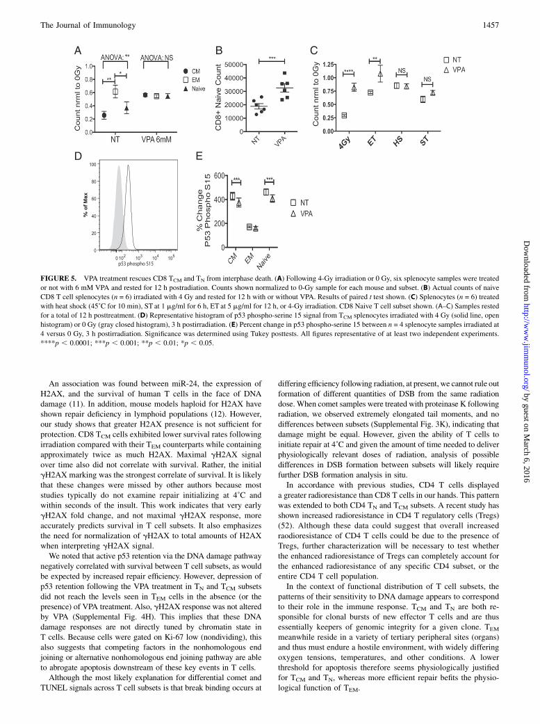
death, we treated cells with 6 mM VPA and subjected them to heatshock, brefeldin A, or ST treatment to induce apoptosis. Asa positive control, we treated cells with ET to induce apoptosis ina radiation-independent, but DSB-dependent manner. As predictedfor a model of enhanced DNA repair through chromatin relaxation,VPA treatment did not affect survival of cells exposed to heatshock, ST (Fig. 5C), or brefeldin A (Supplemental Fig. 4F), butdid affect survival following ET treatment in manner identical tothat following radiation damage, by specifically improving CD8TCM and TN cell subset survival (Fig. 5C, Supplemental Fig. 4G).Because apoptosis following IR in lymphocytes is dependent onp53 (36), we examined p53 retention in these subsets. p53 isprotected from degradation following genomic damage via phos-phorylation of serine 15 by ataxia-telangiectasia mutated kinase(37). Fold change in p53–phospho-S15 was lowest in TEM fol-lowing IR, correlating with increased survival. However, p53 re-tention in TCM and TN p53 was significantly but only slightlylowered by VPA treatment (Fig. 5D, 5E), suggesting that de-creased p53 retention is not the sole mechanism explaining theefficacy of VPA-mediated increase in TCM and TN survival.
DiscussionA fundamental understanding of peripheral immune subset radiationsensitivity is crucial for better understanding of the clinical impactof radiotherapy (38, 39), bone marrow transplantation (40), andimmune reconstitution (41). Moreover, the discovery of factorsresponsible for differential lymphocyte radiation sensitivity hasimplications for diverse areas of immunology and biology: DNAdamage response (42), natural and virus-induced hematopoieticcancers (43, 44), V(D)J recombination (45), cytotoxic killing (46),and possibly age-induced immune senescence (47), inasmuch as thelatter may intersect with DNA damage repair over lifespan.
Despite the breadth and depth of the field of radiobiology, thedifferential response of lymphocyte subsets to radiation in different
compartments remains incompletely understood (48). Previous
studies have demonstrated that radiation-induced T cell apoptosis
requires p53 (36). The constitutive Bcl-2 transgenic mouse model
has established that antiapoptotic machinery can be protective, but
the relevance of these findings for physiological radiosensitivity
remains unclear (49). Our work demonstrates that the TEM cell
populations are intrinsically radioresistant relative to other T cell
subpopulations and that this resistance is linked to repair advantages
fostered by open chromatin. Unlike other subsets, survival of TEM
following DSB is not improved by forcing the chromatin open.
Given that subset sensitivity and genome-wide heterochromatin
content are highly correlated, we propose that chromatin state is the
central arbiter of differential T cell subset radiosensitivity.Aside from bone marrow transplantation, there is no truly ef-
fective treatment for hematopoietic radiation syndrome followinglethal irradiation. The therapeutic use of recombinant cytokines andgrowth factors can be effective but only against exposures on thelower end of the lethal range (50). Treating large swaths of thepopulation with from bone marrow transplantation would be im-possible in the case of mass exposure. When applied in vivo afterpotentially lethal radiation exposure, VPA is able to abrogateT cell death in C57BL/6 mice (51). Our work suggests that thismay in part operate via a mechanism whereby lymphocyte attri-tion is lowered throughout the animal because of enhanced DSBbinding and repair, improving defense against opportunistic andother infection. This indicates that VPA-mediated HDACi pro-vides a temporary enhancement of native repair abilities and thusmay be able to limit the damage to the immune system, althoughthat remains to be tested in vivo.
FIGURE 4. Standing chromatin content, and H3K27me3 downregulation following radiation, correlate with T cell subset radiosensitivity. (A) Standing
H3K27me3 in T cell subsets versus relative survival of those same subsets post-4 Gy and 21 h incubation, averages from n = 5. From highest survival: CD4EM,
CD8EM, CD4CM, CD4N, CD8CM, and CD8N. (B) CD8 H3K27me3 alterations following 4 Gy IR and 5 min incubation at 37˚C. Two-way ANOVA: cell type:
****, radiation ***. (n = 4). (C) Samples as in (B), incubated for 1 h post-IR. Two-way ANOVA: Time: ** Cell subsets: *. Shown are results of Tukey’s posttests
between subsets. (D) Representative histogram of CD8 H3K27me3 in TN (solid line), TCM (dashed line), or TEM (gray). (E) DNAse I hypersensitivity assay was
performed in CD8 spleen samples by flow cytometry (n = 4), and results were expressed as percent decrease in PI MFI. Significance between the subsets was
assessed using the Tukey posttests. (F) Representative histogram of PI staining in Ki-67 negative TEM cells either treated (solid line, left histogram) or untreated
(dashed line, right histogram) with DNAse I. All figures representative of at least two independent experiments. ***p , 0.001; **p , 0.01; *p , 0.05.
1456 HISTONE DEACETYLATION AND T CELL RADIOSENSITIVITY
by guest on March 6, 2016
http://ww
w.jim
munol.org/
Dow
nloaded from
An association was found between miR-24, the expression ofH2AX, and the survival of human T cells in the face of DNAdamage (11). In addition, mouse models haploid for H2AX haveshown repair deficiency in lymphoid populations (12). However,our study shows that greater H2AX presence is not sufficient forprotection. CD8 TCM cells exhibited lower survival rates followingirradiation compared with their TEM counterparts while containingapproximately twice as much H2AX. Maximal gH2AX signalover time also did not correlate with survival. Rather, the initialgH2AX marking was the strongest correlate of survival. It is likelythat these changes were missed by other authors because moststudies typically do not examine repair initializing at 4˚C andwithin seconds of the insult. This work indicates that very earlygH2AX fold change, and not maximal gH2AX response, moreaccurately predicts survival in T cell subsets. It also emphasizesthe need for normalization of gH2AX to total amounts of H2AXwhen interpreting gH2AX signal.We noted that active p53 retention via the DNA damage pathway
negatively correlated with survival between T cell subsets, as wouldbe expected by increased repair efficiency. However, depression ofp53 retention following the VPA treatment in TN and TCM subsetsdid not reach the levels seen in TEM cells in the absence (or thepresence) of VPA treatment. Also, gH2AX response was not alteredby VPA (Supplemental Fig. 4H). This implies that these DNAdamage responses are not directly tuned by chromatin state inT cells. Because cells were gated on Ki-67 low (nondividing), thisalso suggests that competing factors in the nonhomologous endjoining or alternative nonhomologous end joining pathway are ableto abrogate apoptosis downstream of these key events in T cells.Although the most likely explanation for differential comet and
TUNEL signals across T cell subsets is that break binding occurs at
differing efficiency following radiation, at present, we cannot rule outformation of different quantities of DSB from the same radiationdose. When comet samples were treated with proteinase K followingradiation, we observed extremely elongated tail moments, and nodifferences between subsets (Supplemental Fig. 3K), indicating thatdamage might be equal. However, given the ability of T cells toinitiate repair at 4˚C and given the amount of time needed to deliverphysiologically relevant doses of radiation, analysis of possibledifferences in DSB formation between subsets will likely requirefurther DSB formation analysis in situ.In accordance with previous studies, CD4 T cells displayed
a greater radioresistance than CD8 T cells in our hands. This patternwas extended to both CD4 TN and TCM subsets. A recent study hasshown increased radioresistance in CD4 T regulatory cells (Tregs)(52). Although these data could suggest that overall increasedraodioresistance of CD4 T cells could be due to the presence ofTregs, further characterization will be necessary to test whetherthe enhanced radioresistance of Tregs can completely account forthe enhanced radioresistance of any specific CD4 subset, or theentire CD4 T cell population.In the context of functional distribution of T cell subsets, the
patterns of their sensitivity to DNA damage appears to correspondto their role in the immune response. TCM and TN are both re-sponsible for clonal bursts of new effector T cells and are thusessentially keepers of genomic integrity for a given clone. TEM
meanwhile reside in a variety of tertiary peripheral sites (organs)and thus must endure a hostile environment, with widely differingoxygen tensions, temperatures, and other conditions. A lowerthreshold for apoptosis therefore seems physiologically justifiedfor TCM and TN, whereas more efficient repair befits the physio-logical function of TEM.
FIGURE 5. VPA treatment rescues CD8 TCM and TN from interphase death. (A) Following 4-Gy irradiation or 0 Gy, six splenocyte samples were treated
or not with 6 mM VPA and rested for 12 h postradiation. Counts shown normalized to 0-Gy sample for each mouse and subset. (B) Actual counts of naive
CD8 T cell splenocytes (n = 6) irradiated with 4 Gy and rested for 12 h with or without VPA. Results of paired t test shown. (C) Splenocytes (n = 6) treated
with heat shock (45˚C for 10 min), ST at 1 mg/ml for 6 h, ET at 5 mg/ml for 12 h, or 4-Gy irradiation. CD8 Naive T cell subset shown. (A–C) Samples rested
for a total of 12 h posttreatment. (D) Representative histogram of p53 phospho-serine 15 signal from TCM splenocytes irradiated with 4 Gy (solid line, open
histogram) or 0 Gy (gray closed histogram), 3 h postirradiation. (E) Percent change in p53 phospho-serine 15 between n = 4 splenocyte samples irradiated at
4 versus 0 Gy, 3 h postirradiation. Significance was determined using Tukey posttests. All figures representative of at least two independent experiments.
****p , 0.0001; ***p , 0.001; **p , 0.01; *p , 0.05.
The Journal of Immunology 1457
by guest on March 6, 2016
http://ww
w.jim
munol.org/
Dow
nloaded from

AcknowledgmentsWe thank Drs. Giovanni Bosco (Dartmouth College), Ted Weinert, Kirsten
Limesand, Felicia Goodrum (all from the University of Arizona), and the
Goodrum laboratory. We also thank Paula Campbell at the University of
Arizona Flow Core Facility and Doug Cromey at the University of Arizona
Microscopy Core Facility. We give special thanks to Dr. Wendell Lutz (Uni-
versity of Arizona Cancer Center) for radiation dosage calibration and in-
struction, and Drs. Jeff Frelinger, Mike Kuhns, and Richard Wagner
(University of Arizona) for help and advice.
DisclosuresThe authors have no financial conflicts of interest.
References1. Alper, T. 1979. Cellular Radiobiology. Cambridge University Press, Cambridge, U.K.2. Nias, A. 1990. Introduction to Radiobiology. John Wiley and Sons Ltd., Chichester.3. Sprent, J., R. E. Anderson, and J. F. Miller. 1974. Radiosensitivity of T and
B lymphocytes. II. Effect of irradiation on response of T cells to alloantigens.Eur. J. Immunol. 4: 204–210.
4. Anderson, R. E., and N. L. Warner. 1976. Ionizing radiation and the immuneresponse. Adv. Immunol. 24: 215–335.
5. Bergonie, J., and L. Tribondeau. 1959. Interpretation of some results of radio-therapy and an attempt at determining a logical technique of treatment. Radiat.Res. 11: 587–588.
6. Wilkins, R. C., D. Wilkinson, H. P. Maharaj, P. V. Bellier, M. B. Cybulski, andJ. R. McLean. 2002. Differential apoptotic response to ionizing radiation insubpopulations of human white blood cells. Mutat. Res. 513: 27–36.
7. Chambers, K. A., N. P. Harrington, W. M. Ross, and L. G. Filion. 1998. Relativealterations in blood mononuclear cell populations reflect radiation injury in mice.Cytometry 31: 45–52.
8. Seki, H., K. Iwai, H. Kanegane, A. Konno, K. Ohta, K. Ohta, A. Yachie,N. Taniguchi, and T. Miyawaki. 1995. Differential protective action of cytokineson radiation-induced apoptosis of peripheral lymphocyte subpopulations. Cell.Immunol. 163: 30–36.
9. Grayson, J. M., L. E. Harrington, J. G. Lanier, E. J. Wherry, and R. Ahmed.2002. Differential sensitivity of naive and memory CD8+ T cells to apoptosisin vivo. J. Immunol. 169: 3760–3770.
10. Rogakou, E. P., D. R. Pilch, A. H. Orr, V. S. Ivanova, and W. M. Bonner. 1998.DNA double-stranded breaks induce histone H2AX phosphorylation on serine139. J. Biol. Chem. 273: 5858–5868.
11. Brunner, S., D. Herndler-Brandstetter, C. R. Arnold, G. J. Wiegers, A. Villunger,M. Hackl, J. Grillari, M. Moreno-Villanueva, A. B€urkle, and B. Grubeck-Loe-benstein. 2012. Upregulation of miR-24 is associated with a decreased DNAdamage response upon etoposide treatment in highly differentiated CD8+ T cellssensitizing them to apoptotic cell death. Aging Cell 11: 579–587.
12. Celeste, A., S. Petersen, P. J. Romanienko, O. Fernandez-Capetillo, H. T. Chen,O. A. Sedelnikova, B. Reina-San-Martin, V. Coppola, E. Meffre,M. J. Difilippantonio, et al. 2002. Genomic instability in mice lacking histoneH2AX. Science 296: 922–927.
13. Price, B. D., and A. D. D’Andrea. 2013. Chromatin remodeling at DNA double-strand breaks. Cell 152: 1344–1354.
14. Murga, M., I. Jaco, Y. Fan, R. Soria, B. Martinez-Pastor, M. Cuadrado,S. M. Yang, M. A. Blasco, A. I. Skoultchi, and O. Fernandez-Capetillo. 2007.Global chromatin compaction limits the strength of the DNA damage response.J. Cell Biol. 178: 1101–1108.
15. Dispirito, J. R., and H. Shen. 2010. Histone acetylation at the single-cell level: a markerof memory CD8+ T cell differentiation and functionality. J. Immunol. 184: 4631–4636.
16. Roos, W. P., and B. Kaina. 2013. DNA damage-induced cell death: from specificDNA lesions to the DNA damage response and apoptosis. Cancer Lett. 332: 237–248.
17. Jankovic, V., I. Messaoudi, and J. Nikolich-�Zugich. 2003. Phenotypic andfunctional T-cell aging in rhesus macaques (Macaca mulatta): differential be-havior of CD4 and CD8 subsets. Blood 102: 3244–3251.
18. Tough, D. F., and J. Sprent. 1994. Turnover of naive- and memory-phenotypeT cells. J. Exp. Med. 179: 1127–1135.
19. Sallusto, F., D. Lenig, R. Forster, M. Lipp, and A. Lanzavecchia. 1999. Twosubsets of memory T lymphocytes with distinct homing potentials and effectorfunctions. Nature 401: 708–712.
20. Starborg, M., K. Gell, E. Brundell, and C. Hoog. 1996. The murine Ki-67 cellproliferation antigen accumulates in the nucleolar and heterochromatic regionsof interphase cells and at the periphery of the mitotic chromosomes in a processessential for cell cycle progression. J. Cell Sci. 109: 143–153.
21. Marrack, P., and J. Kappler. 2004. Control of T cell viability. Annu. Rev.Immunol. 22: 765–787.
22. Rochman, Y., and W. J. Leonard. 2008. The role of thymic stromal lympho-poietin in CD8+ T cell homeostasis. J. Immunol. 181: 7699–7705.
23. Lopez, F., F. Belloc, F. Lacombe, P. Dumain, J. Reiffers, P. Bernard, andM. R. Boisseau. 1991. Modalities of synthesis of Ki67 antigen during thestimulation of lymphocytes. Cytometry 12: 42–49.
24. Gerdes, J., H. Lemke, H. Baisch, H. H. Wacker, U. Schwab, and H. Stein. 1984.Cell cycle analysis of a cell proliferation-associated human nuclear antigendefined by the monoclonal antibody Ki-67. J. Immunol. 133: 1710–1715.
25. Grahn, D., and K. F. Hamilton. 1957. Genetic variation in the acute lethal response offour inbred mouse strains to whole body X-irradiation. Genetics 42: 189–198.
26. Ciccia, A., and S. J. Elledge. 2010. The DNA damage response: making it safe toplay with knives. Mol. Cell 40: 179–204.
27. Gottschalk, A. J., G. Timinszky, S. E. Kong, J. Jin, Y. Cai, S. K. Swanson,M. P. Washburn, L. Florens, A. G. Ladurner, J. W. Conaway, and R. C. Conaway.2009. Poly(ADP-ribosyl)ation directs recruitment and activation of an ATP-dependent chromatin remodeler. Proc. Natl. Acad. Sci. USA 106: 13770–13774.
28. Wang, M., W. Wu, W. Wu, B. Rosidi, L. Zhang, H. Wang, and G. Iliakis. 2006.PARP-1 and Ku compete for repair of DNA double strand breaks by distinctNHEJ pathways. Nucleic Acids Res. 34: 6170–6182.
29. Gavrieli, Y., Y. Sherman, and S. A. Ben-Sasson. 1992. Identification of programmedcell death in situ via specific labeling of nuclear DNA fragmentation. J. Cell Biol. 119:493–501.
30. Collins, A. R., A. A. Oscoz, G. Brunborg, I. Gaivao, L. Giovannelli, M. Kruszewski,C. C. Smith, and R. Stetina. 2008. The comet assay: topical issues. Mutagenesis 23:143–151.
31. Seo, J., S. C. Kim, H. S. Lee, J. K. Kim, H. J. Shon, N. L. Salleh, K. V. Desai,J. H. Lee, E. S. Kang, J. S. Kim, and J. K. Choi. 2012. Genome-wide profiles ofH2AX and g-H2AX differentiate endogenous and exogenous DNA damagehotspots in human cells. Nucleic Acids Res. 40: 5965–5974.
32. Rawlings, J. S., M. Gatzka, P. G. Thomas, and J. N. Ihle. 2011. Chromatincondensation via the condensin II complex is required for peripheral T-cellquiescence. EMBO J. 30: 263–276.
33. Barski, A., S. Cuddapah, K. Cui, T. Y. Roh, D. E. Schones, Z. Wang, G. Wei,I. Chepelev, and K. Zhao. 2007. High-resolution profiling of histone methyl-ations in the human genome. Cell 129: 823–837.
34. Creyghton, M. P., A. W. Cheng, G. G. Welstead, T. Kooistra, B. W. Carey,E. J. Steine, J. Hanna, M. A. Lodato, G. M. Frampton, P. A. Sharp, et al. 2010.Histone H3K27ac separates active from poised enhancers and predicts devel-opmental state. Proc. Natl. Acad. Sci. USA 107: 21931–21936.
35. Monti, B., E. Polazzi, and A. Contestabile. 2009. Biochemical, molecular and epige-netic mechanisms of valproic acid neuroprotection. Curr. Mol. Pharmacol. 2: 95–109.
36. Seki, H., H. Kanegane, K. Iwai, A. Konno, K. Ohta, A. Yachie, N. Taniguchi, andT. Miyawaki. 1994. Ionizing radiation induces apoptotic cell death in humanTcR-g/d+ T and natural killer cells without detectable p53 protein. Eur. J.Immunol. 24: 2914–2917.
37. Banin, S., L. Moyal, S. Shieh, Y. Taya, C. W. Anderson, L. Chessa, N. I. Smorodinsky,C. Prives, Y. Reiss, Y. Shiloh, and Y. Ziv. 1998. Enhanced phosphorylation of p53 byATM in response to DNA damage. Science 281: 1674–1677.
38. Finnon, P., S. Kabacik, A. MacKay, C. Raffy, R. A’Hern, R. Owen, C. Badie,J. Yarnold, and S. Bouffler. 2012. Correlation of in vitro lymphocyte radiosen-sitivity and gene expression with late normal tissue reactions following curativeradiotherapy for breast cancer. Radiother. Oncol. 105: 329–336.
39. Kitayama, J., K. Yasuda, K. Kawai, E. Sunami, and H. Nagawa. 2011.Circulating lymphocyte is an important determinant of the effectiveness ofpreoperative radiotherapy in advanced rectal cancer. BMC Cancer 11: 64.
40. Baron, F., J. E. Baker, R. Storb, T. A. Gooley, B. M. Sandmaier, M. B. Maris,D. G. Maloney, S. Heimfeld, D. Oparin, E. Zellmer, et al. 2004. Kinetics of en-graftment in patients with hematologic malignancies given allogeneic hematopoieticcell transplantation after nonmyeloablative conditioning. Blood 104: 2254–2262.
41. Peggs, K. S. 2004. Immune reconstitution following stem cell transplantation.Leuk. Lymphoma 45: 1093–1101.
42. Janssens, S., A. Tinel, S. Lippens, and J. Tschopp. 2005. PIDD mediates NF-kBactivation in response to DNA damage. Cell 123: 1079–1092.
43. Hakim, O., W. Resch, A. Yamane, I. Klein, K. R. Kieffer-Kwon, M. Jankovic,T. Oliveira, A. Bothmer, T. C. Voss, C. Ansarah-Sobrinho, et al. 2012. DNAdamage defines sites of recurrent chromosomal translocations in B lymphocytes.Nature 484: 69–74.
44. Chlichlia, K., and K. Khazaie. 2010. HTLV-1 tax: linking transformation, DNAdamage and apoptotic T-cell death. Chem. Biol. Interact. 188: 359–365.
45. Yin, B., V. Savic, M. M. Juntilla, A. L. Bredemeyer, K. S. Yang-Iott,B. A. Helmink, G. A. Koretzky, B. P. Sleckman, and C. H. Bassing. 2009.Histone H2AX stabilizes broken DNA strands to suppress chromosome breaksand translocations during V(D)J recombination. J. Exp. Med. 206: 2625–2639.
46. Lieberman, J. 2010. Granzyme A activates another way to die. Immunol. Rev.235: 93–104.
47. Nijnik, A., L. Woodbine, C. Marchetti, S. Dawson, T. Lambe, C. Liu,N. P. Rodrigues, T. L. Crockford, E. Cabuy, A. Vindigni, et al. 2007. DNA repairis limiting for haematopoietic stem cells during ageing. Nature 447: 686–690.
48. UNSCEAR. 2006. Annex D: effects of ionizing radiation on the immune system.United Nations Scientific Committee on the Effects of Radiation, United NationsPublications, Vienna, Austria.
49. Yao, Z., J. Jones, H. Kohrt, and S. Strober. 2011. Selective resistance of CD44hi
T cells to p53-dependent cell death results in persistence of immunologicmemory after total body irradiation. J. Immunol. 187: 4100–4108.
50. Waselenko, J. K., T. J. MacVittie, W. F. Blakely, N. Pesik, A. L. Wiley,W. E. Dickerson, H. Tsu, D. L. Confer, C. N. Coleman, T. Seed, et al. 2004. Medicalmanagement of the acute radiation syndrome: recommendations of the StrategicNational Stockpile Radiation Working Group. Ann. Intern. Med. 140: 1037–1051.
51. Brown, S. L., A. Kolozsvary, J. Liu, S. Ryu, and J. H. Kim. 2008. Histonedeacetylase inhibitors protect against and mitigate the lethality of total-bodyirradiation in mice. Radiat. Res. 169: 474–478.
52. Qu, Y., S. Jin, A. Zhang, B. Zhang, X. Shi, J. Wang, and Y. Zhao. 2010. Gamma-ray resistance of regulatory CD4+CD25+Foxp3+ T cells in mice. Radiat. Res.173: 148–157.
1458 HISTONE DEACETYLATION AND T CELL RADIOSENSITIVITY
by guest on March 6, 2016
http://ww
w.jim
munol.org/
Dow
nloaded from
Copyright © 2022 FDOKUMEN