Fighting in fiddler crabs< i> Uca mjoebergi: what determines duration?

Supplemental Data Interaction between Oct3/4 and Cdx2 Determines Trophectoderm Differentiation Hitoshi Niwa, Yayoi Toyooka, Daisuke Shimosato, Dan Strumpf, Kadue Takahashi, Rika Yagi, and Janet Rossant
Culture of ES Cells
All ES cells were cultured in the absence of feeder cells in Glasgow minimal essential
medium (GMEM) supplemented with 10% fetal calf serum (FCS), 1 mM sodium
pyruvate, 10-4 M 2-mercaptoethanol, 1x non-essential amino acids and 1000 U/ml of
LIF on gelatin-coated dishes.
Episomal Transfection for Overexpression of Transgenes in ES Cells
The episomal transfection system allows us to obtain stable transfectants in which the
introduced transgenes are expressed strongly and homogeneously (Niwa et al., 1998).
The host ES cell line MGZ5 (Fujikura et al., 2002) was established by introduction of
pOct4IRESzeocinpA KO vector (Nichols et al., 1998) into MG1.19 ES cells (Gassmann
et al., 1995). Cdx2 and Eomeso cDNA were amplified by RT-PCR with high-fidelity Pfx
DNA polymerase (Invitrogen) using TS total RNA as a template with primers Cdx2S:
AAACATGTACGTGAGCTACCTTCC, Cdx2AS:

AAAGTACTGGGTGACAGTGGAGTTT, EomesoS:
AAAGCATGCAGTTGGGAGAGCAGCTCC, and EomesoAS:
AATGCTCTAGGCACTTGTGTAAAAAGCA, and subcloned into pCAG-IP (Niwa et
al., 2002). 2 µg of plasmid DNA was transfected into 3x10⁴ MGZ5 ES cells with
Lipofectoamine 2000 (Invitrogen) and these cells were replated into 60-mm dishes.
Selection with 1 µg/ml of puromycin (Sigma) began 24 hours following transfection
and effects of transgene expression were scored at 6 days after transfection.
Establishment of ES Cells Carrying Hormone-Inducible Cdx2
The entire open reading frame of Cdx2 amplified by PCR with primers
Cdx2S2:CCTCGAGCCAACATGTACGTGAGCTACCTT and Cdx2AS2:
TCAACTAGTCTGGGTGACTGAGGAGTT followed by digestion with XhoI and SpeI
and the ligand binding domain of the mutant form of mouse estrogen receptor (Esr)
amplified by PCR with primers mESRS:
TTCATGACTAGTCGAAATGAAATGGGTGCT and mESRAS:
AGCGGCCGCTCAGATCGTGTTGGGGAAGCC followed by digestion with SpeI and
NotI were subcloned into XhoI-NotI of pCAG-IP, resulting in the generation of
pCAG-Cdx2ER-IP. 40 µg of pCAG-Cdx2ER-IP was linealized by SalI and transfected
into ZHBTc4 ES cells (Niwa et al., 2000) at 800 V and 3 µF in a 0.4-cm cuvette using
Gene Pulser (Bio-Rad) followed by selection with 1 µg/ml of puromycin for 8 days.
Clones in which pCAG-Cdx2ER-IP were randomly integrated into genomic DNA were
selected by the ability to undergo differentiation to TE by addition of 1 µg/ml of
4-hydroxy tamoxifen (Tx; Sigma), resulting establishment of 4CER1 ES cells. The
plasmid pCAG-EGFPCdx2ER-IP was generated by in-frame insertion of EGFP cDNA
(Clonetech) amplified by primers EGFPS:
GACTCGAGCCAGGATGGTGAGCAAGGGCGAGG and EGFPAS:
CTCCCATGGTCTTGTACAGCTCGTCCATGC into XhoI-AflIII of pCAG-Cdx2ER-IP.
40 µg of linealized pCAG-EGFPCdx2ER-IP was introduced into EB5 ES cells
(Kawasaki et al., 2000), which was established by targeted integration of
pOct4IRESBSDpA (Niwa et al., 2000) in E14tg2a ES cells (Thompson et al., 1989), to
establish 5ECER4 ES cells. Because of the weak EGFP fluorescence of this clone, we
then introduced pCAG-EGFP-IZ, in which EGFP cDNA was inserted into the
expression vector pCAG-IZ (Niwa et al., 1998), into 5ECER4 ES cells followed by
selection with 40 µg/ml zeocin (Stratagene) for 8 daysThis resulted in the establishment
of 5ECER4G20. These ES cells showed bright and ubiquitous expression of EGFP that
allowed us to follow the progenies of injected ES cells in chimeric embryos.

Luciferase Assay
The reporter constructs Fgf4tkluc, 6Wtkluc and tkluc were described in our previous
paper (Niwa et al., 2002). Utf1tkluc was constructed by insertion of 1076 bp
SalI-BamHI fragment of mouse Utf1 genomic DNA carrying Oct3/4-Sox2-dependent
enhancer (Nishimoto et al., 1999) into SalI-BamHI of tkluc. A series of luciferase
reporters carrying the Oct3/4 promoter was described by Okumura-Nakanishi et al.
(2005). Cdx2luc was generated by insertion of the 8.2 kb Cdx2 promoter isolated from
129Sv genomic DNA library into pGL3Basic (Promega). The plasmids pCAG-Cdx1-IP
and pCAG-Cdx4-IP were generated by insertion of Cdx1 and Cdx4 cDNA amplified by
RT-PCR using 8.5 dpc embryo total RNA as a template with primers Cdx1S:
CCCTCGAGGACGACTGGGCAGCTGCT, Cdx1AS:
TTGCGGCCGCTAGGGTAGAAACTCCTCC, Cdx4S:
CCCTCGAGTCACCATGTATGGAAGCTG, and Cdx4AS:
TTGCGGCCGCGTTTCATTCAGAAAGTATGACCTGCTG into pCAG-IP.
For luciferase assay, 0.5 µg of luciferase reporters and 0.5 µg of expression
vectors for transcription factors were transfected into 1X10⁴ ES cells using
Lipofectoamine 2000 (Invitrogen) with 0.01 µg of pRL-CMV (Promega) as an internal

control. Transfectants were lysed at 24 hours after transfection and luciferase activities
were estimated using Dual-luciferase assay kit (Promega).
Antibodies for Immunostaining
The first antibodies (Abs) used in our experiment for staining of cells were mouse
anti-Cdh3 monoclonal Ab (Clone56C1, NEOMARKERS), mouse anti-Cytokeratin 7
monoclonal Ab (MAB3226, Chemicon), rabbit anti-Ets2 polyclonal Ab (C-20, Santa
Cruz), mouse anti-Oct3/4 monoclonal Ab (C-10, Santa Cruz) and mouse anti-Cdx2
monoclonal Ab (Cdx2-88, BioGenex).
For immunohistochemical analysis of chimeric placenta, we used goat
anti-GFP Ab (Abcam) and anti-goat IgG-HRP (rabbit polyclonal; Abcam) as the first
and second Abs, respectively. The signal was detected by DAB staining followed by
counterstaining with hematoxylin.
For immunostaining of Cdx2 and Oct3/4 in pre-implantation embryos, mouse
anti-Cdx2 monoclonal Ab (Cdx2-88, BioGenex) and rabbit anti-Oct3/4 antisera raised
by us were used as primary antibodies followed by incubation with the secondary Abs
detecting mouse and rabbit IgG conjugated with Alexa Fluor 488 or 594 (Molecular
Probes), respectively.

FRET Analysis
The Cdx2ECFP and EYFPCdx2 fusion genes were constructed by in-frame fusion of
the full-length Cdx2 with ECFP and EYFP (Clonetech) to the N- and C-terminus,
respectively. nlsECFP was generated by addition of amino acid stretch MPKKKRKV
for nuclear localization signal to the N-terminus of ECFP by PCR technique. The
Venus- (Nagai et al., 2002) and ECFP-Oct3/4 fusion genes were made as described
previously for EGFPOct3/4 (Niwa et al., 2002). The Open reading frame of monomeric
version of Kusabira-Orange (mKO1) (MBL: Karasawa et al., 2004) was fused to
N-terminal of Oct3/4 via a flexible linker peptide (GGSGGGGSGGGSS) to generate
the mKO1Oct3/4 fusion gene. These fusion genes were subcloned into pCAG-IP. VO-1,
CO-1 and KOWT7 ES cells were established by introduction of VenusOct3/4,
ECFPOct3/4 and mKO1Oct3/4 expression vectors into ZHBTc4 (Niwa et al., 2000) in
the presence of Tc, respectively. Self-renewal of these ES cells was maintained by the
expression of these modified Oct3/4 transgenes instead of the wild-type Oct3/4,
confirming the proper function of these fusion gene products.
For FRET analysis, we chose two fluorescent proteins ECFP and mKO1 due to
this combinations apparent suitable for FRET because of (1) very low activation of
mKO1 by excitation at 458 nm of ECFP, (2) high excitation efficiency of mKO1 and (3)
easy photo acceptor-bleaching of mKO1. 2 µg of pCAG-Cdx2ECFP-IP or
pCAG-nlsECFP-IP plasmid DNA was transfected into 1x10⁴ KOWT7 ES cells. At 24
hours after transfection, the FRET analysis was performed by the spectral confocal
scanning system TCS SP2 (Leica). The signals were captured with the following
excitation/emission ranges: 458 nm/465-485nm for ECFP, 514 nm/550-570 nm for
mKO1, and 458nm/550-570 nm for FRET. After acquiring the first series of data (C1
for ECFP, Y1 for EYFP and F1 for FRET), acceptor bleaching was performed by 514
nm light for 2 minutes, the signals were acquired again for ECFP, EYFP and FRET (C2,
F2 and Y2, respectively), and the FRET ratio (F) is calculated as below;
F=1-(C2/F2’)/(C1/F1) ; F2’=F2-{F1x(Y1/Y2)}
Acceptor bleaching was confirmed by the ratio Y2/Y1, and only samples in which this
ratio was under 0.2 were statistically analyzed by t-test between F values from
Cdx2ECFP+mKO1Oct3/4 (n=8) and nlsECFP+mKO1Oct-3/4 (n=7).
Knockout of Cdx2
To construct the Cdx2 targeting vector, the genomic DNA clone was isolated from a
129/SvJ mouse genomic library. A 4.2 kb HindIII-AvaII fragment containing the 5’
untranslated region, a 1.9 kb XbaI-MluI fragment from the first intron of the Cdx2 gene,
and an MC1 promoter-driven diphteria toxin A fragment (DTA) gene with the poly-A
signal (pA) derived from the mouse phosphoglycerokinase (PGK) gene (Yagi et al.,
1993) were introduced into pBluescript KS (-), resulting in the generation of
pCdx2KO-DTA. The unique SpeI site was introduced between the two genomic DNA
fragments, and the floxed PGKpacpA cassette or the β-actin promoter-driven
Salmonella typhimurium gene hisD for resistance to L-histidinol (Yamashita et al.,
2002) were introduced into the SpeI site, generating pCdx2KOfpac-DTA or
pCdx2KOhis-DTA, respectively.
100 µg of the linealized vector pCdx2KOfpac-DTA was electroporated into
ZHBTc4 ES cells at 800 V and 3 µF in a 0.4-cm cuvette using Gene Pulser (Bio-Rad)
followed by selection with 1 µg/ml of puromycin for 8 days. Targeted clones were
identified by Southern blot analysis with external probe in the second intron, and the
correct recombination of 5’ end was then confirmed by PCR. For second round targeting,
the linealized vector pCdx2KOhis-DTA was electroporated into one of the targeted
clones, sko112, followed by selection with 3 µM of L-histidinol (Sigma) for 8 days. The
correct double-knockout clone dko23 was identified by both Southern blot and PCR
analyses. To remove the floxed PGKpacpA cassette from the first knockout allele,
dko23 ES cells were transfected with the Cre expression vector pCAG-Cre-IP by
lipofection and replated at clonal density. The isolated clones were analyzed by
Southern blot, resulting in the identification of the clone dko23-5 in which the
PGKpacpA cassette was correctly removed. The rescued clone 23CER1 was established
by introduction of pCAG-Cdx2ER-IP into dko23-5 ES cells.
Establishment of ES Cells Carrying Hormone-Inducible Eomeso
The entire open reading frame of Eomeso amplified by PCR with primers
EomesoS2:CCTCGAGCCACCATGCAGTTGGGAGAGCAGCTCC and EomesoAS2:
AACCATGGCACTTGTGTAAAAAGCA and followed by digestion with XhoI and
NcoI and the ligand binding domain of the mutant Esr amplified by PCR with primers
mERS and mERAS followed by digestion with BspHI and NotI was subcloned into
XhoI-NotI of pCAG-IP, resulting in the generation of pCAG-EomesoER-IP. ZHBTc4
and dko23-5 ES cells were transfected with the linealized pCAG-EomesoER-IP
followed by selction with puromycin, resulting in the establishment of 4EER3 and
23EER1 ES cells, respectively.

References
Fujikura, J., Yamato, E., Yonemura, S., Hosoda, K., Masui, S., Nakao, K., Miyazaki Ji,
J., and Niwa, H. (2002). Differentiation of embryonic stem cells is induced by GATA
factors. Genes Dev 16, 784-789.
Gassmann, M., Donoho, G. and Berg, P. (1995). Maintenance of an extrachromosomal
plasmid vector in mouse embryonic stem cells. Proc Natl Acad Sci USA 92, 1292-1296.
Karasawa, S., Araki, T., Nagai, T., Mizuno, H. and Miyawaki, A. (2004). Cyan-emitting
and orange-emitting fluorescent proteins as a donor/acceptor pair for fluorescence
resonance energy transfer. Biochem J 381, 307-312.
Kawasaki, H., Mizuseki, K., Nishikawa, S., Kaneko, S., Kuwana, Y., Nakanishi, S.,
Nishikawa, S.-I. and Sasai, Y. (2000). Induction of midbrain dopaminergic neurons from
ES cells by stromal cell-derived inducing activity. Neuron 28, 31-40.
Nagai, T., Ibata, K., Park, E. S., Kubota, M., Mikoshiba, K. and Miyawaki, A. (2002). A
variant yellow fluorescent protein with fast and efficient maturation for cell-biological
applications. Nat Biotechnol 20, 87-90.
Nichols, J., Zevnik, B., Anastassiadis, K., Niwa, H., Klewe-Nebenius, D., Chambers, I.,
Schöler, H. and Smith, A. (1998). Formation of pluripotent stem cells in the mammalian
embryo depends on the POU transcription factor Oct4. Cell 95, 379-391.

Nishimoto, M., Fukushima, A., Okuda, A. and Muramatsu, M. (1999) The gene for the
embryonic stem cell coactivator UTF1 carries a regulatory element which selectively
interacts with a complex composed of Oct3/4 and Sox-2. Mol Cell Biol 19, 5453-5465.
Niwa, H., Burdon, T., Chambers, I., and Smith, A. (1998). Self-renewal of pluripotent
embryonic stem cells is mediated via activation of STAT3. Genes Dev 12, 2048-2060.
Niwa, H., Masui, S., Chambers, I., Smith, A. G., and Miyazaki, J. (2002). Phenotypic
complementation establishes requirements for specific POU domain and generic
transactivation function of Oct-3/4 in embryonic stem cells. Mol Cell Biol 22,
1526-1536.
Niwa, H., Miyazaki, J., and Smith, A. G. (2000). Quantitative expression of Oct-3/4
defines differentiation, dedifferentiation or self-renewal of ES cells. Nat Genet 24,
372-376.
Okumura-Nakanishi, S., Saito, M., Niwa, H. and Ishikawa, F. (2005). Oct-3/4 and Sox2
regulate Oct3/4 gene in ES cells. J. Biol. Chem., 280, 5307-5317.
Thompson, S., Clarke, A.R., Pow, A.M., Hooper, M.L. and Melton, D.W. (1989) Germ
line transmission and expression of a corrected HPRT gene produced by gene targeting
in embryonic stem cells. Cell 56, 313-320.
Yagi, T., Nada, S., Watanabe, N., Tamemoto, H., Kohmura, N., Ikawa, Y. and Aizawa, S.

(1993). A novel negative selection for homologous recombinants using diphtheria toxin
A fragment gene. Anal Biochem 214, 77-86.
Yamashita, Y.M., Okada, T., Matsusaka, T., Sonoda, E., Zhao, G.Y., Araki, K., Tateishi,
S., Yamaizumi, M. and Takeda, S. (2002). RAD18 and RAD54 cooperatively contribute
to maintenance of genomic stability in vertebrate cells. EMBO J 21, 5558-5566.

Supplemental Figures
Supplemental Figure 1. FRET Analysis of Interaction between Oct3/4 and Cdx2
(A) Nuclear Localization of Cdx2ECFP and nlsECFP in KOWT 7 ES Cells
The plasmids pCAG-Cdx2ECFP-IP or pCAG-nlsECFP-IP were introduced into
KOWT7 ES cells expressing the mKO1Oct3/4 fusion gene and the transfectants were
observed by fluorescent microscopy at 24 hours after transfection. The ECFP signals
co-localized in nuclei with the mKO1 signals in both cases.

(B) Detection of the FRET Signal by Confocal Microscopy
Intensity of fluorescent signal excited by 458 nm light was scanned between 470 nm to
645 nm in KOWT7 ES cells with (yellow) or without (green) expression of Cdx2ECFP.
Emission peak of ECFP around 475 nm was observed only in the Cdx2ECFP
transfectant. A small peak around 559 nm derived from mKO1, which could be
generated by either cross-emission or FRET, was observed not only in the Cdx2ECFP
transfectant as a faint hump on the slope but also in the non-transfectant as a small peak.
After photobleaching, these peak around 559 nm disappeared whereas the signal around
475 nm was enhanced in the transfectant, indicating that a FRET event occurred
between mKO1Oct3/4 and Cdx2ECFP
Supplemental Figure 2. Generation of Cdx2 Null ES Cells
(A) Strategy for Generating Cdx2 Null ES Cells from ZHBTc4 ES Cells

Both allele of Cdx2 in ZHBTc4 ES cells were inactivated by targeted integration of the
floxed PGK-pac-pA cassette and the AG-HisD-pA cassette in a stepwise manner,
resulting in the generation of heterozygous sko113 and homozygous dko23 ES cells.
(B) Southern-Blot Hybridization of sko113 and dko23 ES Cells
Loss of the wild-type allele in dko23 cells was confirmed.
Supplemental Figure 3. Relative Amounts of Transcripts for TE-Specific
Transcription Factors in Undifferentiated ES Cells
The expression levels of indicated genes in ZHBTc4 ES cells were estimated by
competition of the endogenous cDNA products with various amounts of purified cDNA
fragments, and relative copy numbers per cell were shown.
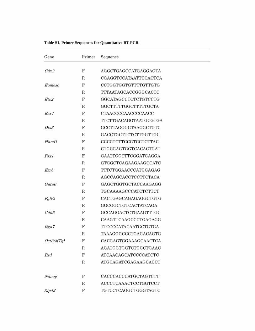
Table S1. Primer Sequences for Quantitative RT-PCR
Gene Primer Sequence Cdx2 F AGGCTGAGCCATGAGGAGTA R CGAGGTCCATAATTCCACTCA Eomeso F CCTGGTGGTGTTTTGTTGTG R TTTAATAGCACCGGGCACTC Ets2 F GGCATAGCCTCTCTGTCCTG R GGCTTTTTGGCTTTTTGCTA Esx1 F CTAACCCCAACCCCAACC R TTCTTGACAGGTAATGCGTGA Dlx3 F GCCTTAGGGGTAAGGCTGTC R GACCTGCTTCTCTTGGTTGC Hand1 F CCCCTCTTCCGTCCTCTTAC R CTGCGAGTGGTCACACTGAT Psx1 F GAATTGGTTTCGGATGAGGA R GTGGCTCAGAAGAAGCCATC Errb F TTTCTGGAACCCATGGAGAG R AGCCAGCACCTCCTTCTACA Gata6 F GAGCTGGTGCTACCAAGAGG R TGCAAAAGCCCATCTCTTCT Fgfr2 F CACTGAGCAGAGAGGCTGTG R GGCGGCTGTCACTATCAGA Cdh3 F GCCAGGACTCTGAAGTTTGC R CAAGTTCAAGCCCTGAGAGG Itga7 F TTCCCCATACAATGCTGTGA R TAAAGGGCCCTGAGACAGTG Oct3/4(Tg) F CACGAGTGGAAAGCAACTCA R AGATGGTGGTCTGGCTGAAC Bsd F ATCAACAGCATCCCCATCTC R ATGCAGATCGAGAAGCACCT Nanog F CACCCACCCATGCTAGTCTT R ACCCTCAAACTCCTGGTCCT Zfp42 F TGTCCTCAGGCTGGGTAGTC

R TGATTTTCTGCCGTATGCAA Pl1 F TACCCTGCTTGGTCTGGACT R GGGCACTCAACATTCGTTCT Tpbp F AAGTTAGGCAACGAGCGAAA R AGTGCAGGATCCCACTTGTC Gapdh F ACCACAGTCCATGCCATCAC R TCCACCACCCTGTTGCTGTA F, forward; R, reverse
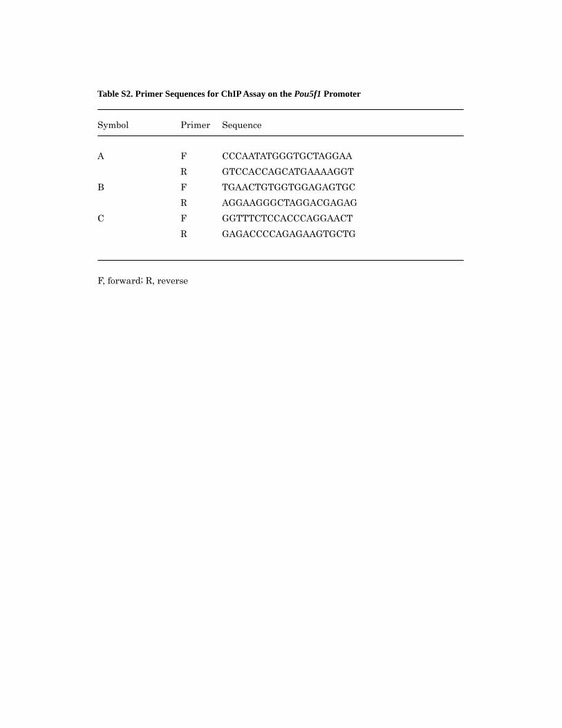
Table S2. Primer Sequences for ChIP Assay on the Pou5f1 Promoter
Symbol Primer Sequence A F CCCAATATGGGTGCTAGGAA R GTCCACCAGCATGAAAAGGT B F TGAACTGTGGTGGAGAGTGC R AGGAAGGGCTAGGACGAGAG C F GGTTTCTCCACCCAGGAACT R GAGACCCCAGAGAAGTGCTG F, forward; R, reverse
Copyright © 2022 FDOKUMEN