T-cell receptor bias and immunity
7
Available online at www.sciencedirect.com T-cell receptor bias and immunity Stephanie Gras 1 , Lars Kjer-Nielsen 2 , Scott R Burrows 3 , James McCluskey 2,* and Jamie Rossjohn 1,* Despite the potentially vast T-cell repertoire, biased ab T-cell receptor (TCR) usage has emerged as a common theme in immunity. Examples of TCR bias are observed in classical polymorphic major histocompatibility complex (MHC)- restricted immune responses as well as in T-cell responses to non-classical, monomorphic Ag-presenting molecules, such as CD1d. Recent data have implicated the structural landscape of these antigen-presenting molecules as one of the drivers of TCR bias. Here we review recent advances in the field, focussing on structural data pertaining to biased TCR usage, and discuss the implications for T-cell repertoire selection, MHC restriction and therapeutic development. Addresses 1 The Protein Crystallography Unit, Department of Biochemistry and Molecular Biology, School of Biomedical Sciences, Monash University, Clayton, Victoria 3800, Australia 2 Department of Microbiology & Immunology, University of Melbourne, Parkville, Victoria 3010, Australia 3 Cellular Immunology Laboratory, Queensland Institute of Medical Research, Brisbane 4029, Australia Corresponding author: McCluskey, James ([email protected]) and Rossjohn, Jamie ([email protected]) Current Opinion in Immunology 2008, 20:119–125 This review comes from a themed issue on Antigen Processing and Recognition Edited by Emil Unanue and James McCluskey Available online 18th January 2008 0952-7915/$ – see front matter # 2007 Elsevier Ltd. All rights reserved. DOI 10.1016/j.coi.2007.12.001 Introduction The ab T-cell receptor (TCR) plays a central role in the adaptive immune response, interacting with foreign pep- tide antigens (Ag) in complex with the major histocom- patibility complex (pMHC). The MHC is highly polymorphic and distinct features within the peptide- binding groove enables the MHC to present a wide array of peptides, but nevertheless TCRs are highly restricted to recognise host MHC molecules. In addition, the TCR can also recognise lipid-based antigens that are presented by the monomorphic CD1 family, which includes CD1d, the only CD1 family member that is present in humans and mice. To cope with this Ag diversity, the host possesses a potentially vast T-cell repertoire and after positive [1– 3] and negative selection [4–6] the number of different TCRs is approximately 10 7 in human and 10 6 in mice. This TCR diversity is generated via gene rearrangement within the variable domains of the TCR; namely the variable (V) and junction (J) gene segments define the Va chain whereas the Vb chain is composed of rearranged V, D (diversity) and J genes segments (Figure 1). The human locus for the TCR a chain is located on chromo- some 14 and contains 43 Va genes (referred to here as TRAV according to the immunogenetics database, IMGT [7]) as well as 58Ja (TRAJ) gene segments. The b TCR genes are located on the chromosome 7 and contain 42Vb, 2Db and 12Jb (TRBV, TRBD and TRBJ, respectively) gene segments. In addition, there is also junctional diversity V-(N)-J, V-(N)-D and D-(N)-J produced by random insertions/deletions at these regions (N represents ‘nucleotide’) [8,9](Figure 1). This diversity is manifested in the third complementarity determining regions (CDR), which is the region of the TCR that interacts with the bound peptide and glycolipid of MHC and CD1d molecules respectively (Figure 1a and c). The CDR3 loops that span the junctional region are highly diverse in sequence and length. Despite this TCR diversity and its importance in protective immunity, there are a growing number of examples of TCR bias in anti- viral immunity in which restricted Va and/or Vb usage as well as restricted CDR3 conserved motif characterises the immune response to defined antigens. A number of different mechanisms may operate to shape the T-cell repertoire, including thymic selection of the naı¨ve T-cell repertoire, TCR avidity for the pMHC, convergent recombination, antigen load, duration of the pMHC– TCR interaction and the pMHC structural landscape [10,11 ,12]. However, the relative contribution of such factors in shaping the TCR repertoire remains unclear. Examples of TCR bias are observed in persistent viral infections including Epstein-Barr virus (EBV) [13], cyto- megalovirus (CMV) [14], human immunodeficiency virus (HIV), SIV [15] and influenza [16,17]. As some of these viruses represent a significant threat to human health, understanding T-cell repertoire selection is important at a therapeutic level for vaccine design and immunotherapy. In addition, TCR bias has been reported in autoimmunity as well as in lipid-based immunity. Recently the struc- tural basis of biased TCR usage has begun to emerge, and we review recent data in this field that encompasses peptide and lipid-based recognition and discuss the implications for biased TCR usage, MHC restriction and immunity. * Joint senior. www.sciencedirect.com Current Opinion in Immunology 2008, 20:119–125
-
Upload
independent -
Category
Documents
-
view
4 -
download
0
Transcript of T-cell receptor bias and immunity

Available online at www.sciencedirect.com
T-cell receptor bias and immunityStephanie Gras1, Lars Kjer-Nielsen2, Scott R Burrows3,James McCluskey2,* and Jamie Rossjohn1,*
Despite the potentially vast T-cell repertoire, biased ab T-cell
receptor (TCR) usage has emerged as a common theme in
immunity. Examples of TCR bias are observed in classical
polymorphic major histocompatibility complex (MHC)-
restricted immune responses as well as in T-cell responses to
non-classical, monomorphic Ag-presenting molecules, such as
CD1d. Recent data have implicated the structural landscape of
these antigen-presenting molecules as one of the drivers of
TCR bias. Here we review recent advances in the field,
focussing on structural data pertaining to biased TCR usage,
and discuss the implications for T-cell repertoire selection,
MHC restriction and therapeutic development.
Addresses1 The Protein Crystallography Unit, Department of Biochemistry and
Molecular Biology, School of Biomedical Sciences, Monash University,
Clayton, Victoria 3800, Australia2 Department of Microbiology & Immunology, University of Melbourne,
Parkville, Victoria 3010, Australia3 Cellular Immunology Laboratory, Queensland Institute of Medical
Research, Brisbane 4029, Australia
Corresponding author: McCluskey,
James ([email protected]) and Rossjohn, Jamie
Current Opinion in Immunology 2008, 20:119–125
This review comes from a themed issue on
Antigen Processing and Recognition
Edited by Emil Unanue and James McCluskey
Available online 18th January 2008
0952-7915/$ – see front matter
# 2007 Elsevier Ltd. All rights reserved.
DOI 10.1016/j.coi.2007.12.001
IntroductionThe ab T-cell receptor (TCR) plays a central role in the
adaptive immune response, interacting with foreign pep-
tide antigens (Ag) in complex with the major histocom-
patibility complex (pMHC). The MHC is highly
polymorphic and distinct features within the peptide-
binding groove enables the MHC to present a wide array
of peptides, but nevertheless TCRs are highly restricted
to recognise host MHC molecules. In addition, the TCR
can also recognise lipid-based antigens that are presented
by the monomorphic CD1 family, which includes CD1d,
the only CD1 family member that is present in humans
and mice.
* Joint senior.
www.sciencedirect.com
To cope with this Ag diversity, the host possesses a
potentially vast T-cell repertoire and after positive [1–
3] and negative selection [4–6] the number of different
TCRs is approximately 107 in human and 106 in mice.
This TCR diversity is generated via gene rearrangement
within the variable domains of the TCR; namely the
variable (V) and junction (J) gene segments define the
Va chain whereas the Vb chain is composed of rearranged
V, D (diversity) and J genes segments (Figure 1). The
human locus for the TCR a chain is located on chromo-
some 14 and contains 43 Va genes (referred to here as
TRAV according to the immunogenetics database,
IMGT [7]) as well as 58Ja (TRAJ) gene segments.
The b TCR genes are located on the chromosome 7
and contain 42Vb, 2Db and 12Jb (TRBV, TRBD and
TRBJ, respectively) gene segments. In addition, there is
also junctional diversity V-(N)-J, V-(N)-D and D-(N)-J
produced by random insertions/deletions at these regions
(N represents ‘nucleotide’) [8,9] (Figure 1). This diversity
is manifested in the third complementarity determining
regions (CDR), which is the region of the TCR that
interacts with the bound peptide and glycolipid of
MHC and CD1d molecules respectively (Figure 1a and
c). The CDR3 loops that span the junctional region are
highly diverse in sequence and length. Despite this TCR
diversity and its importance in protective immunity, there
are a growing number of examples of TCR bias in anti-
viral immunity in which restricted Va and/or Vb usage as
well as restricted CDR3 conserved motif characterises the
immune response to defined antigens. A number of
different mechanisms may operate to shape the T-cell
repertoire, including thymic selection of the naı̈ve T-cell
repertoire, TCR avidity for the pMHC, convergent
recombination, antigen load, duration of the pMHC–
TCR interaction and the pMHC structural landscape
[10,11�,12]. However, the relative contribution of such
factors in shaping the TCR repertoire remains unclear.
Examples of TCR bias are observed in persistent viral
infections including Epstein-Barr virus (EBV) [13], cyto-
megalovirus (CMV) [14], human immunodeficiency virus
(HIV), SIV [15] and influenza [16,17]. As some of these
viruses represent a significant threat to human health,
understanding T-cell repertoire selection is important at a
therapeutic level for vaccine design and immunotherapy.
In addition, TCR bias has been reported in autoimmunity
as well as in lipid-based immunity. Recently the struc-
tural basis of biased TCR usage has begun to emerge, and
we review recent data in this field that encompasses
peptide and lipid-based recognition and discuss the
implications for biased TCR usage, MHC restriction
and immunity.
Current Opinion in Immunology 2008, 20:119–125

120 Antigen Processing and Recognition
Figure 1
The TCR diversity is concentrated in the CDR3. (a) The crystal structure of the TCR ELS4 (a-chain in blue and the b-chain in pink) in complex
with EPLP/HLA-B*3501 (EPLP in maroon, the MHC heavy chain in grey and the b2m in green) and show how a TCR binds a pMHC. (b) The
CDR3a is composed of three residues coded by the V gene segment TRAV1-2*01 (orange), nine residues coded by the J gene segment
TRAJ6*01 (red) and one residue is the product of the random insertion/deletion in the junction region V-(N)-J (yellow). The CDR3b is coded
by the TRBV10-3*01 (purple), the TRBD1 (cyan) and the TRBJ1-5*01 (green). The TRBD1 includes the downstream V-(N1)-D junction region
and the upstream D-(N2)-J region, which are responsible for the high diversity by the random insertion/deletion, with N1 coding for A and
N2 for GTGD in this TCR. (c) The six CDRs loops interacting with the pMHC complex.
Structural overviewThe TCR is composed of two chains, a and b, both of
which comprise a variable domain that interacts with Ag,
and a constant domain that interacts with the CD3
complex [18]. The MHC molecule is also heterodimeric,
composed of either a heavy chain (hc) and b2 micro-
globulin for MHC-I or an a and a b chain for MHC-II
[18]. Antigenic peptides reside within the Ag-binding
cleft, which is bound by two long a-helices (a1 and a2)
(Figure 1c). There has been a number of TCR–pMHC
crystal structures determined to date permitting some
generalisations regarding TCR docking. Although the
basis of MHC-restriction remains unclear, a rough dock-
ing mode is preserved, in which the TCR Va domain is
positioned over the MHC a2-helix and the N-terminal
end of the peptide, whilst the TCR Vb domain contacts
the MHC a1-helix and the C-terminal end of the
Current Opinion in Immunology 2008, 20:119–125
peptide (Figure 1). Within this approximate framework,
either or both of the CDR3 loops can interact with the
peptide and also with the MHC. Likewise whilst the
CDR1 and CDR2 loops generally interact with the
MHC, they have also been observed to interact with
the peptide [19,20�]. Some of these TCR–pMHC com-
plexes utilize biased TCRs (see Table 1) and can there-
fore provide an understanding of how the antigenic
landscape leads to the selection of a biased T-cell
repertoire.
Three different types of TCR bias
There are a number of terms used in the literature to
describe biased TCR usage, including public, restricted,
limited, immunodominant, and for this review we use the
term TCR bias. TCR bias can be sub-divided into three
different operational types according to the degree of bias
www.sciencedirect.com

TCR bias Gras et al. 121
Figure 2
An overview of the TCR-Ag-MHC complex structures that include biased TCRs. The TCR is represented in blue for the a-chain and in pink for
the b-chain, the MHC heavy chain (or the b-chain for MHC class II) is in grey and the b2M (or the a-chain for MHC class II) is in green. The
peptide is represented in stick conformation. All the complexes have been aligned by the a1–a2 domain of the MHC in order to show the
different docking angle for each complex. The structures represented are (a) LC13/FLR/HLA-B8, (b) SB27/LPEP/HLA-B*3508, (c)ELS4/EPLP/HLA-B*3501, (d) JM22/MP58–66/HLA-A2, (e) 172.10/MBP1-11/I-Au and (f) NKT15/a-GalCer/CD1d, respectively.
in Va/Vb and CDR3 usage. Type 1 bias consists of at least
one biased V region selection, but does not display con-
served CDR3 usage. The type 2 bias is defined by
sequence conservation within the CDR3 loop, and can
be characterised by the presence of sequence motifs; and
Type 3 TCR bias displays conserved Va and/or Vb usage
as well as conservation within the CDR3 loop [11�].
Table 1
Examples of TCR bias where structural data are available
Virus Epitope source Epitope MHC TCR–p
struc
(TCR n
EBV EBNA3 FLR193–201 HLA-B8 Yes (LC
EBNA1 HPVG407–417 HLA-B*3508 No
HLA-B*3501
BZLF1 LPEP52–64 HLA-B*3508 Yes (S
BZLF1 EPLP54–64 HLA-B*3501 Yes (EL
Influenza Matrix protein MP58–66 HLA-A2 Yes (JM
Nucleoprotein NP366–374 H2Db No
HIV p24 capsid KF11160–170 HLA-B*5703 No
Other Myelin basic protein MBP1–11 I-Au (class II) Yes (17
Lipid (marine sponge) aGalCer CD1d Yes (N
www.sciencedirect.com
TCR bias and Epstein-Barr virus (EBV) infection
Our laboratories have been interested in understanding
the immune response and T-cell repertoire selection
against EBV, a persistent pathogen that infects more
than 90% of the human population. The immune
response is directed against a number of EBV antigens.
Examples of TCR bias, where structural data exists,
MHC
ture
ame)
TRAV TRBV Bias
type
pMHC
structure
TCR
structure
Reference
13) 26–2*01 7-8*03 3 Yes Yes [21�]
20 or 29 9 3 Yes No [24]
B27) 19 5–6 3 Yes No [26�]
6–1
7–2
20–1
27
28
S4) 1–2 10–3 3 Yes Yes [25]
22) No bias 19*01 3 No No [20�]
No bias 13–1 3 Yes No [17]
5 19 3 Yes No [16]
2.10) No bias 13–2 2 Yes No [31]
KT15) 10 25–1 2 Yes Yes [34��]
Current Opinion in Immunology 2008, 20:119–125

122 Antigen Processing and Recognition
include responses against EBNA 1, EBNA3 and the
BZLF1 antigen.
(i)EBNA3 AgThe first structural correlates into biased TCR usage
were established in the LC13 TCR system, which
recognises the HLA-B*0801-restricted EBNA3 epitope,
FLRGRAYGL (termed FLR). The FLR-specific T-cell
response is characterised by biased Va and Vb usage
(TRAV26-2*01, TRAJ52*01, TRBV7-8*03, TRJB2-
7*01) as well as conservation in the CDR3 loops (type
3 bias) (Table 1). The LC13 TCR was shown to undergo
conformational changes in the CDR 1, 2 and 3 loops
upon binding the Ag, and focus on the P7-Tyr from the
FLR epitope [21�], which was previously shown to be a
Figure 3
Structural features of TCR–Ag–MHC complexes that include biased TCRs. Th
with the CDR1a and CDR2a loops in orange and the CDR3a loop in red. The b
CDR3b loop in green. The contacts between the TCR and the peptide are rep
are red. (a) The LC13/FLR/HLA-B8 complex reveals focusing of the TCR on P
the LC13 TCR, with vdw contacts made via both CDR1a Tyr31 and CDR3b
orthogonal docking mode for the SB27 TCR in order to accommodate the b
MHC a2-helix and a number of contacts are thus established between Va and
as a dashed red line). (c) For clarity, in the ELS4/EPLP/HLA-B*3501 complex
are shown. The conserved SGGS motif of the CDR3a loop acts to cover ov
CDR3b loop is responsible for pushing the peptide towards the a1-helix. (d) T
recognize a notch in the MP58–66 peptide (shown in orange). The JM22 TCR
loop (shown in green) to recognize a notch in the MP58–66/HLA-A2 complex
chain is selected by the MHC through a network of contacts established be
(shown in pale green). The Vb8.2 residues responsible for the conserved inter
shows the NKT15 receptor in a parallel docking mode recognizing a lipid Ag.
by the CDR1a loop (shown in orange) and the CDR3a loop (shown in red). The
with the CD1d molecule.
Current Opinion in Immunology 2008, 20:119–125
critical determinant (Figure 3a). An alanine-scanning
mutagenesis study on the LC13 TCR also revealed that
the CDR3 loops dictate the energetics of the interaction,
with the CDR3a and CDR3b loops primarily
responsible for interacting with HLA-B8 and the FLR
epitope respectively [22,23]. Accordingly TCR bias is
largely driven by interactions mediated via the non-
germline encoded CDR3 regions.
(ii)EBNA1 AgAn 11-mer HPVGEADYFEY epitope from the nuclear
protein EBNA1, presented by HLA-B*3508 or HLA-
B*3501, selects T-cells with a biased TCR repertoire
using TRBV9 [24]. There was also sequence
conservation in the CDR3b and CDR3a of these T-
cell clones (Table 1). Interestingly, the Va usage is
e colour coding is the same as with Figure 2. The a-chain is in pale blue
-chain is in pale pink with the CDR1b and CDR2b loops in purple and the
resented by a blue dash line and the one between the TCR and the MHC
7-Tyr of the FLR peptide (in blue stick format). The P7-Tyr is enveloped by
Tyr100 residues. (b) The SB27/LPEP/HLA-B*3508 structure shows an
ulged 13-mer LPEP peptide. The alpha chain is tilted towards the
the a2-helix, whereas the b-chain primarily contacts the peptide (shown
only the CDR3a and CDR3b loops, each containing a conserved motif,
er the N-terminal part of the peptide. The conserved TGD motif of the
he JM22/MP58–66/HLA-A2 structure reveals how the JM22 TCR is able to
uses a biased Vb chain with a conserved ‘RS’ motif in the CDR3b
. (e) The 172.10/MBP1-11/I-Au structure reveals how the biased Vb8.2
tween the CDR1b and CDR2b loops (shown in purple) and the a1-helix
action are shown in stick format. (f) The NKT15/a-GalCer/CD1d structure
The biased Va chain is responsible for the majority of the contacts, made
biased Vb11 chain makes contact via the CDR2b loop (shown in purple)
www.sciencedirect.com

TCR bias Gras et al. 123
dictated by the HLA type of the virus carrier, being
biased towards TRAV20 or TRAV29 in HLA-B*3501+ or
HLA-B*3508+ individuals respectively. These two HLA
molecules differ by only one amino acid at position 156,
(Arg or Leu in B*3508 or B*3501, respectively) and the
structures of these binary complexes reveal the HPVG
epitope to be highly mobile in B*3501 yet well ordered
in B*3508 [24]. This difference of Ag mobility appears to
be responsible for the biased Va chain selection,
suggesting that biased Vb usage is directed by
interactions with the MHC.
(iii)BZLF1 AgTypically MHC-I molecules bind epitopes of 8–10
residues in length, but approximately 5% of epitopes
can be more than 10 amino acids long and such epitopes
typically bulge centrally from the Ag-binding cleft.
These atypical pMHC landscapes have been associated
with biased TCR usage [24,25,26�]. Tynan et al. have
solved the structure of a TCR, SB27, in complex with a
13-mer peptide (termed LPEP) from the BZLF1 antigen
restricted to HLA-B*3508 [26�] (Figure 2b). The T-cell
response (of which the SB27 TCR was the archetype) to
this epitope exhibits biased a-chain usage (TRAV19 and
TRAJ34), and a preference for some Vb chains (TRBV5-
6, TRBV6-1, TRBV7-2, TRBV20-1, TRBV27 or
TRBV28) [27�] (Table 1). Interestingly, the SB27 TCR
docked orthogonally to accommodate the bulged epitope
(Figure 2b), with the Va domain tilted forward towards
the a2-helix, and as a consequence the Vb domain
formed few interaction with the MHC, and played a
more prominent role in interacting with the peptide
(Figure 3b). The SB27 TCR made many fewer contacts
with the MHC in comparison to TCRs interacting with
canonical length epitopes and as such this ternary
complex provided insight into the minimal basis of
MHC-restriction. Collectively, the CDR1a, CDR2a and
CDR3a loops provided the main contacts with HLA
B*3508, thereby explaining the biased Va usage,
whereas the CDR1b loop played a prominent role in
interacting with the peptide, thereby underscoring the
preferential Vb usage.
The ELS4 TCR is also directed against a bulged epitope
from the BZLF1 Ag (EPLP), but is restricted to HLA-
B*3501 [25]. The T-cell response to this Ag is charac-
terised by TRAV1-2/TRAJ6 and TRBV10-3/TRBJ1-5
usage [28]. Moreover, both the CDR3a and CDR3b loops
contain 9 residues with the conserved motifs of ‘SGGS’ or
‘TGD’, respectively (Table 1 and Figure 3c). Interest-
ingly, whilst conformational changes upon TCR–pMHC
ligation are generally observed for TCRs, in this case the
ELS4 TCR remained relatively rigid upon binding.
Instead, the EPLP peptide was ‘bulldozed’ upon ELS4
ligation, enabling the TCR to make a large number of
contacts with HLA-B*3501. The movement in the pep-
tide is quite marked, and the conserved ‘96TGD98’ motif
www.sciencedirect.com
of the CDR3b loop pushed the peptide towards the a1-
helix, whilst the CDR3a ‘95SGGS98’ motif is largely
involved in interacting with HLA-B*3501 (Figure 3c)
as well as interacting with the P1 peptide residue
(Figure 3c).
TCR bias and influenza A infection
Influenza infection, both in animal models and within
the human population, can also generate a biased TCR
response against defined antigens. A biased TCR
response against the HLA-A2-restricted influenza matrix
protein peptide GILGFVFTL (termed MP58–66) has
been shown [20�]. The canonical TCR, JM22, exhibits
Vb17 (TRBV19*01) usage and a highly conserved
CDR3b sequence ‘98RS99’ (Figure 3d). The biased Vb
selection was reflected in its dominant role in interaction
with the Ag. The MP58–66 epitope was termed a fea-
tureless epitope, and this permitted the insertion of the
highly conserved Arg98 of the CDR3b loop between the
peptide and the HLA-A2 a2-helix (Figure 3d), thereby
providing a basis for the biased TCR usage in this
instance.
This observation led to the hypothesis that a featureless
pMHC landscape was a mechanism that selects a biased
TCR usage. To examine this, Turner and colleagues
examined T-cell repertoire selection in a murine model
of influenza against a featureless NP366–374 epitope and a
more featured PA224–233 epitope. The TCR repertoire
specific for the nucleoprotein peptide NP366–374 exhibits
TCR bias (TRBV13-1-TRBD1-TRBJ2-2) whereas the
response against the more featured PA epitope was more
diverse [17,29]. Interestingly, generating a more feature-
less PA epitope via genetic engineering resulted in a more
biased TCR repertoire [17].
TCR bias and human immunodeficiency virus (HIV)
infection
HLA-B57 has been associated with slower disease pro-
gression in HIV infection, and interestingly, functional
differences in HIV immunity are observed in individuals
who are HLA-B*5701+ or HLA-B*5703+ that respond to
the same HIV determinant. One of the immunodominant
epitopes in HLA-B57+ individuals from HIV is the KF11
peptide (KAFSPEVIPMF) derived from the p24 capsid
protein [16]. The KF11 response selected a biased T-cell
repertoire, displaying TRAV5 and TRBV19 usage and
moreover the TCRs share common CDR3a and CDR3b
sequences. The structure of KF11 in complex with HLA-
B*5703 also revealed a bulged conformation for this
peptide [30]. The authors have solved the structure of
a shorter epitope version (called KF8), which binds the
MHC in a very different conformation. Interestingly the
T-cell clones specific for the KF8 peptide were partially
cross-reactive to the KF11 peptide whereas that the KF11
T-cell specific clones do not show any cross-reactivity to
the KF8 peptide. These data suggest a highly specific
Current Opinion in Immunology 2008, 20:119–125

124 Antigen Processing and Recognition
binding mode by the biased TCR of the bulged KF11
peptide.
TCR bias and autoimmunity
Maynard et al. determined the structure of the 172.10
TCR in complex with a myelin basic protein MBP1–11
peptide bound to the I-Au MHC class II molecule [31]
(Figure 2e). Their study of murine experimental auto-
immune encephalomyelitis (EAE) indicated that the T-
cell response to MBP1–11 exhibited highly biased TCR
Vb8.2 gene usage (TRBV13-2 in IMGT database). The
biased Vb chain dominated the interaction with the
pMHC, with CDR1b and CDR2b interacting with the
a1-helix (Figure 3e). Interestingly the mode of docking of
Vb8.2 of the 172.10 TCR is similar to the docking
strategies used by other TCRs utilizing Vb8.2
[32,33��], including specific interactions between the
TCR and the MHC. Thus these studies provide evidence
for preferred sets of interactions between particular TCR
and pMHC molecules.
TCR bias and lipid presentation
In addition to peptide Ags, lipid-based Ags can also lead
to biased TCR usage, as in the case of the semi-invariant
TCR expressed on NKT cells, which recognises CD1d-
Ag complexes. The archetypal human NKT TCR is
characterised by invariant a-chain usage Va24-Ja18
(TRAV10-TRAJ18) and Vb11 (TRBV25-1) bias, but
lacks CDR3b conservation (Table 1). Recently the struc-
ture of the NKT TCR in complex with CD1d-aGalCer
has been determined, providing a structural basis for the
preferred Va/Vb usage (Figure 2f) [34��]. The invariant
Va usage is consistent with dominant Va domain con-
tacts, and contributes two thirds of the buried surface area
of the TCR at the interface with the CD1d-a-GalCer
complex. The a-GalCer protruded minimally over the
CD1d groove; only the glycosyl head group is solvent
exposed to interact with the NKT TCR. The invariant
Ja18 usage correlates well with the CDR3a loop making
extensive contacts with the CD1d a1 helix, the a2 helix
and a-GalCer whereas the CDR1a loop interacted solely
with the lipid Ag (Figure 3f). The Vb11 bias is reflected in
key contacts established between CDR2b and CD1d.
DiscussionWhilst TCR diversity is clearly a key facet of protective
immunity, TCR bias also represents an important factor
in immunity, with examples of TCR bias occurring in
antiviral immunity, alloreactivity and auto-immunity.
The parameters leading to generation of TCR bias have
begun to emerge, and include the need to recognize
atypical pMHC landscapes, such as featureless and
bulged epitopes. Structural studies on biased TCR usage
have been informative in not only understanding the
underlying basis of TCR bias, but have also shed pro-
found insight into the fundamental basis of TCR–pMHC
interactions. For example the SB27 TCR minimally
Current Opinion in Immunology 2008, 20:119–125
contacted the MHC, and suggested that three positions
on the MHC molecule (residues 65, 69 and 155) are the
minimal requirements for MHC-restriction [26�]. Sim-
ilarly, the study on murine Vb8.2-biased TCRs have
revealed ‘interaction codons’, namely patterns of recog-
nition between TCR and MHC [33��]. Interestingly,
Vb8.2-biased TCRs also interact with CD1d–Ag com-
plexes, and the NKT TCR also represents an example of
biased TCR usage against a relatively featureless CD1d-
Ag landscape. For the TCR to recognize such peptide-
based and lipid-based Ags, docking strategies ranging
from parallel to orthogonal have been observed, high-
lighting the adaptability of the TCR. Subtle changes in
the peptide ligand, such as in the case of viral escape, can
have a profound effect on the T-cell repertoire, and it is
important that we understand the link between the
pMHC landscape and TCR in order to optimize immu-
notherapeutic approaches to treat the plethora of life-
threatening ailments involving T cells.
AcknowledgementsThe authors would like to thank members of their laboratories forcontributions towards these studies. The Australian Research Council(ARC), the Anti-Cancer Council and the National Health and MedicalResearch Council of Australia (NHMRC) supported this research. SB issupported by a NHMRC Senior Research Fellowship and JR by an ARCFederation Fellowship.
References and recommended readingPapers of particular interest, published within the period of review,have been highlighted as:
� of special interest�� of outstanding interest
1. Fink PJ, Bevan MJ: H-2 antigens of the thymus determinelymphocyte specificity. J Exp Med 1978, 148:766-775.
2. Zinkernagel RM, Callahan GN, Klein J, Dennert G: Cytotoxic Tcells learn specificity for self H-2 during differentiation in thethymus. Nature 1978, 271:251-253.
3. Starr TK, Jameson SC, Hogquist KA: Positive and negativeselection of T cells. Annu Rev Immunol 2003, 21:139-176.
4. Viret C, Janeway CA Jr: MHC and T-cell development. RevImmunogenet 1999, 1:91-104.
5. Matzinger P, Zamoyska R, Waldmann H: Self tolerance is H-2-restricted. Nature 1984, 308:738-741.
6. Rammensee HG, Bevan MJ: Evidence from in vitro studies thattolerance to self antigens is MHC-restricted. Nature 1984,308:741-744.
7. Lefranc MP, Giudicelli V, Ginestoux C, Bodmer J, Muller W,Bontrop R, Lemaitre M, Malik A, Barbie V, Chaume D: IMGT, theinternational immunogenetics database. Nucleic Acids Res1999, 27:209-212.
8. Cabaniols JP, Fazilleau N, Casrouge A, Kourilsky P,Kanellopoulos JM: Most alpha/beta T-cell receptor diversity isdue to terminal deoxynucleotidyl transferase. J Exp Med 2001,194:1385-1390.
9. Rock EP, Sibbald PR, Davis MM, Chien YH: CDR3 length inantigen-specific immune receptors. J Exp Med 1994, 179:323-328.
10. Venturi V, Kedzierska K, Price DA, Doherty PC, Douek DC,Turner SJ, Davenport MP: Sharing of T-cell receptors inantigen-specific responses is driven by convergentrecombination. Proc Natl Acad Sci 2006, 103:18691-18696.
www.sciencedirect.com
TCR bias Gras et al. 125
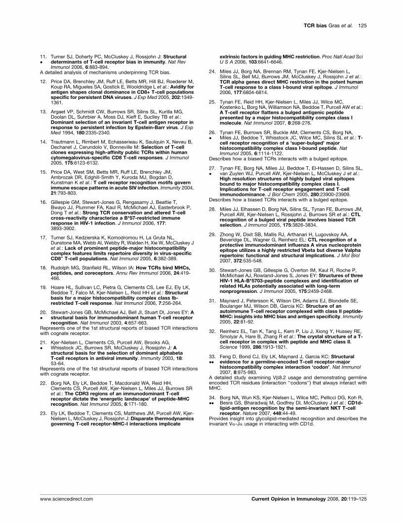
11.�
Turner SJ, Doherty PC, McCluskey J, Rossjohn J: Structuraldeterminants of T-cell receptor bias in immunity. Nat RevImmunol 2006, 6:883-894.
A detailed analysis of mechanisms underpinning TCR bias.
12. Price DA, Brenchley JM, Ruff LE, Betts MR, Hill BJ, Roederer M,Koup RA, Migueles SA, Gostick E, Wooldridge L et al.: Avidity forantigen shapes clonal dominance in CD8+ T-cell populationsspecific for persistent DNA viruses. J Exp Med 2005, 202:1349-1361.
13. Argaet VP, Schmidt CW, Burrows SR, Silins SL, Kurilla MG,Doolan DL, Suhrbier A, Moss DJ, Kieff E, Suclley TB et al.:Dominant selection of an invariant T-cell antigen receptor inresponse to persistent infection by Epstein-Barr virus. J ExpMed 1994, 180:2335-2340.
14. Trautmann L, Rimbert M, Echasserieau K, Saulquin X, Neveu B,Dechanet J, Cerundolo V, Bonneville M: Selection of T-cellclones expressing high-affinity public TCRs within humancytomegalovirus-specific CD8 T-cell responses. J Immunol2005, 175:6123-6132.
15. Price DA, West SM, Betts MR, Ruff LE, Brenchley JM,Ambrozak DR, Edghill-Smith Y, Kuroda MJ, Bogdan D,Kunstman K et al.: T cell receptor recognition motifs governimmune escape patterns in acute SIV infection. Immunity 2004,21:793-803.
16. Gillespie GM, Stewart-Jones G, Rengasamy J, Beattie T,Bwayo JJ, Plummer FA, Kaul R, McMichael AJ, Easterbrook P,Dong T et al.: Strong TCR conservation and altered T-cellcross-reactivity characterize a B*57-restricted immuneresponse in HIV-1 infection. J Immunol 2006, 177:3893-3902.
17. Turner SJ, Kedzierska K, Komodromou H, La Gruta NL,Dunstone MA, Webb AI, Webby R, Walden H, Xie W, McCluskey Jet al.: Lack of prominent peptide-major histocompatibilitycomplex features limits repertoire diversity in virus-specificCD8+ T-cell populations. Nat Immunol 2005, 6:382-389.
18. Rudolph MG, Stanfield RL, Wilson IA: How TCRs bind MHCs,peptides, and coreceptors. Annu Rev Immunol 2006, 24:419-466.
19. Hoare HL, Sullivan LC, Pietra G, Clements CS, Lee EJ, Ely LK,Beddoe T, Falco M, Kjer-Nielsen L, Reid HH et al.: Structuralbasis for a major histocompatibility complex class Ib-restricted T-cell response. Nat Immunol 2006, 7:256-264.
20.�
Stewart-Jones GB, McMichael AJ, Bell JI, Stuart DI, Jones EY: Astructural basis for immunodominant human T-cell receptorrecognition. Nat Immunol 2003, 4:657-663.
Represents one of the 1st structural reports of biased TCR interactionswith cognate receptor.
21.�
Kjer-Nielsen L, Clements CS, Purcell AW, Brooks AG,Whisstock JC, Burrows SR, McCluskey J, Rossjohn J: Astructural basis for the selection of dominant alphabetaT-cell receptors in antiviral immunity. Immunity 2003, 18:53-64.
Represents one of the 1st structural reports of biased TCR interactionswith cognate receptor.
22. Borg NA, Ely LK, Beddoe T, Macdonald WA, Reid HH,Clements CS, Purcell AW, Kjer-Nielsen L, Miles JJ, Burrows SRet al.: The CDR3 regions of an immunodominant T-cellreceptor dictate the ‘energetic landscape’ of peptide-MHCrecognition. Nat Immunol 2005, 6:171-180.
23. Ely LK, Beddoe T, Clements CS, Matthews JM, Purcell AW, Kjer-Nielsen L, McCluskey J, Rossjohn J: Disparate thermodynamicsgoverning T-cell receptor-MHC-I interactions implicate
www.sciencedirect.com
extrinsic factors in guiding MHC restriction. Proc Natl Acad SciU S A 2006, 103:6641-6646.
24. Miles JJ, Borg NA, Brennan RM, Tynan FE, Kjer-Nielsen L,Silins SL, Bell MJ, Burrows JM, McCluskey J, Rossjohn J et al.:TCR alpha genes direct MHC restriction in the potent humanT-cell response to a class I-bound viral epitope. J Immunol2006, 177:6804-6814.
25. Tynan FE, Reid HH, Kjer-Nielsen L, Miles JJ, Wilce MC,Kostenko L, Borg NA, Williamson NA, Beddoe T, Purcell AW et al.:A T-cell receptor flattens a bulged antigenic peptidepresented by a major histocompatibility complex class Imolecule. Nat Immunol 2007, 8:268-276.
26.�
Tynan FE, Burrows SR, Buckle AM, Clements CS, Borg NA,Miles JJ, Beddoe T, Whisstock JC, Wilce MC, Silins SL et al.: T-cell receptor recognition of a ’super-bulged’ majorhistocompatibility complex class I-bound peptide. NatImmunol 2005, 6:1114-1122.
Describes how a biased TCRs interacts with a bulged epitope.
27.�
Tynan FE, Borg NA, Miles JJ, Beddoe T, El-Hassen D, Silins SL,van Zuylen WJ, Purcell AW, Kjer-Nielsen L, McCluskey J et al.:High resolution structures of highly bulged viral epitopesbound to major histocompatibility complex class I.Implications for T-cell receptor engagement and T-cellimmunodominance. J Biol Chem 2005, 280:23900-23909.
Describes how a biased TCRs interacts with a bulged epitope.
28. Miles JJ, Elhassen D, Borg NA, Silins SL, Tynan FE, Burrows JM,Purcell AW, Kjer-Nielsen L, Rossjohn J, Burrows SR et al.: CTLrecognition of a bulged viral peptide involves biased TCRselection. J Immunol 2005, 175:3826-3834.
29. Zhong W, Dixit SB, Mallis RJ, Arthanari H, Lugovskoy AA,Beveridge DL, Wagner G, Reinherz EL: CTL recognition of aprotective immunodominant influenza A virus nucleoproteinepitope utilizes a highly restricted Vbeta but diverse Valpharepertoire: functional and structural implications. J Mol Biol2007, 372:535-548.
30. Stewart-Jones GB, Gillespie G, Overton IM, Kaul R, Roche P,McMichael AJ, Rowland-Jones S, Jones EY: Structures of threeHIV-1 HLA-B*5703-peptide complexes and identification ofrelated HLAs potentially associated with long-termnonprogression. J Immunol 2005, 175:2459-2468.
31. Maynard J, Petersson K, Wilson DH, Adams EJ, Blondelle SE,Boulanger MJ, Wilson DB, Garcia KC: Structure of anautoimmune T-cell receptor complexed with class II peptide-MHC: insights into MHC bias and antigen specificity. Immunity2005, 22:81-92.
32. Reinherz EL, Tan K, Tang L, Kern P, Liu J, Xiong Y, Hussey RE,Smolyar A, Hare B, Zhang R et al.: The crystal structure of a T-cell receptor in complex with peptide and MHC class II.Science 1999, 286:1913-1921.
33.��
Feng D, Bond CJ, Ely LK, Maynard J, Garcia KC: Structuralevidence for a germline-encoded T-cell receptor-majorhistocompatibility complex interaction ‘codon’. Nat Immunol2007, 8:975-983.
A detailed study examining Vb8.2 usage and demonstrating germlineencoded TCR residues (interaction ‘‘codons’’) that always interact withMHC.
34.��
Borg NA, Wun KS, Kjer-Nielsen L, Wilce MC, Pellicci DG, Koh R,Besra GS, Bharadwaj M, Godfrey DI, McCluskey J et al.: CD1d-lipid-antigen recognition by the semi-invariant NKT T-cellreceptor. Nature 2007, 448:44-49.
Provides insight into glycolipid-mediated recognition and describes theinvariant Va-Ja usage in interacting with CD1d.
Current Opinion in Immunology 2008, 20:119–125