Auto Immunity
19
■ Organ-Specific Autoimmune Diseases ■ Systemic Autoimmune Diseases ■ Animal Models for Autoimmune Diseases ■ Evidence Implicating the CD4 + T Cell, MHC, and TCR in Autoimmunity ■ Proposed Mechanisms for Induction of Autoimmunity ■ Treatment of Autoimmune Diseases Autoimmunity , realized that the immune system could go awry and, instead of reacting against foreign antigens, could focus its attack on self-antigens. He termed this con- dition “horror autotoxicus.”We now understand that, while mechanisms of self-tolerance normally protect an individual from potentially self-reactive lymphocytes, there are failures. They result in an inappropriate response of the immune system against self-components termed autoimmunity. In the 1960s, it was believed that all self-reactive lymphocytes were eliminated during their development in the bone mar- row and thymus and that a failure to eliminate these lym- phocytes led to autoimmune consequences. Since the late 1970s, a broad body of experimental evidence has countered that belief, revealing that not all self-reactive lymphocytes are deleted during T-cell and B-cell maturation. Instead, normal healthy individuals have been shown to possess ma- ture, recirculating, self-reactive lymphocytes. Since the pres- ence of these self-reactive lymphocytes in the periphery does not inevitably result in autoimmune reactions, their activity must be regulated in normal individuals through clonal anergy or clonal suppression. A breakdown in this regulation can lead to activation of self-reactive clones of T or B cells, generating humoral or cell-mediated responses against self- antigens. These reactions can cause serious damage to cells and organs, sometimes with fatal consequences. Sometimes the damage to self-cells or organs is caused by antibodies; in other cases, T cells are the culprit. For exam- ple, a common form of autoimmunity is tissue injury by mechanisms similar to type II hypersensitivity reactions. As Chapter 16 showed, type II hypersensitivity reactions in- volve antibody-mediated destruction of cells. Autoimmune hemolytic anemia is an excellent example of such an autoim- mune disease. In this disease, antigens on red blood cells are recognized by auto-antibodies, which results in the destruc- tion of the blood cells, which in turn results in anemia. Auto- antibodies are also the major offender in Hashimoto’s thy- roiditis, in which antibodies reactive with tissue-specific antigens such as thyroid peroxidase and thyroglobulin cause severe tissue destruction. Other autoimmune diseases that involve auto-antibodies are listed in Table 20-1. Many autoimmune diseases are characterized by tissue destruction mediated directly by T cells. A well-known ex- ample is rheumatoid arthritis, in which self-reactive T cells attack the tissue in joints, causing an inflammatory response that results in swelling and tissue destruction. Other exam- ples include insulin-dependent diabetes mellitus and multi- ple sclerosis (see Table 20-1). This chapter describes some common human autoim- mune diseases. These can be divided into two broad cate- gories: organ-specific and systemic autoimmune disease (Table 20-1). Such diseases affect 5%–7% of the human pop- ulation, often causing chronic debilitating illnesses. Several experimental animal models used to study autoimmunity and various mechanisms that may contribute to induction of autoimmune reactions also are described. Finally, current and experimental therapies for treating autoimmune dis- eases are described. Organ-Specific Autoimmune Diseases In an organ-specific autoimmune disease, the immune re- sponse is directed to a target antigen unique to a single organ or gland, so that the manifestations are largely limited to that organ. The cells of the target organs may be damaged di- chapter 20 Kidney Biopsy from Goodpasture’s Syndrone E
Transcript of Auto Immunity

■ Organ-Specific Autoimmune Diseases
■ Systemic Autoimmune Diseases
■ Animal Models for Autoimmune Diseases
■ Evidence Implicating the CD4+ T Cell, MHC, and TCR in Autoimmunity
■ Proposed Mechanisms for Induction ofAutoimmunity
■ Treatment of Autoimmune Diseases
Autoimmunity
,
realized that the immune system could go awryand, instead of reacting against foreign antigens,
could focus its attack on self-antigens. He termed this con-dition “horror autotoxicus.” We now understand that, whilemechanisms of self-tolerance normally protect an individualfrom potentially self-reactive lymphocytes, there are failures.They result in an inappropriate response of the immune system against self-components termed autoimmunity. Inthe 1960s, it was believed that all self-reactive lymphocyteswere eliminated during their development in the bone mar-row and thymus and that a failure to eliminate these lym-phocytes led to autoimmune consequences. Since the late1970s, a broad body of experimental evidence has counteredthat belief, revealing that not all self-reactive lymphocytesare deleted during T-cell and B-cell maturation. Instead,normal healthy individuals have been shown to possess ma-ture, recirculating, self-reactive lymphocytes. Since the pres-ence of these self-reactive lymphocytes in the periphery doesnot inevitably result in autoimmune reactions, their activitymust be regulated in normal individuals through clonalanergy or clonal suppression. A breakdown in this regulationcan lead to activation of self-reactive clones of T or B cells,generating humoral or cell-mediated responses against self-antigens. These reactions can cause serious damage to cellsand organs, sometimes with fatal consequences.
Sometimes the damage to self-cells or organs is caused byantibodies; in other cases, T cells are the culprit. For exam-ple, a common form of autoimmunity is tissue injury bymechanisms similar to type II hypersensitivity reactions. AsChapter 16 showed, type II hypersensitivity reactions in-volve antibody-mediated destruction of cells. Autoimmunehemolytic anemia is an excellent example of such an autoim-mune disease. In this disease, antigens on red blood cells arerecognized by auto-antibodies, which results in the destruc-tion of the blood cells, which in turn results in anemia. Auto-antibodies are also the major offender in Hashimoto’s thy-roiditis, in which antibodies reactive with tissue-specificantigens such as thyroid peroxidase and thyroglobulin causesevere tissue destruction. Other autoimmune diseases thatinvolve auto-antibodies are listed in Table 20-1.
Many autoimmune diseases are characterized by tissuedestruction mediated directly by T cells. A well-known ex-ample is rheumatoid arthritis, in which self-reactive T cellsattack the tissue in joints, causing an inflammatory responsethat results in swelling and tissue destruction. Other exam-ples include insulin-dependent diabetes mellitus and multi-ple sclerosis (see Table 20-1).
This chapter describes some common human autoim-mune diseases. These can be divided into two broad cate-gories: organ-specific and systemic autoimmune disease(Table 20-1). Such diseases affect 5%–7% of the human pop-ulation, often causing chronic debilitating illnesses. Severalexperimental animal models used to study autoimmunityand various mechanisms that may contribute to inductionof autoimmune reactions also are described. Finally, currentand experimental therapies for treating autoimmune dis-eases are described.
Organ-Specific AutoimmuneDiseasesIn an organ-specific autoimmune disease, the immune re-sponse is directed to a target antigen unique to a single organor gland, so that the manifestations are largely limited to thatorgan. The cells of the target organs may be damaged di-
chapter 20
Kidney Biopsy from Goodpasture’s Syndrone
E

rectly by humoral or cell-mediated effector mechanisms.Alternatively, the antibodies may overstimulate or block thenormal function of the target organ.
Some Autoimmune Diseases AreMediated by Direct Cellular DamageAutoimmune diseases involving direct cellular damage occurwhen lymphocytes or antibodies bind to cell-membrane an-tigens, causing cellular lysis and/or an inflammatory responsein the affected organ. Gradually, the damaged cellular struc-ture is replaced by connective tissue (scar tissue), and the func-tion of the organ declines. This section briefly describes a fewexamples of this type of autoimmune disease.
HASHIMOTO’S THYROIDITIS
In Hashimoto’s thyroiditis, which is most frequently seen inmiddle-aged women, an individual produces auto-antibodies
and sensitized TH1 cells specific for thyroid antigens. TheDTH response is characterized by an intense infiltration ofthe thyroid gland by lymphocytes, macrophages, and plasmacells, which form lymphocytic follicles and germinal centers(Figure 20-1). The ensuing inflammatory response causes agoiter, or visible enlargement of the thyroid gland, a physio-logical response to hypothyroidism. Antibodies are formedto a number of thyroid proteins, including thyroglobulin andthyroid peroxidase, both of which are involved in the uptakeof iodine. Binding of the auto-antibodies to these proteinsinterferes with iodine uptake and leads to decreased produc-tion of thyroid hormones (hypothyroidism).
AUTOIMMUNE ANEMIAS
Autoimmune anemias include pernicious anemia, autoim-mune hemolytic anemia, and drug-induced hemolytic ane-mia. Pernicious anemia is caused by auto-antibodies to intrin-sic factor, a membrane-bound intestinal protein on gastricparietal cells. Intrinsic factor facilitates uptake of vitamin B12
Autoimmunity C H A P T E R 20 463
TABLE 20-1 Some autoimmune diseases in humans
Disease Self-antigen Immune response
ORGAN-SPECIFIC AUTOIMMUNE DISEASES
Addison’s disease Adrenal cells Auto-antibodies
Autoimmune hemolytic anemia RBC membrane proteins Auto-antibodies
Goodpasture’s syndrome Renal and lung basement membranes Auto-antibodies
Graves’ disease Thyroid-stimulating hormone receptor Auto-antibody (stimulating)
Hashimoto’s thyroiditis Thyroid proteins and cells TDTH cells, auto-antibodies
Idiopathic thrombocyopenia purpura Platelet membrane proteins Auto-antibodies
Insulin-dependent diabetes mellitus Pancreatic beta cells TDTH cells, auto-antibodies
Myasthenia gravis Acetylcholine receptors Auto-antibody (blocking)
Myocardial infarction Heart Auto-antibodies
Pernicious anemia Gastric parietal cells; intrinsic factor Auto-antibody
Poststreptococcal glomerulonephritis Kidney Antigen-antibody complexes
Spontaneous infertility Sperm Auto-antibodies
SYSTEMIC AUTOIMMUNE DISEASES
Ankylosing sponkylitis Vertebrae Immune complexes
Multiple sclerosis Brain or white matter TH1 cells and TC cells, auto-antibodies
Rheumatoid arthritis Connective tissue, IgG Auto-antibodies, immune complexes
Scleroderma Nuclei, heart, lungs, gastrointestinal tract, kidney Auto-antibodies
Sjogren’s syndrome Salivary gland, liver, kidney, thyroid Auto-antibodies
Systemic lupus erythematosus (SLE) DNA, nuclear protein, RBC and platelet membranes Auto-antibodies, immune complexes

from the small intestine. Binding of the auto-antibody tointrinsic factor blocks the intrinsic factor–mediated absorp-tion of vitamin B12. In the absence of sufficient vitamin B12,which is necessary for proper hematopoiesis, the number offunctional mature red blood cells decreases below normal.Pernicious anemia is treated with injections of vitamin B12,thus circumventing the defect in its absorption.
An individual with autoimmune hemolytic anemia makesauto-antibody to RBC antigens, triggering complement-mediated lysis or antibody-mediated opsonization and phago-cytosis of the red blood cells. One form of autoimmune ane-mia is drug-induced: when certain drugs such as penicillin orthe anti-hypertensive agent methyldopa interact with redblood cells, the cells become antigenic. The immunodiag-nostic test for autoimmune hemolytic anemias generallyinvolves a Coombs test, in which the red cells are incubatedwith an anti–human IgG antiserum. If IgG auto-antibodiesare present on the red cells, the cells are agglutinated by theantiserum.
GOODPASTURE’S SYNDROME
In Goodpasture’s syndrome, auto-antibodies specific forcertain basement-membrane antigens bind to the basementmembranes of the kidney glomeruli and the alveoli of thelungs. Subsequent complement activation leads to direct cel-lular damage and an ensuing inflammatory response medi-ated by a buildup of complement split products. Damage tothe glomerular and alveolar basement membranes leads toprogressive kidney damage and pulmonary hemorrhage.Death may ensue within several months of the onset ofsymptoms. Biopsies from patients with Goodpasture’s syn-drome stained with fluorescent-labeled anti-IgG and anti-C3b reveal linear deposits of IgG and C3b along the base-ment membranes (Figure 20-2).
INSULIN-DEPENDENT DIABETES MELLITUS
A disease afflicting 0.2% of the population, insulin-dependentdiabetes mellitus (IDDM) is caused by an autoimmuneattack on the pancreas. The attack is directed against special-ized insulin-producing cells (beta cells) that are located inspherical clusters, called the islets of Langerhans, scatteredthroughout the pancreas. The autoimmune attack destroysbeta cells, resulting in decreased production of insulin andconsequently increased levels of blood glucose. Several factorsare important in the destruction of beta cells. First, activatedCTLs migrate into an islet and begin to attack the insulin-producing cells. Local cytokine production during this
464 P A R T I V The Immune System in Health and Disease
(a) (b)
FIGURE 20-1 Photomicrographs of (a) normal thyroid gland show-ing a follicle lined by cuboidal follicular epithelial cells and (b) gland in
Hashimoto’s thyroiditis showing intense lymphocyte infiltration. [FromWeb Path, courtesy of E. C. Klatt, University of Utah.]
FIGURE 20-2 Fluorescent anti-IgG staining of a kidney biopsyfrom a patient with Goodpasture’s syndrome reveals linear depositsof auto-antibody along the basement membrane. [From Web Path,courtesy of E. C. Klatt, University of Utah.]
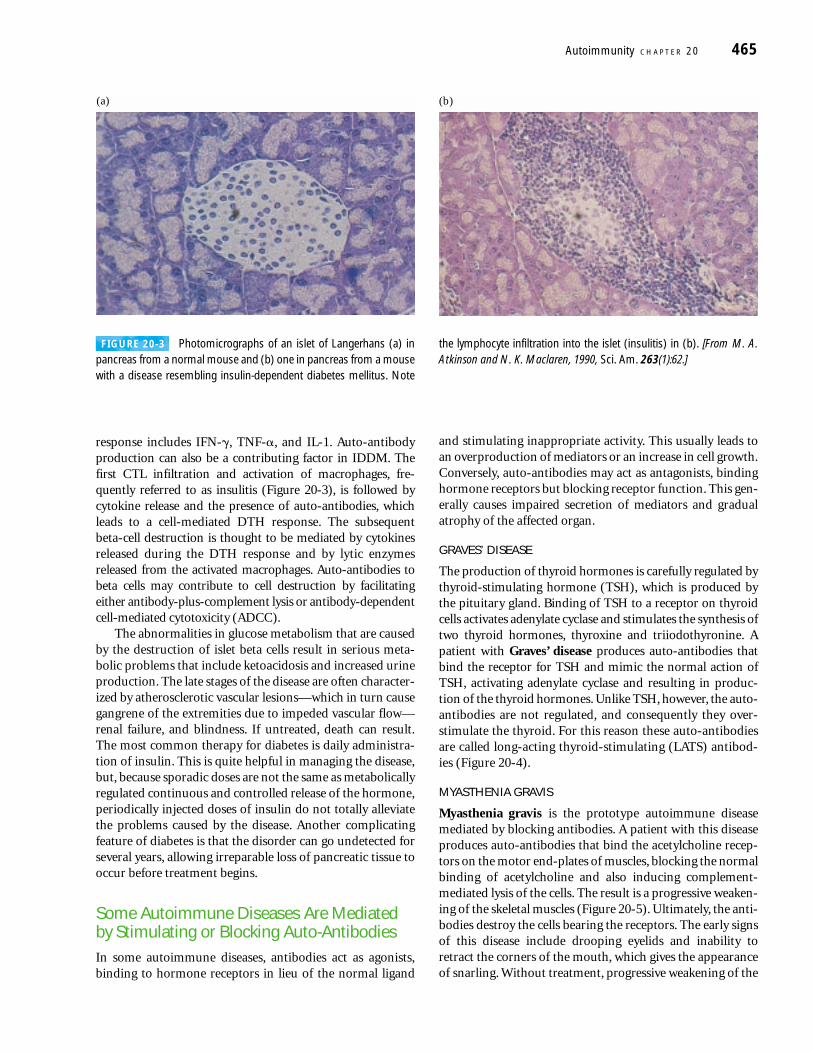
response includes IFN-�, TNF-�, and IL-1. Auto-antibodyproduction can also be a contributing factor in IDDM. Thefirst CTL infiltration and activation of macrophages, fre-quently referred to as insulitis (Figure 20-3), is followed bycytokine release and the presence of auto-antibodies, whichleads to a cell-mediated DTH response. The subsequent beta-cell destruction is thought to be mediated by cytokinesreleased during the DTH response and by lytic enzymes released from the activated macrophages. Auto-antibodies tobeta cells may contribute to cell destruction by facilitatingeither antibody-plus-complement lysis or antibody-dependentcell-mediated cytotoxicity (ADCC).
The abnormalities in glucose metabolism that are causedby the destruction of islet beta cells result in serious meta-bolic problems that include ketoacidosis and increased urineproduction. The late stages of the disease are often character-ized by atherosclerotic vascular lesions—which in turn causegangrene of the extremities due to impeded vascular flow—renal failure, and blindness. If untreated, death can result.The most common therapy for diabetes is daily administra-tion of insulin. This is quite helpful in managing the disease,but, because sporadic doses are not the same as metabolicallyregulated continuous and controlled release of the hormone,periodically injected doses of insulin do not totally alleviatethe problems caused by the disease. Another complicatingfeature of diabetes is that the disorder can go undetected forseveral years, allowing irreparable loss of pancreatic tissue tooccur before treatment begins.
Some Autoimmune Diseases Are Mediatedby Stimulating or Blocking Auto-AntibodiesIn some autoimmune diseases, antibodies act as agonists,binding to hormone receptors in lieu of the normal ligand
and stimulating inappropriate activity. This usually leads toan overproduction of mediators or an increase in cell growth.Conversely, auto-antibodies may act as antagonists, bindinghormone receptors but blocking receptor function. This gen-erally causes impaired secretion of mediators and gradualatrophy of the affected organ.
GRAVES’ DISEASE
The production of thyroid hormones is carefully regulated bythyroid-stimulating hormone (TSH), which is produced bythe pituitary gland. Binding of TSH to a receptor on thyroidcells activates adenylate cyclase and stimulates the synthesis oftwo thyroid hormones, thyroxine and triiodothyronine. Apatient with Graves’ disease produces auto-antibodies thatbind the receptor for TSH and mimic the normal action ofTSH, activating adenylate cyclase and resulting in produc-tion of the thyroid hormones. Unlike TSH, however, the auto-antibodies are not regulated, and consequently they over-stimulate the thyroid. For this reason these auto-antibodiesare called long-acting thyroid-stimulating (LATS) antibod-ies (Figure 20-4).
MYASTHENIA GRAVIS
Myasthenia gravis is the prototype autoimmune diseasemediated by blocking antibodies. A patient with this diseaseproduces auto-antibodies that bind the acetylcholine recep-tors on the motor end-plates of muscles, blocking the normalbinding of acetylcholine and also inducing complement-mediated lysis of the cells. The result is a progressive weaken-ing of the skeletal muscles (Figure 20-5). Ultimately, the anti-bodies destroy the cells bearing the receptors. The early signsof this disease include drooping eyelids and inability toretract the corners of the mouth, which gives the appearanceof snarling. Without treatment, progressive weakening of the
Autoimmunity C H A P T E R 20 465
(a) (b)
FIGURE 20-3 Photomicrographs of an islet of Langerhans (a) inpancreas from a normal mouse and (b) one in pancreas from a mousewith a disease resembling insulin-dependent diabetes mellitus. Note
the lymphocyte infiltration into the islet (insulitis) in (b). [From M. A.Atkinson and N. K. Maclaren, 1990, Sci. Am. 263(1):62.]

muscles can lead to severe impairment of eating as well asproblems with movement. However, with appropriate treat-ment, this disease can be managed quite well and afflictedindividuals can lead a normal life.
Systemic Autoimmune DiseasesIn systemic autoimmune diseases, the response is directedtoward a broad range of target antigens and involves a num-ber of organs and tissues. These diseases reflect a general de-fect in immune regulation that results in hyperactive T cellsand B cells. Tissue damage is widespread, both from cell-mediated immune responses and from direct cellular dam-age caused by auto-antibodies or by accumulation of im-mune complexes.
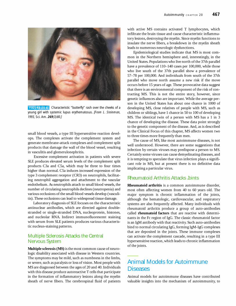
Systemic Lupus Erythematosus Attacks Many TissuesOne of the best examples of a systemic autoimmune disease issystemic lupus erythematosus (SLE), which typically appearsin women between 20 and 40 years of age; the ratio of femaleto male patients is 10:1. SLE is characterized by fever, weak-ness, arthritis, skin rashes, pleurisy, and kidney dysfunction(Figure 20-6). Lupus is more frequent in African-Americanand Hispanic women than in Caucasians, although it is notknown why this is so. Affected individuals may produce auto-antibodies to a vast array of tissue antigens, such as DNA, his-tones, RBCs, platelets, leukocytes, and clotting factors; inter-action of these auto-antibodies with their specific antigensproduces various symptoms. Auto-antibody specific for RBCsand platelets, for example, can lead to complement-mediatedlysis, resulting in hemolytic anemia and thrombocytopenia,respectively. When immune complexes of auto-antibodieswith various nuclear antigens are deposited along the walls of
466 P A R T I V The Immune System in Health and Disease
STIMULATING AUTO-ANTIBODIES (Graves’ disease)
Pituitary gland
TSH
TSH receptor
Thyroid cell
Stimulateshormonesynthesis
Auto-antibodyto receptor
Regulated production ofthyroid hormones
Unregulated overproductionof thyroid hormones
Negativefeedbackcontrol
Stimulateshormonesynthesis
BLOCKING AUTO-ANTIBODIES (Myasthenia gravis)
Nerve Nerve
Acetylcholine
Muscle cell
AChR Auto-antibody to AChR
Muscle activation Muscle activation inhibited
FIGURE 20-4 In Graves’ disease, binding of auto-antibodies to thereceptor for thyroid-stimulating hormone (TSH) induces unregu-lated activation of the thyroid, leading to overproduction of the thy-roid hormones (purple dots).
FIGURE 20-5 In myasthenia gravis, binding of auto-antibodies tothe acetylcholine receptor (right) blocks the normal binding of acetyl-choline (burgandy dots) and subsequent muscle activation (left). In
addition, the anti-AChR auto-antibody activates complement, whichdamages the muscle end-plate; the number of acetylcholine receptorsdeclines as the disease progresses. AChR = acetylcholine receptor.
small blood vessels, a type III hypersensitive reaction devel-ops. The complexes activate the complement system andgenerate membrane-attack complexes and complement splitproducts that damage the wall of the blood vessel, resultingin vasculitis and glomerulonephritis.
Excessive complement activation in patients with severeSLE produces elevated serum levels of the complement splitproducts C3a and C5a, which may be three to four timeshigher than normal. C5a induces increased expression of thetype 3 complement receptor (CR3) on neutrophils, facilitat-ing neutrophil aggregation and attachment to the vascularendothelium. As neutrophils attach to small blood vessels, thenumber of circulating neutrophils declines (neutropenia) andvarious occlusions of the small blood vessels develop (vasculi-tis). These occlusions can lead to widespread tissue damage.
Laboratory diagnosis of SLE focuses on the characteristicantinuclear antibodies, which are directed against double-stranded or single-stranded DNA, nucleoprotein, histones,and nucleolar RNA. Indirect immunofluorescent stainingwith serum from SLE patients produces various characteris-tic nucleus-staining patterns.
Multiple Sclerosis Attacks the CentralNervous SystemMultiple sclerosis (MS) is the most common cause of neuro-logic disability associated with disease in Western countries.The symptoms may be mild, such as numbness in the limbs,or severe, such as paralysis or loss of vision. Most people withMS are diagnosed between the ages of 20 and 40. Individualswith this disease produce autoreactive T cells that participatein the formation of inflammatory lesions along the myelinsheath of nerve fibers. The cerebrospinal fluid of patients
with active MS contains activated T lymphocytes, whichinfiltrate the brain tissue and cause characteristic inflamma-tory lesions, destroying the myelin. Since myelin functions toinsulate the nerve fibers, a breakdown in the myelin sheathleads to numerous neurologic dysfunctions.
Epidemiological studies indicate that MS is most com-mon in the Northern hemisphere and, interestingly, in theUnited States. Populations who live north of the 37th parallelhave a prevalence of 110–140 cases per 100,000, while thosewho live south of the 37th parallel show a prevalence of57–78 per 100,000. And individuals from south of the 37thparallel who move north assume a new risk if the moveoccurs before 15 years of age. These provocative data suggestthat there is an environmental component of the risk of con-tracting MS. This is not the entire story, however, sincegenetic influences also are important. While the average per-son in the United States has about one chance in 1000 ofdeveloping MS, close relatives of people with MS, such aschildren or siblings, have 1 chance in 50 to 100 of developingMS. The identical twin of a person with MS has a 1 in 3chance of developing the disease. These data point stronglyto the genetic component of the disease. And, as is describedin the Clinical Focus of this chapter, MS affects women twoto three times more frequently than men.
The cause of MS, like most autoimmune diseases, is notwell understood. However, there are some suggestions thatinfection by certain viruses may predispose a person to MS.Certainly some viruses can cause demyelinating diseases, andit is tempting to speculate that virus infection plays a signifi-cant role in MS, but at present there is no definitive dataimplicating a particular virus.
Rheumatoid Arthritis Attacks JointsRheumatoid arthritis is a common autoimmune disorder,most often affecting women from 40 to 60 years old. Themajor symptom is chronic inflammation of the joints,although the hematologic, cardiovascular, and respiratorysystems are also frequently affected. Many individuals withrheumatoid arthritis produce a group of auto-antibodiescalled rheumatoid factors that are reactive with determi-nants in the Fc region of IgG. The classic rheumatoid factoris an IgM antibody with that reactivity. Such auto-antibodiesbind to normal circulating IgG, forming IgM-IgG complexesthat are deposited in the joints. These immune complexescan activate the complement cascade, resulting in a type IIIhypersensitive reaction, which leads to chronic inflammationof the joints.
Animal Models for AutoimmuneDiseases Animal models for autoimmune diseases have contributedvaluable insights into the mechanism of autoimmunity, to
Autoimmunity C H A P T E R 20 467
FIGURE 20-6 Characteristic “butterfly” rash over the cheeks of ayoung girl with systemic lupus erythematosus. [From L. Steinman,1993, Sci. Am. 269(3):80.]

our understanding of autoimmunity in humans, and topotential treatments. Autoimmunity develops spontaneouslyin certain inbred strains of animals and can also be inducedby certain experimental manipulations (Table 20-2).
Autoimmunity Can Develop Spontaneously in AnimalsA number of autoimmune diseases that develop sponta-neously in animals exhibit important clinical and pathologicsimilarities to certain autoimmune diseases in humans. Cer-tain inbred mouse strains have been particularly valuablemodels for illuminating the immunologic defects involved inthe development of autoimmunity.
New Zealand Black (NZB) mice and F1 hybrids of NZBand New Zealand White (NZW) mice spontaneously developautoimmune diseases that closely resemble systemic lupus ery-thematosus. NZB mice spontaneously develop autoimmunehemolytic anemia between 2 and 4 months of age, at whichtime various auto-antibodies can be detected, including anti-bodies to erythrocytes, nuclear proteins, DNA, and T lym-phocytes. F1 hybrid animals develop glomerulonephritis fromimmune-complex deposits in the kidney and die prematurelyby 18 months. As in human SLE, the incidence of autoimmu-nity in the (NZB � NZW)F1 hybrids is greater in females.
An accelerated and severe form of systemic autoimmunedisease resembling systemic lupus erythematosus develops in
a mouse strain called MRL/lpr/lpr. These mice are homozy-gous for a gene called lpr, which has been identified as adefective fas gene. The fas-gene product is a cell-surface pro-tein belonging to the TNF family of cysteine-rich membranereceptors (see Figure 12-6d). When the normal Fas proteininteracts with its ligand, it transduces a signal that leads toapoptotic death of the Fas-bearing cells. This mechanismmay operate in destruction of target cells by some CTLs (seeFigure 14-9). Fas is known also to be essential in the death ofhyperactivated peripheral CD4+ cells. Normally, when ma-ture peripheral T cells become activated, they are induced toexpress both Fas antigen and Fas ligand, When Fas-bearingcells come into contact with a neighboring activated cell bear-ing Fas ligand, the Fas-bearing cell is induced to die. It is alsopossible that Fas ligand can engage Fas from the same cell,inducing a cellular suicide. In the absence of Fas, matureperipheral T cells do not die, and these activated cells con-tinue to proliferate and produce cytokines that result ingrossly enlarged lymph nodes and spleen. Defects in fas ex-pression similar to that found in the lpr mouse are observed inhumans, and these can have severe consequences. Howeverthere is no link between fas expression and SLE in humans,which suggests that the lpr mouse may not be a true modelfor SLE.
Another important animal model is the nonobese dia-betic (NOD) mouse, which spontaneously develops a formof diabetes that resembles human insulin-dependent dia-
468 P A R T I V The Immune System in Health and Disease
TABLE 20-2 Experimental animal models of autoimmune diseases
DiseasePossible human transferred
Animal model disease counterpart Inducing antigen by T cells
SPONTANEOUS AUTOIMMUNE DISEASES
Nonobese diabetic (NOD) Insulin-dependent diabetes Unknown Yesmouse mellitus (IDDM)
(NZB � NZW) F1 mouse Systemic lupus erythematosus (SLE) Unknown Yes
Obese-strain chicken Hashimoto’s thyroiditis Thyroglobulin Yes
EXPERIMENTALLY INDUCED AUTOIMMUNE DISEASES*
Experimental autoimmune Myasthenia gravis Acetylcholine receptor Yesmyasthenia gravis (EAMG)
Experimental autoimmune Multiple sclerosis (MS) Myelin basic protein (MBP); Yesencephalomyelitis (EAE) proteolipid protien (PLP)
Autoimmune arthritis (AA) Rheumatoid arthritis M. tuberculosis (proteoglycans) Yes
Experimental autoimmune Hashimoto’s thyroiditis Thyroglobulin Yesthyroiditis (EAT)
* These diseases can be induced by injecting appropriate animals with the indicated antigen in complete Freund’s adjuvant. Except for autoimmune arthritis, the antigens used correspond to the self-antigens associated with the human-disease counterpart. Rheumatoid arthritis involves reaction to proteoglycans, which are self-antigens associated with connective tissue.

betes mellitus (IDDM). Like the human disease, the NODmouse disease begins with lymphocytic infiltration into theislets of the pancreas. Also, as in IDDM, there is a strong asso-ciation between certain MHC alleles and the development ofdiabetes in these mice. Experiments have shown that T cellsfrom diabetic mice can transfer diabetes to nondiabetic re-cipients. For example, when the immune system of normalmice is destroyed by lethal doses of x-rays and then is recon-stituted with an injection of bone-marrow cells from NODmice, the reconstituted mice develop diabetes. Conversely,when the immune system of still healthy NOD mice is de-stroyed by x-irradiation and then reconstituted with normalbone-marrow cells, the NOD mice do not develop diabetes.Various studies have demonstrated a pivotal role for CD4+
T cells in the NOD mouse, and recent evidence implicates theTH1 subset in disease development.
Several other spontaneous autoimmune diseases have beendiscovered in animals that have served as models for similarhuman diseases. Among these are Obese-strain chickens,which develop both humoral and cell-mediated reactivity tothyroglobulin resembling that seen in Hashimoto’s thyroiditis.
Autoimmunity Can Be InducedExperimentally in AnimalsAutoimmune dysfunctions similar to certain human autoim-mune diseases can be induced experimentally in some ani-mals (see Table 20-2). One of the first such animal modelswas discovered serendipitously in 1973 when rabbits wereimmunized with acetylcholine receptors purified from elec-tric eels. The animals soon developed muscular weaknesssimilar to that seen in myasthenia gravis. This experimentalautoimmune myasthenia gravis (EAMG) was shown to resultwhen antibodies to the acetylcholine receptor blocked mus-cle stimulation by acetylcholine in the synapse. Within a year,this animal model had proved its value with the discoverythat auto-antibodies to the acetylcholine receptor were thecause of myasthenia gravis in humans.
Experimental autoimmune encephalomyelitis (EAE) isanother animal model that has greatly improved under-standing of autoimmunity. This is one of the best-studiedmodels of autoimmune disease. EAE is mediated solely by T cells and can be induced in a variety of species by immu-nization with myelin basic protein (MBP) or proteolipidprotein (PLP) in complete Freund’s adjuvant (Figure 20-7).Within 2–3 weeks the animals develop cellular infiltration ofthe myelin sheaths of the central nervous system, resulting in demyelination and paralysis. Most of the animals die, butothers have milder symptoms, and some animals develop achronic form of the disease that resembles chronic relapsingand remitting MS in humans. Those that recover are resistantto the development of disease from a subsequent injection ofMBP and adjuvant.
The mouse EAE model provides a system for testingtreatments for human MS. For example, because MBP- orPLP-specific T-cell clones are found in the periphery, it is
assumed that these clones must have escaped negative selec-tion in the thymus. Recent mouse experiments have suggestedthat orally administered MBP may make these antigen-specificperipheral T-cell clones self-tolerant. These studies have pavedthe way for clinical trials in MS patients.
Experimental autoimmune thyroiditis (EAT) can beinduced in a number of animals by immunizing with thy-roglobulin in complete Freund’s adjuvant. Both humoral anti-bodies and TH1 cells directed against the thyroglobulin de-velop, resulting in thyroid inflammation. EAT appears to bestmimic Hashimoto’s thyroiditis. In contrast to both EAE andEAT, which are induced by immunizing with self-antigens,autoimmune arthritis (AA) is induced by immunizing ratswith Mycobacterium tuberculosis in complete Freund’s adju-vant. These animals develop an arthritis whose features aresimilar to those of rheumatoid arthritis in humans.
Evidence Implicating the CD4+ T Cell,MHC, and TCR in AutoimmunityThe inappropriate response to self-antigens that character-izes all autoimmune diseases can involve either the humoralor cell-mediated branches of the immune system. Identifyingthe defects underlying human autoimmune diseases hasbeen difficult; more success has been achieved in characteriz-ing the immune defects in the various animal models. Eachof the animal models has implicated the CD4+ T cell as theprimary mediator of autoimmune disease. For example, theevidence is quite strong that, in mice, EAE is caused by CD4+
TH1 cells specific for the immunizing antigen. The diseasecan be transferred from one animal into another by T cellsfrom animals immunized with either MBP or PLP or by
Autoimmunity C H A P T E R 20 469
Normal rat
MBP + CFA Lymph-nodecells + MBP
EAE rat(paralysis)
Normal ratEAE rat(most die; some recover)
MBP-specificT-cell clones
FIGURE 20-7 Experimental autoimmune encephalomyelitis (EAE)can be induced in rats by injecting them with myelin basic protein(MBP) in complete Freud’s adjuvant (CFA). MBP-specific T-cellclones can be generated by culturing lymph-node cells from EAE ratswith MBP. When these T cells are injected into normal animals, mostdevelop EAE and die, although a few recover.
cloned T-cell lines from such animals. It also has been shownthat disease can be prevented by treating animals with anti-CD4 antibodies. These data are compelling evidence for theinvolvement of CD4 in the establishment of EAE.
T-cell recognition of antigen, of course, involves a trimol-ecular complex of the T-cell receptor, an MHC molecule, andantigenic peptide (see Figure 9-16). Thus, an individual sus-ceptible to autoimmunity must possess MHC molecules andT-cell receptors capable of binding self-antigens.
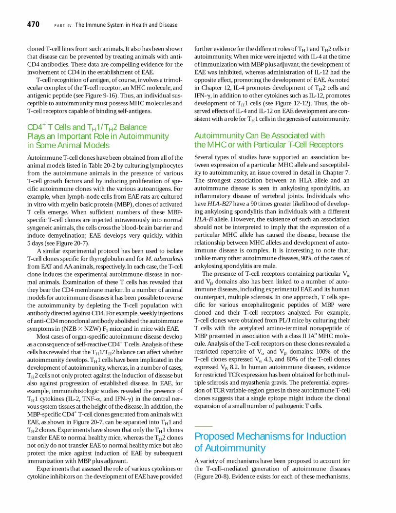
CD4+ T Cells and TH1/TH2 Balance Plays an Important Role in Autoimmunity in Some Animal ModelsAutoimmune T-cell clones have been obtained from all of theanimal models listed in Table 20-2 by culturing lymphocytesfrom the autoimmune animals in the presence of various T-cell growth factors and by inducing proliferation of spe-cific autoimmune clones with the various autoantigens. Forexample, when lymph-node cells from EAE rats are culturedin vitro with myelin basic protein (MBP), clones of activatedT cells emerge. When sufficient numbers of these MBP-specific T-cell clones are injected intravenously into normalsyngeneic animals, the cells cross the blood-brain barrier andinduce demyelination; EAE develops very quickly, within 5 days (see Figure 20-7).
A similar experimental protocol has been used to isolate T-cell clones specific for thyroglobulin and for M. tuberculosisfrom EAT and AA animals, respectively. In each case, the T-cellclone induces the experimental autoimmune disease in nor-mal animals. Examination of these T cells has revealed thatthey bear the CD4 membrane marker. In a number of animalmodels for autoimmune diseases it has been possible to reversethe autoimmunity by depleting the T-cell population withantibody directed against CD4. For example, weekly injectionsof anti-CD4 monoclonal antibody abolished the autoimmunesymptoms in (NZB � NZW) F1 mice and in mice with EAE.
Most cases of organ-specific autoimmune disease developas a consequence of self-reactive CD4+ T cells.Analysis of thesecells has revealed that the TH1/TH2 balance can affect whetherautoimmunity develops. TH1 cells have been implicated in thedevelopment of autoimmunity, whereas, in a number of cases,TH2 cells not only protect against the induction of disease butalso against progression of established disease. In EAE, forexample, immunohistologic studies revealed the presence ofTH1 cytokines (IL-2, TNF-�, and IFN-�) in the central ner-vous system tissues at the height of the disease. In addition, theMBP-specific CD4+ T-cell clones generated from animals withEAE, as shown in Figure 20-7, can be separated into TH1 andTH2 clones. Experiments have shown that only the TH1 clonestransfer EAE to normal healthy mice, whereas the TH2 clonesnot only do not transfer EAE to normal healthy mice but alsoprotect the mice against induction of EAE by subsequentimmunization with MBP plus adjuvant.
Experiments that assessed the role of various cytokines orcytokine inhibitors on the development of EAE have provided
further evidence for the different roles of TH1 and TH2 cells inautoimmunity. When mice were injected with IL-4 at the timeof immunization with MBP plus adjuvant, the development ofEAE was inhibited, whereas administration of IL-12 had theopposite effect, promoting the development of EAE. As notedin Chapter 12, IL-4 promotes development of TH2 cells andIFN-�, in addition to other cytokines such as IL-12, promotesdevelopment of TH1 cells (see Figure 12-12). Thus, the ob-served effects of IL-4 and IL-12 on EAE development are con-sistent with a role for TH1 cells in the genesis of autoimmunity.
Autoimmunity Can Be Associated with the MHC or with Particular T-Cell ReceptorsSeveral types of studies have supported an association be-tween expression of a particular MHC allele and susceptibil-ity to autoimmunity, an issue covered in detail in Chapter 7.The strongest association between an HLA allele and anautoimmune disease is seen in ankylosing spondylitis, aninflammatory disease of vertebral joints. Individuals whohave HLA-B27 have a 90 times greater likelihood of develop-ing ankylosing spondylitis than individuals with a differentHLA-B allele. However, the existence of such an associationshould not be interpreted to imply that the expression of aparticular MHC allele has caused the disease, because therelationship between MHC alleles and development of auto-immune disease is complex. It is interesting to note that,unlike many other autoimmune diseases, 90% of the cases ofankylosing spondylitis are male.
The presence of T-cell receptors containing particular V�
and V� domains also has been linked to a number of auto-immune diseases, including experimental EAE and its humancounterpart, multiple sclerosis. In one approach, T cells spe-cific for various encephalitogenic peptides of MBP werecloned and their T-cell receptors analyzed. For example,T-cell clones were obtained from PL/J mice by culturing theirT cells with the acetylated amino-terminal nonapeptide ofMBP presented in association with a class II IAu MHC mole-cule. Analysis of the T-cell receptors on these clones revealed arestricted repertoire of V� and V� domains: 100% of the T-cell clones expressed V� 4.3, and 80% of the T-cell clonesexpressed V� 8.2. In human autoimmune diseases, evidencefor restricted TCR expression has been obtained for both mul-tiple sclerosis and myasthenia gravis. The preferential expres-sion of TCR variable-region genes in these autoimmune T-cellclones suggests that a single epitope might induce the clonalexpansion of a small number of pathogenic T cells.
Proposed Mechanisms for Inductionof AutoimmunityA variety of mechanisms have been proposed to account forthe T-cell–mediated generation of autoimmune diseases(Figure 20-8). Evidence exists for each of these mechanisms,
470 P A R T I V The Immune System in Health and Disease
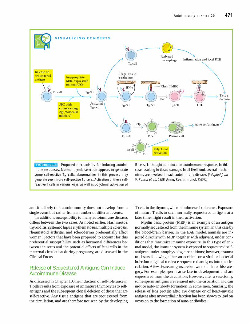
and it is likely that autoimmunity does not develop from asingle event but rather from a number of different events.
In addition, susceptibility to many autoimmune diseasesdiffers between the two sexes. As noted earlier, Hashimoto’sthyroiditis, systemic lupus erythematosus, multiple sclerosis,rheumatoid arthritis, and scleroderma preferentially affectwomen. Factors that have been proposed to account for thispreferential susceptibility, such as hormonal differences be-tween the sexes and the potential effects of fetal cells in thematernal circulation during pregnancy, are discussed in theClinical Focus.
Release of Sequestered Antigens Can InduceAutoimmune DiseaseAs discussed in Chapter 10, the induction of self-tolerance inT cells results from exposure of immature thymocytes to self-antigens and the subsequent clonal deletion of those that areself-reactive. Any tissue antigens that are sequestered fromthe circulation, and are therefore not seen by the developing
T cells in the thymus, will not induce self-tolerance. Exposureof mature T cells to such normally sequestered antigens at alater time might result in their activation.
Myelin basic protein (MBP) is an example of an antigennormally sequestered from the immune system, in this case bythe blood-brain barrier. In the EAE model, animals are in-jected directly with MBP, together with adjuvant, under con-ditions that maximize immune exposure. In this type of ani-mal model, the immune system is exposed to sequestered self-antigens under nonphysiologic conditions; however, traumato tissues following either an accident or a viral or bacterialinfection might also release sequestered antigens into the cir-culation. A few tissue antigens are known to fall into this cate-gory. For example, sperm arise late in development and aresequestered from the circulation. However, after a vasectomy,some sperm antigens are released into the circulation and caninduce auto-antibody formation in some men. Similarly, therelease of lens protein after eye damage or of heart-muscleantigens after myocardial infarction has been shown to lead onoccasion to the formation of auto-antibodies.
Autoimmunity C H A P T E R 20 471
V I S U A L I Z I N G C O N C E P T S
TH cell
Tissue damage
Inflammation and local DTH
Ab to self-antigens
Activatedmacrophage
Target tissueepithelium
IFN-γTH cell
ActivatedTH cell
TH cell CTL
TH cell
Help
Plasma cell
B cell
TC cell
Polyclonalactivation
TH cell
B cell
TH cell
IL-2
Class II MHC
Release ofsequesteredantigen
InappropriateMHC expressionon non-APCs
APC withcross-reactingAg (molecularmimicry)
FIGURE 20-8 Proposed mechanisms for inducing autoim-mune responses. Normal thymic selection appears to generatesome self-reactive TH cells; abnormalities in this process maygenerate even more self-reactive TH cells. Activation of these self-reactive T cells in various ways, as well as polyclonal activation of
B cells, is thought to induce an autoimmune response, in thiscase resulting in tissue damage. In all likelihood, several mecha-nisms are involved in each autoimmune disease. [Adapted fromV. Kumar et al., 1989, Annu. Rev. Immunol. 7:657.]

Data indicate that injection of normally sequestered anti-gens directly into the thymus can reverse the development oftissue-specific autoimmune disease in animal models. Forinstance, intrathymic injection of pancreatic islet beta cellsprevented development of autoimmunity in NOD mice.
Moreover, EAE was prevented in susceptible rats by priorinjection of MBP directly into the thymus. In these experi-ments, exposure of immature T cells to self-antigens thatnormally are not present in the thymus presumably led totolerance to these antigens.
472 P A R T I V The Immune System in Health and Disease
young females. Women tend to havehigher levels of CD4+ T cells and signifi-cantly higher levels of serum IgM.
In mice, whose gender differences areeasier to study, there is a large body of lit-erature documenting gender differencesin immune responses. Female mice aremuch more likely than male mice to de-velop TH1 responses and, in infections towhich pro-inflammatory TH1 responsesare beneficial, are more likely to be re-sistant to the infection. An excellentexample is infection by viruses such asvesicular stomatitis virus (VSV), herpessimplex virus (HSV), and Theiler’s mu-rine encephalomyelitis virus (TMEV).Clearance of these viruses is enhanced byTH1 responses. In some cases, however,a pro-inflammatory response can bedeleterious. For example, a TH1 response to lymphocytic choriomeningitis virus(LCMV) correlates with more severe dis-ease and significant pathology. Thus,female mice are more likely to succumbto infection with LCMV. The fact that gen-der is important in LCMV infection isunderscored by experiments demonstrat-ing that castrated male mice behave likefemales and are more likely to succumbto infection than their un-castrated malelittermates.
Another disease in which genderplays a role is infection by coxsackievirus Type B-3 (CVB-3), an etiologicalagent of immune myocarditis. Male
mice are much more susceptible to thisdisease than females. CVB-3 induces apredominant TH1 response in males,while females, contrary to the situationsdescribed above, respond by mounting aprotective TH2 response. The responseby females can be altered by injectingthem with testosterone, which makesthem susceptible to the disease. Ad-ditionally, the male response can be al-tered by injecting them with estradiol,which makes them resistant to the virus.These data in mice are consistent withthe possibility that basic differences maywell exist between men and women intheir responses to pathogens. We muststress, however, that the particular gen-der differences observed in mice maynot extend to human populations.
How do these gender differencesarise? The evidence cited above thatestradiol or testosterone can alter theoutcome of infection by CVB-3 suggestsa critical role for sex hormones. In hu-mans it appears that estrogen on its owndoes not play a significant role in the eti-ology of either RA or MS, but there areindications that it may be important inSLE. This is suggested by data indicat-ing that estrogen can stimulate auto-antibody production in SLE-prone miceand these effects can be modulated byan anti-estrogenic compound. Such dataimply that, at least in mice, estrogen iscapable of triggering SLE-like autoimmu-nity. Additionally, androgens such astestosterone clearly play an importantrole in some autoimmune diseases. Female NOD mice are much more sus-ceptible to spontaneous diabetes, andcastration significantly increases thesusceptibility of male NOD mice. Fe-male SJL mice are more likely to be sus-ceptible to EAE, a mouse MS-like dis-ease. This indicates that testosteronemay well be effective in amelioratingsome autoimmune responses and so
Of the nearly9 million individuals in the United Stateswith autoimmune disease, approxi-mately 6.7 million are women. This pre-disposition to autoimmunity is moreapparent in some diseases than others.For example, the female:male ratio ofindividuals who suffer from diseasessuch as multiple sclerosis (MS) orrheumatoid arthritis (RA) is approxi-mately two or three females to one male,and there are nine women for every onemale afflicted with systemic lupus ery-thematosus (SLE). However, these sta-tistics do not tell the entire story, since,in some diseases, MS for example, theseverity of the disease can be worse inmen than in women. The fact thatwomen are more susceptible to autoim-mune disease has been recognized forseveral years but the reasons for this in-creased risk are not entirely understood.Here some of the possible explanationsare considered.
Although it may seem unlikely, con-siderable evidence suggests there aresignificant gender differences in im-mune responses in both humans andmice. Immunization studies in both spe-cies suggest that females produce ahigher titer of antibodies than males. Infact, females in general tend to mountmore vigorous immune responses. Inhumans, this is particularly apparent in
C L I N I C A L F O C U S
Why Are Women More Susceptible Than Men to Autoimmunity? Gender Differences in AutoimmuneDisease

Molecular Mimicry May Contribute to Autoimmune Disease
For several reasons, the notion that microbial or viral agentsmight play a role in autoimmunity is very attractive. It is well
accepted that migrant human populations acquire the dis-eases of the area to which they move and that the incidence ofautoimmunity has increased dramatically as populationshave become more mobile. This, coupled with the fact that anumber of viruses and bacteria have been shown to possess
Autoimmunity C H A P T E R 20 473
can have a profound influence on im-mune responses, as demonstrated inmice by removal of the anterior pituitary:this results in a severe immunosuppres-sion, which can be entirely reversed bytreatment with exogenous prolactin. Thepresence of prolactin receptors on peri-pheral T and B cells in humans is furtherevidence that this hormone may play arole in regulating immune responses. Infact, some evidence suggests that pro-lactin may tend to turn cells towardsTH1-dominated immune responses.
Pregnancy may give us a clue to howsex plays a role in regulating immuneresponse. It is clear that, while womennormally mount a normal response toforeign antigens, during pregnancy it iscritical that the mother tolerate the fetus(which is, in fact, a foreign graft). Thismakes it very likely that the femaleimmune system undergoes importantmodifications during pregnancy. Recallthat women normally tend to mountmore TH1-like responses than TH2 re-sponses. During pregnancy, however, wo-men mount more TH2-like responses. Itis thought that pregnancy-associated lev-els of sex steroids may promote an anti-inflammatory environment. In this re-gard, it is notable that diseases enhancedby TH2-like responses, such as SLE,which has a strong antibody-mediatedcomponent, can be exacerbated duringpregnancy, while diseases that involveinflammatory responses, such as RA andMS, sometimes are ameliorated in preg-nant women.
Another effect of pregnancy is thepresence of fetal cells in the maternal cir-culation (see the description of sclero-derma on page 000). It is known thatfetal cells can persist in the maternal cir-culation for decades, so these long-livedfetal cells may play a significant role inthe development of autoimmune dis-ease. Furthermore, the exchange of cells
during pregnancy is bi-directional (cellsof the mother may also appear in thefetal circulatory system), and this has ledsome to postulate that the presence ofmother’s cells in the male circulationcould be a contributing factor in autoim-mune disease.
In summary, women and men differsignificantly in their ability to mount animmune response. Women mount morerobust immune responses, and theseresponses tend to be more TH1-like. Ithas been reported that estrogen is im-munostimulatory; this may be due, inpart, to the ability of the hormone to reg-ulate specific gene expression throughthe estrogen receptor. Furthermore, theincidence of autoimmune diseases issharply higher in women than in men.These observations have generated thecompelling hypothesis that the tendencyof females to mount more TH1-like re-sponses may, in part, explain differencesin susceptibility to autoimmunity. Sincethis type of response is pro-inflammatory,it may enhance the development ofautoimmunity. Whether the bias towardsa TH1 response is due to differences insex steroids between males and femalesis less certain, but surely, in the next sev-eral years, experiments that explore thisidea are likely to be pursued vigorously.
NOTE: The data discussed in thisClinical Focus were extracted from a let-ter to Science (C. C. Whitacre, S. C. Rein-gold, and P. A. O’Looney, 1999, Science283:1277) from the Task Force onGender, MS, and Autoimmunity, a groupconvened by the National Multiple Scle-rosis Society to begin a dialog on issuesof gender and autoimmune disease.Science also has established a Web site(http://www.sciencemag.org/feature/data/983519.shl) that contains more de-tailed data concerning autoimmunityand gender.
may be protective against several auto-immune diseases, including MS, dia-betes, SLE, and Sjogren’s syndrome.
Why do sex steroids affect immuneresponses? This is not well understood,but it is likely that these hormones, whichcirculate throughout the body, alterimmune responses by altering patternsof gene expression. The sex steroids, ahighly lipophilic group of compounds,function by passing through the cellmembrane and binding a cytoplasmicreceptor. Each hormone has a cognatereceptor and binding of hormone to re-ceptor leads to the activation or, in someinstances, repression of gene expression.This is mediated by the binding of thereceptor/hormone complex receptor to a specific DNA sequence. Thus, estro-gen enters a cell, binds to the estrogenreceptor, and induces the binding of theestrogen receptor to a specific DNAsequence, which in turn results in themodulation of transcription. Therefore,in cells that contain hormone receptors,sex hormones can regulate gene expres-sion, and it is highly likely that sexsteroids play an important role in theimmune system through their receptors.Whether various cells of the immune sys-tem contain hormone receptors is notknown at present; to understand how sexhormones mediate immune responses,clearly we must determine which cellsexpress which hormone receptors.
Hormonal effects on immune re-sponses may not be limited to steroidalsex hormones. Prolactin, a hormone thatis expressed in higher levels in womenthan in men, is not a member of thelipophilic sex steroid family that includesestrogen, progesterone, and testoster-one. But prolactin secretion (by the ante-rior pituitary) is stimulated by estrogen,thus explaining the higher levels of pro-lactin in women and the very high levelsobserved during pregnancy. Prolactin
AU
:su
pp
ly 5
e p
age
ref(
or d
elet
e th
is)

antigenic determinants that are identical or similar to normalhost-cell components led Michael Oldstone to propose that apathogen may express a region of protein that resembles aparticular self-component in conformation or primary se-quence. Such molecular mimicry appears in a wide variety oforganisms (Table 20-3). In one study, 600 different mono-clonal antibodies specific for 11 different viruses were testedto evaluate their reactivity with normal tissue antigens. Morethan 3% of the virus-specific antibodies tested also bound tonormal tissue, suggesting that molecular mimicry is a fairlycommon phenomenon.
Molecular mimicry has been suggested as one mecha-nism that leads to autoimmunity. One of the best examples ofthis type of autoimmune reaction is post-rabies encephalitis,which used to develop in some individuals who had receivedthe rabies vaccine. In the past, the rabies virus was grown inrabbit brain-cell cultures, and preparations of the vaccineincluded antigens derived from the rabbit brain cells. In avaccinated person, these rabbit brain-cell antigens couldinduce formation of antibodies and activated T cells, whichcould cross-react with the recipient’s own brain cells, leading
to encephalitis. Cross-reacting antibodies are also thought tobe the cause of heart damage in rheumatic fever, which cansometimes develop after a Streptococcus infection. In thiscase, the antibodies are to streptococcal antigens, but theycross-react with the heart muscle.
There Is Evidence for Mimicry Between MBP and Viral PeptidesSince the encephalitogenic MBP peptides are known, the ex-tent to which they are mimicked by proteins from other organ-isms can be assessed. For example, one MBP peptide (aminoacid residues 61–69) is highly homologous with a peptide inthe P3 protein of the measles virus (see Table 20-3). In onestudy, the sequence of another encephalitogenic MBP peptide(66–75) was compared with the known sequences of a largenumber of viral proteins. This computer analysis revealedsequence homologies between this MBP peptide and a num-ber of peptides from animal viruses, including influenza, poly-oma, adenovirus, Rous sarcoma, Abelson leukemia, polio-myelitis, Epstein-Barr, and hepatitis B viruses.
474 P A R T I V The Immune System in Health and Disease
TABLE 20-3 Molecular mimicry between proteins of infectious organisms and human host proteins
Protein* Residue† Sequence‡
Human cytomegalovirus IE2 79 P D P L G R P D E DHLA-DR molecule 60 V T E L G R P D A E
Poliovirus VP2 70 S T T K E S R G T TAcetylcholine receptor 176 T V I K E S R G T K
Papilloma virus E2 76 S L H L E S L K D SInsulin receptor 66 V Y G L E S L K D L
Rabies virus glycoprotein 147 T K E S L V I I SInsulin receptor 764 N K E S L V I S E
Klebsiella pneumoniae nitrogenase 186 S R Q T D R E D EHLA-B27 molecule 70 K A Q T D R E D L
Adenovirus 12 E1B 384 L R R G M F R P S Q C N�-Gliadin 206 L G Q G S F R P S Q Q N
Human immunodeficiency virus p24 160 G V E T T T P SHuman IgG constant region 466 G V E T T T P S
Measles virus P3 13 L E C I R A L KCorticotropin 18 L E C I R A C K
Measles virus P3 31 E I S D N L G Q EMyelin basic protein 61 E I S F K L G Q E
*In each pair, the human protein is listed second. The proteins in each pair have been shown to exhibit immunologic cross-reactivity.
†Each number indicates the position on the intact protein of the amino-terminal amino acid in the listed sequence.
‡Amino acid residues are indicated by single-letter code. Identical residues are shown in blue.
SOURCE: Adapted from M. B. A. Oldstone, 1987, Cell 50:819.

One peptide from the polymerase enzyme of the hepati-tis B virus was particularly striking, exhibiting 60% homol-ogy with a sequence in the encephalitogenic MBP peptide. Totest the hypothesis that molecular mimicry can generateautoimmunity, rabbits were immunized with this hepatitis Bvirus peptide. The peptide was shown to induce both the for-mation of antibody and the proliferation of T cells that cross-reacted with MBP; in addition, central nervous system tissuefrom the immunized rabbits showed cellular infiltrationcharacteristic of EAE.
These findings suggest that infection with certain virusesexpressing epitopes that mimic sequestered self-components,such as myelin basic protein, may induce autoimmunity tothose components. Susceptibility to this type of autoimmunitymay also be influenced by the MHC haplotype of the individ-ual, since certain class I and class II MHC molecules may bemore effective than others in presenting the homologous pep-tide for T-cell activation.
Another particularly compelling example of molecularmimicry comes from studies of herpes stromal keratinitis(HSK). In these studies, investigators showed that prior in-fection of mice with herpes simplex virus Type 1 leads to adisease known as herpes stromal keratinitis (HSK), an auto-immune-like disease in which T cells specific for a particularviral peptide attack corneal tissue, thus causing blindness.These data demonstrated very clearly that a particular epi-tope of HSV-1 is responsible for the disease and that mutantstrains of HSV-1 that lack this epitope do not cause HSK. Thedata provide strong evidence for molecular mimicry in thedevelopment of a particular autoimmune disease.
Inappropriate Expression of Class II MHCMolecules Can Sensitize Autoreactive T CellsThe pancreatic beta cells of individuals with insulin-dependentdiabetes mellitus (IDDM) express high levels of both class Iand class II MHC molecules, whereas healthy beta cells ex-press lower levels of class I and do not express class II at all.Similarly, thyroid acinar cells from those with Graves’ diseasehave been shown to express class II MHC molecules on theirmembranes. This inappropriate expression of class II MHCmolecules, which are normally expressed only on antigen-presenting cells, may serve to sensitize TH cells to peptidesderived from the beta cells or thyroid cells, allowing activa-tion of B cells or TC cells or sensitization of TH1 cells againstself-antigens.
Other evidence suggests that certain agents can inducesome cells that should not express class II MHC molecules toexpress them. For example, the T-cell mitogen phytohemag-glutinin (PHA) has been shown to induce thyroid cells toexpress class II molecules. In vitro studies reveal that IFN-�also induces increases in class II MHC molecules on a widevariety of cells, including pancreatic beta cells, intestinalepithelial cells, melanoma cells, and thyroid acinar cells. Itwas hypothesized that trauma or viral infection in an organ
may induce a localized inflammatory response and thus in-creased concentrations of IFN-� in the affected organ. If IFN-� induces class II MHC expression on non-antigen-presentingcells, inappropriate TH-cell activation might follow, withautoimmune consequences. It is noteworthy that SLE pa-tients with active disease have higher serum titers of IFN-�than patients with inactive disease. These data suggested thatthe increase in IFN-� in these patients may lead to inappro-priate expression of class II MHC molecules and thus to T-cell activation against a variety of autoantigens.
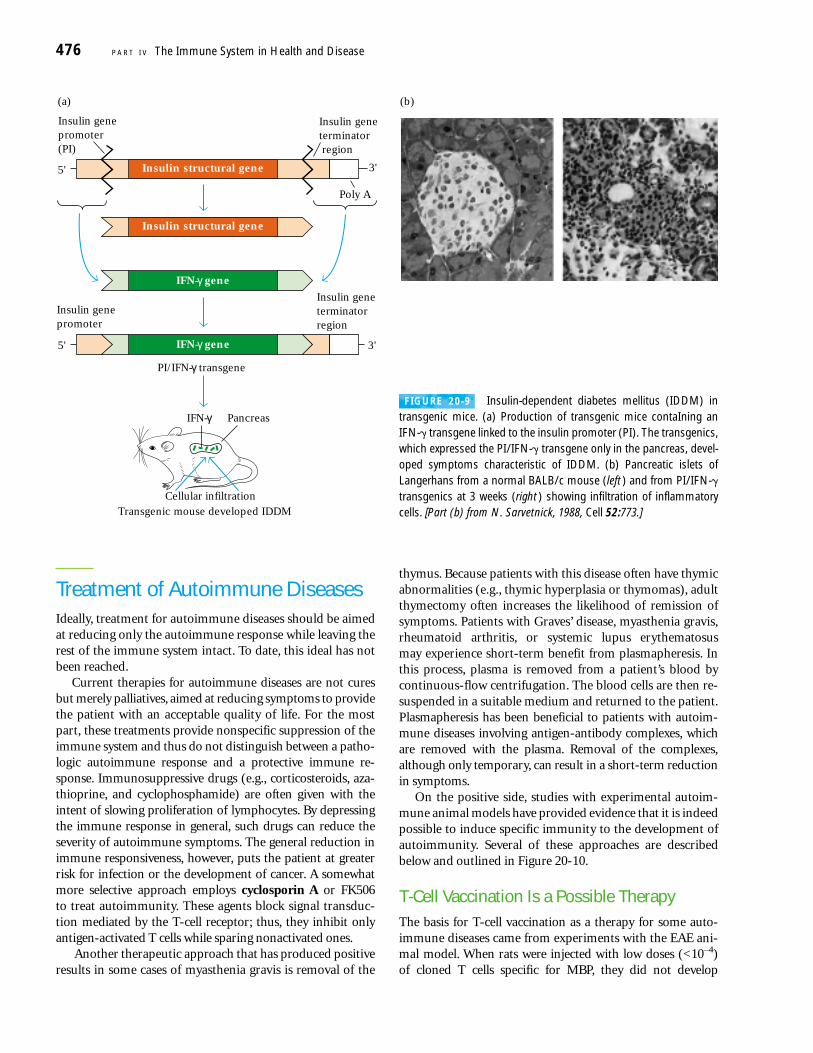
An interesting transgenic mouse system implicates IFN-�and inappropriate class II MHC expression in autoim-munity. In this system, an IFN-� transgene was geneticallyengineered with the insulin promoter, so that the transgenicmice secreted IFN-� from their pancreatic beta cells (Figure20-9a). Since IFN-� up-regulates class II MHC expression,these transgenic mice also expressed class II MHC moleculeson their pancreatic beta cells. The mice developed diabetes,which was associated with cellular infiltration of lympho-cytes and inflammatory cells like the infiltration seen in auto-immune NOD mice and in patients with insulin-dependentdiabetes mellitus (Figure 20-9b).
Although inappropriate class II MHC expression on pan-creatic beta cells may be involved in the autoimmune reactionin these transgenic mice, other factors also may play a role. Forexample, IFN-� is known to induce production of severalother cytokines, including IL-1 and TNF. Therefore, the devel-opment of autoimmunity in this transgenic system may in-volve antigen presentation by class II MHC molecules on pan-creatic beta cells together with a co-stimulatory signal, such asIL-1, that may activate self-reactive T cells. There is also someevidence to suggest that IL-1, IFN-�, and TNF may directlyimpair the secretory function of human beta cells.
Polyclonal B-Cell Activation Can Lead to Autoimmune DiseaseA number of viruses and bacteria can induce nonspecific poly-clonal B-cell activation. Gram-negative bacteria, cytomega-lovirus, and Epstein-Barr virus (EBV) are all known to be suchpolyclonal activators, inducing the proliferation of numer-ous clones of B cells that express IgM in the absence ofTH cells. If B cells reactive to self-antigens are activated bythis mechanism, auto-antibodies can appear. For instance,during infectious mononucleosis, which is caused by EBV,a variety of auto-antibodies are produced, including auto-antibodies reactive to T and B cells, rheumatoid factors, andantinuclear antibodies. Similarly, lymphocytes from patientswith SLE produce large quantities of IgM in culture, suggest-ing that they have been polyclonally activated. Many AIDSpatients also show high levels of nonspecific antibody andauto-antibodies to RBCs and platelets. These patients areoften coinfected with other viruses such as EBV and cyto-megalovirus, which may induce the polyclonal B-cell activa-tion that results in auto-antibody production.
Autoimmunity C H A P T E R 20 475
Treatment of Autoimmune DiseasesIdeally, treatment for autoimmune diseases should be aimedat reducing only the autoimmune response while leaving therest of the immune system intact. To date, this ideal has notbeen reached.
Current therapies for autoimmune diseases are not curesbut merely palliatives, aimed at reducing symptoms to providethe patient with an acceptable quality of life. For the mostpart, these treatments provide nonspecific suppression of theimmune system and thus do not distinguish between a patho-logic autoimmune response and a protective immune re-sponse. Immunosuppressive drugs (e.g., corticosteroids, aza-thioprine, and cyclophosphamide) are often given with theintent of slowing proliferation of lymphocytes. By depressingthe immune response in general, such drugs can reduce theseverity of autoimmune symptoms. The general reduction inimmune responsiveness, however, puts the patient at greaterrisk for infection or the development of cancer. A somewhatmore selective approach employs cyclosporin A or FK506 to treat autoimmunity. These agents block signal transduc-tion mediated by the T-cell receptor; thus, they inhibit onlyantigen-activated T cells while sparing nonactivated ones.
Another therapeutic approach that has produced positiveresults in some cases of myasthenia gravis is removal of the
thymus. Because patients with this disease often have thymicabnormalities (e.g., thymic hyperplasia or thymomas), adultthymectomy often increases the likelihood of remission ofsymptoms. Patients with Graves’ disease, myasthenia gravis,rheumatoid arthritis, or systemic lupus erythematosus may experience short-term benefit from plasmapheresis. Inthis process, plasma is removed from a patient’s blood bycontinuous-flow centrifugation. The blood cells are then re-suspended in a suitable medium and returned to the patient.Plasmapheresis has been beneficial to patients with autoim-mune diseases involving antigen-antibody complexes, whichare removed with the plasma. Removal of the complexes,although only temporary, can result in a short-term reductionin symptoms.
On the positive side, studies with experimental autoim-mune animal models have provided evidence that it is indeedpossible to induce specific immunity to the development ofautoimmunity. Several of these approaches are describedbelow and outlined in Figure 20-10.
T-Cell Vaccination Is a Possible TherapyThe basis for T-cell vaccination as a therapy for some auto-immune diseases came from experiments with the EAE ani-mal model. When rats were injected with low doses (<10–4) of cloned T cells specific for MBP, they did not develop
476 P A R T I V The Immune System in Health and Disease
3'5'
PI/IFN-γ transgene
PancreasIFN-γ
Cellular infiltration
Transgenic mouse developed IDDM
(a) (b)
IFN-γ gene
IFN-γ gene
Insulin genepromoter
Insulin geneterminator region
3'5'
Insulin genepromoter(PI)
Insulin geneterminator region
Insulin structural gene
Insulin structural gene
Poly A
FIGURE 20-9 Insulin-dependent diabetes mellitus (IDDM) intransgenic mice. (a) Production of transgenic mice contaIning anIFN-� transgene linked to the insulin promoter (PI). The transgenics,which expressed the PI/IFN-� transgene only in the pancreas, devel-oped symptoms characteristic of IDDM. (b) Pancreatic islets ofLangerhans from a normal BALB/c mouse (left) and from PI/IFN-�transgenics at 3 weeks (right) showing infiltration of inflammatorycells. [Part (b) from N. Sarvetnick, 1988, Cell 52:773.]

symptoms of EAE. Instead they became resistant to thedevelopment of EAE when later challenged with a lethal doseof activated MBP-specific T cells or MBP in adjuvant. Laterfindings revealed that the efficacy of these autoimmune T-cell clones as a vaccine could be enhanced by crosslinkingthe cell-membrane components with formaldehyde or glu-taraldehyde. When crosslinked T cells were injected into ani-mals with active EAE, permanent remission of symptoms wasobserved. The crosslinked T cells apparently elicit regulatory T cells specific for TCR variable-region determinants of theautoimmune clones. Presumably these regulatory T cells act tosuppress the autoimmune T cells that mediate EAE.
Peptide Blockade of MHC Molecules CanModulate Autoimmune ResponsesIdentification and sequencing of various autoantigens hasled to the development of new approaches to modulate auto-immune T-cell activity. In EAE, for example, the encephalito-genic peptides of MBP have been well characterized. Syn-thetic peptides differing by only one amino acid from theirMBP counterpart have been shown to bind to the appropri-ate MHC molecule. Moreover, when sufficient amounts ofsuch a peptide were administered along with the correspond-ing encephalitogenic MBP peptide, the clinical developmentof EAE was blocked. Presumably, the synthetic peptide acts as
a competitor, occupying the antigen-binding cleft on MHCmolecules and thus preventing binding of the MBP peptide.
In other studies, blocking peptides complexed to solubleclass II MHC molecules reversed the clinical progression ofEAE in mice, presumably by inducing a state of clonal anergyin the autoimmune T cells.
Monoclonal Antibodies May Be Used to Treat AutoimmunityMonoclonal antibodies have been used successfully to treatautoimmune disease in several animal models. For example,a high percentage of (NZB � NZW) F1 mice given weeklyinjections of high doses of monoclonal antibody specific forthe CD4 membrane molecule recovered from their autoim-mune lupus-like symptoms (Figure 20-11). Similar positiveresults were observed in NOD mice, in which treatment withan anti-CD4 monoclonal antibody led to disappearance ofthe lymphocytic infiltration and diabetic symptoms.
Because anti-CD4 monoclonal antibodies block ordeplete all TH cells, regardless of their specificity, they canthreaten the overall immune responsiveness of the recipient.One remedy for this disadvantage is to try to block antigen-activated TH cells only, since these cells are involved in theautoimmune state. To do this, researchers have used mono-clonal antibody directed against the � subunit of the high-affinity IL-2 receptor, which is expressed only by antigen-activated TH cells. Because the IL-2R � subunit is expressedat higher levels on autoimmune T cells, monoclonal anti-body to the � subunit (anti-TAC) might preferentially blockautoreactive T cells. This approach was tested in adult ratsinjected with activated MBP-specific T cells in the presenceor absence of anti-TAC. All the control rats died of EAE,whereas six of the nine treated with anti-TAC had no symp-toms, and the symptoms in the other three were mild.
Autoimmunity C H A P T E R 20 477
IL-2R
IL-2 toxin orAnti-IL-2R
CD4
Anti-CD4
Anti-MHC
Anti-TCR
Peptideblockade
APC AutoreactiveT cell
FIGURE 20-10 Some experimental agents for immunointerven-tion in autoimmune disease.
50
20
40
Surv
ival
, % 60
Treatment started
Anti-CD4
Saline control
Age, months
80
100
6 7 8 9 10 11 12 13 14 15 18
FIGURE 20-11 Weekly injections of anti-CD4 monoclonal anti-body into (NZB � NZW) F1 mice exhibiting autoimmune lupus-likesymptoms significantly increased their survival rate. [Adapted fromD. Wofsy, 1988, Prog. Allergy 45:106.]
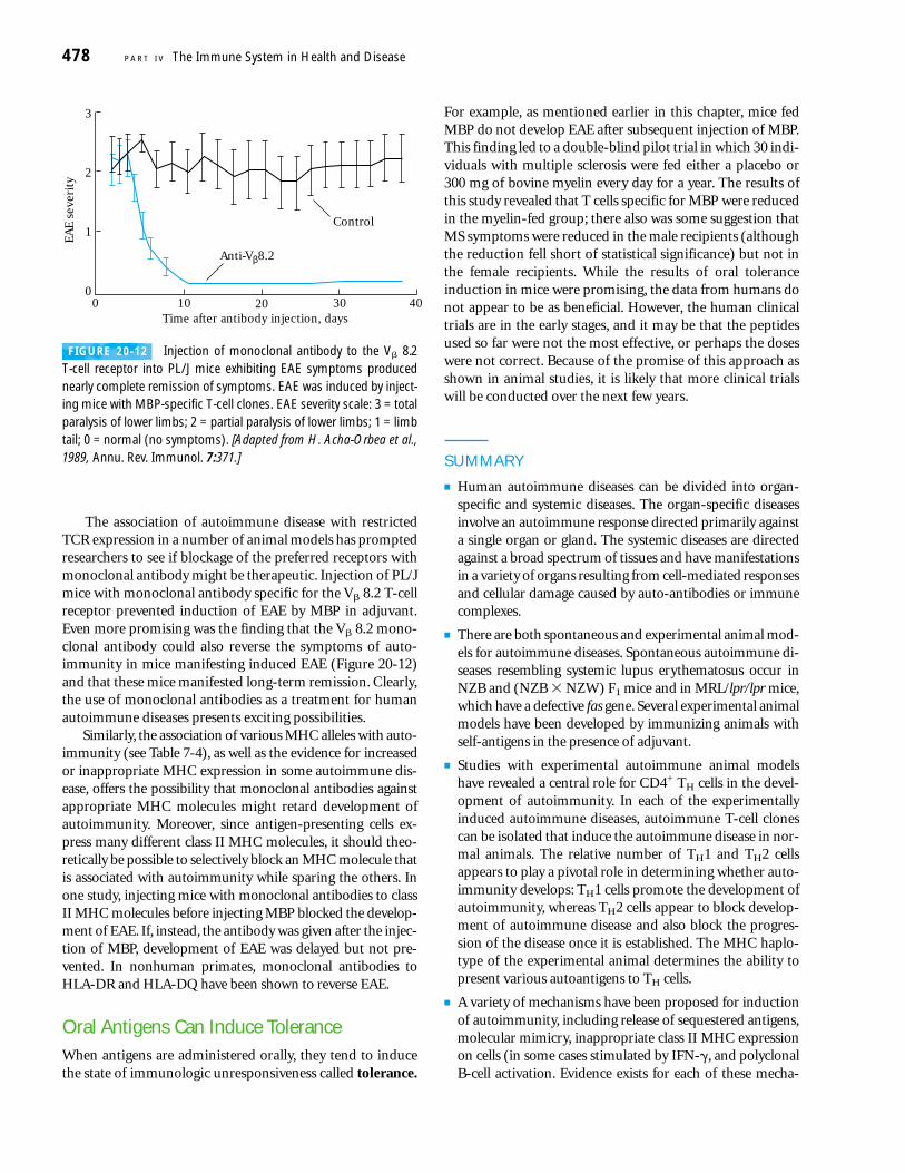
The association of autoimmune disease with restrictedTCR expression in a number of animal models has promptedresearchers to see if blockage of the preferred receptors withmonoclonal antibody might be therapeutic. Injection of PL/Jmice with monoclonal antibody specific for the V� 8.2 T-cellreceptor prevented induction of EAE by MBP in adjuvant.Even more promising was the finding that the V� 8.2 mono-clonal antibody could also reverse the symptoms of auto-immunity in mice manifesting induced EAE (Figure 20-12)and that these mice manifested long-term remission. Clearly,the use of monoclonal antibodies as a treatment for humanautoimmune diseases presents exciting possibilities.
Similarly, the association of various MHC alleles with auto-immunity (see Table 7-4), as well as the evidence for increasedor inappropriate MHC expression in some autoimmune dis-ease, offers the possibility that monoclonal antibodies againstappropriate MHC molecules might retard development ofautoimmunity. Moreover, since antigen-presenting cells ex-press many different class II MHC molecules, it should theo-retically be possible to selectively block an MHC molecule thatis associated with autoimmunity while sparing the others. Inone study, injecting mice with monoclonal antibodies to classII MHC molecules before injecting MBP blocked the develop-ment of EAE. If, instead, the antibody was given after the injec-tion of MBP, development of EAE was delayed but not pre-vented. In nonhuman primates, monoclonal antibodies toHLA-DR and HLA-DQ have been shown to reverse EAE.
Oral Antigens Can Induce ToleranceWhen antigens are administered orally, they tend to inducethe state of immunologic unresponsiveness called tolerance.
For example, as mentioned earlier in this chapter, mice fedMBP do not develop EAE after subsequent injection of MBP.This finding led to a double-blind pilot trial in which 30 indi-viduals with multiple sclerosis were fed either a placebo or300 mg of bovine myelin every day for a year. The results ofthis study revealed that T cells specific for MBP were reducedin the myelin-fed group; there also was some suggestion thatMS symptoms were reduced in the male recipients (althoughthe reduction fell short of statistical significance) but not inthe female recipients. While the results of oral toleranceinduction in mice were promising, the data from humans donot appear to be as beneficial. However, the human clinicaltrials are in the early stages, and it may be that the peptidesused so far were not the most effective, or perhaps the doseswere not correct. Because of the promise of this approach asshown in animal studies, it is likely that more clinical trialswill be conducted over the next few years.
SUMMARY
■ Human autoimmune diseases can be divided into organ-specific and systemic diseases. The organ-specific diseasesinvolve an autoimmune response directed primarily againsta single organ or gland. The systemic diseases are directedagainst a broad spectrum of tissues and have manifestationsin a variety of organs resulting from cell-mediated responsesand cellular damage caused by auto-antibodies or immunecomplexes.
■ There are both spontaneous and experimental animal mod-els for autoimmune diseases. Spontaneous autoimmune di-seases resembling systemic lupus erythematosus occur inNZB and (NZB � NZW) F1 mice and in MRL/lpr/lpr mice,which have a defective fas gene. Several experimental animalmodels have been developed by immunizing animals withself-antigens in the presence of adjuvant.
■ Studies with experimental autoimmune animal modelshave revealed a central role for CD4+ TH cells in the devel-opment of autoimmunity. In each of the experimentallyinduced autoimmune diseases, autoimmune T-cell clonescan be isolated that induce the autoimmune disease in nor-mal animals. The relative number of TH1 and TH2 cellsappears to play a pivotal role in determining whether auto-immunity develops: TH1 cells promote the development ofautoimmunity, whereas TH2 cells appear to block develop-ment of autoimmune disease and also block the progres-sion of the disease once it is established. The MHC haplo-type of the experimental animal determines the ability topresent various autoantigens to TH cells.
■ A variety of mechanisms have been proposed for inductionof autoimmunity, including release of sequestered antigens,molecular mimicry, inappropriate class II MHC expressionon cells (in some cases stimulated by IFN-�, and polyclonalB-cell activation. Evidence exists for each of these mecha-
478 P A R T I V The Immune System in Health and DiseaseEA
E se
veri
ty
3
2
1
00 10 20 30 40
Anti-Vβ8.2
Control
Time after antibody injection, days
FIGURE 20-12 Injection of monoclonal antibody to the V� 8.2T-cell receptor into PL/J mice exhibiting EAE symptoms producednearly complete remission of symptoms. EAE was induced by inject-ing mice with MBP-specific T-cell clones. EAE severity scale: 3 = totalparalysis of lower limbs; 2 = partial paralysis of lower limbs; 1 = limbtail; 0 = normal (no symptoms). [Adapted from H. Acha-Orbea et al.,1989, Annu. Rev. Immunol. 7:371.]

nisms, reflecting the many different pathways leading toautoimmune reactions.
■ Current therapies for autoimmune diseases include treat-ment with immunosuppressive drugs, thymectomy, andplasmapheresis for diseases involving immune complexes.Other therapies include vaccination with T cells specificfor a given autoantigen, administration of synthetic block-ing peptides that compete with autoantigen for binding toMHC molecules, treatment with monoclonal antibodiesthat react with some component specifically involved in anautoimmune reaction, and induction of tolerance toautoantigens by administering them orally.
ReferencesBenoist, C., and D. Matis. 2001. Autoimmunity provoked by
infection: how good is the case for T cell epitope mimicry? NatImmunol. 2:797.
Charlton, B., and K. J. Lafferty. 1995. The TH1/TH2 balance inautoimmunity. Curr. Opin. Immunol. 7:793.
Erikson, J., et al. 1998. Self-reactive B cells in nonautoimmuneand autoimmune mice. Immunol. Res. 17:49.
Hausmann, S., and K. W. Wucherpfennig. 1997. Activation ofautoreactive T cells by peptides from human pathogens. Curr.Opin. Immunol. 9:831.
Horwitz, M. S., et al. 1998. Diabetes induced by Coxsackie virus:initiation by bystander damage and not molecular mimicry.Nature Med. 4:781.
King, C., and N. Sarvetnick. 1997. Organ specific autoimmunity.Curr. Opin. Immunol. 9:863.
Levin, M. C., et al. 2002. Autoimmunity due to molecular mim-icry as a cause of neurological disease. Nat Med. 8:509.
Liblau, R. S., S. M. Singer, and H. O. McDevitt. 1995. TH1 andTH2 CD4+ T cells in the pathogenesis of organ-specific auto-immune diseases. Immunol. Today 16:34.
Lockshin, M. D. 1998. Why women? J. Am. Med. Womens Assoc.53:4.
O’Garra, A., L. Steinman, and K. Gijbels. 1997. CD4+ T-cell sub-sets in autoimmunity. Curr. Opin. Immunol. 9:872.
Rose, N. R. 1998. The role of infection in the pathogenesis ofautoimmune disease. Semin. Immunol. 10:5.
Silverstein, A. M., and N. R. Rose. 1997. On the mystique of theimmunological self. Immunol. Rev. 159:197.
Steinman, L., J. R. Oskenberg, and C. C. A. Bernard. 1992. Asso-ciation of susceptibility to multiple sclerosis with TCR genes.Immunol. Today 13:49.
Streilein, J. W., M. R. Dana, and B. R. Ksander. 1997. Immunitycausing blindness: five different paths to herpes stromal kerati-tis. Immunol. Today 18:443.
Theofilopoulos, A. N. 1995. The basis of autoimmunity. Part I:Mechanisms of aberrant self-recognition. Immunol. Today16:90.
Theofilopoulos, A. N. 1995. The basis of autoimmunity. Part II:Genetic predisposition. Immunol. Today 16:150.
Vyse, T. J., and B. L. Kotzin. 1998. Genetic susceptibility to sys-temic lupus erythematosus. Annu. Rev. Immunol. 16:261.
Weiner, H. L., et al. 1993. Double-blind pilot trial of oral toler-ization with myelin antigens in multiple sclerosis. Science259:1321.
Wilder, R. L. 1995. Neuroendocrine-immune system interac-tions and autoimmunity. Annu. Rev. Immunol. 13:307.
Wucherpfennig, K. W. 2001.Mechanisms for the induction ofautoimmunity by infectious agents. J Clin. Invest. 108:1097.
Zhao, Z-S, et al. 1998. Molecular mimicry by herpes simplexvirus type-1: autoimmune disease after viral infection. Science279:1344.
USEFUL WEB SITES
http://www.lupus.org/index.html
The site for the Lupus Foundation of America contains valu-able information for patients and family members as well ascurrent information about research in this area.
http://www.nih.gov/niams/
Home page for the National Institute for Arthritis and Mus-culoskeletal and Skin Diseases. This site contains links toother arthritis sites.
http://www.niddk.nih.gov/
Home page for the National Institute for Diabetes and Diges-tive and Kidney Diseases. This site contains an exhaustive listof links to other diabetes health-related sites.
http://www.sciencemag.org/feature/data/983519.shl
Link to a Web site that provides specific information concern-ing the role of gender in autoimmune disease.
Study QuestionsCLINICAL FOCUS QUESTION What are some of the possible reasonswhy females are more susceptible to autoimmune diseases thanmales?
1. For each of the following autoimmune diseases (a–m), selectthe most appropriate characteristic (1–13) listed below.
Diseases
a. Experimental autoimmune encephalitis (EAE)
b. Goodpasture’s syndromec. Graves’ diseased. Systemic lupus erythematosus (SLE)d. Insulin-dependent diabetes mellitus
(IDDM)
Autoimmunity C H A P T E R 20 479
Go to www.whfreeman.com/immunology Self-TestReview and quiz of key terms

f. Rheumatoid arthritisg. Hashimoto’s thyroiditish. Experimental autoimmune myasthenia
gravis (EAMG)i. Myasthenia gravisj. Pernicious anemiak. Multiple sclerosisl. Autoimmune hemolytic anemia
Characteristics
(1) Auto-antibodies to intrinsic factor block vitamin B12
absorption
(2) Auto-antibodies to acetylcholine receptor
(3) TH1-cell reaction to thyroid antigens
(4) Auto-antibodies to RBC antigens
(5) T-cell response to myelin
(6) Induced by injection of myelin basic protein plus com-plete Freund’s adjuvant
(7) Auto-antibody to IgG
(8) Auto-antibodies to basement membrane
(9) Auto-antibodies to DNA and DNA-associated protein
(10) Auto-antibodies to receptor for thyroid-stimulatinghormone
(11) Induced by injection of acetylcholine receptors
(12) TH1-cell response to pancreatic beta cells
2. Experimental autoimmune encephalitis (EAE) has proved tobe a useful animal model of autoimmune disorders.
a. Describe how this animal model is made.b. What is unusual about the animals that recover from
EAE?c. How has this animal model indicated a role for T cells in
the development of autoimmunity?
3. Molecular mimicry is one mechanism proposed to accountfor the development of autoimmunity. How has induction ofEAE with myelin basic protein contributed to the understandingof molecular mimicry in autoimmune disease?
4. Describe at least three different mechanisms by which alocalized viral infection might contribute to the development ofan organ-specific autoimmune disease.
5. Transgenic mice expressing the IFN-� transgene linked tothe insulin promoter developed diabetes.
a. Why was the insulin promoter used?b. What is the evidence that the diabetes in these mice is due
to autoimmune damage?c. What is unusual about MHC expression in this system?d. How might this system mimic events that might be
caused by a localized viral infection in the pancreas?
6. Monoclonal antibodies have been administered for therapyin various autoimmune animal models. Which monoclonal anti-bodies have been used and what is the rationale for theseapproaches?
7. Indicate whether each of the following statements is true orfalse. If you think a statement is false, explain why.
a. TH1 cells have been associated with development ofautoimmunity.
b. Immunization of mice with IL-12 prevents induction ofEAE by injection of myelin basic protein plus adjuvant.
c. The presence of the HLA B27 allele is diagnostic forankylosing spondylitis, an autoimmune disease affectingthe vertebrae.
d. Individuals with pernicious anemia produce antibodiesto intrinsic factor.
e. A defect in the gene encoding Fas can reduce pro-grammed cell death by apoptosis.
480 P A R T I V The Immune System in Health and Disease