Anticipating ubiquitous computing: logics to forecast technological ...
Upload
independentCategory
view
0download
0
MOLECULAR AND CELLULAR BIOLOGY, July 2004, p. 6000–6010 Vol. 24, No. 130270-7306/04/$08.00�0 DOI: 10.1128/MCB.24.13.6000–6010.2004Copyright © 2004, American Society for Microbiology. All Rights Reserved.
Functional Domains of the Ubiquitous Chromatin Protein DEKFerdinand Kappes,† Ingo Scholten,† Nicole Richter, Claudia Gruss, and Tanja Waldmann*
Department of Biology, University of Konstanz, 78457 Konstanz, Federal Republic of Germany
Received 11 February 2004 /Returned for modification 3 April 2004 /Accepted 9 April 2004
DEK was originally described as a proto-oncogene protein and is now known to be a major component ofmetazoan chromatin. DEK is able to modify the structure of DNA by introducing supercoils. In order to findinteraction partners and functional domains of DEK, we performed yeast two-hybrid screens and mutationalanalyses. Two-hybrid screening yielded C-terminal fragments of DEK, suggesting that DEK is able to mul-timerize. We could localize the domain to amino acids 270 to 350 and show that multimerization is dependenton phosphorylation by CK2 kinase in vitro. We also found two DNA binding domains of DEK, one on afragment including amino acids 87 to 187 and containing the SAF-box DNA binding motif, which is locatedbetween amino acids 149 and 187. This region is sufficient to introduce supercoils into DNA. The second DNAbinding domain is located between amino acids 270 and 350 and thus overlaps the multimerization domain. Weshow that the two DNA-interacting domains differ in their binding properties and in their abilities to respondto CK2 phosphorylation.
The proto-oncogene protein DEK was first identified as afusion protein with the nucleoporin CAN in patients sufferingfrom a subtype of acute myeloid leukemia (AML) (30). Arecent study showed that more than two-thirds of AML pa-tients expressing the DEK-CAN fusion also have a specificinternal tandem duplication in the FMS-like tyrosine kinase(FLT3) gene (23, 28), resulting in a constitutively active kinase.In contrast to AML with the DEK-CAN rearrangement, theincidence of the activating FLT3 internal tandem duplicationin all AML patients is only around 20%. This suggests thatDEK-CAN might contribute to the generation of FLT3 inter-nal tandem repeats as a possible mechanism leading to carci-nogenesis (4, 19, 28).
In addition to its involvement in leukemia, DEK has beenshown to be a major antigen in several autoimmune diseaseslike systemic lupus erythematosus (6, 7, 33), juvenile rheuma-toid arthritis (7, 25), and sarcoidosis (6, 7). Despite theseassociations with several human disorders, little is knownabout how DEK could functionally be involved in these dis-eases.
In search of the cellular function of DEK, several groups aretrying to find interaction partners of DEK. DEK has beenreported to be involved in transcriptional repression by inter-action with the corepressor Daxx (13), as well as in transcrip-tional activation stimulating the binding of activator proteinAP-2� to its target DNA sequences (5). Furthermore, DEKhas been reported to bind to the pets site, a regulatory elementof the human immunodeficiency virus type 2 enhancer, in asequence-specific manner (8, 10). It has also been reportedthat DEK recognizes the Y box of the class II major histocom-patibility complex gene promoter, a known transcription factorbinding site with sequence similarities to the pets site (1, 26). Incontrast to these findings, we could not detect sequence-spe-
cific binding of DEK to DNA (31). Instead, we observed struc-ture-specific binding of DEK to DNA crossovers, as found insupercoiled or four-way junction DNAs (31). Moreover, DEKbinding causes a change in the superhelical density of closedcircular plasmid DNA and simian virus 40 (SV40) minichro-mosomes in vitro (2, 32).
DEK is a nuclear phosphoprotein (9, 25) that remains asso-ciated with chromatin during the entire cell cycle (14). Whilethe amount of DEK on chromatin does not change during thecell cycle, we detected a moderate increase in DEK phosphor-ylation during the G1 phase (15). It appears that DEK is mainlyphosphorylated by CK2 kinase in vitro and in vivo. Moreover,phosphorylation reduces the DNA binding activity of DEK.This is surprising, because most of the phosphorylation siteswere mapped within the C-terminal region of the protein (15,31, 32), whereas a potential DNA binding motif, the SAF-box(scaffold attachment factor) (11, 18), also known as the SAP-box (SAF-A/B acinus and pias) (3), is located between aminoacids 149 and 187. The SAF-box is the only recognizable fea-ture besides some highly acidic regions of the protein. TheSAF-box was identified as a novel DNA binding domain thatbinds specifically to AT-rich SAR/MAR (scaffold/matrix at-tachment region) DNA (11, 18). SAF-box-containing proteinsare involved in different processes, such as DNA repair (Ku70,PARP), pre-mRNA processing (SAF-A, SAF-B, SPRY), tran-scriptional elongation (Tho1), and nuclear architecture(SAF-A, SAF-B) (3).
To investigate whether the SAF-box is the DNA bindingdomain of DEK, we expressed several deletion mutant formsof DEK and tested them for DNA binding in electrophoreticmobility shift assays (EMSAs) and for the ability to change thesupercoils of closed circular DNA. We show here that a DEKfragment containing the SAF-box is able to bind DNA andappears to be responsible for supercoiling activity. Moreover,the SAF-box-containing fragment induces intermolecular in-teractions between two or more DNA molecules. In addition,we have identified a second functional domain within the C-terminal region of DEK. This domain is able to bind DNA and
* Corresponding author. Mailing address: Department of Biology,University of Konstanz, Universitatsstrasse 10, M1019, 78457 Kon-stanz, Germany. Phone: 49 7531 882125. Fax: 49 7531 884036. E-mail:[email protected].
† F.K. and I.S. contributed equally to this paper.
6000

induce DEK-DEK interactions. These two functional activitiesare regulated by phosphorylation.
MATERIALS AND METHODS
Cloning. Full-length DEK cDNA was subcloned from pRSET-A-dek intopBlueBacHis2-A (Invitrogen) via the BamHI and EcoRI restriction sites. DEKfragments with 5� EcoRI and 3� XhoI cutting sites were generated by PCR. Astop codon was introduced before the XhoI site. The PCR products were di-gested and ligated into pBlueBacHis2-B (Invitrogen) that was cut with EcoRIand SalI, creating a hybrid SalI/XhoI site at the 3� end of the insert. All con-structs were checked by sequencing.
Expression and purification of his-DEK fragments. The different pBlueBac-His2 constructs of DEK were cotransfected with linearized Autographa califor-nica nuclear polyhedrosis virus DNA (Bac-N-Blue DNA) into SF9 cells as de-scribed in the manufacturer’s (Invitrogen) protocol. Passage 3 virus stocks wereused to infect HighFive cells for protein expression. Cells at 3 days postinfectionwere washed twice with phosphate-buffered saline and then lysed with 2 ml oflysis buffer per 175-mm2 flask (100 mM Tris-Cl [pH 7.5], 150 mM NaCl, 5 mMKCl, 0.5 mM MgCl2, 1% NP-40, 5 mM imidazole). To disrupt DNA-protein andprotein-protein interactions, the lysed cells were further incubated in the pres-ence of 1.3 M NaCl for 20 min at room temperature. The lysate was cleared bycentrifugation at 100,000 � g. The supernatant was adjusted to 10% glycerol,diluted with lysis buffer to a final concentration of 700 mM NaCl, and incubatedwith 8 �l of 50% Ni-nitrilotriacetic acid (NTA)-agarose beads (QIAGEN). After1 h of incubation at 4°C, the beads were washed twice with WB-150 (100 mMTris-Cl [pH 7.5], 150 mM NaCl, 50 mM imidazole) and transferred into a 10-mlBio-Rad column. The beads were washed twice with WB-300 (100 mM Tris-Cl[pH 7.5], 300 mM NaCl, 50 mM imidazole) and again with WB-150. The His-tagged proteins were then eluted 10 times with 20 �l of elution buffer (100 mMTris-Cl [pH 7.5], 150 mM NaCl, 500 mM imidazole) per 175-cm2 flask. AllDEK-containing fractions were pooled and stored at �70°C.
Far Western analysis. A protein kinase A (PKA) phosphorylation site (aminoacid motif RRASV) was cloned into pBlueBacHis2-A-DEK with BamHI and theannealed oligonucleotides PKA forward (5� GATCC CGT CGT GCA TCT GTTG) and PKA reverse (5� GATCC AAC AGA TGC ACG ACG G). Correctorientation of the insert was checked by sequencing. After expression and puri-fication, 1 �g of PKA-His-tagged DEK was diluted 1/10 with wash buffer WB-150(see description of expression and purification) and rebound to 100 �l of settledNi-NTA-agarose for 2 h at 4°C. The resin was washed twice with 1� PKAphosphorylation buffer (New England Biolabs) by centrifuging the batch for 15 sat maximum speed. [�-32P]ATP labeling was then carried out on column, with 1�l of PKA (New England Biolabs) in a total volume of 400 �l for 30 min at 37°C.After being washed three times with 1 ml of wash buffer, labeled DEK was elutedfrom the Ni-NTA-agarose with 3� 300 �l of elution buffer. The eluates wereanalyzed by Western blotting and autoradiography; radioactively labeled DEKpeaked in the second eluate.
Two-hybrid screening. For two-hybrid screening, we used the “interactionhunt” protocol of Golemis et al. (12). Full-length DEK and a truncated DEKcDNA coding for amino acids 1 to 350 were cloned into the bait vector pEG202with EcoRI and XhoI, generated by PCR. We used two libraries, an oligo(dT)-primed HeLa cell library (Origene) and a random-primed human kidney library(Invitrogen), for multiple screenings, both with full-length and truncated DEK asbait.
Sedimentation analysis. For sedimentation analysis, 2 �g of recombinantHis-DEK was used either dephosphorylated or phosphorylated. Dephosphory-lation was carried out for 90 min with �-phosphatase (New England Biolabs) at30°C in accordance with the manufacturer’s protocol (for 10 pmol of DEK, 400U of �-phosphatase was used). Phosphorylation with CK2 was performed withCK2 buffer (20 mM HEPES [pH 7.8], 10 mM MgCl2, 10 mM CaCl2, 20 mM NaF,and 1 mM vanadate supplemented with 100 �M ATP) for 90 min at 37°C (for 10pmol of DEK, 40 U of CK2 was used). After incubation for 60 min at 37°C innE100 (20 mM HEPES [pH 7.6], 100 mM NaCl, 10 mM sodium bisulfite, 1 mMEDTA), samples were loaded onto 5 to 30% sucrose gradients in nE100 ornE800 (800 mM NaCl) and centrifuged for 14 h at 36,000 rpm in an SW40 rotor(Beckman). Gradients were fractionated, and proteins were concentrated andthen immunoblotted with DEK-specific antibodies. The pellet fraction was dis-solved in 2% sodium dodecyl sulfate (SDS) and treated in the same way.
Topology assay. Recombinant DEK fragments were dephosphorylated with�-phosphatase (New England Biolabs) as described above. After incubation at30°C for 90 min, the samples were dialyzed on Whatman filters (type VS; poresize, 0.025 �m) against nE300 buffer (20 mM HEPES [pH 7.6], 300 mM NaCl,10 mM sodium bisulfite, 1 mM EDTA) in the presence of 1 �g of bovine serum
albumin (New England Biolabs) per microliter for 90 min at 4°C. SupercoiledSV40 DNA (0.0028 pmol) was incubated with increasing amounts of the differentDEK fragments in the presence of 1 U of topoisomerase I (wheat germ; Pro-mega). The amounts of protein are indicated in the figure legends. The reactionswere performed in buffer L0 (20 mM HEPES [pH 7.6], 10 mM dithiothreitol, 25�g of bovine serum albumin per ml) in a total volume of 90 �l. After proteinaseK (Roche) digestion, DNA was precipitated and analyzed on 0.8% agarose gelsin 0.5� TBE (45 mM Tris-borate, 1 mM EDTA) at 2 V/cm for 16 h. The gelswere stained with SybrGold (MobiTec).
EMSA. Purified DEK fragments were treated as described for the topologyassay. Linearized SV40 DNA (0.05 pmol) was incubated with increasing amountsof DEK as indicated in the figure legends. The reactions were performed in atotal volume of 35 �l, and mixtures were loaded directly onto a 0.6% agarose gelin 0.5� TBE (50 mM Tris-borate, 1 mM EDTA [adjusted to pH 7.8 with boricacid]) and run at 2 V/cm overnight.
Ligase assay. A 123-bp DNA ladder (Pharmacia) was digested with AvaI.Twenty-four nanograms (0.3 pmol) of the 123-bp fragment was incubated with1.2 pmol of the different DEK fragments for 1 h at 37°C in ligase buffer (4 mMTris [pH 7.8], 1 mM dithiothreitol, 0.5 mM ATP, 5 mM MgCl2) in a final volumeof 20 �l. T4 DNA ligase (1 U) was added for 10 min at 37°C; the ligase was theninactivated for 15 min at 65°C. To distinguish between linear and circular prod-ucts, the samples were treated with 50 U of exonuclease III. Samples weredigested with proteinase K and analyzed on 8% polyacrylamide gels for 3 h at 10V/cm. The reaction products were then analyzed by SybrGold (MobiTec) stain-ing.
RESULTS
Multimerization of DEK protein. To identify DEK interac-tion partners, we used the yeast two-hybrid system. Two dif-ferent libraries were screened with full-length DEK or DEK 1-350 as bait. By using full-length DEK as bait, we recoveredsequences containing amino acids 215 to 375 of DEK from anoligo(dT)-primed HeLa library. With DEK 1-350 as bait, weisolated DEK 247-375 from the same cDNA library. To avoidthe bias for carboxy-terminal regions intrinsic to oligo(dT)-primed libraries, we also used a random primed kidney cDNAlibrary and full-length DEK as bait. Again, a cDNA encodinga C-terminal fragment of DEK (amino acids 229 to 375) wasfound (Fig. 1A). These data indicate that DEK�s preferredinteraction partner is DEK itself and that amino acids 247 to350 of DEK contain a di- or multimerization domain.
To confirm this finding and localize the multimerizationdomain more precisely, we tested several fragments of DEKversus the full-length protein in the two-hybrid system (Fig.1B). All fragments containing amino acids 270 to 350 were ableto interact with DEK, showing that the amino-terminal regionup to residue 270 is not involved in di- or multimerization.
We obtained independent support for this conclusion by farWestern blotting. A PKA consensus sequence (amino acidmotif RRASV) was linked 5� to the amino terminus of full-length DEK. The protein was expressed in the baculovirussystem and labeled in vitro with [�-32P]ATP by PKA. Equalamounts of deletion mutant forms and full-length DEK wereseparated by SDS-polyacrylamide gel electrophoresis andtransferred to a nitrocellulose membrane. After incubationwith labeled full-length DEK and extensive washing, the blotwas analyzed by autoradiography (Fig. 2B). Full-length DEKand fragments with amino acids 1 to 350 and 270 to 375 gavepositive autoradiographic signals, whereas amino acids 1 to 310yielded only a faint autoradiographic band (Fig. 2B). Thus,these data confirmed that a protein-protein interaction domainis located between amino acids 270 and 350 of the DEK pro-tein (Fig. 2C). Here it is important to mention that recombi-
VOL. 24, 2004 FUNCTIONAL DOMAINS OF THE DEK PROTEIN 6001

nant His-DEK as purified from the baculovirus system is phos-phorylated (15). Therefore we tested phosphorylatedfragments of DEK and also used phosphorylated PKA-DEK asa probe.
Since DEK-DNA interaction is affected by phosphorylation(15), we tested whether phosphorylation also influences DEKmultimerization. We compared the sedimentation behavior ofphosphorylated and dephosphorylated full-length DEK on su-
crose gradients. We used recombinant full-length DEK thatwas purified from the baculovirus system in a phosphorylatedform. This substrate was first dephosphorylated by �-phospha-tase (Fig. 3A, �P) to remove endogenous phosphates and thenrephosphorylated by CK2 in vitro (Fig. 3A, �PCK2). CK2 is themain kinase responsible for the majority of DEK phosphory-lation in vitro and in vivo (15).
When sucrose gradients were run under nearly physiological
FIG. 1. Two-hybrid screening. (A) Results of three different two-hybrid interaction hunts. Full-length DEK (top and bottom) and DEK 1-350(middle) were used as bait. We screened against two different human libraries and obtained prey that codes for carboxy-terminal fragments ofDEK. (B) Localization of the DEK-DEK multimerization domain. We tested different deletion mutant forms of DEK (indicated on the left side)against full-length DEK in the two-hybrid system. LacZ activation is shown on the right. Dark grey boxes, highly acidic regions; light grey box,SAF-box, a potential DNA binding domain; black box, nuclear localization sequence (NLS).
6002 KAPPES ET AL. MOL. CELL. BIOL.

salt conditions (100 mM NaCl), phosphorylated DEK formedlarge complexes that were found in the pellet of the gradient(Fig. 3A, �PCK2). However, dephosphorylated DEK sedi-mented at physiological salt concentrations and DEK sedi-mented mainly as monomers close to the top of the gradient(Fig. 3A, �P). At 800 mM salt (Fig. 3B), phosphorylated DEKhad sedimentation properties like those of dephosphorylatedDEK at 100 mM NaCl, showing that a high salt concentrationdisrupts the interaction of phosphorylated DEK (compare Fig.3A, �P, and B). Thus, phosphorylation of DEK induces DEK-DEK interactions at moderate salt concentrations, whereas thedephosphorylated protein mainly exists in the monomeric formunder these conditions.
Similar results were obtained by native gel electrophoresis:phosphorylated DEK remained close to the gel slots as a high-molecular-weight complex, while unphosphorylated DEK mi-grated as monomers (data not shown). A conclusion fromsucrose gradient and native gel electrophoretic analyses is that
phosphorylated DEK tends to aggregate in vitro instead offorming discrete multimers.
Characterization of the DNA binding properties of the SAF-box-containing fragment. The region of DEK between aminoacids 149 and 187 has similarity to a DNA binding motif that isknown as the SAF-box (11) and that is also termed the SAP-box (3). To test if the SAF-box is indeed the DNA bindingdomain of the DEK protein, we used DEK deletion mutantforms with or without the SAF-box in EMSAs. As we haveshown before (31, 32), the binding of full-length DEK results inlarge DNA protein complexes that cannot enter the gel (Fig.4A, lanes 2 to 4). To investigate if the multimerization domain(amino acids 270 to 350, Fig. 1 and 2) was responsible for thisbinding mode, we successively removed the C-terminal part ofDEK and tested these fragments by EMSA (Fig. 4A, lanes 5 to13). Surprisingly, we found that even the smallest fragment(amino acids 87 to 187) that contains the SAF-box at its C-terminal region (amino acids 149 to 187) was able to induce
FIG. 2. Far Western analysis. Twenty nanomoles of full-length DEK (wt, lane 1) and different DEK deletion mutant forms were separated ona 10 to 18% polyacrylamide gel and blotted onto a nitrocellulose membrane. The membrane was then incubated with 32P-labeled full-length DEK.A, Ponceau staining; B, autoradiography. Molecular sizes of marker proteins are given in kilodaltons. (C) DEK protein with the multimerizationdomain. Dark grey boxes, highly acidic regions; light grey box, SAF-box, a potential DNA binding domain; black box, nuclear localization sequence(NLS). wt, wild type.
VOL. 24, 2004 FUNCTIONAL DOMAINS OF THE DEK PROTEIN 6003

this high-molecular-weight complex (Fig. 4A, lanes 11 to 13).Therefore, the previously identified protein-protein interactiondomain (amino acids 270 to 350, Fig. 1 and 2) seems to have noinfluence on the DNA binding properties of DEK. This couldindicate that another multimerization domain may exist in theregion of amino acids 87 to 187, probably dependent on DNAbinding, which induces intermolecular interactions betweentwo or more DNA molecules. Interestingly, the SAF-box-con-taining protein SAF-A also multimerizes upon DNA binding(18).
The SAF-box-containing fragment induces intermolecularinteractions and is sufficient for the introduction of supercoils.We have recently shown that full-length DEK (glutathioneS-transferase and His tagged) promotes the linkage of DNAfragments via a ligase-mediated end-to-end joining assay (31).To determine whether this reaction depends on the SAF-box-containing domain (amino acids 87 to 187), we incubated var-ious deletion mutant DEK proteins with 123-bp DNA frag-ments in the presence of T4 ligase. Our data show (Fig. 5A)that all DEK constructs containing the amino acid 87 to 187region stimulated the formation of linear multimers (Fig. 5A,lanes 4 and 10), whereas DEK constructs without the aminoacid 87 to 187 region could not (Fig. 5A, lanes 6 and 8).However, all ligation products were sensitive to exonucleaseIII (Fig. 5A, lanes 5, 7, 9, and 11), showing that none of theDEK fragments were able to bend DNA, as shown before forfull-length DEK (31). These data indicate that the region be-tween amino acids 87 and 187 is responsible for the stimulationof intermolecular interactions and thus might include a DNA-dependent multimerization domain.
Recently, we have shown that DEK causes supercoiling ofcircular DNA in the presence of topoisomerase I (32). In orderto determine which domain is responsible for this activity, wetested recombinant DEK fragments in the topology assay (32).We used dephosphorylated DEK fragments and incubatedthem with circular SV40 DNA in the presence of topoisomer-ase I. Again, only the fragments containing the amino acid 87to 187 region were able to change the superhelicity of the DNA(Fig. 5B, lanes 1 to 4, 5 to 8, and 13 to 16).
Thus, we have shown that the region between amino acids 87and 187, including the SAF-box (amino acids 149 to 187), bindsto DNA, induces intermolecular interactions, probably owingto a DNA-dependent multimerization, and is responsible forthe introduction of supercoils into circular DNA.
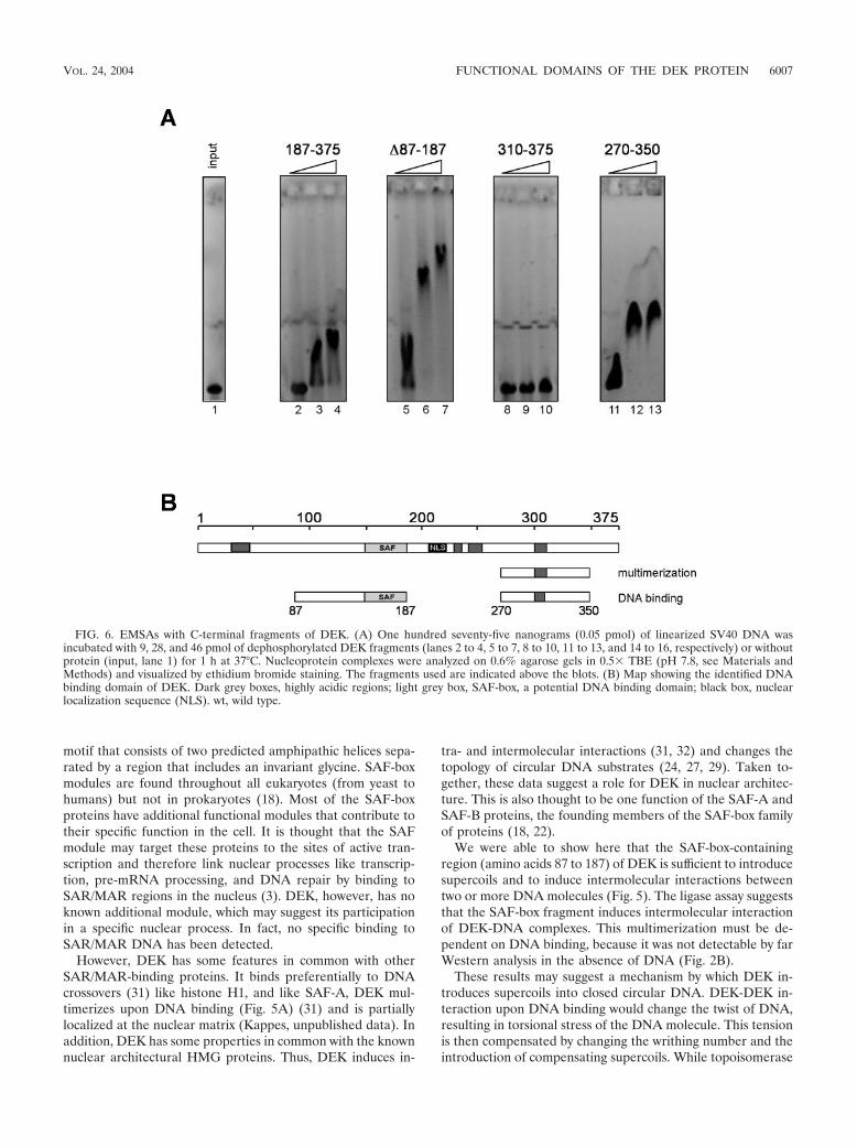
Identification of a second DNA binding domain in the C-terminal region of DEK. During our investigations of differentSAF-box fragments, we also tested the N-terminally truncateddeletion mutant forms of DEK by EMSA. Surprisingly, wefound that these fragments were able to bind DNA (Fig. 6A,lanes 2 to 4), but we detected no large DNA-protein com-plexes, even when we used very large amounts of protein (datanot shown). We then performed EMSAs with shorter C-ter-minal fragments (Fig. 6A) and found that the amino acid 310to 375 fragment showed very weak, if any, binding, even withthe largest amount of protein (Fig. 6A, lane 10), whereas afragment consisting of amino acids 270 to 350 induced a com-plete shift at low protein concentrations (Fig. 6A, lanes 11 to13). Large immobile complexes, like those involving aminoacids 87 to 187, were never observed. We have thus identifieda second DNA binding domain within amino acids 270 to 350
FIG. 3. Sedimentation analysis of phosphorylated and dephosphorylated DEK. Recombinant full-length DEK was dephosphorylated with�-phosphatase (�P), rephosphorylated with CK2 (�PCK2), analyzed on 5 to 30% sucrose gradients, and then centrifuged for 14 h at 36,000 rpm.The gradients were fractionated and analyzed by immunoblotting with DEK-specific antibodies. (A) Sedimentation analysis of dephosphorylatedDEK (�P) and phosphorylated DEK (�PCK2) in the presence of 100 mM NaCl. (B) Sedimentation analysis of phosphorylated DEK in thepresence of 800 mM NaCl. P corresponds to the pellet fraction.
6004 KAPPES ET AL. MOL. CELL. BIOL.

that does not multimerize upon DNA binding like the SAF-box-containing fragments (Fig. 4A).
It appears that the domain between amino acids 270 and 350has several functions. As just shown, it includes a DNA bindingactivity (Fig. 6), but in addition it contains a multimerizationdomain (Fig. 2) and also most of the phosphorylation sites.Above we have shown that phosphorylation affects the inter-action between DEK proteins (Fig. 3), and therefore we asknow whether phosphorylation also influences DNA binding.
We showed that the sequence between amino acids 187 and310 can be phosphorylated in vitro (Fig. 7A). This was ex-pected, as most of the in vitro phosphorylation sites mapped tothis region (15). We then investigated whether phosphoryla-tion affects DNA binding and found that the in vitro-phosphor-ylated C-terminal fragments had completely lost the ability tobind DNA (Fig. 7B; compare with Fig. 6). A control wasfragment 1 to 187, which still bound DNA after CK2 phos-phorylation (Fig. 7B, lanes 5 to 7). Another control was full-length DEK, whose DNA binding activity was reduced afterphosphorylation (Fig. 7B, lanes 2 to 4; compare with Fig. 4).This indicates that phosphorylation has an overall effect onDEK�s ability to bind DNA.
DISCUSSION
We used two-hybrid screening and mutational analysis toidentify interaction partners and functional domains of theproto-oncogene protein DEK. Two-hybrid screening yieldedC-terminal fragments of DEK, suggesting that DEK is able tomultimerize. We localized the multimerization domain toamino acids 270 to 350 and showed furthermore that multim-erization is dependent on phosphorylation by CK2 in vitro. Wealso found two DNA binding domains of DEK, one on afragment consisting of amino acids 87 to 187 that contains aknown DNA binding motif, the SAF-box (amino acids 149 to187) (11). This region is sufficient to introduce supercoils intoDNA and to induce intermolecular interactions, as shown byligase-mediated end-to-end joining. A second DNA bindingdomain is located between amino acids 270 and 350 and thusoverlaps the multimerization domain. The two DNA-interact-ing domains differ in their binding modes and in their abilitiesto be regulated by CK2 phosphorylation.
The SAF-box-containing fragment (amino acids 87 to 187)introduces supercoils and induces intermolecular interac-tions. The SAF-box has been described as a DNA binding
FIG. 4. EMSAs with different dephosphorylated SAF-box fragments. (A) One hundred seventy-five nanograms (0.05 pmol) of linearized SV40DNA was incubated with 9, 28, and 46 pmol of dephosphorylated DEK fragments (lanes 2 to 4, 5 to 7, 8 to 10, 11 to 13, and 14 to 16, respectively)or without protein (input, lane 1) for 1 h at 37°C. Nucleoprotein complexes were analyzed on 0.6% agarose gels in 0.5� TBE (pH 7.8; see Materialsand Methods) and visualized by ethidium bromide staining. The fragments used are indicated above the blots. (B) Map showing the identified DNAbinding domain of DEK. Dark grey boxes, highly acidic regions; light grey box, SAF-box, a potential DNA binding domain; black box, nuclearlocalization sequence (NLS). wt, wild type.
VOL. 24, 2004 FUNCTIONAL DOMAINS OF THE DEK PROTEIN 6005
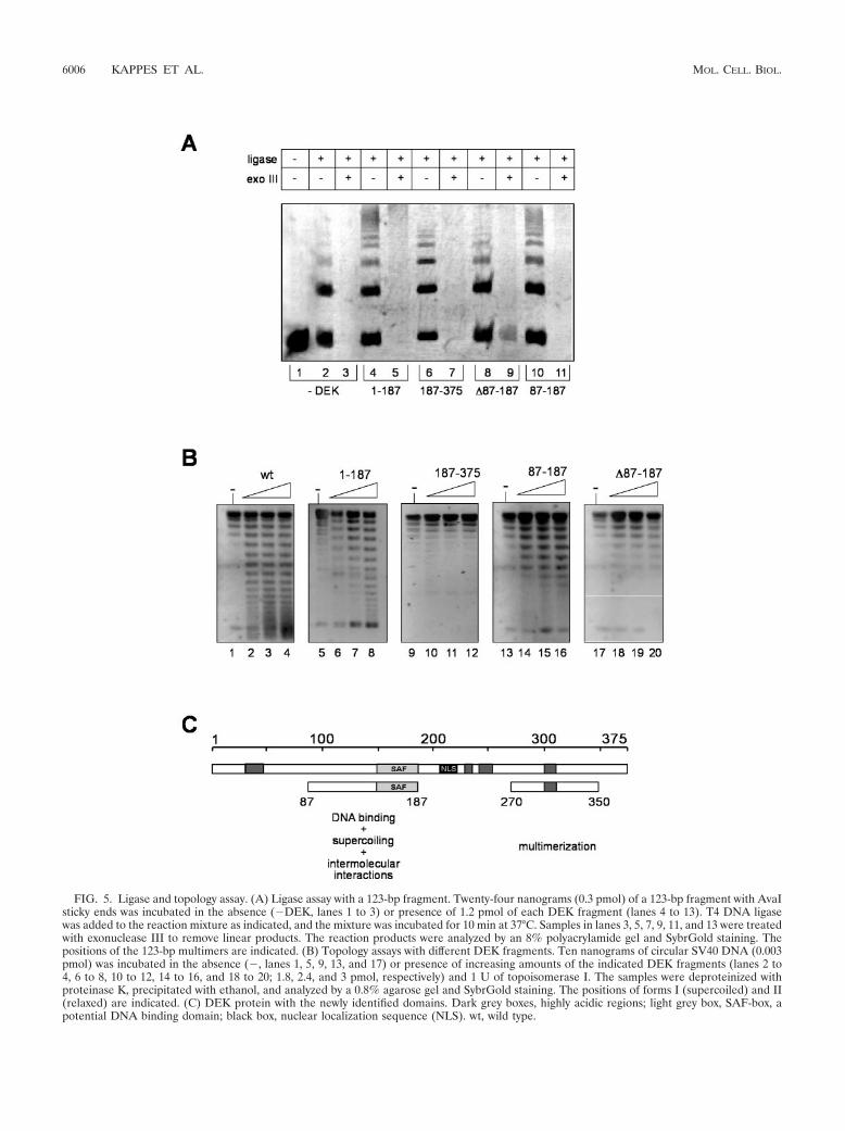
FIG. 5. Ligase and topology assay. (A) Ligase assay with a 123-bp fragment. Twenty-four nanograms (0.3 pmol) of a 123-bp fragment with AvaIsticky ends was incubated in the absence (�DEK, lanes 1 to 3) or presence of 1.2 pmol of each DEK fragment (lanes 4 to 13). T4 DNA ligasewas added to the reaction mixture as indicated, and the mixture was incubated for 10 min at 37°C. Samples in lanes 3, 5, 7, 9, 11, and 13 were treatedwith exonuclease III to remove linear products. The reaction products were analyzed by an 8% polyacrylamide gel and SybrGold staining. Thepositions of the 123-bp multimers are indicated. (B) Topology assays with different DEK fragments. Ten nanograms of circular SV40 DNA (0.003pmol) was incubated in the absence (�, lanes 1, 5, 9, 13, and 17) or presence of increasing amounts of the indicated DEK fragments (lanes 2 to4, 6 to 8, 10 to 12, 14 to 16, and 18 to 20; 1.8, 2.4, and 3 pmol, respectively) and 1 U of topoisomerase I. The samples were deproteinized withproteinase K, precipitated with ethanol, and analyzed by a 0.8% agarose gel and SybrGold staining. The positions of forms I (supercoiled) and II(relaxed) are indicated. (C) DEK protein with the newly identified domains. Dark grey boxes, highly acidic regions; light grey box, SAF-box, apotential DNA binding domain; black box, nuclear localization sequence (NLS). wt, wild type.
6006 KAPPES ET AL. MOL. CELL. BIOL.
motif that consists of two predicted amphipathic helices sepa-rated by a region that includes an invariant glycine. SAF-boxmodules are found throughout all eukaryotes (from yeast tohumans) but not in prokaryotes (18). Most of the SAF-boxproteins have additional functional modules that contribute totheir specific function in the cell. It is thought that the SAFmodule may target these proteins to the sites of active tran-scription and therefore link nuclear processes like transcrip-tion, pre-mRNA processing, and DNA repair by binding toSAR/MAR regions in the nucleus (3). DEK, however, has noknown additional module, which may suggest its participationin a specific nuclear process. In fact, no specific binding toSAR/MAR DNA has been detected.
However, DEK has some features in common with otherSAR/MAR-binding proteins. It binds preferentially to DNAcrossovers (31) like histone H1, and like SAF-A, DEK mul-timerizes upon DNA binding (Fig. 5A) (31) and is partiallylocalized at the nuclear matrix (Kappes, unpublished data). Inaddition, DEK has some properties in common with the knownnuclear architectural HMG proteins. Thus, DEK induces in-
tra- and intermolecular interactions (31, 32) and changes thetopology of circular DNA substrates (24, 27, 29). Taken to-gether, these data suggest a role for DEK in nuclear architec-ture. This is also thought to be one function of the SAF-A andSAF-B proteins, the founding members of the SAF-box familyof proteins (18, 22).
We were able to show here that the SAF-box-containingregion (amino acids 87 to 187) of DEK is sufficient to introducesupercoils and to induce intermolecular interactions betweentwo or more DNA molecules (Fig. 5). The ligase assay suggeststhat the SAF-box fragment induces intermolecular interactionof DEK-DNA complexes. This multimerization must be de-pendent on DNA binding, because it was not detectable by farWestern analysis in the absence of DNA (Fig. 2B).
These results may suggest a mechanism by which DEK in-troduces supercoils into closed circular DNA. DEK-DEK in-teraction upon DNA binding would change the twist of DNA,resulting in torsional stress of the DNA molecule. This tensionis then compensated by changing the writhing number and theintroduction of compensating supercoils. While topoisomerase
FIG. 6. EMSAs with C-terminal fragments of DEK. (A) One hundred seventy-five nanograms (0.05 pmol) of linearized SV40 DNA wasincubated with 9, 28, and 46 pmol of dephosphorylated DEK fragments (lanes 2 to 4, 5 to 7, 8 to 10, 11 to 13, and 14 to 16, respectively) or withoutprotein (input, lane 1) for 1 h at 37°C. Nucleoprotein complexes were analyzed on 0.6% agarose gels in 0.5� TBE (pH 7.8, see Materials andMethods) and visualized by ethidium bromide staining. The fragments used are indicated above the blots. (B) Map showing the identified DNAbinding domain of DEK. Dark grey boxes, highly acidic regions; light grey box, SAF-box, a potential DNA binding domain; black box, nuclearlocalization sequence (NLS). wt, wild type.
VOL. 24, 2004 FUNCTIONAL DOMAINS OF THE DEK PROTEIN 6007

FIG. 7. EMSAs with phosphorylated DEK fragments. (A) Nine, 28, and 46 pmol of the different DEK fragments were phosphorylated with CK2in vitro as described before. The samples were separated by SDS–10 to 18% polyacrylamide gel electrophoresis and analyzed by autoradiography(32P) and Western blotting with DEK-specific antibodies (�-DEK). The positions of the molecular weight markers are indicated. (B) One hundredseventy-five nanograms (0.05 pmol) of linearized SV40 DNA was incubated with 9, 28, and 46 pmol of phosphorylated DEK fragments (lanes 2to 4, 5 to 7, 8 to 10, 11 to 13, and 14 to 16, respectively) or without protein (input, lane 1) for 1 h at 37°C. The nucleoprotein complexes wereanalyzed on 0.6% agarose gels in 0.5� TBE (pH 7.8, see Materials and Methods) and visualized by ethidium bromide staining. (C) DEK proteinwith newly identified regions. Dark grey boxes, highly acidic regions; light grey box, SAF-box, a potential DNA binding domain; black box, nuclearlocalization sequence (NLS). wt, wild type.
6008 KAPPES ET AL. MOL. CELL. BIOL.

I is able to relax these compensating supercoils, the change intwist is constrained when DEK is bound (32). Such a mecha-nism has been discussed for 13S condensin (16, 17) and forbacterial HMf proteins (21), which both introduce positivesupercoils into circular DNA.
The region of amino acids 270 to 350 is responsible formultimerization and DNA binding. We have searched for pro-teins that interact with DEK. However, in several independentyeast two-hybrid screens, we identified only one interactionpartner, namely, DEK itself. Testing different fragments ofDEK against the full-length protein showed that the putativedi- or multimerization domain is located in the C-terminalregion of DEK, between amino acids 270 and 350 (Fig. 1B).These results were confirmed by far Western analysis (Fig. 3).Why were other interacting proteins not detected given thatDEK-binding partners such as histone H2A/H2B, AP-2�,SRm160, or Daxx and HDAC have been described in theliterature (2, 5, 13, 20)?
One possibility is that putative interaction partners are mis-folded or not properly posttranslationally modified in yeast. Itis also possible that the prey libraries do not contain sequencesencoding the respective proteins. Notably, the intrinsic restric-tion of the two-hybrid system for testing one-to-one interac-tions could make it difficult to detect larger protein complexessuch as the complex formed by Daxx, HDAC, histones, andDEK (13).
We showed by EMSA that there exists a second DNA bind-ing domain in the C-terminal part of DEK (Fig. 6A). We couldmap this second DNA binding domain to the region betweenamino acids 270 and 350 (Fig. 6A), which is also the region ofprotein-protein interaction (Fig. 1 and 2). The nucleoproteincomplexes formed by the second DNA binding domain mi-grated into the gel, suggesting that no large DNA-proteincomplexes are induced by this domain (Fig. 6A). Consistentwith this, the second DNA binding domain does not induceintermolecular interactions upon DNA binding (Fig. 5A, lanes8 and 10) and fails to create supercoils into circular DNA (Fig.5B, lanes 9 to 12 and 17 to 20).
Interestingly, the region of amino acids 270 to 350, includingthe multimerization domain and the second DNA binding do-main, also contains most of the mapped CK2 phosphorylationsites (15). Indeed, fragments containing the C-terminal regionof DEK are highly phosphorylated by CK2 in vitro (Fig. 7A,lanes 2, 5, 6, and 7). Furthermore, phosphorylation of aminoacids 270 to 350 disrupts the DNA binding of these fragments(Fig. 7B, lanes 5 to 16) but triggers DEK-DEK interaction(Fig. 3A). Therefore, the functional activity of this domainappears to be changed upon phosphorylation: it binds to DNAwhen dephosphorylated but dissociates and multimerizes uponphosphorylation. Furthermore, phosphorylation of the full-length protein seems to negatively regulate the DNA bindingactivity of the SAF-box (Fig. 7B, lanes 2 to 4; compare with Fig.4A, lanes 2 to 4). This could be due to conformational changesin the whole protein or to a release of the phosphorylatedsecond DNA binding domain (amino acids 270 to 350) fromthe DNA. DEK-DEK interactions could then weaken the bind-ing of the SAF-box domain (amino acids 87 to 187).
In summary, these data could provide an explanation for thediverse and sometimes conflicting data about DEK. Thus,DEK may be involved in nuclear architecture via the SAF-box
region (amino acids 87 to 187). In fact, just like SAF-A, DEKpreferentially binds to DNA crossovers (31) and induces inter-and intramolecular interactions (31, 32).
Published data suggest that DEK may be involved in tran-scriptional regulation of chromatin. This could be connected tothe fact that DEK is released from DNA upon phosphoryla-tion, thereby making target sequences accessible for transcrip-tional activators or repressors. This implies that DEK on chro-matin is dynamic. However, further experiments are needed tosupport this assumption.
ACKNOWLEDGMENTS
We are grateful to Rolf Knippers, Martina Baack, and Anita Krebsfor stimulating discussions and critical reading of the manuscript andLorenzo Pinna for providing CK2.
This work was supported by grants from DFG to C.G. I.S. wassupported by BIF (Boehringer Ingelheim Fonds).
REFERENCES
1. Adams, B. S., H. C. Cha, J. Cleary, T. Haiying, H. Wang, K. Sitwala, andD. M. Markovitz. 2003. DEK binding to class II MHC Y-box sequences isgene- and allele-specific. Arthritis Res. Ther. 5:R226–R233.
2. Alexiadis, V., T. Waldmann, J. Andersen, M. Mann, R. Knippers, and C.Gruss. 2000. The protein encoded by the proto-oncogene DEK changes thetopology of chromatin and reduces the efficiency of DNA replication in achromatin-specific manner. Genes Dev. 14:1308–1312.
3. Aravind, L., and E. V. Koonin. 2000. SAP—a putative DNA binding motifinvolved in chromosomal organization. Trends Biochem. Sci. 25:112–114.
4. Birg, F., M. Courcoul, O. Rosnet, F. Bardin, M. J. Pebusque, S. Marchetto,A. Tabilio, P. Mannoni, and D. Birnbaum. 1992. Expression of the FMS/KIT-like gene FLT3 in human acute leukemias of the myeloid and lymphoidlineages. Blood 80:2584–2593.
5. Campillos, M., M. A. Garcia, F. Valdivieso, and J. Vazquez. 2003. Transcrip-tional activation by AP-2� is modulated by the oncogene DEK. NucleicAcids Res. 31:1571–1575.
6. Dong, X., M. A. Michelis, J. Wang, R. Bose, T. DeLange, and W. H. Reeves.1998. Autoantibodies to DEK oncoprotein in a patient with systemic lupuserythematosus and sarcoidosis. Arthritis Rheum. 41:1505–1510.
7. Dong, X., J. Wang, F. N. Kabir, M. Shaw, A. M. Reed, L. Stein, L. E.Andrade, V. F. Trevisani, M. L. Miller, T. Fujii, M. Akizuki, L. M. Pachman,M. Satoh, and W. H. Reeves. 2000. Autoantibodies to DEK oncoprotein inhuman inflammatory disease. Arthritis Rheum. 43:85–93.
8. Faulkner, N. E., J. M. Hilfinger, and D. M. Markovitz. 2001. Protein phos-phatase 2A activates the HIV-2 promoter through enhancer elements thatinclude the pets site. J. Biol. Chem. 276:25804–25812.
9. Fornerod, M., J. Boer, S. van Baal, M. Jaegle, M. von Lindern, K. G. Murti,D. Davis, J. Bonten, A. Buijs, and G. Grosveld. 1995. Relocation of thecarboxy-terminal part of CAN from the nuclear envelope to the nucleus asa result of leukemia-specific chromosome rearrangements. Oncogene 10:1739–1748.
10. Fu, G. K., G. Grosveld, and D. M. Markovitz. 1997. DEK, an autoantigeninvolved in a chromosomal translocation in acute myelogenous leukemia,binds to the HIV-2 enhancer. Proc. Natl. Acad. Sci. USA 94:1811–1815.
11. Gohring, F., and F. O. Fackelmayer. 1997. The scaffold/matrix attachmentregion binding protein hnRNP-U (SAF-A) is directly bound to chromosomalDNA in vivo: a chemical cross-linking study. Biochemistry 36:8276–8283.
12. Golemis, E. A., et al. 1999. Current protocols in molecular biology. JohnWiley & Sons, Inc., New York, N.Y.
13. Hollenbach, A. D., C. J. McPherson, E. J. Mientjes, R. Iyengar, and G.Grosveld. 2002. Daxx and histone deacetylase II associate with chromatinthrough an interaction with core histones and the chromatin-associatedprotein Dek. J. Cell Sci. 115:3319–3330.
14. Kappes, F., K. Burger, M. Baack, F. O. Fackelmayer, and C. Gruss. 2001.Subcellular localization of the human proto-oncogene protein DEK. J. Biol.Chem. 276:26317–26323.
15. Kappes, F., C. Damoc, R. Knippers, M. Przybylski, L. A. Pinna, and C.Gruss. 2004. Phosphorylation by protein kinase CK2 changes the DNAbinding properties of the human chromatin protein DEK. Mol. Cell. Biol.24:6011–6020.
16. Kimura, K., and T. Hirano. 1997. ATP-dependent positive supercoiling ofDNA by 13S condensin: a biochemical implication for chromosome conden-sation. Cell 90:625–634.
17. Kimura, K., V. V. Rybenkov, N. J. Crisona, T. Hirano, and N. R. Cozzarelli.1999. 13S condensin actively reconfigures DNA by introducing global posi-tive writhe: implications for chromosome condensation. Cell 98:239–248.
18. Kipp, M., F. Gohring, T. Ostendorp, C. M. van Drunen, R. van Driel, M.
VOL. 24, 2004 FUNCTIONAL DOMAINS OF THE DEK PROTEIN 6009

Przybylski, and F. O. Fackelmayer. 2000. SAF-box, a conserved proteindomain that specifically recognizes scaffold attachment region DNA. Mol.Cell. Biol. 20:7480–7489.
19. Libura, M., V. Asnafi, A. Tu, E. Delabesse, I. Tigaud, F. Cymbalista, A.Bennaceur-Griscelli, P. Villarese, G. Solbu, A. Hagemeijer, K. Beldjord, O.Hermine, and E. Macintyre. 2003. FLT3 and MLL intragenic abnormalitiesin AML reflect a common category of genotoxic stress. Blood 102:2198–2204.
20. McGarvey, T., E. Rosonina, S. McCracken, Q. Li, R. Arnaout, E. Mientjes,J. A. Nickerson, D. Awrey, J. Greenblatt, G. Grosveld, and B. J. Blencowe.2000. The acute myeloid leukemia-associated protein, DEK, forms a splic-ing-dependent interaction with exon-product complexes. J. Cell Biol. 150:309–320.
21. Musgrave, D. R., K. M. Sandman, and J. N. Reeve. 1991. DNA binding bythe archaeal histone HMf results in positive supercoiling. Proc. Natl. Acad.Sci. USA 88:10397–10401.
22. Romig, H., F. O. Fackelmayer, A. Renz, U. Ramsperger, and A. Richter.1992. Characterization of SAF-A, a novel nuclear DNA binding protein fromHeLa cells with high affinity for nuclear matrix/scaffold attachment DNAelements. EMBO J. 11:3431–3440.
23. Rosnet, O., M. G. Mattei, S. Marchetto, and D. Birnbaum. 1991. Isolationand chromosomal localization of a novel FMS-like tyrosine kinase gene.Genomics 9:380–385.
24. Sheflin, L. G., and S. W. Spaulding. 1989. High mobility group protein 1preferentially conserves torsion in negatively supercoiled DNA. Biochemis-try 28:5658–5664.
25. Sierakowska, H., K. R. Williams, I. S. Szer, and W. Szer. 1993. The putativeoncoprotein DEK, part of a chimera protein associated with acute myeloid
leukaemia, is an autoantigen in juvenile rheumatoid arthritis. Clin. Exp.Immunol. 94:435–439. [Erratum, 96:177, 1994.]
26. Sitwala, K. V., K. Adams, and D. M. Markovitz. 2002. YY1 and NF-Ybinding sites regulate the transcriptional activity of the dek and dek-canpromoter. Oncogene 21:8862–8870.
27. Stros, M., J. Stokrova, and J. O. Thomas. 1994. DNA looping by the HMG-box domains of HMG1 and modulation of DNA binding by the acidicC-terminal domain. Nucleic Acids Res. 22:1044–1051.
28. Thiede, C., C. Steudel, B. Mohr, M. Schaich, U. Schakel, U. Platzbecker, M.Wermke, M. Bornhauser, M. Ritter, A. Neubauer, G. Ehninger, and T.Illmer. 2002. Analysis of FLT3-activating mutations in 979 patients withacute myelogenous leukemia: association with FAB subtypes and identifica-tion of subgroups with poor prognosis. Blood 99:4326–4335.
29. Thomas, J. O. 2001. HMG1 and 2: architectural DNA binding proteins.Biochem. Soc. Trans. 29:395–401.
30. von Lindern, M., M. Fornerod, S. van Baal, M. Jaegle, T. de Wit, A. Buijs,and G. Grosveld. 1992. The translocation (6;9), associated with a specificsubtype of acute myeloid leukemia, results in the fusion of two genes, dekand can, and the expression of a chimeric, leukemia-specific dek-can mRNA.Mol. Cell. Biol. 12:1687–1697.
31. Waldmann, T., M. Baack, N. Richter, and C. Gruss. 2003. Structure-specificbinding of the proto-oncogene protein DEK to DNA. Nucleic Acids Res.31:7003–7010.
32. Waldmann, T., C. Eckerich, M. Baack, and C. Gruss. 2002. The ubiquitouschromatin protein DEK alters the structure of DNA by introducing positivesupercoils. J. Biol. Chem. 277:24988–24994.
33. Wichmann, I., N. Respaldiza, J. R. Garcia-Lozano, M. Montes, J. Sanchez-Roman, and A. Nunez-Roldan. 2000. Autoantibodies to DEK oncoprotein insystemic lupus erythematosus (SLE). Clin. Exp. Immunol. 119:530–532.
6010 KAPPES ET AL. MOL. CELL. BIOL.
Copyright © 2022 FDOKUMEN