Natural Dietary Pigments: Potential Mediators against Hepatic ...
Upload
khangminh22Category
view
1download
0
1Scientific RepoRts | 6:34320 | DOI: 10.1038/srep34320
www.nature.com/scientificreports
Sequestration of ubiquitous dietary derived pigments enables mitochondrial light sensingDan Zhang, Kiera Robinson, Doina M. Mihai & Ilyas Washington
Animals alter their physiological states in response to their environment. We show that the introduction of a chlorophyll metabolite, a light-absorbing pigment widely consumed in human diets, to Caenorhabditis elegans results in animals whose fat mass can be modulated by exposure to light, despite the worm consuming the same amount of food. In the presence of the chlorophyll metabolite, exposing the worms to light increased adenosine triphosphate, reduced oxidative damage, and increased median life spans, without an effect on animal reproduction. Mice fed a dietary metabolite of chlorophyll and exposed to light, over several months, showed reductions in systemic inflammation as measured by plasma α-macroglobulin. We propose that dietary chlorophyll metabolites can enable mitochondria to use light as an environmental cue, by absorbing light and transferring the energy to mitochondrial coenzyme Q.
Interactions with environmental factors such as light, gravity, the earth’s magnetic field, food, microbes, and the atmosphere are, for all practical purposes, inescapable. One's genetic makeup, expression patterns of genes, com-position of molecules, biochemistry, physiology and morphology can all be viewed as culminations of adaptations to the environment. Evolutionarily conserved environmental cues can be defined as environmental factors that humans have become to rely upon for normal function. If so, then the identification of such cues and the deter-mination of how the frequency of their exposure can affect physiology can be expected to be important towards maintaining human health, wellbeing, and disease prevention. Nutrition and electromagnetic radiation from the sun are conserved environmental cues that affect every living organism. In this work, we asked how these two cues might interact inside the body to perturb physiology.
Respiration, in animal cells, is enabled by transmembrane proteins linked by diffusible coenzyme Q10 (CoQ10) molecules in the mitochondrial inner membrane space. We proposed that animal mitochondria could seques-ter certain metabolites of dietary chlorophyll, where once inside, the metabolites could absorb long wavelength light (LWL) and transfer the absorbed energy to CoQ10, resulting in the photoreduction of CoQ10
1. In the pro-posed scenario, dietary metabolites of chlorophyll would play a similar role, in animals, as chlorophyll plays in the chloroplatst of plants: where photon absorption by chlorophyll results in the photoreduction of plastoqui-none, the plant equivalent to CoQ10. The potential photoreduction of CoQ10 mediated by dietary pigments is of interest because mitochondria play a central role in coordinating physiology with environmental demands2–4, and the oxidation state of CoQ10 provides a known molecular basis for signal origin in cell signaling5. Thus, the photoreduction of CoQ10 by an accessory pigment would offer a way to potentially modulate physiology in response to environmental light. For example, by sequestration of the chlorophyll metabolite pheophorbide-a (PA), Caenorhabditis elegans are able to absorb LWL, resulting in increased ATP concentrations and median life spans1. Here, we investigated additional changes induced by LWL in the presence of PA. We were interested in the extent to which wavelengths of light and dietary pigments, which are both present in the body, but have largely been regarded as benign, might affect the functioning of an organism. This work has implications as to how the frequency of exposure to light and chlorophyll plant pigments may enable normal function.
ResultsLight and a dietary pigment modulates CoQ10 ratios and adenine nucleotides in animal mitochondria. To show that metabolites of dietary chlorophyll, or PA, could catalyze the photoreduction of CoQ10, we incorporated CoQ10 into liposomes with less than stoichiometric amount of PA, in the presence of vita-min C, used as a hydrogen donor. Upon exposure of the solution to light centered at λ max = 660 nm to simulate
Columbia University Medical Center, Ophthalmology, New York, NY 10032, USA. Correspondence and requests for materials should be addressed to I.W. (email: [email protected])
received: 16 May 2016
Accepted: 09 September 2016
Published: 12 October 2016
OPEN

www.nature.com/scientificreports/
2Scientific RepoRts | 6:34320 | DOI: 10.1038/srep34320
the long wavelength portion of environmental lighting, ubiquinone (the oxidized form of CoQ10) was increasingly reduced to ubiquinol (the reduced form of CoQ10) with increasing light dose (Fig. 1A,B). In the absence of vita-min C, PA, or light, no reduction occurred. A few hundred times more molecules of ubiquinone were reduced than molecules of added PA, suggesting that PA acted catalytically. Similarly, when PA-treated isolated mitochon-dria were exposed to LWL, 86% of mitochondrial CoQ10 was reduced to ubiquinol, consistent with the results in liposomes (Fig. 1C). Before LWL exposure, nearly all the CoQ10 was in its oxidized form. When the light was turned off, the generated ubiquinol was oxidized to ubiquinone. LWL had no effect on the ubiquinone:ubiquinol ratios in the absence of PA.
After demonstrating that photon absorption by PA could photoreduce ubiquinone, we sought to determine the extent to which the generated ubiquinol were in a position to donate electrons to complex III, the next step in electron transport. We measured complex III activity by its ability to reduce cytochrome c in the presence of rotenone, to limit the reduction of CoQ10 by complex I, and by potassium cyanide to limit the oxidation of cytochrome c by complex IV. In the presence of PA, within 15 minutes of LWL exposure, 45% of cytochrome c was reduced (Fig. 1D). However, when kept in the dark or in the absence of added PA or mitochondria, signifi-cantly less cytochrome c was reduced. This suggested that the photoreduced CoQ10 was available to participate in electron transport.
To show that ubiquinone photoreduction and subsequent cytochrome c reduction could be coupled with ATP synthesis, we measured the concentrations of adenosine nucleotides in the same mitochondria. Quantification of ATP by the luciferase assay revealed that ATP concentrations increased with light exposure in PA -treated mitochondria but not in PA naïve mitochondria (Fig. 1E). The number of photons put into 50 μ L of the reaction mixture was approximately 52 pmol per second (the light intensity was 4 μ mol s−1 m−2, and 50 μ L is 0.000013 m2). The total rate of ATP synthesis in 50 μ L was 0.0037 pmol per second (total protein in 50 μ L was 81 ± 6 μ g). Quantification of adenosine nucleotides by Ultra Performance Liquid Chromatography (UPLC) was consistent
Figure 1. Photoreduction of coenzyme Q (CoQ10) activates complex III, which reduces cytochrome c, resulting in generation of ATP, in animal mitochondria. (A) Schematic of pheophorbide-a (PA) catalyzed photoreduction of coenzyme Q (also known as ubiquinone/ubiquinol). (B) Photoreduction of ubiquinone in liposomes catalyzed by PA. The x-axis represents the amount of time the liposome mixture was exposed to red light. (C) Photoreduction of ubiquinone in isolated, intact heart mitochondria. Mitochondria were either treated with PA (+ ) or not treated with PA (− ). When exposed to light, 86% of CoQ10 was reduced to ubiquinol in PA treated mitochondria (red line) but not for control mitochondria (blue line). When the light was turned off, ubiquinol was reoxidized. (D) Photoreduction of ubiquinone activates complex III resulting in the reduction of cytochrome C. Reaction conditions noted on the x-axis. Values represent the amount of cytochrome c reduced upon exposure to light minus the amount of cytochrome c reduced when the same samples were kept in the dark. Averages of 8 replicates and standard deviations are shown. CytC: cytochrome c; Mito: mitochondria; *P-value < 0.05 determined by unpaired Student’s t-test. (E) Storage of photonic energy by animal mitochondria. ATP synthesis in the PA treated (+ ) and control (− ) mitochondria exposed to red light as measured by the luciferase assay. ATP synthesis was coupled to CoQ10 photoreduction. (F) ATP concentrations and ATP/AMP and ATP/ADP ratios, as measured by UPLC in the light exposed, PA treated heart mitochondria (+ , red lines), were consistent with ATP synthesis from ADP and AMP pools. PA-naïve, light exposed mitochondria (− , blue lines) showed no change in adenosine nucleotides.

www.nature.com/scientificreports/
3Scientific RepoRts | 6:34320 | DOI: 10.1038/srep34320
with that of the firefly luciferase assay (Fig. 1F). As ATP increased, ATP:ADP and ATP:AMP ratios increased, consistent with an increase in ATP from ADP and AMP pools. In contrast, in control PA naïve mitochondria, no changes in ATP, ADP, and AMP were observed upon light exposure, further confirming measurements by the luciferase assay. The concentrations of adenosine nucleotides remained unchanged when the PA treated mito-chondria were kept in the dark. The above data suggested that the isolated mitochondria were able to convert approximately 0.01% of incident photons into stored energy as ATP (i.e. 14,000 photons of incident light could produce one molecule of ATP).
Light and a dietary pigment modulate metabolism in an animal model. C. elegans provide a con-venient animal model to study energy homeostasis. A number of pathways central to regulation of mammalian metabolism appear to have similar function in C. elegans6. In the wild, C. elegans coexist with rich microbial communities7 which dictate almost every aspect of C. elegans biology8. We therefore conducted experiments with worms raised on a wild culture of bacteria/fungi (Supplementary Fig. 1A). Upon co-incubation, PA fluorescence was seen by fluorescence microscopy as a diffuse staining throughout the animal (Supplementary Fig. 1B,C) indicating that C. elegans readily incorporated PA. Subsequent exposure to LWL increased ATP concentrations by 75% in PA treated animals but not in PA naïve animals or in animals treated with fluoxetine (Fig. 2A), a drug that leads to alterations in C. elegans fat stores9. This indicated that LWL, in the presence of PA, could alter energy homeostasis in the worm in vivo, as well as in isolated mitochondria.
Figure 2. Light and PA regulates leanness and fat mass in C. elegans. *P-value < 0.05 determined by unpaired, two sided, Student’s t-test. Replicate measures are shown with averages and 95% confidence intervals. (A) ATP quantification in animals kept in the dark or exposed to LWL in the presence or absence of PA and/or the drug fluoxetine. (B) Oil Red-O staining of fat mass. From hatching, animals were raised on wild bacteria/fungi and exposed to light centered at λ max = 660 nm, to simulate the long wavelength portion environmental lighting, for 12 hours followed by 12 hours of darkness (cylic light). Representative images taken at 400 x magnification. (C) Quantification of Oil Red-O staining at 2 days of age. (D) Food consumption in light and PA treated animals compared to control cohorts. (E) Representative images of a 2-day old worm raised on wild bacteria/fungi under cyclic light in the presence of PA (lean worm) compared to a PA naïve animal raised under cyclic light. (F) Average body thickness measured at terminal bulb. (G) Average body thickness measured at terminal bulb. Comparison of leanness triggered by light/PA with that by fluoxetine after 2 days of treatment. (H) Quantification of Oil Red-O staining in worms treated with light/PA or fluoxetine for 2-days. (I) Average body thickness measured at terminal bulb in the presence of complex III blocker antimycin.
www.nature.com/scientificreports/
4Scientific RepoRts | 6:34320 | DOI: 10.1038/srep34320
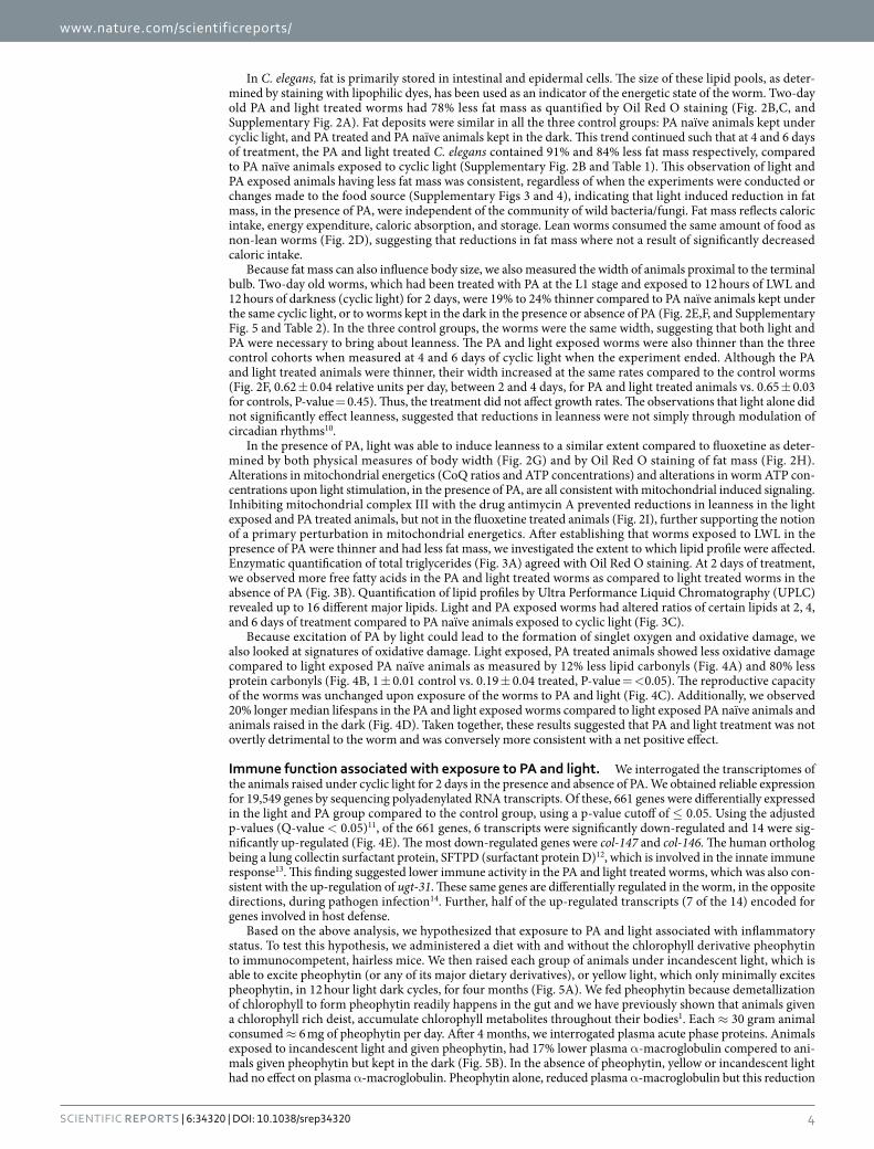
In C. elegans, fat is primarily stored in intestinal and epidermal cells. The size of these lipid pools, as deter-mined by staining with lipophilic dyes, has been used as an indicator of the energetic state of the worm. Two-day old PA and light treated worms had 78% less fat mass as quantified by Oil Red O staining (Fig. 2B,C, and Supplementary Fig. 2A). Fat deposits were similar in all the three control groups: PA naïve animals kept under cyclic light, and PA treated and PA naïve animals kept in the dark. This trend continued such that at 4 and 6 days of treatment, the PA and light treated C. elegans contained 91% and 84% less fat mass respectively, compared to PA naïve animals exposed to cyclic light (Supplementary Fig. 2B and Table 1). This observation of light and PA exposed animals having less fat mass was consistent, regardless of when the experiments were conducted or changes made to the food source (Supplementary Figs 3 and 4), indicating that light induced reduction in fat mass, in the presence of PA, were independent of the community of wild bacteria/fungi. Fat mass reflects caloric intake, energy expenditure, caloric absorption, and storage. Lean worms consumed the same amount of food as non-lean worms (Fig. 2D), suggesting that reductions in fat mass where not a result of significantly decreased caloric intake.
Because fat mass can also influence body size, we also measured the width of animals proximal to the terminal bulb. Two-day old worms, which had been treated with PA at the L1 stage and exposed to 12 hours of LWL and 12 hours of darkness (cyclic light) for 2 days, were 19% to 24% thinner compared to PA naïve animals kept under the same cyclic light, or to worms kept in the dark in the presence or absence of PA (Fig. 2E,F, and Supplementary Fig. 5 and Table 2). In the three control groups, the worms were the same width, suggesting that both light and PA were necessary to bring about leanness. The PA and light exposed worms were also thinner than the three control cohorts when measured at 4 and 6 days of cyclic light when the experiment ended. Although the PA and light treated animals were thinner, their width increased at the same rates compared to the control worms (Fig. 2F, 0.62 ± 0.04 relative units per day, between 2 and 4 days, for PA and light treated animals vs. 0.65 ± 0.03 for controls, P-value = 0.45). Thus, the treatment did not affect growth rates. The observations that light alone did not significantly effect leanness, suggested that reductions in leanness were not simply through modulation of circadian rhythms10.
In the presence of PA, light was able to induce leanness to a similar extent compared to fluoxetine as deter-mined by both physical measures of body width (Fig. 2G) and by Oil Red O staining of fat mass (Fig. 2H). Alterations in mitochondrial energetics (CoQ ratios and ATP concentrations) and alterations in worm ATP con-centrations upon light stimulation, in the presence of PA, are all consistent with mitochondrial induced signaling. Inhibiting mitochondrial complex III with the drug antimycin A prevented reductions in leanness in the light exposed and PA treated animals, but not in the fluoxetine treated animals (Fig. 2I), further supporting the notion of a primary perturbation in mitochondrial energetics. After establishing that worms exposed to LWL in the presence of PA were thinner and had less fat mass, we investigated the extent to which lipid profile were affected. Enzymatic quantification of total triglycerides (Fig. 3A) agreed with Oil Red O staining. At 2 days of treatment, we observed more free fatty acids in the PA and light treated worms as compared to light treated worms in the absence of PA (Fig. 3B). Quantification of lipid profiles by Ultra Performance Liquid Chromatography (UPLC) revealed up to 16 different major lipids. Light and PA exposed worms had altered ratios of certain lipids at 2, 4, and 6 days of treatment compared to PA naïve animals exposed to cyclic light (Fig. 3C).
Because excitation of PA by light could lead to the formation of singlet oxygen and oxidative damage, we also looked at signatures of oxidative damage. Light exposed, PA treated animals showed less oxidative damage compared to light exposed PA naïve animals as measured by 12% less lipid carbonyls (Fig. 4A) and 80% less protein carbonyls (Fig. 4B, 1 ± 0.01 control vs. 0.19 ± 0.04 treated, P-value = < 0.05). The reproductive capacity of the worms was unchanged upon exposure of the worms to PA and light (Fig. 4C). Additionally, we observed 20% longer median lifespans in the PA and light exposed worms compared to light exposed PA naïve animals and animals raised in the dark (Fig. 4D). Taken together, these results suggested that PA and light treatment was not overtly detrimental to the worm and was conversely more consistent with a net positive effect.
Immune function associated with exposure to PA and light. We interrogated the transcriptomes of the animals raised under cyclic light for 2 days in the presence and absence of PA. We obtained reliable expression for 19,549 genes by sequencing polyadenylated RNA transcripts. Of these, 661 genes were differentially expressed in the light and PA group compared to the control group, using a p-value cutoff of ≤ 0.05. Using the adjusted p-values (Q-value < 0.05)11, of the 661 genes, 6 transcripts were significantly down-regulated and 14 were sig-nificantly up-regulated (Fig. 4E). The most down-regulated genes were col-147 and col-146. The human ortholog being a lung collectin surfactant protein, SFTPD (surfactant protein D)12, which is involved in the innate immune response13. This finding suggested lower immune activity in the PA and light treated worms, which was also con-sistent with the up-regulation of ugt-31. These same genes are differentially regulated in the worm, in the opposite directions, during pathogen infection14. Further, half of the up-regulated transcripts (7 of the 14) encoded for genes involved in host defense.
Based on the above analysis, we hypothesized that exposure to PA and light associated with inflammatory status. To test this hypothesis, we administered a diet with and without the chlorophyll derivative pheophytin to immunocompetent, hairless mice. We then raised each group of animals under incandescent light, which is able to excite pheophytin (or any of its major dietary derivatives), or yellow light, which only minimally excites pheophytin, in 12 hour light dark cycles, for four months (Fig. 5A). We fed pheophytin because demetallization of chlorophyll to form pheophytin readily happens in the gut and we have previously shown that animals given a chlorophyll rich deist, accumulate chlorophyll metabolites throughout their bodies1. Each ≈ 30 gram animal consumed ≈ 6 mg of pheophytin per day. After 4 months, we interrogated plasma acute phase proteins. Animals exposed to incandescent light and given pheophytin, had 17% lower plasma α -macroglobulin compered to ani-mals given pheophytin but kept in the dark (Fig. 5B). In the absence of pheophytin, yellow or incandescent light had no effect on plasma α -macroglobulin. Pheophytin alone, reduced plasma α -macroglobulin but this reduction

www.nature.com/scientificreports/
5Scientific RepoRts | 6:34320 | DOI: 10.1038/srep34320
was more pronounced in animals exposed to incandescent light. A 17% reduction in α -macroglobulin is similar, in magnitude, to the increase in plasma α -macroglobulin in mice injected with zymosan or LPS, known stimu-lants of the innate immune system15. We did not obverse significant changes in plasma, adipsin, C-reactive pro-tein, haptoglobin, amyloid P, and α -1 acid glycoprotein amongst the 4 groups of animals (Fig. S6).
DiscussionLWL, of approximately 660 nm, penetrates most biological tissues, relatively unabsorbed. Here we propose a potential mechanism by which this photonic energy might be utilized by the sequestration of dietary pigments, which are able to absorb this energy. The measured 14,000 photons to generate one molecule of ATP was esti-mated using total incident flux (as opposed to the fraction of light absorbed), and is presumably on the low end taking into account changes in mitochondria architecture during isolation and handling. To gain insight into the extent to which the absorption of this additional energy, by an exogenous chromosphere, might affect an animal, we looked at lipid regulation in C. elegans because lipids act as energy reserves. We were able to show that the absorbed photonic energy resulted in changes in the worm’s lipid deposits, the concentrations of total triglyc-erides and free fatty acids, lipid ratios, and worm thickness. We observed sustained metabolic perturbations in lipids up to 6 days of age. This seems rare for this model where reductions in fat mass are mostly reported up to around 2 to 3 days of age9,16–23. The observed light induced reductions in fat mass16–20 and alterations of lipid profiles21,22 were greater or about the same in magnitude as reported for changes in response to genetic engi-neering or through exogenous control of metabolism. For example, out of a screen of 3,200 chemicals, 5 (0.16%) lowered fat mass more than two-fold relative to control-treated animals9. However, in contrast to foreign chem-icals, here we are able to show control of metabolism resulting from exposure to environmental cues that have a normal presence inside the body. This opens the possibility that similar chemistry may be taking place in animals and may constitute normal function. Differences in ATP, transcriptomes, a decrease in oxidative damage, and increased in lifespans, all suggest that the additional absorbed energy can impact animal physiology.
In the majority of metabolic studies, C. elegans were raised on what can be considered purified diets or in simple environments: using E. coli OP50 as a sole food source and controlling or eliminating bacterial and fungal growth. However, animals raised on purified diets are generally more susceptible to external perturbations24,25 and C. elegans are naturally colonizers of nutrient- and bacteria-rich substrates7 and their body size, fat stores, lipid composition, behavior, and life-spans are all dependent on the bacteria diet8. Here, we observed changes in
Figure 3. Light and PA regulates lipid composition in C. elegans. (A) Triglyceride quantification in light exposed animals in the presence or absence of PA at 2 days of age. Replicate measures are shown with averages and 95% confidence intervals. *P-value < 0.05 determined by unpaired, two sided, Student’s t-test. (B) Total free fatty acid quantification in worms described above. (C) UPLC profiles illustrating the relative amounts of fatty acids in light exposed animals in the presence or absence of PA at 2, 4, and 6 days of age. Free fatty acids represent the free fatty acids in the worm, while the total fatty acid profile represents free fatty acids obtained after saponification of a worm extract. Each peak represents a different free fatty acids. Peaks eluting later in time represent longer fatty acid chain lengths or more hydrophobic lipids. Numbers above the peaks represent the percent change in peak area (i.e. fatty acid amount) between the light exposed PA treated and PA naïve animals. Arrows point to peaks with the same area, to which curves where normalized.

www.nature.com/scientificreports/
6Scientific RepoRts | 6:34320 | DOI: 10.1038/srep34320
energetics for C. elegans raised in a complex environment rich in bacteria/fungi, suggesting that these invoked changes were not restricted to C. elegans cultured on strict artificial diets/environments and the invoked changes were larger than diet induced variability.
Upon excitation by LWL, PA can either lose their excitation energy as heat, or transfer the excitation energy to a neighboring molecule. Transfer of excitation energy to oxygen can result in the formation of singlet oxy-gen (a reactive oxygen species), which could have potentially played a role in the observed phenotypes through increased oxidative stress. However, measurements of oxidative damage suggested that light, in the presence of PA, did not obviously increase oxidative stress. To the contrary, we observed a decrease in lipid and protein car-bonyls in the light and PA treated animals. We also did not observe any obvious deleterious effects of light and PA on ATP levels, feeding, growth, reproduction, or life spans. Taken together, these results suggest that the observed phenotypes were not the result of general sickness, altered feeding behavior, or oxidative damage, although the possibility of stress response mechanisms cannot be ruled out at this time.
A fundamental reaction of chlorins is the transfer of excitation energy to quinones. This energy transfer pro-vides the initial step in the transformation of photonic energy to chemical energy in photosynthesis and thus sets the stage for life on earth. It is thus tempting to speculate on the existence of a similar process in animal cells. Data in liposomes and isolated mitochondria demonstrate that mitochondrial CoQ10 can be a primary recipient of PA's excitation energy leading to the reduction of CoQ10. CoQ10 photoreduction is also consistent with our observed increases in complex III activity in isolated mitochondria and subsequent increase in ATP concentrations in both isolated mitochondria and C. elegans. Taken together, we suggest that, in the worm, light excites PA and this excitation energy is transferred to CoQ10. This transfer of excitation energy from PA to CoQ10 could serve as the origin of a signaling cascade that results in reductions in fat mass (Fig. 6). This hypothesis was supported by experiments in which partially blocking complex III, which is upstream of CoQ10, with low dose antimycin
Figure 4. Light and PA did not show overt toxicity. (A) Quantification of lipid peroxides in animals raised on wild bacteria/fungi under cyclic light in the presence or absence of PA for 2 days. Replicate measures are shown with averages and 95% confidence intervals. *P-value < 0.05 determined by unpaired, two sided, Student’s t-test. (B) Quantification of protein carbonyls in the worms described in panel A. (C) Number of offspring (eggs and worms) per adult worm at 4 and 5-days of treatment. (D) Medium life spans. (E) Fold change in RNA transcripts as determined by RNA-sequencing in light exposed, 2-day old animals in the presence PA relative to light exposed animals in the absence of PA. Significance was defined by a Q-value < 0.05.

www.nature.com/scientificreports/
7Scientific RepoRts | 6:34320 | DOI: 10.1038/srep34320
A inhibited fat mass reduction upon light and PA exposure, but not upon Fluoxetine exposure. It is also in-line with the notions that the redox state of CoQ10 regulates mitochondrial uncoupling26, that there is less reduced CoQ10 in proportion to oxidized CoQ10 in adipose from obese animals27 and people28, and links between the CoQ10-cytochrome c reductase core protein and obesity29. CoQ10 photoreduction could provide a chemical entry point by which the long wavelength component of light and the chlorophyll component of chlorophyll-rich food may interact in the body to impact normal function.
In C. elegans, we used alterations in lipid as an indicator to gauge the potential significance of the absorption of additional photonic energy through the use of accessory, dietary derived pigments. The relationship between the observed phenotypes (reduced fat mass, altered lipid profiles, reduced oxidative stress, altered transcriptomes, increased ATP levels, and increased life span) may be coincidental. Further, the worm phenotypes may or may not present in higher order animals. The value of the worm is that it can be used as a simple model to suggest a likelihood that light and dietary derived pigments contribute to normal physiology. Cell types, in higher order animals, susceptible to similar perturbations would presumably be those exposed to chlorins and LWL. It has been estimated that, in humans, approximately 5% of dietary chlorophyll is absorbed30. Dietary derived chlo-rophyll pigments can be found in the liver, bile, and lining of the intestines31–34. We have shown that chlorins may also accumulate in adipose1. There are, however, additional routes of chlorin exposure including oral sup-plements and/or injections. Adipose tissue is bathed in LWL35–37. Light between 600 to 700 nm can penetrate a 1.5-inch-thick abdominal wall with three-to-five orders of magnitude of attenuation38,39. The amount of LWL in
Figure 5. Exposure to light and pheophytin associated with a reduction in inflammation. (A) SKH1-Elite, hairless mice, pictured, were fed a chlorophyll free diet. For two groups of mice, we added pheophytin to the diet; the absorption spectrum of which is shown. Animals were then raised either under yellow light, which minimally excites pheophytin, or incandescent light, which contains red wavelengths, that can excite pheophytin. Approximate absorption bands of the lights used are shown. (B) Plasma α -2 macroglobulin levels in the above described, 4 groups of mice (n = 5 to 7), after 4 months of treatment. Averages and 95% confidence intervals are shown. *P-value < 0.05, determined by unpaired, two sided, Student’s t-test.
Figure 6. Mitochondrial CoQ10 photoreduction as potential mechanism to sense light. Almost all of the cell’s redox reactions culminate to reduce coenzyme Q (CoQ10), shown in the schematic in its three oxidations states. I and II (Complexes I and II): transfer electrons from NADH and FADH2, respectively, to CoQ10 mGPDH (Mitochondrial glycerol-3-phosphate dehydrogenase): oxidizes glycerol-3-phosphate to dihydroxyacetone phosphate with concurrent reduction of flavin adenine dinucleotide (FAD) to FADH2 and transfers electrons to CoQ10 DHODH (Dihydroorotate Dehydrogenase): catalyzes the oxidation of dihydroorotate to orotate for de novo pyrimidine biosynthesis, using CoQ10 as an electron acceptor. ChDH (Choline dehydrogenase): oxidizes choline to glycine-betaine with CoQ10 serving as the primary electron acceptor. EFT (Electron-transferring-flavoprotein dehydrogenase): links the oxidation of fatty acids and some amino acids to oxidative phosphorylation by the oxidation of electron-transferring-flavoprotein using CoQ10 as an electron acceptor. The oxidation state of the mitochondrial CoQ10 pool can be used as a means of signal origin. We propose that dietary chlorophyll metabolites (green circle), such as PA, can enter the mitochondria, where they can absorb long wavelength light, resulting in the photoreduction of CoQ10, which could then propagate the input photonic energy via several potential pathways (wavy lines).

www.nature.com/scientificreports/
8Scientific RepoRts | 6:34320 | DOI: 10.1038/srep34320
the body can also be tuned by various means ranging from bathing under sun or red light, both activities cur-rently practiced, to wearing light-emitting diodes. There are several potential routes by which the body may be exposed to both chlorins and light to potentially photoreduce CoQ10.
In mice, plasma α -macroglobulin is a major acute phase protein and is increased during inflammation15. The magnitude of reduction of α -macroglobulin was significant given that known activators of the innate immune system trigger a similar increase in α -macroglobulin15. The reduction in α -macroglobulin upon exposure to light and pheophytin was further significant considering we used outbread mice, in which each animal had a degree of genetic variability. Our data suggest that acute phase proteins may serve as indicators of exposure to LWL and dietary chlorophyll, and that α -macroglobulin, in particular, may be useful for quantifying this exposure in mice. As the major acute phase proteins in humans are different than those in mice, other acute phase proteins may correlate with exposure to LWL and dietary chlorophyll in humans. The phenotypic significance of a reduction in α -macroglobulin in the LWL and pheophytin treated animals, and the extent to which the relationship between the treatment and α -macroglobulin is causal or casual, remains to be elucidated.
The state of an organism is largely dictated by its environment. Elucidating these environmental determinants and how they affect function are important for establishing lifestyle recommendations to prolong human health and wellbeing. Evolutionary conserved environmental pressures – those that the body has become dependent on - provide rational starting points of inquiry. The above data suggest that, like in plants, the longer wavelength component of visible light and the chlorophyll component of chlorophyll-rich foods may act together to affect animal function, as well.
MethodsPhotoreduction of Coenzyme Q10 in Liposome Mimics. Pheophorbide-a (PA) [Frontier Scientific, Catalog number: Pha-592, Logan, UT] (≈ 0.20 mg) and coenzyme Q [Sigma-Aldrich, Catalog number: C9538, St. Louis, MO] (50 mg) were dissolved in 1 mL of ethanol in a round bottom flask. L-α -Phosphatidylcholine [Sigma-Aldrich, Catalog number: P5638, St. Louis, MO] (112 mg), cholesterol (18 mg), Tween-80 (8 mg) and 5 mL of chloroform were then added to the flask. The solvent was removed under vacuum with a rotary evap-orator at 25 °C, and trace solvent was removed by placing the flask under high vacuum for 1 hour. The flask was then charged with argon and 25 mL of saline was added. The solution was stirred at 60 °C for 1 hour to produce a suspension, which was transferred to a 50 mL conical tube, placed on ice, and then sonicated with a probe sonicator (15 seconds on/15 seconds off) for 5 minutes to yield a transparent solution. The tube was filled to 50 mL with saline, centrifuged at 11,000x g for 30 minutes, and the resulting supernatant filtered through a 0.2 μ m filter. Final concentrations were: 325 μ g of ubiquinone and 1.0 μ g of pheophorbide-a per 500 μ L of solution. The concentration of PA was measured by mixing 50 μ L of the liposome in 950 μ L of acetone, vortexing the solution for 1 minute, centrifugation for 5 minutes at 16,000 rpm and reading the absorbance of the supernatant at 665 nm using an extinction coefficient of 47,000 cm−1/M40. The concentration of ubiquinone was determined by UPLC as described below.
For the photoreduction, we added ascorbic acid (10 molar equivalents in comparison to CoQ10) to 5 mL of the liposome solution in a round bottom flask. The solution was purged with argon to prevent re-oxidation of ubiquinol, and exposed to red light from a 1.70 W, 660 nm LED light bulb. Light intensity was measured using a LI-250A light meter (LI-COR Biosciences, Lincoln, NE). At the times shown in Fig 1B, aliquots were withdrawn, mixed with 1 mL of methanol containing 1% of 80% phosphoric acid, and ubiquinol and ubiquinone were quan-tified by UPLC as described below.
Quantification of ubiquinol and ubiquinone by UPLC. Stock solution of 5 mg of ubiquinone in 5 mL of butanol was prepared. To prepare ubiquinol, 1 mL of the ubiquinone stock solution was added to 100 mg of sodium borohydride. The solution was shaken for 3 minutes, 1 mL of 1 M HCL was added to decompose the unreacted sodium borohydride and the solution was centrifuged on high for one minute. 20 μ L of the upper butanol layer containing ubiqunole was added to 1 mL of methanol containing 1% of 80% phosphoric acid to give a 23 pmol/mL stock solution of ubiquinol. To make a corresponding 23 pmol/mL stock solution of ubiquinone, 20 μ L of the ubiquinone from 5 mg/5 mL of stock solution was added to 1 mL of the methanolic phosphoric acid. Both 23 pmol/mL stock solutions were combined and made serial dilutions down to 0.2 pmol/mL. For UPLC analysis, we injected 20 L of the above stock solutions (8 in total) into a Flexar FX-15 system with a PDA Detector. The system contained a 50 × 2.18 mm, 2.6 μ m particle size, C18 column, the oven was set to 45 °C, samples were eluted isocratically with methanol:isopropanol: trifluoroacetic acid (800:200:0.1) and detected at 205 nm. The limit of detection for both ubiquinone and ubiquinol were greater than 0.004 pmol on column. The generated standard curve was used to determine ubiquinone:ubiquinol ratios in unknown sample.
Complex III Activity. Complex III activity was measured as described by Birch-Machin and Turnbull41. However, using a 96-well plate format, 1,600 pmols of cytochrome c (from bovine heart; Sigma-Aldrich) per well, 20 μ M pheophorbide-a, and using isolated mouse liver mitochondria42. For each of the shown treatment group in Fig. 1D, reactions were performed in 8 replicates. For each group, 4 of the replicates were kept in the dark and 4 were exposed to light. We used a 2-tailed, unpaired t-test to determine significance (P-values < 0.05).
Photoreduction of Coenzyme Q10 and ATP Generation in Isolated Mitochondria. All solu-tions were made using type I water. All instrumentation that contacted the mitochondria or heart tissue were rinsed with type I water before use. For mitochondrial isolation, a domestic pigeon (Columbidae) was decapi-tated and the heart was put in an ice-cold solution of wash buffer (EDTA (0.001 M), Sucrose (0.25 M), Tris HCL (0.010 M), pH 7.4, made with type I water) within 20 seconds of death. While in the wash buffer, the heart was

www.nature.com/scientificreports/
9Scientific RepoRts | 6:34320 | DOI: 10.1038/srep34320
minced into approximately 5 mm of pieces with scissors and freed of any connective tissues. The minced heart was washed 5 times with the ice-cold wash buffer or until the buffer wash was clean, transferred into a blender tube (C Tube Miltenyi Biotec Inc. Auburn, CA) containing 10 mL of isolation buffer [D-mannitol (0.225 M), sucrose (0.075 M), EDTA (0.001 M), ATP (0.0017 M), succinate (0.002 M), glutamic acid (0.005 M), malic acid (0.0025 M), 2-amino-2-hydroxymethyl-propane-1,3-diol (0.006 M)/3-morpholinopropane-1-sulfonic acid (0.003 M), 2-mercaptoethanol (0.005 M), fatty acid free-bovine serum albumin (1 g/L), pH 7.3] and homoge-nized (gentleMACS™ Dissociator, program D, Miltenyi Biotec Inc. Auburn, CA). We used approximately 10 mL of isolation buffer for each gram of minced heart. The homogenate was transferred to a 50 mL tube and larger cell fragments were removed by a centrifugal spin at 600 relative centrifugal force for 10 minutes in a refrigerated centrifuge held at 4 °C. The pellet was collected and re-homogenized, as described above, in half the amount of the original buffer, and the larger cell fragments were removed from this second homogenate by another centrifugal spin at 600 relative centrifugal force for 10 minutes at 4 °C. The two supernatants were combined and mitochon-dria were pelleted with a 10 minute spin at 7,000 relative centrifugal force. All floating fat and fat on the side of the tube was removed. The pelleted mitochondria were washed once by re-suspending the pellet in 10 mL of reaction buffer (0.25 M Mannitol, 0.02 M HEPES, 0.01 M KCl, 0.01 M KH2PO4, 0.005 M MgAc2, 0.001 M EGTA, 1 mg/mL fatty acid free-BSA, pH 7.2) and centrifugation at 7,000 relative centrifugal force for 10 minutes. Between washes, the mitochondria pellets were re-suspended with 3 strokes of a custom pestle designed for the 50 mL tube.
The mitochondria pellet was suspended in 7 mL of reaction buffer. To this, we added ascorbic acid (10 mg) and ADP (20 mg) pre-dissolved in 1 mL of the reaction buffer. The suspension was divided into two equals, 4 mL of portions and 4 μ L of a stock solution of PA (2.4 mg/400 μ L) and pyropheophorbide-a [Frontier Scientific, Logan UT] (2.4 mg/400 μ L) in dimethyl sulfoxide (DMSO) was added to one suspension to give final metabolite con-centrations of 10 μ M each. We added 4 μ L of DMSO to the control suspension. The solutions were incubated under an atmosphere of argon, on ice and in the dark for 1 hour. We then removed the samples from the ice, replaced the atmosphere with approximately 1.5% oxygen to better simulate oxygen tensions in the mitochon-dria, allowed the suspensions to warm to room temperature for 10 minutes, then exposed them to 660 nm light of 4 μ mol s−1 m−2.
For ATP, ADP, and AMP quantification, we withdrew 50 μ L of reaction buffer, added it to a mixture of 500 μ L of chloroform and 150 μ L of phosphate buffer (0.1 M disodium phosphate, pH 10) and immediately vortexed the sample for 30 seconds. After all samples were collected, we centrifuged them to separate the layers, withdrew 50 μ L of the aqueous layer and added it to 200 μ L of a glycine buffer (0.01 M glycine, pH 10). For chromatograpic detection, 2 mL of the sample in glycine buffer was injected into the UPLC system, which was equipped with a C18, 1.7 μ m particle size, 150 × 2.1 mm column held at 45 °C. We eluted at a flow of 0.7 mL per minute with a solution of potassium dihydrogen phosphate (0.107 M), tetrabutylammonium hydrogensulphate (0.0012 M) and 3.5% acetonitrile adjusted to pH 6.25 with KOH, and detected at 260 nm. For ATP, ADP, and AMP quantification, 4 point standard curves were generated for each analyte.
For detection of ATP using the luciferase assay, 5 μ L of the aqueous extract was added to 50 μ L of water. To this mixture, 50 μ L of assay buffer was added and ATP was determined according to the manufactures instructions (CellTiter-Glo® Luminescent Cell Viability, Promega, Madison, WI). For comparisons and plotting, all data was normalized to 1.
To determine the ratio of ubiquinone and ubiquinol, we withdrew 200 μ L of the suspension and added to 600 μ L of methanol containing 1% perchloric acid and 200 μ L of heptane containing 1 mg of BHT per 10 mL, and vortexed the tube for 1 minute, centrifuged at 1,400 rpm for 5 minutes and injected 5 μ L of the organic layer into the UPLC system. The UPLC conditions have been described1.
For protein determination, we withdrew three 50 μ L aliquots from each sample, added it to 1,000 μ L of water, precipitated the protein by centrifugation at 14,000 rpm for 5 minutes, washed the pellet with 1 mL of water and spun down again at 14,000 rpm. The resulting pellet was dissolved in 50 μ L of 9 M urea solution, and the absorbance at 260 and 280 nm were measured (NanoVue Plus spectrophotometer, GE Healthcare Bio-Sciences, Pittsburgh, PA), and concentrations calculated with the Christian and Warburg Equation (Protein (mg/ml) = 1.55 * Abs280 − 0.76 * Abs260)43.
Routine Caenorhabditis elegans maintenance. The C. elegans were as previously described1 and grown under non-sterile conditions on a substrate of an equal weight of mushrooms (Agaricus bisporus), bovine liver, and Red Delicious apple all blended into a fine paste with a high sheer mixer. The substrate was spread onto petri dishes, worms were added, and dishes were maintained at 25 °C under 12 hour light-dark cycles from fluores-cent lights. Animals were transferred to new plates every 4 to 5 days. Rich microbial communities grew on this substrate.
Worm synchronization. Sucrose flotation - Worms were washed from the above-described plates with ice-cold phosphate-buffered saline (PBS). An equal volume of the resulting worm suspension was added to an equal volume of ice-cold solution of 60% sucrose in PBS. Worms were then floated by centrifugation in a refrig-erated centrifuge at 300 rpm for 3 minutes. The upper layer containing worms was removed and added to an equal volume of ice-cold PBS, the worms were pelleted by centrifugation at 3,000 rpm for 1 minute, and the pellet washed with ice-cold PBS.
For every ≈ 500 μ L of worm pellet, overlaid to approximately 1 mL with saline, was added to 6 mL of a bleach-ing solution (1 mL of 8% sodium hypochlorite and 5 mL of 1 M sodium hydroxide) in a 50 mL conical tube. The solution was swirled for 9 minutes at room temperature, filled to 50 mL with saline, the resulting eggs pelleted with a one-minute spin, and then washed twice with saline.

www.nature.com/scientificreports/
1 0Scientific RepoRts | 6:34320 | DOI: 10.1038/srep34320
General Treatments. Substrate (0.2 g) was added to a 20 mL suspension of synchronized, one-day old worms at a density of 5 to 10 worms per microliter in S-buffer (IPM Scientific, Inc., Eldersburg, MD, Cat No.11006-505). A 7,482 μ M stock solution of the PA in DMSO was prepared. From this DMSO stock, 2x concen-trations of the PA in S-media were made. For the reaction, an equal volume of the 5 to 10 worms per microliter worm suspension was added to an equal volume of the 2x PA solution in an Erlenmeyer flask. For the vehicle control, DMSO was used without added PA. The resulting worm suspensions (10 mL at 2.5 to 5 worms per µL) were aerated on an orbital shaker for 1 to 6 days. For light treatments, animals were exposed to red light of 660 nm (LEDwholesalers, CA, USA) at 0.25 to 0.65 W/m2 for 10 hours, followed by 14 hours of darkness. For dark rearing, flasks were covered with aluminum foil. For experiments lasting past reproductive age, 5-Fluoro-2’-deoxyuridine (Sigma, St. Louis, MO, USA) was added at a concentration of 200 μ M.
Leanness measurements. Worms were imaged via light microscopy at 1,000x magnification. Widths were measured at the terminal bulb using the ImageJ software suite.
Oil Red O staining and quantification. Each cohort of worms was collected into a 15 mL conical tube and immediately cooled on ice. The worms were pelleted by centrifugation at 3,000 rpm for 1 minute. The supernatant was withdrawn to give a 3 mL of liquid. To this, 7 mL of ice-cold isopropanol was added, to give 70% isopro-panol, and the resulting worm suspensions were kept on ice for approximately 10 minutes. During this time, a pre-prepared solution of 1% solution of Oil Red O in isopropanol was mixed with water to give 0.7% Oil Red O and 70% isopropanol (i.e. 7 mL of the 1% solution with 3 mL of water). The mixture was vortexed, slightly heated, placed on ice for 10 minutes, then filtered through a 0.20 μ m filter. The worms in the 70% isopropanol were pel-leted with brief spin, the solution was withdrawn to 1 mL, then 5 mL of the filtered dye solution was added. The worms were kept on ice for 3 hours, then washed 3 times with water and visualized. For quantification, we meas-ured particle density using ImageJ, 1.7 cm behind the terminal bulb.
Biochemical Quantification of Triglycerides and Free Fatty Acids. Worms were collected and bac-teria were removed by sucrose flotation. To each worm pellet in a 1.5 mL heavy walled tube, we added 0.32 g of 0.5 mm zirconium oxide beads, and 500 μ L of butanol. Worms were then homogenized with use of a Bullet Blender® Tissue Homogenizer (Next Advance, Inc. Averill Park, NY), setting the speed to 7 for 25 minutes. Triglycerides and free fatty acids in the resulting homogenates were quantified using commercial kits according to the manufacture’s instructions (Biovision Inc. Milpitas, CA, Catalog numbers: K622-100 for triglycerides and K612-100 for free fatty acids).
Lipid quantification of fats by HPLC. Lipid quantification was conducted as described44 with modifica-tions. Briefly, worms were collected and bacteria were removed by sucrose flotation. For lipid extraction, to each worm pellet in a 1.5 mL centrifuge tube, we added 500 μ L of a modified Dole’s mixture (Isopropanol, 7.843 mL; heptane, 1.961 mL; and 2 M phosphoric acid, 0.96 mL) and homogenized the worms in the Bullet Blender® as described above. Then, 400 μ L of heptane and 600 μ L of water were added to each tube. The tube was vortexed for 1 minute, the heptane layer was separated by centrifugation at 14,000 rpm for 5 minutes and 150 μ L of heptane transferred to a screw-top tube and the heptane was evaporated under reduced pressure at 45 °C.
For free fatty acids, to each tube of evaporated heptane extract, 150 μ L of derivatization buffer (p-Bromophenacylbromide, 8 mg; 18-crown-6, 0.8 mg; and 50 mL acetonitrile), 1 mg of K2CO3 and a magnetic stir bar was added. The tube was heated for 85 °C for 30 minutes while siring at 300 rpm, cooled to room temper-ature, centrifuged at 14,000 g for 5 minutes, and 100 μ L of clear supernatant was transferred to a UPLC-injecting tube for analysis. For UPLC, we used an isocratic mobile phase of acetonitrile and water (75:25 both with 0.1% trifluoroacetic acid with a 50 × 2.18 mm, 2.6 μ m particle size, C18 column.
For total extracted lipids, the lipid extract was hydrolyzed by adding 500 μ L of KOH solution (KOH, 0.5 g; water, 6 mL; isopropanol, 4 mL) to each tube of the evaporated heptane extract. The tube was heated for 85 °C for 30 minutes while stirring at 300 rpm, cooled to room temperature, then 100 μ L of 6 M HCl, 400 μ L of water, and 300 μ L of heptane were added. The sample was vortexed for 1 minute, centrifuged at 14,000 g for 5 minutes, and 200 μ L of the top heptane layer was transferred to a to screw-top tube and the heptane was evaporated under reduced pressure at 45 °C. The resulting released free fatty acids were then derivatizatized as above.
RNA Sequencing. Worms were collected and pelleted by centrifugation in a 1.5 mL tube. To the pellet, we added 500 μ L of RNAlater vortexed to disrupt the pellet, and kept the samples at 4 °C for overnight. Worms were pelleted by centrifugation, the RNA latter was removed, the worms were homogenized in a Bullet Blender® Tissue Homogenizer as previously described in a commercial homogenization buffer (RNeasy Mini Kit, Qiagen, Valencia, CA) and total RNA was isolated according to the manufacture’s instructions. RNA sequencing and bioinformatics were performed by the Otogenetics Corporation (Norcross, GA, Catalog numbers: Oto-Rseq2-20 and Oto-Rseq-Bio1).
Lipid Carbonyls. Was performed according to literature procedures45,46 with the below modifications. To a 1.5 mL screw-top tube, we added 200 μ L of the worm homogenate, 25 μ L of thiobarbituric acid solution (thio-barbituric acid, 0.8 g; 0.1 M sodium hydroxide, 50 mL; 25 μ L of butylated hydroxytoluene solution (71.4 mg of butylated hydroxytoluene in 1.5 mL of ethanol) and 200 μ L of 0.2 M phosphoric acid). The reaction was heated for 45 minutes at 90 °C then cooled to room temperature with an ice bath. To each tube, we then added 500 μ L of butanol containing 5% pyridine and 50 μ L of a saturated sodium chloride solution. We vortexed each tube for 1 minute, centrifuged at 12,000 rpm for 5 minutes and transferred 250 μ L of upper butanol phase into flat-bottom

www.nature.com/scientificreports/
1 1Scientific RepoRts | 6:34320 | DOI: 10.1038/srep34320
96-well plate and measured fluorescence using 540 nm excitation and 590 nm emission. Total protein was assayed in the original worm homogenate sample by UV-vis spectroscopy as described above.
Protein Carbonyls. Was performed according to literature procedures47 with the below modifications. Worms were collected, bacteria were removed by sucrose flotation, and worms homogenized in PBS using a Bullet Blender® , as described above. To a 2 mL centrifuge tube, we added 200 μ L of worm homogenate (at protein concentration was about 10 μ g per μ L of homogenate) and 200 μ L of 0.2 mM of fluorescein thiosemicarbazide (FTC). The tubes were incubated at room temperature on a shaker for 24 hours in the dark. To each tube, we then added 1.6 mL of ice-cold 20% trichloroacetic acid, incubated the solution for 10 minutes on ice to precipitate protein, and pelleted the protein by centrifugation at 12,000 rpm at 4 °C for 10 minutes. The supernatant was aspi-rated and discarded, and the pellet was washed 3 times with 1 mL of acetone at 4 °C until no fluorescence signal, at 495 nm excitation and 535 nm emission, was detected in the acetone wash. For washing, the pellet was dislodged during each wash and re-pelleted by centrifugation at 12,000 rpm at 4 °C for 10 minutes. The resulting pellet was air dried and dissolved in 300 μ L of 6 M guanidine hydrochloride. The resulting labeled protein carbonyls were measured by placing 250 μ L into a flat-bottom 96-well plate and measuring fluorescence using 495 nm excitation and 535 nm emission. Total protein was assayed in the labeled sample by UV-vis spectroscopy as described above.
Life Spans. To a 25 mL Erlenmeyer flask, was added 10 mL of synchronized L1 worms at concentration of 300 worms/mL, 25 μ L of food substrate (to give an OD600 = 2.5), 200 μ L of a 100 × penicillin/streptomycin (Amresco, Solon, OH, USA) solution in S-media, and 10 μ L of a 5 mg/mL amphotericin B (Amresco, Solon, OH, USA) solution in S-media. The resulting worm suspension (100 μ L) was added to each well of a 96-well plate, the plates were wrapped with paraffin to prevent evaporation, and then incubated at 20 °C on an orbital shaker at a speed of 110 rpm. At 1 day of age, we added 100 μ L of a PA stock solution (24 μ M of PA with 0.16% DMSO in S-media) or DMSO vehicle to each well. We used 8 replicates or 8 wells for each cohort. At 2.5 days of age, to each well, we added 10 μ L of a 4.2 mM solution of FUdR in S-media. Beginning at 5 days of age, the worms were exposed to LWL (660 nm at 0.25 W/m2, 5 h per day light and 19 hours darkness) or kept in the dark by covering the plate with aluminum foil. Worms were counted and scored dead or alive every 2 or 3 days until all worms were dead.
Food Consumption. We followed literature methods48 with the following modifications. The food substrate was sonicated with a probe sonicator and filtered through a 8.0 μ m millicell filter (Millipore, Billerica, MA, USA) to give a homogenous solution. To 2 mL of the homogenous food substrate, we then added 14 μ L of 7,482 μ M PA stock solution in DMSO or DMSO vehicle. Worms were harvested and bacteria were removed through gradient sucrose (30% and 15%). To each well of a 24 well plate, we added 20 μ L of food substrate (containing either PA or DMSO vehicle), 3 μ L of a 10 × penicillin/streptomycin stock solution in S-media, and 23 μ g/mL of an ampho-tericin B stock solution in S-media, and 5 μ L of a 60 worms/μ L of worm suspension. The plates were wrapped with paraffin and placed on shaker at a speed of 110 rpm, and exposed to LWL (0.5 W/m2, 10 hours per day light and 14 hours darkness). For food quantification, 300 μ L of a 9 M urea buffer was added to each well to lyse the food substrate (the urea did not lyse the worms). Then, 250 μ L of the resulting solution was added to a 96-well plate and the absorbance at 500 nm was measured. The absorbance at 500 nm was dependent on the concentration of the food substrate and neither the worms, nor the PA, significantly contributed to the absorbance at 500 nm (Fig. S7).
Reproduction. PA, in a stock solution in DMSO, was mixed with food substrate to give a concentration of 25 μ M PA. 50 μ L of this substrate or 50 μ L or substrate without PA, but balanced in DMSO, were added to each well of a 96-well plate, followed by a 2 μ L suspension of L1 worms in S-media, to give approximately 5 worms per well. The plate was covered with a clear plate cover and placed under LWL of 0.35 W/m2 for 6 h per day. For the dark groups, aluminum foil was used to block the light. At the times shown, 100 μ L of PBS where added to each well, the number of adult worms per well along with all offspring and eggs were counted in 10 μ L aliquots from each well.
Nile Red Staining. A 150 μ L suspension of L1 worms in S-media containing heat inactivated E. coli K-12 was placed into each well of a 96-well plate followed by a solution of Nile red, to give a 150 nM final concentration of Nile red, and PA or DMSO vehicle. 32 replicates were used for each group. The plate was put onto a shaker and exposed to 0.25 W/m2 LWL for 6 h per day for 3 days. Nile red fluorescence (Ex/Em = 485/535 nm) from each well was then measured with use of a plate reader.
In vivo ATP. To a 50 mL Erlenmeyer flask was added, 30 mL of synchronized L1 worms at concentration of 15,000 worms/mL, and 75 μ L of food substrate (to give an OD600 of 2.5). Then to each well of a 12-well plate was added, 2 mL of the above worm suspension, and either 500 μ L of a 1 mM fluoxetine solution in S-media, 8.5 μ L of 7,482 μ M PA stock solution in DMSO, or DMSO vehicle (all reaction contained the same volume of media and DMSO). We used 6 replicates for each cohort. The worms solutions were then incubated in dark for 2 days on orbital shaker at a speed of 110 rpm. For ATP measurement, 80 μ L of worm suspension was pipetted into each well of a 96-well plate and the worms were exposed to LWL (0.25 W/m2) for 30 minutes. Then, 80 μ L of a 2 × lucif-erin buffer (citrate phosphate buffer, pH 6.5, plus 1% DMSO, 0.05% Triton X-100, and 100 μ M D-luciferin) was quickly added to each well, the plate agitated to mix the solution, and incubated under the LWL for an additional 25 minutes before recording the luminesces signal from each well in a plate reader.
Mice. All experiments were conducted in accordance with the current Guide for the Care and Use of Laboratory Animals, published by the US National Academies Press. Columbia University’s Institutional Animal Care and Use

www.nature.com/scientificreports/
1 2Scientific RepoRts | 6:34320 | DOI: 10.1038/srep34320
Committee (IACUC) approved animal protocols. Four week old, male mice (SKH1-Elite Mouse, Crl:SKH1-Hrhr, Charles River) were fed a purified diet (Product# F3282, Bioserve, Flemington, NJ). Pheophytin was isolated from chlorella according to standard methods49 and was administered in the drinking water at a concentration of 1 mg of pheophytin per mL water. The water or liquid diet was prepared by mixing 900 mL water, 300 g sucrose, 50 mL vegetable oil, 5 g sucrose fatty acid esters, and 1 g pheophytin in a high sheer mixture. Pheophytin naïve animals received the same diet without added pheophytin. Animals were housed under either yellow (580 nm) LED light or an incandescent light. The light intensities at the bottom of the cages were between 2 and 5 μ mol s−1 m−2. Otherwise, all animals were kept as described in the “Guide for the Care and Use of Laboratory Animals”. Blood was collected via submandibular bleeding. A MILLIPLEX® MAP Magnetic Bead Panel (EMD Millipore Corporation., Darmstadt, Germany) was used to determine all acute phase proteins.
References1. Xu, C., Zhang, J., Mihai, D. M. & Washington, I. Light-harvesting chlorophyll pigments enable mammalian mitochondria to capture
photonic energy and produce ATP. Journal of cell science 127, 388–399, doi: 10.1242/jcs.134262 (2014).2. Arnould, T., Michel, S. & Renard, P. Mitochondria Retrograde Signaling and the UPR(mt): Where Are We in Mammals?
International journal of molecular sciences 16, 18224–18251, doi: 10.3390/ijms160818224 (2015).3. Aon, M. A. & Camara, A. K. Mitochondria: hubs of cellular signaling, energetics and redox balance. A rich, vibrant, and diverse
landscape of mitochondrial research. Frontiers in physiology 6, 94, doi: 10.3389/fphys.2015.00094 (2015).4. Zhang, F., Pracheil, T., Thornton, J. & Liu, Z. Adenosine Triphosphate (ATP) Is a Candidate Signaling Molecule in the Mitochondria-
to-Nucleus Retrograde Response Pathway. Genes 4, 86–100, doi: 10.3390/genes4010086 (2013).5. Crane, F. L. Biochemical functions of coenzyme Q10. Journal of the American College of Nutrition 20, 591–598 (2001).6. Mullaney, B. C. & Ashrafi, K. C. elegans fat storage and metabolic regulation. Biochim Biophys Acta 1791, 474–478, doi: 10.1016/j.
bbalip.2008.12.013 (2009).7. Felix, M. A. & Braendle, C. The natural history of Caenorhabditis elegans. Current biology: CB 20, R965–969, doi: 10.1016/j.
cub.2010.09.050 (2010).8. Cabreiro, F. & Gems, D. Worms need microbes too: microbiota, health and aging in Caenorhabditis elegans. EMBO molecular
medicine 5, 1300–1310, doi: 10.1002/emmm.201100972 (2013).9. Lemieux, G. A. et al. A whole-organism screen identifies new regulators of fat storage. Nature chemical biology 7, 206–213,
doi: 10.1038/nchembio.534 (2011).10. Saigusa, T. et al. Circadian behavioural rhythm in Caenorhabditis elegans. Current biology: CB 12, R46–47 (2002).11. Storey, J. D. & Tibshirani, R. Statistical significance for genomewide studies. Proceedings of the National Academy of Sciences of the
United States of America 100, 9440–9445, doi: 10.1073/pnas.1530509100 (2003).12. Shaye, D. D. & Greenwald, I. OrthoList: a compendium of C. elegans genes with human orthologs. PLoS One 6, e20085, doi: 10.1371/
journal.pone.0020085 (2011).13. Haczku, A. Protective role of the lung collectins surfactant protein A and surfactant protein D in airway inflammation. J Allergy Clin
Immunol 122, 861–879; quiz 880-861, doi: 10.1016/j.jaci.2008.10.014 (2008).14. Twumasi-Boateng, K. & Shapira, M. Dissociation of immune responses from pathogen colonization supports pattern recognition in
C. elegans. PLoS One 7, e35400, doi: 10.1371/journal.pone.0035400 (2012).15. Isaac, L., Florido, M. P., Fecchio, D. & Singer, L. M. Murine alpha-2-macroglobulin increase during inflammatory responses and
tumor growth. Inflamm Res 48, 446–452, doi: 10.1007/s000110050485 (1999).16. Cohen, M. et al. Coordinated regulation of foraging and metabolism in C. elegans by RFamide neuropeptide signaling. Cell
metabolism 9, 375–385, doi: 10.1016/j.cmet.2009.02.003 (2009).17. Wang, M. C., O’Rourke, E. J. & Ruvkun, G. Fat metabolism links germline stem cells and longevity in C. elegans. Science 322,
957–960, doi: 10.1126/science.1162011 (2008).18. Mak, H. Y., Nelson, L. S., Basson, M., Johnson, C. D. & Ruvkun, G. Polygenic control of Caenorhabditis elegans fat storage. Nature
genetics 38, 363–368, doi: 10.1038/ng1739 (2006).19. Mukhopadhyay, A., Deplancke, B., Walhout, A. J. & Tissenbaum, H. A. C. elegans tubby regulates life span and fat storage by two
independent mechanisms. Cell metabolism 2, 35–42, doi: 10.1016/j.cmet.2005.06.004 (2005).20. Ashrafi, K. et al. Genome-wide RNAi analysis of Caenorhabditis elegans fat regulatory genes. Nature 421, 268–272, doi: 10.1038/
nature01279 (2003).21. Ma, D. K. et al. Acyl-CoA Dehydrogenase Drives Heat Adaptation by Sequestering Fatty Acids. Cell 161, 1152–1163, doi: 10.1016/j.
cell.2015.04.026 (2015).22. Brock, T. J., Browse, J. & Watts, J. L. Genetic regulation of unsaturated fatty acid composition in C. elegans. PLoS genetics 2, e108,
doi: 10.1371/journal.pgen.0020108 (2006).23. Srinivasan, S. et al. Serotonin regulates C. elegans fat and feeding through independent molecular mechanisms. Cell metabolism 7,
533–544, doi: 10.1016/j.cmet.2008.04.012 (2008).24. McClain, R. M. & Rohrs, J. M. Potentiation of the teratogenic effects and altered disposition of diphenylhydantoin in mice fed a
purified diet. Toxicology and applied pharmacology 77, 86–93 (1985).25. Gans, J. H. Dietary influences on theobromine-induced toxicity in rats. Toxicology and applied pharmacology 63, 312–320 (1982).26. Echtay, K. S., Winkler, E. & Klingenberg, M. Coenzyme Q is an obligatory cofactor for uncoupling protein function. Nature 408,
609–613, doi: 10.1038/35046114 (2000).27. Galinier, A. et al. Site specific changes of redox metabolism in adipose tissue of obese Zucker rats. FEBS letters 580, 6391–6398, doi:
10.1016/j.febslet.2006.10.052 (2006).28. Grenier-Larouche, T., Galinier, A., Casteilla, L., Carpentier, A. C. & Tchernof, A. Omental Adipocyte Hypertrophy relates to
Coenzyme Q10 Redox State and Lipid Peroxidation in Obese Women. Journal of lipid research, doi: 10.1194/jlr.P058578 (2015).29. Xie, W. D. et al. Proteomic profile of visceral adipose tissues between low-fat diet-fed obesity-resistant and obesity-prone C57BL/6
mice. Molecular medicine reports 3, 1047–1052, doi: 10.3892/mmr.2010.354 (2010).30. Baxter, J. H. Absorption of chlorophyll phytol in normal man and in patients with Refsum's disease. Journal of lipid research 9,
636–641 (1968).31. Hsu, C. Y. et al. Organ-specific distribution of chlorophyll-related compounds from dietary spinach in rabbits. Indian journal of
biochemistry & biophysics 51, 388–395 (2014).32. Rothemund, P., McNary, R. R. & Inman, O. L. Occurrence of decomposition products of chlorophyll II Decomposition products of
chlorophyll in the stomach walls of herbivorous animals. J Am Chem Soc 56, 2400–2403 (1934).33. Femandes, T. M., Gomes, B. B. & Lanfer-Marquez, U. M. Apparent absorption of chlorophyll from spinach in an assay with dogs.
Innov Food Sci Emerg 8, 426–432 (2007).34. Yoshida, A., Yokono, O. & Oda, T. The effects of intravenously administered chlorophyll-A on naturally occurring serum protease
inhibitors in rabbits. Gastroenterologia Japonica 15, 41–48 (1980).

www.nature.com/scientificreports/
13Scientific RepoRts | 6:34320 | DOI: 10.1038/srep34320
35. Zourabian, A. et al. Trans-abdominal monitoring of fetal arterial blood oxygenation using pulse oximetry. J Biomed Opt 5, 391–405, doi: 10.1117/1.1289359S1083-3668(00)00604-3 [pii] (2000).
36. Bachem, A. & Reed, C. I. The penetration of light through human skin. Am J Physiol 97, 86–91 (1931).37. Barun, V. V., Ivanov, A. P., Volotovskaya, A. V. & Ulashchik, V. S. Absorption spectra and light penetration depth of normal and
pathologically altered human skin. j Appl Spectroscopy 74, 430–439 (2007).38. Wan, S., Parrish, J. A., Anderson, R. R. & Madden, M. Transmittance of nonionizing radiation in human tissues. Photochem
Photobiol 34, 679–681 (1981).39. Bearden, E. D., Wilson, J. D., Zharov, V. P. & Lowery, C. L. Deep penetration of light into biotissue. Proceedings of SPIE 4257,
417–425 (2001).40. Ashby, K. D. et al. Fluorescence of dietary porphyrins as a basis for real-time detection of fecal contamination on meat. Journal of
agricultural and food chemistry 51, 3502–3507, doi: 10.1021/jf0211736 (2003).41. Birch-Machin, M. A. & Turnbull, D. M. Assaying mitochondrial respiratory complex activity in mitochondria isolated from human
cells and tissues. Methods in cell biology 65, 97–117 (2001).42. Frezza, C., Cipolat, S. & Scorrano, L. Organelle isolation: functional mitochondria from mouse liver, muscle and cultured fibroblasts.
Nature protocols 2, 287–295, doi: 10.1038/nprot.2006.478 (2007).43. Warburg, O. & Christian, W. Isolierung und Kristallisation des Gärungsferments Enolase. Naturwissenschaften 29, 589–590 (1941).44. Puttmann, M., Krug, H., von Ochsenstein, E. & Kattermann, R. Fast HPLC determination of serum free fatty acids in the picomole
range. Clinical chemistry 39, 825–832 (1993).45. Jentzsch, A. M., Bachmann, H., Furst, P. & Biesalski, H. K. Improved analysis of malondialdehyde in human body fluids. Free radical
biology & medicine 20, 251–256 (1996).46. Hammond, K. S. & Papermaster, D. S. Fluorometric assay of sialic acid in the picomole range: a modification of the thiobarbituric
acid assay. Analytical biochemistry 74, 292–297 (1976).47. Chaudhuri, A. R. et al. Detection of protein carbonyls in aging liver tissue: A fluorescence-based proteomic approach. Mechanisms
of ageing and development 127, 849–861, doi: 10.1016/j.mad.2006.08.006 (2006).48. Gomez-Amaro, R. L. et al. Measuring Food Intake and Nutrient Absorption in Caenorhabditis elegans. Genetics 200, 443–454,
doi: 10.1534/genetics.115.175851 (2015).49. Schertz, F. M. Isolation of Chlorophyll, Cartene, and Xanthophyll by Improved Methods. Ind. Eng. Chem. 30, 1073–1075 (1938).
AcknowledgementsWe thank the U.S. National Institutes of Health, National Eye Institute (grant number 1R01EY021207 and 5P30EY019007), The Office of Naval Research (N00014-08-10150), and Research to Prevent Blindness (RPB) Inc., New York, NY, USA, for funding.
Author ContributionsD.Z., D.M.M. and K.R. and I.W. performed the experiments, designed the experiments, analyzed the data, and wrote the manuscript. I.W. conceived the project.
Additional InformationSupplementary information accompanies this paper at http://www.nature.com/srepCompeting financial interests: The authors declare no competing financial interests.How to cite this article: Zhang, D. et al. Sequestration of ubiquitous dietary derived pigments enables mitochondrial light sensing. Sci. Rep. 6, 34320; doi: 10.1038/srep34320 (2016).
This work is licensed under a Creative Commons Attribution 4.0 International License. The images or other third party material in this article are included in the article’s Creative Commons license,
unless indicated otherwise in the credit line; if the material is not included under the Creative Commons license, users will need to obtain permission from the license holder to reproduce the material. To view a copy of this license, visit http://creativecommons.org/licenses/by/4.0/ © The Author(s) 2016
Copyright © 2022 FDOKUMEN