Artefacts as Mediators of Distributed Social Cognition: A Case Study
Upload
khangminh22Category
view
5download
0
University of Groningen
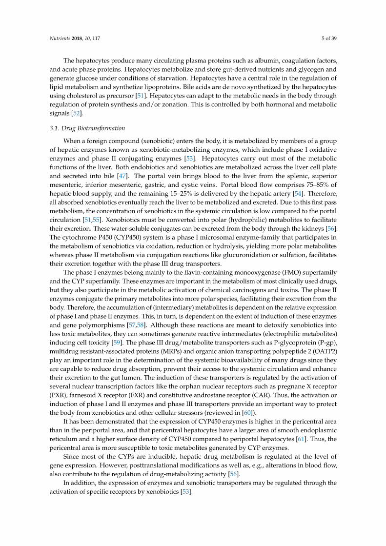
Natural Dietary PigmentsGonzález-Ponce, Herson Antonio; Rincón-Sánchez, Ana Rosa; Jaramillo-Juárez, Fernando;Moshage, HanPublished in:Nutrients
DOI:10.3390/nu10020117
IMPORTANT NOTE: You are advised to consult the publisher's version (publisher's PDF) if you wish to cite fromit. Please check the document version below.
Document VersionPublisher's PDF, also known as Version of record
Publication date:2018
Link to publication in University of Groningen/UMCG research database
Citation for published version (APA):González-Ponce, H. A., Rincón-Sánchez, A. R., Jaramillo-Juárez, F., & Moshage, H. (2018). NaturalDietary Pigments: Potential Mediators against Hepatic Damage Induced by Over-The-Counter Non-Steroidal Anti-Inflammatory and Analgesic Drugs. Nutrients, 10(2), [117].https://doi.org/10.3390/nu10020117
CopyrightOther than for strictly personal use, it is not permitted to download or to forward/distribute the text or part of it without the consent of theauthor(s) and/or copyright holder(s), unless the work is under an open content license (like Creative Commons).
The publication may also be distributed here under the terms of Article 25fa of the Dutch Copyright Act, indicated by the “Taverne” license.More information can be found on the University of Groningen website: https://www.rug.nl/library/open-access/self-archiving-pure/taverne-amendment.
Take-down policyIf you believe that this document breaches copyright please contact us providing details, and we will remove access to the work immediatelyand investigate your claim.
Downloaded from the University of Groningen/UMCG research database (Pure): http://www.rug.nl/research/portal. For technical reasons thenumber of authors shown on this cover page is limited to 10 maximum.
Download date: 01-06-2022
nutrients
Review
Natural Dietary Pigments: Potential Mediatorsagainst Hepatic Damage Induced byOver-The-Counter Non-Steroidal Anti-Inflammatoryand Analgesic Drugs
Herson Antonio González-Ponce 1 ID , Ana Rosa Rincón-Sánchez 2 ID , Fernando Jaramillo-Juárez 3
and Han Moshage 1,4,* ID
1 Department of Gastroenterology and Hepatology, University Medical Center Groningen,University of Groningen, 9713GZ Groningen, The Netherlands; [email protected]
2 Department of Molecular Biology and Genomics, University Center of Health Sciences,Universidad de Guadalajara, Guadalajara 44340, Mexico; [email protected]
3 Department of Physiology and Pharmacology, Basic Science Center, Universidad Autónoma de Aguascalientes,Aguascalientes 20131, Mexico; [email protected]
4 Department of Laboratory Medicine, University Medical Center Groningen, University of Groningen,9713GZ Groningen, The Netherlands
* Correspondence: [email protected]; Tel.: +31-(0)50-361-2364
Received: 7 December 2017; Accepted: 14 December 2017; Published: 24 January 2018
Abstract: Over-the-counter (OTC) analgesics are among the most widely prescribed and purchaseddrugs around the world. Most analgesics, including non-steroidal anti-inflammatory drugs (NSAIDs)and acetaminophen, are metabolized in the liver. The hepatocytes are responsible for drug metabolismand detoxification. Cytochrome P450 enzymes are phase I enzymes expressed mainly in hepatocytesand they account for ≈75% of the metabolism of clinically used drugs and other xenobiotics.These metabolic reactions eliminate potentially toxic compounds but, paradoxically, also result in thegeneration of toxic or carcinogenic metabolites. Cumulative or overdoses of OTC analgesic drugscan induce acute liver failure (ALF) either directly or indirectly after their biotransformation. ALF isthe result of massive death of hepatocytes induced by oxidative stress. There is an increased interestin the use of natural dietary products as nutritional supplements and/or medications to preventor cure many diseases. The therapeutic activity of natural products may be associated with theirantioxidant capacity, although additional mechanisms may also play a role (e.g., anti-inflammatoryactions). Dietary antioxidants such as flavonoids, betalains and carotenoids play a preventive roleagainst OTC analgesics-induced ALF. In this review, we will summarize the pathobiology of OTCanalgesic-induced ALF and the use of natural pigments in its prevention and therapy.
Keywords: analgesics; liver; acute liver failure; oxidative stress; antioxidant capacity
1. Introduction
Humans have always relied on nature for their basic needs. For thousands of years, plantsand their derivatives have formed the basis of sophisticated traditional medicine and have been aninvaluable source of bioactive compounds with therapeutic potential. They play an important role allover the world in the treatment and prevention of human diseases [1,2]. The first records of the use ofplants in medicine are from Mesopotamia and date from about 2600 BC. Most of the plant derivativesreported and used by the Mesopotamians are still in use as antibiotics and anti-inflammatorytreatments [3]. Plant organs such as roots, leaves, fruits, seeds, and other sub-products contain avast array of biological activities related to the presence of many chemically-diverse components.
Nutrients 2018, 10, 117; doi:10.3390/nu10020117 www.mdpi.com/journal/nutrients
Nutrients 2018, 10, 117 2 of 39
Therefore, plants represent an enormous resource for many kinds of bioactive molecules with highlyspecific biological activities for different diseases [4]. Many of the pharmacological activities ofnatural products are related to the presence of bioactive compounds with excellent capacity toreduce oxidative stress [5–8]. Although bioactive components are not always essential for the normaldevelopment and/or reproduction of a plant, they may play an important role as protective agentsagainst environmental factors and predators, thus enhancing their survival [9,10]. The potentialbeneficial health effects of these dietary constituents are highly dependent upon their uptake fromnatural sources, their metabolism and their disposition in target tissues and cells [11,12]. Thus, dietaryphytochemicals are important in human nutrition, medicinal chemistry and drug development [13].It has been estimated that 25–50% of marketed drugs are derived from natural products and almost 50%of novel FDA-approved drugs between 1981–2006 have a natural product origin [2,14]. Since bioactivecomponents from natural sources have evolved through natural selection, they are often perceivedas showing more “drug-likeness and biological friendliness than totally synthetic molecules” [15].Because these compounds have been selected for optimal interactions with cellular macromoleculesin plants, they are likely to induce highly specific biological actions in mammals making them goodcandidates for further drug development [16]. Therefore, these compounds have proven to be a richsource of novel compounds for biological studies and an essential source for drug discovery [13].
In recent years, the self-consumption of over-the-counter (OTC) drugs such as non-steroidalanti-inflammatory and analgesic drugs has rapidly increased. It now represents a serious healthproblem around the world due to the high rates of morbidity and mortality both from consciousand unconscious overdoses [17–20]. Most of these intoxications cause either acute liver damage orchronic gastrointestinal, cardiovascular and renal diseases, with severe oxidative stress as a cause orconsequence [21,22]. In this review, we will discuss the importance of dietary plant pigments in humanhealth and their use as a preventive or therapeutic modality in the treatment of OTC drugs-inducedacute liver failure.
2. Over-The-Counter (OTC) Non-Steroidal Anti-Inflammatory and Analgesic Drugs
Pain in daily life is the most common complaint among patients seeking care in an emergencydepartment [23]. It is also a common experience for adolescents who suffer frequently from headache,abdominal and musculoskeletal types of pain [24]. There are two main classes of drugs recognizedby the FDA: prescription and non-prescription (OTC) drugs. OTC drugs can be purchased andself-administered without a prescription or guidance of a general practitioner [25]. OTC drugs aresold worldwide, although the regulatory systems differ between countries [26]. OTC medicationsare the most commonly purchased and used drugs in the United States for the control of pain inpatients with arthritis, minor surgery, headache, dysmenorrhea, backache, strains and sprains [27].Due to the limited health care access of patients in developing countries, these drugs are oftenused inappropriately, increasing the risk of adverse effects, acute intoxications, and deaths [28].OTC medications can be classified according to the World Health Organization Anatomical TherapeuticChemical (ATC) classification into ten categories: analgesics, laxatives, antithrombotic agents, antacids,cough and cold preparations, antihistamines, dermatologicals, throat preparations, nasal preparationsand antidiarrheals [29]. These medications are normally safe when used properly, but when usedfor extended periods or at high doses the incidence of adverse effects increases [30]. The OTCnon-steroidal anti-inflammatory drugs (NSAIDs) ibuprofen, naproxen, and aspirin, as well as theanalgesic acetaminophen are the most frequently used medications. They are used by approximately23% of the population around the world. OTC analgesics and NSAIDs are mainly used by elderlypatients to relief pain and inflammation [31]. However, there is a lack of information about the potentialtoxicity or adverse drug interactions associated with the long-term use and misuse of OTC analgesics.Not all consumers realize that prolonged daily use and intake of high doses of single OTC analgesicsor combinations dramatically increase the risk of toxicity or adverse drug events, particularly for thehepatic, gastrointestinal, cardiovascular and renal systems. In addition, patients may not be aware
Nutrients 2018, 10, 117 3 of 39
that common cough, cold, or flu medications may contain OTC analgesics again increasing the risk oftoxicity [18,32]. It is well known that use of analgesics is critically relevant for public health, but thereare no representative population-based data on their actual use [33]. The use and prescription of OTCanalgesics in patients known to be at high risk to develop adverse effects has been regulated to reducethe incidence of intoxications and mortality [34,35].
Epidemiology of OTC Non-Steroidal Anti-Inflammatory and Analgesic Drug (Over) Use
Pain in the United States is one of the most important causes of lost labor productivity. In 1985,it was estimated that the total loss of work days due to pain equaled $55 billion [36]. Likewise,a significant percentage of the adult population in Canada lost work days because of the highprevalence of headaches, including migraine [37]. Because of the high incidence of pain, the useand sales of OTC analgesic drugs has steeply increased. A survey in UK demonstrated that 60% ofOTC NSAIDs prescribed were for elderly patients. Thirty-eight percent were taking drugs that caninteract with NSAIDs, 46% had one or more conditions that may be aggravated by NSAIDs, and 18%had side effects [38]. Most of these analgesics and NSAIDs are sold over-the-counter [39]. In the UK,acetaminophen (APAP) has been reported as the most commonly drug used for self-poisoning andoverdoses with an increase of ≈28% from 1976 to 1990, and in 1993, 48% of all overdoses reported inthe UK involved acetaminophen or acetaminophen-containing drugs (reviewed in [40]). A survey ofmedication use in the United States under participants over 18 years old demonstrated that 81% ofthem used at least one OTC or prescription drug in the preceding week, and the highest prevalence ofmedication use was observed in women over 65 years old. In addition, six of the ten most frequentlyused drugs are OTC drugs. The most frequently used drugs were acetaminophen, ibuprofen andaspirin [31]. An Emergency Department survey at the USA showed that 56.2% of 546 patientsinterviewed took OTC analgesics, including acetaminophen (53%), ibuprofen (34%), aspirin (17%) andnaproxen (7.8%); and 6.2% of these 546 patients exceeded the manufacturer’s maximum recommendeddaily dose for at least one medication for at least one day during the three days preceding theevaluation [28]. A study in the Czech Republic from 2007 to 2011 to determine the toxicologicalcharacteristics of suicide attempts by deliberate self-poisoning reported that acetaminophen, diclofenacand ibuprofen were related to ≈16% of the cases from 2393 calls concerning children and adolescentsand 30.3% of these cases were related to drug combinations including acetaminophen [41]. A Dutchsurvey performed in 2014 revealed that almost one-third of the general population used NSAIDs priorto the survey and 31% of those used two or more NSAIDs. In addition, 23% of NSAID users consumedthese drugs for more than seven days and 9% of this population exceeded the daily maximumdose. These results suggest that at least 333,000 Dutch adults exceeded the maximum dose of OTCNSAIDs [35]. In Germany, analgesic use increased from 19.2% of the population in 1998 to 21.4% in theperiod 2008–2011. This increase is due exclusively to the increase in OTC analgesic use from 10.0% to12.2%. Ibuprofen was the most commonly used analgesic. In the period 2008–2011, the use of analgesicswas significantly higher in women than men (25.1% vs. 17.6%) with ibuprofen and acetaminophenbeing the most commonly consumed analgesics. From all the analgesic users, 4.9% (74/1490) useda combination of acetaminophen and ibuprofen. NSAIDs were used mostly in combination by 6.0%of the participants during the seven-day period [33]. A recent Swedish survey concluded that it isimportant to inform the population about the therapeutic use and risks of the consumption of OTCanalgesics since there is a significant influence of parents and peers on the young population [26].Due to the risk of unintentional overdoses with OTC medications, the prevalence of the problemand the frequent lack of an expert to guide and inform consumers on the proper use, a report inthe USA concluded that OTC (over)consumption is a serious public health threat. This health issuerequires urgent attention because many consumers are not able to identify or differentiate the activecomponent(s) in OTC analgesic medications and their biological activities, nor do they adhere to therecommended intake instructions [42]. In addition, there is a high prevalence of drug–drug interactionsresulting from the co-administration of NSAIDs and other commonly used medications by patients
Nutrients 2018, 10, 117 4 of 39
with osteoarthrosis (OA) and rheumatoid arthritis (RA). Therefore, it is necessary to maintain medicalsupervision of those patients with OA and RA receiving OTC NSAIDs and other medications, as wellas to inform them about the risk of toxicity and how to identify toxicity [43].
In summary, when OTC analgesics are taken as recommended by the general practitioner, theyare a safe, effective, and economical treatment to relief pain, inflammation, and fever. Nevertheless,because of their wide availability and perceived safety, OTC analgesics are frequently overconsumedresulting in their hepatic, gastrointestinal and cardiovascular side effects [32,44].
3. Liver Histology and Structures
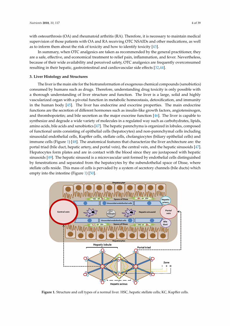
The liver is the main site for the biotransformation of exogenous chemical compounds (xenobiotics)consumed by humans such as drugs. Therefore, understanding drug toxicity is only possible witha thorough understanding of liver structure and function. The liver is a large, solid and highlyvascularized organ with a pivotal function in metabolic homeostasis, detoxification, and immunityin the human body [45]. The liver has endocrine and exocrine properties. The main endocrinefunctions are the secretion of different hormones such as insulin-like growth factors, angiotensinogen,and thrombopoietin; and bile secretion as the major exocrine function [46]. The liver is capable tosynthesize and degrade a wide variety of molecules in a regulated way such as carbohydrates, lipids,amino acids, bile acids and xenobiotics [47]. The hepatic parenchyma is organized in lobules, composedof functional units consisting of epithelial cells (hepatocytes) and non-parenchymal cells includingsinusoidal endothelial cells, Kupffer cells, stellate cells, cholangiocytes (biliary epithelial cells) andimmune cells (Figure 1) [48]. The anatomical features that characterize the liver architecture are: theportal triad (bile duct, hepatic artery, and portal vein), the central vein, and the hepatic sinusoids [47].Hepatocytes form plates and are in contact with the blood since they are juxtaposed with hepaticsinusoids [49]. The hepatic sinusoid is a microvascular unit formed by endothelial cells distinguishedby fenestrations and separated from the hepatocytes by the subendothelial space of Disse, wherestellate cells reside. This mass of cells is pervaded by a system of secretory channels (bile ducts) whichempty into the intestine (Figure 1) [50].
Nutrients 2018, 10, x FOR PEER REVIEW 4 of 38
those patients with OA and RA receiving OTC NSAIDs and other medications, as well as to inform them about the risk of toxicity and how to identify toxicity [43].
In summary, when OTC analgesics are taken as recommended by the general practitioner, they are a safe, effective, and economical treatment to relief pain, inflammation, and fever. Nevertheless, because of their wide availability and perceived safety, OTC analgesics are frequently overconsumed resulting in their hepatic, gastrointestinal and cardiovascular side effects [32,44].
3. Liver Histology and Structures
The liver is the main site for the biotransformation of exogenous chemical compounds (xenobiotics) consumed by humans such as drugs. Therefore, understanding drug toxicity is only possible with a thorough understanding of liver structure and function. The liver is a large, solid and highly vascularized organ with a pivotal function in metabolic homeostasis, detoxification, and immunity in the human body [45]. The liver has endocrine and exocrine properties. The main endocrine functions are the secretion of different hormones such as insulin-like growth factors, angiotensinogen, and thrombopoietin; and bile secretion as the major exocrine function [46]. The liver is capable to synthesize and degrade a wide variety of molecules in a regulated way such as carbohydrates, lipids, amino acids, bile acids and xenobiotics [47]. The hepatic parenchyma is organized in lobules, composed of functional units consisting of epithelial cells (hepatocytes) and non-parenchymal cells including sinusoidal endothelial cells, Kupffer cells, stellate cells, cholangiocytes (biliary epithelial cells) and immune cells (Figure 1) [48]. The anatomical features that characterize the liver architecture are: the portal triad (bile duct, hepatic artery, and portal vein), the central vein, and the hepatic sinusoids [47]. Hepatocytes form plates and are in contact with the blood since they are juxtaposed with hepatic sinusoids [49]. The hepatic sinusoid is a microvascular unit formed by endothelial cells distinguished by fenestrations and separated from the hepatocytes by the subendothelial space of Disse, where stellate cells reside. This mass of cells is pervaded by a system of secretory channels (bile ducts) which empty into the intestine (Figure 1) [50].
Figure 1. Structure and cell types of a normal liver. HSC, hepatic stellate cells; KC, Kupffer cells.
The hepatocytes produce many circulating plasma proteins such as albumin, coagulation factors, and acute phase proteins. Hepatocytes metabolize and store gut-derived nutrients and glycogen and generate glucose under conditions of starvation. Hepatocytes have a central role in the
Figure 1. Structure and cell types of a normal liver. HSC, hepatic stellate cells; KC, Kupffer cells.
Nutrients 2018, 10, 117 5 of 39
The hepatocytes produce many circulating plasma proteins such as albumin, coagulation factors,and acute phase proteins. Hepatocytes metabolize and store gut-derived nutrients and glycogen andgenerate glucose under conditions of starvation. Hepatocytes have a central role in the regulation oflipid metabolism and synthetize lipoproteins. Bile acids are de novo synthetized by the hepatocytesusing cholesterol as precursor [51]. Hepatocytes can adapt to the metabolic needs in the body throughregulation of protein synthesis and/or zonation. This is controlled by both hormonal and metabolicsignals [52].
3.1. Drug Biotransformation
When a foreign compound (xenobiotic) enters the body, it is metabolized by members of a groupof hepatic enzymes known as xenobiotic-metabolizing enzymes, which include phase I oxidativeenzymes and phase II conjugating enzymes [53]. Hepatocytes carry out most of the metabolicfunctions of the liver. Both endobiotics and xenobiotics are metabolized across the liver cell plateand secreted into bile [47]. The portal vein brings blood to the liver from the splenic, superiormesenteric, inferior mesenteric, gastric, and cystic veins. Portal blood flow comprises 75–85% ofhepatic blood supply, and the remaining 15–25% is delivered by the hepatic artery [54]. Therefore,all absorbed xenobiotics eventually reach the liver to be metabolized and excreted. Due to this first passmetabolism, the concentration of xenobiotics in the systemic circulation is low compared to the portalcirculation [51,55]. Xenobiotics must be converted into polar (hydrophilic) metabolites to facilitatetheir excretion. These water-soluble conjugates can be excreted from the body through the kidneys [56].The cytochrome P450 (CYP450) system is a phase I microsomal enzyme-family that participates inthe metabolism of xenobiotics via oxidation, reduction or hydrolysis, yielding more polar metaboliteswhereas phase II metabolism via conjugation reactions like glucuronidation or sulfation, facilitatestheir excretion together with the phase III drug transporters.
The phase I enzymes belong mainly to the flavin-containing monooxygenase (FMO) superfamilyand the CYP superfamily. These enzymes are important in the metabolism of most clinically used drugs,but they also participate in the metabolic activation of chemical carcinogens and toxins. The phase IIenzymes conjugate the primary metabolites into more polar species, facilitating their excretion from thebody. Therefore, the accumulation of (intermediary) metabolites is dependent on the relative expressionof phase I and phase II enzymes. This, in turn, is dependent on the extent of induction of these enzymesand gene polymorphisms [57,58]. Although these reactions are meant to detoxify xenobiotics intoless toxic metabolites, they can sometimes generate reactive intermediates (electrophilic metabolites)inducing cell toxicity [59]. The phase III drug/metabolite transporters such as P-glycoprotein (P-gp),multidrug resistant-associated proteins (MRPs) and organic anion transporting polypeptide 2 (OATP2)play an important role in the determination of the systemic bioavailability of many drugs since theyare capable to reduce drug absorption, prevent their access to the systemic circulation and enhancetheir excretion to the gut lumen. The induction of these transporters is regulated by the activation ofseveral nuclear transcription factors like the orphan nuclear receptors such as pregnane X receptor(PXR), farnesoid X receptor (FXR) and constitutive androstane receptor (CAR). Thus, the activation orinduction of phase I and II enzymes and phase III transporters provide an important way to protectthe body from xenobiotics and other cellular stressors (reviewed in [60]).
It has been demonstrated that the expression of CYP450 enzymes is higher in the pericentral areathan in the periportal area, and that pericentral hepatocytes have a larger area of smooth endoplasmicreticulum and a higher surface density of CYP450 compared to periportal hepatocytes [61]. Thus, thepericentral area is more susceptible to toxic metabolites generated by CYP enzymes.
Since most of the CYPs are inducible, hepatic drug metabolism is regulated at the level ofgene expression. However, posttranslational modifications as well as, e.g., alterations in blood flow,also contribute to the regulation of drug-metabolizing activity [56].
In addition, the expression of enzymes and xenobiotic transporters may be regulated through theactivation of specific receptors by xenobiotics [53].
Nutrients 2018, 10, 117 6 of 39
3.2. Free Radicals and Reactive Oxygen Species
Production of energy, ATP, in cells requires oxygen consumption. Free radicals are produced as aresult of aerobic ATP production via the mitochondrial electron transport chain [62–64].
Harman proposed the “free-radical theory” of ageing in the mid-1950s, suggesting thatendogenous reactive oxygen species, produced by the metabolism of mammalian cells, inducecumulative damage [65]. This concept was initially controversial until superoxide dismutase (SOD)was identified [66]. SOD is an enzyme that inactivates superoxide anions produced by the aerobicmetabolism of cells, providing a mechanistic link to support Harman’s hypothesis. Ageing and thedevelopment of age-related diseases appears to be a consequence of increased levels of intracellularoxidants that induce significant effects such as the activation of signaling pathways and the damage ofcellular components [67].
Reactive oxygen species and reactive nitrogen species (ROS and RNS, respectively) arefundamental in modulating mitochondrial functions via the regulation of electron transfer chainenzymes and mitochondrial membrane potential [68]. ROS are crucial for various cellularprocesses, including cell proliferation [69], apoptosis [70,71], cytotoxicity against bacteria and otherpathogens [72], cell adhesion and immune responses [73]. ROS and RNS also act as second messengersin redox signaling [74].
Mitochondrial metabolism, although essential for cellular homeostasis, is also consideredthe main source of intracellular ROS: superoxide radicals are mainly generated by complex I(NADH:ubiquinone oxidoreductase) and complex III (ubiquinol-cytochrome c reductase) of theelectron transport chain [75]. However, mitochondrial metabolism is not the only source of oxidants.Under physiological conditions, cytosolic enzyme systems including NAPDH oxidases (NOX),microsomal monooxygenases (cytochromes P450), xanthine oxidase (XO), nitric oxide synthases(NOS), lipoxygenases (LOX), cyclooxygenases (COX) and myeloperoxidases can also produce ROSand RNS [67,76–78]. ROS and RNS are generated in excess in some pathological conditions such asneurodegenerative disorders, cancer, diabetes and cardiovascular and liver diseases, and cause celldamage due to their high reactivity with cellular biomolecules [79].
ROS and RNS comprise a group of different molecules, including free radicals such as superoxideanion (O2
•−), hydroxyl radical (•OH) and nitric oxide (NO•), and non-radicals, such as hydrogenperoxide (H2O2), singlet oxygen (1O2) and peroxynitrite (ONOO−). Many free radicals are extremelyunstable, whereas others are freely diffusible and relatively long-lived [64,79].
Additional endogenous non-mitochondrial sources of free radicals include Fenton’s reaction,peroxisomal beta-oxidation, and the respiratory burst of phagocytic cells [22,80]. In addition, theproduction of pro-inflammatory cytokines by activated macrophages and neutrophils and viral proteinsstimulate the generation of ROS [81]. The auto-oxidation of many biologically important moleculesand the electron delocalization that takes place in reactions of heme-containing proteins, also resultsin the production of oxidants [64]. The most relevant exogenous sources of free radical productionare pollutants/toxins such as cigarette smoke, alcohol, ionizing and UV radiation, pesticides, andozone [22]. Moreover, several OTC anti-inflammatory and analgesic drugs induce excess generationof ROS and RNS when used at high or prolonged doses due to their metabolism in the liver, e.g.,acetaminophen [82,83], diclofenac [84,85], aspirin [86], and ibuprofen [87].
3.3. Cellular Oxidative Stress
The excessive generation of ROS and RNS cause damage to cellular macromolecules such asnuclear and mitochondrial DNA, RNA, lipids and proteins by nitration, oxidation, and halogenationreactions, leading to impaired cellular functions and increased mutagenesis [88,89]. Oxidative damageto essential cellular components (macromolecules, organelles) is generally considered as an importantmechanism in the pathophysiology of inflammatory diseases [90]. Oxidative stress is the result ofeither increased generation of ROS and RNS by endogenous and/or exogenous factors, or the result ofa decline of the cellular antioxidant capacity (Figure 2) [91].
Nutrients 2018, 10, 117 7 of 39Nutrients 2018, 10, x FOR PEER REVIEW 7 of 38
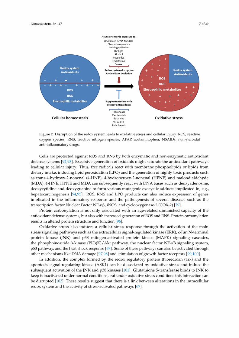
Figure 2. Disruption of the redox system leads to oxidative stress and cellular injury. ROS, reactive oxygen species; RNS, reactive nitrogen species; APAP, acetaminophen; NSAIDs, non-steroidal anti-inflammatory drugs.
Cells are protected against ROS and RNS by both enzymatic and non-enzymatic antioxidant defense systems [92,93]. Excessive generation of oxidants might saturate the antioxidant pathways leading to cellular injury. Thus, free radicals react with membrane phospholipids or lipids from dietary intake, inducing lipid peroxidation (LPO) and the generation of highly toxic products such as trans-4-hydroxy-2-nonenal (4-HNE), 4-hydroperoxy-2-nonenal (HPNE) and malondialdehyde (MDA). 4-HNE, HPNE and MDA can subsequently react with DNA bases such as deoxyadenosine, deoxycytidine and deoxyguanine to form various mutagenic exocyclic adducts implicated in, e.g., hepatocarcinogenesis [94,95]. ROS, RNS and LPO products can also induce expression of genes implicated in the inflammatory response and the pathogenesis of several diseases such as the transcription factor Nuclear Factor NF-κβ, iNOS, and cyclooxygenase-2 (COX-2) [79].
Protein carbonylation is not only associated with an age-related diminished capacity of the antioxidant defense systems, but also with increased generation of ROS and RNS. Protein carbonylation results in altered protein structure and function [96].
Oxidative stress also induces a cellular stress response through the activation of the main stress signaling pathways such as the extracellular signal-regulated kinase (ERK), c-Jun N-terminal protein kinase (JNK) and p38 mitogen-activated protein kinase (MAPK) signaling cascades, the phosphoinositide 3-kinase (PI(3)K)/Akt pathway, the nuclear factor NF-κB signaling system, p53 pathway, and the heat shock response [67]. Some of these pathways can also be activated through other mechanisms like DNA damage [97,98] and stimulation of growth-factor receptors [99,100].
In addition, the complex formed by the redox regulatory protein thioredoxin (Trx) and the apoptosis signal-regulating kinase (ASK1) can be dissociated by oxidative stress and induce the subsequent activation of the JNK and p38 kinases [101]. Glutathione S-transferase binds to JNK to keep it inactivated under normal conditions, but under oxidative stress conditions this interaction can be disrupted [102]. These results suggest that there is a link between alterations in the intracellular redox system and the activity of stress-activated pathways [67].
4. Drug-Induced Liver Injury
Drug-induced oxidative stress is a frequent cause of hepatotoxicity, liver injury and failure. In this regard, drug-induced oxidative stress is considered an important event that can lead to the initiation or progression of liver injury [103]. It is frequently accompanied by clinical signs of acute hepatitis and/or cholestasis [104]. Drug-induced liver injury (DILI) accounts for almost 50% of the
Figure 2. Disruption of the redox system leads to oxidative stress and cellular injury. ROS, reactiveoxygen species; RNS, reactive nitrogen species; APAP, acetaminophen; NSAIDs, non-steroidalanti-inflammatory drugs.
Cells are protected against ROS and RNS by both enzymatic and non-enzymatic antioxidantdefense systems [92,93]. Excessive generation of oxidants might saturate the antioxidant pathwaysleading to cellular injury. Thus, free radicals react with membrane phospholipids or lipids fromdietary intake, inducing lipid peroxidation (LPO) and the generation of highly toxic products suchas trans-4-hydroxy-2-nonenal (4-HNE), 4-hydroperoxy-2-nonenal (HPNE) and malondialdehyde(MDA). 4-HNE, HPNE and MDA can subsequently react with DNA bases such as deoxyadenosine,deoxycytidine and deoxyguanine to form various mutagenic exocyclic adducts implicated in, e.g.,hepatocarcinogenesis [94,95]. ROS, RNS and LPO products can also induce expression of genesimplicated in the inflammatory response and the pathogenesis of several diseases such as thetranscription factor Nuclear Factor NF-κβ, iNOS, and cyclooxygenase-2 (COX-2) [79].
Protein carbonylation is not only associated with an age-related diminished capacity of theantioxidant defense systems, but also with increased generation of ROS and RNS. Protein carbonylationresults in altered protein structure and function [96].
Oxidative stress also induces a cellular stress response through the activation of the mainstress signaling pathways such as the extracellular signal-regulated kinase (ERK), c-Jun N-terminalprotein kinase (JNK) and p38 mitogen-activated protein kinase (MAPK) signaling cascades,the phosphoinositide 3-kinase (PI(3)K)/Akt pathway, the nuclear factor NF-κB signaling system,p53 pathway, and the heat shock response [67]. Some of these pathways can also be activated throughother mechanisms like DNA damage [97,98] and stimulation of growth-factor receptors [99,100].
In addition, the complex formed by the redox regulatory protein thioredoxin (Trx) and theapoptosis signal-regulating kinase (ASK1) can be dissociated by oxidative stress and induce thesubsequent activation of the JNK and p38 kinases [101]. Glutathione S-transferase binds to JNK tokeep it inactivated under normal conditions, but under oxidative stress conditions this interaction canbe disrupted [102]. These results suggest that there is a link between alterations in the intracellularredox system and the activity of stress-activated pathways [67].
Nutrients 2018, 10, 117 8 of 39
4. Drug-Induced Liver Injury
Drug-induced oxidative stress is a frequent cause of hepatotoxicity, liver injury and failure. In thisregard, drug-induced oxidative stress is considered an important event that can lead to the initiationor progression of liver injury [103]. It is frequently accompanied by clinical signs of acute hepatitisand/or cholestasis [104]. Drug-induced liver injury (DILI) accounts for almost 50% of the cases of acuteliver failure (ALF) in the United States [105,106], and for more than 50% in UK. Several factors havebeen identified that predict drug-induced liver injury (DILI), e.g., dose, alcohol consumption, use ofconcomitant drugs, nature of the drug, time of exposure, age, preconditioning diseases, and congenitalanomalies [104,107]. DILI may be classified as non-idiosyncratic or idiosyncratic. Idiosyncratic drugreactions are unpredictable and independent, and can occur from intermediate to long periods ofexposure by an activation of the immune response, inflammation, and cell death (mostly apoptosis).Drug-induced predictable liver injury, such as from acetaminophen, can occur within few hoursor days and are mediated by the production of free radicals or electrophilic metabolites from drugbiotransformation inducing organelle stress and cell death (both necrosis and apoptosis) [104,108,109].Necrosis involves the depletion or inactivation of endogenous antioxidants and the induction ofcellular stress including mitochondrial stress and decreased ATP synthesis which leads to cellulardysfunction and ATP-independent death. In contrast, apoptosis is an ATP-dependent mechanisminvolving activation of nucleases [108,110]. Therefore, the activation of death-signaling pathways suchas JNK is an important event in DILI [111].
The liver plays a critical role in the disposition of orally administered therapeutic agents. It is theport-of-entry of most orally taken drugs and it represents the major site of drug biotransformationmaking the liver susceptible to drug-induced toxicity. Products of drug biotransformation (electrophiliccompounds and free radicals) have been implicated as causative agents of liver toxicity through directinjury to the hepatocytes by interfering with critical cellular functions (e.g., ATP production), modifyingimportant biomolecules (e.g., proteins, lipids, or nucleic acids), depleting cellular antioxidants,inducing cellular oxidative stress [112,113].
Cellular functions can be affected by both direct effects on organelles (e.g., the mitochondria, theendoplasmic reticulum, the cytoskeleton, microtubules, or the nucleus), and indirectly modulatingsignaling kinases, transcription factors, and gene expression. These cellular effects can activatethe immune response via the release of pro-inflammatory cytokines and/or cell debris into theblood-stream, resulting in the recruitment of immune cells (neutrophils), cellular stress and hepatocytedeath that ultimately induce liver injury and failure [56,114–116].
Mitochondria play an important role in the development of DILI since they are an importantregulator of cellular homeostasis and their dysfunction can trigger liver cell toxicity resulting inmild to fulminant hepatic failure [117]. Very often, cell death is associated with depletion ofmitochondrial glutathione and not with loss of cytoplasmic glutathione [118]. Therefore, it is importantto elucidate whether drug metabolites have direct effects on mitochondrial function (e.g., via inhibitionof electron transport chain or increasing lipid peroxidation and membrane permeability) resulting inhepatocellular death or indirect effects via activation of the mitochondrial pathways of programmedcell death [119].
4.1. OTC Non-Steroidal Anti-Inflammatory and Analgesic Drugs-Induced Acute Liver Injury
NSAIDs are the most widely used OTC drugs as well as the most prescribed class of drugs fora variety of conditions [120,121]. The group of NSAIDs is composed of a large class of chemicalcompounds with the same biological activity: blocking the production of prostaglandins (PGs) throughthe inhibition of the enzyme cyclooxygenase (COX). The COX enzyme is present as two isoforms,each with distinct functions. COX-1 is an isoenzyme constitutively expressed in the stomach, kidney,intestinal mucosa, and other tissues, and is involved in the biosynthesis of PGs serving homeostaticfunctions. It plays an important role in vasoconstriction and platelet aggregation.
Nutrients 2018, 10, 117 9 of 39
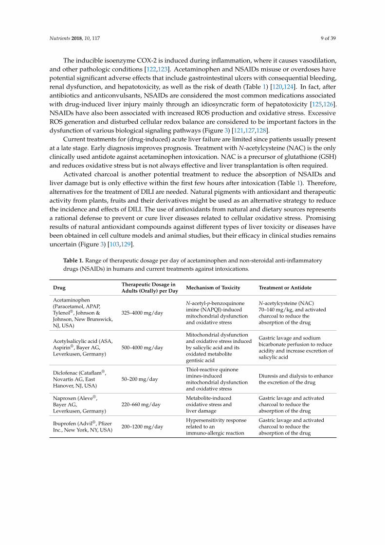
The inducible isoenzyme COX-2 is induced during inflammation, where it causes vasodilation,and other pathologic conditions [122,123]. Acetaminophen and NSAIDs misuse or overdoses havepotential significant adverse effects that include gastrointestinal ulcers with consequential bleeding,renal dysfunction, and hepatotoxicity, as well as the risk of death (Table 1) [120,124]. In fact, afterantibiotics and anticonvulsants, NSAIDs are considered the most common medications associatedwith drug-induced liver injury mainly through an idiosyncratic form of hepatotoxicity [125,126].NSAIDs have also been associated with increased ROS production and oxidative stress. ExcessiveROS generation and disturbed cellular redox balance are considered to be important factors in thedysfunction of various biological signaling pathways (Figure 3) [121,127,128].
Current treatments for (drug-induced) acute liver failure are limited since patients usually presentat a late stage. Early diagnosis improves prognosis. Treatment with N-acetylcysteine (NAC) is the onlyclinically used antidote against acetaminophen intoxication. NAC is a precursor of glutathione (GSH)and reduces oxidative stress but is not always effective and liver transplantation is often required.
Activated charcoal is another potential treatment to reduce the absorption of NSAIDs andliver damage but is only effective within the first few hours after intoxication (Table 1). Therefore,alternatives for the treatment of DILI are needed. Natural pigments with antioxidant and therapeuticactivity from plants, fruits and their derivatives might be used as an alternative strategy to reducethe incidence and effects of DILI. The use of antioxidants from natural and dietary sources representsa rational defense to prevent or cure liver diseases related to cellular oxidative stress. Promisingresults of natural antioxidant compounds against different types of liver toxicity or diseases havebeen obtained in cell culture models and animal studies, but their efficacy in clinical studies remainsuncertain (Figure 3) [103,129].
Table 1. Range of therapeutic dosage per day of acetaminophen and non-steroidal anti-inflammatorydrugs (NSAIDs) in humans and current treatments against intoxications.
Drug Therapeutic Dosage inAdults (Orally) per Day Mechanism of Toxicity Treatment or Antidote
Acetaminophen(Paracetamol, APAP,Tylenol®, Johnson &Johnson, New Brunswick,NJ, USA)
325–4000 mg/day
N-acetyl-p-benzoquinoneimine (NAPQI)-inducedmitochondrial dysfunctionand oxidative stress
N-acetylcysteine (NAC)70–140 mg/kg, and activatedcharcoal to reduce theabsorption of the drug
Acetylsalicylic acid (ASA,Aspirin®, Bayer AG,Leverkusen, Germany)
500–4000 mg/day
Mitochondrial dysfunctionand oxidative stress inducedby salicylic acid and itsoxidated metabolitegentisic acid
Gastric lavage and sodiumbicarbonate perfusion to reduceacidity and increase excretion ofsalicylic acid
Diclofenac (Cataflam®,Novartis AG, EastHanover, NJ, USA)
50–200 mg/day
Thiol-reactive quinoneimines-inducedmitochondrial dysfunctionand oxidative stress
Diuresis and dialysis to enhancethe excretion of the drug
Naproxen (Aleve®,Bayer AG,Leverkusen, Germany)
220–660 mg/dayMetabolite-inducedoxidative stress andliver damage
Gastric lavage and activatedcharcoal to reduce theabsorption of the drug
Ibuprofen (Advil®, PfizerInc., New York, NY, USA)
200–1200 mg/dayHypersensitivity responserelated to animmuno-allergic reaction
Gastric lavage and activatedcharcoal to reduce theabsorption of the drug
Nutrients 2018, 10, 117 10 of 39Nutrients 2018, 10, x FOR PEER REVIEW 10 of 38
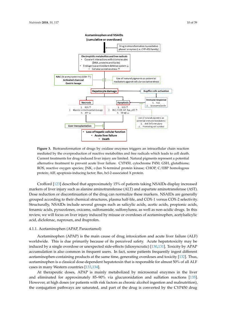
Figure 3. Biotransformation of drugs by oxidase enzymes triggers an intracellular chain reaction mediated by the overproduction of reactive metabolites and free radicals which leads to cell death. Current treatments for drug-induced liver injury are limited. Natural pigments represent a potential alternative treatment to prevent acute liver failure. CYP450, cytochrome P450; GSH, glutathione; ROS, reactive oxygen species; JNK, c-Jun N-terminal protein kinase; CHOP, C/EBP homologous protein; AIF, apoptosis-inducing factor; Bax, bcl-2-associated X protein.
Crofford [123] described that approximately 15% of patients taking NSAIDs display increased markers of liver injury such as alanine aminotransferase (ALT) and aspartate aminotransferase (AST). Dose reduction or discontinuation of the drug can normalize these markers. NSAIDs are generally grouped according to their chemical structures, plasma half-life, and COX-1 versus COX-2 selectivity. Structurally, NSAIDs include several groups such as salicylic acids, acetic acids, propionic acids, fenamic acids, pyrazolones, oxicams, sulfonamide, sulfonylurea, as well as non-acidic drugs. In this review, we will focus on liver injury induced by misuse or overdoses of acetaminophen, acetylsalicylic acid, diclofenac, naproxen, and ibuprofen.
4.1.1. Acetaminophen (APAP, Paracetamol)
Acetaminophen (APAP) is the main cause of drug intoxication and acute liver failure (ALF) worldwide. This is due primarily because of its perceived safety. Acute hepatotoxicity may be induced by a single overdose or unexpected side-effects (idiosyncratic) [130,131]. Toxicity by APAP accumulation is also common in frequent users. In fact, some patients frequently ingest different acetaminophen-containing products at the same time, generating overdoses and toxicity [132]. Thus, acetaminophen is a classical dose-dependent hepatotoxin that is responsible for almost 50% of all ALF cases in many Western countries [133,134].
At therapeutic doses, APAP is mainly metabolized by microsomal enzymes in the liver and eliminated for approximately 85–90% via glucuronidation and sulfation reactions [135]. However, at high doses (or patients with risk factors as chronic alcohol ingestion and malnutrition), the conjugation pathways are saturated, and part of the drug is converted by the CYP450 drug
Figure 3. Biotransformation of drugs by oxidase enzymes triggers an intracellular chain reactionmediated by the overproduction of reactive metabolites and free radicals which leads to cell death.Current treatments for drug-induced liver injury are limited. Natural pigments represent a potentialalternative treatment to prevent acute liver failure. CYP450, cytochrome P450; GSH, glutathione;ROS, reactive oxygen species; JNK, c-Jun N-terminal protein kinase; CHOP, C/EBP homologousprotein; AIF, apoptosis-inducing factor; Bax, bcl-2-associated X protein.
Crofford [123] described that approximately 15% of patients taking NSAIDs display increasedmarkers of liver injury such as alanine aminotransferase (ALT) and aspartate aminotransferase (AST).Dose reduction or discontinuation of the drug can normalize these markers. NSAIDs are generallygrouped according to their chemical structures, plasma half-life, and COX-1 versus COX-2 selectivity.Structurally, NSAIDs include several groups such as salicylic acids, acetic acids, propionic acids,fenamic acids, pyrazolones, oxicams, sulfonamide, sulfonylurea, as well as non-acidic drugs. In thisreview, we will focus on liver injury induced by misuse or overdoses of acetaminophen, acetylsalicylicacid, diclofenac, naproxen, and ibuprofen.
4.1.1. Acetaminophen (APAP, Paracetamol)
Acetaminophen (APAP) is the main cause of drug intoxication and acute liver failure (ALF)worldwide. This is due primarily because of its perceived safety. Acute hepatotoxicity may beinduced by a single overdose or unexpected side-effects (idiosyncratic) [130,131]. Toxicity by APAPaccumulation is also common in frequent users. In fact, some patients frequently ingest differentacetaminophen-containing products at the same time, generating overdoses and toxicity [132]. Thus,acetaminophen is a classical dose-dependent hepatotoxin that is responsible for almost 50% of all ALFcases in many Western countries [133,134].
At therapeutic doses, APAP is mainly metabolized by microsomal enzymes in the liverand eliminated for approximately 85–90% via glucuronidation and sulfation reactions [135].However, at high doses (or patients with risk factors as chronic alcohol ingestion and malnutrition),the conjugation pathways are saturated, and part of the drug is converted by the CYP450 drug
Nutrients 2018, 10, 117 11 of 39
metabolizing system (mainly CYP1A2, CYP2E1 and CYP3A4) to the highly reactive metaboliteN-acetyl-p-benzoquinone imine (NAPQI) that reacts with protein sulfhydryl groups of cysteine.Once generated, NAPQI is immediately inactivated by endogenous reduced glutathione (GSH) to formNAPQI-GSH conjugates which are excreted through the urine. However, when the hepatic reservoirof GSH is depleted, cellular organelles (e.g., mitochondria and endoplasmic reticulum) are exposed tothe highly reactive metabolite NAPQI. NAPQI reacts with (membrane) biomolecules forming adductsand resulting in the disruption of cellular homeostasis [7,81,136,137].
Activation of the JNK pathway plays an important role in APAP-induced liver injury andhepatocyte death. Models of genetic JNK knock-out and hepatoprotective compounds with JNKsuppressing activity may prevent APAP-induced oxidative stress, cell death and liver failure.Once activated, JNK translocates to the mitochondria inducing mitochondrial permeability transition(MPT), mitochondrial dysfunction and cell death [118]. Protein kinase C (PKC) may also play acritical role in APAP-induced hepatotoxicity via the JNK signaling pathway since treatment with PKCinhibitors (Ro-31-8245, Go6983) protected primary mouse hepatocytes. Ro-31-8245 treatment increasedp-AMPK levels (phosphorylated AMP-activated kinase), and promoted autophagy. Treatment withthe PKC inhibitor Go6976 inhibits JNK activation and translocation, protecting hepatocytes againstAPAP cytotoxicity [138].
In mouse models and in human hepatocytes, APAP-induced liver injury involves mitochondrialdamage, oxidative stress, JNK activation, and nuclear DNA fragmentation. Thus, protein adducts inmitochondria damage the electron transport chain, increase oxidative stress and disturb the innateimmune system of the liver [139,140]. Of note, liver injury is aggravated by subsequent oxidant stressvia ROS. The enhanced superoxide formation leads to generation of the potent oxidant peroxynitrite inmitochondria [141].
In the mouse model, APAP toxicity produces very early activation (phosphorylation) of JNKin the cytoplasm. Activated JNK translocates to the mitochondria and increases oxidant stress.This causes the formation of the mitochondrial permeability transition pore and collapse of themitochondrial membrane potential, as well as a drop in ATP production [140,142,143]. In addition todepleting intracellular GSH, APAP treatment also increases lipid peroxidation and causes hepatic DNAfragmentation. The combination of massive mitochondrial dysfunction and nuclear disintegrationleads to cellular necrosis [144–146]. Alterations in hepatic innate immunity and inflammation also playa significant role in the progression of hepatic failure after acetaminophen overdose [135,147].
The primary mechanism of cell death in acetaminophen-induced liver failure is thought to benecrosis, however, some reports have shown that apoptosis may also play a significant role [140]. In thehuman hepatoblastoma cell line (HuH7), activation of caspases was observed and the manifestationof apoptosis was preceded by a translocation of cytochrome c from mitochondria to the cytosol [148].It remains to be elucidated whether hepatoma cell lines accurately reflect the in vivo metabolism ofAPAP. Recent studies also demonstrated increased serum markers of apoptosis, such as caspase-cleavedcytokeratin-18 (M30), in the early phase of acetaminophen-induced ALF in humans [149]. The exactmechanism(s) and optimal management of APAP-induced acute liver failure still need to be clearlyelucidated to reduce the high morbidity and mortality of APAP-induced ALF [150].
4.1.2. Acetylsalicylic Acid (ASA, Aspirin)
Acetylsalicylic acid (ASA) is a widely used NSAID due to its pharmacological properties(including analgesic, antipyretic, anti-inflammatory and anti-platelets effects), as well as its easyavailability. ASA is an irreversible inhibitor of both cyclooxygenase isoenzymes, COX-1 and COX-2.After oral administration, ASA is absorbed from the stomach and small intestine, primarily by passivediffusion across the gastrointestinal tract [151]. ASA is rapidly deacetylated to salicylic acid byesterases in the gastrointestinal mucosa, in the blood and in the liver. The oxidation of salicylic acidin human liver microsomes produces two metabolites, 2,5-dihydroxybenzoic acid (gentisic acid) and
Nutrients 2018, 10, 117 12 of 39
2,3-dihydroxybenzoic acid. The major human cytochrome P450 involved in both biotransformationreactions is CYP2E1 [152].
Liver toxicity induced by ASA is considered to be dose-dependent, although predisposingconditions may exist that increase the individual risk of liver damage [153,154]. Oxidative stress is oneof the mechanisms associated with the adverse effects of ASA and salicylic acid may induce cytochromeP450-mediated lipid peroxidation in liver microsomes [155]. On the other hand, antioxidant propertiesof this drug have also been reported [156]. Doi and Horie [86] also analyzed mitochondrial dysfunctionand oxidative stress in salicylic acid-induced liver injury. In rat hepatocytes, salicylic acid significantlyincreased the leakage of lactate dehydrogenase and increased thiobarbituric acid reactive substances(TBARS) formation, a marker of lipid peroxidation, whereas antioxidants (promethazine and DPPD(N,N′-diphenyl-p-phenylenediamine)) suppressed both harmful effects. TBARS formation in rat livermicrosomes was also suppressed by diethyldithiocarbamate (a specific inhibitor of CYP2E1) anddiclofenac (a specific inhibitor of CYP2C11). Salicylic acid also significantly decreased ATP content inisolated rat hepatocytes and mitochondrial respiration. The authors suggest that salicylic acid impairsmitochondrial function leading to lethal liver cell injury by lipid peroxidation.
Raza et al. [157] analyzed the oxidative effects of ASA in cultured human hepatoma cells (HepG2)and reported a cascade of adverse events starting with the overproduction of cellular ROS through theuncoupling of the complex I (NADH:ubiquinone oxidoreductase) and IV (cytochrome c oxidase) of themitochondrial electron transport chain and ultimately resulting in reduced levels of GSH. The alteredMPT disrupted the mitochondrial ATP synthesis, decreased the expression of the anti-apoptoticprotein Bcl-2 and induced the activation and release of pro-apoptotic proteins to induce cell death.ASA-induced cytotoxicity was augmented by inhibition of GSH synthesis and attenuated by increasingthe GSH pool. The authors conclude that ASA-induced toxicity in human HepG2 cells is mediatedby increased metabolic and oxidative stress, accompanied by mitochondrial dysfunction, resulting inapoptosis [158].
Tassone et al. [159] reported a pilot study in 22 newly diagnosed diabetic patients treated withASA (100 mg/daily for four weeks). The authors suggest that ASA treatment for primary preventionin diabetic patients causes oxidative stress and impairs vascular function.
In addition to mitochondrial dysfunction, ASA treatment can also lead to accumulation of freefatty acids in the liver leading to massive hepatic steatosis [154]. The mechanism of ASA-inducedhepatotoxicity is different from that of other NSAIDs. As described above, ASA is first hydrolyzed bynon-specific esterases into salicylic acid. In mitochondria, salicylic acid may form salicylyl-coenzymeA (CoA) conjugates, thus sequestering extramitochondrial CoA. This conjugate indirectly inhibitsβ-oxidation of long-chain fatty acids since CoA is necessary to transport free fatty acids into themitochondria [160,161]. The inhibition of mitochondrial β-oxidation of long-chain fatty acids by ASAmay lead to microvesicular steatosis known as Reye’s syndrome [113,162]. Salicylic acid can alsoinhibit Krebs cycle enzymes such as α-ketoglutarate dehydrogenase and succinate dehydrogenaseleading to mitochondrial dysfunction [162].
Finally, Jain et al. [151] published that treatment of female rats with ASA (100 mg/kg b.w. (bodyweight)) caused significant histopathological alterations in the liver, including degenerative andpyknotic changes in the nuclei, vacuolization and clear dilatations in the sinusoids and hypertrophyof hepatocytes.
4.1.3. Diclofenac
Diclofenac is a commonly prescribed NSAID of the phenyl-acetic acid class. Diclofenac has beenused in a variety of inflammatory conditions and has strong anti-inflammatory activity, althoughanalgesic and antipyretic properties have also been reported [163]. In contrast to traditionalNSAIDs, diclofenac appears to have a higher selectivity for COX-2 than COX-1. Diclofenac hasbeen associated with serious dose-dependent gastrointestinal, cardiovascular, and renal toxicity [164].Diclofenac-induced liver injury has been used as a model of drug-related toxicity. The hepatotoxicity
Nutrients 2018, 10, 117 13 of 39
induced by diclofenac is mainly due to its metabolites, but genetic factors can also increase thesusceptibility to produce and accumulate the reactive acylglucuronide metabolite which triggersan immune response and liver injury [84,154,165–167]. The bioactivation of diclofenac by CYP2C9or CYP3A4 yields thiol-reactive quinone-imines which in turn are conjugated by UDP (Uridine5′-diphospho)-glucuronosyltransferase (UGT2B7) into protein-reactive acyl-glucuronides. Therefore,both disruption of mitochondrial function and alterations in the redox state due to oxidative ornitrosative stress appear to be the main mechanisms of diclofenac-induced cell death and liver injury.
Laine et al. [168] conducted a long-term prospective clinical trial to analyze the frequency ofdiclofenac-induced adverse hepatic effects. A total of 17,289 arthritis patients received diclofenacfor a mean duration of 18 months. Increased serum transaminase occurred primarily within thefirst 4–6 months of therapy and was observed in 3.1% of arthritic patients. Of note, ALT/AST ratiosof >10× the upper limit of normal (ULN) were only observed in 0.5% of cases. The clinical liversymptoms requiring hospitalization were relatively rare (23/100,000 patients or 0.023% of cases).No liver failure or death was observed. These and other results indicate that diclofenac rarely causessevere liver injury in humans [169].
The United States (U.S.) Drug Induced Liver Injury Network (DILIN) is a prospective registryof severe idiosyncratic drug hepatotoxicity. Schmeltzer et al. [126] reported a study on liver injurycaused by NSAIDs in the U.S. The authors conclude that hepatocellular injury is the most commonmanifestation seen with NSAID toxicity and diclofenac is the most frequently implicated NSAID agent(16/30 cases). Bort et al. [170] analyzed acute diclofenac cytotoxicity on human and rat hepatocytes andhepatic cell lines (HepG2, FaO). Diclofenac impaired ATP synthesis by mitochondria and the authorssuggested that toxicity might be related to drug metabolism because diclofenac was more cytotoxicto drug metabolizing cells (rat and human primary hepatocytes) than to non-metabolizing cell lines(HepG2, FaO). The toxic effect was reduced by the addition of cytochrome P450 inhibitors and in vitrocytotoxicity of diclofenac correlated well with the generation of two metabolites: 5-hydroxydiclofenacand N,5-dihydroxydiclofenac.
Gómez-Lechón et al. [85] also analyzed the generation of ROS and the apoptotic effect of diclofenacafter exposure of human and rat hepatocytes to diclofenac. Antioxidants were able to prevent caspase-8and -9 activation by diclofenac and maintain mitochondrial integrity. The authors concluded that themitochondrial pathway of apoptosis is the only (or major) pathway involved in diclofenac-inducedapoptosis and that the strongest apoptotic effect was produced by the metabolite 5-hydroxydiclofenac.
Other studies reported similar findings, such as diclofenac (metabolite)-induced ROS generation,lipid peroxidation, mitochondrial injury, ATP depletion, GSH depletion, lysosomal fragmentation andDNA fragmentation [171–173]. Many of these signs of toxicity were reversed by antioxidants, MPT poresealing agents, lysosomotropic agents and inhibitors of cytochrome P450 isoenzymes. The final commonpathway of all these events is the leakage of cytochrome c from the mitochondrial intermembrane spaceinto the cytosol, resulting in the activation of caspases-9 and 3, mitochondrial/lysosomal cross-talkand apoptosis.
Yano et al. [169] investigated the immune response in diclofenac-induced idiosyncratichepatotoxicity in mice. Gene expression of the main interleukins (ILs) and chemokines involvedin the inflammatory response and the expression of helper T (Th) 17 cell-derived factors in theliver were significantly increased, as well as the levels of IL-17 in plasma. The results suggest atleast a partial involvement of IL-17 in the development of diclofenac-induced liver injury sinceantagonizing IL-17 reduced toxicity. In addition, both gene expression and plasma levels of IL-1β wererapidly increased after diclofenac administration suggesting its involvement in the pathogenesis ofdiclofenac-induced hepatotoxicity.
Interaction between drug-induced toxicity pathways and the pro-inflammatory cytokinetumor necrosis factor alpha (TNFα) was investigated in HepG2 cells by Fredriksson et al. [174].Transcriptomics of the stress response pathways initiated by diclofenac and carbamazepine, revealedthe endoplasmic reticulum (ER) stress/translational initiation signaling and nuclear factor-erythroid
Nutrients 2018, 10, 117 14 of 39
2 (NF-E2)-related factor 2 (Nrf2) antioxidant signaling as two important affected pathways. Inhibitionof the Nrf2-dependent adaptive oxidative stress response enhanced drug/TNFα-induced cytotoxicitybut did not affect C/EBP homologous protein (CHOP) expression. Both hepatotoxic drugs enhancedexpression of the translational initiation factor EIF4A1, which was essential for CHOP expressionand drug/TNFα-mediated cell killing. The authors conclude from their data that diclofenac initiatesPERK-mediated CHOP signaling in an EIF4A1 dependent manner, thereby sensitizing the hepatocytetowards caspase-8-dependent TNFα-induced apoptosis.
4.1.4. Naproxen
Naproxen is a propionic acid derivative NSAID and has been available as OTC medication since1994. Naproxen is an analgesic, antipyretic and anti-inflammatory drug. It is a non-selective inhibitorof the enzymes COX-1 and COX-2 and decreases synthesis of prostaglandins, important mediatorsin inflammatory and pain pathways. Currently, more than 10 million prescriptions for naproxen arefiled each year but these numbers do not include the large-scale OTC sales. Side effects of naproxeninclude dizziness, dyspepsia, nausea, and abdominal discomfort, but rarely liver injury. Naproxenis metabolized by the cytochrome P450 system and idiosyncratic liver injury may be due to a toxicmetabolite although the mechanism has not been completely elucidated yet [175,176].
The absorption of naproxen is rapid and complete when given orally and the biotransformationof this drug includes demethylation and glucuronidation, as well as sulfate conjugation reactions.The metabolites are excreted in urine, with only a small proportion of the drug being eliminatedunchanged [177]. In human liver, CYP2C9 and CYP1A2 are involved in naproxen metabolism [178,179]and subsequent glucuronidation takes place via UDP-glucuronosyltransferase (UGT2B7) [180]. There issome evidence that naproxen metabolism is related to hepatotoxicity: Yokoyama et al. [181] reportedthat naproxen induces lipid peroxidation in isolated rat hepatocytes, resulting in cell death andformation of high molecular weight protein aggregates in the hepatocytes. Oxidative stress wasdemonstrated by the formation of TBARS, a marker of lipid peroxidation. The increase of TBARSstrongly correlated with the decrease of intracellular GSH. The authors concluded that ROS productionand lipid peroxidation are induced by the metabolism of naproxen in rat primary hepatocytes.
A follow-up study confirmed the pivotal role of the GSH/GSSG (glutathione/glutathionedisulfide) ratio in naproxen-induced toxicity. Increased GSSG levels preceded lipid peroxidationand LDH release [182]. Ji et al. [183] reported that ferrous iron release contributes to naproxen-inducedmicrosomal lipid peroxidation and that naproxen and salicylic acid are not uncouplers of cytochromeP450. Naproxen toxicity and disposition were also investigated in the isolated perfused liver.Lo et al. [184] investigated the disposition of naproxen, its reactive acyl glucuronide metabolite (NAG)and a mixture of NAG rearrangement isomers (isoNAG) and concluded that covalent protein-adductswere formed in the liver, with isoNAG being the more important substrate for adduct formation.Using the same experimental model, Yokoyama et al. [185] demonstrated that naproxen increasedliver damage (AST, ALT) and peroxidation (TBARS). GSSG and TBARS content were significantlyincreased in naproxen-perfused liver. In addition, the biliary excretion (clearance) of indocyanine green,a compound used for testing liver function, was decreased. The authors concluded that the biliaryexcretion system was disrupted due to naproxen-induced hepatic oxidative stress. Naproxen-inducedhepatotoxicity and liver injury is very rare (≈1–3 per 100,000 users), although cases of acute hepatitishave been reported within six weeks after the start of naproxen intake. Once naproxen intake isterminated, biochemical markers of liver injury such as AST and ALT usually return to normallevels [186].
Andrejak et al. [175] described a patient using naproxen at 500 mg/day with nausea, abdominalpain, malaise, jaundice and increased AST, ALT and alkaline phosphatase levels. Histology showedmoderate hepatocellular necrosis. After termination of naproxen, the patient recovered rapidly.Ali et al. [176] described a patient who developed jaundice and intractable pruritus shortly after takingnaproxen. Histological analysis showed inflammatory infiltration and a progressive loss of the small
Nutrients 2018, 10, 117 15 of 39
interlobular bile ducts (ductopenia). The authors suggest that normalization of liver function andhistology after termination of naproxen consumption may take up to 10 years.
4.1.5. Ibuprofen
Ibuprofen was the first member of propionic acid derivatives to be introduced in 1969 as a betteralternative to ASA. Ibuprofen is the most frequently prescribed NSAID and it is also an OTC drug.Ibuprofen has excellent analgesic and anti-inflammatory properties as well as antipyretic activitybecause it inhibits both cyclooxygenases (COX-1 and COX-2). Ibuprofen is frequently used to reliefpain related to dysmenorrhea, headache, and osteoarthritis or rheumatoid arthritis. Adverse reactionsto ibuprofen appear to be dose and duration dependent and the major adverse effects are related to thegastrointestinal tract, the kidneys and blood coagulation. In addition, ibuprofen may produce dizziness,dyspepsia, bronchospasm, and hypersensitivity reactions, but rarely causes clinically apparent andserious acute liver injury [43,187,188].
In fact, ibuprofen at OTC doses does not represent a risk for developing liver injury, because it hasa short plasma half-life and it does not give rise to toxic metabolites (e.g., covalent modification of liverproteins by the quinine-imine metabolites of paracetamol or irreversible acetylation of biomoleculesby ASA) [189]. High doses of ibuprofen (2400 to 3200 mg daily) may produce increased ALT plasmalevels (<100 U/L), although clinically apparent liver injury due to ibuprofen is very rare. Only a fewcases of ibuprofen-induced acute liver toxicity have been reported. The mechanism of toxicity hasnot been completely elucidated and may be multifactorial. Most of the ibuprofen-induced cases ofhepatotoxicity have a rapid onset suggesting the production of a reactive metabolite as well as theinvolvement of a hypersensitivity response related to an immuno-allergic reaction [188]. Underlyingliver diseases like hepatitis C may increase the risk of ibuprofen-induced acute liver injury [154,190].Finally, Basturk et al. [191] published a case report of a seven-year-old patient who developed toxicepidermal necrolysis and vanishing bile duct syndrome (VBDS) after oral ibuprofen intake. AcuteVBDS is a rare disease with unknown etiology. The patient was treated with supportive care (a steroidand ursodeoxycholic acid), with complete recovery after eight months.
5. Endogenous Antioxidant Defense Systems
Reactive Oxygen Species (ROS) are produced during normal intracellular metabolism and fromexogenous substances. They play an important role in a range of biological processes, e.g., inthe defense against microorganisms, as second messengers in several signaling pathways, and inmodulating gene expression. Moreover, when generated in excess and when redox balance is disturbed,these free radicals can damage cellular organelles and induce inflammation, ischemia, apoptosis, andnecrosis. Since living organisms are continuously exposed to free radicals, cells have developedantioxidant defense mechanisms. These antioxidant defense mechanisms include molecules andenzymes formed endogenously and bioactive molecules obtained from food (reviewed in [192]).
Any substance or compound that scavenges free radicals or non-radical ROS or RNS, or inhibitscellular oxidation reactions can be considered an antioxidant [193]. Endogenous antioxidants arecapable to counteract the deleterious effects of free radicals and maintain cellular homeostasis.These endogenous antioxidants include both enzymatic antioxidants such as catalase, superoxidedismutases, glutathione peroxidases, peroxiredoxins, and thioredoxins [194] as well as non-enzymaticantioxidants (glutathione, urate, bilirubin, melatonin). The coordinated action of antioxidant enzymesensures efficient ROS removal and promotes repair [195].
Free radicals, ROS and RNS are very reactive and since they are generated in different cellorganelles, enzymatic defense systems have evolved in different cellular compartments. One of themost effective intracellular enzymatic antioxidants is superoxide dismutase (SOD) which catalyzes thedismutation of O2
•− (superoxide anions) to O2 and to the less-reactive species H2O2 with remarkablyhigh reaction rates. This is accomplished by successive oxidation and reduction of the transition metalions incorporated in the SOD enzymes (reviewed in [196]). The enzyme SOD is widespread in nature
Nutrients 2018, 10, 117 16 of 39
and present in all oxygen-metabolizing cells [197]. In humans, there are three superoxide dismutases:mitochondrial Mn-SOD, cytosolic Cu/Zn-SOD, and extracellular ecSOD. The main function of allSODs is to protect the cell from harmful effects of superoxide anions. In addition, extracellular SOD(ecSOD) is known to affect endothelial cells by preventing NO from reacting with superoxide anions(reviewed by [198]).
Glutathione peroxidase (GPx), catalase (CAT) and peroxiredoxins (Prx) control the ultimate fateof hydrogen peroxide produced from the superoxide anions by SODs.
Peroxisomes are the major storage site of catalase, an enzyme that catalyzes the biotransformationof H2O2 into water and O2. In addition, catalase has a peroxidative activity promoting the reactionbetween H2O2 and hydrogen donors to generate water and oxidize the reduced donor [199]. Althoughcatalase might not be essential for all cell types, under normal conditions its deficiency in conditions ofoxidative stress can increase cell damage and death [200].
GPx enzymes protect the cells against oxidative stress through the reduction of hydroperoxidesusing GSH as electron donor, yielding water and oxidized GSSG (glutathione disulfide).This antioxidant property is important for cellular homeostasis since hydroperoxides can be substratesfor the Fenton reaction which induces oxidative stress through the production of the highly reactivehydroxyl radical. Five different isoenzymes of GPx have been identified but their expression dependson the tissue and species (reviewed in [200]).
Peroxiredoxins (Prx) are a group of 25-kDa proteins present in organisms of all kingdomsincluding humans. They contain a conserved cysteine (Cys) residue giving them the capacity todonate electrons and inactivate hydroperoxides and peroxynitrite. Six different isoforms of Prx (PrxIto PrxVI) have been identified in mammals, varying in number and position of the Cys residues. Theseenzymes may protect cellular components against hydroperoxides produced by normal metabolismand prevent cellular oxidative damage (reviewed in [201]).
The thioredoxin system is composed of thioredoxin (Trx) and thioredoxin reductases (TrxR),a group of enzymes that belong to the pyridine nucleotide-disulfide oxidoreductases family.The thioredoxin system plays an important role in DNA synthesis, defense against oxidative stress,redox signaling and apoptosis. Trx expression is very high in the intestine and has an important role inthe gut immune response [202].
The tripeptide GSH (γ-glutamylcysteinylglycine) is synthetized in the cytosol and is an essentialnon-enzymatic regulator of intracellular redox homeostasis. The cysteine residue present in thiscompound allows GSH to inactivate oxidants through the reversible oxidation of its active thiol formingthe oxidized form GSSG. GSH is a ubiquitous antioxidant present in many organelles and has twoimportant functions: (1) to scavenge or inactivate ROS, RNS and electrophilic compounds generated incellular metabolism; and (2) to function as a substrate for GPx to inactivate hydroperoxides and preventthe generation of hydroxyl radicals. Mitochondria contain large stores of GSH. Mitochondria areimportant organelles that produce a substantial amount of ROS. It has been reported that mitochondrialGSH (mGSH) has a vital role in maintaining the integrity and function of the mitochondria and itsdepletion may trigger undesirable events that promote mitochondrial dysfunction and cell death(reviewed in [203,204]).
Other important molecules in the cell that have antioxidant capacity include metal-bindingproteins. The function of these proteins is to sequester metals such as iron and copper, preventingtransition-metal catalyzed generation of radicals, e.g., transferrin and lactoferrin bind iron whilealbumin binds copper. Other molecules, such as bilirubin, melatonin, lipoic acid, coenzyme Q and uricacid, have also been proposed to act as antioxidants [192].
Protection of cellular systems against oxidative stress is also achieved via transcriptionalregulation of antioxidant enzymes. Various transcription factors are involved in the regulation of theexpression of antioxidant enzymes such as SOD, catalase, GPx, Prx and Trx. These transcription factorsinclude Nrf1/2, PGC1-α and Foxo3a, often acting together, e.g., as co-activators [195,205,206].
Nutrients 2018, 10, 117 17 of 39
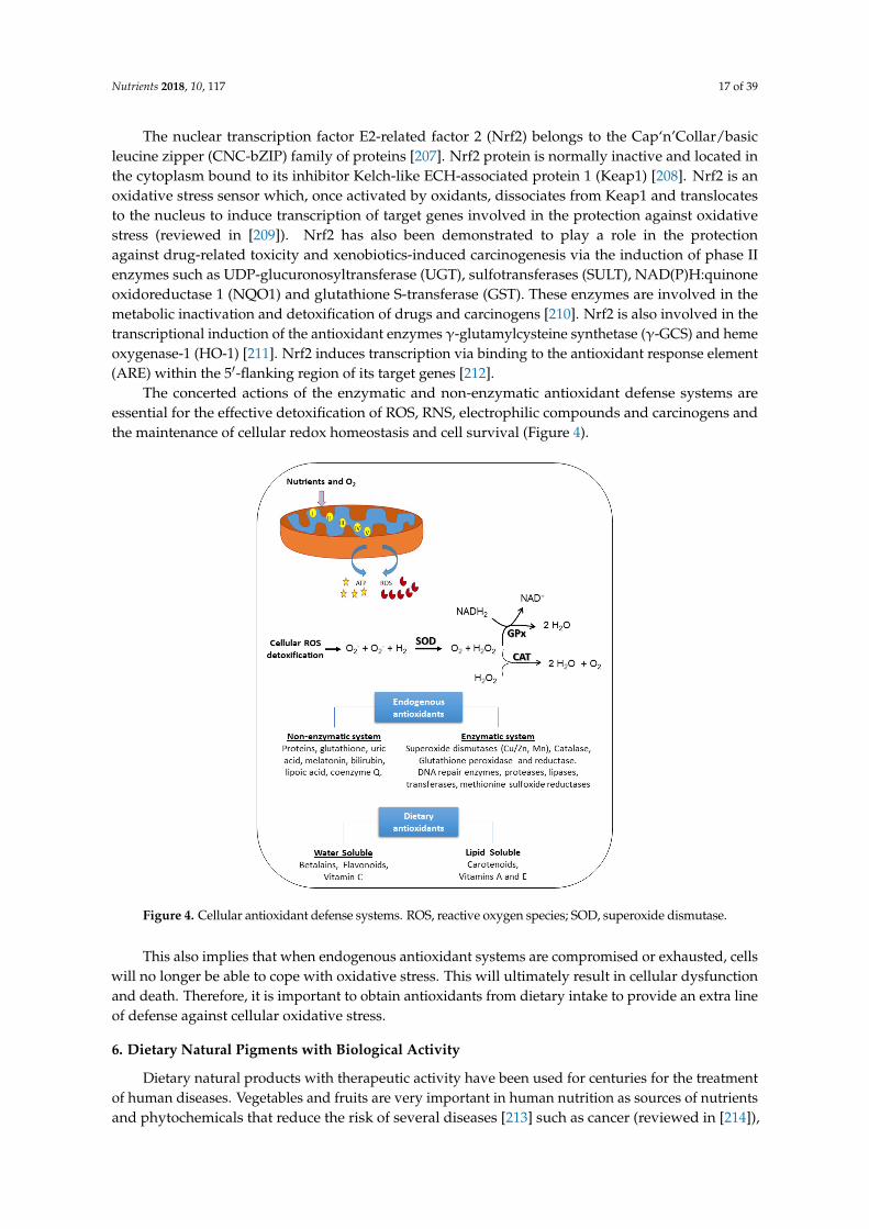
The nuclear transcription factor E2-related factor 2 (Nrf2) belongs to the Cap‘n’Collar/basicleucine zipper (CNC-bZIP) family of proteins [207]. Nrf2 protein is normally inactive and located inthe cytoplasm bound to its inhibitor Kelch-like ECH-associated protein 1 (Keap1) [208]. Nrf2 is anoxidative stress sensor which, once activated by oxidants, dissociates from Keap1 and translocatesto the nucleus to induce transcription of target genes involved in the protection against oxidativestress (reviewed in [209]). Nrf2 has also been demonstrated to play a role in the protectionagainst drug-related toxicity and xenobiotics-induced carcinogenesis via the induction of phase IIenzymes such as UDP-glucuronosyltransferase (UGT), sulfotransferases (SULT), NAD(P)H:quinoneoxidoreductase 1 (NQO1) and glutathione S-transferase (GST). These enzymes are involved in themetabolic inactivation and detoxification of drugs and carcinogens [210]. Nrf2 is also involved in thetranscriptional induction of the antioxidant enzymes γ-glutamylcysteine synthetase (γ-GCS) and hemeoxygenase-1 (HO-1) [211]. Nrf2 induces transcription via binding to the antioxidant response element(ARE) within the 5′-flanking region of its target genes [212].
The concerted actions of the enzymatic and non-enzymatic antioxidant defense systems areessential for the effective detoxification of ROS, RNS, electrophilic compounds and carcinogens andthe maintenance of cellular redox homeostasis and cell survival (Figure 4).
Nutrients 2018, 10, x FOR PEER REVIEW 17 of 38
related toxicity and xenobiotics-induced carcinogenesis via the induction of phase II enzymes such as UDP-glucuronosyltransferase (UGT), sulfotransferases (SULT), NAD(P)H:quinone oxidoreductase 1 (NQO1) and glutathione S-transferase (GST). These enzymes are involved in the metabolic inactivation and detoxification of drugs and carcinogens [210]. Nrf2 is also involved in the transcriptional induction of the antioxidant enzymes γ-glutamylcysteine synthetase (γ-GCS) and heme oxygenase-1 (HO-1) [211]. Nrf2 induces transcription via binding to the antioxidant response element (ARE) within the 5′-flanking region of its target genes [212].
The concerted actions of the enzymatic and non-enzymatic antioxidant defense systems are essential for the effective detoxification of ROS, RNS, electrophilic compounds and carcinogens and the maintenance of cellular redox homeostasis and cell survival (Figure 4).
Figure 4. Cellular antioxidant defense systems. ROS, reactive oxygen species; SOD, superoxide dismutase.
This also implies that when endogenous antioxidant systems are compromised or exhausted, cells will no longer be able to cope with oxidative stress. This will ultimately result in cellular dysfunction and death. Therefore, it is important to obtain antioxidants from dietary intake to provide an extra line of defense against cellular oxidative stress.
6. Dietary Natural Pigments with Biological Activity
Dietary natural products with therapeutic activity have been used for centuries for the treatment of human diseases. Vegetables and fruits are very important in human nutrition as sources of nutrients and phytochemicals that reduce the risk of several diseases [213] such as cancer (reviewed in [214]), cardiovascular disease [215], neurodegenerative diseases (reviewed in [216]), type 2 diabetes [217] and hypertension [218].
It is estimated that the frequent intake of vegetables and fruits might prevent up to one third of cancer-related deaths in the United States [219]. Several in vitro and animal studies suggest that the biological activities of vegetables, fruits and derivatives are frequently related to their antioxidant capacity (reviewed in [40,220]). Therefore, the health benefits of diets rich in vegetables, fruits and
Figure 4. Cellular antioxidant defense systems. ROS, reactive oxygen species; SOD, superoxide dismutase.
This also implies that when endogenous antioxidant systems are compromised or exhausted, cellswill no longer be able to cope with oxidative stress. This will ultimately result in cellular dysfunctionand death. Therefore, it is important to obtain antioxidants from dietary intake to provide an extra lineof defense against cellular oxidative stress.
6. Dietary Natural Pigments with Biological Activity
Dietary natural products with therapeutic activity have been used for centuries for the treatmentof human diseases. Vegetables and fruits are very important in human nutrition as sources of nutrientsand phytochemicals that reduce the risk of several diseases [213] such as cancer (reviewed in [214]),
Nutrients 2018, 10, 117 18 of 39
cardiovascular disease [215], neurodegenerative diseases (reviewed in [216]), type 2 diabetes [217] andhypertension [218].
It is estimated that the frequent intake of vegetables and fruits might prevent up to one third ofcancer-related deaths in the United States [219]. Several in vitro and animal studies suggest that thebiological activities of vegetables, fruits and derivatives are frequently related to their antioxidantcapacity (reviewed in [40,220]). Therefore, the health benefits of diets rich in vegetables, fruits andderived products such as beverages are due not only to fibers, vitamins and minerals, but also to adiversity of plant pigments and secondary metabolites with potential biological activity in humans(reviewed in [12]). Wang et al. reviewed different studies of natural extracts such as Dunaliella salina,Hydnora africana, Calotropis procera, Zingiber officinale Rosc, Hibiscus sabdariffa L., Aloe barbadensis Miller,Lycium barbarum and food-derived compounds such as resveratrol, apigenin, silymarin, lupeol, sesamol,ellagic acid, gallic acid, picroliv, curcumin, β-carotene, sulforaphane, and α-lipoic acid, againstacetaminophen-induced hepatotoxicity. All of them showed a protective effect against the cellularoxidative stress induced by the highly reactive acetaminophen metabolite NAPQI but they appearto have different mechanisms of detoxification depending on the method of extraction, reagents anddosage used (reviewed in [40]). In addition, an in vitro study with 20 fruits commonly consumed inthe American diet (apple, avocado, banana, blueberry, cantaloupe, cherry, cranberry, red and whitegrape, grapefruit, lemon, melon, nectarine, orange, peach, pear, pineapple, plum, strawberry, andwatermelon) suggest that there is synergism between these compounds that improves the antioxidantcapacity compared with single antioxidant compounds [221].
Vitamins, pigments and other phenolic compounds are widely distributed in the plant kingdomand can be found in vegetables, fruits, roots, seeds, leaves, and other natural products such ashoney, propolis and royal jelly. Natural products show a vast diversity of phytochemical compounds,including carotenoids, betalains, flavonoids, and other phenolic compounds [222]. They includestructurally simple molecules as well as complex oligo/polymeric assemblies with very specificbio-physicochemical properties making them important candidates for new drugs [223].
Colored compounds or pigments, are generated from natural sources such as plants, animals,fungi and microorganisms and serve vital functions in cellular processes such as photosynthesis,protection of cells against UV light and oxidative stress and transport of oxygen in blood (heme).Dietary natural pigments can be classified by their structural characteristics as benzopyran derivatives(e.g., flavonoids), N-heterocyclic compounds (e.g., betalains) and isoprenoid derivatives (e.g.,carotenoids) (reviewed in [224]).
The presence or absence of many bioactive compounds in natural products depends ongeographical and environmental factors, such as humidity, temperature, season, pollution and altitude,as well as conditions of growth and storage, and the presence of genetically different varieties [225].Therefore, it is difficult to standardize the chemical composition of natural products, but it is possibleto link their biological activity to the presence of specific compounds with demonstrated therapeuticactivity [226].
Plant-derived antioxidants, in particular pigments, have gained considerable importance dueto their health benefits. Recently, plant foods and food-derived antioxidants have received growingattention, because they are known to function as chemopreventive agents and to protect againstoxidative damage and genotoxicity [227]. The National Research Council (NRC) has recommendedeating five or more servings of fruits and vegetables to increase the health benefits [228].
In addition, there is still a lack of knowledge about the potential therapeutic effect of plant-derivedcompounds against OTC analgesic and NSAIDs-induced acute liver failure. Therefore, additionalanimal studies and clinical trials are needed to discover new antidotes and therapeutic doses fromnatural sources to prevent or reduce drug-induced liver toxicity and acute failure.
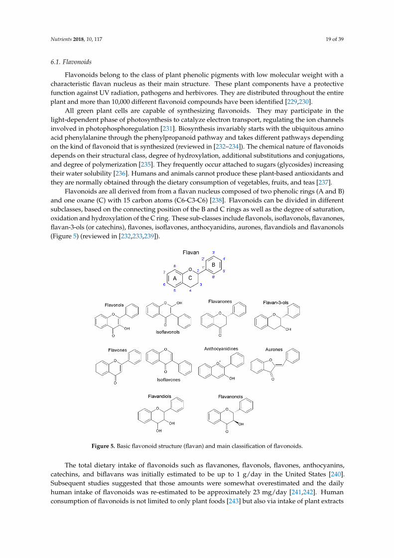
Nutrients 2018, 10, 117 19 of 39
6.1. Flavonoids
Flavonoids belong to the class of plant phenolic pigments with low molecular weight with acharacteristic flavan nucleus as their main structure. These plant components have a protectivefunction against UV radiation, pathogens and herbivores. They are distributed throughout the entireplant and more than 10,000 different flavonoid compounds have been identified [229,230].
All green plant cells are capable of synthesizing flavonoids. They may participate in thelight-dependent phase of photosynthesis to catalyze electron transport, regulating the ion channelsinvolved in photophosphoregulation [231]. Biosynthesis invariably starts with the ubiquitous aminoacid phenylalanine through the phenylpropanoid pathway and takes different pathways dependingon the kind of flavonoid that is synthesized (reviewed in [232–234]). The chemical nature of flavonoidsdepends on their structural class, degree of hydroxylation, additional substitutions and conjugations,and degree of polymerization [235]. They frequently occur attached to sugars (glycosides) increasingtheir water solubility [236]. Humans and animals cannot produce these plant-based antioxidants andthey are normally obtained through the dietary consumption of vegetables, fruits, and teas [237].
Flavonoids are all derived from from a flavan nucleus composed of two phenolic rings (A and B)and one oxane (C) with 15 carbon atoms (C6-C3-C6) [238]. Flavonoids can be divided in differentsubclasses, based on the connecting position of the B and C rings as well as the degree of saturation,oxidation and hydroxylation of the C ring. These sub-classes include flavonols, isoflavonols, flavanones,flavan-3-ols (or catechins), flavones, isoflavones, anthocyanidins, aurones, flavandiols and flavanonols(Figure 5) (reviewed in [232,233,239]).
Nutrients 2018, 10, x FOR PEER REVIEW 19 of 38
flavonoids depends on their structural class, degree of hydroxylation, additional substitutions and conjugations, and degree of polymerization [235]. They frequently occur attached to sugars (glycosides) increasing their water solubility [236]. Humans and animals cannot produce these plant-based antioxidants and they are normally obtained through the dietary consumption of vegetables, fruits, and teas [237].
Flavonoids are all derived from from a flavan nucleus composed of two phenolic rings (A and B) and one oxane (C) with 15 carbon atoms (C6-C3-C6) [238]. Flavonoids can be divided in different subclasses, based on the connecting position of the B and C rings as well as the degree of saturation, oxidation and hydroxylation of the C ring. These sub-classes include flavonols, isoflavonols, flavanones, flavan-3-ols (or catechins), flavones, isoflavones, anthocyanidins, aurones, flavandiols and flavanonols (Figure 5) (reviewed in [232,233,239]).
Figure 5. Basic flavonoid structure (flavan) and main classification of flavonoids.
The total dietary intake of flavonoids such as flavanones, flavonols, flavones, anthocyanins, catechins, and biflavans was initially estimated to be up to 1 g/day in the United States [240]. Subsequent studies suggested that those amounts were somewhat overestimated and the daily human intake of flavonoids was re-estimated to be approximately 23 mg/day [241,242]. Human consumption of flavonoids is not limited to only plant foods [243] but also via intake of plant extracts such as tea, coffee, and red wine [244]. Bioavailability of flavonoids depends on the food source, dosage and their physicochemical properties such as molecular size, lipophilicity, solubility and pKa. In nature, flavonoids are usually bound to sugars such as β-glycosides except for flavan-3-ols and proanthocyanidins. Glycoside conjugates need to be hydrolyzed in the small intestine to release the aglycone flavonoid and be absorbed by passive diffusion through the epithelium. After absorption, flavonoids can react with oxidants to inactivate them or they are metabolized in the liver, e.g., via conjugation reactions. Therefore, aglycone flavonoids cannot be detected in urine or plasma (reviewed in [239]). Recent evidence suggests that the colon plays an important role in the bioavailability of flavonoids since analysis of urinary flavonoid metabolites indicate the presence of colonic metabolites. These results suggest that some of these compounds and their metabolites may play an important role in the protective properties of a vegetable and fruit-rich diet (reviewed in [12]).
Figure 5. Basic flavonoid structure (flavan) and main classification of flavonoids.
The total dietary intake of flavonoids such as flavanones, flavonols, flavones, anthocyanins,catechins, and biflavans was initially estimated to be up to 1 g/day in the United States [240].Subsequent studies suggested that those amounts were somewhat overestimated and the dailyhuman intake of flavonoids was re-estimated to be approximately 23 mg/day [241,242]. Humanconsumption of flavonoids is not limited to only plant foods [243] but also via intake of plant extracts
Nutrients 2018, 10, 117 20 of 39
such as tea, coffee, and red wine [244]. Bioavailability of flavonoids depends on the food source,dosage and their physicochemical properties such as molecular size, lipophilicity, solubility and pKa.In nature, flavonoids are usually bound to sugars such as β-glycosides except for flavan-3-ols andproanthocyanidins. Glycoside conjugates need to be hydrolyzed in the small intestine to release theaglycone flavonoid and be absorbed by passive diffusion through the epithelium. After absorption,flavonoids can react with oxidants to inactivate them or they are metabolized in the liver, e.g., viaconjugation reactions. Therefore, aglycone flavonoids cannot be detected in urine or plasma (reviewedin [239]). Recent evidence suggests that the colon plays an important role in the bioavailability offlavonoids since analysis of urinary flavonoid metabolites indicate the presence of colonic metabolites.These results suggest that some of these compounds and their metabolites may play an importantrole in the protective properties of a vegetable and fruit-rich diet (reviewed in [12]). Flavonoids havea short half-life (≈2–3 h) and they do not accumulate in the body explaining their low toxicity evenwhen ingested for prolonged periods. Despite this, flavonoids might interfere in the biotransformationof many commonly used drugs such as losartan, digoxin, cyclosporine, vinblastine and fexofenadineby the inhibition of CYP450 enzymes, thus increasing their pharmacological potency (reviewedin [232,245]).
Flavonoids have played a major role as medical treatment in ancient times and their usehas continued to present day [232]. Flavonoids can inhibit several important enzymes includingATPase, aldose reductase, hexokinase and tyrosine kinase [246]. Flavonoids can also induce severalenzymes, e.g., aryl hydroxylase and epoxide hydroxylase. Thus, flavonoids seem to possess severalpharmacological properties that make them excellent agents to serve as natural biological responsemodifiers [231]. In addition, modulation of the activity of pro-inflammatory enzymes is one of themost important mechanisms of action for flavonoids. Pro-inflammatory enzymes, such as cytosolicphospholipase A2 (cPLA2), cyclooxygenases (COX), lipoxygenases (LOX), and inducible NO synthase(iNOS), produce very potent inflammatory mediators and therefore their inhibition contributes to theoverall anti-inflammatory potential of flavonoids (reviewed in [247]).
The flavonoid-related antioxidant activity is mainly due to their free radical scavenging potential.The rate and efficiency of this biological activity is determined by the presence of structural featuressuch as the number and position of hydroxyl groups (A, B and C rings) and 2,3-double bond conjugatedwith a 4-oxo function (C ring). These structural features facilitate electron delocalization and radicalabsorption [248]. A quantum chemical study of flavonoids explains that the 3-hydroxyl group in ring Cplays an important role in the activity of flavonols due to its capacity to interact with the positions 2′ and6′ of ring B and the 4-keto group of ring C, making them the most potent flavonoid antioxidants [249].In addition, in vivo studies have demonstrated that flavonoids can also indirectly induce antioxidantenzymes and increase the concentration of uric acid in plasma (reviewed in [250]). Based on thesestructural characteristics and biological activities, flavonoids have been evaluated in various diseasesand toxicity models, including liver injury and toxicity to determine their protective effect (reviewedin [239]). Quercetin, one of the most abundant flavonoids, has been tested in different models of livertoxicity including carbon tetrachloride, ethanol, clivorine, thioacetamide and paracetamol. In thesestudies, orally or intraperitoneally administered quercetin, at doses of 40–90 mg/kg for mice and20–50 mg/kg for rat, showed a strong inhibition of oxidative stress-induced injury, enhancing theredox status and reducing inflammation [251–255]. A mixture of flavonoids from German chamomile(160 mg/kg p.o. (per os) in vivo, and 250–750 µg/mL in vitro) and apigenin-7-glucoside (AP7Glu)(10–60 µM in vitro) protected against ethanol- and CCl4-induced hepatocellular injury in rats andprimary rat hepatocytes by normalizing ceramide content [256]. Another study showed that a mixtureof nine flavonoids identified from Laggera alata extract had hepatoprotective effects at doses (orally)of 50–200 mg/kg in vivo and 1–100 µg/mL in vitro against CCl4-induced liver injury in rats andprimary rat hepatocytes due to its potent antioxidative and anti-inflammatory activity. These resultsalso suggest that flavonoids can scavenge reactive oxygen species by non-enzymatic mechanisms andenhance the activity of hepatic antioxidant enzymes [257]. High glucose-induced oxidative stress
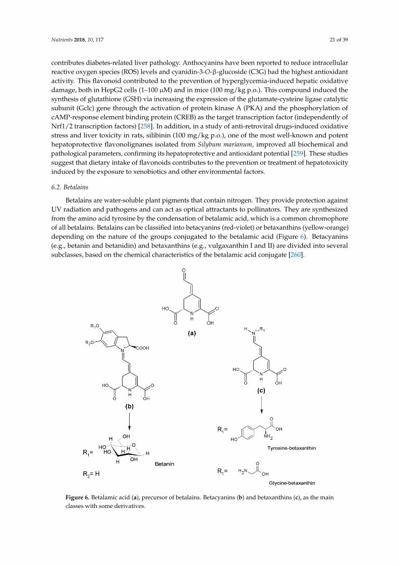
Nutrients 2018, 10, 117 21 of 39
contributes diabetes-related liver pathology. Anthocyanins have been reported to reduce intracellularreactive oxygen species (ROS) levels and cyanidin-3-O-β-glucoside (C3G) had the highest antioxidantactivity. This flavonoid contributed to the prevention of hyperglycemia-induced hepatic oxidativedamage, both in HepG2 cells (1–100 µM) and in mice (100 mg/kg p.o.). This compound induced thesynthesis of glutathione (GSH) via increasing the expression of the glutamate-cysteine ligase catalyticsubunit (Gclc) gene through the activation of protein kinase A (PKA) and the phosphorylation ofcAMP-response element binding protein (CREB) as the target transcription factor (independently ofNrf1/2 transcription factors) [258]. In addition, in a study of anti-retroviral drugs-induced oxidativestress and liver toxicity in rats, silibinin (100 mg/kg p.o.), one of the most well-known and potenthepatoprotective flavonolignanes isolated from Silybum marianum, improved all biochemical andpathological parameters, confirming its hepatoprotective and antioxidant potential [259]. These studiessuggest that dietary intake of flavonoids contributes to the prevention or treatment of hepatotoxicityinduced by the exposure to xenobiotics and other environmental factors.
6.2. Betalains
Betalains are water-soluble plant pigments that contain nitrogen. They provide protection againstUV radiation and pathogens and can act as optical attractants to pollinators. They are synthesizedfrom the amino acid tyrosine by the condensation of betalamic acid, which is a common chromophoreof all betalains. Betalains can be classified into betacyanins (red-violet) or betaxanthins (yellow-orange)depending on the nature of the groups conjugated to the betalamic acid (Figure 6). Betacyanins(e.g., betanin and betanidin) and betaxanthins (e.g., vulgaxanthin I and II) are divided into severalsubclasses, based on the chemical characteristics of the betalamic acid conjugate [260].
Nutrients 2018, 10, x FOR PEER REVIEW 21 of 38
the prevention or treatment of hepatotoxicity induced by the exposure to xenobiotics and other environmental factors.
6.2. Betalains
Betalains are water-soluble plant pigments that contain nitrogen. They provide protection against UV radiation and pathogens and can act as optical attractants to pollinators. They are synthesized from the amino acid tyrosine by the condensation of betalamic acid, which is a common chromophore of all betalains. Betalains can be classified into betacyanins (red-violet) or betaxanthins (yellow-orange) depending on the nature of the groups conjugated to the betalamic acid (Figure 6). Betacyanins (e.g., betanin and betanidin) and betaxanthins (e.g., vulgaxanthin I and II) are divided into several subclasses, based on the chemical characteristics of the betalamic acid conjugate [260].
Figure 6. Betalamic acid (a), precursor of betalains. Betacyanins (b) and betaxanthins (c), as the main classes with some derivatives.
Betalamic acid can be conjugated with cyclo-3,4-dihydroxyphenylalanine (cyclo-Dopa) to form betacyanins and with amino acids or amines to form betaxanthins. The groups conjugated to the betalamic acid determine the absorption wavelength of around 480 nm for betaxanthins and around 540 nm for betacyanins. Structural modifications and the presence of side chains (e.g., sugars and amino acids) cause hypso- or bathochromic shifts (shorter or longer) in the absorption wavelengths [260–262].
Betalain pigments are particularly abundant in the Caryophyllales order and can be found in roots, flowers, fruits and some vegetative tissues of plants [263]. However, betalains are not produced in the Caryophyllaceae and Molluginaceae families, since these families accumulate anthocyanins, flavonoid-derived pigments. Betalains and anthocyanins have never been reported together in the same plant, suggesting they are mutually exclusive [264]. This appears to be an evolutionary mechanism of adaptation to protect against environmental factors and predators [265].
Figure 6. Betalamic acid (a), precursor of betalains. Betacyanins (b) and betaxanthins (c), as the mainclasses with some derivatives.
Nutrients 2018, 10, 117 22 of 39
Betalamic acid can be conjugated with cyclo-3,4-dihydroxyphenylalanine (cyclo-Dopa) to formbetacyanins and with amino acids or amines to form betaxanthins. The groups conjugated to thebetalamic acid determine the absorption wavelength of around 480 nm for betaxanthins and around540 nm for betacyanins. Structural modifications and the presence of side chains (e.g., sugars and aminoacids) cause hypso- or bathochromic shifts (shorter or longer) in the absorption wavelengths [260–262].
Betalain pigments are particularly abundant in the Caryophyllales order and can be found inroots, flowers, fruits and some vegetative tissues of plants [263]. However, betalains are not producedin the Caryophyllaceae and Molluginaceae families, since these families accumulate anthocyanins,flavonoid-derived pigments. Betalains and anthocyanins have never been reported together in the sameplant, suggesting they are mutually exclusive [264]. This appears to be an evolutionary mechanism ofadaptation to protect against environmental factors and predators [265].
Betalains are cationic compounds with a high affinity for negatively charged membranes, thusimproving their efficacy as antioxidants. The active cyclic amine group of betalains functions ashydrogen donor and confers reducing properties to these compounds. In addition, the betacyanins suchas betanin and betanidin have an enhanced antioxidant capacity in vitro compared to betaxanthins,catechin and α-tocopherol. This is due to the presence of a phenolic ring which increases their electrontransfer capability, making them superior antioxidants [266]. It has been suggested that the free radicalscavenging activity of betanin is pH-dependent. It is also important to consider the contribution ofanionic forms of cyclo-DOPA-5-O-β-D-glucoside to the antioxidant activity of betanin at basic pH.These data suggest that betanin is a better hydrogen and electron donor at higher pH, contributing toits enhanced free radical-scavenging activity [267].
Several studies have demonstrated potent radical scavenging activity of betalains and theirderivatives in vitro [268–272]. The antioxidant protection against reactive radical species may bemediated either by direct free radical scavenging or by the induction of endogenous antioxidantdefense mechanisms under the control of redox-regulated transcription factors such as Nrf2 [266].Nrf2 orchestrates the expression of genes encoding antioxidant and phase II enzymes such asheme-oxygenase 1 (HO-1), NAD(P)H:quinone oxidoreductase 1 (NQO1) and glutathione S-transferases(GST). The chemo-preventive effect of betanin is due to the induction of endogenous redox-systemenzymes and glutathione synthesis mediated by Nrf2 dependent signal transduction pathways [272].Chemopreventive compounds may enhance the transcriptional activity of Nrf2 independently fromKeap1 [273].
The dietary intake of betalains in humans is limited, since they can be obtained only from red beet,swiss chard, amaranthus, cactus pear, pitaya and some tubers and their derived products. They arealso used as food colorants providing another dietary source of these phytochemicals. The intakeof red beet juice or cactus pear revealed the bioavailability of betalains. They can be absorbedby the small intestine into the systemic circulation in their intact forms, indicating that hydrolysisreactions are not always necessary for absorption as reported for some glycosylated flavonoids,although the bioavailability of betalains has been reported to be only ≈1% of the consumed amount.The maximum concentration of betalains is detected in plasma 3 h after intake. Moreover, someunknown metabolites have been detected in urine after betalain intake. The biological actions ofthese metabolites are unknown. Betalains are capable to bind to biological membranes reducing theirbioavailability and capacity to react with other macromolecules. The major commercially exploitedsource of betalain is red beetroot (Beta vulgaris), which contains two major soluble pigments: betanin(red) and vulgaxanthine I (yellow). Previous studies reported that the betacyanin and betaxanthincontent of red beet roots varies between 0.04–0.21% and 0.02–0.14%, respectively, depending on thecultivar, although some new varieties have higher betalain content (reviewed in [274]). Red beet rootscontain a large amount of betanin, 300–600 mg/kg, and lower concentrations of isobetanin, betanidinand betaxanthins [266]. The prickly pear (Opuntia ficus indica) contains about 50 mg/kg of betanin and26 mg/kg of indicaxanthin [269]. Opuntia robusta and Opuntia streptacantha fruit juices also containlarge amounts of betacyanins (333 and 87 mg/L, respectively) and betaxanthin (134 and 36 mg/L,
Nutrients 2018, 10, 117 23 of 39
respectively) [7]. The concentration of betacyanins in red pitaya (Hylocereus cacti), expressed as betaninequivalents ranges from 100–400 mg/kg depending on the species [275,276].
Thanks to the increasing interest in the antioxidant properties of betalains, some studieshave focused on their benefits against oxidative stress-related organ damage, although there areonly a few studies on the hepatoprotective effect of betalains. Red beet juice and other red beetproducts, frequently used in dietary products, contain many bioactive compounds, the most importantbeing betalains and may provide protection against certain oxidative stress-related disorders inhumans [266]. Betalains from the berries of Rivina humilis showed dose-dependent cytotoxicity(0–40 µg/mL) in HepG2 hepatoma cells [277]. Frequent consumption of purple Opuntia cactus fruitjuices rich in betalains protects hepatocytes against oxidative stress and improves the redox balancein acetaminophen-induced acute liver toxicity in vivo (800 mg/kg of lyophilized juice p.o.) andin vitro (8% v/v) [7]. In vitro studies have subsequently demonstrated that the free radical scavengingcapacity of betanin (1–35 µM) via activation of the Nrf2 pathway and subsequent induction of Nrf2target genes, is responsible for its hepatoprotective and anticarcinogenic effects [272,273]. Betanin(1–4% in fodder) also attenuated CCl4-induced liver damage in common carp (Cyprinus carpio L.)by the inhibition of CYP2E1 activity and reduction of oxidative stress [278]. The role of betalains ashepatoprotective and chemopreventive compounds can be summarized by their ability to stabilizecellular membranes, scavenge free radicals or electrophilic metabolites, and improve the cellularredox-balance. Nevertheless, the optimal daily intake of betalains to achieve hepatoprotection inhumans has not been elucidated yet due to their apparent poor stability and bioavailability. In addition,there is still a lack of information regarding the safety of consumption of processed betalains and theirpossible pharmacological interactions, necessitating more toxicological and clinical studies.
6.3. Carotenoids
Carotenoids are colored (red, orange, and yellow) pigments widely distributed in naturewith over 700 structurally different compounds [279]. These lipophilic molecules are synthesizedin photosynthetic organelles as well as in fruits, flowers, seeds, and storage roots. Carotenoidshave important functions in photosynthesis in plant and algae, protection against UV light and inphotomorphogenesis. Some microorganisms such as yeast and bacteria can also produce carotenoidsthat function in alleviating photo-oxidative damage [280,281]. Structurally, carotenoids are composedof eight isoprenoid units and can be considered lycopene (C40H56) derivatives. The synthesis of thisgroup of pigments involves hydrogenation, dehydrogenation, cyclization, oxidation, double bondmigration, methyl migration, chain elongation or chain shortening [282].
The natural functions and actions of carotenoids are determined by their physical and chemicalproperties, which are defined by the molecular structure. Carotenoids must have a specific moleculargeometry to ensure their biological activity. The presence and number of double bonds in theirstructures define to a large extent the chemical properties, e.g., lipophilicity that facilitates theirability to inactivate free radicals and protect cellular membranes [283]. The carotenoid classification isbased on their chemical composition: carotenoids composed of carbon and hydrogen and carotenoidscomposed of carbon, hydrogen and oxygen, also called oxycarotenoids or xanthophylls. Alternatively,carotenoids may also be classified as primary carotenoids that are required for plant photosynthesis(β-carotene, violaxanthin, and neoxanthin) and secondary carotenoids that serve as UV protectors andattractants present in flowers and fruits (α-carotene, β-cryptoxanthin, zeaxanthin, lutein, astaxanthin,and capsanthin) [284] (Figure 7).
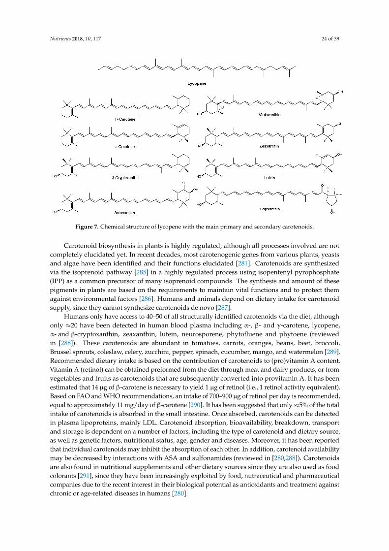
Nutrients 2018, 10, 117 24 of 39
Nutrients 2018, 10, x FOR PEER REVIEW 23 of 38
genes, is responsible for its hepatoprotective and anticarcinogenic effects [272,273]. Betanin (1–4% in fodder) also attenuated CCl4-induced liver damage in common carp (Cyprinus carpio L.) by the inhibition of CYP2E1 activity and reduction of oxidative stress [278]. The role of betalains as hepatoprotective and chemopreventive compounds can be summarized by their ability to stabilize cellular membranes, scavenge free radicals or electrophilic metabolites, and improve the cellular redox-balance. Nevertheless, the optimal daily intake of betalains to achieve hepatoprotection in humans has not been elucidated yet due to their apparent poor stability and bioavailability. In addition, there is still a lack of information regarding the safety of consumption of processed betalains and their possible pharmacological interactions, necessitating more toxicological and clinical studies.
6.3. Carotenoids
Carotenoids are colored (red, orange, and yellow) pigments widely distributed in nature with over 700 structurally different compounds [279]. These lipophilic molecules are synthesized in photosynthetic organelles as well as in fruits, flowers, seeds, and storage roots. Carotenoids have important functions in photosynthesis in plant and algae, protection against UV light and in photomorphogenesis. Some microorganisms such as yeast and bacteria can also produce carotenoids that function in alleviating photo-oxidative damage [280,281]. Structurally, carotenoids are composed of eight isoprenoid units and can be considered lycopene (C40H56) derivatives. The synthesis of this group of pigments involves hydrogenation, dehydrogenation, cyclization, oxidation, double bond migration, methyl migration, chain elongation or chain shortening [282].
The natural functions and actions of carotenoids are determined by their physical and chemical properties, which are defined by the molecular structure. Carotenoids must have a specific molecular geometry to ensure their biological activity. The presence and number of double bonds in their structures define to a large extent the chemical properties, e.g., lipophilicity that facilitates their ability to inactivate free radicals and protect cellular membranes [283]. The carotenoid classification is based on their chemical composition: carotenoids composed of carbon and hydrogen and carotenoids composed of carbon, hydrogen and oxygen, also called oxycarotenoids or xanthophylls. Alternatively, carotenoids may also be classified as primary carotenoids that are required for plant photosynthesis (β-carotene, violaxanthin, and neoxanthin) and secondary carotenoids that serve as UV protectors and attractants present in flowers and fruits (α-carotene, β-cryptoxanthin, zeaxanthin, lutein, astaxanthin, and capsanthin) [284] (Figure 7).
Figure 7. Chemical structure of lycopene with the main primary and secondary carotenoids. Figure 7. Chemical structure of lycopene with the main primary and secondary carotenoids.
Carotenoid biosynthesis in plants is highly regulated, although all processes involved are notcompletely elucidated yet. In recent decades, most carotenogenic genes from various plants, yeastsand algae have been identified and their functions elucidated [281]. Carotenoids are synthesizedvia the isoprenoid pathway [285] in a highly regulated process using isopentenyl pyrophosphate(IPP) as a common precursor of many isoprenoid compounds. The synthesis and amount of thesepigments in plants are based on the requirements to maintain vital functions and to protect themagainst environmental factors [286]. Humans and animals depend on dietary intake for carotenoidsupply, since they cannot synthesize carotenoids de novo [287].
Humans only have access to 40–50 of all structurally identified carotenoids via the diet, althoughonly ≈20 have been detected in human blood plasma including α-, β- and γ-carotene, lycopene,α- and β-cryptoxanthin, zeaxanthin, lutein, neurosporene, phytofluene and phytoene (reviewedin [288]). These carotenoids are abundant in tomatoes, carrots, oranges, beans, beet, broccoli,Brussel sprouts, coleslaw, celery, zucchini, pepper, spinach, cucumber, mango, and watermelon [289].Recommended dietary intake is based on the contribution of carotenoids to (pro)vitamin A content.Vitamin A (retinol) can be obtained preformed from the diet through meat and dairy products, or fromvegetables and fruits as carotenoids that are subsequently converted into provitamin A. It has beenestimated that 14 µg of β-carotene is necessary to yield 1 µg of retinol (i.e., 1 retinol activity equivalent).Based on FAO and WHO recommendations, an intake of 700–900 µg of retinol per day is recommended,equal to approximately 11 mg/day of β-carotene [290]. It has been suggested that only≈5% of the totalintake of carotenoids is absorbed in the small intestine. Once absorbed, carotenoids can be detectedin plasma lipoproteins, mainly LDL. Carotenoid absorption, bioavailability, breakdown, transportand storage is dependent on a number of factors, including the type of carotenoid and dietary source,as well as genetic factors, nutritional status, age, gender and diseases. Moreover, it has been reportedthat individual carotenoids may inhibit the absorption of each other. In addition, carotenoid availabilitymay be decreased by interactions with ASA and sulfonamides (reviewed in [280,288]). Carotenoidsare also found in nutritional supplements and other dietary sources since they are also used as foodcolorants [291], since they have been increasingly exploited by food, nutraceutical and pharmaceuticalcompanies due to the recent interest in their biological potential as antioxidants and treatment againstchronic or age-related diseases in humans [280].
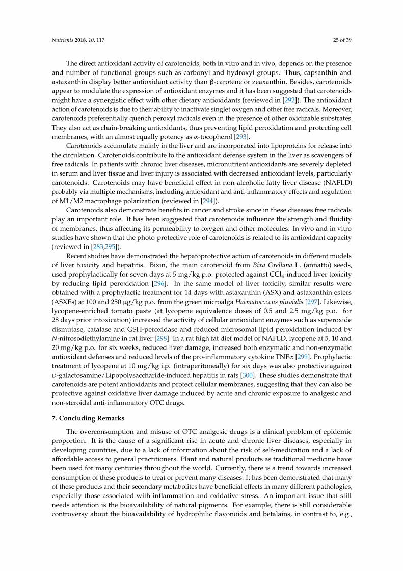
Nutrients 2018, 10, 117 25 of 39
The direct antioxidant activity of carotenoids, both in vitro and in vivo, depends on the presenceand number of functional groups such as carbonyl and hydroxyl groups. Thus, capsanthin andastaxanthin display better antioxidant activity than β-carotene or zeaxanthin. Besides, carotenoidsappear to modulate the expression of antioxidant enzymes and it has been suggested that carotenoidsmight have a synergistic effect with other dietary antioxidants (reviewed in [292]). The antioxidantaction of carotenoids is due to their ability to inactivate singlet oxygen and other free radicals. Moreover,carotenoids preferentially quench peroxyl radicals even in the presence of other oxidizable substrates.They also act as chain-breaking antioxidants, thus preventing lipid peroxidation and protecting cellmembranes, with an almost equally potency as α-tocopherol [293].
Carotenoids accumulate mainly in the liver and are incorporated into lipoproteins for release intothe circulation. Carotenoids contribute to the antioxidant defense system in the liver as scavengers offree radicals. In patients with chronic liver diseases, micronutrient antioxidants are severely depletedin serum and liver tissue and liver injury is associated with decreased antioxidant levels, particularlycarotenoids. Carotenoids may have beneficial effect in non-alcoholic fatty liver disease (NAFLD)probably via multiple mechanisms, including antioxidant and anti-inflammatory effects and regulationof M1/M2 macrophage polarization (reviewed in [294]).
Carotenoids also demonstrate benefits in cancer and stroke since in these diseases free radicalsplay an important role. It has been suggested that carotenoids influence the strength and fluidityof membranes, thus affecting its permeability to oxygen and other molecules. In vivo and in vitrostudies have shown that the photo-protective role of carotenoids is related to its antioxidant capacity(reviewed in [283,295]).
Recent studies have demonstrated the hepatoprotective action of carotenoids in different modelsof liver toxicity and hepatitis. Bixin, the main carotenoid from Bixa Orellana L. (annatto) seeds,used prophylactically for seven days at 5 mg/kg p.o. protected against CCl4-induced liver toxicityby reducing lipid peroxidation [296]. In the same model of liver toxicity, similar results wereobtained with a prophylactic treatment for 14 days with astaxanthin (ASX) and astaxanthin esters(ASXEs) at 100 and 250 µg/kg p.o. from the green microalga Haematococcus pluvialis [297]. Likewise,lycopene-enriched tomato paste (at lycopene equivalence doses of 0.5 and 2.5 mg/kg p.o. for28 days prior intoxication) increased the activity of cellular antioxidant enzymes such as superoxidedismutase, catalase and GSH-peroxidase and reduced microsomal lipid peroxidation induced byN-nitrosodiethylamine in rat liver [298]. In a rat high fat diet model of NAFLD, lycopene at 5, 10 and20 mg/kg p.o. for six weeks, reduced liver damage, increased both enzymatic and non-enzymaticantioxidant defenses and reduced levels of the pro-inflammatory cytokine TNFα [299]. Prophylactictreatment of lycopene at 10 mg/kg i.p. (intraperitoneally) for six days was also protective againstD-galactosamine/Lipopolysaccharide-induced hepatitis in rats [300]. These studies demonstrate thatcarotenoids are potent antioxidants and protect cellular membranes, suggesting that they can also beprotective against oxidative liver damage induced by acute and chronic exposure to analgesic andnon-steroidal anti-inflammatory OTC drugs.
7. Concluding Remarks
The overconsumption and misuse of OTC analgesic drugs is a clinical problem of epidemicproportion. It is the cause of a significant rise in acute and chronic liver diseases, especially indeveloping countries, due to a lack of information about the risk of self-medication and a lack ofaffordable access to general practitioners. Plant and natural products as traditional medicine havebeen used for many centuries throughout the world. Currently, there is a trend towards increasedconsumption of these products to treat or prevent many diseases. It has been demonstrated that manyof these products and their secondary metabolites have beneficial effects in many different pathologies,especially those associated with inflammation and oxidative stress. An important issue that stillneeds attention is the bioavailability of natural pigments. For example, there is still considerablecontroversy about the bioavailability of hydrophilic flavonoids and betalains, in contrast to, e.g.,
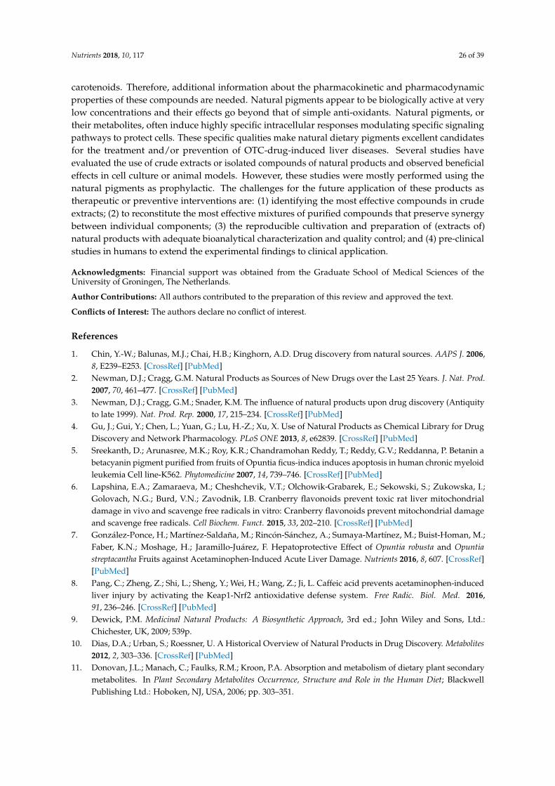
Nutrients 2018, 10, 117 26 of 39
carotenoids. Therefore, additional information about the pharmacokinetic and pharmacodynamicproperties of these compounds are needed. Natural pigments appear to be biologically active at verylow concentrations and their effects go beyond that of simple anti-oxidants. Natural pigments, ortheir metabolites, often induce highly specific intracellular responses modulating specific signalingpathways to protect cells. These specific qualities make natural dietary pigments excellent candidatesfor the treatment and/or prevention of OTC-drug-induced liver diseases. Several studies haveevaluated the use of crude extracts or isolated compounds of natural products and observed beneficialeffects in cell culture or animal models. However, these studies were mostly performed using thenatural pigments as prophylactic. The challenges for the future application of these products astherapeutic or preventive interventions are: (1) identifying the most effective compounds in crudeextracts; (2) to reconstitute the most effective mixtures of purified compounds that preserve synergybetween individual components; (3) the reproducible cultivation and preparation of (extracts of)natural products with adequate bioanalytical characterization and quality control; and (4) pre-clinicalstudies in humans to extend the experimental findings to clinical application.
Acknowledgments: Financial support was obtained from the Graduate School of Medical Sciences of theUniversity of Groningen, The Netherlands.
Author Contributions: All authors contributed to the preparation of this review and approved the text.
Conflicts of Interest: The authors declare no conflict of interest.
References
1. Chin, Y.-W.; Balunas, M.J.; Chai, H.B.; Kinghorn, A.D. Drug discovery from natural sources. AAPS J. 2006,8, E239–E253. [CrossRef] [PubMed]
2. Newman, D.J.; Cragg, G.M. Natural Products as Sources of New Drugs over the Last 25 Years. J. Nat. Prod.2007, 70, 461–477. [CrossRef] [PubMed]
3. Newman, D.J.; Cragg, G.M.; Snader, K.M. The influence of natural products upon drug discovery (Antiquityto late 1999). Nat. Prod. Rep. 2000, 17, 215–234. [CrossRef] [PubMed]
4. Gu, J.; Gui, Y.; Chen, L.; Yuan, G.; Lu, H.-Z.; Xu, X. Use of Natural Products as Chemical Library for DrugDiscovery and Network Pharmacology. PLoS ONE 2013, 8, e62839. [CrossRef] [PubMed]
5. Sreekanth, D.; Arunasree, M.K.; Roy, K.R.; Chandramohan Reddy, T.; Reddy, G.V.; Reddanna, P. Betanin abetacyanin pigment purified from fruits of Opuntia ficus-indica induces apoptosis in human chronic myeloidleukemia Cell line-K562. Phytomedicine 2007, 14, 739–746. [CrossRef] [PubMed]
6. Lapshina, E.A.; Zamaraeva, M.; Cheshchevik, V.T.; Olchowik-Grabarek, E.; Sekowski, S.; Zukowska, I.;Golovach, N.G.; Burd, V.N.; Zavodnik, I.B. Cranberry flavonoids prevent toxic rat liver mitochondrialdamage in vivo and scavenge free radicals in vitro: Cranberry flavonoids prevent mitochondrial damageand scavenge free radicals. Cell Biochem. Funct. 2015, 33, 202–210. [CrossRef] [PubMed]
7. González-Ponce, H.; Martínez-Saldaña, M.; Rincón-Sánchez, A.; Sumaya-Martínez, M.; Buist-Homan, M.;Faber, K.N.; Moshage, H.; Jaramillo-Juárez, F. Hepatoprotective Effect of Opuntia robusta and Opuntiastreptacantha Fruits against Acetaminophen-Induced Acute Liver Damage. Nutrients 2016, 8, 607. [CrossRef][PubMed]
8. Pang, C.; Zheng, Z.; Shi, L.; Sheng, Y.; Wei, H.; Wang, Z.; Ji, L. Caffeic acid prevents acetaminophen-inducedliver injury by activating the Keap1-Nrf2 antioxidative defense system. Free Radic. Biol. Med. 2016,91, 236–246. [CrossRef] [PubMed]
9. Dewick, P.M. Medicinal Natural Products: A Biosynthetic Approach, 3rd ed.; John Wiley and Sons, Ltd.:Chichester, UK, 2009; 539p.
10. Dias, D.A.; Urban, S.; Roessner, U. A Historical Overview of Natural Products in Drug Discovery. Metabolites2012, 2, 303–336. [CrossRef] [PubMed]
11. Donovan, J.L.; Manach, C.; Faulks, R.M.; Kroon, P.A. Absorption and metabolism of dietary plant secondarymetabolites. In Plant Secondary Metabolites Occurrence, Structure and Role in the Human Diet; BlackwellPublishing Ltd.: Hoboken, NJ, USA, 2006; pp. 303–351.
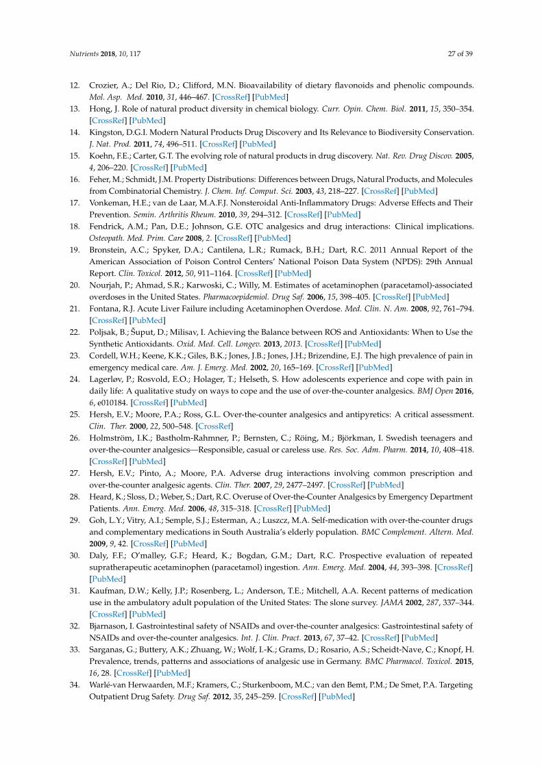
Nutrients 2018, 10, 117 27 of 39
12. Crozier, A.; Del Rio, D.; Clifford, M.N. Bioavailability of dietary flavonoids and phenolic compounds.Mol. Asp. Med. 2010, 31, 446–467. [CrossRef] [PubMed]
13. Hong, J. Role of natural product diversity in chemical biology. Curr. Opin. Chem. Biol. 2011, 15, 350–354.[CrossRef] [PubMed]
14. Kingston, D.G.I. Modern Natural Products Drug Discovery and Its Relevance to Biodiversity Conservation.J. Nat. Prod. 2011, 74, 496–511. [CrossRef] [PubMed]
15. Koehn, F.E.; Carter, G.T. The evolving role of natural products in drug discovery. Nat. Rev. Drug Discov. 2005,4, 206–220. [CrossRef] [PubMed]
16. Feher, M.; Schmidt, J.M. Property Distributions: Differences between Drugs, Natural Products, and Moleculesfrom Combinatorial Chemistry. J. Chem. Inf. Comput. Sci. 2003, 43, 218–227. [CrossRef] [PubMed]
17. Vonkeman, H.E.; van de Laar, M.A.F.J. Nonsteroidal Anti-Inflammatory Drugs: Adverse Effects and TheirPrevention. Semin. Arthritis Rheum. 2010, 39, 294–312. [CrossRef] [PubMed]
18. Fendrick, A.M.; Pan, D.E.; Johnson, G.E. OTC analgesics and drug interactions: Clinical implications.Osteopath. Med. Prim. Care 2008, 2. [CrossRef] [PubMed]
19. Bronstein, A.C.; Spyker, D.A.; Cantilena, L.R.; Rumack, B.H.; Dart, R.C. 2011 Annual Report of theAmerican Association of Poison Control Centers’ National Poison Data System (NPDS): 29th AnnualReport. Clin. Toxicol. 2012, 50, 911–1164. [CrossRef] [PubMed]
20. Nourjah, P.; Ahmad, S.R.; Karwoski, C.; Willy, M. Estimates of acetaminophen (paracetamol)-associatedoverdoses in the United States. Pharmacoepidemiol. Drug Saf. 2006, 15, 398–405. [CrossRef] [PubMed]
21. Fontana, R.J. Acute Liver Failure including Acetaminophen Overdose. Med. Clin. N. Am. 2008, 92, 761–794.[CrossRef] [PubMed]
22. Poljsak, B.; Šuput, D.; Milisav, I. Achieving the Balance between ROS and Antioxidants: When to Use theSynthetic Antioxidants. Oxid. Med. Cell. Longev. 2013, 2013. [CrossRef] [PubMed]
23. Cordell, W.H.; Keene, K.K.; Giles, B.K.; Jones, J.B.; Jones, J.H.; Brizendine, E.J. The high prevalence of pain inemergency medical care. Am. J. Emerg. Med. 2002, 20, 165–169. [CrossRef] [PubMed]
24. Lagerløv, P.; Rosvold, E.O.; Holager, T.; Helseth, S. How adolescents experience and cope with pain indaily life: A qualitative study on ways to cope and the use of over-the-counter analgesics. BMJ Open 2016,6, e010184. [CrossRef] [PubMed]
25. Hersh, E.V.; Moore, P.A.; Ross, G.L. Over-the-counter analgesics and antipyretics: A critical assessment.Clin. Ther. 2000, 22, 500–548. [CrossRef]
26. Holmström, I.K.; Bastholm-Rahmner, P.; Bernsten, C.; Röing, M.; Björkman, I. Swedish teenagers andover-the-counter analgesics—Responsible, casual or careless use. Res. Soc. Adm. Pharm. 2014, 10, 408–418.[CrossRef] [PubMed]
27. Hersh, E.V.; Pinto, A.; Moore, P.A. Adverse drug interactions involving common prescription andover-the-counter analgesic agents. Clin. Ther. 2007, 29, 2477–2497. [CrossRef] [PubMed]
28. Heard, K.; Sloss, D.; Weber, S.; Dart, R.C. Overuse of Over-the-Counter Analgesics by Emergency DepartmentPatients. Ann. Emerg. Med. 2006, 48, 315–318. [CrossRef] [PubMed]
29. Goh, L.Y.; Vitry, A.I.; Semple, S.J.; Esterman, A.; Luszcz, M.A. Self-medication with over-the-counter drugsand complementary medications in South Australia’s elderly population. BMC Complement. Altern. Med.2009, 9, 42. [CrossRef] [PubMed]
30. Daly, F.F.; O’malley, G.F.; Heard, K.; Bogdan, G.M.; Dart, R.C. Prospective evaluation of repeatedsupratherapeutic acetaminophen (paracetamol) ingestion. Ann. Emerg. Med. 2004, 44, 393–398. [CrossRef][PubMed]
31. Kaufman, D.W.; Kelly, J.P.; Rosenberg, L.; Anderson, T.E.; Mitchell, A.A. Recent patterns of medicationuse in the ambulatory adult population of the United States: The slone survey. JAMA 2002, 287, 337–344.[CrossRef] [PubMed]
32. Bjarnason, I. Gastrointestinal safety of NSAIDs and over-the-counter analgesics: Gastrointestinal safety ofNSAIDs and over-the-counter analgesics. Int. J. Clin. Pract. 2013, 67, 37–42. [CrossRef] [PubMed]
33. Sarganas, G.; Buttery, A.K.; Zhuang, W.; Wolf, I.-K.; Grams, D.; Rosario, A.S.; Scheidt-Nave, C.; Knopf, H.Prevalence, trends, patterns and associations of analgesic use in Germany. BMC Pharmacol. Toxicol. 2015,16, 28. [CrossRef] [PubMed]
34. Warlé-van Herwaarden, M.F.; Kramers, C.; Sturkenboom, M.C.; van den Bemt, P.M.; De Smet, P.A. TargetingOutpatient Drug Safety. Drug Saf. 2012, 35, 245–259. [CrossRef] [PubMed]
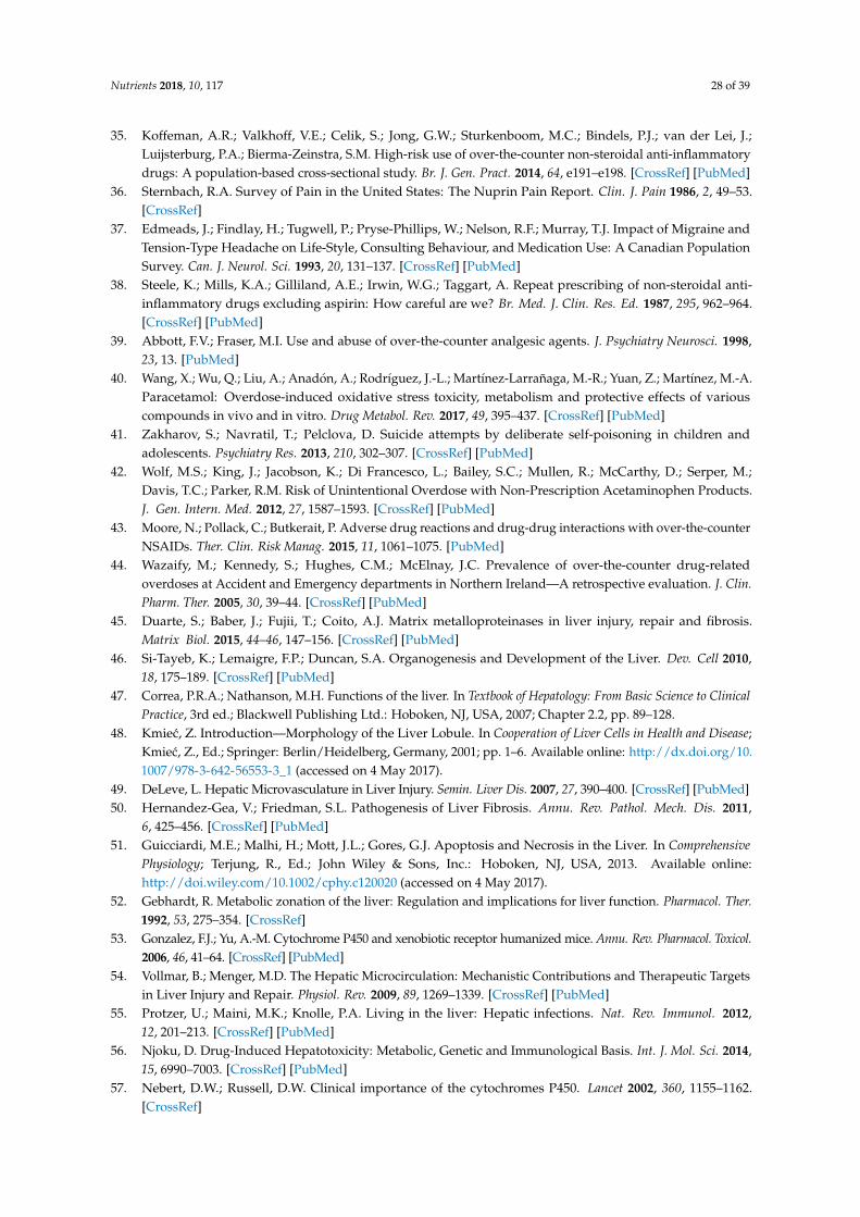
Nutrients 2018, 10, 117 28 of 39
35. Koffeman, A.R.; Valkhoff, V.E.; Celik, S.; Jong, G.W.; Sturkenboom, M.C.; Bindels, P.J.; van der Lei, J.;Luijsterburg, P.A.; Bierma-Zeinstra, S.M. High-risk use of over-the-counter non-steroidal anti-inflammatorydrugs: A population-based cross-sectional study. Br. J. Gen. Pract. 2014, 64, e191–e198. [CrossRef] [PubMed]
36. Sternbach, R.A. Survey of Pain in the United States: The Nuprin Pain Report. Clin. J. Pain 1986, 2, 49–53.[CrossRef]
37. Edmeads, J.; Findlay, H.; Tugwell, P.; Pryse-Phillips, W.; Nelson, R.F.; Murray, T.J. Impact of Migraine andTension-Type Headache on Life-Style, Consulting Behaviour, and Medication Use: A Canadian PopulationSurvey. Can. J. Neurol. Sci. 1993, 20, 131–137. [CrossRef] [PubMed]
38. Steele, K.; Mills, K.A.; Gilliland, A.E.; Irwin, W.G.; Taggart, A. Repeat prescribing of non-steroidal anti-inflammatory drugs excluding aspirin: How careful are we? Br. Med. J. Clin. Res. Ed. 1987, 295, 962–964.[CrossRef] [PubMed]
39. Abbott, F.V.; Fraser, M.I. Use and abuse of over-the-counter analgesic agents. J. Psychiatry Neurosci. 1998,23, 13. [PubMed]
40. Wang, X.; Wu, Q.; Liu, A.; Anadón, A.; Rodríguez, J.-L.; Martínez-Larrañaga, M.-R.; Yuan, Z.; Martínez, M.-A.Paracetamol: Overdose-induced oxidative stress toxicity, metabolism and protective effects of variouscompounds in vivo and in vitro. Drug Metabol. Rev. 2017, 49, 395–437. [CrossRef] [PubMed]
41. Zakharov, S.; Navratil, T.; Pelclova, D. Suicide attempts by deliberate self-poisoning in children andadolescents. Psychiatry Res. 2013, 210, 302–307. [CrossRef] [PubMed]
42. Wolf, M.S.; King, J.; Jacobson, K.; Di Francesco, L.; Bailey, S.C.; Mullen, R.; McCarthy, D.; Serper, M.;Davis, T.C.; Parker, R.M. Risk of Unintentional Overdose with Non-Prescription Acetaminophen Products.J. Gen. Intern. Med. 2012, 27, 1587–1593. [CrossRef] [PubMed]
43. Moore, N.; Pollack, C.; Butkerait, P. Adverse drug reactions and drug-drug interactions with over-the-counterNSAIDs. Ther. Clin. Risk Manag. 2015, 11, 1061–1075. [PubMed]
44. Wazaify, M.; Kennedy, S.; Hughes, C.M.; McElnay, J.C. Prevalence of over-the-counter drug-relatedoverdoses at Accident and Emergency departments in Northern Ireland—A retrospective evaluation. J. Clin.Pharm. Ther. 2005, 30, 39–44. [CrossRef] [PubMed]
45. Duarte, S.; Baber, J.; Fujii, T.; Coito, A.J. Matrix metalloproteinases in liver injury, repair and fibrosis.Matrix Biol. 2015, 44–46, 147–156. [CrossRef] [PubMed]
46. Si-Tayeb, K.; Lemaigre, F.P.; Duncan, S.A. Organogenesis and Development of the Liver. Dev. Cell 2010,18, 175–189. [CrossRef] [PubMed]
47. Correa, P.R.A.; Nathanson, M.H. Functions of the liver. In Textbook of Hepatology: From Basic Science to ClinicalPractice, 3rd ed.; Blackwell Publishing Ltd.: Hoboken, NJ, USA, 2007; Chapter 2.2, pp. 89–128.
48. Kmiec, Z. Introduction—Morphology of the Liver Lobule. In Cooperation of Liver Cells in Health and Disease;Kmiec, Z., Ed.; Springer: Berlin/Heidelberg, Germany, 2001; pp. 1–6. Available online: http://dx.doi.org/10.1007/978-3-642-56553-3_1 (accessed on 4 May 2017).
49. DeLeve, L. Hepatic Microvasculature in Liver Injury. Semin. Liver Dis. 2007, 27, 390–400. [CrossRef] [PubMed]50. Hernandez-Gea, V.; Friedman, S.L. Pathogenesis of Liver Fibrosis. Annu. Rev. Pathol. Mech. Dis. 2011,
6, 425–456. [CrossRef] [PubMed]51. Guicciardi, M.E.; Malhi, H.; Mott, J.L.; Gores, G.J. Apoptosis and Necrosis in the Liver. In Comprehensive
Physiology; Terjung, R., Ed.; John Wiley & Sons, Inc.: Hoboken, NJ, USA, 2013. Available online:http://doi.wiley.com/10.1002/cphy.c120020 (accessed on 4 May 2017).
52. Gebhardt, R. Metabolic zonation of the liver: Regulation and implications for liver function. Pharmacol. Ther.1992, 53, 275–354. [CrossRef]
53. Gonzalez, F.J.; Yu, A.-M. Cytochrome P450 and xenobiotic receptor humanized mice. Annu. Rev. Pharmacol. Toxicol.2006, 46, 41–64. [CrossRef] [PubMed]
54. Vollmar, B.; Menger, M.D. The Hepatic Microcirculation: Mechanistic Contributions and Therapeutic Targetsin Liver Injury and Repair. Physiol. Rev. 2009, 89, 1269–1339. [CrossRef] [PubMed]
55. Protzer, U.; Maini, M.K.; Knolle, P.A. Living in the liver: Hepatic infections. Nat. Rev. Immunol. 2012,12, 201–213. [CrossRef] [PubMed]
56. Njoku, D. Drug-Induced Hepatotoxicity: Metabolic, Genetic and Immunological Basis. Int. J. Mol. Sci. 2014,15, 6990–7003. [CrossRef] [PubMed]
57. Nebert, D.W.; Russell, D.W. Clinical importance of the cytochromes P450. Lancet 2002, 360, 1155–1162.[CrossRef]
Nutrients 2018, 10, 117 29 of 39
58. Guengerich, F.P. Cytochromes P450, drugs, and diseases. Mol. Interv. 2003, 3, 194–204. [CrossRef] [PubMed]59. Shimada, T. Xenobiotic-Metabolizing Enzymes Involved in Activation and Detoxification of Carcinogenic
Polycyclic Aromatic Hydrocarbons. Drug Metab. Pharmacokinet. 2006, 21, 257–276. [CrossRef] [PubMed]60. Xu, C.; Li, C.Y.-T.; Kong, A.-N.T. Induction of phase I, II and III drug metabolism/transport by xenobiotics.
Arch. Pharm. Res. 2005, 28, 249. [CrossRef] [PubMed]61. Kanai, K.; Kanamura, S.; Watanabe, J. Peri- and postnatal development of heterogeneity in the amounts of
endoplasmic reticulum in mouse hepatocytes. Am. J. Anat. 1986, 175, 471–480. [CrossRef] [PubMed]62. Forman, H.J.; Kennedy, J.A. Role of superoxide radical in mitochondrial dehydrogenase reactions.
Biochem. Biophys. Res. Commun. 1974, 60, 1044–1050. [CrossRef]63. Loschen, G.; Azzi, A.; Richter, C.; Flohé, L. Superoxide radicals as precursors of mitochondrial hydrogen
peroxide. FEBS Lett. 1974, 42, 68–72. [CrossRef]64. Halliwell, B. Reactive Species and Antioxidants. Redox Biology Is a Fundamental Theme of Aerobic Life.
Plant Physiol. 2006, 141, 312–322. [CrossRef] [PubMed]65. Harman, D. Aging: A Theory Based on Free Radical and Radiation Chemistry. J. Gerontol. 1956, 11, 298–300.
[CrossRef] [PubMed]66. McCord, J.M.; Fridovich, I. Superoxide dismutase an enzymic function for erythrocuprein (hemocuprein).
J. Biol. Chem. 1969, 244, 6049–6055. [PubMed]67. Finkel, T.; Holbrook, N.J. Oxidants, oxidative stress and the biology of ageing. Nature 2000, 408, 239–247.
[CrossRef] [PubMed]68. Zorov, D.B.; Filburn, C.R.; Klotz, L.-O.; Zweier, J.L.; Sollott, S.J. Reactive oxygen species (ROS-induced) ROS
release. J. Exp. Med. 2000, 192, 1001–1014. [CrossRef] [PubMed]69. Boonstra, J.; Post, J.A. Molecular events associated with reactive oxygen species and cell cycle progression in
mammalian cells. Gene 2004, 337, 1–13. [CrossRef] [PubMed]70. Wong, G.H. Protective roles of cytokines against radiation: Induction of mitochondrial MnSOD.
Biochim. Biophys. Acta (BBA) Mol. Basis Dis. 1995, 1271, 205–209. [CrossRef]71. Mokim Ahmed, K.; Li, J.J. NF-κB-mediated adaptive resistance to ionizing radiation. Free Radic. Biol. Med.
2008, 44, 1–13. [CrossRef] [PubMed]72. Muriel, P. Role of free radicals in liver diseases. Hepatol. Int. 2009, 3, 526–536. [CrossRef] [PubMed]73. Dröge, W. Free Radicals in the Physiological Control of Cell Function. Physiol. Rev. 2002, 82, 47–95. [CrossRef]
[PubMed]74. Forman, H.J. Redox signaling: Thiol chemistry defines which reactive oxygen and nitrogen species can act as
second messengers. Am. J. Physiol. 2004, 287, C246–C256. [CrossRef] [PubMed]75. Turrens, J.F. Superoxide production by the mitochondrial respiratory chain. Biosci. Rep. 1997, 17, 3–8.
[CrossRef] [PubMed]76. Halliwell, B. Reactive Oxygen Species and the Central Nervous System. J. Neurochem. 1992, 59, 1609–1623.
[CrossRef] [PubMed]77. Geiszt, M.; Kopp, J.B.; Várnai, P.; Leto, T.L. Identification of Renox, an NAD(P)H oxidase in kidney. Proc. Natl.
Acad. Sci. USA 2000, 97, 8010–8014. [CrossRef] [PubMed]78. Suh, Y.-A.; Arnold, R.S.; Lassegue, B.; Shi, J.; Xu, X.; Sorescu, D.; Chung, A.; Griendling, K.; Lambeth, J. Cell
transformation by the superoxide-generating oxidase Mox1. Nature 1999, 401, 79–82. [CrossRef] [PubMed]79. Bartsch, H.; Nair, J. Chronic inflammation and oxidative stress in the genesis and perpetuation of cancer:
Role of lipid peroxidation, DNA damage, and repair. Langenbeck’s Arch. Surg. 2006, 391, 499–510. [CrossRef][PubMed]
80. Boveris, A.; Oshino, N.; Chance, B. The cellular production of hydrogen peroxide. Biochem. J. 1972, 128, 617–630.[CrossRef] [PubMed]
81. Jaeschke, H. Reactive oxygen and mechanisms of inflammatory liver injury: Present concepts: Reactiveoxygen and liver inflammation. J. Gastroenterol. Hepatol. 2011, 26, 173–179. [CrossRef] [PubMed]
82. Adamson, G.M.; Harman, A.W. Oxidative stress in cultured hepatocytes exposed to acetaminophen.Biochem. Pharmacol. 1993, 45, 2289–2294. [CrossRef]
83. Knockaert, L.; Descatoire, V.; Vadrot, N.; Fromenty, B.; Robin, M.-A. Mitochondrial CYP2E1 is sufficientto mediate oxidative stress and cytotoxicity induced by ethanol and acetaminophen. Toxicol. In Vitro 2011,25, 475–484. [CrossRef] [PubMed]
Nutrients 2018, 10, 117 30 of 39
84. Boelsterli, U. Diclofenac-induced liver injury: A paradigm of idiosyncratic drug toxicity. Toxicol. Appl.Pharmacol. 2003, 192, 307–322. [CrossRef]
85. Gómez-Lechón, M.J.; Ponsoda, X.; O’Connor, E.; Donato, T.; Castell, J.V.; Jover, R. Diclofenac inducesapoptosis in hepatocytes by alteration of mitochondrial function and generation of ROS. Biochem. Pharmacol.2003, 66, 2155–2167. [CrossRef] [PubMed]
86. Doi, H.; Horie, T. Salicylic acid-induced hepatotoxicity triggered by oxidative stress. Chem. Biol. Interact.2010, 183, 363–368. [CrossRef] [PubMed]
87. Al-Nasser, I.A. Ibuprofen-induced liver mitochondrial permeability transition. Toxicol. Lett. 2000, 111, 213–218.[CrossRef]
88. Vázquez-Medina, J.P.; Zenteno-Savín, T.; Elsner, R.; Ortiz, R.M. Coping with physiological oxidative stress:A review of antioxidant strategies in seals. J. Comp. Physiol. B 2012, 182, 741–750. [CrossRef] [PubMed]
89. Jones, D.P. Redefining oxidative stress. Antioxid. Redox Signal. 2006, 8, 1865–1879. [CrossRef] [PubMed]90. Seven, A.; Güzel, S.; Aslan, M.; Hamuryudan, V. Lipid, protein, DNA oxidation and antioxidant status in
rheumatoid arthritis. Clin. Biochem. 2008, 41, 538–543. [CrossRef] [PubMed]91. Toyokuni, S.; Okamoto, K.; Yodoi, J.; Hiai, H. Persistent oxidative stress in cancer. FEBS Lett. 1995, 358, 1–3.
[CrossRef]92. Conde de la Rosa, L.; Schoemaker, M.H.; Vrenken, T.E.; Buist-Homan, M.; Havinga, R.; Jansen, P.L.M.;
Moshage, H. Superoxide anions and hydrogen peroxide induce hepatocyte death by different mechanisms:Involvement of JNK and ERK MAP kinases. J. Hepatol. 2006, 44, 918–929. [CrossRef] [PubMed]
93. Halliwell, B. Free radicals and antioxidants—Quo vadis? Trends Pharmacol. Sci. 2011, 32, 125–130. [CrossRef][PubMed]
94. Marnett, L.J. Oxy radicals, lipid peroxidation and DNA damage. Toxicology 2002, 181–182, 219–222.[CrossRef]
95. Zhou, L.; Yang, Y.; Tian, D.; Wang, Y. Oxidative stress-induced 1, N6-ethenodeoxyadenosine adduct formationcontributes to hepatocarcinogenesis. Oncol. Rep. 2013, 29, 875–884. [CrossRef] [PubMed]
96. Dukan, S.; Farewell, A.; Ballesteros, M.; Taddei, F.; Radman, M.; Nyström, T. Protein oxidation in response toincreased transcriptional or translational errors. Proc. Natl. Acad. Sci. USA 2000, 97, 5746–5749. [CrossRef][PubMed]
97. Johnson, T.M.; Yu, Z.-X.; Ferrans, V.J.; Lowenstein, R.A.; Finkel, T. Reactive oxygen species are downstreammediators of p53-dependent apoptosis. Proc. Natl. Acad. Sci. USA 1996, 93, 11848–11852. [CrossRef][PubMed]
98. Polyak, K.; Xia, Y.; Zweier, J.L.; Kinzler, K.W.; Vogelstein, B. A model for p53-induced apoptosis. Nature 1997,389, 300–305. [CrossRef] [PubMed]
99. Knebel, A.; Rahmsdorf, H.J.; Ullrich, A.; Herrlich, P. Dephosphorylation of receptor tyrosine kinases as targetof regulation by radiation, oxidants or alkylating agents. EMBO J. 1996, 15, 5314–5325. [PubMed]
100. Wang, X.; McCullough, K.D.; Franke, T.F.; Holbrook, N.J. Epidermal growth factor receptor-dependentAkt activation by oxidative stress enhances cell survival. J. Biol. Chem. 2000, 275, 14624–14631. [CrossRef][PubMed]
101. Saitoh, M.; Nishitoh, H.; Fujii, M.; Takeda, K.; Tobiume, K.; Sawada, Y.; Kawabata, M.; Miyazono, K.; Ichijo, H.Mammalian thioredoxin is a direct inhibitor of apoptosis signal-regulating kinase (ASK) 1. EMBO J. 1998,17, 2596–2606. [CrossRef] [PubMed]
102. Adler, V.; Yin, Z.; Fuchs, S.Y.; Benezra, M.; Rosario, L.; Tew, K.D.; Pincus, M.; Sardana, M.; Henderson, C.;Wolf, C.R.; et al. Regulation of JNK signaling by GSTp. EMBO J. 1999, 18, 1321–1334. [CrossRef] [PubMed]
103. Li, S.; Tan, H.-Y.; Wang, N.; Zhang, Z.-J.; Lao, L.; Wong, C.-W.; Feng, Y. The Role of Oxidative Stress andAntioxidants in Liver Diseases. Int. J. Mol. Sci. 2015, 16, 26087–26124. [CrossRef] [PubMed]
104. Kaplowitz, N. Drug-induced liver injury. Clin. Infect. Dis. 2004, 38 (Suppl. 2), S44–S48. [CrossRef] [PubMed]105. Reuben, A.; Koch, D.G.; Lee, W.M. Drug-induced acute liver failure: Results of a U.S. Multicenter, prospective
study. Hepatology 2010, 52, 2065–2076. [CrossRef] [PubMed]106. Larson, A.M.; Polson, J.; Fontana, R.J.; Davern, T.J.; Lalani, E.; Hynan, L.S.; Reisch, J.S.; Schiødt, F.V.;
Ostapowicz, G.; Shakil, A.O.; et al. Acetaminophen-induced acute liver failure: Results of a United Statesmulticenter, prospective study. Hepatology 2005, 42, 1364–1372. [CrossRef] [PubMed]
107. Bernal, W.; Wendon, J. Acute Liver Failure. N. Engl. J. Med. 2013, 369, 2525–2534. [CrossRef] [PubMed]

Nutrients 2018, 10, 117 31 of 39
108. Grattagliano, I. Biochemical mechanisms in drug-induced liver injury: Certainties and doubts. World J.Gastroenterol. 2009, 15, 4865. [CrossRef] [PubMed]
109. Iorga, A.; Dara, L.; Kaplowitz, N. Drug-Induced Liver Injury: Cascade of Events Leading to Cell Death,Apoptosis or Necrosis. Int. J. Mol. Sci. 2017, 18, 1018. [CrossRef] [PubMed]
110. Roth, A.D.; Lee, M.-Y. Idiosyncratic Drug-Induced Liver Injury (IDILI): Potential Mechanisms and PredictiveAssays. BioMed Res. Int. 2017, 2017, 9176937. [CrossRef] [PubMed]
111. Han, D.; Dara, L.; Win, S.; Than, T.A.; Yuan, L.; Abbasi, S.Q.; Liu, Z.-X.; Kaplowitz, N. Regulation ofdrug-induced liver injury by signal transduction pathways: Critical role of mitochondria. Trends Pharmacol. Sci.2013, 34, 243–253. [CrossRef] [PubMed]
112. Cohen, S.D.; Pumford, N.R.; Khairallah, E.A.; Boekelheide, K.; Pohl, L.R.; Amouzadeh, H.R.; Hinson, J.A.Selective Protein Covalent Binding and Target Organ Toxicity. Toxicol. Appl. Pharmacol. 1997, 143, 1–12.[CrossRef] [PubMed]
113. Baillie, T.A.; Rettie, A.E. Role of biotransformation in drug-induced toxicity: Influence of intra-and inter-speciesdifferences in drug metabolism. Drug Metab. Pharmacokinet. 2011, 26, 15–29. [CrossRef] [PubMed]
114. Kaplowitz, N. Mechanisms of liver cell injury. J. Hepatol. 2000, 32, 39–47. [CrossRef]115. Singal, A.K.; Jampana, S.C.; Weinman, S.A. Antioxidants as therapeutic agents for liver disease. Liver Int.
2011, 31, 1432–1448. [CrossRef] [PubMed]116. Han, D.; Shinohara, M.; Ybanez, M.D.; Saberi, B.; Kaplowitz, N. Signal Transduction Pathways Involved
in Drug-Induced Liver Injury. In Adverse Drug Reactions; Uetrecht, J., Ed.; Springer: Berlin/Heidelberg,Germany, 2010; pp. 267–310. Available online: http://dx.doi.org/10.1007/978-3-642-00663-0_10 (accessed on27 June 2017).
117. Begriche, K.; Massart, J.; Robin, M.-A.; Borgne-Sanchez, A.; Fromenty, B. Drug-induced toxicity onmitochondria and lipid metabolism: Mechanistic diversity and deleterious consequences for the liver.J. Hepatol. 2011, 54, 773–794. [CrossRef] [PubMed]
118. Meredith, M.J.; Reed, D.J. Status of the mitochondrial pool of glutathione in the isolated hepatocyte.J. Biol. Chem. 1982, 257, 3747–3753. [PubMed]
119. Jones, D.P.; Lemasters, J.J.; Han, D.; Boelsterli, U.A.; Kaplowitz, N. Mechanisms of pathogenesis in drughepatotoxicity putting the stress on mitochondria. Mol. Interv. 2010, 10, 98–111. [CrossRef] [PubMed]
120. Simon, L.S. Nonsteroidal anti-inflammatory drugs and their risk: A story still in development. BMC ArthritisRes. Ther. 2013, 15. [CrossRef] [PubMed]
121. Ghosh, R.; Alajbegovic, A.; Gomes, A.V. NSAIDs and Cardiovascular Diseases: Role of Reactive OxygenSpecies. Oxid. Med. Cell. Longev. 2015, 2015, 536962. [CrossRef] [PubMed]
122. Dubois, R.N.; Abramson, S.B.; Crofford, L.; Gupta, R.A.; Simon, L.S.A.; Van De Putte, L.B.; Lipsky, P.E.Cyclooxygenase in biology and disease. FASEB J. 1998, 12, 1063–1073. [CrossRef] [PubMed]
123. Crofford, L.J. Use of NSAIDs in treating patients with arthritis. BMC Arthritis Res. Ther. 2013, 15. [CrossRef]124. Schmeltzer, P.A.; Kosinski, A.S.; Kleiner, D.E.; Hoofnagle, J.H.; Stolz, A.; Fontana, R.J.; Russo, M.W. Liver
injury from nonsteroidal anti-inflammatory drugs in the United States. Liver Int. 2016, 36, 603–609. [CrossRef][PubMed]
125. Leise, M.D.; Poterucha, J.J.; Talwalkar, J.A. Drug-Induced Liver Injury. Mayo Clin. Proc. 2014, 89, 95–106.[CrossRef] [PubMed]
126. Unzueta, A.; Vargas, H.E. Nonsteroidal Anti-Inflammatory Drug–Induced Hepatoxicity. Drug Hepatotoxic.2013, 17, 643–656. [CrossRef] [PubMed]
127. Liou, J.-Y.; Wu, C.-C.; Chen, B.-R.; Yen, L.B.; Wu, K.K. Nonsteroidal Anti-Inflammatory Drugs InducedEndothelial Apoptosis by Perturbing Peroxisome Proliferator-Activated Receptor-δ Transcriptional Pathway.Mol. Pharmacol. 2008, 74, 1399–1406. [CrossRef] [PubMed]
128. Watson, A.J.M.; Askew, J.N.; Benson, R.S.P. Poly(adenosine diphosphate ribose) polymerase inhibitionprevents necrosis induced by H2O2 but not apoptosis. Gastroenterology 1995, 109, 472–482. [CrossRef]
129. Eugenio-Pérez, D.; Montes de Oca-Solano, H.A.; Pedraza-Chaverri, J. Role of food-derived antioxidant agentsagainst acetaminophen-induced hepatotoxicity. Pharm. Biol. 2016, 54, 2340–2352. [CrossRef] [PubMed]
130. Castro, M.P.V. Intoxicación por acetaminofén en adultos. Med. Leg. Costa Rica 2016, 33, 103–109.131. Bunchorntavakul, C.; Reddy, K.R. Acetaminophen-related Hepatotoxicity. Drug Hepatotoxic. 2013, 17, 587–607.
[CrossRef] [PubMed]
Nutrients 2018, 10, 117 32 of 39
132. Hodgman, M.J.; Garrard, A.R. A Review of Acetaminophen Poisoning. Toxicology 2012, 28, 499–516.[CrossRef] [PubMed]
133. Lee, W. Acute Liver Failure. Semin. Respir. Crit. Care Med. 2012, 33, 36–45. [CrossRef] [PubMed]134. Jaeschke, H. Acetaminophen: Dose-Dependent Drug Hepatotoxicity and Acute Liver Failure in Patients.
Dig. Dis. 2015, 33, 464–471. [CrossRef] [PubMed]135. Lancaster, E.M.; Hiatt, J.R.; Zarrinpar, A. Acetaminophen hepatotoxicity: An updated review. Arch. Toxicol.
2015, 89, 193–199. [CrossRef] [PubMed]136. Jaeschke, H. The role of oxidant stress and reactive nitrogen species in acetaminophen hepatotoxicity.
Toxicol. Lett. 2003, 144, 279–288. [CrossRef]137. McGill, M.R.; Jaeschke, H. Metabolism and Disposition of Acetaminophen: Recent Advances in Relation to
Hepatotoxicity and Diagnosis. Pharm. Res. 2013, 30, 2174–2187. [CrossRef] [PubMed]138. Saberi, B.; Ybanez, M.D.; Johnson, H.S.; Gaarde, W.A.; Han, D.; Kaplowitz, N. Protein kinase C (PKC)
participates in acetaminophen hepatotoxicity through JNK dependent and independent signaling pathways.Hepatology 2014, 59, 1543–1554. [CrossRef] [PubMed]
139. Jaeschke, H.; McGill, M.R.; Ramachandran, A. Oxidant stress, mitochondria, and cell death mechanismsin drug-induced liver injury: Lessons learned from acetaminophen hepatotoxicity. Drug Metab. Rev. 2012,44, 88–106. [CrossRef] [PubMed]
140. Jaeschke, H.; Xie, Y.; McGill, M.R. Acetaminophen-induced Liver Injury: From Animal Models to Humans.J. Clin. Transl. Hepatol. 2014, 2, 153–161. [PubMed]
141. Cover, C. Peroxynitrite-Induced Mitochondrial and Endonuclease-Mediated Nuclear DNA Damage inAcetaminophen Hepatotoxicity. J. Pharmacol. Exp. Ther. 2005, 315, 879–887. [CrossRef] [PubMed]
142. Masubuchi, Y.; Suda, C.; Horie, T. Involvement of mitochondrial permeability transition in acetaminophen-induced liver injury in mice. J. Hepatol. 2005, 42, 110–116. [CrossRef] [PubMed]
143. Hanawa, N.; Shinohara, M.; Saberi, B.; Gaarde, W.A.; Han, D.; Kaplowitz, N. Role of JNK Translocation toMitochondria Leading to Inhibition of Mitochondria Bioenergetics in Acetaminophen-induced Liver Injury.J. Biol. Chem. 2008, 283, 13565–13577. [CrossRef] [PubMed]
144. Gujral, J.S.; Knight, T.R.; Farhood, A.; Bajt, M.L.; Jaeschke, H. Mode of Cell Death after AcetaminophenOverdose in Mice: Apoptosis or Oncotic Necrosis? Toxicol. Sci. 2002, 67, 322–328. [CrossRef] [PubMed]
145. Das, J.; Ghosh, J.; Manna, P.; Sil, P.C. Acetaminophen induced acute liver failure via oxidative stress andJNK activation: Protective role of taurine by the suppression of cytochrome P450 2E1. Free Radic. Res. 2010,44, 340–355. [CrossRef] [PubMed]
146. Arai, T.; Koyama, R.; Yuasa, M.; Kitamura, D.; Mizuta, R. Acrolein, a highly toxic aldehyde generated underoxidative stress in vivo, aggravates the mouse liver damage after acetaminophen overdose. Biomed. Res.2014, 35, 389–395. [CrossRef] [PubMed]
147. Kon, K.; Kim, J.-S.; Jaeschke, H.; Lemasters, J.J. Mitochondrial permeability transition in acetaminophen-induced necrosis and apoptosis of cultured mouse hepatocytes. Hepatology 2004, 40, 1170–1179. [CrossRef][PubMed]
148. Kass, G.E.N.; Macanas-Pirard, P.; Lee, P.C.; Hinton, R.H. The Role of Apoptosis in Acetaminophen-InducedInjury. Ann. N. Y. Acad. Sci. 2003, 1010, 557–559. [CrossRef] [PubMed]
149. Possamai, L.A.; McPhail, M.J.; Quaglia, A.; Zingarelli, V.; Abeles, R.D.; Tidswell, R.; Puthucheary, Z.; Rawal, J.;Karvellas, C.; Leslie, E.M.; et al. Character and Temporal Evolution of Apoptosis in Acetaminophen-InducedAcute Liver Failure. Crit. Care Med. 2013, 41, 2543–2550. [CrossRef] [PubMed]
150. Yoon, E.; Babar, A.; Choudhary, M.; Kutner, M.; Pyrsopoulos, N. Acetaminophen-Induced Hepatotoxicity:A Comprehensive Update. J. Clin. Transl. Hepatol. 2016, 4, 131–142. [PubMed]
151. Jain, N.; Shrivastava, R.; Raghuwanshi, A.; Shrivastava, V. Aspirin induced changes in serum ACP, ALP,GOT, GPT, bilirubin and creatinine in corelation with histopathological changes in liver and kidney of femalealbino rat. Int. J. Appl. Pharm. 2012, 4, 9–11.
152. Bojic, M.; Sedgeman, C.A.; Nagy, L.D.; Guengerich, F.P. Aromatic Hydroxylation of Salicylic Acid andAspirin by Human Cytochromes P450. Eur. J. Pharm. Sci. 2015, 73, 49–56. [CrossRef] [PubMed]
153. Zimmerman, H.J. Aspirin-induced hepatic injury. Ann. Intern. Med. 1974, 80, 103–105. [CrossRef] [PubMed]154. Bessone, F. Non-steroidal anti-inflammatory drugs: What is the actual risk of liver damage? World J.
Gastroenterol. 2010, 16, 5651–5661. [CrossRef] [PubMed]
Nutrients 2018, 10, 117 33 of 39
155. Doi, H.; Masubuchi, Y.; Narimatsu, S.; Nishigaki, R.; Horie, T. Salicylic acid-induced lipid peroxidation in ratliver microsomes. Res. Commun. Mol. Pathol. Pharmacol. 1998, 100, 265–271. [PubMed]
156. Berk, M.; Dean, O.; Drexhage, H.; McNeil, J.J.; Moylan, S.; O’Neil, A.; Davey, C.; Sanna, L.; Maes, M. Aspirin:A review of its neurobiological properties and therapeutic potential for mental illness. BMC Med. 2013,11, 74. [CrossRef] [PubMed]
157. Raza, H.; John, A.; Benedict, S. Acetylsalicylic acid-induced oxidative stress, cell cycle arrest, apoptosis andmitochondrial dysfunction in human hepatoma HepG2 cells. Eur. J. Pharmacol. 2011, 668, 15–24. [CrossRef][PubMed]
158. Raza, H.; John, A. Implications of Altered Glutathione Metabolism in Aspirin-Induced Oxidative Stress andMitochondrial Dysfunction in HepG2 Cells. PLoS ONE 2012, 7, e36325. [CrossRef] [PubMed]
159. Tassone, E.J.; Perticone, M.; Sciacqua, A.; Mafrici, S.F.; Settino, C.; Malara, N.; Mollace, V.; Sesti, G.;Perticone, F. Low dose of acetylsalicylic acid and oxidative stress-mediated endothelial dysfunction indiabetes: A short-term evaluation. Acta Diabetol. 2015, 52, 249–256. [CrossRef] [PubMed]
160. Deschamps, D.; Fisch, C.; Fromenty, B.; Berson, A.; Degott, C.; Pessayre, D. Inhibition by salicylic acid of theactivation and thus oxidation of long chain fatty acids. Possible role in the development of Reye’s syndrome.J. Pharmacol. Exp. Ther. 1991, 259, 894. [PubMed]
161. Lauterburg, B.; Grattagliano, I.; Gmür, R.; Stalder, M.; Hildebrand, P. Noninvasive assessment of the effect ofxenobiotics on mitochondrial function in human beings: Studies with acetylsalicylic acid and ethanol withthe use of the carbon 13-labeled ketoisocaproate breath test. J. Lab. Clin. Med. 1995, 125, 378–383. [PubMed]
162. Fromenty, B.; Pessayre, D. Inhibition of mitochondrial beta-oxidation as a mechanism of hepatotoxicity.Pharmacol. Ther. 1995, 67, 101–154. [CrossRef]
163. Gan, T.J. Diclofenac: An update on its mechanism of action and safety profile. Curr. Med. Res. Opin. 2010,26, 1715–1731. [CrossRef] [PubMed]
164. Altman, R.; Bosch, B.; Brune, K.; Patrignani, P.; Young, C. Advances in NSAID Development: Evolution ofDiclofenac Products Using Pharmaceutical Technology. Drugs 2015, 75, 859–877. [CrossRef] [PubMed]
165. Aithal, G.P.; Day, C.P. Nonsteroidal Anti-Inflammatory Drug–Induced Hepatotoxicity. Drug Induc. Liver Dis.2007, 11, 563–575. [CrossRef] [PubMed]
166. Watkins, P.B.; Seligman, P.J.; Pears, J.S.; Avigan, M.I.; Senior, J.R. Using controlled clinical trials to learn moreabout acute drug-induced liver injury. Hepatology 2008, 48, 1680–1689. [CrossRef] [PubMed]
167. Banks, A.T.; Zimmerman, H.J.; Ishak, K.G.; Harter, J.G. Diclofenac-associated hepatotoxicity: Analysis of180 cases reported to the food and drug administration as adverse reactions. Hepatology 1995, 22, 820–827.[CrossRef] [PubMed]
168. Laine, L.; Goldkind, L.; Curtis, S.P.; Connors, L.G.; Yanqiong, Z.; Cannon, C.P. How Common IsDiclofenac-Associated Liver Injury? Analysis of 17,289 Arthritis Patients in a Long-Term ProspectiveClinical Trial. Am. J. Gastroenterol. 2009, 104, 356–362. [CrossRef] [PubMed]
169. Yano, A.; Higuchi, S.; Tsuneyama, K.; Fukami, T.; Nakajima, M.; Yokoi, T. Involvement of immune-relatedfactors in diclofenac-induced acute liver injury in mice. Toxicology 2012, 293, 107–114. [CrossRef] [PubMed]
170. Bort, R.; Ponsoda, X.; Jover, R.; Gómez-Lechón, M.J.; Castell, J.V. Diclofenac Toxicity to Hepatocytes: A Rolefor Drug Metabolism in Cell Toxicity. J. Pharmacol. Exp. Ther. 1999, 288, 65–72. [PubMed]
171. Amin, A.; Hamza, A.A. Oxidative stress mediates drug-induced hepatotoxicity in rats: A possible role ofDNA fragmentation. Toxicology 2005, 208, 367–375. [CrossRef] [PubMed]
172. Vickers, A.E.M. Tissue slices for the evaluation of metabolism-based toxicity with the example of diclofenac.Eval. Metab. Based Drug Toxic Drug Dev. 2009, 179, 9–16. [CrossRef] [PubMed]
173. Pourahmad, J.; Mortada, Y.; Eskandari, M.R.; Shahraki, J. Involvement of Lysosomal Labilisation andLysosomal/mitochondrial Cross-Talk in Diclofenac Induced Hepatotoxicity. Iran. J. Pharm. Res. 2011,10, 877–887. [PubMed]
174. Fredriksson, L.; Wink, S.; Herpers, B.; Benedetti, G.; Hadi, M.; de Bont, H.; Groothuis, G.; Luijten, M.;Danen, E.; de Graauw, M.; et al. Drug-Induced Endoplasmic Reticulum and Oxidative Stress ResponsesIndependently Sensitize toward TNFα-Mediated Hepatotoxicity. Toxicol. Sci. 2014, 140, 144–159. [CrossRef][PubMed]
175. Andrejak, M.; Davion, T.; Gineston, J.L.; Capron, J.P. Cross hepatotoxicity between non-steroidal anti-inflammatory drugs. Br. Med. J. Clin. Res. Ed. 1987, 295, 180–181. [CrossRef] [PubMed]
Nutrients 2018, 10, 117 34 of 39
176. Ali, S.; Pimentel, J.D.; Ma, C. Naproxen-induced liver injury. Hepatobiliary Pancreat. Dis. Int. 2011, 10, 552–556.[CrossRef]
177. Davies, N.M.; Anderson, K.E. Clinical Pharmacokinetics of Naproxen. Clin. Pharmacokinet. 1997, 32, 268–293.[CrossRef] [PubMed]
178. Miners, J.O.; Coulter, S.; Tukey, R.H.; Veronese, M.E.; Birkett, D.J. Cytochromes P450, 1A2, and 2C9 areresponsible for the human hepatic O-demethylation of R- and S-naproxen. Biochem. Pharmacol. 1996,51, 1003–1008. [CrossRef]
179. Tracy, T.S.; Marra, C.; Wrighton, S.A.; Gonzalez, F.J.; Korzekwa, K.R. Involvement of multiple cytochromeP450 isoforms in naproxen O-demethylation. Eur. J. Clin. Pharmacol. 1997, 52, 293–298. [CrossRef] [PubMed]
180. Bowalgaha, K.; Elliot, D.J.; Mackenzie, P.I.; Knights, K.M.; Swedmark, S.; Miners, J.O. S-Naproxenand desmethylnaproxen glucuronidation by human liver microsomes and recombinant humanUDP-glucuronosyltransferases (UGT): Role of UGT2B7 in the elimination of naproxen. Br. J. Clin. Pharmacol.2005, 60, 423–433. [CrossRef] [PubMed]
181. Yokoyama, H.; Horie, T.; Awazu, S. Oxidative stress in isolated rat hepatocytes during naproxen metabolism.Biochem. Pharmacol. 1995, 49, 991–996. [CrossRef]
182. Yokoyama, H.; Horie, T.; Awazu, S. Glutathione disulfide formation during naproxen metabolism in theisolated rat hepatocytes. Res. Commun. Mol. Pathol. Pharmacol. 1998, 99, 143–154. [PubMed]
183. Ji, B.; Masubuchi, Y.; Horie, T. A Possible Mechanism of Naproxen-Induced Lipid Peroxidation in Rat LiverMicrosomes. Pharmacol. Toxicol. 2001, 89, 43–48. [CrossRef] [PubMed]
184. Lo, A.; Addison, R.S.; Hooper, W.D.; Dickinson, R.G. Disposition of naproxen, naproxen acyl glucuronideand its rearrangement isomers in the isolated perfused rat liver. Xenobiotica 2001, 31, 309–319. [CrossRef][PubMed]
185. Yokoyama, H.; Horie, T.; Awazu, S. Naproxen-induced oxidative stress in the isolated perfused rat liver.Chem. Biol. Interact. 2006, 160, 150–158. [CrossRef] [PubMed]
186. Naproxen. LiverTox, 2017. Available online: https://livertox.nlm.nih.gov//Naproxen.htm (accessed on27 June 2017).
187. Bushra, R.; Aslam, N. An Overview of Clinical Pharmacology of Ibuprofen. Oman Med. J. 2010, 25, 155–1661.[PubMed]
188. Ibuprofen. LiverTox. 2017. Available online: https://livertox.nlm.nih.gov//Ibuprofen.htm (accessed on27 June 2017).
189. Rainsford, K.D. Ibuprofen: Pharmacology, efficacy and safety. Inflammopharmacology 2009, 17, 275–342.[CrossRef] [PubMed]
190. Riley, T.R.; Smith, J.P. Ibuprofen-induced hepatotoxicity in patients with chronic hepatitis c: A case series.Am. J. Gastroenterol. 1998, 93, 1563–1565. [CrossRef] [PubMed]
191. Basturk, A.; Artan, R.; Yılmaz, A.; Gelen, M.T.; Duman, O. Acute vanishing bile duct syndrome after the useof ibuprofen. Arab. J. Gastroenterol. 2016, 17, 137–139. [CrossRef] [PubMed]
192. Sánchez-Valle, V.; Carlos Chavez-Tapia, N.; Uribe, M.; Méndez-Sánchez, N. Role of oxidative stress andmolecular changes in liver fibrosis: A review. Curr. Med. Chem. 2012, 19, 4850–4860. [CrossRef] [PubMed]
193. Halliwell, B. Biochemistry of oxidative stress. Biochem. Soc. Trans. 2007, 35, 1147–1150. [CrossRef] [PubMed]194. Matés, J.M. Effects of antioxidant enzymes in the molecular control of reactive oxygen species toxicology.
Toxicology 2000, 153, 83–104. [CrossRef]195. Marí, M.; Colell, A.; Morales, A.; von Montfort, C.; Garcia-Ruiz, C.; Fernández-Checa, J.C. Redox control of
liver function in health and disease. Antioxid. Redox Signal. 2010, 12, 1295–1331. [CrossRef] [PubMed]196. Flora, S.J. Structural, chemical and biological aspects of antioxidants for strategies against metal and metalloid
exposure. Oxid. Med. Cell. Longev. 2009, 2, 191–206. [CrossRef] [PubMed]197. Gregory, E.M.; Goscin, S.A.; Fridovich, I. Superoxide dismutase and oxygen toxicity in a eukaryote. J. Bacteriol.
1974, 117, 456–460. [PubMed]198. Gao, F.; Kinnula, V.L.; Myllärniemi, M.; Oury, T.D. Extracellular Superoxide Dismutase in Pulmonary Fibrosis.
Antioxid. Redox Signal. 2008, 10, 343–354. [CrossRef] [PubMed]199. Ha, H.-L. Oxidative stress and antioxidants in hepatic pathogenesis. World J. Gastroenterol. 2010, 16, 6035.
[CrossRef] [PubMed]200. Matés, J.M.; Pérez-Gómez, C.; De Castro, I.N. Antioxidant enzymes and human diseases. Clin. Biochem. 1999,
32, 595–603. [CrossRef]
Nutrients 2018, 10, 117 35 of 39
201. Rhee, S.G.; Chae, H.Z.; Kim, K. Peroxiredoxins: A historical overview and speculative preview of novelmechanisms and emerging concepts in cell signaling. Free Radic. Biol. Med. 2005, 38, 1543–1552. [CrossRef][PubMed]
202. Gasdaska, J.R.; Gasdaska, P.Y.; Gallegos, A.; Powis, G. Human thioredoxin reductase gene localization tochromosomal position 12q23–q24.1 and mRNA distribution in human tissue. Genomics 1996, 37, 257–259.[CrossRef] [PubMed]
203. Fernandez-Checa, J.C.; Kaplowitz, N. Hepatic mitochondrial glutathione: Transport and role in disease andtoxicity. Toxicol. Appl. Pharmacol. 2005, 204, 263–273. [CrossRef] [PubMed]
204. Masella, R.; Di Benedetto, R.; Varì, R.; Filesi, C.; Giovannini, C. Novel mechanisms of naturalantioxidant compounds in biological systems: Involvement of glutathione and glutathione-related enzymes.J. Nutr. Biochem. 2005, 16, 577–586. [CrossRef] [PubMed]
205. St Pierre, J.; Drori, S.; Uldry, M.; Silvaggi, J.M.; Rhee, J.; Jäger, S.; Handschin, C.; Zheng, K.; Lin, J.;Yang, W.; et al. Suppression of Reactive Oxygen Species and Neurodegeneration by the PGC-1 TranscriptionalCoactivators. Cell 2006, 127, 397–408. [CrossRef] [PubMed]
206. Olmos, Y.; Valle, I.; Borniquel, S.; Tierrez, A.; Soria, E.; Lamas, S.; Monsalve, M. Mutual Dependence ofFoxo3a and PGC-1α in the Induction of Oxidative Stress Genes. J. Biol. Chem. 2009, 284, 14476–14484.[CrossRef] [PubMed]
207. Moi, P.; Chan, K.; Asunis, I.; Cao, A.; Kan, Y.W. Isolation of NF-E2-related factor 2 (Nrf2), a NF-E2-like basicleucine zipper transcriptional activator that binds to the tandem NF-E2/AP1 repeat of the beta-globin locuscontrol region. Proc. Natl. Acad. Sci. USA 1994, 91, 9926–9930. [CrossRef] [PubMed]
208. Kaspar, J.W.; Niture, S.K.; Jaiswal, A.K. Nrf2:INrf2 (Keap1) signaling in oxidative stress. Free Radic. Biol. Med.2009, 47, 1304–1309. [CrossRef] [PubMed]
209. Zhang, H.; Davies, K.J.A.; Forman, H.J. Oxidative stress response and Nrf2 signaling in aging. Free Radic.Biol. Med. 2015, 88, 314–336. [CrossRef] [PubMed]
210. Shen, G.; Kong, A.-N. Nrf2 plays an important role in coordinated regulation of Phase II drug metabolismenzymes and Phase III drug transporters. Biopharm. Drug Dispos. 2009, 30, 345–355. [CrossRef] [PubMed]
211. Nguyen, T.; Sherratt, P.J.; Pickett, C.B. Regulatory mechanisms controlling gene expression mediated by theantioxidant response element. Annu. Rev. Pharmacol. Toxicol. 2003, 43, 233–260. [CrossRef] [PubMed]
212. Rushmore, T.H.; Morton, M.R.; Pickett, C.B. The antioxidant responsive element. Activation by oxidativestress and identification of the DNA consensus sequence required for functional activity. J. Biol. Chem. 1991,266, 11632–11639. [PubMed]
213. Boeing, H.; Bechthold, A.; Bub, A.; Ellinger, S.; Haller, D.; Kroke, A.; Leschik-Bonnet, E.; Müller, M.J.;Oberritter, H.; Schulze, M.; et al. Critical review: Vegetables and fruit in the prevention of chronic diseases.Eur. J. Nutr. 2012, 51, 637–663. [CrossRef] [PubMed]
214. Roleira, F.M.F.; Tavares-da-Silva, E.J.; Varela, C.L.; Costa, S.C.; Silva, T.; Garrido, J.; Borges, F. Plant derivedand dietary phenolic antioxidants: Anticancer properties. Food Chem. 2015, 183, 235–258. [CrossRef][PubMed]
215. Rimm, E.B.; Ascherio, A.; Giovannucci, E.; Spiegelman, D.; Stampfer, M.J.; Willett, W.C. Vegetable, fruit,and cereal fiber intake and risk of coronary heart disease among men. JAMA 1996, 275, 447–451. [CrossRef][PubMed]
216. Kelsey, N.A.; Wilkins, H.M.; Linseman, D.A. Nutraceutical Antioxidants as Novel Neuroprotective Agents.Molecules 2010, 15, 7792–7814. [CrossRef] [PubMed]
217. Liu, Y.-J.; Zhan, J.; Liu, X.-L.; Wang, Y.; Ji, J.; He, Q.-Q. Dietary flavonoids intake and risk of type 2 diabetes:A meta-analysis of prospective cohort studies. Clin. Nutr. 2014, 33, 59–63. [CrossRef] [PubMed]
218. Ascherio, A.; Stampfer, M.J.; Colditz, G.A.; Willett, W.C.; McKinlay, J. Nutrient intakes and blood pressure innormotensive males. Int. J. Epidemiol. 1991, 20, 886–891. [CrossRef] [PubMed]
219. Doll, R.; Peto, R. The causes of cancer: Quantitative estimates of avoidable risks of cancer in the UnitedStates today. J. Natl. Cancer Inst. 1981, 66, 1192–1308. [CrossRef]
220. Ames, B.N.; Shigenaga, M.K.; Hagen, T.M. Oxidants, antioxidants, and the degenerative diseases of aging.Proc. Natl. Acad. Sci. USA 1993, 90, 7915–7922. [CrossRef] [PubMed]
221. Vinson, J.A.; Su, X.; Zubik, L.; Bose, P. Phenol Antioxidant Quantity and Quality in Foods: Fruits. J. Agric.Food Chem. 2001, 49, 5315–5321. [CrossRef] [PubMed]
Nutrients 2018, 10, 117 36 of 39
222. Heber, D. Vegetables, fruits and phytoestrogens in the prevention of diseases. J. Postgrad. Med. 2004,50, 145–149. [PubMed]
223. Quideau, S.; Deffieux, D.; Douat-Casassus, C.; Pouységu, L. Plant Polyphenols: Chemical Properties,Biological Activities, and Synthesis. Angew. Chem. Int. Ed. 2011, 50, 586–621. [CrossRef] [PubMed]
224. Delgado-Vargas, F.; Jiménez, A.R.; Paredes-López, O. Natural pigments: Carotenoids, anthocyanins, andbetalains—Characteristics, biosynthesis, processing, and stability. Crit. Rev. Food Sci. Nutr. 2000, 40, 173–289.[CrossRef] [PubMed]
225. Hartmann, A.; Patz, C.-D.; Andlauer, W.; Dietrich, H.; Ludwig, M. Influence of Processing on QualityParameters of Strawberries. J. Agric. Food Chem. 2008, 56, 9484–9489. [CrossRef] [PubMed]
226. Kujumgiev, A.; Tsvetkova, I.; Serkedjieva, Y.; Bankova, V.; Christov, R.; Popov, S. Antibacterial, antifungal andantiviral activity of propolis of different geographic origin. J. Ethnopharmacol. 1999, 64, 235–240. [CrossRef]
227. Brahmi, D.; Bouaziz, C.; Ayed, Y.; Mansour, H.B.; Zourgui, L.; Bacha, H. Chemopreventive effect of cactusOpuntia ficus indica on oxidative stress and genotoxicity of aflatoxin B1. Nutr. Metabol. 2011, 8, 73. [CrossRef][PubMed]
228. Dewanto, V.; Wu, X.; Adom, K.K.; Liu, R.H. Thermal Processing Enhances the Nutritional Value of Tomatoesby Increasing Total Antioxidant Activity. J. Agric. Food Chem. 2002, 50, 3010–3014. [CrossRef] [PubMed]
229. Harborne, J.B.; Williams, C.A. Advances in flavonoid research since 1992. Phytochemistry 2000, 55, 481–504.[CrossRef]
230. Pollastri, S.; Tattini, M. Flavonols: Old compounds for old roles. Ann. Bot. 2011, 108, 1225–1233. [CrossRef][PubMed]
231. Das, D.K. Naturally occurring flavonoids: Structure, chemistry, and high-performance liquid chromatographymethods for separation and characterization. Oxyg. Radic. Biol. Syst. Part D 1994, 234, 410–420.
232. Havsteen, B.H. The biochemistry and medical significance of the flavonoids. Pharmacol. Ther. 2002, 96, 67–202.[CrossRef]
233. Falcone Ferreyra, M.L.; Rius, S.P.; Casati, P. Flavonoids: Biosynthesis, biological functions, andbiotechnological applications. Front. Plant. Sci. 2012, 3, 222. [CrossRef] [PubMed]
234. Martens, S.; Preuß, A.; Matern, U. Multifunctional flavonoid dioxygenases: Flavonol and anthocyaninbiosynthesis in Arabidopsis thaliana L. Phytochemistry 2010, 71, 1040–1049. [CrossRef] [PubMed]
235. Heim, K.E.; Tagliaferro, A.R.; Bobilya, D.J. Flavonoid antioxidants: Chemistry, metabolism andstructure-activity relationships. J. Nutr. Biochem. 2002, 13, 572–584. [CrossRef]
236. Croft, K.D. The chemistry and biological effects of flavonoids and phenolic acids. Ann. N. Y. Acad. Sci. 1998,854, 435–442. [CrossRef] [PubMed]
237. Hässig, A.; Linag, W.X.; Schwabl, H.; Stampfli, K. Flavonoids and tannins: Plant-based antioxidants withvitamin character. Med. Hypotheses. 1999, 52, 479–481. [CrossRef] [PubMed]
238. Stalikas, C.D. Extraction, separation, and detection methods for phenolic acids and flavonoids. J. Sep. Sci.2007, 30, 3268–3295. [CrossRef] [PubMed]
239. Kumar, S.; Pandey, A.K. Chemistry and Biological Activities of Flavonoids: An Overview. Sci. World J. 2013,2013, 162750. [CrossRef] [PubMed]
240. Kühnau, J. The flavonoids. A class of semi-essential food components: Their role in human nutrition.Word Rew. Nutr. Diet. 1976, 24, 117–191.
241. Hertog, M.G.L.; Hollman, P.C.H.; Katan, M.B.; Kromhout, D. Intake of potentially anticarcinogenic flavonoidsand their determinants in adults in the Netherlands. Nutr. Cancer 1993, 20, 21–29. [CrossRef] [PubMed]
242. Hertog, M.L.; Kromhout, D.; Aravanis, C.; Blackburn, H.; Buzina, R.; Fidanza, F.; Giampaoli, S.; Jansen, A.;Menotti, A.; Nedeljkovic, S.; et al. Flavonoid intake and long-term risk of coronary heart disease and cancerin the seven countries study. Arch. Intern. Med. 1995, 155, 381–386. [CrossRef] [PubMed]
243. Crozier, A.; Lean, M.E.; McDonald, M.S.; Black, C. Quantitative analysis of the flavonoid content ofcommercial tomatoes, onions, lettuce, and celery. J. Agric. Food Chem. 1997, 45, 590–595. [CrossRef]
244. Hertog, M.G.; Hollman, P.C.; Van de Putte, B. Content of potentially anticarcinogenic flavonoids of teainfusions, wines, and fruit juices. J. Agric. Food Chem. 1993, 41, 1242–1246. [CrossRef]
245. Bjørklund, G.; Dadar, M.; Chirumbolo, S.; Lysiuk, R. Flavonoids as detoxifying and pro-survival agents:What’s new? Food Chem. Toxicol. 2017, 110, 240–250. [CrossRef] [PubMed]
Nutrients 2018, 10, 117 37 of 39
246. Ferry, D.R.; Smith, A.; Malkhandi, J.; Fyfe, D.W.; Anderson, D.; Baker, J.; Kerr, D.J. Phase I clinical trial of theflavonoid quercetin: Pharmacokinetics and evidence for in vivo tyrosine kinase inhibition. Clin. Cancer Res.1996, 2, 659–668. [PubMed]
247. García-Lafuente, A.; Guillamón, E.; Villares, A.; Rostagno, M.A.; Martínez, J.A. Flavonoids as anti-inflammatory agents: Implications in cancer and cardiovascular disease. Inflamm. Res. 2009, 58, 537–552.[CrossRef] [PubMed]
248. Bors, W.; Heller, W.; Michel, C.; Saran, M. Flavonoids as antioxidants: Determination of radical-scavengingefficiencies. Oxyg. Radic. Biol. Syst. Part B Oxyg. Radic. Antioxid. 1990, 186, 343–355.
249. Van Acker, S.A.; de Groot, M.J.; van den Berg, D.-J.; Tromp, M.N.; Donné-Op den Kelder, G.;van der Vijgh, W.J.; Bast, A. A quantum chemical explanation of the antioxidant activity of flavonoids.Chem. Res. Toxicol. 1996, 9, 1305–1312. [CrossRef] [PubMed]
250. Georgiev, V.; Ananga, A.; Tsolova, V. Recent Advances and Uses of Grape Flavonoids as Nutraceuticals.Nutrients 2014, 6, 391–415. [CrossRef] [PubMed]
251. Domitrovic, R.; Jakovac, H.; Marchesi, V.V.; Vladimir-Kneževic, S.; Cvijanovic, O.; Tadic, Ž.; Romic, Ž.;Rahelic, D. Differential hepatoprotective mechanisms of rutin and quercetin in CCl4-intoxicated BALB/cNmice. Acta Pharmacol. Sin. 2012, 33, 1260–1270. [CrossRef] [PubMed]
252. Jie, Q.; Tang, Y.; Deng, Y.; Li, Y.; Shi, Y.; Gao, C.; Xing, M.; Wang, D.; Liu, L.; Yao, P. Bilirubin participatesin protecting of heme oxygenase-1 induction by quercetin against ethanol hepatotoxicity in cultured rathepatocytes. Alcohol 2013, 47, 141–148. [CrossRef] [PubMed]
253. Ji, L.; Ma, Y.; Wang, Z.; Cai, Z.; Pang, C.; Wang, Z. Quercetin Prevents Pyrrolizidine AlkaloidClivorine-Induced Liver Injury in Mice by Elevating Body Defense Capacity. PLoS ONE 2014, 9, e98970.[CrossRef] [PubMed]
254. De David, C.; Rodrigues, G.; Bona, S.; Meurer, L.; González-Gallego, J.; Tuñón, M.J.; Marroni, M. Role ofQuercetin in Preventing Thioacetamide-Induced Liver Injury in Rats. Toxicol. Pathol. 2011, 39, 949–957.[CrossRef] [PubMed]
255. Yousef, M.I.; Omar, S.A.M.; El-Guendi, M.I.; Abdelmegid, L.A. Potential protective effects of quercetinand curcumin on paracetamol-induced histological changes, oxidative stress, impaired liver and kidneyfunctions and haematotoxicity in rat. Food Chem. Toxicol. 2010, 48, 3246–3261. [CrossRef] [PubMed]
256. Babenko, N.A.; Shakhova, E.G. Effects of flavonoids on sphingolipid turnover in the toxin-damaged liverand liver cells. Lipids Health Dis. 2008, 7. [CrossRef] [PubMed]
257. Wu, Y.; Wang, F.; Zheng, Q.; Lu, L.; Yao, H.; Zhou, C.; Wu, X.; Zhao, Y. Hepatoprotective effect of totalflavonoids from Laggera alata against carbon tetrachloride-induced injury in primary cultured neonatal rathepatocytes and in rats with hepatic damage. J. Biomed. Sci. 2006, 13, 569–578. [CrossRef] [PubMed]
258. Zhu, W.; Jia, Q.; Wang, Y.; Zhang, Y.; Xia, M. The anthocyanin cyanidin-3-O-β-glucoside, a flavonoid,increases hepatic glutathione synthesis and protects hepatocytes against reactive oxygen species duringhyperglycemia: Involvement of a cAMP-PKA-dependent signaling pathway. Free Radic. Biol. Med. 2012,52, 314–327. [CrossRef] [PubMed]
259. Raghu, R.; Karthikeyan, S. Zidovudine and isoniazid induced liver toxicity and oxidative stress: Evaluationof mitigating properties of silibinin. Environ. Toxicol. Pharmacol. 2016, 46, 217–226. [CrossRef] [PubMed]
260. Strack, D.; Vogt, T.; Schliemann, W. Recent advances in betalain research. Phytochemistry 2003, 62, 247–269.[CrossRef]
261. Jackman, R.L.; Smith, J.L. Anthocyanins and betalains. In Natural Food Colorants; Hendry, G.A.F.,Houghton, J.D., Eds.; Springer: Boston, MA, USA, 1996; pp. 244–309. Available online: http://dx.doi.org/10.1007/978-1-4615-2155-6_8 (accessed on 16 June 2017).
262. Stintzing, F.C.; Schieber, A.; Carle, R. Identification of Betalains from Yellow Beet (Beta vulgaris L.) and CactusPear [Opuntia ficus-indica (L.) Mill.] by High-Performance Liquid Chromatography−Electrospray IonizationMass Spectrometry. J. Agric. Food Chem. 2002, 50, 2302–2307. [CrossRef] [PubMed]
263. Steglich, W.; Strack, D. Chapter 1 Betalains. Alkaloids Chem. Pharmacol. 1990, 39, 1–62.264. Stafford, H.A. Anthocyanins and betalains: Evolution of the mutually exclusive pathways. Plant Sci. 1994,
101, 91–98. [CrossRef]265. Clement, J.S.; Mabry, T.J. Pigment evolution in the Caryophyllales: A systematic overview. Plant Biol. 1996,
109, 360–367. [CrossRef]
Nutrients 2018, 10, 117 38 of 39
266. Kanner, J.; Harel, S.; Granit, R. Betalains. A New Class of Dietary Cationized Antioxidants. J. Agric.Food Chem. 2001, 49, 5178–5185. [CrossRef] [PubMed]
267. Gliszczynska-Swiglo, A.; Szymusiak, H.; Malinowska, P. Betanin, the main pigment of red beet: Molecularorigin of its exceptionally high free radical-scavenging activity. Food Addit. Contam. 2006, 23, 1079–1087.[CrossRef] [PubMed]
268. Escribano, J.; Pedreño, M.A.; García-Carmona, F.; Muñoz, R. Characterization of the antiradical activity ofbetalains from Beta vulgaris L. roots. Phytochem. Anal. 1998, 9, 124–127. [CrossRef]
269. Butera, D.; Tesoriere, L.; Di Gaudio, F.; Bongiorno, A.; Allegra, M.; Pintaudi, A.M.; Kohen, R.; Livrea, M.A.Antioxidant Activities of Sicilian Prickly Pear (Opuntia ficus indica) Fruit Extracts and Reducing Properties ofIts Betalains: Betanin and Indicaxanthin. J. Agric. Food Chem. 2002, 50, 6895–6901. [CrossRef] [PubMed]
270. Pavlov, A.; Kovatcheva, P.; Tuneva, D.; Ilieva, M.; Bley, T. Radical Scavenging Activity and Stability ofBetalains from Beta vulgaris Hairy Root Culture in Simulated Conditions of Human Gastrointestinal Tract.Plant Foods Hum. Nutr. 2005, 60, 43–47. [CrossRef] [PubMed]
271. Swarna, J.; Lokeswari, T.S.; Smita, M.; Ravindhran, R. Characterisation and determination of in vitroantioxidant potential of betalains from Talinum triangulare (Jacq.) Willd. Food Chem. 2013, 141, 4382–4390.[CrossRef] [PubMed]
272. Esatbeyoglu, T.; Wagner, A.E.; Motafakkerazad, R.; Nakajima, Y.; Matsugo, S.; Rimbach, G. Free radicalscavenging and antioxidant activity of betanin: Electron spin resonance spectroscopy studies and studies incultured cells. Food Chem. Toxicol. 2014, 73, 119–126. [CrossRef] [PubMed]
273. Krajka-Kuzniak, V.; Paluszczak, J.; Szaefer, H.; Baer-Dubowska, W. Betanin, a beetroot component, inducesnuclear factor erythroid-2-related factor 2-mediated expression of detoxifying/antioxidant enzymes inhuman liver cell lines. Br. J. Nutr. 2013, 110, 2138–2149. [CrossRef] [PubMed]
274. Moreno, D.A.; García-Viguera, C.; Gil, J.I.; Gil-Izquierdo, A. Betalains in the era of global agri-food science,technology and nutritional health. Phytochem. Rev. 2008, 7, 261–280. [CrossRef]
275. Wybraniec, S.; Mizrahi, Y. Fruit Flesh Betacyanin Pigments in Hylocereus Cacti. J. Agric. Food Chem. 2002,50, 6086–6089. [CrossRef] [PubMed]
276. Wu, L.; Hsu, H.-W.; Chen, Y.-C.; Chiu, C.-C.; Lin, Y.-I.; Ho, J.A. Antioxidant and antiproliferative activities ofred pitaya. Food Chem. 2006, 95, 319–327. [CrossRef]
277. Khan, M.I.; Sri Harsha, P.S.C.; Giridhar, P.; Ravishankar, G.A. Pigment identification, nutritional composition,bioactivity, and in vitro cancer cell cytotoxicity of Rivina humilis L. berries, potential source of betalains.LWT Food Sci. Technol. 2012, 47, 315–323. [CrossRef]
278. Han, J.; Gao, C.; Yang, S.; Wang, J.; Tan, D. Betanin attenuates carbon tetrachloride (CCl4)-induced liverinjury in common carp (Cyprinus carpio L.). Fish Physiol. Biochem. 2014, 40, 865–874. [CrossRef] [PubMed]
279. Britton, G.; Liaaen-Jensen, S.; Pfander, H. Introduction and guidelines on the use of the Handbook.In Carotenoids: Handbook; Britton, G., Liaaen-Jensen, S., Pfander, H., Eds.; Birkhäuser Basel: Basel,Switzerland, 2004; pp. 1–33. Available online: http://dx.doi.org/10.1007/978-3-0348-7836-4_1 (accessed on27 June 2017).
280. Fraser, P.D.; Bramley, P.M. The biosynthesis and nutritional uses of carotenoids. Prog. Lipid Res. 2004,43, 228–265. [CrossRef] [PubMed]
281. Stange, C. (Ed.) Carotenoids in Nature; Subcellular Biochemistry Book Series; Springer International Publishing:Cham, Switzerland, 2016; Available online: http://link.springer.com/10.1007/978-3-319-39126-7 (accessed on20 June 2017).
282. Goodwin, T.W. Nature and Properties. In The Biochemistry of the Carotenoids: Volume I Plants; Goodwin, T.W.,Ed.; Springer: Dordrecht, The Netherlands, 1980; pp. 1–32. Available online: http://dx.doi.org/10.1007/978-94-009-5860-9_1 (accessed on 21 June 2017).
283. Britton, G. Structure and properties of carotenoids in relation to function. FASEB J. 1995, 9, 1551–1558.[CrossRef] [PubMed]
284. Lichtenthaler, H.K. Chlorophylls and carotenoids: Pigments of photosynthetic biomembranes. Plant Cell Membr.1987, 148, 350–382.
285. Gray, J.C. Control of Isoprenoid Biosynthesis in Higher Plants. Adv. Bot. Res. 1988, 14, 25–91.286. McGarvey, D.J.; Croteau, R. Terpenoid metabolism. Plant Cell 1995, 7, 1015–1026. [CrossRef] [PubMed]
Nutrients 2018, 10, 117 39 of 39
287. Goodwin, T.W. Mammals. In The Biochemistry of the Carotenoids: Volume II Animals Goodwin; Goodwin, T.W., Ed.;Springer: Dordrecht, The Netherlands, 1984; pp. 173–195. Available online: http://dx.doi.org/10.1007/978-94-009-5542-4_11 (accessed on 21 June 2017).
288. Fiedor, J.; Burda, K. Potential Role of Carotenoids as Antioxidants in Human Health and Disease. Nutrients2014, 6, 466–488. [CrossRef] [PubMed]
289. O’Neill, M.E.; Carroll, Y.; Corridan, B.; Olmedilla, B.; Granado, F.; Blanco, I.; Van den Berg, H.; Hininger, I.;Rousell, A.-M.; Chopra, M.; et al. A European carotenoid database to assess carotenoid intakes and its use ina five-country comparative study. Br. J. Nutr. 2001, 85, 499–507.
290. Xavier, A.A.O.; Pérez-Gálvez, A. Carotenoids as a Source of Antioxidants in the Diet. In Carotenoids in Nature:Biosynthesis, Regulation and Function; Stange, C., Ed.; Springer International Publishing: Cham, Switzerland, 2016;pp. 359–375. Available online: http://dx.doi.org/10.1007/978-3-319-39126-7_14 (accessed on 21 June 2017).
291. Bramley, P. The genetic enhancement of phytochemicals: The case of carotenoids. In PhytochemicalFunctional Foods; Woodhead Publishing: Cambridge, UK, 2003; pp. 253–279. Available online: http://www.sciencedirect.com/science/article/pii/B9781855736726500174 (accessed on 21 June 2017).
292. Palozza, P.; Krinsky, N.I. Antioxidant effects of carotenoids in vivo and in vitro: An overview. Methods Enzymol.1992, 213, 403–420. [PubMed]
293. Liebler, D.C. Antioxidant Reactions of Carotenoids. Ann. N. Y. Acad. Sci. 1993, 691, 20–31. [CrossRef][PubMed]
294. Ni, Y.; Zhuge, F.; Nagashimada, M.; Ota, T. Novel Action of Carotenoids on Non-Alcoholic Fatty LiverDisease: Macrophage Polarization and Liver Homeostasis. Nutrients 2016, 8, 391. [CrossRef] [PubMed]
295. Hong, W.K.; Sporn, M.B. Recent Advances in Chemoprevention of Cancer. Science 1997, 278, 1073–1077.[CrossRef] [PubMed]
296. Moreira, P.R.; Maioli, M.A.; Medeiros, H.C.; Guelfi, M.; Pereira, F.T.; Mingatto, F.E. Protective effect of bixinon carbon tetrachloride-induced hepatotoxicity in rats. Biol. Res. 2014, 47, 49. [CrossRef] [PubMed]
297. Rao, A.R.; Sarada, R.; Shylaja, M.D.; Ravishankar, G.A. Evaluation of hepatoprotective and antioxidantactivity of astaxanthin and astaxanthin esters from microalga-Haematococcus pluvialis. J. Food Sci. Technol.2015, 52, 6703–6710. [CrossRef] [PubMed]
298. Kujawska, M.; Ewertowska, M.; Adamska, T.; Sadowski, C.; Ignatowicz, E.; Jodynis-Liebert, J. Antioxidanteffect of lycopene-enriched tomato paste on N-nitrosodiethylamine-induced oxidative stress in rats.J. Physiol. Biochem. 2014, 70, 981–990. [CrossRef] [PubMed]
299. Jiang, W.; Guo, M.-H.; Hai, X. Hepatoprotective and antioxidant effects of lycopene on non-alcoholic fattyliver disease in rat. World J. Gastroenterol. 2016, 22, 10180–10188. [CrossRef] [PubMed]
300. Sheriff, S.A.; Devaki, T. Lycopene stabilizes lipoprotein levels during D-galactosamine/lipopolysaccharideinduced hepatitis in experimental rats. Asian Pac. J. Trop. Biomed. 2012, 2, 975–980. [CrossRef]
© 2018 by the authors. Licensee MDPI, Basel, Switzerland. This article is an open accessarticle distributed under the terms and conditions of the Creative Commons Attribution(CC BY) license (http://creativecommons.org/licenses/by/4.0/).
Copyright © 2022 FDOKUMEN