Nursing Management of Hepatic Encephalopathy
13
VOLUME 43 | NUMBER 2 | MARCH/APRIL 2020 E35 Background Hepatic encephalopathy is a common complication of liver disease that affects not only the quality of life of patients but also their families and caregivers. In the United States (U.S.), liver disease is most commonly caused by hepatitis C as well as fatty liver disease (Scaglione et al., 2015). Significant liver disease also results from excessive alcohol intake, nonalcoholic steatohepatitis, and autoimmune disorders. The quick- est rising type of liver disease is nonalcoholic fatty liver disease due to the increased rate of obesity in Western countries (Burnham et al., 2014). However, cirrhosis is a final common pathway for many patients with severe liver disease, no matter what the cause. Cirrhosis Cirrhosis results from chronic liver disease due to a combination of inflammation, tissue fibrosis, nodule formation, and alteration or obstruction of hepatic vasculature (Tsochatzis, Bosch, & Burrough, 2014). Hepatic encephalopathy has been termed a “silent epi- demic” by the U.S. Department of Health & Human Services. A study done by Scaglione and colleagues estimated the prevalence of cirrhosis in the U.S. to be 0.27%, which amounts to approximately 630,000 adults, of which 69% were unable to recall even being diagnosed with liver disease (Scaglione et al., 2015). L iver disease affects a large number of people all over the world. Hepatic encephalopathy (HE) is a complication of the disease that can progress quickly. The purpose of this article is to give an overview of the pathophysiology of HE, while reviewing treatment options providing insight to nurses caring for the patients. Then they may better teach the patient and family signs and symptoms along with management at home. Nursing Management of Hepatic Encephalopathy ABSTRACT This article provides a background of hepatic encephalopathy, its relation to liver disease as well as its prevalence in the United States. A literature review provides an overview of HE discussing the pathophysiology, evidence-based diagnosis, and grading of the disease severity as well as treatment options and interventions. A large emphasis of the article is placed on nursing’s role of identifying and managing hepatic encephalopathy. The authors hope to provide clinical nurses with the tools and information needed to provide evidence-based care to this patient population. Phar- macologic therapies, as well as nutrition for these patients, are other topics reviewed. Education for nurses on the management of hepatic encephalopathy is important, as well as education needed for patients and families to support them through the treatment and follow-up care needed to manage hepatic encephalopathy. The authors also hope to provide nurses with education tips to provide the patients and families they care for during their hospitalization with HE, as well as on discharge from the hospital to prevent reoccurrence of symptoms. Diana Rodenbaugh, MSN, RN, CMSRN, WCC Christina T. Vo, MSN, AGPCNP-BC Rhoda Redulla, DNP, RN-BC Kathleen McCauley, PhD, RN, FAAN, FAHA Received May 17, 2018; accepted October 7, 2018. About the authors: Diana Rodenbaugh, MSN, RN, CMSRN, WCC, is Clinical Nurse IV, The Hospital of the University of Pennsylvania, Philadelphia. Christina T. Vo, MSN, AGPCNP-BC, is Medical Transplant–Advanced Lung Disease Nurse Practitioner, The Hospital of the University of Pennsylvania, Philadelphia. Rhoda Redulla, DNP, RN-BC, is Weill Cornell Magnet Program Director, New York–Presbyterian/Weill Cornell Medical Center, New York. Kathleen McCauley, PhD, RN, FAAN, FAHA, is Professor of Cardiovascular Nursing/New Courtland Center for Transitions in Health, University of Pennsylvania School of Nursing, Philadelphia. The authors declare no conflicts of interest. Correspondence to: Diana Rodenbaugh, MSN, RN, CMSRN, WCC, The Hospital of the University of Pennsylvania, 3400 Spruce Street, Silverstein Building, 11th Floor, Philadelphia, PA 19104 (Diana.Rodenbaugh@ uphs.upenn.edu). DOI: 10.1097/SGA.0000000000000434 1.5 ANCC Contact Hours 2.0 1.0 Pharmacology Contact Hour Copyright © 2020 Society of Gastroenterology Nurses and Associates. Unauthorized reproduction of this article is prohibited.
-
Upload
khangminh22 -
Category
Documents
-
view
4 -
download
0
Transcript of Nursing Management of Hepatic Encephalopathy

VOLUME 43 | NUMBER 2 | MARCH/APRIL 2020 E35
BackgroundHepatic encephalopathy is a common complication of liver disease that affects not only the quality of life of patients but also their families and caregivers. In the United States (U.S.), liver disease is most commonly caused by hepatitis C as well as fatty liver disease (Scaglione et al., 2015). Significant liver disease also results from excessive alcohol intake, nonalcoholic steatohepatitis, and autoimmune disorders. The quick-est rising type of liver disease is nonalcoholic fatty liver disease due to the increased rate of obesity in Western countries (Burnham et al., 2014). However, cirrhosis is a final common pathway for many patients with severe liver disease, no matter what the cause.
CirrhosisCirrhosis results from chronic liver disease due to a combination of inflammation, tissue fibrosis, nodule formation, and alteration or obstruction of hepatic vasculature (Tsochatzis, Bosch, & Burrough, 2014). Hepatic encephalopathy has been termed a “silent epi-demic” by the U.S. Department of Health & Human Services. A study done by Scaglione and colleagues estimated the prevalence of cirrhosis in the U.S. to be 0.27%, which amounts to approximately 630,000 adults, of which 69% were unable to recall even being diagnosed with liver disease (Scaglione et al., 2015).
Liver disease affects a large number of people all over the world. Hepatic encephalopathy (HE) is a complication of the disease that can progress quickly. The purpose of this article is
to give an overview of the pathophysiology of HE, while reviewing treatment options providing insight to nurses caring for the patients. Then they may better teach the patient and family signs and symptoms along with management at home.
Nursing Management of Hepatic Encephalopathy
ABSTRACTThis article provides a background of hepatic encephalopathy, its relation to liver disease as well as its prevalence in the United States. A literature review provides an overview of HE discussing the pathophysiology, evidence-based diagnosis, and grading of the disease severity as well as treatment options and interventions. A large emphasis of the article is placed on nursing’s role of identifying and managing hepatic encephalopathy. The authors hope to provide clinical nurses with the tools and information needed to provide evidence-based care to this patient population. Phar-macologic therapies, as well as nutrition for these patients, are other topics reviewed. Education for nurses on the management of hepatic encephalopathy is important, as well as education needed for patients and families to support them through the treatment and follow-up care needed to manage hepatic encephalopathy. The authors also hope to provide nurses with education tips to provide the patients and families they care for during their hospitalization with HE, as well as on discharge from the hospital to prevent reoccurrence of symptoms.
Diana Rodenbaugh, MSN, RN, CMSRN, WCCChristina T. Vo, MSN, AGPCNP-BC
Rhoda Redulla, DNP, RN-BCKathleen McCauley, PhD, RN, FAAN, FAHA
Received May 17, 2018; accepted October 7, 2018.
About the authors: Diana Rodenbaugh, MSN, RN, CMSRN, WCC, is Clinical Nurse IV, The Hospital of the University of Pennsylvania, Philadelphia.
Christina T. Vo, MSN, AGPCNP-BC, is Medical Transplant–Advanced Lung Disease Nurse Practitioner, The Hospital of the University of Pennsylvania, Philadelphia.
Rhoda Redulla, DNP, RN-BC, is Weill Cornell Magnet Program Director, New York–Presbyterian/Weill Cornell Medical Center, New York.
Kathleen McCauley, PhD, RN, FAAN, FAHA, is Professor of Cardiovascular Nursing/New Courtland Center for Transitions in Health, University of Pennsylvania School of Nursing, Philadelphia.
The authors declare no conflicts of interest.
Correspondence to: Diana Rodenbaugh, MSN, RN, CMSRN, WCC, The Hospital of the University of Pennsylvania, 3400 Spruce Street, Silverstein Building, 11th Floor, Philadelphia, PA 19104 ([email protected]).
DOI: 10.1097/SGA.0000000000000434
1.5 ANCC Contact Hours
2.0ANCCContact Hours
1.0 Pharmacology
Contact Hour
Copyright © 2020 Society of Gastroenterology Nurses and Associates. Unauthorized reproduction of this article is prohibited.

Nursing Management of Hepatic Encephalopathy
E36 Copyright © 2020 Society of Gastroenterology Nurses and Associates Gastroenterology Nursing
The same study showed that there was a prevalence of cirrhosis in patient autopsies, meaning that almost one third of patients with cirrhosis are undiagnosed. Cirrhosis and chronic liver disease combined are the 12th leading cause of death in the U.S. overall and fourth leading cause of death for patients aged 45–54 years (Heron, 2015). Analyses of economic factors revealed that hospitalization costs for patients admit-ted for cirrhosis were about 30% higher than those for other common illnesses such as heart failure and chronic obstructive pulmonary disease. The complexi-ty of managing complications such as HE and ascites results in longer lengths of stay (DiPascoli et al., 2017).
Hepatic EncephalopathyThe American Association for the Study of Liver Diseases (AASLD) and the European Association for the Study of the Liver (EASL) defines hepatic encepha-lopathy as “a brain dysfunction caused by liver insuf-ficiency and/or portosystemic shunting; it manifests as a wide spectrum of neurological and psychiatric abnor-malities ranging from subclinical alterations to coma” (Vilstrup et al., 2014, p. 717).
Patients with cirrhosis have up to a 50% chance of developing a milder form of HE (termed minimal or covert), during their course of care (Ferenci, 2017). Minimal or covert HE (mHE) produces very mild alterations in awareness and mood with symptoms such as anxiety or euphoria, sleep disturbances, trem-ors, and short attention span (Vilstrup et al., 2014). Patients with cirrhosis have a 47%–57% chance of developing serious HE (termed overt) within their first 5 years of diagnosis (Sharma & Maharshi, 2015). Of the patients with overt HE (OHE), 40% are likely to have a recurrence within 1 year (Sharma, Sharma, Agrawal, & Sarin, 2009). Overt HE is manifested by obvious personality changes, lethargy, disorientation, and neurological signs such as asterixis (flapping trem-ors), slow and slurred speech, progressing to somno-lence and coma. Hepatic encephalopathy can be asso-ciated with and exacerbated by a variety of clinical factors such as constipation, infection, dehydration, and gastrointestinal (GI) bleeding (Table 1), though hyperammonemia is a central factor (Frederick, 2011).
In this article, we describe how to differentiate between severity levels of HE, monitor and correct precipitating factors, and promote patient safety. Nurses have central roles in determining the patient-specific therapeutic plan, evaluating the effectiveness of therapies to restore cognitive function and serving as key advocates. Moreover, nurses are central to ena-bling patients and families to learn to prevent, detect, and manage this serious complication.
In addition to societal costs, HE places a significant burden on patients and their families. During periods
of HE, patients can exhibit atypical behavior, becom-ing belligerent, aggressive, and agitated, increasing the anxieties and fears of families and patients (Hansen, Sasaki, & Zucker, 2010). Hepatic encephalopathy also increases patient falls, motor vehicle accidents, and lost work days while significantly decreasing quality of life (Bajaj, 2010a; Sanyal et al., 2011). Therefore, HE exerts a significant impact on the physical, emotional, and financial well-being of both patients and families. The process of managing HE begins with accurate and timely diagnosis as well as determining the level of severity of the disease.
Pathophysiology of HEDespite over 100 years of research, the exact patho-genesis of HE had been difficult to pinpoint (Ferenci, 2017). In-depth exploration of HE has been hindered by the inability to study the brains of patients with HE in vivo and from varying definitions and nomenclature of HE. The definition of HE, nomenclature to describe levels of severity, and standards of diagnosis and man-agement have since been clarified by the 2014 AASLD and EASL practice guidelines (Vilstrup et al., 2014). An extensive discussion of the multifaceted pathogen-esis of HE is beyond the scope of this article, but the key factors associated with HE and the nursing man-agement of HE will be discussed. (Please see Cordoba, 2014; Ferenci, 2017; Wijdicks, 2016; and Vilstrup et al. for more detailed information.)
Role of AmmoniaAmmonia has been implicated as a key neurotoxin responsible for many of the symptoms of HE. The GI tract is the primary source of ammonia because it is produced by the digestion of protein by colonic bacte-ria and mucosal enzymes. From the GI tract, high
Copyright © 2020 Society of Gastroenterology Nurses and Associates. Unauthorized reproduction of this article is prohibited.
TABLE 1. Potential Precipitating Factors of Hepatic EncephalopathyPrecipitating Factors of HE
Gastrointestinal bleeding
Sepsis/infection, including spontaneous bacterial perito-nitis and urinary tract infection
Hypokalemia and/or metabolic alkalosis
Renal failure
Hypovolemia
Sedatives or tranquilizers
Constipation
Transjugular intrahepatic porto-systemic shunting (TIPS)
Note. Christina T. Vo, MSN, AGPCNP-BC. From Frederick (2011); Shawcross et al. (2016); and Vilstrup et al. (2014).
Nursing Management of Hepatic Encephalopathy
VOLUME 43 | NUMBER 2 | MARCH/APRIL 2020 E37
levels of ammonia proceed to portal blood. In a normal functioning liver, ammonia undergoes a high first-pass hepatic extraction of ∼80%, with the rest converted to urea via the urea cycle, which is then rid from the body via urinary elimination (Cordoba 2014; Ferenci 2017).
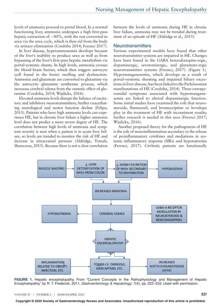
In liver disease, hyperammonemia develops because of the liver’s inability to produce urea as well as from bypassing of the liver’s first-pass hepatic metabolism via portal-systemic shunts. In high levels, ammonia crosses the blood–brain barrier, which then triggers astrocyte (cell found in the brain) swelling and dysfunction. Ammonia and glutamate are converted to glutamine via the astrocytic glutamine synthetase process, which increases cerebral edema from the osmotic effect of glu-tamine (Cordoba, 2014; Wijdicks, 2016).
Elevated ammonia levels disrupt the balance of excita-tory and inhibitory neurotransmitters, further exacerbat-ing neurological and motor function decline (Felipo, 2013). Patients who have high ammonia levels can expe-rience HE, but in chronic liver failure a higher ammonia level does not predict a more severe degree of HE. The correlation between high levels of ammonia and symp-tom severity is seen when a patient is in acute liver fail-ure, so levels are trended to monitor the risk of HE and increase in intracranial pressure (Aldridge, Tranah, Shawcross, 2015). Because there is not a clear correlation
between the levels of ammonia during HE in chronic liver failure, ammonia may not be trended during treat-ment of an episode of HE (Aldridge et al., 2015).
NeurotransmittersVarious experimental models have found that other neurotransmitter systems are impaired in HE. Changes have been found in the GABA benzodiazepine-ergic, dopaminergic, serotoninergic, and glutamate-ergic neurotransmitter systems (Ferenci, 2017). (Figure 1). Hypermanganesemia, which develops as a result of portal–systemic shunting and impaired biliary excre-tions in liver disease, has been linked to the Parkinsonian manifestations of HE (Cordoba, 2014). These extrapy-ramidal symptoms associated with hypermangane-semia are linked to altered dopaminergic function. Some initial studies have examined the role that neuro-steroids, flumazenil, and bromocriptine or levodopa play in the treatment of HE with inconsistent results; further research is needed in this area (Ferenci 2017; Wijdicks, 2016).
Another proposed theory for the pathogenesis of HE is the role of neuroinflammation secondary to the release of proinflammatory cytokines and mediations in sys-temic inflammatory response (SIRs) and hyponatremia (Ferenci, 2017). Cirrhotic patients are functionally
Copyright © 2020 Society of Gastroenterology Nurses and Associates. Unauthorized reproduction of this article is prohibited.
FIGURE 1. Hepatic encephalopathy. From “Current Concepts in the Pathophysiology and Management of Hepatic Encephalopathy,” by R. T. Frederick, 2011, Gastroenterology & Hepatology, 7(4), pp. 222–233. Used with permission.

Nursing Management of Hepatic Encephalopathy
E38 Copyright © 2020 Society of Gastroenterology Nurses and Associates Gastroenterology Nursing
Copyright © 2020 Society of Gastroenterology Nurses and Associates. Unauthorized reproduction of this article is prohibited.
immunocompromised, but it is unclear if the infections or the neuroinflammatory response worsen HE symp-toms. Additionally, hyponatremia may enhance the effects of hyperammonemia on cerebral edema (Cordoba, 2014).
Overall, the symptoms of HE have been attributed to cerebral edema and impaired neurotransmitter sys-tems, which have been linked to infections, hypona-tremia, and hypermanganesemia. However, hyperam-monemia with its effects on cerebral edema and reduced neural metabolic function remains the central factor in our current understanding of HE (Cordoba, 2014; Ferenci, 2017; Vilstrup et al., 2014; Wijdicks, 2016). As such, much of the research and treatment developed for HE has aimed at reducing the absorp-tion of ammonia from the GI tract with non-absorba-ble disaccharides such as lactulose (Wijdicks, 2016). In addition to the concern for infections that can increase ammonia in cirrhotic patients, GI bleeding can increase levels due to a large nitrogenous source for ammonia production; dehydration can lead to hyponatremia and worsening cerebral edema; and constipation results in decreased ammonia excretion, leading to hyperam-monemia (Cordoba, 2014; Ferenci, 2017; Wijdicks, 2016).
Other than liver transplantation, the only surgical procedure with any documented effectiveness in modi-fying the underlying cause if HE is a transjugular intra-hepatic portosystemic shunt (TIPS), which may reduce portal hypertension and decrease the risk of GI
bleeding and recurrent ascites (Punamiya & Amarapurkar, 2011). However, HE development is highest within 30 days of a TIPS procedure and has been associated with larger diameter shunts due to decreased blood flow to the liver, which leads to hyperammonemia from bypassing the liver’s first-pass metabolism (Punamiya & Amarapurkar, 2011). An understanding of the pathogenesis and precipitating factors of HE enables nurses to target assessments, anticipate impending complications, treat precipitating factors, and titrate therapies in collaboration with the healthcare team.
Grading of HEAmong clinicians, the most widely accepted diagnostic tool for HE is the West Haven Criteria, which rates the neurological status according to five grades (Table 2): mHE or Covert HE may produce some alterations in psychometric or neuropsychological testing with no obvious clinical alterations; Grade I produces very mild alterations in awareness, personality and mood changes such as anxiety and euphoria, altered sleep and shorter attention span, along with tremor. West Haven criteria of minimal and Grade I exhibit the same symptoms of covert HE. OHE begins with Grade II and higher ratings. Patients with Grade II HE display more obvious personality changes, including lethargy, disorientation to time, inappropriate behavior, and neurological signs such as asterixis (flapping tremors) and slow and slurred speech. Patients rated as Grade
TABLE 2. Hepatic Encephalopathy GradesGRADE Impairment SONIC Criteria
0 Normal Normal Normal
MHEa Normal examination findings. Subtle changes in work or driving
Minor abnormalities of visual perception or on psycho-metric or number tests } Covert
1 Personality changes, attention deficits, irritability, depressed state
Tremor and incoordination
2 Changes in sleep–wake cycle, lethargy, mood and behavioral changes, cognitive dysfunction
Asterixis, ataxic gait, speech abnormalities (slow and slurred)
} Overt3 Altered level of consciousness
(somnolence), confusion, diso-rientation, and amnesia
Muscular rigidity, nystag-mus, clonus Babinski sign, hyporeflexia
4 Stupor and coma Oculocephalic reflex, unre-sponsiveness to noxious stimuli
Note. MHE = minimal hepatic encephalopathy. From “Management of Hepatic Encephalopathy in the Hospital,” by M. D. Leise, J. J. Poterucha, P. S. Kamath, & W. R. Kim, 2014, Mayo Clinic Proceedings, 89 (2), pp. 241–253. Adapted with permission.

Nursing Management of Hepatic Encephalopathy
VOLUME 43 | NUMBER 2 | MARCH/APRIL 2020 E39
Copyright © 2020 Society of Gastroenterology Nurses and Associates. Unauthorized reproduction of this article is prohibited.
III tend to be somnolent, confused, severely disorient-ed, and display bizarre behavior. When coma is pre-sent, patients are rated at Grade IV on the West Haven scale (Bajaj, 2010b; Vilstrup et al., 2014). The West Haven criteria have been used in numerous trials for the classification of OHE, but is less precise in detect-ing mHE since mHE lacks the classical symptoms of OHE.
Even mHE in decompensated cirrhosis patients has a negative impact on their quality of life. The preva-lence of mHE has not been extensively studied, and was originally thought to affect about 24%–30% of patients. Now with additional research and more descriptive inclusion criteria, mHE in decompensated cirrhosis is estimated to occur in 44% of patients (Mina, Moran, Ortiz-Olvera, Mera, & Uribe, 2013). Although numbers vary, it is important to know that mHE is difficult to diagnose and is not usually tested at outpatient appointments. mHE should be assessed for during hospitalization, even for an unrelated condi-tion. Minimal or covert HE can lead to an increased risk for hospitalization. Prior diagnosis of mHE is associated with the development of a patient’s first episode of OHE. Therefore, detection of covert HE informs the clinician of the increased risk for OHE, and the need to provide family and caregivers with education about signs, symptoms, and management of HE to identify it earlier, and even prevent future hospi-talization (Patidar et al., 2014).
Tools Used for GradingThe Psychometric Hepatic Encephalopathy Score (PHES), a battery of five paper-and-pencil tests, is the current recommended tool to test for mHE (Vilstrup et al., 2014). Despite being validated for use in the mHE population, applicability of this tool in a typical clinical setting is low since these tests must be admin-istered by a neuropsychologist. Although this test is recommended for the diagnosis of mHE, because it has not been validated, it is not currently used in the inpa-tient or outpatient setting for diagnosis. Multiple other tools may be helpful, such as the Hepatic Encephalopathy Scoring Algorithm, the Critical Flicker Frequency test, and a novel smartphone application called the Stroop App. These tools are a bit challenging, and only some-times used by providers as they still require further validation for this population (Vilstrup et al., 2014).
Nursing Management of HEPatients who are admitted to the hospital typically have at least Grade II classification of OHE. General supportive care of OHE includes providing adequate nutritional support, maintaining fluid and electrolyte balance, administering pharmacologic agents to man-age high ammonia levels, preventing and treating
constipation, and ensuring patient safety. Identification of the factors that precipitate worsening of symptoms is critical and nurses play a key role in both this process and in educating patients and families to reduce future episodes (Vilstrup et al., 2014) (Figures 2 and 3).
Nursing AssessmentHepatic encephalopathy may occur with other compli-cations of liver disease such as ascites, hyponatremia, pulmonary vascular complications, and esophageal varices. A comprehensive nursing assessment is crucial to ensuring appropriate care and patient safety. It should include the patient’s level of responsiveness; sensory and motor abnormalities; fluid, electrolyte, and acid–base imbalances and the expected plus prob-lematic effects of treatment measures. Regular evalua-tion of these behaviors, lab values, and deviations from normal baseline status are assessment priorities in HE (Lewis, Heitkemper, & Dirksen, 2008).
Identification of factors precipitating the current episode of OHE should be identified as well. Common problems contributing to acute worsening of HE include infections, GI bleeding, overuse of diuretics, electrolyte imbalances, and constipation (Vilstrup et al. 2014). Therefore, nurses should be alert for signs of HE when patients with underlying liver failure are admitted for other conditions, such heart failure, pneu-monia, or surgical procedures that can disrupt normal fluid and electrolyte balance or pose a risk for infec-tion. It is important to assess patient responses to treat-ment to collaborate with the medical team about needed modifications in the treatment plan. Also, the patient’s response will inform the discharge planning process to prepare patients and families for preventing future episodes.
Level of ResponsivenessLevel of responsiveness may range from mild confusion and slurred speech to coma. Regardless of the severity, family members and significant others are key inform-ants to this specific assessment by providing data on baseline status and the timeframe of worsening symp-toms. OHE can be identified on examination, but Minimal HE usually is not. Minimal HE is noticed by subtler changes to behavior, mood, or sleep disturbances in the patient and may require psychometric or neuropsy-chological testing to diagnose (Vilstrup et al., 2014).
Specific alterations in cognitive status of patients with OHE include impairments in attention, reaction time, confusion, and agitation (Shawcross et al., 2016). Baseline information on these parameters is important in planning care and monitoring patient’s progress. Family members or significant others can provide invaluable input about the patient’s baseline status, related to orientation and general behavior to aid in

Nursing Management of Hepatic Encephalopathy
E40 Copyright © 2020 Society of Gastroenterology Nurses and Associates Gastroenterology Nursing
Copyright © 2020 Society of Gastroenterology Nurses and Associates. Unauthorized reproduction of this article is prohibited.
early detection of mHE. Examples of probe questions are as follows: “Has Mr. A recently been sleepier?”; “I noticed that Mr. A is very slow to respond to ques-tions, how is he at home?” “Tell me about how Mr. A.’s mental status has changed over the past few days.” A comprehensive nursing assessment including family perceptions can aid in the detection of subtle changes like excessive sleepiness, sleep disturbances, and agitation.
Sensory and Motor AbnormalitiesAsterixis (flapping tremor), muscle twitching, and hyperreflexia may be observed in patients with OHE. These can be accompanied by other neuromuscular impairments such as bradykinesia and hyperactive deep tendon reflexes. Bradykinesia means “slow move-ment” and can present as decreased facial expressions, increased stillness, or difficulty with performing repeti-tive tasks such as finger tapping.
Fluid and Electrolyte ImbalancesPatients who are placed on disaccharides (lactulose) are expected to have more bowel movements since these agents remove ammonia through the GI tract. Although bowel movements are needed to clear the ammonia, monitoring the patient for diarrhea is essen-tial since it can contribute to dehydration and electro-lyte disturbances. The goal for medication manage-ment is for the patient to have 2–3 bowel movements a day (Vilstrup et al., 2014). The medication should be titrated to achieve this goal and diminish complica-tions due to lactulose overuse (Vilstrup et al., 2014). Either hypokalemia or hyperkalemia may also occur in hypovolemic patients. Dehydration and hypovolemia are another potential cause of HE, so fluid lost through bowel movements or loose stools should be monitored closely. Consequently, daily weights and accurate monitoring of intake and output are required espe-cially since the patient can be a poor historian.
FIGURE 2. Treatment algorithm for hepatic encephalopathy. From “Management of Hepatic Encephalopathy in the Hospital,” by M. D. Leise, J. J. Poterucha, P. S. Kamath, and W. R. Kim, 2014, Mayo Clinic Proceedings, 89(2), pp. 241–253 Adapted with permission.
Nursing Management of Hepatic Encephalopathy
VOLUME 43 | NUMBER 2 | MARCH/APRIL 2020 E41
Copyright © 2020 Society of Gastroenterology Nurses and Associates. Unauthorized reproduction of this article is prohibited.
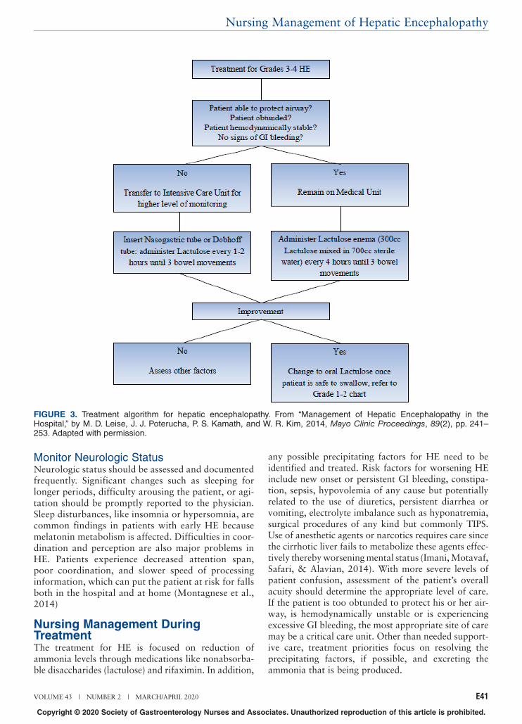
Monitor Neurologic StatusNeurologic status should be assessed and documented frequently. Significant changes such as sleeping for longer periods, difficulty arousing the patient, or agi-tation should be promptly reported to the physician. Sleep disturbances, like insomnia or hypersomnia, are common findings in patients with early HE because melatonin metabolism is affected. Difficulties in coor-dination and perception are also major problems in HE. Patients experience decreased attention span, poor coordination, and slower speed of processing information, which can put the patient at risk for falls both in the hospital and at home (Montagnese et al., 2014)
Nursing Management During TreatmentThe treatment for HE is focused on reduction of ammonia levels through medications like nonabsorba-ble disaccharides (lactulose) and rifaximin. In addition,
any possible precipitating factors for HE need to be identified and treated. Risk factors for worsening HE include new onset or persistent GI bleeding, constipa-tion, sepsis, hypovolemia of any cause but potentially related to the use of diuretics, persistent diarrhea or vomiting, electrolyte imbalance such as hyponatremia, surgical procedures of any kind but commonly TIPS. Use of anesthetic agents or narcotics requires care since the cirrhotic liver fails to metabolize these agents effec-tively thereby worsening mental status (Imani, Motavaf, Safari, & Alavian, 2014). With more severe levels of patient confusion, assessment of the patient’s overall acuity should determine the appropriate level of care. If the patient is too obtunded to protect his or her air-way, is hemodynamically unstable or is experiencing excessive GI bleeding, the most appropriate site of care may be a critical care unit. Other than needed support-ive care, treatment priorities focus on resolving the precipitating factors, if possible, and excreting the ammonia that is being produced.
FIGURE 3. Treatment algorithm for hepatic encephalopathy. From “Management of Hepatic Encephalopathy in the Hospital,” by M. D. Leise, J. J. Poterucha, P. S. Kamath, and W. R. Kim, 2014, Mayo Clinic Proceedings, 89(2), pp. 241–253. Adapted with permission.

Nursing Management of Hepatic Encephalopathy
E42 Copyright © 2020 Society of Gastroenterology Nurses and Associates Gastroenterology Nursing
Copyright © 2020 Society of Gastroenterology Nurses and Associates. Unauthorized reproduction of this article is prohibited.
Drug Therapy for HELactuloseWhile the exact mechanism of action of lactulose is still being discussed, the currently accepted benefit results from the fermentation that this nonabsorbable disaccha-ride undergoes in the colon thereby increasing acid pro-duction leading to evacuation of ammonia (Leise, Poterucha, Kamath, & Kim, 2014, p. 544). Administration, while inpatient for an episode of HE, has shown improvement in patient mental status while simul-taneously treating potential precipitating factors (Bajaj, 2010a).
Lactulose is usually taken orally until 2–3 bowel movements within 24 hours are achieved (Bajaj, Sanyal, Bell, Gilles & Heuman, 2010; Vilstrup et al., 2014). Once the goal bowel movements are achieved for the day, the patient may stop taking the lactulose and then resume doses the next day.
Although lactulose use has had good outcomes for inpatient and outpatient use, there remain some chal-lenges with medication adherence. In one cohort of patients observed, 69% were readmitted with HE because of failure to take the drug appropriately and consistently, attributed to lack of education on lactu-lose (Leise et al., 2014). The adverse effects of lactulose include GI discomfort such as nausea, diarrhea and abdominal cramping, dehydration, hypernatremia, and perianal skin irritation (Vilstrup et al., 2014). Loose stools and more frequent bowel movements are changes that some patients struggle with depending on their degree of mobility and cognitive function. Strategies for managing these expected results should be integrated into the education plan.
Enabling patients and caregivers to fully engage in the treatment plan requires that they understand the pur-pose and benefit of the medication and have helped in planning how to manage its expected effects and sustain their participation in treatment. Patients are likely to need assistance in titrating the appropriate dose. If there are zero bowel movements, the dose should be increased. If there are greater than four large bowel movements a day, then the drug should be stopped for that day to prevent dehydration. The patient would then restart lactulose the following day. Given the cognitive prob-lems associated with HE, recruiting a committed family member or caregiver to support the patient can make an important contribution to success.
RifaximinOnce patient-precipitating factors of HE are corrected and confusion has resolved, a plan for long-term preven-tion of HE must be established. This plan may include the use of rifaximin, an antibiotic that is minimally absorbed by the gut. Rifaximin’s antibiotic properties
remain concentrated to the GI tract, decreasing the amount of bacteria in the gut that produce ammonia (Welliver, 2013). The drug binds to the enzyme that replicates ribonucleic acid (RNA) and blocks deoxyribo-nucleis acid (DNA) replication and ultimately protein synthesis causing bacterial cell death (Welliver, 2013). There are no data proving that this antibiotic is as effec-tive as lactulose in improving HE when used alone, but when used in combination these drugs can lengthen the time between HE occurrences (Vilstrup et al., 2014).
The Food and Drug Administration (FDA) has not yet approved rifaximin as a first-line therapy for epi-sodic OHE, but has approved it as a secondary preven-tion for OHE (Leise et al., 2014). The combination therapy of lactulose and rifaximin has been shown to reverse acute HE and decrease length of hospital stay and therefore is commonly used in the hospital and continues into outpatient treatment (Leise et al., 2014).
SupplementsAnother potential supplemental treatment option is Zinc. While research on zinc as a treatment agent is sparse, cirrhotic patients commonly have a zinc defi-ciency (Leise et al., 2014). There are enzymes in the body that assist in ammonia detoxification; these are found in the muscle and liver and are zinc dependent. A decrease in liver function and muscle mass can lead to lower zinc levels, leading to an increase in ammonia (Amodio et al., 2013). Zinc supplementation with a dose of 200 mg t.i.d. (Mohammad, Zhou, Cave, Barve, & McClain, 2012) has shown benefits in liver function and in nutrition levels but more clinical trials are needed to determine the best zinc dosing (Table 3).
Pain Management
Analgesics in CirrhosisPain management proves to be challenging in patients with liver disease for all clinicians. Many commonly used analgesics are metabolized via the liver, and when it is not functioning properly, it can result in adverse side effects. Analgesics can cause an increased risk for oversedation and some clinicians may feel unsafe to use, leading to undertreatment of pain in patients with cirrhosis. Other risk factors, in addition to overseda-tion, are worsening renal function, inducing HE, portal hypertension, and GI bleeding (Imani, Motavaf, Safari, & Alavian, 2014).
Hepatic clearance of analgesics usually depends on three things: hepatic blood flow, plasma binding pro-teins, and hepatic enzymes. Analgesic drugs are usually are absorbed from the GI tract and then passed directly to the liver, being metabolized before it even enters the body’s systemic blood flow. If the hepatic flow is altered in anyway, possibly reduced, or bypassed by

Nursing Management of Hepatic Encephalopathy
VOLUME 43 | NUMBER 2 | MARCH/APRIL 2020 E43
Copyright © 2020 Society of Gastroenterology Nurses and Associates. Unauthorized reproduction of this article is prohibited.
portal shunts, more of the drug enters the systemic system. Therefore, if a patient has impaired hepatic blood flow, the initial dose of an analgesic should be reduced. Some patients with liver disease may have more impairment with hepatic excretion, rather than shunting, so maintenance doses may need to be reduced to prevent accumulation. Some analgesics are protein bound, and if a patient has low albumin that may cause elevated drug levels, so doses in these patient may need to be reduced. The drug half-life has also been proven to increase because of the drug accu-mulating in the plasma (Soleimanpour, Safari, Nia, Sanaie, & Alavian, 2016). All these factors combined
make prescribing and managing pain difficult for clini-cians caring for this patient population.
AcetaminophenAcetaminophen is one of the most widely used analge-sics for pain relief, but sometimes avoided in patients with liver disease because of the risk of liver damage with potential overdosing. Studies show that acetami-nophen intake in conservative doses may be safe to use in patients with liver disease. FDA guidelines recom-mend that long-term use of acetaminophen in inpa-tients with liver disease, not currently drinking, should be at a reduced dose between 2 and 3g per day (Imani
TABLE 3. Hepatic Encephalopathy and Associated MedicationsMedications
Name of Medication Lactulose Rifaximin Zinc Sulfate
Mechanism in hepatic encephalopathy (HE)
• Metabolized in the gut by bacteria; the metabolism of lactulose creates an acidic pH in the stomach/colon; this acidic pH prevents ammonia from diffusing into the blooda
• Enhances elimination of am-monia by expediting expulsion of stoola
• Broad spectrum: covers Gram negative and posi-tive bacteriaa
• Eliminates gut bacteria that produce ammoniaa
• Liver is main organ responsible for zinc metabolismb
• Low serum zinc con-centrations compared to high blood ammonia concentrationsb
• Zinc deficiency contrib-utes to liver diseaseb
Adverse reactions • Endocrine: dehydration, hyper-natremia, hypokalemiac
• GI: abdominal cramps/ distension/distress, diarrheac
• Cardio: peripheral edema (15%)d
• CNS: dizziness (13%), fatigue (12%)d
• GI: Nausea (14%); irrita-ble bowel syndrome with diarrhea (2%–3%)d
Frequency not defined• CNS: dizziness, head-
achee
• GI: abdominal cramps, diarrhea, nausea, vom-itinge
Monitoring parameters • Changes in mental statusc
• Serum electrolytesc
• Serum ammoniac
• Bowel movements, fluid statusc
• Changes in mental statusd
• Hypersensitivity reac-tions (less than 2% in-cidence of angioedema, anaphylaxis, skin rash)d
• Changes in mental statuse
Administration considerations
• Recommended dose: 25 mL twice dailya
• Available as oral solution 10 g/15 mLc
• May be mixed with fruit juice, water, or milkc
• Titrate for production of 2–3 bowel movements dailyc
• Recommended dose: 550 mg twice dailya
• May be taken with or without foodd
• Tablets may be crushedd
• Recommended dose: 220 mg (elemental zinc 50 mg)e
Note. Ingrid Pan, Lauren Simonds, Pharm D., BCPS Pharmacy Department, Hospital of the University of Pennsylvania.aFrom “Hepatic Encephalopathy,” by E. F. Wijdicks, 2016, New England Journal of Medicine, 375 (17), pp. 1660–1670.bFrom “The Role of Zinc in Liver Cirrhosis,” by K. Grüngreiff, D. Reinhold, and H. Wedemeyer, 2016, Annals of Hepatology, 15 (1), pp. 7–16.cFrom “Lactulose” Lexi-Drugs. Lexicomp. Riverwoods, IL: Wolters Kluwer Health. Retrieved February 1, 2017, from http://online.lexi.comdFrom “Rifaximin,” Lexi-Drugs. Lexicomp. Riverwoods, IL: Wolters Kluwer Health. Retrieved February 1, 2017, from http://online.lexi.comeFrom “Zinc Sulfate,” Lexi-Drugs. Lexicomp. Riverwoods, IL: Wolters Kluwer Health. Retrieved February 1, 2017, from http://online.lexi.com
Nursing Management of Hepatic Encephalopathy
E44 Copyright © 2020 Society of Gastroenterology Nurses and Associates Gastroenterology Nursing
Copyright © 2020 Society of Gastroenterology Nurses and Associates. Unauthorized reproduction of this article is prohibited.
et al., 2014, p. 3) and American Liver Foundation rec-ommends less than 3g per day (Hayward, Powell, Irvine, & Martin, 2015, p. 213).
Nonsteroidal Anti-Inflammatory DrugsUse of nonsteroidal anti-inflammatory drugs (NSAIDs) poses a high risk to patients already having liver dis-ease because of how significant their side effects can be. The reduction of platelet synthesis that NSAIDs can have on the body can lead to thrombocytopenia and coagulopathy problems in cirrhotic patients. This side effect can be so significant in the liver patient that it has the potential to cause varices, both esophageal and cardiac (Imani et al., 2014). Because of this, NSAIDs in liver disease should be used with caution, or avoided if possible (Imani et al., 2014).
OpioidsMost of the commonly prescribed opioids are metabo-lized by the liver. Because of reduced or impaired hepatic blood flow, protein binding and hepatic enzymes, and the half-life of these drugs increased in this population, opioid therapy is extremely challenging. With an increased half-life, drugs like morphine, hydromorphone, and oxy-codone cause increased levels of the drugs systemically, which may result in a greater risk for side effects and drug toxicity (Soleimanpour et al., 2016).
Interestingly, codeine is not metabolized in patients with hepatic failure and has no effect on these patients with liver disease because of its relation to enzyme reduction. The effects may not relieve any pain, but they can still result in the drug accumulating and res-piratory depression side effects (Imani et al., 2014; Soleimanpour et al., 2016).
Methadone and fentanyl are protein-bound drugs, so dose reduction is needed in patients with hypoalbu-minemia. They also are dependent on enzyme activity, so may not even have therapeutic effects on patients with liver disease. Tramadol is another opioid used for pain control but because the “metabolic enzymes of the liver are reduced in patients with liver failure, the drug may not be metabolized properly, reducing the effects of the enzymes” (Soleimanpour et al., 2016, p. 8)
It is suggested to proceed with caution when pre-scribing and administering any opioids for pain man-agement because of their high-risk effects on patient with chronic liver disease like sedation, constipation, and confusion, which are precipitating factors to induce HE. Doses may need to be lower with longer intervals between these doses for patient safety. Patients using opioids for pain control should be closely moni-tored by a provider to assess for any side effects (Imani et al., 2014). Close attention should be paid to patients prescribed opioids with a history of alcohol abuse for possibility of cross-addiction.
Nutritional SupportNutrition has a close correlation to health in cirrhotic patients. Moderate to severe malnutrition is seen in 75% of patients with HE (Romeiro & Augusti, 2015, p. 2943; Vilstrup et al., 2014) and is considered a risk factor for development of HE. Serious complications of malnutrition include refractory ascites, spontaneous bacterial peritonitis, hepatorenal syndrome, and variceal hemorrhage (Chadalavada, Sappati Biyyani, Maxwell, & Mullen, 2010, p. 258).
Prior recommendation to reduce protein intake to prevent or manage HE is no longer considered valid because protein deficiency can cause muscle break-down, thereby increasing the release of amino acids and ammonia production causing or worsening HE (Amodio et al., 2013). Nutritional support for patients with HE should include maintaining an energy intake of 35–40 kcal/kg/day, with a protein intake of 1.2–1.5 g/kg/day (Vilstrup et al., 2014). Protein restriction is reserved for patients who are severely protein-intoler-ant or for very short periods for patients with GI bleed-ing until symptoms resolve (Amodio et al., 2013). Dairy and vegetable proteins are preferred but are usually much less palatable. A fiber-rich diet is recom-mended to encourage fecal ammonia excretion, while avoiding diarrhea that could potentially induce HE in patients already taking lactulose.
Small meals throughout the day are encouraged with a late-night snack. Frequent meals eliminate the need for a patient to become overly hungry throughout the day, possibly using their glucose stores, and releas-ing more amino acids and elevating ammonia levels (Amodio et al., 2013). Having a late evening snack can be another source of caloric intake, shortening the time between eating overnight.
Patient and Family EducationSince the patient may have cognitive deficits, the family or primary caregiver should be included in all teaching. Both patient and family benefit from an education on the general overview of HE and practical pointers about integrating treatments such as lactulose administration and prevention of precipitating problems such as dehy-dration and infection into daily activities. The manage-ment and educational plan should be individualized to the patient and be driven by a thorough nursing assess-ment. It is also beneficial to educate the patient and fam-ily on the precipitating factors of HE in order to prevent reoccurrences and spot HE early, in order to get sooner treatment and possibly prevent a hospital admission. A primary safety concern for patients with HE, and more pronounced in mHE, is their fitness to drive. Patients with HE or mHE can overestimate their driving abilities. This should be included when educating patients and families.

Nursing Management of Hepatic Encephalopathy
VOLUME 43 | NUMBER 2 | MARCH/APRIL 2020 E45
Copyright © 2020 Society of Gastroenterology Nurses and Associates. Unauthorized reproduction of this article is prohibited.
Patients should fill their prescriptions immediately upon discharge as to not miss any doses. An under-standing of the reasoning for taking the medications, what to do if a dose is missed, and how to titrate lactu-lose are needed for effective home management (Donnelly, 2015). Education on polypharmacy should also be included. Polypharmacy is the simultaneous use of multiple drugs to treat one or more medical condi-tions and is associated with poor medication adherence and greater chance for patient–clinician miscommuni-cation about medications. (Patel, 2017). Patients with liver disease can have multimorbidities and are prone to polypharmacy.
Weight tracking is also important, and should be recorded and reviewed at provider visits. If a large weight fluctuation, more than 10 pounds, is seen in a week, the patient should contact their provider (Donnelly, 2015). Patients should abstain from alcohol and avoid aspirin, ibuprofen and acetaminophen (Tylenol) as they may be harmful to the liver, and always consult with a provider before taking any of these (Donnelly, 2015). Good hand washing and avoiding sick contacts can help decrease the risk for infection.
TransplantationAfter all treatment options for HE have been exhausted, a patient may be referred for liver transplantation. The evaluation not only includes a battery of tests similar to other solid organ transplants but also includes a Model for End stage Liver Disease (MELD) score in determin-ing the patient’s eligibility for a transplant and its urgency. The MELD is a calculation using a patient’s blood chemistry laboratory values including serum cre-atinine, bilirubin levels, INR, and recently serum sodi-um. The score of the calculation can range from 6 to 40 and relates to prognosis of liver disease. A higher MELD score increases prioritization for an organ transplant (Martin, DiMartini, Feng, Brown, & Fallon, 2014). Serum sodium levels have been incorporated into the equation to prioritize those patients with increased mor-tality because of hyponatremia (Martin et al., 2014).
ConclusionHepatic encephalopathy is a chronic and often-pro-gressive consequence of liver failure. However, patients can keep hyperammonemia under control with careful management of food and fluid intake, consistent use of agents such as lactulose and rifaximin, and early detec-tion and reporting of problems likely to worsen the condition. Effective self-management is challenging. The patient’s altered mental status coupled with the complexity of the treatment can make every day adher-ence challenging and problem solving very difficult. Optimal outcomes rely on active participation of a knowledgeable caregiver and strong partnership with
outpatient and, when needed, inpatient teams. Nurses are a critical link in helping patients and caregivers understand what is needed and in supporting their efforts to manage a complex set of problems.
Case StudyAlice is a nurse on a busy medical-surgical unit, her assignment today includes a new patient, Mr. Martin, who came to the emergency department, complaining of fever and increased fatigue. Mr. Martin has advanced Hepatitis C cirrhosis with a MELD score of 27 and is awaiting a donor for a liver transplant. Other signifi-cant problems include GI bleeding from esophageal varices within the past year and Type II diabetes.
The night shift nurse reported that he is sleepy but able to ambulate in his room without assistance. Alice introduces herself to Mr. Martin as he is trying to eat breakfast and notices that his hand tremors are pro-nounced. His wife is sleeping in a chair nearby. While doing her assessment, Alice notes that Mr. Martin is very slow to respond, and his speech is slow and slurred. He seems to be falling asleep as he is eating.
Although the patient is lethargic, he is easily arous-able. He has a gag reflex, and his airway is not com-promised. He is warm to the touch with easily palpable pulses throughout. Neurologically, the patient is slow to respond, disoriented to date and time but knows that he is in the hospital. Asterixis is noted; pupils are PERRLA. Muscle strength is even throughout. HEENT presents as dry mucous membranes and poor skin tur-gor. Sclera are jaundiced. Respiratory assessment was clear to auscultation bilaterally. Cardiovascular assess-ment reveals tachycardia but with a regular rhythm. His abdomen is distended, soft, and nontender. Mr. Martin has +2 bilateral lower extremity edema and his skin is jaundiced. His vital signs are as follows: tem-perature: 101°F, heart rate 110 beats/minute, blood pressure 104/78 mmHg, respiration rate 18 breaths/minute, and SpO2 94% on room air. This blood pres-sure is within normal range for the patient.
Mr. Martin does not remember why he came to the hospital. Thankfully, his wife awakens and notes that Mr. Martin has not been feeling well for a few days at home, but was reluctant to go to the hospital. He was having intermittent fevers, chills, and fatigue. She is anxious and provides important information that since he started feeling poorly, his food and liquid intake has been minimal, and he refused to take his medications including his lactulose and rifaximin. She denies any vomiting blood (hematemesis) or frank red blood in his stool (hematochezia) as well as use of any new pain or anxiety medications. With this helpful information from Mrs. Martin and her own nursing assessment, Alice is now ready to inform the physi-cians/advanced practitioners of the situation and to

Nursing Management of Hepatic Encephalopathy
E46 Copyright © 2020 Society of Gastroenterology Nurses and Associates Gastroenterology Nursing
Copyright © 2020 Society of Gastroenterology Nurses and Associates. Unauthorized reproduction of this article is prohibited.
collaboratively plan Mr. Martin’s monitoring and treatment plan.
With the information from Alice, the team acknowl-edges that Mr. Martin’s mental status has deteriorated since admission. According to West Haven Criteria, this patient most likely has Grade II HE. As the bedside nurse, Alice is asked if she feels comfortable caring for this patient on a general medical–surgical unit. Alice references the Treatment Algorithm for HE and because Mr. Martin’s HE is a Grade II and he can protect his airway, she believes that he is appropriate for this unit. Plan of care at this point will be to restart the lactulose, monitor his oral intake and correct any dehydration, assess number and quality of bowel movements, identify and treat the source of the fever, and encourage the patient’s participation in care. Alice is concerned about patient safety since he is more disoriented. He had been steady on his feet, but this needs to be continually reas-sessed and he will be assisted to and from the bathroom to prevent falls.
Mr. Martin was found to have spontaneous bacterial peritonitis a bacterial infection in his ascites fluid, con-firmed by diagnostic paracentesis. He was treated with antibiotics and his fevers resolved. Lactulose medica-tion treatment was continued until Mr. Martin had an adequate number of bowel movements each day, and his oral intake improved with return of his mental sta-tus to baseline. He responded well to the team’s treat-ment plan and is now ready for discharge. ✪
ACKNOWLEDGMENTSWith our sincerest gratitude, we thank Diane Peyton, BSN, and Rebecca Trotta, PhD, RN, for their as-sistance with the development of this paper. We also thank Kathleen McCauley, PhD, RN, FAAN, FAHA, for her mentorship through this paper.
REFERENCESAldridge, D. R., Tranah, E. J., & Shawcross, D. L. (2015). Pathogen-
esis of hepatic encephalopathy: role of ammonia and systemic inflammation. Journal of Clinical and Experimental Hepatology, 5(1), S7–S20.
Amodio, P., Bemeur, C., Butterworth, R., Cordoba, J., Kato, A., Mon-tagnese, S., … Morgan, M. Y. (2013). The nutritional management of hepatic encephalopathy in patients with cirrhosis: International Society for Hepatic Encephalopathy and Nitrogen Metabolism consensus. Hepatology (Baltimore, MD), 58(1), 325–336.
Bajaj, J. S. (2010a). Review article: The modern management of hepatic encephalopathy. Alimentary Pharmacology & Thera-peutics, 31(5), 537–547.
Bajaj, J. S. (2010b). Current and future diagnosis of hepatic encepha-lopathy. Metabolic Brain Disease, 25(1), 107–110.
Bajaj, J. S., Sanyal, A. J., Bell, D., Gilles, H., & Heuman, D. M. (2010). Predictors of the recurrence of hepatic encephalopathy in lactulose-treated patients. Alimentary Pharmacology & Ther-apeutics, 31(9), 1012–1017.
Burnham, B., Wallington, S., Jillson, I. A., Trandafili, H., Shetty, K., Wang, J., & Loffredo, C. A. (2014). Knowledge, attitudes, and beliefs of patients with chronic liver disease. American Journal of Health Behavior, 38(5), 737–744.
Chadalavada, R., Sappati Biyyani, R. S., Maxwell, J., & Mullen, K. (2010). Nutrition in hepatic encephalopathy. Nutrition in Clini-cal Practice: Official Publication of the American Society for Parenteral and Enteral Nutrition, 25(3), 257–264.
Cordoba, J. (2014). Hepatic encephalopathy: From the pathogenesis to the new treatments. ISRN Hepatology, 2014, 236–268.
DiPascoli, M., Ceranto, E., De Nardi, P., Donata, D., Gatta, A., An-geli, P., & Pontisso, P. (2017). Hospitalizations due to cirrho-sis: clinical aspects in a large cohort of Italian patients and cost analysis report. Digestive Diseases (Basel, Switzerland), 35(5), 433–438. doi: 10.1159/000458722. Accessed May 17, 2017.
Donnelly, A., (2015). Taking care of yourself at home when you have liver disease. Penn medicine patient and family educa-tion. https://extranet.uphs.upenn.edu/hupnursing/patientfami lyed/sheets/,DanaInfo=uphsxnet.uphs.upenn.edu±Liver%20tips%20sheet%20Silverstein%2011%20%202-17-2014.pdf
Felipo, V. (2013). Hepatic encephalopathy: Effects of liver failure on brain function. Nature Reviews. Neuroscience, 14(12), 851–858.
Ferenci, P. (2017). Hepatic encephalopathy. Gastroenterology Re-port, 5(2), 138–147. Doi: 10.1093/gastro/gox013
Frederick, R. T. (2011). Current concepts in the pathophysiology and management of hepatic encephalopathy. Gastroenterology & Hepatology, 7(4), 222–233.
Hansen, L., Sasaki, A., & Zucker, B. (2010). End-stage liver disease: Challenges and practice implications. The Nursing Clinics of North America, 45(3), 411–426.
Hayward, K. L., Powell, E. E., Irvine, K. M., & Martin, J. H. (2015). Can paracetamol (Acetaminophen) be administered to patients with liver impairment? British Journal of Clinical Pharmacol-ogy, 81(2), 210–222. doi:10.1111/bcp.12802
Heron, M. (2015). Deaths: Leading causes for 2012. National Vi-tal Statistics Reports: From the Centers for Disease Control and Prevention, National Center for Health Statistics, National Vital Statistics System, 64(10), 1–93.
Imani, F., Motavaf, M., Safari, S., & Alavian, S. M. (2014). The therapeutic use of analgesics in patients with liver cirrhosis: A literature review and evidence-based recommendations. Hepati-tis Monthly, 14(10). doi:10.5812/hepatmon.23539
Leise, M. D., Poterucha, J. J., Kamath, P. S., & Kim, W. R. (2014). Management of hepatic encephalopathy in the hospital. Mayo Clinic Proceedings, 89(2), 241–253.
Lewis, S. L., Heitkemper, M. M., & Dirksen, S. R., (2008). Medical surgical nursing (9th ed.). Philadelphia, PA: Elsevier.
Martin, P., DiMartini, A., Feng, S., Brown, R., Jr., & Fallon, M. (2014). Evaluation for liver transplantation in adults: 2013 practice guideline by the American Association for the Study of Liver Diseases and the American Society of Transplantation. Hepatology (Baltimore, MD), 59(3), 1144–1165.
Mina, A., Moran, S., Ortiz-Olvera, N., Mera, R., & Uribe, M. (2013). Prevalence of minimal hepatic encephalopathy and qual-ity of life in patients with decompensated cirrhosis. Hepatology Research, 44(10), E92–E99. doi:10.1111/hepr.12227.
Mohammad, M. K., Zhou, Z., Cave, M., Barve, A., & McClain, C. J. (2012). Zinc and liver disease. Nutrition in Clinical Practice: Official Publication of the American Society for Parenteral and Enteral Nutrition, 27(1), 8–20.

Nursing Management of Hepatic Encephalopathy
VOLUME 43 | NUMBER 2 | MARCH/APRIL 2020 E47
For 10 additional continuing education articles related to liver disease, go to www.NursingCenter.com.
Instructions for Taking the CE Test Online:• Readthearticle.ThetestforthisCEactivitycanbe
takenonlineatwww.nursingcenter.com.Testscannolongerbemailedorfaxed.
• YouwillneedtocreateafreelogintoyourpersonalCEPlanneraccountbeforetakingonlinetests.YourplannerwillkeeptrackofallyourLippincottProfessionalDevelopmentonlineCEactivitiesforyou.
• Thereisonlyonecorrectanswerforeachquestion.Apassingscoreforthistestis13correctanswers.Ifyoupass,youcanprintyourcertificateofearnedcon-tacthoursandtheanswerkey.Ifyoufail,youhavetheoptionoftakingthetestagainatnoadditionalcost.
• Forquestions,contactLippincottProfessionalDevelopment:1-800-787-8985.
Registration Deadline: March 4, 2022
Disclosure Statement:Theauthorsandplannershavedisclosedthattheyhavenofinancialrelationshipsrelatedtothisarticle.
Provider Accreditation:LippincottProfessionalDevelopmentwillaward1.5contacthours,including1.0pharmacologycredit,forthiscontinuingnursingeducationactivity.
LippincottProfessionalDevelopmentisaccreditedasaproviderofcontinuingnursingeducationbytheAmericanNursesCredentialingCenter’sCommissiononAccreditation.ThisactivityisalsoproviderapprovedbytheCaliforniaBoardofRegistered
Nursing,ProviderNumberCEP11749for1.5contacthours.LippincottProfessionalDevelopmentisalsoanapprovedproviderofcontinuingnursingeducationbytheDistrictofColumbia,Georgia,andFlorida,CEBroker#50-1223.Yourcertificateisvalidinallstates.
Payment: •Theregistrationfeeforthistestis$10.50formembers;
$15.00fornonmembers.
DOI:10.1097/SGA.0000000000000521
Copyright © 2020 Society of Gastroenterology Nurses and Associates. Unauthorized reproduction of this article is prohibited.
Montagnese, S., De Pitta, C., De Rui, M., Corrias, M., Turco, M., Merkel, C., … Gatta, A. (2014). Sleep–wake abnormalities in patients with cirrhosis. Hepatology, 59(2), 705–712. Doi:10.1002/hep.26555
Patel, P. J. (2017). Multimorbidity and polypharmacy in diabetic pa-tients with NAFLD. Medicine, 96(26) e6761.
Patidar, K. R., Thacker, L. R., Wade, J. B., Sterling, R. K., Sanyal, A. J., Siddiqui, M. S., … Bajaj, J. S. (2014). Covert hepatic encephalopa-thy is independently associated with poor survival and increased risk of hospitalization. The American Journal of Gastroenterology, 109(11), 1757–1763. doi:10.1038/ajg.2014.264
Punamiya, S. J., & Amarapurkar, D. N. (2011). Role of TIPS in im-proving survival of patients with decompensated liver disease. International Journal of Hepatology, 2011, 398291.
Romeiro, F. G., & Augusti, L. (2015). Nutritional assessment in cir-rhotic patients with hepatic encephalopathy. World Journal of Hepatology, 7(30), 2940–2954.
Sanyal, A., Younossi, Z. M., Bass, N. M., Mullen, K. D., Poordad, F., Brown, R. S., … Forbes, W. P. (2011). Randomized clinical trial: Rifaximin improves health-related quality of life in cirrhotic patients with hepatic encephalopathy—a double-blind placebo-controlled study. Alimentary Pharmacology & Therapeutics, 34(8), 853–861.
Scaglione, S., Kliethermes, S., Cao, G., Shoham, D., Durazo, R., Luke, A., & Volk, M. L. (2015). The epidemiology of cirrhosis in the United States: A population-based study. Journal of Clini-cal Gastroenterology, 49(8), 690–696.
Sharma, B. C., & Maharshi, S. (2015), Prevention of hepatic en-cephalopathy recurrence. Clinical Liver Disease, 5(3), 64–67. doi:10.1002/cld.446
Sharma, B. C., Sharma, P., Agrawal, A., & Sarin, S. K. (2009). Sec-ondary prophylaxis of hepatic encephalopathy: An open-label randomized controlled trial of lactulose versus placebo. Gastro-enterology, 137(3), 885-891, 891.e1.
Shawcross, D. L., Dunk, A. A., Jalan, R., Kircheis, G., de Knegt, R. J., Laleman, W., … New Insights Steering Committee (2016). How to diagnose and manage hepatic encephalopathy: A con-sensus statement on roles and responsibilities beyond the liver specialist. European Journal of Gastroenterology & Hepatol-ogy, 28(2), 146–152.
Soleimanpour, H., Safari, S., Shahsavari Nia, K, Sanaie, S., & Ala-vian, S. M. (2016). Opioid drugs in patients with liver dis-ease: A systematic review. Hepatitis Monthly, 16(4), e32636. doi:10.5812/hepatmon.32636
Tsochatzis, E. A., Bosch, J., & Burroughs, A. K. (2014). Liver cir-rhosis. The Lancet, 383(9930), 1749–1761. doi:10.1016/s0140-6736(14)60121-5
Vilstrup, H., Amodio, P., Bajaj, J., Cordoba, J., Ferenci, P., Mullen, K. D., … Wong, P. (2014). Hepatic encephalopathy in chronic liver disease: 2014 practice guideline by the American Associa-tion for the Study of Liver Diseases and the European Associa-tion for the Study of the Liver. Hepatology (Baltimore, MD), 60(2), 715–735.
Welliver, M. (2013). Rifaximin: A nonsystemic antibiotic for hepatic encephalopathy. Gastroenterology Nursing: The Official Jour-nal of the Society of Gastroenterology Nurses and Associates, 36(2), 140-142.
Wijdicks, E. F. (2016). Hepatic encephalopathy. The New England Journal of Medicine, 375(17), 1660–1670.