Isolating Photosynthetic Pigments
13
Biology 43: Plant Physiology Laboratory Activity VI Isolating Photosynthetic Pigments Krizea Marie Kiamco Duron BS Biology IV [email protected]
Transcript of Isolating Photosynthetic Pigments
Biology 43: Plant Physiology
Laboratory Activity VI
Isolating Photosynthetic Pigments
Krizea Marie Kiamco Duron
BS Biology IV
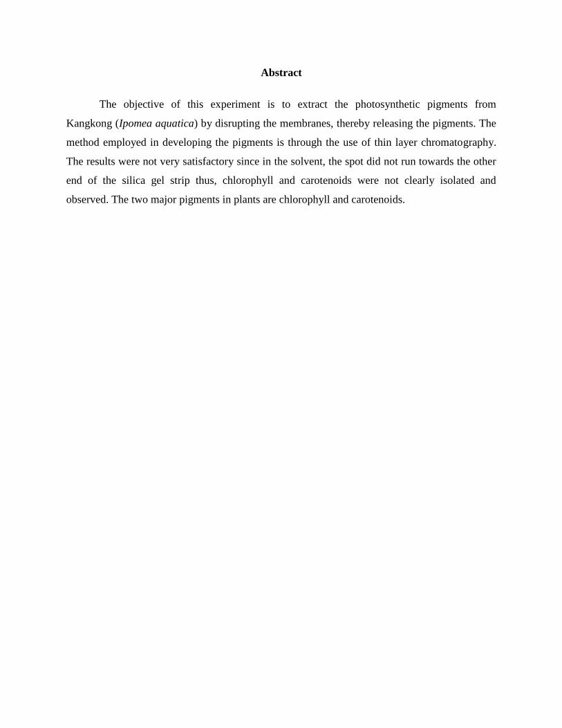
Abstract
The objective of this experiment is to extract the photosynthetic pigments from
Kangkong (Ipomea aquatica) by disrupting the membranes, thereby releasing the pigments. The
method employed in developing the pigments is through the use of thin layer chromatography.
The results were not very satisfactory since in the solvent, the spot did not run towards the other
end of the silica gel strip thus, chlorophyll and carotenoids were not clearly isolated and
observed. The two major pigments in plants are chlorophyll and carotenoids.
Introduction
The efficiency of plants at capturing light for photosynthesis varies. One means by which a
plant can increase its photosynthetic productivity is to use a variety of pigments to harvest a
wider range of light wavelengths. Pigments are chemical compounds that reflect only certain
wavelengths of visible light. There are three basic groups of pigments. Chlorophylls are greenish
pigments that contain a porphyrin ring around which electrons freely migrate. Carotenoids are
typically yellow, orange, or red pigments that contain two 6-carbon rings connected by a
hydrocarbon chain and do not directly transfer light energy to the reaction center and electron
transport chain.
Many of the colors associated with higher plants (green leaves in the spring and summer,
yellow or red leaves in the fall, the orange color of carrots, some colors in flower petals) are due
to the presence of pigment molecules. The major photosynthetic pigments of higher plants can be
divided into two groups, the chlorophylls and the carotenoids. Both types of pigments are
present in the subcellular organelles called chloroplasts, where they are bound to proteins in the
thylakoids, the photochemically active photosynthetic biomembranes. Intact pigment-protein
complexes, which are held together by weak, noncovalent bonds, have been isolated from
chloroplasts and characterized b~( polyacrylamide gel electrophoresis and isoelectric focusing.
The pigments are released in a protein-free form by grinding plant tissue in solvents such as
acetone, methanol or hexane. Since the chlorophylls and the carotenoids are readily soluble in
organic solvents, they are classified biochemically as lipids.
Chromatography is one technique that can be used to separate and identify a variety of
compounds. The separation of the components within the mixture is dependent on their different
affinities for a stationary phase (the silica) and a moving phase (the solvent). The migration
distance of a specific compound, a pigment in our case, is dependent on its affinity for the
solvent (a hydrophobic mixture of acetone, chloroform, and ether) versus its affinity for the silica
gel (hydrophilic). Under a specific set of conditions, the distance moved is a characteristic that
may be used to help identify the compound. The ratio of the distance traveled by a compound to
that of the solvent front is known as the Rf value.
The purpose of this lab experiment was to separate plant pigments using paper
chromatography, and to measure the rate of photosynthesis in isolated chloroplasts. Because of
capillary action the solvent moves up the paper causing the pigments to become visible at certain
distances.
The substances visible on the paper are called pigments. Chlorophyll a is the main
pigment that makes up about 75% of the pigmentation in plants. Chlorophyll b makes up about
25% of the pigmentation. And carotenes and xanthophylls are accessory pigments that make up
the rest of the pigmentation. Carotene is the most soluble of the pigments and as a result will be
carried the farthest by the solvent. The paper will display a spectrum of the pigments found in the
spinach leaves. Using the formula Rf one can determine the relationship between the distance the
solvent traveled to the distance the pigment traveled.
Materials and Methods
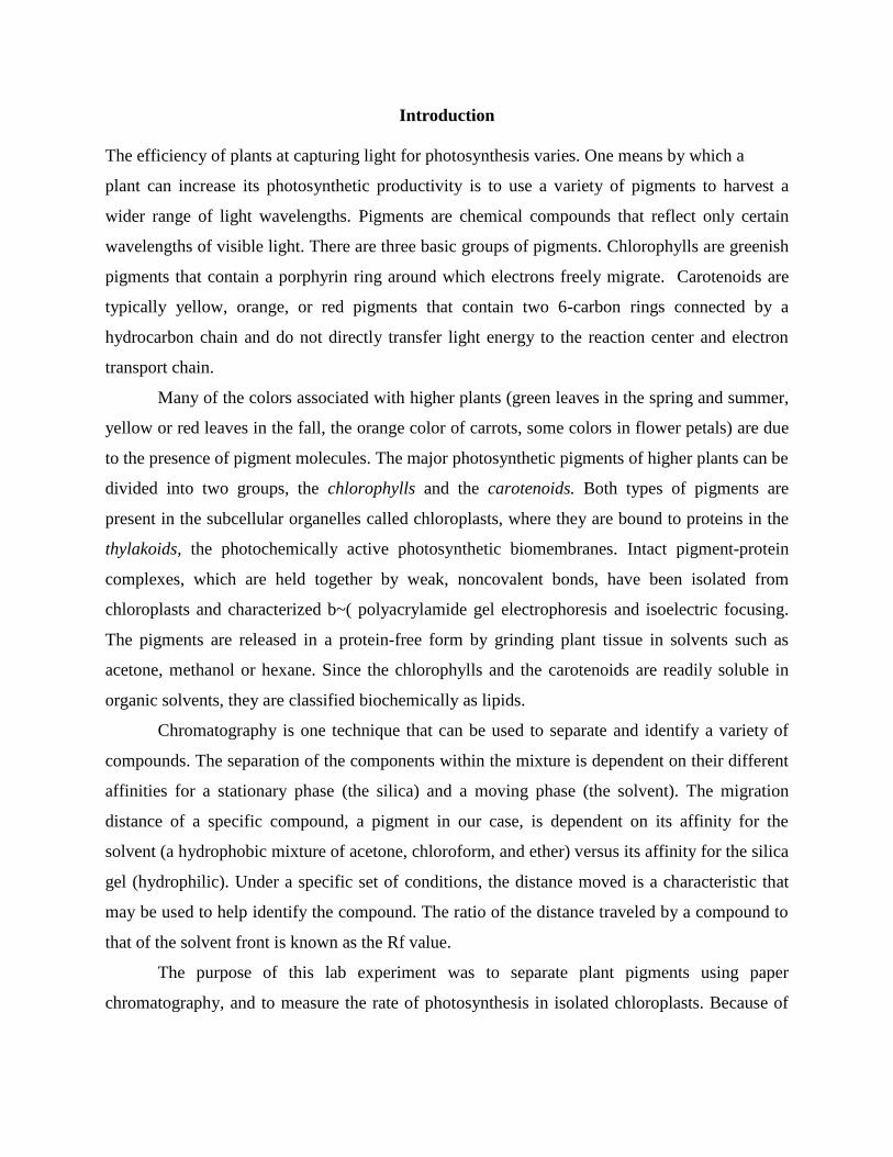
In this study, ethanol extraction was employed. We removed the petioles from the leaves
and weighed about 5g of our kangkong leaves. We used a blender to liquefy our leaves and
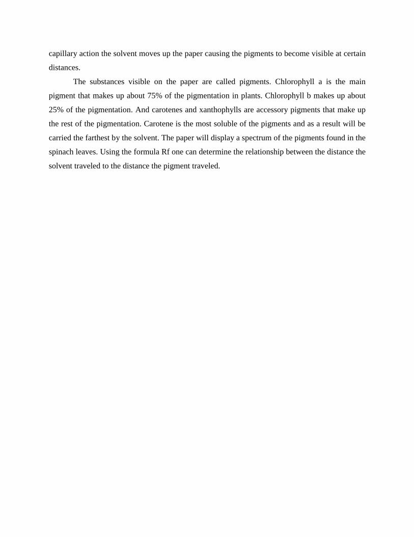
centrifuged for 2 minutes. We mounted a small portion on a microscope slide with 10% NaCl.
The appearance of the chloroplast was observed. We also put it under UV light to observe
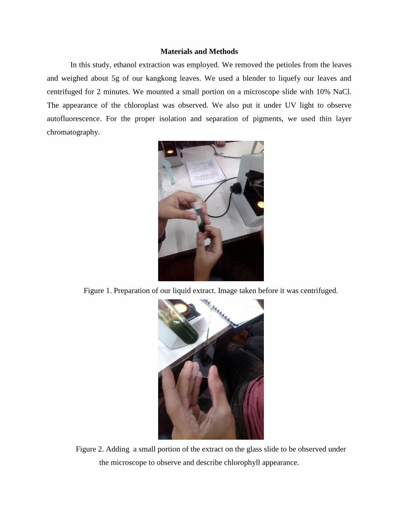
autofluorescence. For the proper isolation and separation of pigments, we used thin layer
chromatography.
Figure 1. Preparation of our liquid extract. Image taken before it was centrifuged.
Figure 2. Adding a small portion of the extract on the glass slide to be observed under
the microscope to observe and describe chlorophyll appearance.
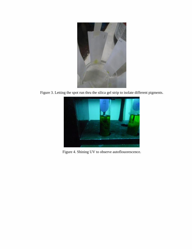
Figure 3. Letting the spot run thru the silica gel strip to isolate different pigments.
Figure 4. Shining UV to observe autoflouorescence.
Results
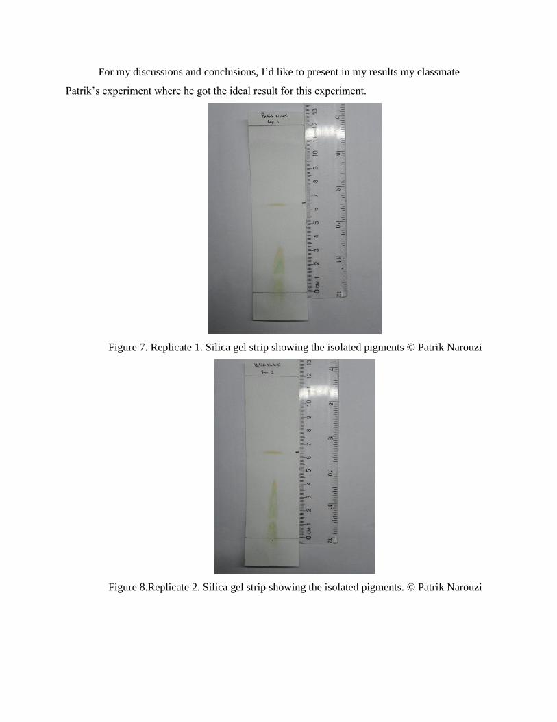
The results basically were not satisfactory since the spot did not run through the
silica gel strip. Perhaps not enough extract were dropped into the strip. But for my results, I
borrowed Patrik’s reults so that I can conclude and discuss further the different types of
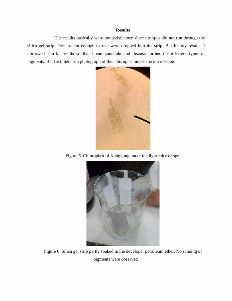
pigments. But first, here is a photograph of the chloroplast under the microscope.
Figure 5. Chloroplast of Kangkong under the light microscope.
Figure 6. Silica gel strip partly soaked in the developer petroleum ether. No running of
pigments were observed.
For my discussions and conclusions, I’d like to present in my results my classmate
Patrik’s experiment where he got the ideal result for this experiment.
Figure 7. Replicate 1. Silica gel strip showing the isolated pigments © Patrik Narouzi
Figure 8.Replicate 2. Silica gel strip showing the isolated pigments. © Patrik Narouzi
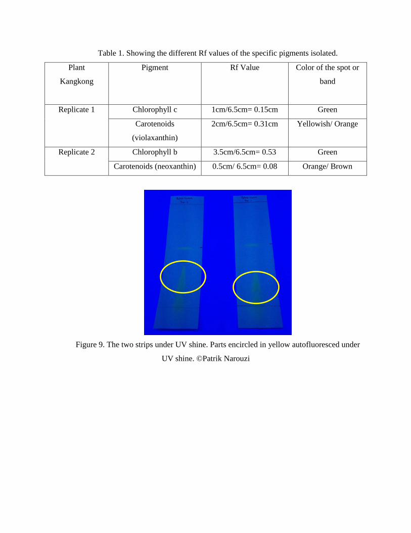
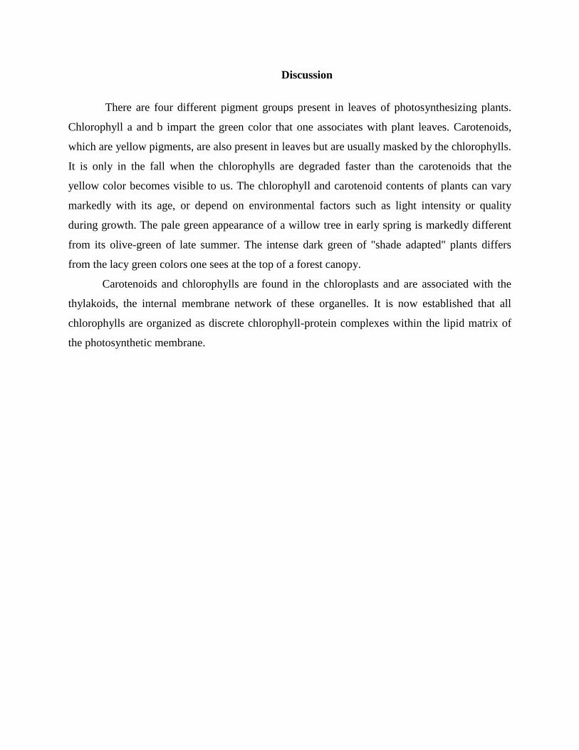
Table 1. Showing the different Rf values of the specific pigments isolated.
Plant
Kangkong
Pigment Rf Value Color of the spot or
band
Replicate 1 Chlorophyll c 1cm/6.5cm= 0.15cm Green
Carotenoids
(violaxanthin)
2cm/6.5cm= 0.31cm Yellowish/ Orange
Replicate 2 Chlorophyll b 3.5cm/6.5cm= 0.53 Green
Carotenoids (neoxanthin) 0.5cm/ 6.5cm= 0.08 Orange/ Brown
Figure 9. The two strips under UV shine. Parts encircled in yellow autofluoresced under
UV shine. ©Patrik Narouzi
Discussion
There are four different pigment groups present in leaves of photosynthesizing plants.
Chlorophyll a and b impart the green color that one associates with plant leaves. Carotenoids,
which are yellow pigments, are also present in leaves but are usually masked by the chlorophylls.
It is only in the fall when the chlorophylls are degraded faster than the carotenoids that the
yellow color becomes visible to us. The chlorophyll and carotenoid contents of plants can vary
markedly with its age, or depend on environmental factors such as light intensity or quality
during growth. The pale green appearance of a willow tree in early spring is markedly different
from its olive-green of late summer. The intense dark green of "shade adapted" plants differs
from the lacy green colors one sees at the top of a forest canopy.
Carotenoids and chlorophylls are found in the chloroplasts and are associated with the
thylakoids, the internal membrane network of these organelles. It is now established that all
chlorophylls are organized as discrete chlorophyll-protein complexes within the lipid matrix of
the photosynthetic membrane.
Conclusion
Leaves contain usually 7 different carotenoids (neoxanthin, violaxanthin, lutein,
zeaxanthin, antheraxanthin, β-carotene and α-carotene) that function in light harvesting.
Although most plant leaves appear green to our eyes, several pigments of different color are
usually present in the chloroplasts. Chlorophylls a and b provide the green color and absorb the
light energy needed for photosynthesis. However, other accessory pigments, such as yellow
xanthophylls and orange carotenes are also present in the chloroplasts and collect additional light
energy for photosynthesis.
Acknowledgement
In the conduct of this study, I’d like to thank first, the God Almighty for giving us the drive, the
good health, and the spirit to perform the experiment.
I’d also like to thank the following individuals and institutions who helped us in this
activity:
To Professor Hilconida Calumpong for being our instructor and giving us the proper
instructions fit for our experiment.
To Sir Ian Canlas for helping us set up our developer and also for helping us in handling
the apparatuses we used.
To my classmate, Patrik Narouzi, for sharing with me his results and thoughts regarding
why our experiment failed to get the ideal results.
To the Stock room of the the Biology Department for providing us with the materials
needed for this experiment.
And of course, to our parents, for their never-ending financial support.
References
Edwards-Knox Central School Science Department -
http://ekcsk12.org/science/lelab/chromatographylab.html.
Tomkins, S. P. and Miller, M. B. (1994). A rapid extraction and fast separation of leaf pigments
using thin layer chromatography. School Science review75 (273), 69 - 72.
CSU Stanislaus Chemistry http://wwwchem.csustan.edu/chem1102H/Green_Pigments.html.
http://sps.k12.ar.us/massengale/lab_4_plant_pigments.htm
http://www.ualr.edu/~biology/botany/pigmentlab.html
http://gened.emc.maricopa.edu/bio/bio181/BIOBK/BioBookPS.html