A synthetic triacylated pseudo-dipeptide molecule promotes Th1/TReg immune responses and enhances...
11
Vaccine (2007) 26, 108—118 available at www.sciencedirect.com journal homepage: www.elsevier.com/locate/vaccine A synthetic triacylated pseudo-dipeptide molecule promotes Th1/TReg immune responses and enhances tolerance induction via the sublingual route Laurent Mascarell a,1 , Laurence Van Overtvelt a,1 , Vincent Lombardi a , Alain Razafindratsita a , H´ el` ene Moussu a , St´ ephane Horiot a , Henri Chabre a , David Limal b , St´ ephane Moutel b , Jacques Bauer b , Carlo Chiavaroli b , Philippe Moingeon a,∗ a Stallerg` enes SA, Research and Development, 6 rue Alexis de Tocqueville, 92160 Antony, France b OM PHARMA, 22 rue du Bois-du-Lan, CH-1217 Meyrin, Switzerland Received 5 July 2007; received in revised form 15 October 2007; accepted 18 October 2007 KEYWORDS Sublingual immunotherapy; Lipid A derivatives; Th1; Adjuvant; Tolerance induction Summary In this study, we tested two triacylated pseudo-dipeptidic molecules, OM-197-MP- AC and OM-294-BA-MP as candidate adjuvants for allergy vaccines. Both molecules induce human dendritic cell (h-DC) maturation and polarize na¨ ıve T cells toward the Th1 type with IFN pro- duction. Only OM-294-BA-MP induces IL10 gene expression both in monocyte-derived DCs and CD4+ na¨ ıve T cells. Sublingual administration of OM-294-BA-MP plus the antigen enhances tol- erance induction in BALB/c mice with established asthma to ovalbumin with an impact on both airways hyperresponsiveness and lung inflammation. Given its Th1/Treg polarizing properties, OM-294-BA-MP is a valid candidate for sublingual allergy vaccines. © 2007 Elsevier Ltd. All rights reserved. Introduction Allergen-specific immunotherapy is currently the only causal treatment for allergy. Subcutaneous immunotherapy (SCIT) ∗ Corresponding author. Tel.: +33 1 55 59 25 20; fax: +33 1 55 59 21 02. E-mail address: [email protected] (P. Moingeon). 1 These authors contributed equally to this work. has been considered as a reference therapy to treat aller- gies against insect venoms, house dust mites, grass and tree pollens or animal epithelia for almost a century [1—3]. However, the need for repeated injections over several years makes this treatment inconvenient, particularly for children. In addition, SCIT can be associated with severe, albeit infrequent side effects, including anaphylactic shocks [4]. In this context, sublingual immunotherapy (SLIT) has been developed 15 years ago as a safer and non-invasive alternative to the treatment of respiratory allergies [5—8]. 0264-410X/$ — see front matter © 2007 Elsevier Ltd. All rights reserved. doi:10.1016/j.vaccine.2007.10.050
-
Upload
independent -
Category
Documents
-
view
2 -
download
0
Transcript of A synthetic triacylated pseudo-dipeptide molecule promotes Th1/TReg immune responses and enhances...

V
Apet
LAHC
a
b
R
I
At
f
0d
accine (2007) 26, 108—118
avai lab le at www.sc iencedi rec t .com
journa l homepage: www.e lsev ier .com/ locate /vacc ine
synthetic triacylated pseudo-dipeptide moleculeromotes Th1/TReg immune responses andnhances tolerance induction viahe sublingual route
aurent Mascarell a,1, Laurence Van Overtvelt a,1, Vincent Lombardia,lain Razafindratsitaa, Helene Moussua, Stephane Horiota,enri Chabrea, David Limalb, Stephane Moutelb, Jacques Bauerb,arlo Chiavaroli b, Philippe Moingeona,∗
Stallergenes SA, Research and Development, 6 rue Alexis de Tocqueville, 92160 Antony, FranceOM PHARMA, 22 rue du Bois-du-Lan, CH-1217 Meyrin, Switzerland
eceived 5 July 2007; received in revised form 15 October 2007; accepted 18 October 2007
KEYWORDSSublingualimmunotherapy;
Summary In this study, we tested two triacylated pseudo-dipeptidic molecules, OM-197-MP-AC and OM-294-BA-MP as candidate adjuvants for allergy vaccines. Both molecules induce humandendritic cell (h-DC) maturation and polarize naıve T cells toward the Th1 type with IFN� pro-
Lipid A derivatives;Th1;Adjuvant;Tolerance induction
duction. Only OM-294-BA-MP induces IL10 gene expression both in monocyte-derived DCs andCD4+ naıve T cells. Sublingual administration of OM-294-BA-MP plus the antigen enhances tol-erance induction in BALB/c mice with established asthma to ovalbumin with an impact on bothairways hyperresponsiveness and lung inflammation. Given its Th1/Treg polarizing properties,OM-294-BA-MP is a valid candidate for sublingual allergy vaccines.
ts re
h
© 2007 Elsevier Ltd. All righ
ntroduction
llergen-specific immunotherapy is currently the only causalreatment for allergy. Subcutaneous immunotherapy (SCIT)
∗ Corresponding author. Tel.: +33 1 55 59 25 20;ax: +33 1 55 59 21 02.
E-mail address: [email protected] (P. Moingeon).1 These authors contributed equally to this work.
gtHyca[ba
264-410X/$ — see front matter © 2007 Elsevier Ltd. All rights reserved.oi:10.1016/j.vaccine.2007.10.050
served.
as been considered as a reference therapy to treat aller-ies against insect venoms, house dust mites, grass andree pollens or animal epithelia for almost a century [1—3].owever, the need for repeated injections over severalears makes this treatment inconvenient, particularly for
hildren. In addition, SCIT can be associated with severe,lbeit infrequent side effects, including anaphylactic shocks4]. In this context, sublingual immunotherapy (SLIT) haseen developed 15 years ago as a safer and non-invasivelternative to the treatment of respiratory allergies [5—8].
alwwsultoafc(uToMs(tOal
A
HmmhoP(gasFrfahfmenvaCrCwcdp(t
A lipid A derivative enhances sublingual tolerance induction
Recently, a meta-analysis based on 22 randomized clin-ical studies concluded that SLIT of allergies to eitherhouse dust mites, pollens (e.g. grass, parietaria, olive, rag-weed, cupressus) or cat dander significantly reduces bothsymptoms (rhino-conjunctivitis, asthma), as well as theneed for symptomatic medication in both adults and chil-dren [5—8]. The clinical efficacy of SLIT in humans wasrecently confirmed in a phase IIb/III randomized, double-blind placebo controlled trial involving more than 630patients [9]. In this study, patients treated with grass pollentablets exhibited a significant reduction in both symp-toms (37%; p < 0.0001) and medication use (46%; p < 0.02).Given its well-established safety profile in humans, withmore than a billion doses administered [6,10—13], sub-lingual immunotherapy is presently considered as a validapproach to induce antigen-specific tolerance in humans[14].
During SLIT, a natural allergen extract is placed underthe tongue for at least 1—2 min prior to being swal-lowed. At the mucosal surface, allergens are captured byLangerhans-like dendritic cells [15], which subsequentlymigrate to draining cervical lymph nodes to presentthe antigen to naıve T cells [16,17]. Although allergicpatients develop Th2 polarized allergen-specific CD4+ Tcell responses characterized by IL4, IL5 and IL13 secretion[18,19], successful allergen-specific immunotherapy ratherelicits Th1 responses [13,20—29], as well as IL10 producingT cells [30—33], likely regulatory T cells belonging to theCD4+CD25+ or Tr1 subsets.
Appropriate mucosal immunomodulators capable toinduce antigen-specific Th1 and/or T regulatory immuneresponses are likely to enhance allergen-specific tolerancefollowing sublingual immunization. In this context, we focusin the present study on two triacylated, non-toxic pseudo-dipeptidic analogues of lipid-A, named OM-197-MP-AC andOM-294-BA-MP as candidate adjuvants. Whereas the TLR4agonist, OM-197-MP-AC is known to induce the maturationof human monocyte-derived DC and to increase ovalbumin-specific humoral and T helper cell responses [34,35], theimmunomodulatory properties of OM-294-BA-MP remain tobe assessed.
Herein, we demonstrate that OM-197-MP-AC inducesIFN�, whereas OM-294-BA-MP rather induces both IFN� andIL10 gene expression by naıve human CD4+ T cells. OM-294-BA-MP, but not OM-197-MP-AC, significantly enhances SLITefficacy in establishing tolerance in a therapeutic model ofovalbumin (OVA)-induced asthma in BALB/c mice.
Materials and method
Reagents
Recombinant human IL4 and human/mouse GM-CSF wereobtained from Gentaur (Brussels, Belgium). Phosphate-buffer saline (PBS) and RPMI 1640 were purchased fromInvitrogen (Carlsbad, CA). Alum was obtained from Pierce
(Rockford, IL). LPS from Escherichia coli 0128:B12 and OVAgrade V were purchased from Sigma (St. Louis, MO). Fordesensitization purposes, a purification step was added inorder to remove contaminant endotoxin (LPS) from OVA. Tothis aim, the OVA protein was diluted in PBS at 20 mg/mlhcotT
109
nd filtered with a 0.45 �m filter (Milex SLHV 025 NS, Mil-ipore). A 16/20 XK column (GE Health Care) was filledith 200 ml endotoxin removing gel (20339, Pierce), thenashed with 3 column volumes of apyrogen water, 1%
odium deoxycholate (D5670, Sigma) and water. The col-mn was equilibrated with 3 volumes of PBS, prior tooading the filtered OVA solution for 1 h to allow endo-oxin binding and eluting OVA with PBS. Endotoxin contentf such detoxified OVA was determined by Endochrom-Kssay (R1708K, Charles River, Wilmington, MA) and alwaysound to be less than 0.1 EU/�g of protein. Protein con-entrations were determined using a spectrophotometerSecomam, Ales, France), measuring absorption at 278 nmsing a molecular extinction coefficient of 141 mM−1 cm−1.he lactic acid bacterium, Lactobacillus plantarum wasbtained from the American Type Culture Collection (ATCC,anassas, VA, USA; No. 8014). OM-197-MP-AC (MP-AC
tands for MonoPhosphate AminoCaproyl) and OM-294-BA-MPBA-MP stands for Butyric Acid MonoPhosphate) are syn-hetic triacylated pseudo-dipeptidic molecules, [34] fromM-Pharma (Geneve, Switzerland). For in vitro assays,djuvants were used at 1—20 �g/ml as indicated in figureegends.
djuvant testing in human immune cells
uman-DCs (h-DCs) were generated from peripheral bloodononuclear cells (PBMCs) as previously described, withinor modifications [36]. Briefly, PBMCs were isolated from
ealthy volunteers by centrifugation of heparinized bloodver a Ficoll-Paque plus gradient (Amersham Biosciencesiscataway, NJ). Following resuspension in culture mediumRPMI 1640 supplemented with 2 mM L-glutamine, 20 �g/mlentamicin, 50 �M 2-mercaptoethanol, 1% nonessentialmino acids (all from Invitrogen), and 10% fetal bovineerum (Gentaur)), cells were allowed to adhere to 75 mlalcon flasks. After 2 h at 37 ◦C, nonadherent cells wereemoved and adherent cells were recovered and culturedor 7 days in 20 ml medium containing GM-CSF (80 ng/ml)nd IL4 (25 ng/ml). Nonadherent cells corresponding to the-DC-enriched fraction were harvested, washed, and usedor subsequent experiments. Human-DC purity assessed byorphology and flow cytometry analysis (based on CD1a
xpression) was always superior to 90%. Allogeneic CD4+aıve T lymphocytes were purified from PBMCs of healthyolunteers by magnetic bead separations (Invitrogen) usingmix of antibodies against CD14, CD16, CD56, CDw123,
D36, CD8, HLA Class II DR/DP, glycophorin A (all from Invit-ogen) and CD45RO molecules (BD Biosciences, San Jose,A). The purity of naıve CD45 RA+ T cells analyzed by FACSas always higher than 90%. To investigate the effect ofandidate adjuvants on DC maturation and cytokine pro-uction, 5 × 10⁵ h-DCs were incubated in 24-well plates inresence of either medium, adjuvants (20 �g/ml) or LPS1 �g/ml). After 24 h, h-DCs were washed, and tested inerms of viability and surface phenotype. Supernatants were
arvested and cytokine levels were determined using theytokine beads array (CBA) technology. To assess an effectn T cell polarization, 104 h-DCs/well were further cul-ured in triplicate in presence of allogeneic CD4+ naıvecells (105 cells/well) in 96-well flat-bottom microtiter
1
pst
A
Bocs5auDshsrr9Fw(P
Divwtwt(fS4fhPaDOPCfbc
Fp
FvtcpIIiht
CICBm(c
Ibaa
R
TcsRiBcoTwMmsc7ng
S
Sf(daldWOs2OawEaaota[
10
lates under a final volume of 200 �l. Cells and cultureupernatants were collected after 4 h, 1 or 5 days of cul-ure.
djuvant testing in murine immune cells
one marrow-derived dendritic cells (BM-DCs) werebtained as previously described [37]. Briefly, bone marrowells (2 × 10⁵ cells per ml) were incubated in RPMI 1640,upplemented with 10% foetal calf serum, antibiotics,× 10−5 M beta-2-mercaptoethanol (all from Invitrogen)nd 10 ng/ml GM-CSF. On day 7, BM-DCs were harvestedsing 10 ml of PBS/EDTA 5 mM for 5 min. CD4+ T cells fromO11.10 mice [38], transgenic for the T cell receptorpecific for the OVA 323—339 epitope restricted to the H2daplotype, were purified from spleens by magnetic beadeparation using the mouse CD4 negative isolation kit (Invit-ogen) according to the manufacturer’s instructions. Theesulting DO11.10 T cell preparations contained routinely5—99% pure CD4+ T cells as confirmed by FACS analysis.or proliferation assays, DO11.10 T cells were labelledith 1 �M carboxy-fluorescein diacetate, succinimidyl ester
CFSE; Molecular Probes, Eugene, OR) for 5 min at 37 ◦C inBS and washed twice.
To assess the effect of candidate adjuvants on BM-C maturation and cytokine production, 106 BM-DCs were
ncubated in presence of either complete medium, adju-ant (10 �g/ml) or LPS (2 �g/ml). After 24 h, BM-DCs wereashed, and tested in terms of viability and surface pheno-
ype. Supernatants were recovered and cytokine productionas determined by ELISA. For in vitro antigen presen-
ation assays, BM-DCs (5 × 10⁴) were incubated with OVA25 �g/ml) for 4 h, washed and co-cultured with 5 × 10⁵
reshly isolated DO11.10 T cells in 200 �l complete medium.upernatants were recovered at various time points (i.e.h, 1, or 3 days) during the culture in order to per-
orm cytokine secretion assays. In parallel, cells werearvested and lysed for mRNA isolation and quantitativeCR analysis (see Section RNA isolation and real-time PCRnalysis). For in vitro proliferation assays, CFSE labelledO11.10 CD4+ T cells were stimulated with BM-DCs andVA (10 �g/ml) for 3 days. Cells were stained with theE-KJ1.26 anti clonotypic mAb (BD Biosciences, San Jose,A) directed to the TCR expressed on OVA-specific T cellsrom DO11.10 mice [38]. Proliferating T cells were detectedy FACS on the basis of decreased CFSE-associated fluores-ence.
ACS analysis of surface phenotype and cytokineroduction
or immunofluorescence staining of h-DCs, cells were har-ested, washed in PBS and incubated for 20 min at roomemperature with the following mAbs: fluorescein isothio-yanate (FITC) labelled anti-CD80 IgG1, or anti-HLA-DR IgG1hycoerythrin (PE) labelled anti-CD86 IgG2b or anti-CD40
gG1, phycoerythrin-cyanin 5.1 (PC5) labelled anti-CD83gG2b (all from Beckman Coulter, Villepinte, France). Formmunofluorescence staining of murine BM-DCs, cells werearvested, washed in PBS and incubated for 20 min at roomemperature with the following mAbs: PE-labelled anti-tfsd
L. Mascarell et al.
D11c IgG, allophycocyanin (APC) labelled anti-MHC ClassI (I-A/I-E) IgG2b or anti-CD40 IgG, anti-CD80 IgG or anti-D86 IgG2a (all from eBiosciences). As a control, h-DCs andM-DCs were stained with corresponding isotype-matchedAbs. Samples were analyzed using a FC500 flow cytometer
Beckman Coulter). Results were expressed as mean fluores-ence intensity.
For cytokine production measurement, IL1�, IL6, IL10,L12p70 and TNF� were analyzed in culture supernatantsy flow cytometry using the human or mouse inflammationnd Th1/Th2 CBA kit from BD Biosciences (San Jose, CA), asppropriate, according to the manufacturer’s instructions.
NA isolation and real-time PCR analysis
otal RNA was isolated from 106 human or murine Tells (RNeasy Mini kit, Qiagen) and cDNAs were synthe-ized using random hexamers (Taqman Reverse Transcriptioneagents, Applied Biosystems, Foster city, CA), accord-ng to the manufacturer’s protocols. PCR probes (Appliediosystems) were selected from a gene bank from Appliedustom assays and synthesized by Applied Biosystems, inrder to amplify Tbet, GATA3, Foxp3, IFN�, IL4, IL10 andGF� human or murine genes, respectively. Real-time PCRas performed with Applied Biosystems Predesigned Taq-an Gene Expression Assays and reagents, according to theanufacturer’s specifications, using a 7300 Real-time PCR
ystem (Applied Biosystems). PCR was performed for 30—40ycles under the following conditions: 95 ◦C, 30 s; 55 ◦C, 30 s;2 ◦C, 30 s. The level of transcription for each gene wasormalized in comparison with the housekeeping �-actinene.
ublingual immunotherapy in BALB/c mice
ix- to eight-weeks-old BALB/c female mice were obtainedrom Charles River (L’Arbresle, France). For sensitizationFig. 1), mice were immunized intraperitoneally (i.p.) onays 0 and 14 with 10 �g OVA adsorbed on 2 mg Al(OH)3,dministered in 100 �l PBS. At day 21, a 20 min aerosol chal-enge was performed with 1% (w/v) OVA on 4 consecutiveays using an aerosol delivery system (Buxco Europe; Ltd.,inchester, UK). For tolerance induction (Fig. 1), 500 �gVA with or without 5 �g of each adjuvant were appliedublingually to groups of eight mice twice a week duringmonths in 20 �l of PBS. Control mice were sensitized withVA followed by sham tolerization with PBS or adjuvantlone. Measurements of airway hyperresponsiveness (AHR)ere performed by whole body plethysmography (Buxcourope; Ltd., Winchester, UK) and results were expresseds enhanced pause (Penh). The Penh index, expressed asn increase relative to the baseline airway resistance, wasbtained by dividing the Penh value measured after exposureo 50 mg/ml inhaled metacholine with the one measuredfter inhalation of nebulized PBS, as previously described39].
For lung histology, mice were killed by cervical disloca-ion, lungs were recovered and fixed in phosphate bufferedormalin-zinc and embedded in paraffin wax. Sections weretained with hematoxylin, eosin and safran (HES) for theetermination of cellular infiltrates.
A lipid A derivative enhances sublingual tolerance induction 111
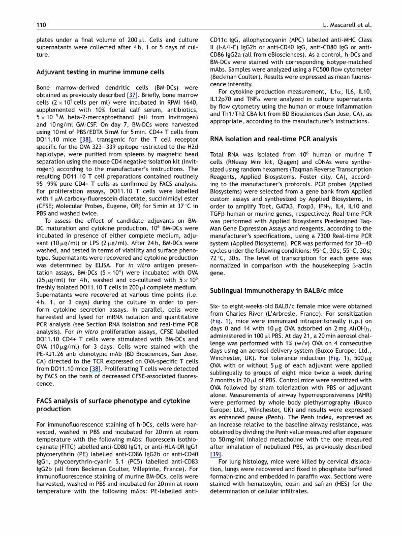
Figure 1 Experimental design. Mice were sensitized by intra-peritoneal (i.p.) injections with OVA/Alum (days 0 and 14)followed by an aerosol challenge (days 21—24) with OVA.BALB/c mice were sublingually treated twice a week with eitherPBS, OM-197-MP-AC (5 �g/dose) or OM-294-BA-MP (5 �g/dose),with or without OVA (500 �g/dose) (days 28—88). After an
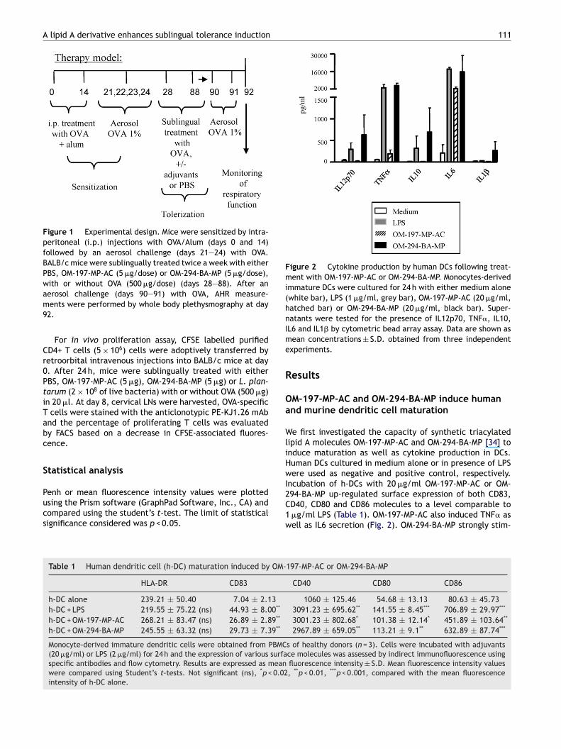
Figure 2 Cytokine production by human DCs following treat-ment with OM-197-MP-AC or OM-294-BA-MP. Monocytes-derivedimmature DCs were cultured for 24 h with either medium alone(white bar), LPS (1 �g/ml, grey bar), OM-197-MP-AC (20 �g/ml,hatched bar) or OM-294-BA-MP (20 �g/ml, black bar). Super-natants were tested for the presence of IL12p70, TNF�, IL10,Ime
R
Oa
WliHwI
aerosol challenge (days 90—91) with OVA, AHR measure-ments were performed by whole body plethysmography at day92.
For in vivo proliferation assay, CFSE labelled purifiedCD4+ T cells (5 × 10⁶) cells were adoptively transferred byretroorbital intravenous injections into BALB/c mice at day0. After 24 h, mice were sublingually treated with eitherPBS, OM-197-MP-AC (5 �g), OM-294-BA-MP (5 �g) or L. plan-tarum (2 × 10⁸ of live bacteria) with or without OVA (500 �g)in 20 �l. At day 8, cervical LNs were harvested, OVA-specificT cells were stained with the anticlonotypic PE-KJ1.26 mAband the percentage of proliferating T cells was evaluatedby FACS based on a decrease in CFSE-associated fluores-cence.
Statistical analysis
Penh or mean fluorescence intensity values were plottedusing the Prism software (GraphPad Software, Inc., CA) andcompared using the student’s t-test. The limit of statisticalsignificance considered was p < 0.05.
2C1w
Table 1 Human dendritic cell (h-DC) maturation induced by OM-
HLA-DR CD83
h-DC alone 239.21 ± 50.40 7.04 ± 2.13h-DC + LPS 219.55 ± 75.22 (ns) 44.93 ± 8.00**
h-DC + OM-197-MP-AC 268.21 ± 83.47 (ns) 26.89 ± 2.89**
h-DC + OM-294-BA-MP 245.55 ± 63.32 (ns) 29.73 ± 7.39**
Monocyte-derived immature dendritic cells were obtained from PBMC(20 �g/ml) or LPS (2 �g/ml) for 24 h and the expression of various surfaspecific antibodies and flow cytometry. Results are expressed as meanwere compared using Student’s t-tests. Not significant (ns), *p < 0.02intensity of h-DC alone.
L6 and IL1� by cytometric bead array assay. Data are shown asean concentrations ± S.D. obtained from three independent
xperiments.
esults
M-197-MP-AC and OM-294-BA-MP induce humannd murine dendritic cell maturation
e first investigated the capacity of synthetic triacylatedipid A molecules OM-197-MP-AC and OM-294-BA-MP [34] tonduce maturation as well as cytokine production in DCs.uman DCs cultured in medium alone or in presence of LPSere used as negative and positive control, respectively.
ncubation of h-DCs with 20 �g/ml OM-197-MP-AC or OM-
94-BA-MP up-regulated surface expression of both CD83,D40, CD80 and CD86 molecules to a level comparable to�g/ml LPS (Table 1). OM-197-MP-AC also induced TNF� asell as IL6 secretion (Fig. 2). OM-294-BA-MP strongly stim-197-MP-AC or OM-294-BA-MP
CD40 CD80 CD86
1060 ± 125.46 54.68 ± 13.13 80.63 ± 45.733091.23 ± 695.62** 141.55 ± 8.45*** 706.89 ± 29.97***
3001.23 ± 802.68* 101.38 ± 12.14* 451.89 ± 103.64**
2967.89 ± 659.05** 113.21 ± 9.1** 632.89 ± 87.74***
s of healthy donors (n = 3). Cells were incubated with adjuvantsce molecules was assessed by indirect immunofluorescence usingfluorescence intensity ± S.D. Mean fluorescence intensity values, **p < 0.01, ***p < 0.001, compared with the mean fluorescence

112 L. Mascarell et al.
Figure 3 Gene expression in T cells co-cultured with human or murine DCs pre-treated by OM-197-MP-AC or OM-294-BA-MP. (A)Naıve CD4+ T lymphocytes were co-cultured for 4 h (white bar), 1 day (grey bar) or 5 days (black bar) with allogeneic h-DCs pre-treated with either medium alone, LPS (1 �g/ml), OM-197-MP-AC (20 �g/ml) or OM-294-BA-MP (20 �g/ml). Gene expression wasanalyzed by quantitative PCR. Data are expressed as relative amounts of mRNA in T cells co-cultured with h-DC pre-treated withLPS (upper panel), OM-197-MP-AC (center panel) or OM-294-BA-MP (lower panel) in comparison with T cells incubated with h-DCalone. Data are normalized to amounts of �-actin, and shown as the mean ± S.D. of three independent experiments. (B) OVA-specificCD4+ T lymphocytes from DO11.10 mice were co-cultured for 4 h (white bar), 1 day (grey bar) or 3 days (black bar) with murineBM-DCs that were pre-treated with either medium alone, LPS (2 �g/ml), OM-197-MP-AC (10 �g/ml) or OM-294-BA-MP (10 �g/ml) inpresence of OVA (10 �g/ml). Gene expression was analyzed by quantitative PCR. Data are expressed as relative amounts of mRNA inT el), Oc normo
ubiMs
EC
WThANw
ct4iao(icup
cells co-cultured with BM-DCs pre-treated with LPS (upper panomparison with T cells incubated with BM-DCs alone. Data aref three independent experiments.
lated both IL12p70, TNF�, IL10, IL6 and IL1� productiony h-DCs (Fig. 2). Both OM-197-MP-AC and OM-294-BA-MPnduced murine BM-DC maturation, with up-regulation ofHC-II, CD40, CD80 and CD86 surface expression (data not
hown).
ffect of OM-197-MP-AC and OM-294-BA-MP onD4+ T cell polarization
e next analyzed the polarization of allogeneic naıve CD4+
cells incubated with adjuvant-treated h-DCs. To this aim,-DCs were incubated for 24 h with either OM-197-MP-C (20 �g/ml), OM-294-BA-MP (20 �g/ml) or LPS (1 �g/ml).on-activated h-DCs were used as a control. Human DCsere washed and incubated with allogeneic naıve CD4+ T
a(
tM
M-197-MP-AC (center panel) or OM-294-BA-MP (lower panel) inalized to amounts of �-actin and are shown as the mean ± S.D.
ells. The expression of genes encoding transcription fac-ors or cytokines was subsequently monitored after eitherh, 1 or 5 days by quantitative RT-PCR. As expected, LPS
nduces IFN� gene expression in naıve T cells as early asfter 4 h of culture (Fig. 3A, upper panel). OM-197-MP-ACnly slightly up-regulates IFN� gene expression, after 4 hFig. 3A, center panel). In contrast, OM-294-BA-MP markedlyncreases IFN� gene expression by at least 80-fold whenompared with T cells incubated with h-DC alone, and thisp-regulation is maintained throughout the 5-day monitoringeriod (Fig. 3A, lower panel). Interestingly, OM-294-BA-MP
lso induces within 24 h a 5-fold increase in IL10 mRNAFig. 3A, lower panel).These results were also confirmed in murine T cells. Tohis aim, murine BM-DCs were incubated with either OM-197-P-AC (10 �g/ml) or OM-294-BA-MP (10 �g/ml) for 24 h, then

A lipid A derivative enhances sublingual tolerance induction 113
washed and co-cultured with OVA-specific CD4+ T cells fromDO11.10 transgenic mice. Non-activated or LPS (2 �g/ml)treated BM-DCs served as negative and positive controls,respectively. Fig. 3B shows that both OM-197-MP-AC (cen-ter panel) and OM-294-BA-MP (lower panel) were able tostimulate IFN� gene expression. IL10 gene expression wasonly found in T cells co-cultured with OM-294-BA-MP treatedBM-DCs (Fig. 3B, lower panel). Taken together, these resultsobtained in both humans and murine immune cells, indicatethat OM-294-BA-MP induces a Th1/Treg CD4+ T cell responsewhereas OM-197-MP-AC rather elicits a Th1 pattern of dif-ferentiation.
We further tested OM-197-MP-AC and OM-294-BA-MP fortheir capacity to modulate T cell stimulation by DCs. MurineBM-DCs were incubated with either medium, LPS (1 �g/ml),OM-197-MP-AC (10 �g/ml) or OM-294-BA-MP (10 �g/ml).After 24 h, cells were washed and co-cultured for 3 dayswith CFSE-labelled DO11.10 CD4+ T cells with or withoutdetoxified OVA (10 �g/ml). OVA-specific T cell proliferationwas barely detectable in all groups when OVA was omitted(Fig. 4). BM-DCs co-cultured with T cells in the presence ofOVA showed a moderate proliferation (with only 25% pro-liferating cells, Fig. 4). In contrast, BM-DCs activated witheither LPS, OM-197-MP-AC or OM-294-BA-MP increased T cellproliferation significantly (Fig. 4).
Figure 4 OM-197-MP-AC or OM-294-BA-MP pre-treatment ofBM-DCs enhances T cell proliferation. OVA-specific CD4+ T lym-phocytes from DO11.10 mice were labelled with 0.5 �M CFSEand co-cultured with BM-DCs pre-treated with either mediumalone, LPS (2 �g/ml), OM-197-MP-AC (10 �g/ml) or OM-294-BA-MP (10 �g/ml) with or without OVA (10 �g/ml). After 3 days,cells were harvested, stained with PE-anti TCR (clone KJ1-26)mAbs. T cell proliferation was evaluated by FACS based on adecrease in CFSE-associated fluorescence using a FC500 flowcytometer. Data are shown as the mean ± S.D. of three inde-pendent experiments.
OM-294-BA-MP, but not OM-197-MP-AC, improvesclinical efficacy in a murine model of sublingualimmunotherapy
As described elsewhere [40], mice sensitized with OVAusing the protocol summarized in Fig. 1 develop airwayshyper responsiveness associated with elevated Penh val-ues detectable by whole body plethysmography, as wellas signs of lung inflammation with cellular infiltrates. OM-197-MP-AC and OM-294-BA-MP were tested as candidateadjuvants for immunotherapy in this in vivo murine model ofestablished asthma. OM-197-MP-AC (5 �g per dose) and OM-294-BA-MP (5 �g per dose) were administered sublinguallywith or without OVA (500 �g per dose) in a total volume of20 �l, in order to test their capacity to enhance the effi-cacy of allergen-specific immunotherapy in lowering AHR.Healthy mice used as a control (i.e. non-sensitized) in thoseexperiments exhibit, as expected, low Penh values (Fig. 5),whereas OVA-sensitized animals treated sublingually withPBS alone display a high AHR. Sublingual treatment withsoluble OVA alone twice a week for two months has avery moderate impact on AHR (Fig. 5). In contrast, sub-lingual treatment with OVA administered together withOM-294-BA-MP, but not OM-197-MP-AC, induces a statisti-cally significant (p < 0.05) reduction of AHR in most animals
Figure 5 OM-294-BA-MP enhances tolerance induction viathe sublingual route. Airway responsiveness was determinedby measuring the Penh value in response to metacholine(50 mg/ml) as described in methods. Between four to eight indi-vidual mice were analyzed within each group. Horizontal barsrepresent the median response within each group with each dotrepresenting the Penh value obtained in a single animal. Resultsare representative of two independent experiments.

114 L. Mascarell et al.
F roncw ithera ltrat
wIimm
owms2(a(
Aa
Wr1cncT
igure 6 Therapeutic SLIT with OM-294-BA-MP + OVA reduces bith HES are shown. Samples obtained from mice treated with ere shown (100-fold magnification). Arrows indicate cellular infi
hen compared with mice treated with PBS (Fig. 5).mportantly, neither OM-197-MP-AC nor OM-294-BA-MP alonemproves AHR, suggesting that antigen-specific immuneechanisms are involved in tolerance induction in thisodel.The evaluation of bronchial inflammation by lung histol-
gy in those animals confirmed that sublingual treatmentith OM-294-BA-MP + OVA leads to a reduction of lung inflam-ation, with less cellular infiltrates detectable in tissue
ections (Fig. 6 and Table 2). Importantly, neither PBS, OM-
94-BA-MP, nor OVA has any effect on lung inflammationFig. 6 and Table 2). Treatment with OM-197-MP-AC as andjuvant had no effect on established lung inflammationdata not shown).ina
Table 2 Perivascular and alveolar inflammation analysis in OVA-BA-MP or OM-294-BA-MP + OVA
Groups n Perivascular inflammatio
+ ++
PBS 7OVA 8 4/8OM-294-BA-MP 8OM-294-BA-MP + OVA 8 1/8 5/8
BALB/c mice (7—8 per group) were sensitized to OVA as described in Fiwith either PBS, or OM-294-BA-MP (5 �g/dose), with or without OVA (days 90 and 91) with 1% (w/v) OVA, lungs were removed and a semi-qassessment of the presence of cellular infiltrates (i.e. lymphoytes, panimals exhibiting signs of inflammation ranked from low (+), moderat
hial inflammation. Representative paraffin lung sections stainedPBS, soluble OVA, OM-294-BA-MP alone or OM-294-BA-MP + OVAes.
ssessment of immune responses after sublingualdministration of OM-197-MP-AC or OM-294-BA-MP
hereas sensitized BALB/c mice exhibit OVA-specific Th2esponses [40], sublingually treated mice with OVA plus OM-97-MP-AC or OM-294-BA-MP did not exhibit any detectablehanges in OVA-specific IgE, IgG1 or IgG2a seric titers (dataot shown). We also failed to detect by Elispot any significanthanges in the number of OVA-specific IL5 or IL10 producingcells in the spleen (data not shown).
To evaluate whether adjuvant molecules promote ornhibit CD4+ T cell priming within draining cervical lymphode after sublingual administration [40,41], we performeddoptive transfer experiments. To this aim, CFSE labelled
sensitized mice treated sublingually with PBS, OVA, OM-294-
n Alveolar inflammation
+++ + ++ +++ ++++
7/7 2/7 5/74/8 1/8 3/8 4/88/8 1/8 4/8 3/82/8 2/8 4/8 1/8 1/8
g. 1. OVA-sensitized mice were sublingually treated twice a week500 �g/dose) (days 28—88). After an aerosol challenge (both onuantitative analysis of inflammation was performed with a blindlasmocytes and eosinophils). Data are expressed as numbers ofe (++), important (+++), to severe (++++).
A lipid A derivative enhances sublingual tolerance induction 115
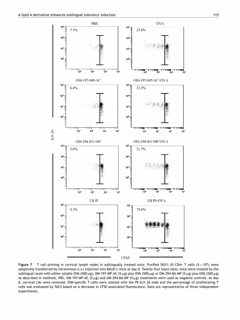
Figure 7 T cell priming in cervical lymph nodes in sublingually treated mice. Purified DO11.10 CD4+ T cells (5 × 10⁶) wereadoptively transferred by intravenous (i.v.) injection into BALB/c mice at day 0. Twenty-four hours later, mice were treated by thesublingual route with either soluble OVA (500 �g), OM-197-MP-AC (5 �g) plus OVA (500 �g) or OM-294-BA-MP (5 �g) plus OVA (500 �g)as described in methods. PBS, OM-197-MP-AC (5 �g) and OM-294-BA-MP (5 �g) treatments were used as negative controls. At day8, cervical LNs were removed. OVA-specific T cells were stained with the PE-KJ1.26 mAb and the percentage of proliferating Tcells was evaluated by FACS based on a decrease in CFSE-associated fluorescence. Data are representative of three independentexperiments.
1
DitLOObvtcomFPTfwOt
iCian
D
TataC[blcaeo
taFMlamIMcigItb[
wiu
t[aBBmcBbDFrstbfOTriA
m[tcccipmitlaAtLspe
vwTsB
R
16
O11.10 CD4+ T cells (5 × 10⁶) were injected intravenouslynto BALB/c mice at day 0. After 24 h, mice were sublinguallyreated with either PBS, OM-197-MP-AC, OM-294-BA-MP or. plantarum (LB-Pl) as a control [41] with or withoutVA. At day 8, cervical LNs were harvested and in vivoVA-specific T cell proliferative responses were analyzedy FACS. T cell proliferation was not detectable in cer-ical LNs in PBS, OM-197-MP-AC, OM-294-BA-MP or LB-Plreated mice with only 7.1, 6.4, 5.6 and 4.3% proliferating Tells, respectively. In contrast, OVA, OM-197-MP-AC plus OVAr OM-294-BA-MP plus OVA treatments similarly induced aoderate proliferation (23.0, 23.5 and 21.7%, respectively,
ig. 7) in cervical LN cells. As previously described [41] LB-l + OVA strongly increased T cell proliferation up to 70%.hese results confirm a local priming of OVA-specific T cellsollowing sublingual immunization under those conditions,ithout any further detectable impact of OM-197-MP-AC orM-294-BA-MP on proliferation when co-administrated withhe antigen.
We also analyzed by FACS the impact of sublingual admin-stration of OM-294-BA-MP plus OVA on Foxp3 expression inFSE labelled DO11.10 CD4+ T cells adoptively transferred
nto BALB/c mice in cervical lymph nodes. We did not detectny increase in Foxp3 expression in both proliferating andon-proliferating T cells (data not shown).
iscussion
he sublingual route of administration is now establisheds a safe and efficacious route to induce allergen-specificolerance in humans [5—14]. Several studies suggest thatllergen-specific immunotherapy redirects allergen-specificD4+ T cell responses from a Th2 to a Th1 response13]. Also successful allergen immunotherapy appears toe associated with the induction of IL10-producing regu-atory T-cells including both Foxp3 + CD4 + CD25+ and Tr1ells [30—33]. In this context, adjuvants promoting IFN�nd/or IL10 production by naıve T cells are expected tonhance allergen-specific tolerance induction in the coursef immunotherapy.
Triggering toll like receptor (TLR) with lipid A deriva-ives is known to elicit both systemic and mucosal responses,nd TLR4 ligands usually represent powerful Th1 adjuvants.or example, nasal or oral administration of antigens withonophosphoryl lipid A (MPL) or RC-529 (a synthetic MPL-
ike acylated monosaccharide) induces seric and local IgAs,s well as IFN�-producing cytotoxic T lymphocytes (CTL) inice [3,42,43]. The capacity of MPL to induce both IFN� and
L10 production by immune cells is well documented [42,44].PL has been successfully used in humans in grass pollen spe-ific subcutaneous immunotherapy and is currently testedn SLIT [45,46]. The co-administration of grass pollen aller-ens in combination with MPL enhances allergen-specificgG1 and IgG4 antibody responses, with potentially induc-ion of blocking antibodies which prevent histamine releasey basophils and reduce IgE-mediated allergen presentation
47].A triacylated pseudo-dipeptidic molecule, OM-197-MP-ACas also shown to increase antigen-specific Th1 responses
n mice [35]. In addition, OM-197-MP-AC induces the mat-ration of human DCs, as well as their capacity to support
L. Mascarell et al.
he expansion of IFN-� secreting T cells from naıve donors34]. In this study, we analyzed OM-197-MP-AC as well asnother triacylated pseudo-dipeptidic molecule, OM-294-A-MP, as potential candidate adjuvants for allergy vaccines.oth OM-197-MP-AC and OM-294-BA-MP similarly induceurine and h-DCs maturation. However, the patterns of
ytokine released are clearly distinct, since only OM-294-A-MP stimulates IL1�, IL12p70, TNF� and IL10 productiony DCs. Although, OM-197-MP-AC and OM-294-BA-MP-treatedCs do not alter transcription factors (i.e. Tbet, GATA3,oxp3) specific for Th1, Th2 or regulatory T lymphocytes,espectively (Fig. 3), we do not exclude a potential tran-ient up-regulation of those genes. However, OM-197-MP-ACreated-DCs elicits a moderate expression of the IFN� geney naıve T cells. As a Th1 adjuvant, it is nonetheless inef-ective in reducing established AHR in mice sensitized withVA. In contrast, OM-294-BA-MP treated-DCs trigger primaryh1/Treg responses with high IFN� gene expression and aeadily detectable induction of the IL10 gene. When admin-stered sublingually in mice, OM-294-BA-MP reduces bothHR and lung inflammation.
Given the known effect of IFN� in inducing tolerance inurine models of transplantation and autoimmune diseases
48—50], the relative efficacy of the two lipid A deriva-ives in our murine SLIT model might be related to theirapacity to induce sufficient amounts of IFN� by naıve Tells. IFN� mediates tolerance by inhibiting Th2 effector Tells or inducing both nitric oxide synthetase (iNOS) and/orndoleamine-2,3-dioxygenase (IDO) expression in antigenresenting cell (APCs) [48—50]. As a non-mutually exclusiveechanism, the induction of the IL10 gene by OM-294-BA-MP
s likely to be critical in the immunomodulatory function ofhis adjuvant. In support of this hypothesis, a diphosphory-ated and triacetylated lipid A analogue, OM-174 which actss a strong Th1 adjuvant in mice [51—53], had no impact onHR in our SLIT model (data not shown). Also, we observedhat only IL10-inducing adjuvants (e.g. selected strains ofactobacilli, 1-�25-(dihydroxy)vitamin D3 plus dexametha-one [41] or the TLR2 ligand Pam3CSK4 (manuscript inreparation)) enhance SLIT efficacy in a murine model ofstablished asthma.
The development of second-generation sublingual allergyaccines, most particularly based on recombinant allergens,ill likely rely upon safe and efficient adjuvants inducingcell responses with a Th1/Treg profile. We conclude that
elected synthetic triacyl lipid A derivatives such as OM-294-A-MP represent valid adjuvants to improve SLIT in humans.
eferences
[1] Noon L. Prophylactic inoculation against hay fever. Lancet1911;2(2):1572—3.
[2] Bousquet J, Lockey R, Malling HJ. Allergen immunotherapy:therapeutic vaccines for allergic diseases. A WHO positionpaper. J Allergy Clin Immunol 1998;102(4 Pt 1):558—62.
[3] Mascarell L, Van Overtvelt L, Moingeon P. Novel ways for
immune intervention in immunotherapy: mucosal allergy vac-cines. Immunol Allergy Clin North Am 2006;26(2):283—306[vii—viii].[4] Medicines CoSo. Desensitising vaccines: an allergist’s view. BrMed J 1986;293(6555):1169—70.

[
[
[
[
[
[
[
[
[
[
[
[
[
[
[
A lipid A derivative enhances sublingual tolerance induction
[5] Wilson DR, Torres LI, Durham SR. Sublingual immunother-apy for allergic rhinitis. Cochrane Database Syst Rev 2003;(2)[CD002893].
[6] Passalacqua G, Guerra L, Pasquali M, Lombardi C, Canonica GW.Efficacy and safety of sublingual immunotherapy. Ann AllergyAsthma Immunol 2004;93(1):3—12 [quiz 12—3, 103].
[7] Pajno GB, Peroni DG, Vita D, Pietrobelli A, Parmiani S, Boner AL.Safety of sublingual immunotherapy in children with asthma.Paediatr Drugs 2003;5(11):777—81.
[8] Penagos M, Compalati E, Tarantini F, Baena-Cagnani R, HuertaJ, Passalacqua G, et al. Efficacy of sublingual immunother-apy in the treatment of allergic rhinitis in pediatric patients3 to 18 years of age: a meta-analysis of randomized, placebo-controlled, double-blind trials. Ann Allergy Asthma Immunol2006;97(2):141—8.
[9] Didier A, Malling H, Worm M, Horak F, Jager S, Mon-tagut A, et al. Optimal dose, efficacy, and safety ofonce-daily sublingual immunotherapy with 5-grass pollentablet for seasonal allergic rhinitis. J Allergy Clin Immunol,doi:10.1016/j.jaci.2007.07.046.
[10] Frew AJ, Smith HE. Sublingual immunotherapy. J Allergy ClinImmunol 2001;107(3):441—4.
[11] Agostinis F, Tellarini L, Canonica GW, Falagiani P, PassalacquaG. Safety of sublingual immunotherapy with a monomeric aller-goid in very young children. Allergy 2005;60(1):133.
[12] Grosclaude M, Bouillot P, Alt R, Leynadier F, Scheinmann P, RufinP, et al. Safety of various dosage regimens during induction ofsublingual immunotherapy. A preliminary study. Int Arch AllergyImmunol 2002;129(3):248—53.
[13] Norman PS. Immunotherapy: 1999—2004. J Allergy ClinImmunol 2004;113(6):1013—23 [quiz 24].
[14] Khinchi MS, Poulsen LK, Carat F, Andre C, Hansen AB,Malling HJ. Clinical efficacy of sublingual and subcutaneousbirch pollen allergen-specific immunotherapy: a random-ized, placebo-controlled, double-blind, double-dummy study.Allergy 2004;59(1):45—53.
[15] Allam JP, Novak N, Fuchs C, Asen S, Berge S, Appel T, et al.Characterization of dendritic cells from human oral mucosa: anew Langerhans’ cell type with high constitutive FcepsilonRIexpression. J Allergy Clin Immunol 2003;112(1):141—8.
[16] van Wilsem EJ, Breve J, Savelkoul H, Claessen A, Scheper RJ,Kraal G. Oral tolerance is determined at the level of draininglymph nodes. Immunobiology 1995;194(4—5):403—14.
[17] van Helvoort JM, Samsom JN, Chantry D, Jansen W, Schadee-Eestermans I, Thepen T, et al. Preferential expression of IgG2bin nose draining cervical lymph nodes and its putative role inmucosal tolerance induction. Allergy 2004;59(11):1211—8.
[18] Durham SR, Till SJ. Immunologic changes associatedwith allergen immunotherapy. J Allergy Clin Immunol1998;102(2):157—64.
[19] El Biaze M, Boniface S, Koscher V, Mamessier E, Dupuy P, Milhe F,et al. T cell activation, from atopy to asthma: more a paradoxthan a paradigm. Allergy 2003;58(9):844—53.
[20] Ippoliti F, De Santis W, Volterrani A, Lenti L, CanitanoN, Lucarelli S, et al. Immunomodulation during sublin-gual therapy in allergic children. Pediatr Allergy Immunol2003;14(3):216—21.
[21] Mavroleon G. Restoration of cytokine imbalance byimmunotherapy. Clin Exp Allergy 1998;28(8):917—20.
[22] Hamid QA, Schotman E, Jacobson MR, Walker SM, DurhamSR. Increases in IL-12 messenger RNA+ cells accompanyinhibition of allergen-induced late skin responses after suc-cessful grass pollen immunotherapy. J Allergy Clin Immunol
1997;99(2):254—60.[23] Benjaponpitak S, Oro A, Maguire P, Marinkovich V, DeKruyffRH, Umetsu DT. The kinetics of change in cytokine productionby CD4 T cells during conventional allergen immunotherapy. JAllergy Clin Immunol 1999;103(3 Pt 1):468—75.
[
117
24] Ebner C, Siemann U, Bohle B, Willheim M, WiedermannU, Schenk S, et al. Immunological changes during specificimmunotherapy of grass pollen allergy: reduced lymphoprolif-erative responses to allergen and shift from TH2 to TH1 in T-cellclones specific for Phl p 1, a major grass pollen allergen. ClinExp Allergy 1997;27(9):1007—15.
25] Secrist H, Chelen CJ, Wen Y, Marshall JD, Umetsu DT. Allergenimmunotherapy decreases interleukin 4 production in CD4+ Tcells from allergic individuals. J Exp Med 1993;178(6):2123—30.
26] Laaksonen K, Junikka M, Lahesmaa R, Terho EO, SavolainenJ. In vitro allergen-induced mRNA expression of signaling lym-phocytic activation molecule by PBMC of patients with allergicrhinitis is increased during specific pollen immunotherapy. JAllergy Clin Immunol 2003;112(6):1171—7.
27] Meissner N, Kochs S, Coutelle J, Kussebi F, Baumgarten C,Lowenstein H, et al. Modified T-cell activation pattern duringspecific immunotherapy (SIT) in cat-allergic patients. Clin ExpAllergy 1999;29(5):618—25.
28] Majori M, Caminati A, Corradi M, Brianti E, Scarpa S, Pesci A.T-cell cytokine pattern at three time points during specificimmunotherapy for mite-sensitive asthma. Clin Exp Allergy2000;30(3):341—7.
29] Gabrielsson S, Soderlund A, Paulie S, van der Pouw Kraan TC,Troye-Blomberg M, Rak S. Specific immunotherapy preventsincreased levels of allergen-specific IL-4- and IL-13-producingcells during pollen season. Allergy 2001;56(4):293—300.
30] Jutel M, Akdis M, Budak F, Aebischer-Casaulta C, Wrzyszcz M,Blaser K, et al. IL-10 and TGF-beta cooperate in the regulatoryT cell response to mucosal allergens in normal immunity andspecific immunotherapy. Eur J Immunol 2003;33(5):1205—14.
31] Gardner LM, Thien FC, Douglass JA, Rolland JM, O’Hehir RE.Induction of T ’regulatory’ cells by standardized house dustmite immunotherapy: an increase in CD4+ CD25+ interleukin-10+ T cells expressing peripheral tissue trafficking markers.Clin Exp Allergy 2004;34(8):1209—19.
32] Taylor A, Verhagen J, Akdis CA, Akdis M. T regulatory cellsin allergy and health: a question of allergen specificity andbalance. Int Arch Allergy Immunol 2004;135(1):73—82.
33] Ciprandi G, Fenoglio D, Cirillo I, Vizzaccaro A, Ferrera A,Tosca MA, et al. Induction of interleukin 10 by sublingualimmunotherapy for house dust mites: a preliminary report. AnnAllergy Asthma Immunol 2005;95(1):38—44.
34] Byl B, Libin M, Bauer J, Martin OR, De Wit D, Davies G, et al.OM197-MP-AC induces the maturation of human dendritic cellsand promotes a primary T cell response. Int Immunopharmacol2003;3(3):417—25.
35] Savoy F, Nicolle DM, Rivier D, Chiavaroli C, Ryffel B, Ques-niaux VF. Synthetic triacylated lipid a derivative activatesantigen presenting cells via the TLR4 pathway and pro-motes antigen-specific responses in vivo. Immunobiology2006;211(10):767—77.
36] Romani N, Reider D, Heuer M, Ebner S, Kampgen E, Eibl B, etal. Generation of mature dendritic cells from human blood. Animproved method with special regard to clinical applicability.J Immunol Methods 1996;196(2):137—51.
37] Inaba K, Inaba M, Romani N, Aya H, Deguchi M, IkeharaS, et al. Generation of large numbers of dendritic cellsfrom mouse bone marrow cultures supplemented with gran-ulocyte/macrophage colony-stimulating factor. J Exp Med1992;176(6):1693—702.
38] Murphy KM, Heimberger AB, Loh DY. Induction by antigen ofintrathymic apoptosis of CD4+CD8+TCRlo thymocytes in vivo.
Science 1990;250(4988):1720—3.39] Kline JN, Kitagaki K, Businga TR, Jain VV. Treatmentof established asthma in a murine model using CpGoligodeoxynucleotides. Am J Physiol Lung Cell Mol Physiol2002;283(1):L170—9.

1
[
[
[
[
[
[
[
[
[
[
[
[
[
18
40] Razafindratsita A, Saint-Lu N, Mascarell L, Berjont N, Bardon T,Betbeder D, et al. Improvement of sublingual immunotherapyefficacy with a mucoadhesive allergen formulation. J AllergyClin Immunol 2007;120:278—85.
41] Van Overtvelt L, Lombardi V, Razafindratsita A, Saint-Lu N,Horiot S, Moussu H, et al. IL-10-inducing adjuvants enhancesublingual immunotherapy efficacy in a murine asthma model.Int Arch Allergy Immunol 2007;145(2):152—62.
42] Baldridge JR, Yorgensen Y, Ward JR, Ulrich JT. Monophos-phoryl lipid A enhances mucosal and systemic immunity tovaccine antigens following intranasal administration. Vaccine2000;18(22):2416—25.
43] Burdin N, Guy B, Moingeon P. Immunological foundations to thequest for new vaccine adjuvants. BioDrugs 2004;18(2):79—93.
44] Martin M, Michalek SM, Katz J. Role of innate immune factors inthe adjuvant activity of monophosphoryl lipid A. Infect Immun2003;71(5):2498—507.
45] Drachenberg KJ, Heinzkill M, Urban E, Woroniecki SR. Efficacyand tolerability of short-term specific immunotherapy withpollen allergoids adjuvanted by monophosphoryl lipid A (MPL)for children and adolescents. Allergol Immunopathol (Madr)2003;31(5):270—7.
46] Drachenberg KJ, Wheeler AW, Stuebner P, Horak F. A well-tolerated grass pollen-specific allergy vaccine containing a
novel adjuvant, monophosphoryl lipid A, reduces allergicsymptoms after only four preseasonal injections. Allergy2001;56(6):498—505.47] Mothes N, Heinzkill M, Drachenberg KJ, Sperr WR, KrauthMT, Majlesi Y, et al. Allergen-specific immunotherapy with a
[
L. Mascarell et al.
monophosphoryl lipid A-adjuvanted vaccine: reduced season-ally boosted immunoglobulin E production and inhibition ofbasophil histamine release by therapy-induced blocking anti-bodies. Clin Exp Allergy 2003;33(9):1198—208.
48] Sawitzki B, Kingsley CI, Oliveira V, Karim M, Herber M, WoodKJ. IFN-gamma production by alloantigen-reactive regulatoryT cells is important for their regulatory function in vivo. J ExpMed 2005;201(12):1925—35.
49] Kelchtermans H, De Klerck B, Mitera T, Van Balen M, Bullens D,Billiau A, et al. Defective CD4+CD25+ regulatory T cell func-tioning in collagen-induced arthritis: an important factor inpathogenesis, counter-regulated by endogenous IFN-gamma.Arthritis Res Ther 2005;7(2):R402—15.
50] Wood KJ, Sawitzki B. Interferon gamma: a crucial role in thefunction of induced regulatory T cells in vivo. Trends Immunol2006;27(4):183—7.
51] Pajak B, Garze V, Davies G, Bauer J, Moser M, Chiavaroli C. Theadjuvant OM-174 induces both the migration and maturation ofmurine dendritic cells in vivo. Vaccine 2003;21(9—10):836—42.
52] Meraldi V, Audran R, Romero JF, Brossard V, Bauer J, LopezJA, et al. OM-174, a new adjuvant with a potential forhuman use, induces a protective response when adminis-tered with the synthetic C-terminal fragment 242—310 fromthe circumsporozoite protein of Plasmodium berghei. Vaccine
2003;21(19—20):2485—91.53] D’Agostini C, Pica F, Febbraro G, Grelli S, Chiavaroli C, GaraciE. Antitumour effect of OM-174 and cyclophosphamide onmurine B16 melanoma in different experimental conditions.Int Immunopharmacol 2005;5(7—8):1205—12.