"Réflexions sémiologiques sur le relief Louvre B7", BdE 145, 2008, p.43-51.

Cell, Vol. 80, 707-718, March 10, 1995, Copyright © 1995 by Cell Press
B7-1 and B7-2 Costimulatory Molecules Activate Differentially the Thl/Th2 Developmental Pathways: Application to Autoimmune Disease Therapy
Vijay K. Kuchroo,*t Mercy Prabhu Das,*t Julia A. Brown,~ Ann M. Ranger,$ Scott S. Zamvil,* Raymond A. Sobel,§ Howard L. Weiner,* Nasrin Nabavi,II and Laurie H. Glimcher$ *Department of Neurology Harvard Medical School and Center for Neurological Diseases Department of Medicine Brigham and Women's Hospital Boston, Massachusetts 02115 SDepartment of Cancer Biology Harvard School of Public Health and Department of Medicine Harvard Medical School Boston, Massachusetts 02115 §Department of Pathology Stanford University School of Medicine
and Laboratory Service Veterans' Affairs Medical Center Palo Alto, California 94304 IIDepartment of Inflammation and Autoimmune Disease Hoffmann-LaRoche Research Center Nutley, New Jersey 07110
Summary
CD4 T helper precursor cells mature along two alterna- tive pathways, Thl and Th2. Here we show that these pathways are differentially activated by two costimula- tory molecules, B7-1 and B7-2. Using anti-B7 antibod- ies, this developmental step was manipulated both in vitro and in vivo in experimental allergic encephalomy- elitis (EAE). Anti-B7-1 reduced the incidence of dis- ease while anti-B7-2 increased disease severity. Nei- ther antibody affected overall T cell induction but rather altered cytokine profile. Administration of anti- B7-1 at immunization resulted in predominant genera- tion of Th2 clones whose transfer both prevented in- duction of EAE and abrogated established disease. Since cotreatment with anti-lL-4 antibody prevented disease amelioration, costim ulatory molecules may di- rectly affect initial cytokine secretion. Thus, interac- tion of B7-1 and B7-2 with shared counterreceptors CD28 and CTLA-4 results in very different outcomes in clinical disease by influencing commitment of pre- cursors to a Thl or Th2 lineage.
Introduction
CD4 ÷ T helper (Th) cells, upon antigenic stimulation, differ- entiate into two distinct subpopulations, each producing its own set of cytokines and mediating separate effector functions (Mosmann and Coffman, 1989; Seder and Paul, 1994). Type 1 Th cells (Thl) produce interleukin-2 (IL-2),
tThe first two authors contributed equally to this work.
tumor necrosis factor 13 (TNFI3), and interferon-7 (IFNT), thereby activating macrophages and inducing delayed- type hypersensitivity responses. Type 2 Th cells (Th2) pro- duce IL-4, IL-5, and IL-10, stimulating production of mast cells, eosinophils, and immunoglobulin G1 (IgG1) and IgE antibodies and possibly suppressing cell-mediated immu- nity (Powrie and Coffman, 1993; Mosmann and Coffman, 1989). Because their respective cytokines act antagonisti- cally, these two cell populations mutually regulate the function of the other (Seder and Paul, 1994).
The differentiation of Th precursor (Thp) cells into Thl or Th2 cells has important biologic implications in terms of susceptibility or resistance to a particular disease. In Leishmania major parasitic infections, there is reciprocal expression of IFN~, and IL-4 in mice of different back- grounds that correlates with resolution or progression of disease (Heinzel et al., 1989). Further, human immunode- ficiency virus-infected individuals switch from a Thl to a Th2 phenotype as symptoms worsen (Cohen, 1993), while patients with rheumatoid arthritis have predominantly Th 1 cells in synovial tissue (Simon et al., 1994). Several organ- specific autoimmune diseases, including experimental al- lergic encephalomyelitis (EAE), are induced by autoreac- rive Thl cells (Kuchroo et al., 1993; Baron et al., 1993). Furthermore, regulatory T cells that suppress the develop- ment of EAE produce Th2 cytokines (Chen et al., 1994), and recovery from EAE in mice and rats is associated with an increase in Th2 cells and cytokines in the central nervous system (CNS) (Khoury et al., 1992). These find- ings, along with the observation that Th2 cytokines can inhibit the actions of the inflammatory Thl cytokines, sug- gest that the modalities that induce and activate Th2 cells and cytokines may prevent EAE and other autoimmune diseases mediated by Thl cells.
In spite of the importance of Th lineage commitment in disease, mechanisms that determine whether an immune response will be dominated by Thl or Th2 cells are not well understood (Seder and Paul, 1994). The dominant factors that control the differentiation of Thl and Th2 cells from Thp cells may be cytokines. Thus, IFN'y and IL-4 reciprocally autoregulate the differentiation of Thp into ma- ture Thl and Th2 effector cells; IFN7 enhances Thl while inhibiting Th2 development, while IL-4 has the opposite effect. However, the initial T cell signaling events that de- termine whether Thp cells produce IL-4 or IFN7 and thus initiate Thl versus Th2 differentiation are unknown, al- though secretion of IL-12 from macrophages and IL-4 from CD4 + NK-like cells may be important (Trinchieri, 1993; Yoshimoto and Paul, 1994).
Induction and activation of T lymphocytes require two signals from antigen-presenting cells (APCs). The first sig- nal, the binding of the T cell receptor (TCR) to its antigen- major histocompatibility complex (MHC) ligand, provides specificity. The second signal is provided by costimulatory molecules expressed on APCs. Of the known costimula- tory molecules, the family of proteins termed B7 appears to be the most potent. The B7 costimulatory pathway in-

Cell 708
volves at least two molecules, B7-1 (CD80) (Freeman et al., 1991) and B7-2 (CD86) (Azuma et al., 1993; Freeman et al., 1993a, 1993b), both of which can interact with their counterreceptors, CD28 and CTLA-4, on T cells (Linsley et al., 1991; Freeman et al., 1993a, 1993b). Blocking B7-1 interactions during T cell activation induces functional in- activation in Thl cells, leading to a state of hyporespon- siveness or anergy (Schwartz, 1992; Chen and Nabavi, 1994), and B7-1 knockout mice have significantly reduced immune responses (Freeman et al., 1993a). Although B7-2 is predicted to have a function similar to that of B7-1, the difference in the temporal expression of these two costim- ulatory molecules on T cells and APCs (Nabavi et al., 1992; Chen et al., 1994; Hathcock et al., 1994) suggests that these molecules may have different functions.
In this report we describe the role of B7 costimulatory molecules (B7-1 and B7-2) in the development of Thl and Th2 cells from Thp cells. Using an in vitro T cell differentia- tion assay of T cells from a myelin basic protein (MBP) transgenic mouse, we show that anti-B7-1 increases the production of IL-4, whereas anti-B7-2 enhances the pro- duction of IFNy. Furthermore, the majority of Th clones derived from anti-B7-1-injected mice were of the Th2 phe- notype. This information was used to manipulate the two subsets selectively in vivo to affect the induction of the autoimmune disease EAE. Administration of anti-B7-1 anti- body significantly reduced the incidence of EAE, an effect prevented by simultaneous administration of anti-lL-4 monoclonal antibody (MAb), whereas injection of anti-B7-2 antibody substantially increased disease severity. Injec- tion of anti-B7 antibody did not significantly affect the in- duction of T cells to the immunizing antigen, but rather altered the cytokine profile of the responding T cells. In- deed, transfer of proteolipid protein (PLP)-specific Th2 clones prevented induction of EAE and abrogated estab- lished disease. These data suggest that treatment with anti-B7 antibodies can alter the course of autoimmune disease by differentially influencing the development of mature Thl and Th2 effector cells from uncommitted Thp cells and raise therapeutic options for the immunoselec- tive treatment of human organ-specific autoimmunity.
Results
Anti-B7-1 and Anti-B7-2 Antibodies Differentially Affect T Cell Development In Vitro To test whether B7-1 and B7-2 molecules may affect the generation of Th 1 versus Th2 cells/cytokines in peripheral lymphoid tissue, we studied the differentiation of naive T cells from two separate MBP TCR transgenic mouse lines in the presence of anti-B7-1 or anti-B7-2 antibodies in vitro (Lafaille et al., 1994; Goverma.n et al., 1993). That these resting MBP TCR transgenic T cells were indeed naive or precursor cells was suggested by their phenotype of lower level expression of two T cell markers, CD44 (pgp-1) and IL-2 receptor (CD25), more characteristic of mature/mem- ory T cells than TCR transgenic T cells from MBP-activated cultures (data not shown). Spleen cells from a separate MBP-specific TCR transgenic mouse line (Goverman et al., 1993) were obtained, equally divided into four aliquots,
15,0001
IO,OOC
500C
6000
4000
2000
IFN-7
i IL-4
rE00 -
_BT-I = 3 6 I000 B7-2 " ,:~-~!~;'~'~;i:
oo_
~°°' I ~ ?~! 200 ~ ?~i:;;
i i ~%ii O~ OR
@ @
1[_-2
4001
500,
200q
100'
Ih-10
Anl&ody
Figure 1. Effect of Anti-B7 Antibodies on In Vitro Differentiation of Th Ceils Spleen cells from a naive MBP-specific TCR transgenic mouse were activated with MBP Ac(1-11) and various anti-B7 antibodies or with control rat immunoglobulin. After 21 days of resting, the T cells from each culture were purified and then reactivated with antigen and syn- geneic APCs in the presence of the respective anti-B7 or control anti- bodies• The amount of cytokine produced by each culture after 48 hr of activation with MBP Ac(1-11) or control peptide PLP(139-151) was determined by ELISA.
and activated in the presence of MBP Ac(1-11) peptide with the addition of anti-B7 or control reagents. Cells were then rested for 21 days in vitro, harvested, and activated again in the presence of antigen and anti-B7 antibodies (secondary stimulation), and cytokine production was de- termined. The data presented in Figure 1 show quantita- tive differences in IFN~ and IL-4 production but not in IL-2 and IL-10 production in culture supernatants following sec- ondary stimulation, but not after primary stimulation that produced only Thl cytokines (data not shown). A dramatic increase in IFN~, production in cultures treated with anti- B7-2 antibody (11.7 ng/ml) was observed when compared with phosphate-buffered saline (PBS) (5.5 ng/ml) or rat immunoglobulin (3.8 ng/ml) controls. The amount of IFN~, produced in the presence of anti-B7-1 was similar to that seen in the control cultures. Conversely, T cell lines de- rived in the presence of anti-B7-1 produced 4- to 5-fold more IL-4 than T cells from cultures treated with PBS or anti-B7-2. There was a small increase in IL-4 production in the presence of rat immunoglobulin, but this was sig- nificantly lower than with anti-B7-1. The data can be expressed in terms of ratios of Thl to Th2 cytokines in B7-1- versus B7-2-treated cultures, Thus, IFN~ ratios are B7-1/B7-2 of 0.34 while IL-4 ratios are B7-1/B7-2 of 3.6. This results in a skewing of Th l /Th2 cytokines of 10.6-fold. There was no significant decrease in IFN~, production in cultures treated with anti-B7-1 antibody despite an in-

Costimulators Affect Th Development and Autoimmunity 709
A 25 PBS L~GIO
,:b 0',
0 5 10 15 20 25 50 0 5 I0 15 20 25 50
Oo/s
E 1 5~B ,PBs ~C ~ ,PBs i ~, / ,TR3,O ! ! ,,gG2o I
Q ! '
So~, , ~ ~ ~ ~'T "~ AJ 0 5 10 15 20 25 50 0 10 20 50
Do/s
Figure 2. EffectoflnjectionofAnti-B7andControlAntibodiesonClini- cal EAE (A) Groups of ten animals each were immunized with PLP(139-151) peptide in CFA (50 ~.g/mouse) and injected intraperitoneally with 100 ~g/mouse of anti-B7 antibodies (anti-B7-1 or anti-B7-2), with a mixture of both anti-B7 antibodies, or with PBS every other day. The treatment was stopped at day 20, as indicated by the arrow. The data are pre- sented as the mean disease score in each group on different days of observation. (B and C) Groups of 5-15 mice were immunized with PLP(139-151) in CFA (25 ~g/mouse) and injected intraperitoneally every other day for 30 days with 100 p~g/mouse of anti-B7 antibodies or isotype- matched rat antibody as control. The data are presented as mean disease scores in each group on different days of observation.
crease in IL-4 production. It is possible that the T cells from the MBP TCR transgenic mice that have differentiated to Thl cells in vivo do not revert and continue to produce IFN~, in vitro following antigen-specific stimulation. Indeed, in an experiment (data not shown) in which we failed to drive Th differentiation with anti-B7 antibodies, concurrent fluorescence-activated cell sorting analysis of resting cells showed extremely high levels of CD44 and IL-2 receptor expression, as well as high levels of IFN7 secretion, sug- gesting that these T cells were no longer Thp cells and, thus, could not be further differentiated. In the experiment in Figure 1, there were no significant differences in IL-10 production in various treatment groups, suggesting that it may be regulated differently from IL-4. However, in addi- tional experiments using cells from three separate TCR transgenic lines, a substantial Th 1/-I-h2 shift was achieved by the dramatic effect of anti-B7-2 in decreasing or abro- gating production of IL-4, IL-10, or both. IL-2 also de- creased in two of the three experiments with anti-B7-2. In these experiments, anti-B7-1 did not increase IL-4 or decrease IFN7 production (A. M. R. and L. H. G., unpub- lished data). Overall, these data demonstrate that antigen- specific activation of T cells in the presence of either anti-
B7-1 or B7-2 results in the polarization of Th cells to T cells that predominantly produce Thl or Th2 cytokines.
MAbs to Costimulatory Molecules B7-1 and B7-2 Have Opposite Effects on the Development of EAE We then used this information to manipulate Th cell differ- entiation in vivo in EAE. To test the effect of anti-B7 anti- bodies on the development of EAE, we immunized SJL mice with various amounts of peptide PLP(139-151) in complete Freund's adjuvant (CFA). The mice were also injected with anti-B7-1, anti-B7-2, or control antibodies. In the first series Of experiments, SJL mice were immunized with 50 p.g of PLP(139-151) in CFA and injected every other day with 100 i,g/mouse of anti-B7-1, anti-B7-2, a mixture of anti-B7-1 plus anti-B7-2, or PBS. The antibody treatment was continued up to day 20. The data in Figure 2A give the mean disease scores of each group over a period of 1 month. In the PBS-injected control group, mice started showing signs of EAE on day 12 with peak disease (mean disease score of 2.4) on day 15. By days 17 and 18, disease began to remit, and the majority of the mice in this group were free of disease by day 20. In the anti-B7-1 (1G10)-treated group, there was a delay in the onset of disease with peak disease (mean score of 1) on day 19; by day 20 mice that were sick began to remit. Interestingly, at day 20, when the antibody treatment was halted, mice that had not previously shown signs of disease became symptomatic, and a second peak of disease (mean dis- ease score of 1.5) appeared on day 24. Treatment with anti-B7-2 (2D10) alone did not have any ameliorating effect on disease. Similarly, treatment with a mixture of anti-B7-1 and anti-B7-2 had only a slight effect on delaying disease onset, and disease peaked on day 18 after immunization. The results from this initial experiment suggested that in- jection of anti-B7-1 alone had the most effect in ameliorat- ing disease. The data further suggested that this was di- rectly related to the antibody treatment, since stopping treatment on day 20 resulted in the appearance of disease in the remainder of the animals.
Accordingly, in the next experiment, the antigen dose was reduced (25 i,g/mouse of PLP peptide in CFA) to in- duce milder disease, and mice were injected intraperitone- ally with 100 ~g/mouse of anti-B7 or control antibodies every other day for the entire length of the experiment. The data (Table 1; Figures 2B and 2C) demonstrate that, when compared with PBS or control antibody (TR-310), administration of anti-B7-1 antibody significantly inhibited EAE induction. A significant decrease in the incidence of disease (43% as compared with 100% in PBS controls) was observed, but there was no difference in the day of onset or the severity of disease in those mice that did develop EAE (experiment 1 in Table 1). Conversely, the same dose of two different anti-B7-2 antibodies (2D10 and GL-1) significantly increased disease severity. Isotype- matched antibody controls (rat IgG2a and lgG2b) did not significantly alter disease course or severity (experiments 1 and 2 in Table 1) when compared with PBS. The course of disease shown as cumulative clinical disease scores in different groups of mice on each day over the course

Cell 710
Table 1. Effect of Anti-B7-1 and Anti-B7-2 Antibody Treatment on EAE
Histological Lesions
Clinical Disease Number of Foci in Mice
TreatmenP Incidence Day of Onset Severity b Incidence With Disease Without Disease
First Experiment PBS 13 of 13 14.2 _ 0.7 2.8 - 0.2 13 of 13 61.9 -*- 16.2 NA TR-310 14 of 15 13.2 +_ 1.1 2.8 _ 0.3 15 of 15 52.8 __. 10.5 NA Anti-B7-1 (1G10) 6 of 1~4 c 14.5 _ 1.0 2.7 _ 0.6 12 of 14 64.4 ± 22.9 7.8 Anti-BT-2 (2D10) 9 of 9 12.6 __. 0.3 4.7 - 0.1 ¢ 6 of 6 95.0 ± 10.4 NA
Second experiment PBS 5 of 6 13.8 ± 0.6 3.1 ± 0.5 ND ND ND IgG2a 5 of 5 . 13.8 ± 0.2 3.5 __- 0.4 ND ND ND IgG2b 5 of 6 13.0 - 0.0 3.2 ± 0.7 ND ND ND Anti-B7-2 (2D10) 5 of 5 13.0 ± 0.0 5.0 ± 0.0 ~ ND ND ND Anti-BT-2 (GL-1) 4 of 5 13.5 _+ 0.3 4.5 ± 0.3 ° ND ND ND
± 2.2
Abbreviations: ND, not determined; NA, not applicable. "Animals were immunized with PLP(139-151) in CFA and injected intraperitoneally with 100 ~.g/mouse of various antibodies or PBS. b Data are presented as mean clinical scores for the animals showing clinical disease. ° Statistically significant; P < 0.001.
of an entire month is shown in Figures 2B and 2C. In an additional experiment, animals immunized with a larger dose of antigen (100 i~g/mouse of PLP[139-151] in CFA) and injected with anti-B7-1 or anti-B7-2 antibodies devel- oped a severe EAE that could not be suppressed by the anti-B7 antibody treatment.
Histopathological analysis of the brains and spinal cords of the mice was also performed. In general, there was a close correlation between clinical scores and the number of inflammatory foci in brain and spinal cords (experiment 1 in Table 1). In particular, 1G10-treated mice that did not develop EAE had few or no lesions while those that did develop disease had significantly fewer lesions than 2D 10- treated and control-treated (PBS or TR-310 antibody) mice (Figure 3).
Cytokine-specific polymerase chain reaction (PCR) from brain-derived cDNA revealed a uniform distribution of Th 1- and Th2-specific cytokines in all mice with the exception of one disease-free animal from the 1G10 group, which expressed IL-4 but negligible amounts of other cytokines by PCR, consistent with the presence of Th2 cells (data not shown). Careful quantitative determinations of cytokine levels at multiple timepoints over the course of disease progression would be required to detect differences in Thl / ' rh2 ratios in control versus anti-B7-treated animals.
Anti-B7 Antibodies Do Not Inhibit the Induction of T Cells In Vivo but Influence Cytokine Profile To study the immunological basis for the opposing effects of anti-B7 antibodies on disease induction, we tested for specific proliferative responses and production of various cytokines (IL-2, IFNy, IL-4, and IL-10) following in vitro stim- ulation with PLP using lymph node cells (LNCs) from mice immunized with peptide PLP(139-151) and treated with anti-B7 or control antibodies. The LNCs from each group showed significant proliferative responses to the immuniz- ing antigen and not to the control peptide PLP(190-209)
(data not shown). The data, presented as stimulation indi- ces for each group (Figure 4A), showed mean stimulation indices of 10.8 _ 3.4, 12.1 -4- 3.5, 8.4 _+ 1.8, and 10.4 _+ 2.9, respectively, for the PBS-treated, TR-310-treated, anti-B7-1-treated, and anti-B7-2-treated groups.
LNCs from PLP-immunized and antibody-treated mice were also cultured with media alone, with immunizing pep- tide PLP(139-151), or with control peptide P LP(190-209). Culture supernatants were harvested 24 and 48 hr later, and the amount of cytokine present was measured by cytokine-specific enzyme-linked immunosorbent assay (ELISA). Data presented in Figure 4B show no differ- ence in IL-2 production in PBS-treated (470 _+ 170 pg/ml), TR-310-treated (535 _ 230 pg/ml), or anti-B7-1-treated (491 _ 180 pg/ml) groups. There was a slight but not significant increase in IL-2 production (1045 --. 460)in the anti-B7-2-treated group. The amount of IFN'y in the same supernatants, however, varied widely among individual animals. Animals injected with PBS and control antibody TR-310 produced, on average, similar amounts of IFN~, (PBS at 1750 __. 730 versus TR-310 at 1770 _ 490 pg/ml; Figure 4C). Animals injected with anti-B7-1 (1G10) gener- ally produced little or no IFNy. However, LNCs from 1G10- treated mice that did develop EAE produced large quanti- ties of IFN,/following antigen activation (up to about 8200 pg/ml), similar to that seen in the control groups. The mean IFN7 production in the anti-B7-1-treated group was about half (850 _ 540 pg/ml) that seen in the control groups. In contrast, LNCs from anti-B7-2-treated mice generally showed substantially increased production of IFN'y: the mean IFN~ (4860 ± 1240 pg/ml) production in this group was approximately 3- to 4-fold higher than control groups. No detectable levels of I L-4 or I L-10 were observed, consis- tent with previous difficulties in detecting Th2 cytokines in bulk lymph node cultures (Kuchroo et al., 1993; data not shown). In a separate experiment, we determined by depletion that the cytokines measured were being pro- duced by CD4 ÷ T cells and not by CD8 ÷ or NK cells in the culture (data not shown).
Costimulators Affect Th Development and Autoimmunity 711
Figure 3. Histology
(A) Anti-B7-2-treated SJL mouse with severe FAE. Prominent menin- geal and perivascular parenchymal mononuclear cell infiltrates are present in the spinal cord. Stained with Luxol fast blue-hematoxylin and eosin. Magnification, 83.8x. (B) Meningeal and perivascular inflammatory infiltrate in the spinal cord of an anti-TR-310 control MAb-treated mouse with severe EAE. Stained with Luxol fast blue-hematoxylin and eosin. Magnification, 126.1 x . (C) Spinal cord white matter (central portion) and gray matter of an SJL mouse sensitized for EAE but treated with anti-B7-1 antibody. No inflammatory infiltrates are present. Stained with Luxol fast blue- hematoxylin and eosin. Magnification, 83.8 x .
I A Proliferative Response a
3° r i
20~ • • •
~o,ooo) C [FN-7 ,
° i ~ 5000 | •
~4°°if
| •
PBS TR510 1610 2D10 (a-B7-1) (C~ 8~2}
[B lb2 >2000~- •
1750~- i ¢
150
PBS TR3IO IGIO 2D10 (a-B74) (e-BT-2)
Figure 4. Proliferation and Cytokine Production by the LNCs of Mice Immunized with PLP(139-151) and Injected with Anti-B7 Antibodies
Groups of SJL mice were immunized with PLP(139-151) and injected with 100 I~g/mouse of anti-B7-1 or anti-B7-2, TR-310 (isotype control), or PBS. Lymph nodes were harvested at different times during the course of the experiment or on day 10 after immunization. The LNCs were then activated in triplicate wells with 100 i~g/ml PLP(139-151) or a control antigen. Proliferation (A) was measured in a 72 hr activation assay, and 1 p.Ci of [3H]thymidine was added during the last 16 hr of culture. The data were calculated as mean radioactivity incorporated in triplicate wells, The data have been presented as a stimulation index: mean counts per minute in test wells divided by mean counts per minute in wells with media. Levels of specific cytokines (IL-2 [B] and IFN~, [C]) were determined in 48 hr culture supernatant of the LNCs following antigen-specific activation. Means and standard errors of each group are shown.
AnU-B7-1 Treatment In Viva Induces IL-4-Producing Th2 Cells The data suggested that anti-B7-1 did not mediate its ef- fects by inhibiting the overall generation of T cells. Since there was a reduction in the production of IFNy in the bulk cultures, we hypothesized that anti-BT-1 may ameliorate disease by either directly reducing the number of IFN~,- producing cells or, alternatively, by promoting the genera- tion of Th2 cells that may in turn inhibit IFN7 production. To address this issue, we initially plated purified lymph node T cells from the PLP(139-151)-immunized antibody- or control-treated mice, at 104 and 105 cells/ml (100 i~1/ well). The number of wells producing IFN'y and IL-4 was determined after restimulation with specific antigen in each treatment group. Interestingly, PBS-, TR-310-, and anti-B7-2-treated groups had approximately the same fre- quency of IL-4-producing cells (1 x 10⁸ to 2 x 10⁸ T cells) while there were five times more wells (5 x 10⁶ to 6 x 10⁶ cells) that produced IL-4 following in viva administration of anti-B7-1. This limited analysis suggested that, following anti-B7-1 treatment in viva, T cells producing Th2 cyto- kines are induced, but these cytokines are not detected in the lymph node bulk cultures, possibly owing to the dominant inhibition by IFN'y.
To address directly whether Th2 cells are preferentially generated following in viva administration of anti-B7-1 anti- body, we derived T cell clones from the PLP(139-151)- immunized, anti-B7-1-treated mice. A total of ten stable PLP(139-151)-specific clones were established from the anti-B7-1-treated mice, and the clones were classified by cytokine analysis as Thl (IFNy) or Th2 (IL-4 and IL-10) phenotypes. Table 2 summarizes the data on the cytokine
Cell 712
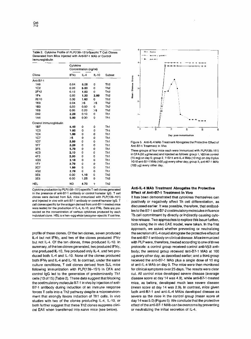
Table 2. Cytokine Profile of PLP(139-151)-Specific T Cell Clones Generated from Mice Injected with Anti-B7-1 MAb or Control Immunoglobulin
Cytokine Concentration (ng/ml)
Clone IFN~, IL-4 IL-10 Subset
Anti-B7-1 1A8 0.04 5.00 0 Th2 1 C2 0.20 5.00 0 Th2 2F12 0.10 1.60 0 Th2 1 F4 0.60 1.30 2.60 Th2 2E10 0.30 1.60 0 Th2 1 E3 0.04 >5 >5 Th2 1 B3 0.50 0.50 0 Th2 1 E6 0.00 0.20 >5 Th2 2A2 2.20 0.10 0 Thl 1A4 5.00 0.30 0 Thl
Control immunoglobulin 1 B7 5.40 0 0 Th 1 1C3 1.00 0 0 Thl 1C4 1.00 0 0 Thl 1C7 >5 0 0 Thl 1 D7 5.80 0 0 Thl 1F7 3.20 0 0 Thl 3F5 5.70 0 0 Thl 4C3 2.10 0 0 Thl 4F3 2.90 0 0 Thl 4G3 3.10 0 0 Thl 1 F7 2.70 0 0 Thl 2C7 1.80 0 0 Thl 4G2 2.70 0 0 Thl 3E6 0.00 1.10 0 Th2 3E5 0.00 1.25 0 Th2
HEL ND 4.70 1. Th2
Cytokine production by PLP(139-151)-specific T cell clones generated in the presence of anti-B7-1 antibody or control hamster IgG. T cell clones were derived from SJL mice immunized with PLP(139-151) and injected in vivo with anti-B7-1 antibody or control hamster IgG. T cell clones specific for the antigen derived from anti-B7-1-treated mice were tested for the production of IL-4, IL-10, and IFN'y. Data are pre- sented as the concentration of various cytokines produced by each individual clone. HEL is a hen-egg white lysozyme-specific T cell line.
profi le of these clones. Of the ten clones, seven produced IL-4 but not IFNy, and two of the c lones produced IFNy but not IL-4. Of the ten clones, three produced IL-10. In summary, of the ten clones generated, two produced I FNy, one produced IL-10, f ive produced only IL-4, and two pro- duced both IL-4 and IL-10. None of the c lones produced both IFNy and IL-4 and IL-10. In contrast, under the same culture condit ions, T cell c lones der ived from SJL mice fol lowing immunizat ion with PLP(139-151) in CFA and control IgG led to the generat ion of predominant ly Th l cells (13 of 15) (Table 2). These data suggest that blocking the cost imulatory molecule B7-1 in v ivo by injection of anti- B7-1 ant ibody during induct ion of an immune response forces T cells into a Th2 pa thway despi te a microenviron- ment that strongly favors induct ion of Th l cells. In v ivo studies with two of the c lones producing IL-4, IL-10, or both further suggest that these Th2 clones suppress clini- cal EAE when transferred into naive mice (see below).
6 t Control Anti-lL4 ÷ anti-BT-1
.... e--.- An|i-BT-1
4"
3
2-
1 -
o m
Day post.immunization
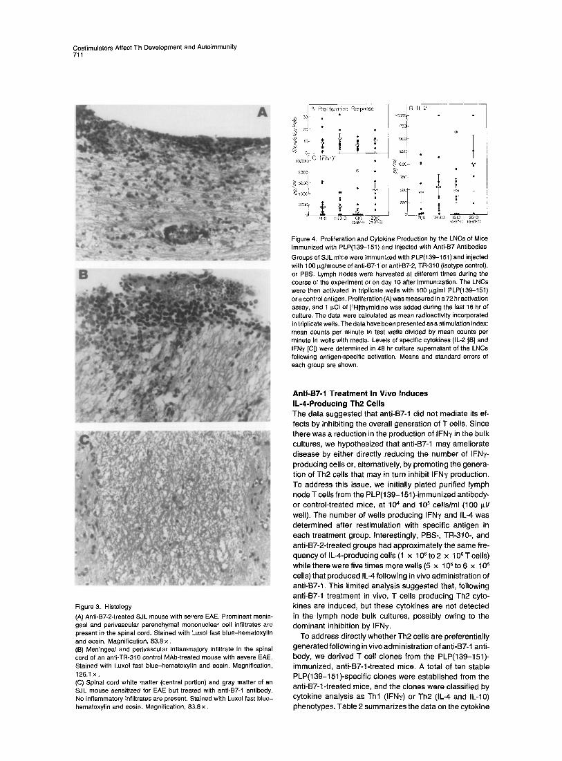
Figure 5. Anti-lL-4 MAb Treatment Abrogates the Protective Effect of Anti-B7-1 Treatment In Vivo Three groups of four mice each were immunized with PLP(t39-151) in CFA (50 p.g/mouse) and injected as follows: group 1, Vl33 as control (10 mg) on day 0; group 2, 11 B11 anti-lL-4 MAb (10 rag) on day 0 plus 1G 10 anti-B7-1 MAb (100 Ilg) every other day; group 3, anti-B7-1 MAb (100 p~g) every other day.
ii: 'i i .
Anti-lL-4 MAb Treatment Abrogates the Protective Effect of Anti-B7-1 Treatment In Vivo It has been demonst ra ted that cytokines themselves can posit ively or negat ive ly affect Th cell di f ferentiat ion, as discussed earl ier. It was possible, therefore, that ant ibod- ies to the B7-1 and B7-2 cost imulatory molecules inf luence Th cell commi tment by direct ly or indirect ly causing cyto-
kine release. Two approaches to explore this issue further, both using the in v ivo EAE model , were taken. In the first approach, we asked whether prevent ing or neutral iz ing the secret ion of I L-4 could abrograte the protect ive effect of the anti-B7-1 ant ibody on clinical disease. Mice immunized with PLP were, therefore, t reated according to one of three protocols: a control group received control anti-VI33 anti- body; the second group received anti-B7-1 MAb at 100 ~g every other day, as descr ibed earlier; and a third group received the anti-BT-1 MAb plus a single dose of 10 mg of anti- lL-4 MAb on day 0. The mice were then moni tored for clinical symptoms over 25 days. The results were clear cut. All control mice deve loped severe disease (average disease score at day 14 was 4.9), whi le anti-B7-1-treated mice, as before, developed much less severe disease (mean score at day 14 was 2.9). In contrast, mice given both anti-B7-1 and anti- lL-4 MAbs deve loped disease as severe as the mice in the control group (mean score at day 14 was 5.0) (Figure 5). We conclude that the protect ive effect of the anti-B7-1 MAb can be overcome by prevent ing or neutral iz ing the initial secret ion of IL-4.

Costimulators Affect Th Development and Autoimmunity 713
A
4 1A4 & 1E3
2 ~ . •
o
Day p o s t - i m m u n i z a t i o n
3 & 1E3
2
Day post-immunization
1 E6
Day p o s t - i m m u n i z a t i o n
Figure 6. Adoptively Transferred Th2 Clones Protect Mice from Development of EAE and Abrogate Established Disease Groups of five mice each were immunized with PLP(139-151) in CFA and injected with 5 x 10⁶ Thl or Th2 clones at the time of immuniza- tion (A) or at the first signs of disease (B).
B
4 '! == ~3
0
Day p o s t - i m m u n i z a t i o n
--O ....
~k ........
Immun i za t i on T cell c lone
PLP 139-151/CFA
PLP 139-151/CFA 1E3 (Th2)
PLP139-151/CFA 1A4 (Thl)
PLP 139-151/CFA 1E6 (Th2)
PEP 139-151/CFA HEL (Th2) tine
Adoptive Transfer of PLP-Specific Th2 Clones Both Prevents EAE and Abrogates Established Disease In the second approach, we directly tested the effects of Th2 clones generated by blocking the B7-1 molecule in vivo on EAE. In this experiment, we transferred Th2 clones to mice at the same time that they were immunized for the development of EAE or at the first appearance of clini- cal signs of EAE. Clones transferred into mice (1A4, 1E3, and 1E6) were PLP(139-151) specific and were I-A s re- stricted, as determined by blocking with MAb 10.2.16 (data not shown). Th2 clones (1E3 and 1E6) transferred into mice on the day of immunization with PLP(139-151 ) signif- icantly inhibited EAE. Mice injected with clone 1E3 (an IL-4 and IL-10 producer) showed little (mean score of 0.3) or no disease as compared with controls (Figure 6A, left and middle panels), whereas a Tht clone (1A4, an IFNy producer) that was transferred into mice increased the severity of disease above that of the controls, with a mean maximal score of 3 (Figure 6A, left panel). The transfer of a control Th2 line (H EL specific) did not ameliorate disease that was just as severe as in mice immunized with peptide alone, with a mean maximal score of 3 (Figure 6A, middle panel). When a second Th2 clone, 1E6, was introduced into mice again, no signs of disease at all were observed (Figure 6A, right panel). We conclude that transfer of Th2 clones generated following treatment with anti-B7-1 MAb can prevent the induction of disease in an antigen-specific manner.
To determine the possible therapeutic efficacy of this approach, we wished to determine whether established disease could be ameliorated or abrogated by Th2 clone transfer. Therefore, the Th2 clone 1E3 was transferred into mice after onset of initial signs of disease (limp tail or weight loss). This clone was injected on day 14, and the incidence of disease in the clone-injected group was
40% compared with the controls (100%) (Figure 6B). In the clone-injected mice, the disease peaked (mean disease score of 0.6) on day 16-17, whereas in the control group, all the animals showed complete paralysis of fore and hind limbs (mean score of 4). These data suggest that an anti- gen-specific Th2 clone that produces IL-4, IL-10, or both is capable of treating mice that manifest clinical disease when introduced after the onset of disease. These data (Figures 5 and 6) taken together suggest that initial secre- tion of IL-4 by Th2 or CD4 ÷ NK1.1 ÷ cells may directly en- hance Th2 generation in an autocrine fashion and may also inhibit the generation of encephalitogenic Thl cells.
Discussion
We have examined the role of the costimulatory molecules B7-1 and B7-2 in the differentiation of Thp cells to mature Thl and Th2 effector cells and in the induction of an auto- immune disease, EAE. Utilizing MAbs specific for these two costimulatory molecules, we have shown that anti- B7-1 drives naive MBP-specific Thp cells along aTh2 path- way while anti-B7-2 favors Thl development. Administra- tion of anti-B7-1 antibody ameliorated an organ-specific autoimmune disease, EAE, whereas injection of anti-B7-2 antibody significantly worsened clinical and histological disease. Blocking B7 molecules in vivo did not inhibit the generation of antigen-specific T cells, consistent with the in vitro differentiation results, but affected the cytokine profile of the responding T cells. Our results further sug- gest that the ability of anti-B7 antibodies to inhibit or en- hance EAE relates to the capacity of these antibodies dif- ferentially to activate Thl or Th2 cells/cytokines upon contact with Thp cells in the peripheral lymphoid compart- ment. Indeed, prevention of the initial secretion of IL-4 abrogates the protective effect of anti-B7-1. In addition, we provide evidence that transfer of antigen-specific Th2

Cell 714
clones generated by blocking the B7-1 molecule can pre- vent an organ-specific autoimmune disease and can abro- gate established disease.
B7-Based Immunotherapy Interactions with costimulators expressed on APCs are required for T cell activation and cytokine secretion and in the prevention of tolerance (Harding et al., 1992; Lenschow et al., 1992). A number of studies have sug- gested that manipulating the interaction of B7 molecules with their counterreceptors on T cells provides effective immunotherapy in vivo. For example, a soluble CTLA-4 immunoglobulin fusion protein prolonged allograft rejec- tion (Turka et al., 1992) and prevented xenograft rejection (Lenschow et al., 1992). Furthermore, CTLA-4 immuno- globulin inhibited antibody production (Linsley et al., 1992) and ameliorated murine lupus (Finck et al., 1994). Con- versely, we and others have recently demonstrated that provision of B7-1 to malignant tumor cells results in their rejection and can reduce tumor burden and inhibit tumor metastasis (Baskar et al., 1995; Townsend and Allison, 1993; Chen et al., 1992) and that overexpression of B7-1 in pancreatic islets results in autoimmune insulitis (Guarder et al., 1994). Here we have extended these appli- cations to include the treatment of an organ-specific auto- immune disease.
EAE has been used as an experimental model of an autoimmune disease to study basic immune mechanisms and to develop immunotherapeutics to prevent or treat autoimmunity. A number of immunotherapeutic strategies have been successfully used in the treatment of EAE (re- viewed by Zamvil and Steinman, 1990). These include anti-MHC class II, anti-CD4, and specific anti-TCR V~ anti- bodies. These treatment strategies were aimed at blocking the generation of all Th cells or at least the specific T cells involved in the induction of EAE. In contrast, our approach was aimed at skewing the T cell response to a nonpatho- genic Th2 pathway. Both approaches inhibit the acute dis- ease; however, it is possible that they may have different effects on the incidence and intensity of relapsing disease (Jiang et al., 1992). Of particular interest is the observation that only anti-B7-1 and not anti-B7-2 inhibited EAE. In- deed, anti-B7-2 treatment worsened disease signs and increased the amount of CNS inflammation. If the B7-1 costimulatory molecule is essential for the induction of encephalitogenic Thl cells, then its blockade would inhibit the generation of or induce anergy in these encephalito- genic T cells. This would also result in a compensatory increase of Th2 cells with the end result being an overall reduction in the production of Thl cytokines, including IFNy. However, this alone cannot account for the worsen- ing of EAE and increase in the production of Thl cells/ cytokines (IFN'y) by the administration of anti-B7-2 anti- body. An explanation might be provided by the temporal patterns of expression of these molecules during the de- velopment of a given immune response or by a preferential inhibition of IL-4 secretion by the anti-B7-2 MAb (see below).
EAE Is Induced by Thl Cells, and Th2 Cells Can Prevent Induction of Disease and Abrogate Established Disease Myelin antigen (MBP or PLP)-reactive T cells that induce EAE have thus far all been shown to display a Thl pheno- type (Kuchroo et al., 1993; Baron et al., 1993). In addition, during progression of disease, Thl cyokines (IL-2, TNFI3, and IFN~,) are present in inflammatory EAE lesions, while Th2 cytokines are absent (Khoury et al., 1992), strongly suggesting that Thl cytokines play a role in the pathogene- sis of the disease. This is consistent with the presentdata, since mice immunized with PLP(139-151) and injected with either PBS or the control TR-310 antibody generate T cells that produced significant amounts of Thl (IL-2 and IFNT) but not detectable levels of Th2 (IL-4 and IL-10) cy- tokines. Administration of anti-B7-2 antibody significantly enhanced the severity of EAE, and this was accompanied by a marked increase in antigen-specific IFNy production by LNCs, suggesting that injection of anti-B7-2 antibody leads to the hyperinduction or activation of IFNT-producing Thl cells. On the other hand, administration of anti-B7-1 antibody leads to amelioration of disease with a commen- surate decrease in the production of IFN~,, indicating that blocking the costimulatory B7-1 molecule in vivo inhibits (directly or indirectly) the generation of Thl cells/cyto- kines. Thus, our data indicate that anti-B7-1 and anti-B7-2 antibodies have opposing effects in the induction of EAE and concurrently in the generation of Thl and Th2 cells/ cytokines. The presence of IL-4 alone in the brain in an asymptomatic treated animal, as well as our experiments with Th2 clones generated by the administration of anti- B7-1 antibody, further strengthens the argument that these cells/clones do not induce EAE on their own but can protect mice from the development of EAE. In other systems, regulatory T cells that suppress the development of EAE have also been shown to produce cytokines that correspond to the Th2 profile (Chen et al., 1994). Further- more, recovery from EAE in mice and rats is associated with an increase in the presence of Th2 cells and cytokines in the CNS (Khoury et al., 1992). The present findings demonstrate that an antigen-specific Th2 clone can pre- vent.the induction of an organ-specific autoimmune dis- ease and can abrogate established disease. These find- ings, together with the observation that administration of anti-B7-1 results in the preferential induction of antigen- specific Th2 cells, strongly suggests that the protective effects of anti-B7-1 antibody may be due to activation of the Th2 pathway and thus offer the basis for novel immuno- therapeutic strategies. Such strategies might rely on the manipulation of cytokines such as IL-4, since blocking the initial production of IL-4 abrogated the protective effect of the anti-B7-1 antibody.
Role of the Costimulatory Molecules B7-1 and B7-2 in Th Cell Differentiation Differentiation of Thp cells into Thl or Th2 ceils upon anti- genic stimulation in vivo has important biologic conse- quences. However, the processes that dictate whether an

Costimulators Affect Th Development and Autoimmunity 715
IL2 1FN- 7
ILniY ~ @ IL-4 [L-10
Figure 7. Interaction of B7-1 and B7-2withtheSameCounterreceptor on a Thp Cell Results in Different Th Subset Generations
immune response will be dominated by Th l versus Th2 cells are not well understood (Paul and Seder, 1994; Seder and Paul, 1994), although initial cytokine release, antigen concentrat ion, and the route of antigen administrat ion may all affect the outcome. The data presented here are direct evidence that interaction of the cost imulatory molecules B7-1 or B7-2, with their counterreceptors CD28 and CTLA-4, on Thp cells during antigen presentation leads to polariza- tion of Th responses. The simplest interpretation of our data is that B7-1 preferential ly acts as a cost imulator for the generation of Th l cells while B7-2 cost imulates and induces Th2 cells (see model in Figure 7). Thus, blocking B7-1 will inhibit the generat ion of Th l cells, while enhanc- ing the generat ion of Th2 cells and blocking B7-2 will have the opposite effect. If this hypothesis is correct, then the interaction of B7-1 and B7-2 molecules with their T cell counterreceptors likely generates different intracellular signals that lead to the differentiation of Thp cells along a Th l or Th2 pathway. The recent evidence (Nun,s et al., 1994) that tr iggering of CD28 with anti-CD28 antibody, but not with the l igand B7-1, can activate p21 r " is intriguing in this regard. This raises the possibil ity that a differential effect on p21 ra" or other molecules impl icated in CD28 signaling (such as phosphatidyl inositol 3-kinase) (Prasad et al., 1994) would also be observed when interaction with B7-1 versus B7-2 l igands is compared. It is also possible that differential interaction of B7-1 and B7-2 with their two counterreceptors, CD28 and CTLA-4, may occur. Al- though several structural motifs are shared between the two amino acid sequences (Freeman et al., 1993a, 1993b; Azuma et al., 1993), binding studies have shown that the affinity of B7-1 for CTLA-4 is of 10- to 20-fold higher affinity than to CD28. Similar measurements have recently been published for B7-2 (Linsley et al., 1994), which demon- strate that the two cost imulatory molecules do not bind equivalently to CTLA-4. Since CTLA-4 has recently been shown to function as a negative regulator of T cell activa- tion (Walunas et al., 1994), the more rapid dissociation of B7-2 from CTLA-4 could result in the effects observed. One outcome of this differential signaling could be the initial secretion of opposing cytokines that would drive Th l ineage commitment . The observat ions that t reatment with anti-lL-4 MAb abrogates the protective effect of anti-B7-1 ant ibody together with the prevention of EAE by transfer of PLP-specific Th2 clones is consistent with a direct effect of cost imulatory molecules on cytokine secretion. Alterna-
tively, the polarization of Th cell responses by the addition of anti-B7 antibodies, observed in this study, may be an indirect effect of the ant ibodies themselves. A number of cell types, including T cells, macrophages, and dendrit ic cells, constitutively express B7-2 and can up-regulate ex- pression of B7-1 upon activation. Cross-linking costimula- tory molecules on the surface of these cells by ant ibody may lead to the production of specific cytokines and change the cytokine milieu in the microenvironment, thereby polarizing Th cell responses. If the latter hypothe- sis is correct, then cross-linking B7-1 versus B7-2 would lead to the production of different cytokines, such as IL-12, that may promote or inhibit generat ion of Th l versus Th2 cells. The identif ication of intracellular signals that are gen- erated by interaction of B7-1 and B7-2 with the same coun- terreceptors (CD28 and CTLA-4) on a Thp cell may provide insight into the molecular mechanisms responsible for Th cell differentiation, al lowing selective manipulat ion of the immune response in disease.
Experimental Procedures
Mice Female SJI./J mice 4 to 8 weeks of age (Jackson Laboratory, Bar Harbor, ME) were maintained in accordance with the guidelines of the Committee on Animals of the Harvard Medical School and the Committee on Care and Use of Laboratory Animals of the Institute of Laboratory Animal Resources, National Research Council, as stated in Department of Health and Human Services publication 85-23 (re- vised in 1985).
Transgenic B10.PL mice expressing TCRs for MBP have been de- scribed previously (Goverman et al., 1993) and express Va2.3- and VI38.2-containing TCR genes from a B10.PL-derived T cell hybrid spe- cific for MBP Ac(1-11). Separate TCR ~ and ~ chain transgenic lines were established and the lines bred together to generate mice express- ing a complete transgenic TCR. Expression of transgenes was evalu- ated by genotyping tail DNA and by flow cytometry of peripheral blood T cells utilizing MAbs specific for TCR Va2 and V~8.2 regions. An additional MBP TCR transgenic line expressing V~4 and V1~8.2 that recognizes MBP Ac(1-17) was also utilized (Lafaille et al., 1994) (gift of Dr. C. Janeway, Yale University, New Haven, CT).
Antigens PLP(139-151) (HSLGKWLGHPDKF), PLP(190-209) (SKTSASIGSL- CADARMYYGVL), and MBP Ac(1-11) (AcASQKRPSQRHG) peptides were prepared according to published sequences (Laursen et al., 1984; Newman et al., 1987) except that serine was always substituted for cysteine at position 140 in the peptide PLP(139-151) (luohy et al., 1989). Peptides were synthesized in the laboratory of Dr. R. Laursen (Department of Chemistry, Boston University, Boston, MA) on a Milli- gen model 9050 synthesizer using Fmoc chemistry or by H. Freisheim (Medical College of Ohio, Toledo, OH) on an Applied Biosystems model 430A synthesizer using Milligen PAL resins or RINK amide resins.
Antibodies Anti-B7-1 (1G10) and anti-B7-2 (2D10) have been described previously (Nabavi et al., 1992; Chen et al., 1994) and were derived from the same somatic cell fusion. The MAbs were purified from ascitic fluid on protein G columns. The anti-B7-1 and B7-2 antibodies are of the IgG2a and IgG2b isotypes, respectively. A second anti-B7-2 antibody (GL-1) of rat IgG2a isotype was obtained from ATCC. Hybridoma cells were grown as ascites and purified over a protein G column. Purified rat IgG2a and IgG2b antibodies (Pharmingen, San Diego, CA) were used as isotype controls. A rat anti-Vlt,7 (TR-310) antibody (rat IgG2a) was purified from ascitic fluid on a protein G column and used as an additional control. The rat MAb to mouse IL-4 was the gift of Dr. W. E.

Cell 716
Paul (National Institutes of Health, Bethesda, MD), and the anti-V~3 antibody KJ25a, used as a control, was the gift of Dr. J. Kappler (Na- tional Jewish Hospital and Research Center, Denver, CO).
Induction of EAE SJL mice were injected subcutaneously in the flank with 25-100 ~g of peptide PLP(139-151) and 400 p_g of Mycobacterium tuberculosis H37Ra (Difco Laboratories, Detroit, MI) in an emulsion consisting of equal volumes of PBS and CFA. Each mouse was also injected intrave- nously on day 0 and day 2 or 3 with 10 ~ heat-killed Bordetella pertussis bacilli (Massachusetts Public Health Biological Laboratories, Boston, MA). To test the efficacy of the antibodies in the induction and inhibition of EAE in vivo, we injected mice with anti-B7 or control antibodies (100 p.g/mouse) intraperitoneally every other day following immunization, according to various protocols as described in Results.
Clinical assessment was carried out daily according to the following criteria: 0, no disease; 1, decreased tail tone or slightly clumsy gait; 2, tail atony, moderately clumsy gait, poor righting ability, or some combination of these symptoms; 3, limb weakness; 4, limb paralysis; 5, moribund state. Animals were sacrificed at the termination of the experiment or at the peak of the disease. Mice that showed no clinical signs were sacrificed at the conclusion of the experiment. Brains and spinal cords were removed and fixed in 10% formalin, and paraffin- embedded sections were stained with Luxol fast blue-hematoxylin and eosin for light microscopy. Histological disease was quantified by counting inflammatory loci in meninges and parenchyma (Sobel et al., 1989).
In Vitro Proliferative Response and Cytokine Production of Antigen-Primed LNCs Groups of mice immunized subcutaneously with peptide PLP(139-151) in CFA for the induction of EAE were sacrificed at various timepoints during the course of disease. Inguinal, prescapular, and para-aortic lymph nodes were removed and dissociated in Dulbecco's minimum essential medium (DMEM). LNCs from these mice were used for in vitro proliferative assays and production of cytokines following in vitro stimulation. Proliferative Assays For in vitro proliferation, LNCs were cultured for 72 hr in flat-bottomed 96-well plates in the presence of various concentrations of peptide and then pulsed with 1 p, Ci of [3H]thymidine for the last 16-18 hr of incubation, and mean thymidine incorporation in triplicate wells was calculated. The data of the proliferative assays are presented as stimu- lation indices for easy comparison of the proliferative responses: mean cycles per minute in the test wells divided by mean counts per minute in the control wells (with medium). Cytokine ELISA The amount of cytokines produced by LNCs was determined by ELISA. LNCs obtained from mice immunized with PLP(139-151) in CFA and injected with the various antibodies were activated with PLP(139-151) control peptide in vitro, and culture supernatants were collected from separate wells on day 1 and day 2. In brief, 96-well plates were coated overnight with primary anti-cytokine capture antibody (1 p.g/ml) specific for a particular cytokine. The plates were washed twice and, after a 2 hr incubation with PBS plus 15% blocking reagent (Kirkegaard and Perry Laboratories, Gaithersburg, MD), the supernatants from the acti- vated clones and the standards were added. After overnight incubation at 4°C, the plates were washed, and diluted biotinylated anti-cytokine- detecting MAb (1 p.g/ml) was added and incubated at room tempera- ture for 1 hr. The plates were washed and further developed by adding avidin-peroxidase and its substrate, and absorbance was measured on a 450 nm wavelength spectrophotometer. The amount of cytokine in each supernatant was extrapolated from the standard curve. The antibody pairs used were as follows, listed by capture/biotinylated de- tection: I L-2, JES6-1A12/JE5H4; I L-4, BVD4-1 D11/BVD6-24G2; IL-10, JES5-2A5/SXC-1; IFNy, R4-6A2/XMG1.2 (all from Pharmingen, San Diego, CA). The standards were recombinant cytokine curves gener- ated in 1:2 dilutions from 39 to 2500 pg/ml for IL-2, IL-4, and IL-10 and from 78 to 5000 pg/ml for IFNy with detectable readings at the lowest dilutions.
In Vitro Differentiation and Phenotype of Transgenic T Cells Spleen cells (5 x 10⁶/ml) obtained from a naive MBP TCR transgenic
mouse (Goverman et al., 1993) were equally divided into four aliquots and cultured in a 24-well plate with one of the following antibody re- agents at 20 ~g/ml: anti-B7-1 (1G10), anti-B7-2 (2D10), purified rat immunoglobulin, or PBS, with or without addition of the antigen MBP Ac(1-11) (10 I~g/ml). Culture supernatants were harvested 2 days after activation and tested for cytokine production by ELISA. The cultures were further propagated for an additional 21 days by addition of media containing 5% human IL-2. The live T cells were purified, resuspended at 5 x 10⁸ cells/ml, and activated in the presence ofthe corresponding antibody reagent and syngeneic spleen cells, such as APCs (10qml), with or without addition of MBP Ac(1-11) (10 ~g/ml). The culture super- natants were harvested 48 hr after activation, and cytokine content was determined by ELISA.
Derivation and Maintenance of T Cell Clones SJL mice were injected subcutaneously in each flank with 100 I~g of PLP(139-151) in CFA plus 400 p.g of M. tuberculosis H37RA and then injected with anti-BT-1 antibody (100 I~g intravenously on days 0 and 2) (Razi-Wolf et el., 1992) or control hamster IgG (Cappel, Durham, NC). LNCs were removed from mice 10 days after immunization and cultured for 5 days with PLP(139-151) peptide (20 ~g/ml) in DMEM containing 1% autologous serum. Live cells were isolated on a Ficoll- Hypaque gradient and grown in 5% 11_-2 medium (T-STIM, Collabora- tive Biomedical Products, Bedford, MA) for 14 days. The cells were activated once again in the presence of PLP(139-151) and SJL spleen cells as APCs, The T cells were cloned by limiting dilution 3 days after the last activation and individual T cell clones expanded to obtain sufficient numbers. The cloned cells were activated with PLP(139- 151), and the cytokine profile of each T cell clone was established by cytokine ELISAs.
Adoptive Transfer of Clones T cell clones were activated with APCs and peptide PLP(139-151) (20 ~g/ml) for 3 days, after which live cells were isolated by FicolI-Hypaque density gradient centrifugation. Clones (Thl or Th2; 5 x 10⁶/mouse) were injected intravenously into mice that were injected subcutane- ously with 50 p,g of PLP(139-151) in CFA at the time of transfer. A control line secreting IL-4 and IL-10 specific for hen-egg white lyso- zyme (HEL) was also transferred into PLP peptide-immunized mice. A Th2 clone (1E3, producing IL-4 and IL-10) was also transferred into mice after the first sign of disease (limp tail). The disease course was followed for 25-30 days.
Acknowledgments
Correspondence should be addressed to L. H. G. The authors thank D. Kaul and T. Howard for technical help; C. Freedman for manuscript preparation; Drs. L. Hood, J. Goverman, and C. Janeway for providing MBP transgenic mice; Drs. W. E. Paul and J. Kappler for MAbs to IL-4 and V~3; and Drs. H. Cantor and A. Abbas for thoughtful review of the manuscript. This work was supported by grants from the National Multiple Sclerosis Society (RG2571A2 and RG2320B3) and the Na- tional Institutes of Health (NS-30843 to V. K. K., NS-26773 to R. A. S., and AI-21569 to L. H. G.).
Received September 9, 1994; revised December 19, 1994.
References
Azuma, M., Ito, D., Yagita, H., Okumura, K., Phillips, J. H., Lanier, L. L., and Somoza, C. (1993). B70 antigen is a second ligand for CTLA-4 and CD28. Nature 366, 76-79.
Baron, J. L., Mardi, J. A., Ruddle, N. H., Hashim, G., and Janeway, C. A., Jr. (1993). Surface expression of ~4 integrin by CD4 T cells is required for their entry into brain parenchyma. J. Exp. Med. 177, 57- 68. Baskar, S., Glimcher, L., Nabavi, N., and Ostrand-Rosenberg, S. (1995). MHC class I1÷B7-1 ÷ tumor cells are potent vaccines for stimulat- ing tumor rejection in tumor-bearing mice. J. Exp. Med., in press.
Chen, C., and Nabavi, N. (1994). In vitro induction of T cell anergy by blocking B7 and early T cell costimulatory molecule ETC-1/B7-2. Immunity 1, 147-154.

Costimulators Affect Th Development and Autoimmunity 717
Chen, C., Faherty, D. A., Gault, A., Connaughton, S. E., Powers, G. D., Godfrey, D. I., and Nabavi, N. (1994). Monoclonal antibody2D10 recog- nizes a novel T cell costimulatory molecule on activated murine B lymphocytes. J. Immunol. 152, 2105-2114.
Chen, L., Ashe, S., Brady, W., Hellstrom, I., Hellstrom, K., Ledbetter, J., McGowan, P., and Linsley, P. (1992). Costimulation of antitumor immunity by the B7 counterreceptor for the T lymphocyte molecules CD28 and CTLA-4. Cell 71, 1093-1102.
Chen, Y., Kuchroo, V., Inobe, J.-I., Hailer, D. A., and Weiner, H. L. (1994). Regulatory T cell clones induced by oral tolerance and suppres- sion of autoimmune encephalomyelitis. Science 265, 1237-1240.
Cohen, J. (1993). T cell shift: key to AIDS therapy? Science 262, 175- 176.
Finck, B. K., Linsley, P. S., and Wofsy, D./1994). Treatment of murine lupus with CTLA41g. Science 265, 1225-1227.
Freeman, G. J., Gray, G. S., Gimmi, C. D., Lombard, D. B., Zhou, L. J., White, M., Fingeroth, J. D., Gribben, J. G., and Nadler, L. M (1991). Structure, expression, and T cell costimulatory activity of the murine homologue of the human B lymphocyte activation antigen B7. J. Exp. Med. 174, 625-631.
Freeman, G. J., Borriello, F., Hodes, R. J., Reiser, H., Hathcock, K. S., Laszlo, G., McKnight, A. J., Kim, J., Du, L., Lombard, D. B., Gray, G. S., Nadler, L. M., and Sharpe, A. H. (1993a). Uncovering of functional alternative CTLA-4 counter-receptor in B7-deficient mice. Science 262, 907-909.
Freeman, G. J., Gribben, J. G., Boussiotis, V. A., Ng, J. W., Restivo, V. A., Jr., Lombard, L. A., Gray, G. S., and Nadler, L M. (1993b). Cloning of B7-2: a CTLA-4 counter-receptor that costimulates human T cell proliferation. Science 262, 909-911.
Goverman, J., Woods, A., Larson, L., Weiner, L. P., Hood, L., and Zaller, D. M. (1993). Transgenic mice that express a myelin basic protein-specific T cell receptor develop spontaneous autoimmunity. Cell 72, 551-560.
Guerder, S., Meyerhoff, J., and Flavell, R. (1994). The role of the T cell costimulator B7-1 in autoimmunity and the induction and mainte- nance of tolerance to peripheral antigen. Immunity 1, 155-166.
Harding, F. A., McArthur, J. G., Gross, J. A., Raulet, D. H., and Allison, J. P. (1992). CD28-mediated signalling co-stimulates murine T cells and prevents induction of anergy in T-cell clones. Nature 356, 607- 609.
Hathcock, K. S., Laszlo, G., Pucillo, C., Linsley, P., and Hodes, R. J. (1994). Comparative analysis of B7-1 and B7-2 costimulatory ligands: expression and function. J. Exp. Med. 180, 631-640.
Heinzel, F. P., Sadick, M. D., Holaday, B. J., Coffman, R. L., and Locksley, R. M. (1989). Reciprocal expression of interferon ~, or in- terleukin 4 during the resolution or progression of murine leishmania- sis: evidence for expansion of distinct helper T cell subsets. J. Exp. Med. 169, 59-72.
Jiang, H., Zhang, S.-L., and Pernis, B. (1992). Role of CD8 ÷ T cells in murine experimental allergic encephalomyelitis. Science 256, 1213- 1215.
Khoury, S.J., Hancock, W. W., and Weiner, H. L. (1992). Oral tolerance to myelin basic protein and natural recovery from experimental allergic encephalomyelitis are associated with downregulation of inflammatory cytokines and differential upregutation of transforming growth factor-13, interleukin 4, and prostaglandin E expression in the brain. J. Exp. Med. 176, 1355-1364.
Kuchroo, V. K., Martin, C. A., Greer, J. M., Ju, S.-T., Sobel, R. A., and Dorf, M. E. (1993). Cytokines and adhesion molecules contribute to the ability of myelin proteolipid protein-specific T celt clones to mediate experimental allergic encephalomyelitis. J. immunol. 151, 4371-4382.
Lafaille, J. J., Nagashima, K, Katsuki, M., and Tonegawa, S. (1994). High incidence of spontaneous autoimmune encephalomyelitis in im- munodeficient anti-myelin basic protein T cell receptor transgenic mice. Cell 78, 399-408.
Laursen, R. A., Samiullah, M., and Lees, M. B. (1984). The structure of bovine myelin proteolipid and its organization in myelin. Proc. Natl. Acad. Sci. USA 81, 2912-2916.
Lenschow, D. J., Zeng, Y., Thistlethwaite, J. R., Montag, A., Brady, W., Gibson, M. G., Linsley, P. S., and Bluestone, J. A. (1992). Long-term survival of xenogeneic pancreatic islet grafts induced by CTLA4ig. Science 257, 789-792.
Linsley, P. S., Brady, W., Urnes, M., Grosmaire, L. S., Damle, N. K, and Ledbetter, J. A. (1991). CTLA-4 is a second receptor for the B cell activation antigen B7. J. Exp. Med. 174, 561-569,
Linsley, P. S., Wallace, P. M., Johnson, J., Gibson, M. G., Greene, J. L, Ledbetter, J. A., Singh, C., and Tepper, M. A. (1992). Immunosup- pression in vivo by a soluble form of the CTLA-4 T cell activation mole- cule. Science 257, 792-795.
Linsley, P. S., Greene, J. L., Brady, W., Bajorath, J., Ledbetter, J. A., and Peach, R. (1994). Human B7-2 (CD80) and B7-2 (CD86) bind with similar avidities but distinct kinetics to CD28 and CTLA-4 receptors. Immunity 1,793-801.
Mosmann, T. R., and Coffman, R. L. (1989). Thl and Th2 cells: different patterns of lymphokine secretion lead to different functional properties. Annu. Rev. Immunol. 7, 145-173.
Nabavi, N., Freeman, G. J., Gault, A., Godfrey, D., Nadler, L. M., and Glimcher, L. H. (1992). Signalling th rough the MHC class II cytoplasmic domain is required for antigen presentation and induces B7 expres- sion. Nature 360, 266-268.
Newman, S., Kitamura, K., and Campagnoni, A. T. (1987). Identifica- tion of cDNA coding for a fifth form of myelin basic protein. Proc. Natl. Acad. Sci. USA 84, 886-890.
Nun,s, J. A., Collette, Y., Truneh, A., Olive, D., and Cantrell, D. A. (1994). The role of p21 '~ in CD28 signal transduction: triggering of CD28 with antibodies, but not the ligand B7-1, activates p21 ra'. J. Exp. Med. 180, 1067-1076.
Paul, W. E., and Seder, R. A. (1994). Lymphocyte responses and cytokines. Cell 76, 241-251.
Powrie, F., and Coffman, R. L. (1993). Cytokine regulation of T-celt function: potential for therapeutic intervention. Immunol. Today 14, 270-274.
Prasad, K. V. S., Cai, Y.-C., Raab, M., Duckworth, B., Cantley, L, Shoelson, S. E., and Rudd, C. E. (1994). T-cell antigen CD28 interacts with the lipid kinase phosphatidylinositol 3-kinase by a cytoplasmic Tyr(P)-Met-Xaa-Met motif. Proc. Natl. Acad. Sci. USA 91, 2834-2838.
Razi-Wolf, Z., Freeman, G. J., Galvin, F., Benacerraf, B., Nadler, L., and Reiser, H. (1992). Expression and function of the murine B7 anti- gen, the major costimulatory molecule expressed by peritoneal exu- date cells. Proc. Natl. Acad. Sci. USA 89, 4210-4214.
Schwartz, R. H. (1992). Costimulation of T lymphccytes: the role of CD28, CTLA-4, and B7/BB1 in IL-2 production and immunotherapy. Cell 71, 1065-1068.
Seder, R. A., and Paul, W. E. (1994). Acquisition of lymphokine- producing phenotype by CD4 + T cells. Annu. Rev. Immunol. 12, 635- 673.
Simon, A. K., Seipelt, E., and Sieper, J. (1994). Divergent T-cell cyto- kine patterns in inflammatory arthritis. Proc. Natl. Acad. Sci. USA 91, 8562-8566.
Sobel, R. A., Tuohy, V. K., Lu, Z., Laursen, R., and Lees, M. B. (1989). Acute allergic encephalomyelitis in SJIEJ mice induced by a synthetic peptide of myelin proteolipid protein. J. Neuropathol. Exp. Neuroi. 49, 468-474.
Townsend, S. E., and Allison, J. P. (1993). Tumor rejection after direct costimulation of CD8 ÷ T cells by B7-transfected melanoma cells. Sci- ence 259, 368-370.
Trinchieri, G. (1993). Intedeukin-12 and its role in the generation of Thl cells. Immunol. Today 14, 335-338.
Tuohy, V. K., Lu, Z., Sobel, R. A., Laursen, R. A., and Lees, M. B. (1989). Identification of an encephalitogenic determinant of myelin proteolipid protein for SJLrnice. J. Immunol. 142, 1523-1527.
Turka, L A., Linsley, P. S., Lin, H., Brady, W., Leiden, J. M, Wei, R., Gibson, M. L., Zhen, X., Myrdal, S., Gordon, D., Bailey, T., Boiling, S. F., and Thompson, C. B. (1992). T-cell activation by the CD28 tigand B7 is required for cardiac allograft rejection in vivo. Proc. Natl. Acad. Sci. USA 89, 11102-11105.

Cell 718
Walunas, T. L., Lenschow, D. J., Bakker, C. Y., Linsley, P. S., Free- man, G. J., Green, J. M., Thompson, C. B., and Bluestone, J. A. (1994). CTLA-4 can function as a negative regulator of T cell activation. Immu- nity 1,405-413.
Yoshimoto, T., and Paul, W. E. (1994). CD4 ~ , N K1.1 pos T cells promptly produce interleukin 4 in response to in vivo challenge with anti-CD3. J. Exp. Med. 179, 1285-1295.
Zamvil, S., and Steinman, L. (1990). The T lymphocyte in experimental allergic encephalomyelitis. Annu. Rev. Immunol. 8, 579-621.
Copyright © 2022 FDOKUMEN