MAPK target networks in Arabidopsis thaliana revealed using functional protein microarrays
Upload
independentCategory
view
4download
0
Arabinosylated Lipoarabinomannan Skews Th2Phenotype towards Th1 during Leishmania Infection byChromatin Modification: Involvement of MAPK SignalingParna Bhattacharya1, Gaurav Gupta1, Saikat Majumder1, Anupam Adhikari1, Sayantan Banerjee1, Kuntal
Halder1, Suchandra Bhattacharya Majumdar1, Moumita Ghosh2, Shubho Chaudhuri1, Syamal Roy2,
Subrata Majumdar1*
1 Division of Molecular Medicine, Bose Institute, Kolkata, India, 2 Division of Infectious Diseases and Immunology, Indian Institute of Chemical Biology, Council of Scientific
and Industrial Research, Kolkata, India
Abstract
The parasitic protozoan Leishmania donovani is the causative organism for visceral leishmaniasis (VL) which persists in thehost macrophages by deactivating its signaling machinery resulting in a critical shift from proinflammatory (Th1) to an anti-inflammatory (Th2) response. The severity of this disease is mainly determined by the production of IL-12 and IL-10 whichcould be reversed by use of effective immunoprophylactics. In this study we have evaluated the potential of ArabinosylatedLipoarabinomannan (Ara-LAM), a cell wall glycolipid isolated from non pathogenic Mycobacterium smegmatis, in regulatingthe host effector response via effective regulation of mitogen-activated protein kinases (MAPK) signaling cascades inLeishmania donovani infected macrophages isolated from BALB/C mice. Ara-LAM, a Toll-like receptor 2 (TLR2) specific ligand,was found to activate p38 MAPK signaling along with subsequent abrogation of extracellular signal–regulated kinase (ERKs)signaling. The use of pharmacological inhibitors of p38MAPK and ERK signaling showed the importance of these signalingpathways in the regulation of IL-10 and IL-12 in Ara-LAM pretreated parasitized macrophages. Molecular characterization ofthis regulation of IL-10 and IL-12 was revealed by chromatin immunoprecipitation assay (CHIP) which showed that in Ara-LAM pretreated parasitized murine macrophages there was a significant induction of IL-12 by selective phosphorylation andacetylation of histone H3 residues at its promoter region. While, IL-10 production was attenuated by Ara-LAM pretreatmentvia abrogation of histone H3 phosphorylation and acetylation at its promoter region. This Ara-LAM mediated antagonisticregulations in the induction of IL-10 and IL-12 genes were further correlated to changes in the transcriptional regulatorsSignal transducer and activator of transcription 3 (STAT3) and Suppressor of cytokine signaling 3 (SOCS3). These resultsdemonstrate the crucial role played by Ara-LAM in regulating the MAPK signaling pathway along with subsequent changesin host effector response during VL which might provide crucial clues in understanding the Ara-LAM mediated protectionduring Leishmania induced pathogenesis.
Citation: Bhattacharya P, Gupta G, Majumder S, Adhikari A, Banerjee S, et al. (2011) Arabinosylated Lipoarabinomannan Skews Th2 Phenotype towards Th1during Leishmania Infection by Chromatin Modification: Involvement of MAPK Signaling. PLoS ONE 6(9): e24141. doi:10.1371/journal.pone.0024141
Editor: Jerome Nigou, French National Centre for Scientific Research - Universite de Toulouse, France
Received April 1, 2011; Accepted August 1, 2011; Published September 14, 2011
Copyright: � 2011 Bhattacharya et al. This is an open-access article distributed under the terms of the Creative Commons Attribution License, which permitsunrestricted use, distribution, and reproduction in any medium, provided the original author and source are credited.
Funding: This study was funded by Council of Scientifc Industrial Research, India (Grant No 37/1280/07/EMR-II) and Division of Molecular Medicine. The fundershad no role in study design, data collection and analysis, decision to publish, or preparation of the manuscript.
Competing Interests: The authors have declared that no competing interests exist.
* E-mail: [email protected]
Introduction
The parasitic protozoan Leishmania donovani, the causative agent
of visceral leishmaniasis (VL), resides and multiplies in host
macrophages [1]. Activated macrophages eliminate the intracel-
lular parasite [2] by initiating host-protective anti-leishmanial
responses. To counteract these anti-leishmanial responses parasites
deploys different immune evasion strategies to survive within the
macrophages [3]. Leishmania-induced macrophage dysfunctions
have been correlated mainly with depletion of microbicidal
molecules (nitric oxide (NO) and reactive oxygen species) [4,5],
and altered signaling events resulting in skewing the T helper cells
(Th) to disease promoting Th2 subset that consequently suppress
the host protective Th1 subset [6]. The outcome of infection in
leishmaniasis is mainly determined by the Th1 versus Th2 effector
response and the generation of IL-12 and IL-10 by the infected
macrophages is important for this decision [7].
Several immunoprophylactics have been pursued to combat VL,with varying degrees of success. Recently Arabinosylated-lipoar-abinomannan (Ara-LAM), an immunomodulator has emerged asan effective immunoprophylactic tool against VL. It confersprotection during leishmanial pathogenesis via Toll-like receptor 2(TLR2) signaling mediated nuclear factor-kB (NF-kB) translocationand concomitant induction of the proinflammatory mediators [8].
In addition to NF-kB activation, TLR signaling can also
activate mitogen-activated protein kinases (MAPK) signaling
cascades which include extracellular signal–regulated kinase
(ERKs), p38 MAPKs, and c-Jun NH2-terminal kinases (JNK)
[9]. Most of the effector functions in response to extracellular cues
are regulated by (MAPK) [10,11]. The parasite-triggered recip-
rocal MAPK signaling via p38MAPK and ERK1/2 govern the
counteracting immune response of the host cell resulting in
differential expression of IL-12 and IL-10 in macrophages during
Leishmania infection [12]. p38 MAPK activation results in histone
PLoS ONE | www.plosone.org 1 September 2011 | Volume 6 | Issue 9 | e24141

modifications at the IL-12p40 promoter loci, making it more
accessible for the recruitment of NF-kB leading to transcriptional
induction of IL-12 [13]. In contrast, enhanced IL-10 transcription
is associated with ERK1/2 activation leading to phosphorylation
and acetylation of histone H3 at the IL-10 promoter loci which
facilitates the binding of Signal transducer and activator of
transcription 3 (STAT3) to the IL-10 promoter resulting in
enhanced IL-10 transcription [14]. Moreover activated STAT3
attenuates the transcription of proinfllammatory mediators with
the help of Suppressor of cytokine signaling 3 (SOCS3) inductions
[15,16, and 17]. Previous work from our laboratory has shown
that Ara-LAM is involved in IL-12 induction and IL-10
attenuation during infection demonstrating the suitability of it as
a potential candidate for immunotherapy to cure VL. But, how
Ara-LAM treatment of parasitized macrophages leads to epige-
netic modification at the locus of these two counteractive cytokine
genes leading to their transcriptional regulation and the involve-
ment of MAPK signaling in this regard is yet to be explored.
In the present study, we have found that Ara-LAM, a TLR-2
ligand confers protection against leishmanial pathogenesis via
reciprocal regulation of MAPK signaling. This Ara-LAM
mediated regulation of MAPK signaling resulted in antagonistic
regulation of IL-12 and IL-10 in host macrophages. Detailed
investigation at the molecular level showed that Ara-LAM could
induce IL-12 by selective phosphorylation and acetylation of
histone H3 residues at the IL-12p40 promoter region while
attenuated IL-10 production by abrogating such histone H3
modification at IL-10 promoter in parasitized macrophages. This
antagonistic regulation of effector response by Ara-LAM in the
form of IL-10 and IL-12 was further linked to STAT3 and SOCS3
which were found to be crucial in regulating the host protective
immune response in leishmania infected macrophages.
Results
1. ERK and p38 MAP kinases differentially regulate Ara-LAM-mediated generation of macrophage effectormolecules in Leishmania donovani infected macrophages
Ara-LAM has been reported to confer protection against
leishmanial pathogenesis via TLR2 signaling–mediated induction
of the proinflammatory response [8]. However, it is unclear
whether Ara-LAM can modulate the p38 and ERK1/2 MAPK
signaling molecules which play differential role in the leishmanial
pathogenesis [12]. We found that at an early time point, Ara-LAM
stimulated phosphorylation of p38MAPK was much higher than
infected macrophages; in contrast, ERK1/2 phosphorylation was
abrogated in Ara-LAM treated parasitized macrophages com-
pared to that in infected macrophages (figure 1A). Interestingly,
gene silencing of TLR-2 in infected macrophages reverses the Ara-
LAM mediated regulation of MAPK family (figure 1B). The
MAPKs are key regulators of IL-10, IL-12 generation and NO
production [18,19]; leishmanial parasite leads to impaired effector
response by suppressing p38MAPK induced IL-12, NO secretion
while augmenting ERK-1/2 induced IL-10 production [12]. As
Figure 1. Ara-LAM mediated changes in MAPK signaling cascade in Leishmania donovani–infected macrophages. Macrophages werepretreated with Ara-LAM for 3 hr or in some cases followed by Leishmania infection for 15, 30, 60, and 120 min. The cells were then lysed and subjected toWestern blot with anti-pp38MAPK, p38MAPK and pERK1/2, ERK1/2, as described in Materials and Methods (A). In separate experimental sets, cells weretransfected with control siRNA or TLR2-specific siRNA for 24 hr washed and then treated with Ara-LAM followed by L. donovani for 30 min. Western blotanalysis was performed to analyze the expression of anti-pp38MAPK, p38MAPK and pERK1/2, ERK1/2 (B).doi:10.1371/journal.pone.0024141.g001
Ara-LAM Mediated Protection in Leishmaniasis
PLoS ONE | www.plosone.org 2 September 2011 | Volume 6 | Issue 9 | e24141

Ara-LAM leads to significant protection during leishmania infection
via a Th1 polarized anti-parasitic response [8], we further probed
Ara-LAM induced leishmanicidal activity in the presence of
p38MAPK and ERK inhibitors. Interestingly, preincubation of
cells with PD098059 (an ERK inhibitor) followed by Ara-LAM
treatment in parasitized macrophages caused slight increase in
host protective IL-12 (figure 2A, 2B) and NO generation
(figure 2E, 2F) along with concomitant decrease in IL-10
production (figure 2C, 2D) both at the protein and mRNA level
resulting in reduction in intracellular parasite load compared to
Figure 2. Ara-LAM mediated effector function depends on the reciprocal activation of p38MAPK and ERK–1/2. Peritoneal macrophages(26106cells/mL) were treated with SB203580 (SB) or PD098059 (PD) for 2 h, followed by Ara-LAM treatment for 3 hr. The cells were then infected withLeishmania parasite for 24 h and assayed for the levels of IL-12 (A) and IL-10 (C) in the culture supernatant by ELISA as described in Methods. ELISA dataare expressed as means standard deviations of values from triplicate experiments that yielded similar observations. Macrophages cultured in a 24-wellplate (16106 cells/mL) were pretreated and infected as described above and assayed for the levels of extracellular NO as described in the Materials andMethods (E). Asterisks indicate statistically significant induction of nitrite generation, compared with Ara-LAM–pretreated infected macrophages. *P,.001.From a separate set of cells (26106 cells/mL) RNA was isolated and levels of mRNA expression for IL-12p40 (B), IL-10 (D), inducible nitric oxide synthase 2(iNOS2) (F) were determined by quantitative RT-PCR. Results are presented as changes (nfold) relative to uninfected control cells. The data represent themean values6 standard deviation of results from 3 independent experiments that all yielded similar results. In a separate experiment, the macrophageswere cultured in coverglasses treated with SB or PD for 2 h, subsequently followed by 3 hr of Ara-LAM treatment and 4 h of Leishmania infection. Afterindicated time of incubation intracellular parasite number were assessed as described in methods. Pretreatment with SB significantly inhibited Ara-LAM –mediated parasite killing compared with levels in corresponding Ara-LAM pretreated infected controls (G). *P,.001 for SB.doi:10.1371/journal.pone.0024141.g002
Ara-LAM Mediated Protection in Leishmaniasis
PLoS ONE | www.plosone.org 3 September 2011 | Volume 6 | Issue 9 | e24141
Ara-LAM treated parasitized macrophages (figure 2G). In contrast
SB203580 (p38 MAPK inhibitor) treatment of infected macro-
phages almost completely abrogated Ara-LAM induced genera-
tion of IL-12 (figure 2A,2B) and NO (figure 2E,2F) whilst
enhancing IL-10 production (figure 2C,2D) both at the protein
and mRNA level resulting in drastic increase in intracellular
parasite load (figure 2G), compared to Ara-LAM treated
parasitized macrophages. In contrast, treatment of cells with the
drugs (PD098059 and SB203580) after adding Ara-LAM and
parasite resulted in similar levels of IL-12 p40, NO and IL-10
(both at the protein as well as the mRNA level) as compared to
only Ara-LAM treated parasitized macrophages (data not shown).
Interestingly, PD098059 and SB203580 itself had no effect on
the levels of NO, IL-12p40 and IL-10 in Leishmania infected
macrophages both at the protein as well as the mRNA level
(figure 2).
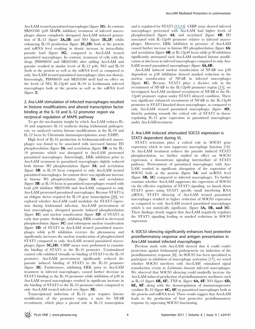
2. Ara-LAM stimulation of infected macrophages resultedin histone modifications and altered transcription factorbinding at the IL-10 and IL-12 promoter region viareciprocal regulation of MAPK pathway
To get the mechanistic insight by which Ara-LAM reduces IL-
10 and augmentes IL-12 synthesis during leishmanial pathogen-
esis, we analyzed various histone modifications at the IL-10 and
IL-12 locus by Chromatin immunoprecipitation assay (CHIP).
High level of IL-10 production in leishmania-infected macro-
phages was found to be associated with increased histone H3
phosphorylation (figure 3A) and acetylation (figure 3B) at the IL-
10 promoter which was abrogated in Ara-LAM pretreated
parasitized macrophages. Interestingly, ERK inhibition prior to
Ara-LAM treatment in parasitized macrophages slightly reduced
both histone H3 phosphorylation (figure 3A) and acetylation
(figure 3B) at IL-10 locus compared to only Ara-LAM treated
parasitized macrophages. In contrast there was significant increase
in histone H3 phosphorylation (figure 3A) and acetylation
(figure 3B) at IL-10 locus in parasitized macrophages treated with
both p38 inhibitor SB203580 and Ara-LAM, compared to only
Ara-LAM pretreated parasitized macrophages. Because STAT3 is
an obligate factor required for IL-10 gene transcription [20], we
explored whether Ara-LAM could modulate the STAT3 expres-
sion during leishmanial infection. Ara-LAM pretreatment of
host macrophages, abrogated parasite induced phosphorylation
(figure 3C) and nuclear translocation (figure 3D) of STAT3 at
early time points. Strikingly, inhibiting ERK resulted in decreased
phosphorylation (figure 3C) and subsequent nuclear translocation
(figure 3D) of STAT3 in Ara-LAM treated parasitized macro-
phages while p-38 inhibition reverses the phenomena and
significantly increases the nuclear translocation of phosphorylated
STAT3 compared to only Ara-LAM treated parasitized macro-
phages (figure 3C,3D). CHIP assays were performed to examine
the binding of STAT3 to the IL-10 promoter. Unstimulated
control cells exhibited virtually no binding of STAT3 to the IL-10
promoter. Ara-LAM pretreatment significantly reduced the
parasite induced binding of STAT3 to the IL-10 promoter
(figure 3E). Furthermore, inhibiting ERK prior to Ara-LAM
treatment in infected macrophages, caused further decrease in
STAT3 binding to the IL-10 promoter while inhibition of p38 in
Ara-LAM treated macrophages resulted in significant increase in
the binding of STAT3 to the IL-10 promoter when compared to
only Ara-LAM treated infected sets (figure 3E).
Transcriptional induction of IL-12 gene requires histone
modification of the promoter region, a mast for NF-kB
recruitment, which plays a pivotal role in IL-12 transcription
and is regulated by STAT3 [13,15]. CHIP assay showed infected
macrophages pretreated with Ara-LAM had higher levels of
phosphorylated (figure 4A) and acetylated (figure 4B) H3
associated with IL-12p40 promoter relative to infected macro-
phages. Moreover, ERK inhibition in presence of Ara-LAM
caused further increase in histone H3 phosphorylation (figure 4A)
and acetylation (figure 4B) at IL-12p40 locus while p-38 inhibition
significantly attenuated such Ara-LAM mediated histone modifi-
cation at this locus in infected macrophages compared to only Ara-
LAM treated parasitized macrophages (figure 4A,4B).
Ara-LAM induced nuclear translocation of NF-kB was p38
dependent as p38 inhibition showed marked reduction in the
nuclear translocation of NF-kB in infected macrophages
(figure 4C). Because, STAT3 plays a decisive role in the
recruitment of NF-kB to the IL-12p40 promoter region [15], we
investigated Ara-LAM mediated recruitment of NF-kB to the IL-
12p40 promoter region under STAT3 silenced condition. There
was significant enhanced recruitment of NF-kB to the IL-12p40
promoter in STAT3 knocked down macrophages, as compared to
only Ara-LAM treated parasitized macrophages (figure 4D),
thereby pointing towards the critical role of STAT3 in down-
regulating IL-12 gene expression in parasitized macrophages
under Ara-LAM-treatment.
3. Ara-LAM induced attenuated SOCS3 expression isSTAT3 dependent during VL
STAT3 activation plays a critical role in SOCS3 gene
expression which in turn suppresses macrophage function [16].
As Ara-LAM treatment reduces the parasite induced STAT3
phosphorylation, we further studied its effect on SOCS3
expression, a downstream signaling intermediate of STAT3
pathway. Pretreatment of parasitized macrophages with Ara-
LAM resulted in significant abrogation of the expression of
SOCS3 both at the protein (figure 5A) and m-RNA level
(figure 5B, 5C) compared to infected macrophages. To further
examine whether Ara-LAM suppresses the expression of SOCS3
via the effective regulation of STAT3 signaling, we knock down
STAT3 genes using STAT3 specific small interfering RNA
(siRNA). STAT3 silencing of Ara-LAM treated parasitized
macrophages resulted in higher reduction of SOCS3 expression
as compared to only Ara-LAM treated parasitized macrophages
which is not statistically significant. (figure 5A, 5B, and 5C).
These findings clearly suggest that Ara-LAM negatively regulates
the STAT3 signaling leading to marked reduction in SOCS3
expression.
4. SOCS3 silencing significantly enhances host protectiveproinflammatory response and antigen presentation inAra-LAM treated infected macrophages
Previous study with Ara-LAM showed that it could confer
protection against leishmanial pathogenesis via induction of the
proinflammatory response [8]. As SOCS3 has been speculated to
participate in inhibition of macrophage activation [17], we tested
whether SOCS3 interferes with Ara-LAM -stimulated signal
transduction events in Leishmania donovani infected macrophages.
We observed that SOCS3 silencing could markedly increase the
Ara-LAM induced production of proinflammatory mediators such
as IL-12 (figure 6B, 6F), TNF-a, (figure 6A, 6F) NO (figure 6D,6E, 6F) along with the downregulation of immunosupressive
cytokine IL-10 (figure 6C, 6F) in parasitized macrophages both at
the protein and mRNA level. These results suggest that Ara-LAM
leads to the production of host protective proinflammatory
response by supressing SOCS3 functioning.
Ara-LAM Mediated Protection in Leishmaniasis
PLoS ONE | www.plosone.org 4 September 2011 | Volume 6 | Issue 9 | e24141

Figure 3. Histone H3 modifications at the IL-10 promoter in Ara-LAM treated infected macrophages. Murine macrophages (16106 cells/mL) were treated with SB or PD for 2 h, subsequently followed by Ara-LAM treatment for 3 hr and Leishmania infection for 45 min. After 45 min ofincubation, ChIP assays were conducted as described in Materials and Methods. Immunoprecipitations were performed using Abs specific tophosphorylated H3 (IP phospho-H3) (A) or IP acetyl-H3 (B), and conventional RTPCR or quantitative real-time PCR was performed using primers specific tothe IL-10 promoter. *P,.001 compared with Ara-LAM–pretreated infected macrophages. In a separate experiment, the macrophages were treated asdescribed above followed by infection with Leishmania parasite. After 24 hr of incubation, the cells were lysed and subjected to Western blot with anti-pSTAT3, and STAT3 as described in Materials and Methods (C). Peritoneal macrophages (36106 (cells/mL) were treated and infected by Leishmania for 15,30 min as described in the legends of figure 1. Cytosolic and nuclear protein extracts were prepared for Western blot analysis as described in materialmethod to analyze the nuclear translocation of pSTAT3 and STAT3 (D). The blot shown is a representative of experiments performed in triplicate. Bandintensities were analyzed by densitometry (i,ii). (inset). In a separate experiment murine macrophages (16106 cells/mL) were treated and infected withLeishmania parasite as described above. After 45 min of incubation, Immunoprecipitations were conducted using STAT3 (IP: STAT3) specific Abs.Conventional RT-PCR or quantitative real-time PCR was performed for amplifying the putative STAT3 binding sites of the IL-10 promoter. *P,.001compared with Ara-LAM–pretreated infected macrophages.doi:10.1371/journal.pone.0024141.g003
Ara-LAM Mediated Protection in Leishmaniasis
PLoS ONE | www.plosone.org 5 September 2011 | Volume 6 | Issue 9 | e24141

SOCS3 has also been reported to down regulate major histo-
compatibility -II (MHC-II) expression resulting in decreased
antigen presentation by macrophages [21], a hallmark of
leishmanial pathogenesis [22]. Fascinatingly, Ara-LAM treatment
prominently restores MHC-II expression in L. donovani–infected
macrophages (figure 7A). We also investigated the role of Ara-
LAM on the antigen presentation capability of infected macro-
phages by measuring their ability to activate l peptide stimulated
T cell hybridoma 9H3.5 cells in terms of IL-2 production which is
important for T effector cell functioning. In case of infected
macrophage, there was a significant impairment of antigen
presentation compared with uninfected macrophages (figure 7C)
along with lower IL-2 secretion from T cell hybridoma 9H3.5
when co-cultured with infected macrophages (figure 7B). Inter-
estingly the release of IL-2 from 9H3.5 cells and antigen
presentation ability could be effectively increased under Ara-
LAM pretreatment in infected macrophages. (figure 7B,7C).
Moreover, parasitized macrophages in which SOCS3 was knocked
Figure 4. Hstone H3 modifications at the IL-12 promoter in Ara-LAM treated infected macrophages. Murine macrophages (16106cells/mL) were treated and infected as described in the Figure 3 legend. After 45 min of incubation, ChIP assays were conducted as described in Materials andMethods. Immunoprecipitations were performed using Abs specific to phosphorylated H3 (IP phospho-H3) (A) or IP acetyl-H3 (B), and conventional RT-PCRor quantitative real-time PCR was performed using primers specific to the IL-12p40 promoter. *P,.001 compared with Ara-LAM–pretreated infectedmacrophages. In a separate experiment peritoneal macrophages were treated and infected by Leishmania for 15, 30 min as described in Figure 3 legend.Cytosolic and nuclear protein extracts were analyzed for the nuclear translocation of NF-kB (C). The blot shown is a representative of experimentsperformed in triplicate. Band intensities were analyzed by densitometry (i,ii) (inset). Peritoneal macrophages (16106 cells/mL) were transfected with controlsiRNA or STAT3-specific siRNA, subsequently followed by Ara-LAM treatment (for 3 hr) and Leishmania infection for 45 min. Immunoprecipitations wereconducted using NF-kB specific Abs. Conventional RT-PCR or quantitative real-time PCR was performed for amplifying the putative NF-kB binding sites ofthe IL-12p40 promoter. *P,.001 compared with Ara-LAM–pretreated infected macrophages.doi:10.1371/journal.pone.0024141.g004
Ara-LAM Mediated Protection in Leishmaniasis
PLoS ONE | www.plosone.org 6 September 2011 | Volume 6 | Issue 9 | e24141

down prior to Ara-LAM treatment resulted in enhanced MHC-II
expression (figure 7A) as reflected by greatly increased antigen
presentation (figure 7C) and higher IL-2 production from 9H3.5
cells (figure 7B) demonstrating that SOCS3 interferes with antigen
presentation and decreased SOCS3 was further potentiating
Ara-LAM mediated antigen presentation ability of infected
macrophages.
Discussion
Leishmania-induced deactivation of macrophage functions are
due to defects in the signaling pathways, which facilitates the
parasite to infect and propagate within the cell [23]. Recently the
capacity of Leishmania to suppress macrophage function has been
linked to alterations in MAPK signaling cascades [12,24,25]. To
revert this parasite induced changes in MAPK signaling, novel
immunomodulators are being evaluated for their functionality in
VL. Ara-LAM, a potential immunomodulator strongly initiates the
host protective immune response during VL via TLR-2 mediated
proinflammatory responses [8]. But its role in regulating the
MAPK signaling pathway has not been explored till date.
Recently it has been demonstrated that the differential
activation of the MAPKs regulates cytokine production. CD40,
a costimulatory molecule with host-protective anti-leishmanial
function, could induce both IL-10 and IL-12 by differential
activation of p38 MAPK and ERK-1/2, respectively. This
reciprocal regulation of p38 MAPK and ERK1/2 has been
shown to be responsible for the protection versus susceptibility to
infection [12,24].
Ara-LAM pretreatment restored the impaired p38MAPK
phosphorylation in parasitized macrophages and abrogated the
ERK1/2 phosphorylation via TLR-2 mediated downstream
signaling (figure 1) thus shifting the macrophageal microenviron-
ment from anti-inflammatory (IL-10) towards proinflammatory
(IL-12) host protective immune response (figure 2). Our finding is
consistent with other studies which have also highlighted the
involvement of this novel molecule, Ara-LAM, in inducing
proinflammatory responses (IL-8, TNF-a) in macrophages via
interacting with TLR-2 [26,27]. Additionally, Ara-LAM has been
demonstrated as a major component responsible for the pro-
inflammatory activity of the whole bacteria (M. smegmatis and M.
fortuitum) [28].
Having identified the reciprocal alteration in Ara-LAM
mediated production of IL-12 and IL-10 during VL, we studied
whether Ara-LAM could modulate the chromatin (specifically
histone H3) at the locus of these two counteractive cytokine genes
since covalent modification to histones of chromatin can lead to
differential gene expression. Our study revealed that Ara-LAM
pretreatment leads to IL-12 induction via histone H3 phosphor-
ylation (figure 4A) and acetylation (figure 4B) of chromatin at IL-
12 locus along with IL-10 attenuation by abrogating the parasite
induced histone H3 phosphorylation (figure 3A) and acetylation
(figure 3B) at IL-10 locus during leishmanial pathogenesis. Thus
Ara-LAM mediated gene-specific chromatin modifications are
found to be associated with transient silencing of IL-10 genes and
priming of the IL-12 gene.
To further ascertain the role of MAPKs in the regulation of
counteracting cytokines IL-10 and IL-12, we investigated Ara-
LAM mediated transcriptional alteration of these cytokine genes in
the presence of their respective pharmacological inhibitors. Our
results implicated that the inhibition of p38 MAPK pathway in
Ara-LAM treated parasitized macrophages abrogates chromatin
remodeling and nuclear translocation of NF-kB thereby attenuat-
ing IL-12 transcription (figure 4A, 4B, 4C) and simultaneously
reverses Ara-LAM mediated attenuated IL-10 production in
parasitized macrophages via increased histone H3 phosphoryla-
tion, acetylation and enhanced STAT3 recruitment to the IL-10
promoter leading to the induction of IL-10 transcription
(figure 3A,3B,3D,3E). In contrast ERK inhibition of Ara-LAM
treated parasitized macrophages leads to further increase in IL-12
transcription (figure 4A, 4B, 4C) along with significant reduction
of the IL-10 (figure 3A,3B,3D,3E) compared to Ara-LAM
treatment only. Therefore, this study suggested a crucial role of
Figure 5. STAT3 silencing potentiate Ara-LAM mediated abrogation of SOCS3 expression in Leishmania-donovani infectedmacrophages. Peritoneal macrophages (26106 cells/mL) were transfected with control siRNA or STAT3-specific siRNA, subsequently followed by Ara-LAMtreatment (for 3 hr) and Leishmania infection for 24 hr. The cells were then lysed and subjected to Western blot with anti-SOCS3 antibody as described inMaterials and Methods (A). In a separate experiment macrophages were transfected with either control-siRNA or STAT3 specific siRNA, subsequentlyfollowed by Ara-LAM treatment for 3 hr and Leishmania infection for another 3 h. RNA was isolated and semi quantitative RT-PCR analyses for SOCS3 andGAPDH were done. Data represented here are from one of three independent experiments, all of which yielded similar results (B). Changes in expression ofSOCS3 mRNA were also determined by quantitative real-time PCR. Results are presented as changes (n-fold) relative to uninfected control cells. Theexperiment was repeated 3 times, yielding similar results (C). *P,.001 compared with infected macrophages. The comparison of SOCS3 expression betweenAra-LAM treated infected macrophages and STAT3 siRNA group did not show any statistical significance.doi:10.1371/journal.pone.0024141.g005
Ara-LAM Mediated Protection in Leishmaniasis
PLoS ONE | www.plosone.org 7 September 2011 | Volume 6 | Issue 9 | e24141

MAPK in Ara-LAM mediated modulation of IL-12 and IL-10
gene transcription during leishmanial pathogenesis.
The signaling pathways which direct IL-10-mediated inhibition of
IL-12 production at the level of transcription is well documented [12].
Recent observations have suggested that absence of STAT3 resulted
in enhanced lipopolysacharide induced IL-12p40 gene expression in
IL-10_/_ mice due to enhanced NF-kB recruitment to the IL-12p40
promoter [15]. Our observation also indicated that STAT3 silencing
Figure 6. SOCS3 silencing significantly enhances host protective proinflammatory response generation by Ara-LAM in infectedmacrophages. Peritoneal macrophages (26106 cells/mL) were transfected with control siRNA or SOCS3-specific siRNA followed by Ara-LAM treatment(for 3 hr) and Leishmania infection for 24 h and assayed for the levels of TNF- a (A), IL-12 (B), and IL-10 (C) in the culture supernatant by ELISA, as describedin Methods. ELISA data are expressed as means standard deviations of values from triplicate experiments that yielded similar observations. *P,.001compared with infected macrophages, **P,.005 compared with Ara-LAM–pretreated infected macrophages. ***,.01 compared to Ara-LAM–pretreatedinfected macrophages. In a separate experiment macrophages were cultured and treated as described above and assayed for the levels of extracellular NOas described in the Materials and Methods (D). *P,.001 compared with infected macrophages, **P,.005 compared with Ara-LAM–pretreated infectedmacrophages. In a separate set macrophages were transfected and treated with Ara-LAM as described above followed by Leishmania infection for 24 hr.Western blot analysis was performed to analyze the expression of inducible nitric oxide synthase. The blot shown is a representative of experimentsperformed in triplicate. Band intensities were analyzed by densitometry (E). Peritoneal macrophages were cultured and treated with Ara-LAM as describedabove followed by Leishmania infection for 3 hr. Changes in mRNA expression of IL-12p40, TNF- a, IL-10, NO were determined by real-time PCR analysisResults are presented as changes (n-fold) relative to uninfected control cells. The experiment was repeated 3 times, yielding similar results; data areexpressed as means 6 standard deviations. (F). *P,.001 compared with infected macrophages, **P,.005 compared with Ara-LAM–pretreated infectedmacrophages.doi:10.1371/journal.pone.0024141.g006
Ara-LAM Mediated Protection in Leishmaniasis
PLoS ONE | www.plosone.org 8 September 2011 | Volume 6 | Issue 9 | e24141

significantly enhances Ara-LAM mediated recruitment of NF-kB to
the IL-12 p40 promoter (figure 4D) indicating that STAT3 negatively
regulates Ara-LAM mediated transcription of IL-12 gene during
leishmanial pathogenesis.
Moreover, STAT3 activation leads to the induction of SOCS3,
which plays a decisive role in inactivation of macrophage function
[16]. SOCS3 is critical for the survival strategy of mycobacteria
[29], leishmania [30], listeria [31] and RNA viruses [32] in the host
macrophages. Moreover silencing of the SOCS3 gene resulted in a
marked reversal of the immunosuppressive effect of IL-10 on many
inflammatory mediators including cytokines, chemokines, and IFN-
inducible, apoptotic and TNF ligand/receptor genes [33].
Ara-LAM treatment lead to significant reduction in SOCS3
expression by inactivation of STAT3 (figure 5A, 5B, 5C). These
changes resulted in significant induction of host protective
proinflammatory responses (figure 6), MHC-II expression, and
antigen presentation capability of infected macrophages (figure 7A,7C) along with enhanced T cell effector functioning as evident
from higher IL-2 production from co-cultured T cells (figure 7B).
Further confirmation about the role of SOCS3 in the generation
of host protective immune responses in Ara-LAM treated infected
sets was done by SOCS3 silencing which was suggestive of SOCS3
hindrance in Ara-LAM mediated induction of proinflammatory
response (figure 6, 7A, 7B, 7C).
In summary, our current findings provide a detailed molecular
mechanistic insight of Ara-LAM mediated IL-12 and IL-10
production in Leishmania donovani infected macrophages where
MAPK signaling via differential regulation of p38 and ERK plays
an important role in generating host protective immune response
during leishmanial pathogenesis.
Materials and Methods
Reagents and ChemicalsRPMI-1640 medium, M-199 medium (M199), penicillin and
streptomycin, SB203580 (p38MAPK inhibitor), PD098059 (ERK
inhibitor) and TRI Reagent were from Sigma (St Louis, MO,
USA). Fetal calf serum (FCS) was obtained from Gibco BRL
Figure 7. Ara-LAM induced MHC-II expression, antigen presentation ability of infected macrophages increased under SOCS3silenced condition. Peritoneal macrophages (26106 cells/mL) were transfected with control siRNA or SOCS3-specific siRNA, subsequently followed byAra-LAM treatment (for 3 hr) and Leishmania infection for 4 hr as mentioned above. After 24 hr of incubation treated macrophages were analyzed by flowcytometry for MHCII (FL2-H) expression as described in material method (A). Data are from 1 of 3 experiments conducted in the same way with similarresults. In a separate set, normal and treated macrohages either uninfected or infected were pulsed with lR12–26, and then were incubated with T-cellhybridoma 9H 3.5. The culture supernatants were analysed for the presence of IL-2 by ELISA as described in the methods (B). Incorporation of 3H-Thymidinein the IL-2 dependent cell line HT-2 was assessed in presence of the cultured supernatant. Results are expressed as mean 6 SD of 5 replicate experiments(C). *P,.001, **P,.005 compared with infected macrophages.doi:10.1371/journal.pone.0024141.g007
Ara-LAM Mediated Protection in Leishmaniasis
PLoS ONE | www.plosone.org 9 September 2011 | Volume 6 | Issue 9 | e24141

(Grand Island, NY, USA) and ELISA Assay Kit (Quantikine M)
for tumour necrosis factor (TNF)-a, IL-12, IL-10 were from R&D
Systems (Minneapolis, MN, USA). dNTPs, RevertAidTM M-
MuLV Reverse Transcriptase, oligo dT, RNase inhibitor and
other chemicals required for cDNA synthesis were from Fermentas
(USA). Anti-phospho-H3 and acetyl-H3 Abs were purchased from
Abcam and chromatin immunoprecipitation (ChIP) assay kits
were purchased from Millipore. SOCS3, STAT3 siRNA were
procured from Santa Cruze biotech.
T-cell hybridoma and lambda repressor peptide9H3.5 (I-Ad restricted T-cell hybridoma) specific for lambda
repressor N-terminal sequence 12–26 [LEDARRLKAIYEKKK,
defined as lR 12–26]. T-cell hybridomas and peptides were kind
gifts of Professor M. L. Gefter, Massachusetts Institute of
Technology, Cambridge, MA, USA. The IL-2 dependent cell
line HT-2 (mouse T helper cell line) was obtained from American
Type Culture Collection. All cells were maintained in RPMI 1640
medium supplemented with 10% fetal calf serum (FCS) and 2-ME
(561025 M) at 5% CO2 in humidified atmosphere.
Animals and parasitesBALB/c mice were purchased from the National Centre for
Laboratory Animal Sciences, India. For each experiment 8–10
mice (4–6 weeks old) were used, regardless of sex. L. donovani
organisms (strain MHOM/IN/1983/AG-83) were maintained
in Medium 199 (Sigma) plus 10% fetal calf serum (Gibco).
Amastigotes were prepared as described elsewhere [34]. Station-
ary-phase promastigotes obtained by suitable transformation were
used for experiments. All experimental protocols were given prior
approval by the institutional animal ethics committee.
Isolation and purification of Ara-LAMAra-LAM was isolated as described elsewhere [35] Lipopoly-
saccharide contamination was checked by the Limulus test and
was ,25 ng/mg in Ara-LAM. The noncytotoxic dose of Ara-
LAM was 3 mg/mL [36].
Peritoneal macrophage preparationMacrophages were isolated by peritoneal lavage with ice-cold
phosphate-buffered saline at 48 h after intraperitoneal injection
of 1.0 mL of sterile 4% thioglycolate broth (Difco). Cells were
cultured as described elsewhere [37].The adherent cell population
was cultured for 48 h prior to any treatment, to achieve the resting
state.
Uptake and intracellular multiplicationFor assessing the activity of Ara-LAM against the amastigote
stage of parasite, peritoneal macrophages cultured on glass cover
slips were pretreated with Ara-LAM (3 mg/ml) for 3 h, followed by
infection with L.donovani promastigotes at a ratio of 1:10 for the
indicated time periods; macrophages were then fixed and stained
as described elsewhere [38] for calculation of the number of
intracellular parasites. When indicated, the uptake and multipli-
cation of L.donovani was studied in the presence and absence of the
p38MAPK inhibitor SB203580 (10 mg/mL), or ERK inhibitor
PD098059 (100 mmol/L; Sigma) [12].
Cytokine enzyme-linked immunosorbent assay (ELISA)The conditioned medium of macrophage culture was assayed
for mouse cytokines and chemokines with use of the sandwich
ELISA kit (Quantikine M; R&D Systems). The assay was
performed according to the manufacturer’s instructions.
Preparation of cell lysate and immunoblot analysisCell lysates were prepared as described elsewhere [39]. Equal
amounts of protein (50 mg) were subjected to 10% sodium dodecyl
sulfate polyacrylamide gel electrophoresis, and were subsequently
transferred to a nitrocellulose membrane. The membrane was
blocked overnight with 3% bovine serum albumin in Tris-saline
buffer (pH, 7.5), and immunoblotting was performed to detect
Inducible Nitric Oxide Synthase 2 (iNOS2) and phosphorylated or
dephosphorylated forms of p38MAPK, ERK1/2, STAT3 and
b-Actin as described elsewhere [40].
Nitrite generationNitrite level in culture was measured using the Nitric Oxide
Colorimetric Assay kit (Boehringer Mannheim Biochemicals) [41].
Cell-free supernatants were collected from different experimental
sets at different time points of infection, and nitrite levels were
estimated in accordance with the manufacturer’s instructions.
Data were expressed in micromoles of nitrite.
Antigen presentation assayAntigen presenting ability of infected macrophages was studied
by their ability to present lR12–26 to T-cell hybridoma (9H 3.5).
The antigen presenting cells (APC) were incubated for 24 h with
specific peptide and T-cell hybridoma in complete RPMI medium
in a 37uC incubator. The culture supernatants were analysed for
the presence of IL-2 by growing an IL-2 dependent cell line HT-2
in the supernatants. HT-2 (104 cells per well) was incubated with a
50% concentration of culture supernatant for 48 h. The cells were
then pulsed with 1 mCi of 3H-Thymidine [6.7 Ci/mmole, New
England Nuclear, Boston, NE, USA)] for the last 18 h [42]. The
incorporation of radioactive thymidine was then assessed by a
scintillation counter (Packard).
Flow cytometryBALB/c-derived macrophages were stained with phycoerythrin
(PE) labeled anti-MHCII antibodies. The cells were analyzed by a
FACSVantageTM flow cytometer (Becton Dickinson).
Preparation of nuclear and cytoplasmic extractsThe nuclear extracts were prepared from normal and infected
macrophages as described elsewhere [43]. Briefly, sedimented cells
were resuspended in hypotonic buffer (10 mM HEPES (pH 7.9),
1.5 mM MgCl2, 10 mM KCl, 0.2 mM PMSF, and 0.5 mM
DTT) and allowed to swell on ice for 10 min. Cells were
homogenized in a homogenizer. The nuclei were separated by
spinning at 33006g for 5 min at 4uC. The supernatant was used as
the cytoplasmic extract. The nuclear pellet was extracted in
nuclear extraction buffer (20 mM HEPES (pH 7.9), 0.4 M NaCl,
1.5 mM MgCl2, 0.2 mM EDTA, 25% glycerol, 0.5 mM PMSF,
and 0.5 mM DTT) for 30 min on ice and centrifuged at 12,0006g
for 30 min. The supernatant was used as nuclear extract.
CHIP assayCHIP assays were conducted using the CHIP Assay kit
following the manufacturers Protocol (Millipore). Briefly, 16106
peritoneal macrophages were plated overnight in six-well plates.
Cells were stimulated as described in figures, and then fixed for
10 min at 37uC in 1% paraformaldehyde. Cells were washed on
ice with ice-cold HBSS containing 1 mM PMSF, harvested and
then lysed in SDS lysis buffer. DNA was sheared by ultrasonica-
tion using a High Intensity Ultrasonic Processor (hielscher) for
3610 s pulses at 20% amplitude. Lysates were cleared by
centrifugation and diluted in ChIP dilution buffer. Lysates were
Ara-LAM Mediated Protection in Leishmaniasis
PLoS ONE | www.plosone.org 10 September 2011 | Volume 6 | Issue 9 | e24141

precleared using salmon sperm DNA/protein A-agarose and a
sample of ‘‘input DNA’’ was collected at this point. Protein-DNA
complexes were immunoprecipitated with 5 mg of Ab overnight at
4uC. Ab-protein-DNA complexes were then captured using
salmon sperm DNA/protein A-agarose for 1 h at 4uC. After
washing beads with low and high salt, LiCl, and TE buffers, the
protein/DNA complexes were eluted using 1% SDS, 0.1 M
NaHCO3 buffer and disrupted by heating at 65uC for 4 h. DNA
was then extracted using phenol/chloroform extraction and
ethanol precipitation. PCR was conducted using promoter specific
primers: IL-10 promoter (STAT3 binding region): sense 59-
TCATGCTGGGATCTGAGCTTCT-39, antisense 59-CGGAA-
GTCACCTTAGCACTCAGT-39 (94uC, 15 s; 56uC, 30 s; 72uC,
1 min, 35cycles); IL-12p40 promoter (NF-kB binding site): sense
59-AGTATCTC TGCCTCCTTCCTT-39, antisense 59-GCAA-
CACTGAAAACTAGTGTC-39 (initial denaturation at 95uC for
3 min; amplification cycles at 95uC for 30 s, 58uC for 1 min,
and 72uC for 1 min; and a final extension at 72uC for 10 min,
40cycles). PCR amplified product was subsequently size fractioned
on 2% agarose gel, stained with ethidium bromide and visualized
under UV-light. For relative quantitation of promoter levels, real-
time PCR was also performed. For real-time PCR samples were
normalized to input DNA controls.
Isolation of RNA and real-time polymerase chain reactionTotal RNA extracted from macrophages (TRI reagent; Sigma)
according to the standard protocol [44,45] was reverse transcribed
using Revert Aid M-MuLV reverse transcriptase (Fermentas).
Real-time polymerase chain reaction (PCR) was performed using
SYBR Green mix and the ABI 7500 real-time PCR system
(Applied Biosystems). Glyceraldehyde- 3-phosphate dehydroge-
nase (GAPDH) was used as a reference. Sequences of the PCR
primers are listed in Table 1. The reaction conditions consisted of
an initial activation step (5 min at 95uC) and cycling step
(denaturation for 30 s at 94uC, annealing for 30 s at 58uC, and
extension for 1 min at 72uC for 40 cycles), after which melt curve
analysis was performed. Detection of the dequenched probe,
calculation of threshold cycles, and further analysis of these data
were done using Sequence Detector software (version 1.4; Applied
Biosystems). Relative changes in iNOS2 and cytokine messenger
RNA (mRNA) expression were compared with unstimulated
control, normalized to GAPDH, and quantified by the 2DDCt
method.
Isolation of RNA and RT-PCRRNA was isolated according to the standard protocol [44,45].
Briefly, total RNA extracted from macrophages (TRI reagent;
Sigma) according to the standard protocol [44,45] was reverse
transcribed using Revert Aid M-MuLV reverse transcriptase
(Fermentas). The cDNA encoding the SOCS3, GAPDH gene was
amplified using specific primers as listed in Table 1. PCR
amplification was conducted in a reaction volume of 50 ml using
a Perkin Elmer Gen Amp PCR system 2400 and 0.5 unit of Taq
polymerase set for 35 cycles (denaturation: at 94uC for 30 s;
annealing: at 58uC for 30 s; extension: at 72uC for 30 s) PCR
amplified product was subsequently size fractioned on 2% agarose
gel, stained with ethidium bromide and visualizedunder UV-light.
Densitometry analysisImmunoblots were analyzed using a model GS-700 Imaging
Densitometer and Molecular Analyst (version 1.5; Bio-Rad
Laboratories).
Statistical analysisThe in vitro cultures were performed in triplicate. The data,
represented as mean values 6standard deviations, are from 1
experiment that was performed at least 3 times. Student’s t test was
employed to assess the significance of the differences between the
mean values of control and experimental groups. A P value of.05
was considered to be significant, and a P value ,.001 was
considered to be highly significant.
Ethics StatementThis study was carried out in strict accordance with the
recommendations in the Guide for the Care and Use of
Laboratory Animals of the National Institutes of Health. All
experimental animal protocols received prior approval from the
Institutional Animal Ethical Committee (Bose Institute, Registra-
tion Number: 95/99/CPCSEA).
Acknowledgments
We are grateful to Director, Bose Institute (Kolkata) for his continuous
encouragement. We cordially thank Mr. Debasish Majumdar and Mr.
Prabal Gupta for their technical assistance.
Author Contributions
Conceived and designed the experiments: S. Majumdar PB SC. Performed
the experiments: PB GG S. Majumder AA SB KH SBM. Analyzed the
data: S. Majumdar PB. Contributed reagents/materials/analysis tools: S.
Majumdar SC SR. Wrote the paper: S. Majumdar.
References
1. Descoteaux A, Matlashewski G (1989) c-fos and tumor necrosis factor gene
expression in Leishmania donovani-infected macrophages. Mol Cell Biol 9: 5223–7.
2. Murray HW, Spitalny GL, Nathan CF (1985) Activation of mouse peritoneal
macrophages in vitro and in vivo by interferon-gamma. J Immunol 134:
1619–1622.
3. Russell DG, Talamas-Rohana P (1989) Leishmania and the macrophage: a
marriage of Inconvenience. Immunol Today 10: 328–33.
4. Murray HW (1982) Cell-mediated immune response in experimental visceral
leishmaniasis. II. Oxygen-dependent killing of intracellular Leishmania dono-
vani amastigotes. J Immunol 129: 351–7.
Table 1. Sequences of the PCR primers.
Gene Sequence of Primers
iNOS2 Forward 59-CCCTTCCGAAGTTTCTGGCAGCAGC-39
Reverse 59-GGCTGTCAGAGCCTCG TGGCTTTGG-39
IL-10 Forward 59-CGGGAAGACAATAACTG-39
Reverse 59-CATTTCCGATAAGGCTTGG-39
IL-12p40 Forward 59-CAACATCAAGAGCAGTAGCAG-39
Reverse 59TACTCCCAGCTGACCTCCAC-39
TNF-a Forward 59-GGCAGGTCTACTTTGGAGTCATTGC-39
Reverse 59-ACATTCGAGGCTCCAGTGAATTCGG-39
SOCS-3 Forward 59-GCGGGCACCTTTCTTATCC-39
Reverse 59-TCCCCGACTGGGTCTTGAC-39
GAPDH Forward 59-CAAGGCTGTGGGCAAGGTCA-39
Reverse 59-AGGTGGAAGAGTGGGAGTTGCTG-39
doi:10.1371/journal.pone.0024141.t001
Ara-LAM Mediated Protection in Leishmaniasis
PLoS ONE | www.plosone.org 11 September 2011 | Volume 6 | Issue 9 | e24141
5. Liew FY, Millott S, Parkinson C, Palmer RM, Moncada S (1990) Macrophage
killing of Leishmania parasite in vivo is mediated by nitric oxide from L-arginine.J Immunol 144: 4794–7.
6. Reiner SL, Locksley RM (1995) The regulation of immunity to Leishmania
major. Annu Rev Immunol 13: 151–177.7. Bacellar O, D’oliveira A, Jr., Jeronimo S, Carvalho EM (2000) IL-10 and IL-12
are the main regulatory cytokines in visceral leishmaniasis. Cytokine 12:1228–31.
8. Bhattacharya P, Bhattacharjee S, Gupta G, Majumder S, Adhikari A, et al.
(2010) Arabinosylated lipoarabinomannan-mediated protection in visceralleishmaniasis through up-regulation of toll-like receptor 2 signaling: an
immunoprophylactic approach. J Infect Dis 202: 145–55.9. Pan ZK (2004) Toll-like receptors and TLR-mediated signaling: more questions
than answers. Am J Physiol Lung Cell Mol Physiol 286(5): L918–20.10. Cano E, Mahadevan LC (1995) Parallel signal processing among mammalian
MAPKs. Trends Biochem Sci 20: 117–22.
11. Clayton AL, Mahadevan LC (2003) MAP kinase-mediated phosphoacetylationof histone H3 and inducible gene regulation. FEBS Lett 546: 51–8.
12. Mathur RK, Awasthi A, Wadhone P, Ramanamurthy B, Saha B (2004)Reciprocal CD40 signals through p38MAPK and ERK-1/2 induce counter-
acting immune responses. Nat Med 10: 540–4.
13. Saccani S, Pantano S, Natoli G (2002) p38-Dependent marking of inflammatorygenes for increased NF-kappa B recruitment. Nat Immunol 1: 69–75.
14. Lucas M, Zhang X, Prasanna V, Mosser DM (2005) ERK activation followingmacrophage FcgammaR ligation leads to chromatin modifications at the IL-10
locus. J Immunol 175: 469–77.15. Hoentjen F, Sartor RB, Ozaki M, Jobin C (2005) STAT3 regulates NF-kappaB
recruitment to the IL-12p40 promoter in dendritic cells. Blood 105: 689–96.
16. Qasimi P, Ming-Lum A, Ghanipour A, Ong CJ, Cox ME, et al. (2006)Divergent mechanisms utilized by SOCS3 to mediate interleukin-10 inhibition
of tumor necrosis factor alpha and nitric oxide production by macrophages.J Biol Chem 281: 6316–24.
17. Qin H, Roberts KL, Niyongere SA, Cong Y, Elson CO, et al. (2007) Molecular
mechanism of lipopolysaccharide-induced SOCS3 gene expression in macro-phages and microglia. J Immunol 179: 5966–76.
18. Feng GJ, Goodridge HS, Harnett MM, Wei XQ, Nikolaev AV, et al. (1999)Extracellular signal-related kinase (ERK) and p38 mitogen-activated protein
(MAP) kinases differentially regulate the lipopolysaccharide-mediated inductionof inducible nitric oxide synthase and IL-12 in macrophages: Leishmania
phosphoglycans subvert macrophage IL-12 production by targeting ERK MAP
kinase. J Immunol 163: 6403–12.19. Suttles J, Milhorn DM, Miller RW, Poe JC, Wahl LM, et al. (1999) CD40
signaling of monocyte inflammatory cytokine synthesis through an ERK1/2-dependent pathway. A target of interleukin (il)-4 and il-10 anti-inflammatory
action. J Biol Chem 274: 5835–42.
20. Murray PJ (2006) STAT3-mediated anti-inflammatory signaling. Biochem SocTrans 34: 1028–31.
21. Li Y, Chu N, Rostami A, Zhang GX (2006) Dendritic cells transduced withSOCS3 exhibit a tolerogenic/DC2 phenotype that directs type 2 Th cell
differentiations in vitro and in vivo. J Immunol 177: 1679–88.22. Chakraborty D, Banerjee S, Sen A, Banerjee KK, Das P, et al. (2005)
Leishmania donovani affects antigen presentation of macrophage by disrupting
lipid rafts. J Immunol 175: 3214–24.23. Reiner NE (1994) Altered cell signaling and mononuclear phagocyte
deactivation during intracellular infection. Immunol Today 15: 374–381.24. Awasthi A, Mathur R, Khan A, Joshi BN, Jain N, et al. (2003) CD40 signaling is
impaired in L. major-infected macrophages and is rescued by a p38MAPK
activator establishing a host-protective memory T cell response. J Exp Med 197:1037–43.
25. Yang Z, Mosser DM, Zhang X (2007) Activation of the MAPK, ERK, followingLeishmania amazonensis infection of macrophages. J Immunol 178: 1077–1085.
26. Briken V, Porcelli SA, Besra GS, Kremer L (2004) Mycobacterial lipoarabino-mannan and related lipoglycans: from biogenesis to modulation of the immune
response. Mol Microbiol 53: 391–403.
27. Nigou J, Gilleron M, Puzo G (2003) Lipoarabinomannans: from structure tobiosynthesis. Biochimie 85: 153–66.
28. Bohsali A, Abdalla H, Velmurugan K, Briken V (2010) The non-pathogenic
mycobacteria M. smegmatis and M. fortuitum induce rapid host cell apoptosisvia a caspase-3 and TNF dependent pathway. BMC Microbiol 10: 237.
29. Imai K, Kurita-Ochiai T, Ochiai K (2003) Mycobacterium bovis bacillus
Calmette-Guerin infection promotes SOCS induction and inhibits IFN-gamma-stimulated JAK/STAT signaling in J774 macrophages. FEMS Immunol Med
Microbiol 39: 173–80.
30. de Veer MJ, Curtis JM, Baldwin TM, DiDonato JA, Sexton A, et al. (2003)MyD88 is essential for clearance of Leishmania major: possible role for
lipophosphoglycan and Toll-like receptor 2 signaling. Eur J Immunol 33:
2822–31.
31. Stoiber D, Stockinger S, Steinlein P, Kovarik J, Decker T (2001) Listeria
monocytogenes modulates macrophage cytokine responses through STAT
serine phosphorylation and the induction of suppressor of cytokine signaling 3.J Immunol 166: 466–72.
32. Suhrbier A, La Linn M (2003) Suppression of antiviral responses by antibody-
dependent enhancement of macrophage infection. Trends Immunol 24: 165–8.
33. Dennis V, Kaushal D, Dixit S, Mehra S, Singh S, et al. (2008) SOCS3 and IL-10
anti-inflammatory activity in Lyme disease. The FASEB Journal 22: 860.17.
34. Hart DT, Vickerman K, Coombs GH, Quick A (1981) Simple method for
purifying L. mexicana amastigotes in large numbers. Parasitology 82: 345–355.
35. Majumder N, Dey R, Mathur RK, Datta S, Maitra M, et al. (2006) An unusualpro-inflammatory role of interleukin-10 induced by arabinosylated lipoarabino-
mannan in murine peritoneal macrophages. Glycoconj J 23: 675–686.
36. Bhattacharjee S, Majumder N, Bhattacharyya P, Bhattacharyya S, Majumdar S(2007) Immunomodulatory role of arabinosylated lipoarabinomannan on
Leishmania donovani infected murine macrophages. Indian J Biochem Biophys
44: 366–372.
37. Fahey TJ, Tracey KJ, Tekamp-Olson P, Cousens LS, Jones WG, et al. (1992)
Macrophage inflammatory protein-1 modulates macrophage function.
J Immunol 148: 2764–2769.
38. Bhattacharyya S, Ghosh S, Dasgupta B, Mazumder D, Roy S, Majumdar S
(2002) Chemokine-induced leishmanicidal activity in murine macrophages via
the generation of nitric oxide. J Infect Dis 185: 1704–8.
39. Majumdar S, Kane LH, Rossi MW, Volpp BD, Nauseef WM, et al. (1993)
Protein kinase C isotypes and signal transduction in human neutrophils: selective
substrate specificity of calcium dependent b-PKC and novel calciumindependent n-PKC. Biochim Biophys Acta 1176: 276–286.
40. Ghosh S, Bhattacharyya S, Sirkar M, Sa GS, Das T, et al. (2002) Leishmaniadonovani suppresses activator protein-1 and NF-kB in host macrophages via
ceramide generation: involvement of extracellular signal regulated kinase. Infect
Immun 70: 6828–6838.
41. Green LC, Wagner DA, Glogowski J, Skipper PL, Wishnok JS, et al. (1982)
Analysis of nitrate, nitrite, and [15N] nitrate in biological fluids. Anal Biochem
126: 131–138.
42. Roy S, Scherer MT, Briner TJ, Smith JA, Gefter LM (1989) Murine MHC
polymorphism and T cell specificities. Science 244: 572–5.
43. Yaron A, Gonen H, Alkalay I, Hatzobai A, Jung S, et al. (1997) Inhibition ofNF-k-B cellular function via specific targeting of the I-k-B-ubiquitin ligase.
EMBO J 16: 6486–6494.
44. Chomezynasky P, Sacchi N (1987) A single step method of RNA isolation byguanidium thiocyanate-phenol-chloroform extraction. Anal Biochem 162:
156–9.
45. Sambrook J, Fritsch EF, Maniatis T (1989) Molecular Cloning: A LaboratoryManual. Cold Spring Harbor, NY: Cold Spring Harbor Laboratory Press.
Ara-LAM Mediated Protection in Leishmaniasis
PLoS ONE | www.plosone.org 12 September 2011 | Volume 6 | Issue 9 | e24141
Copyright © 2022 FDOKUMEN