TH1/TH2 Cytokine profile in relapsing-remitting multiple sclerosis patients treated with Glatiramer...
13
This Provisional PDF corresponds to the article as it appeared upon acceptance. Fully formatted PDF and full text (HTML) versions will be made available soon. TH1/TH2 Cytokine profile in relapsing-remitting multiple sclerosis patients treated with Glatiramer acetate or Natalizumab BMC Neurology 2012, 12:95 doi:10.1186/1471-2377-12-95 Celia Oreja-Guevara ([email protected]) Jaime Ramos-Cejudo ([email protected]) Luis Stark Aroeira ([email protected]) Beatriz Chamorro ([email protected]) Exuperio Diez-Tejedor ([email protected]) ISSN 1471-2377 Article type Research article Submission date 9 April 2012 Acceptance date 11 September 2012 Publication date 18 September 2012 Article URL http://www.biomedcentral.com/1471-2377/12/95 Like all articles in BMC journals, this peer-reviewed article can be downloaded, printed and distributed freely for any purposes (see copyright notice below). Articles in BMC journals are listed in PubMed and archived at PubMed Central. For information about publishing your research in BMC journals or any BioMed Central journal, go to http://www.biomedcentral.com/info/authors/ BMC Neurology © 2012 Oreja-Guevara et al. ; licensee BioMed Central Ltd. This is an open access article distributed under the terms of the Creative Commons Attribution License ( http://creativecommons.org/licenses/by/2.0), which permits unrestricted use, distribution, and reproduction in any medium, provided the original work is properly cited.
-
Upload
fuenlabrada -
Category
Documents
-
view
0 -
download
0
Transcript of TH1/TH2 Cytokine profile in relapsing-remitting multiple sclerosis patients treated with Glatiramer...

This Provisional PDF corresponds to the article as it appeared upon acceptance. Fully formattedPDF and full text (HTML) versions will be made available soon.
TH1/TH2 Cytokine profile in relapsing-remitting multiple sclerosis patientstreated with Glatiramer acetate or Natalizumab
BMC Neurology 2012, 12:95 doi:10.1186/1471-2377-12-95
Celia Oreja-Guevara ([email protected])Jaime Ramos-Cejudo ([email protected])Luis Stark Aroeira ([email protected])
Beatriz Chamorro ([email protected])Exuperio Diez-Tejedor ([email protected])
ISSN 1471-2377
Article type Research article
Submission date 9 April 2012
Acceptance date 11 September 2012
Publication date 18 September 2012
Article URL http://www.biomedcentral.com/1471-2377/12/95
Like all articles in BMC journals, this peer-reviewed article can be downloaded, printed anddistributed freely for any purposes (see copyright notice below).
Articles in BMC journals are listed in PubMed and archived at PubMed Central.
For information about publishing your research in BMC journals or any BioMed Central journal, go to
http://www.biomedcentral.com/info/authors/
BMC Neurology
© 2012 Oreja-Guevara et al. ; licensee BioMed Central Ltd.This is an open access article distributed under the terms of the Creative Commons Attribution License (http://creativecommons.org/licenses/by/2.0),
which permits unrestricted use, distribution, and reproduction in any medium, provided the original work is properly cited.

TH1/TH2 Cytokine profile in relapsing-remitting
multiple sclerosis patients treated with Glatiramer
acetate or Natalizumab
Celia Oreja-Guevara1,3,4,*
Email: [email protected]
Jaime Ramos-Cejudo2
Email: [email protected]
Luiz Stark Aroeira2
Email: [email protected]
Beatriz Chamorro1
Email: [email protected]
Exuperio Diez-Tejedor1,2
Email: [email protected]
1 Department of Neurology, Neuroimmunology and Multiple Sclerosis Unit,
University Hospital La Paz, Madrid, Spain
2 Neuroscience Research Laboratory. Health Research Institute (IdiPAZ),
University Hospital La Paz, Autónoma University of Madrid, Madrid, Spain
3 Department of Neurology, IdISSC, University Hospital San Carlos, Madrid,
Spain
4 Servicio de Neurología. Hospital Clínico San Carlos, Profesor Martin Lagos s/n,
28040 Madrid, EU, Spain
* Corresponding author. Servicio de Neurología. Hospital Clínico San Carlos,
Profesor Martin Lagos s/n, 28040 Madrid, EU, Spain
Abstract
Background
The balance between T helper cells Th2- and Th1-related cytokines plays a key role in
multiple sclerosis (MS). A shift from a Th1 towards a Th2 cytokine profile could have a
beneficial effect on the clinical course of the disease. The objective of this study was to
assess Th2/Th1 cytokine profile in relapsing-remitting MS (RRMS) patients receiving an
immunosuppressive treatment with natalizumab (NAT), or an immunomodulatory treatment
with glatiramer acetate (GA) after one year of treatment.

Methods
This was an observational cross-sectional study. All consecutive patients diagnosed with
RRMS who had received GA or NAT for 12 months were included in the study. We
determined serum levels of Th1 and Th2 cytokines (interleukin [IL]-1a, IL-1b, IL-2, IL-4, IL-
5, IL-6, IL-8, IL-10, IL-12p70, IL-13, monocyte chemotactic protein [MCP]-1, tumor-
necrosis factor [TNF]-α, interferon [IFN]-γ and granulocyte macrophage colony stimulating
factor [GM-CSF]) by flow cytometry. Th2/Th1 bias was defined based on the ratio of IL-4,
IL-5, IL-6 or IL-10 Th2 cytokines and proinflammatory INF-γ or TNF-α Th1 cytokines.
Results
Eleven patients under treatment with NAT and 12 patients treated with GA were evaluated.
RRMS patients treated with NAT showed significantly higher levels of IL-6 (p < 0.05), MCP-
1 (p < 0.01), and GM-CSF (p < 0.05) compared to GA patients after one year of treatment. A
trend for increasing of IL-12p70, IL-1b, TNF- α and IFN- γ levels was also found in patients
receiving NAT compared to GA patients. IL-4/IFN-γ, IFN-γ/TNF-α and IL-10/IFN-γ ratios as
markers of Th2/Th1 ratio were significantly elevated in GA patients compared to those
receiving NAT (p < 0.05).
Conclusion
In conclusion, our findings suggest that GA promotes a superior Th2-biased anti-
inflammatory response as compared with NAT in the systemic circulation of RRMS patients.
Future studies with larger cohorts will determine whether this immune Th2 shift in GA
patients is associated with a beneficial effect on disease outcome.
Keywords
Multiple sclerosis, Th2, Th1, Cytokines, Glatiramer acetate, Natalizumab
Background
Multiple sclerosis (MS) is an inflammatory demyelinating disease of the central nervous
system (CNS). T helper (Th) cells appear to play a pivotal role in the autoreactive immune
response of the CNS in MS, primarily characterized by inflammation and demyielination
[1,2]. Cytokines are key factors in the regulation of inflammatory responses and may reflect
the disease process in MS [3,4]. Th cells can be divided into subtypes based on the
characteristic cytokine secretion patterns and their effector functions. While Th2-related
cytokines such as interleukin IL-4, IL-10, or IL-5 have been associated with inflammation
reduction and improvement of symptoms in MS patients, Th1 cytokines such as interferon-
gamma (INF-γ) and tumor necrosis factor-alpha (TNF-α) have been shown to increase
inflammation, therefore leading to disease progression and worsening of symptoms [5-10].
Th2 and Th1 cytokines can cross-inhibit each other and the progression of disease may
depend on the balance between both types of cytokines. Induction of Th1 cytokines toward
Th2 could achieve the suppression of undesirable autoimmunity and have a beneficial effect
on the clinical course of the disease [11]. In this scenario, effective treatments should induce
a shift toward Th2 cytokine anti-inflammatory response.
Treatment of relapsing-remitting MS (RRMS) with glatiramer acetate (GA) (Copaxone®)
reduces frequency of relapses and the appearance of new lesions in gadolinium enhanced
magnetic resonance imaging (MRI) [12,13]. As other immunomodulatory treatments, GA
seems to interfere with the Th1/Th2 balance by promoting a shift from the Th1 to the Th2
anti-inflammatory cytokine pathway [14-22]. Natalizumab (NAT) (Tysabri®), a humanized
monoclonal antibody against very late activation antigen (VLA)-4 on leukocytes, has
demonstrated to reduce the relapse rate by about 70% in RRMS patients [23,24]. The widely
proposed mode of action of NAT is based on the reduction of transmigration of leukocytes
into the CNS [25,26]. However, other immunological effects may also be operative
accounting for changes of some Th1/Th2 cytokines levels in plasma of RRMS patients
treated with NAT [27,28].
In the present study, we aimed to evaluate the patterns of Th2/Th1 cytokines in RRMS
patients receiving an immunosuppressive treatment with NAT, or an immunomodulatory
treatment with GA after one year of treatment.
Methods
The present study was a cross-sectional, observational study conducted at a University
Hospital. Written informed consent was obtained from all patients before they were included
in the study. The study was approved by the Ethics Committee of the University Hospital La
Paz, Madrid.
Patients
All consecutive patients diagnosed with RRMS who had received GA (Copaxone®) or NAT
(Tysabri®) for 12 months were included in the study. All patients fulfilled the McDonald
criteria for RRMS [29]. Patients underwent a complete neurological examination to quantify
patients’ disability by the Kutzke's Expanded Disability Status Scale (EDSS) every 6 months.
All patients treated with NAT had been previously treated with immunomodulatory
treatments (GA or β-interferon; minimum wash-out period of 1 month). The GA-treated
patients were naïve for previous treatments.
Cytokine analysis
Briefly, blood samples were collected from patients after 12 months of treatment and serum
was obtained by centrifugation and stored at −80°C until cytokine determination. All samples
were collected before each drug administration.
Serum levels of Th1- and Th2-related, MS-relevant cytokines (interleukin [IL]-1a, IL-1b, IL-
2, IL-4, IL-5, IL-6, IL-8, IL-10, IL-12p70, IL-13, monocyte chemotactic protein [MCP]-1,
tumor-necrosis factor [TNF]-α, interferon [IFN]-γ and granulocyte macrophage colony
stimulating factor [GM-CSF]) were determined by flow cytometry (FacsCalibur, BD
Biosciense, CA, USA) using CBA Flex Set kit (BD Bioscience, Bedford, MA, USA)
following manufacturer’s instructions. CBA Flex Set analysis was performed using FCAP
array v1.0 software (Soft Flow Inc., USA). Protein values were converted to NIBSC/WHO
protein standards for further comparisons.

Statistical analysis
Median serum cytokines levels between GA-treated patients and those receiving NAT were
compared using Student´s t-test.
Th2/Th1 ratio was defined based on the ratio of IL-4, IL-5, IL-6 or IL-10 Th2-related
cytokines and proinflammatory INF-γ or TNF-α Th1-related cytokines. The median Th2/Th1
ratio was calculated for each group. The non-parametric Mann-Whitney U test was
performed in order to compare both groups using GraphPad Prism 5.0 software (San Diego,
CA, USA), and t-test under log transformation of Th2/Th1 ratio is presented.
Results
Patient characteristics
A total of 23 RRMM patients treated with GA or NAT for 12 months were included in the
study. Eleven patients under treatment with NAT and 12 patients treated with GA were
evaluated.
The study population was comprised by 9 females in each group (82% and 75% in NAT and
GA group, respectively) and 2 and 3 males (18% and 25%) in NAT and GA group,
respectively. The mean age was 37.73 ± 7.24 and 37.67 ± 10.58 years old for NAT and GA-
treated patients correspondingly.
Mean disease duration for patients treated with NAT and GA was 7.90 ± 3.42 and 4.25 ± 2.70
years, respectively. The mean EDSS score at the start of treatment ranged from 0 to 6.50 for
all patients, with a mean of 4.05 ± 1.67 in NAT-treated patients and 1.67 ± 1.77 in those
receiving GA (p = 0.005). After 12 months of treatment, mean EDSS decreased by 0.2 and
0.1 in the NAT and GA groups, respectively.
Th1/Th2 cytokine profile
Overall, patients receiving NAT showed significantly higher levels of proinflammatory
cytokines IL-6 (p < 0.05), MCP-1 (p < 0.01) and GM-CSF (p < 0.05) compared to GA-treated
patients after one year of treatment. Median levels of IL-6, MCP-1 and GM-CSF in NAT
patients were 5.21 pg/L (3.80-6.36), 121.23 pg/L (95.86-157.68) and 4.05 pg/L (1.38-12.19),
respectively, whereas GA patients showed median levels of 2.88 pg/L (1.05-5.75), 65.41 pg/L
(41.70-75.89) and 1.56 pg/L (0.68-3.23) for IL-6, MCP-1 and GM-CSF, respectively.
Moreover, serum levels of Th1-related cytokines IL-12p70, IL-1b, TNF-α, and IFN-γ showed
a clear tendency to be higher in patients receiving NAT as compared to GA patients (Figure
1).
Figure 1 Serum levels of cytokines after one year of treatment with GA or NAT in
RRMS patients. Cytokines are grouped as Th1-related (A) or Th2-related (B). Cytokines
were determined by Flow cytometry. All concentrations are given in pg/mL and were
converted to international WHO/MIBBSC standards. Data are shown as median and
interquartile ranges. NAT-treated patients showed significantly higher levels of
proinflammatory cytokines MCP-1, and GM-CSF compared to those patients treated with
GA. IL-6 Th2-related cytokine was significantly higher in NAT group. *p < 0.05; **p < 0.01.
GA = glatiramer acetate; NAT = natalizumab
IL-4/IFN-γ, IL-4/TNF-α and IL-10/IFN-γ ratio as markers of Th2/Th1 ratio were significantly
higher for GA-treated patients as compared to those receiving NAT after one year of
treatment (p < 0.05) (Figure 2).
Figure 2 Th2/Th1 ratio in GA and NAT patients. Comparative Th2/Th1 ratios considering
anti-inflammatory IL-4, IL-5, IL-6, IL-10 cytokines and pro-inflammatory cytokine INF-γ
(A) or TNF-α (B). Box and whiskers plots showing median and interquartile ranges are
presented. IL-4/IFN-γ, IL-4/TNF-α and IL-10/IFN-γ ratio as markers of Th2/Th1 ratio are
significantly higher for GA patients compared to patients receiving NAT. *p < 0.05.
GA = glatiramer acetate; NAT = natalizumab
Discussion
The results of the present study suggest a Th2/Th1 balance shift in favour of a Th2 cytokine
profile on GA-treated patients, while NAT causes a predominant Th1-biased response. This
superior anti-inflammatory shift of GA seems to be mainly mediated by raising IL-4 and IL-
10 levels which could lead to a down-regulation of Th1 cytokine secretion. Accordingly, the
enhancement of circulating IL-4 and IL-10 and the subsequent detrimental effect on IFN-γ
and TNF-α seen in GA-treated RRMS patients may play a protective role from inflammatory
response that could affect the clinical course of disease in these patients. In this regard, a
stabilization of disability score was found in both NAT and GA patients after one year of
treatment. The potential clinical implications of immune response in GA-treated patients have
been previously assessed [30-32]. Indeed, Valenzuela et al. [31] reported an increased IL-
4/IFN-γ ratio to be associated with a favourable clinical outcome in a study of 36 RRMS
patients treated with GA. However, the available findings suggesting the potential association
between specific cytokine patterns and clinical response to GA were controversial primarily
due to the short follow-up period. A recent study with a longer follow-up period of 3 years
has demonstrated that IL-2 + IFN-γ/IL-10 + IL-4 ratio was significantly elevated in those
patients with RRMS that suffered from relapses and progressing brain atrophy, suggesting
that a specific pattern of Th2/Th1 cytokines may predict clinical response to GA therapy [33].
Moreover, this study suggests that the quotient IL-4 + IL-10/IL-2 + IFN-γ could be a
promising parameter to identify patients associated with a highly beneficial response to GA
therapy. However, although follow-up data over 3 years were available in this study, the
sample size was relatively small to draw firm conclusions. Consequently, further studies
including larger cohorts of patients will be required to validate that clinical and immune
response correlate in patients treated with GA. Additionally, the mechanisms underlying the
relation between cytokine response and clinical outcome in GA treated patients remain as a
matter of debate.
The results of the present study show that patients treated with NAT exhibit higher levels of
circulating proinflammatory cytokines and chemokines than those treated with GA. These
findings are in agreement with previous studies where NAT treatment has been associated
with an increased expression of proinflammatory cytokines in peripheral blood mononuclear
cells [34,35]. Accordingly, an increase in activated leukocytes producing proinflammatory
cytokines has been found in peripheral blood of NAT-treated patients [34,36]. Although it is
not clear, these findings could probably be due to the inhibition of transmigration of
lymphocytes into CNS, resulting in sequestration of activated T cells in the peripheral
circulation [36]. The prolonged T-cell activation could result in decreased local
immunosurveillance, reactivation of latent viral infections or opportunistic CNS infections, as
evidenced by the rare but severe occurrence of progressive multifocal leukoencephalopathy
caused by JC virus in NAT-treated patients [37]. Recent evidence has suggested that NAT
seem to exert its beneficial effect without affecting regulatory T cell function [28].
Interestingly, NAT therapy has been associated with an increase in some pro-inflammatory
and anti-inflammatory cytokines within the first 2 months of therapy, whereas relevant
cytokines for MS such as IL-2, IL-7, or IL-1β have been found to be increased after one year
of treatment, suggesting different immunological mechanisms [28]. The changes in the
Th1/Th2 paradigm do not appear to be applicable to explain the beneficial effect of NAT.
The increase of circulating Th1 cytokines could be related to a “rebound” effect that led to
the development of new and enlarging T2 lesions previously seen in cohorts of patients
discontinuing NAT due to safety issues related to this therapy, particularly regarding PML
[38]. This finding has led to the concern that cessation of NAT might promote a worsening of
MS disease by increasing inflammatory activity. The most likely explanation is that short
exposure to NAT (e.g., 2 infusions) results in blockade of migration and accumulation of
activated lymphocytes in the periphery that retain their capacity to cause CNS disease [39].
The main limitations of this study include the small sample size and a one-point measurement
of cytokine patterns. However, despite these limitations, our findings are potentially
interesting given that to our knowledge, this is the first study to compare the Th1/Th2 bias
between GA and NAT treated RRMS patients.
Conclusion
In summary, GA seems to modulate Th1/Th2 balance in the systemic circulation with a shift
toward the anti-inflammatory Th2 profile response in RRMS patients. This effect may have a
beneficial effect on disease activity in these patients. Further studies including larger cohorts
of patients and a larger follow-up are needed in order to establish whether this immune Th2
shift in GA patients correlates with a favourable clinical response. NAT seems to exert is
beneficial effect through different mechanisms than immunomodulators such as GA.
Competing interests
E. Diez-Tejedor has collaborated as clinical advisor investigator in clinical trials and as
speaker with the following companies: Astra-Zeneca, Bayer, Bristol-Myers
Squibb,Boehringer Ingelheim, Cellerix, Ferrer Grupo, Knoll, Lilly, Parke-Davis, Pfizer,
Sanofi-Synthelabo, Servier, UCB Pharma, Uriach, EVER Neuro Pharma. C. Oreja- Guevara
has collaborated as speaker and in clinical trials with Biogen Idec, Merck Serono, Teva,
Sanofi, Bayer-Schering and Novartis.
Authors’ contributions
All authors had full access to all the data in the study and take responsibility for the integrity
of the data and the accuracy of the data analysis. Study concept and design: COG.
Acquisition of data: COG, BC. Analysis and interpretation of data: COG, JRC, LSA. Drafting

of the manuscript: COG, JRC, EDT. Statistical analysis: COG, JRC. All authors read and
approved the final manuscript.
Acknowledgements
We thank the Neuroscience Research Laboratory staff at La Paz University Hospital and La
Paz Research Institute (Idipaz). We thank Cristina Vidal and Antonio Torres who provided
editorial and medical writing services.
We also would like to thank all the MS patients who voluntarily and generously participated
in the study.
References
1. Sospedra M, Martin R: Immunology of multiple sclerosis. Annu Rev Immunol 2005,
23:683–747.
2. Weiner HL: Multiple sclerosis is an inflammatory T-cell-mediated autoimmune
disease. Arch Neurol 2004, 61:1613–1615.
3. Hamann I, Zipp F, Infante-Duarte C: Therapeutic targeting of chemokine signaling in
Multiple Sclerosis. J Neurol Sci 2008, 274:31–38.
4. Ubogu EE, Cossoy MB, Ransohoff RM: The expression and function of chemokines
involved in CNS inflammation. Trends Pharmacol Sci 2006, 27:48–55.
5. Murphy KM, Reiner SL: The lineage decisions of helper T cells. Nat Rev Immunol 2002,
2:933–944.
6. Segal BM: Experimental autoimmune encephalomyelitis: cytokines, effector T cells,
and antigen-presenting cells in a prototypical Th1-mediated autoimmune disease. Curr
Allergy Asthma Rep 2003, 3:86–93.
7. Vartanian T, Li Y, Zhao M, Stefansson K: Interferon-gamma-induced oligodendrocyte
cell death: implications for the pathogenesis of multiple sclerosis. Mol Med 1995, 1:732–
743.
8. Sharief MK, Hentges R: Association between tumor necrosis factor-alpha and disease
progression in patients with multiple sclerosis. N Engl J Med 1991, 325:467–472.
9. Miller A, Glass-Marmor L, Abraham M, Grossman I, Shapiro S, Galboiz Y: Bio-markers
of disease activity and response to therapy in multiple sclerosis. Clin Neurol Neurosurg
2004, 106:249–254.
10. Imitola J, Chitnis T, Khoury SJ: Cytokines in multiple sclerosis: from bench to
bedside. Pharmacol Ther 2005, 106:163–177.
11. Steinman L, Conlon P: Antigen specific immunotherapy of multiple sclerosis. J Clin
Immunol 2001, 21:93–98.
12. Johnson KP, Brooks BR, Cohen JA, Ford CC, Goldstein J, Lisak RP, Myers LW, Panitch
HS, Rose JW, Schiffer RB: Copolymer 1 reduces relapse rate and improves disability in
relapsing-remitting multiple sclerosis: results of a phase III multicenter, double-blind
placebo-controlled trial. The Copolymer 1 Multiple Sclerosis Study Group. Neurology
1995, 45:1268–1276.
13. Mancardi GL, Sardanelli F, Parodi RC, Melani E, Capello E, Inglese M, Ferrari A,
Sormani MP, Ottonello C, Levrero F, Uccelli A, Bruzzi P: Effect of copolymer-1 on serial
gadolinium-enhanced MRI in relapsing remitting multiple sclerosis. Neurology 1998,
50:1127–1133.
14. Ochi H, Feng-Jun M, Osoegawa M, Minohara M, Murai H, Taniwaki T, Kira J: Time-
dependent cytokine deviation toward the Th2 side in Japanese multiple sclerosis
patients with interferon beta-1b. J Neurol Sci 2004, 222:65–73.
15. Sellner J, Greeve I, Findling O, Kamm CP, Minten C, Engelhardt B, Grandgirard D, Leib
SL, Mattle HP: Effect of interferon-beta and atorvastatin on Th1/Th2 cytokines in
multiple sclerosis. Neurochem Int 2008, 53:17–21.
16. Krakauer M, Sorensen P, Khademi M, Olsson T, Sellebjerg F: Increased IL-10 mRNA
and IL-23 mRNA expression in multiple sclerosis: interferon-beta treatment increases
IL-10 mRNA expression while reducing IL-23 mRNA expression. Mult Scler 2008,
14:622–630.
17. Schrempf W, Ziemssen T: Glatiramer acetate: mechanisms of action in multiple
sclerosis. Autoimmun Rev 2007, 6:469–475.
18. Sega S, Wraber B, Mesec A, Horvat A, Ihan A: IFN-beta1a and IFN-beta1b have
different patterns of influence on cytokines. Clin Neurol Neurosurg 2004, 106:255–258.
19. Duda PW, Schmied MC, Cook SL, Krieger JI, Hafler DA: Glatiramer acetate
(Copaxone) induces degenerate, Th2-polarized immune responses in patients with
multiple sclerosis. J Clin Invest 2000, 105:967–976.
20. Farina C, Weber MS, Meinl E, Wekerle H, Hohlfeld R: Glatiramer acetate in multiple
sclerosis: update on potential mechanisms of action. Lancet Neurol 2005, 4:567–575.
21. Neuhaus O, Farina C, Yassouridis A, Wiendl H, Then BF, Dose T, Wekerle H, Hohlfeld
R: Multiple sclerosis: comparison of copolymer-1- reactive T cell lines from treated and
untreated subjects reveals cytokine shift from T helper 1 to T helper 2 cells. Proc Natl
Acad Sci USA 2000, 97:7452–7457.
22. Vieira PL, Heystek HC, Wormmeester J, Wierenga EA, Kapsenberg ML: Glatiramer
acetate (copolymer-1, copaxone) promotes Th2 cell development and increased IL-10
production through modulation of dendritic cells. J Immunol 2003, 170:4483–4488.
23. Miller DH, Khan OA, Sheremata WA, Blumhardt LD, Rice GP, Libonati MA, Willmer-
Hulme AJ, Dalton CM, Miszkiel KA, O'Connor PW: A controlled trial of natalizumab for
relapsing multiple sclerosis. N Engl J Med 2003, 348:15–23.
24. Polman CH, O'Connor PW, Havrdova E, Hutchinson M, Kappos L, Miller DH, Phillips
JT, Lublin FD, Giovannoni G, Wajgt A, Toal M, Lynn F, Panzara MA, Sandrock AW: A
randomized, placebo-controlled trial of natalizumab for relapsing multiple sclerosis. N
Engl J Med 2006, 354:899–910.
25. Niino M, Bodner C, Simard ML, Alatab S, Gano D, Kim HJ, Trigueiro M, Racicot D,
Guerette C, Antel JP, Fournier A, Grand’Maison F, Bar-Or A: Natalizumab effects on
immune cell responses in multiple sclerosis. Ann Neurol 2006, 59:748–754.
26. Rice GP, Hartung HP, Calabresi PA: Anti-alpha4 integrin therapy for multiple
sclerosis: mechanisms and rationale. Neurology 2005, 64:1336–1342.
27. Mellergard J, Edstrom M, Vrethem M, Ernerudh J, Dahle C: Natalizumab treatment in
multiple sclerosis: marked decline of chemokines and cytokines in cerebrospinal fluid. Mult Scler 2010, 16:208–217.
28. Ramos-Cejudo J, Oreja-Guevara C, Stark AL, de Rodriguez AL, Chamorro B, Diez-
Tejedor E: Treatment with natalizumab in relapsing-remitting multiple sclerosis
patients induces changes in inflammatory mechanism. J Clin Immunol 2011, 31:623–631.
29. Polman CH, Reingold SC, Edan G, Filippi M, Hartung HP, Kappos L, Lublin FD, Metz
LM, McFarland HF, O'Connor PW, Sandberg-Wollheim M, Thompson AJ, Weinshenker BG,
Wolinsky JS: Diagnostic criteria for multiple sclerosis: 2005 revisions to the "McDonald
Criteria". Ann Neurol 2005, 58:840–846.
30. Farina C, Then BF, Albrecht H, Meinl E, Yassouridis A, Neuhaus O, Hohlfeld R:
Treatment of multiple sclerosis with Copaxone (COP): Elispot assay detects COP-
induced interleukin-4 and interferon-gamma response in blood cells. Brain 2001,
124:705–719.
31. Valenzuela RM, Costello K, Chen M, Said A, Johnson KP, Dhib-Jalbut S: Clinical
response to glatiramer acetate correlates with modulation of IFN-gamma and IL-4
expression in multiple sclerosis. Mult Scler 2007, 13:754–762.
32. Weder C, Baltariu GM, Wyler KA, Gober HJ, Lienert C, Schluep M, Radu EW, De LG,
Kappos L, Duda PW: Clinical and immune responses correlate in glatiramer acetate
therapy of multiple sclerosis. Eur J Neurol 2005, 12:869–878.
33. Tumani H, Kassubek J, Hijazi M, Lehmensiek V, Unrath A, Sussmuth S, Lauda F, Kapfer
T, Fang L, Senel M, Brettschneider J: Patterns of TH1/TH2 cytokines predict clinical
response in multiple sclerosis patients treated with glatiramer acetate. Eur Neurol 2011,
65:164–169.
34. Khademi M, Stol D, Olsson T, Wallstrom E: Induction of systemic TNFalpha in
natalizumab-treated multiple sclerosis. Eur J Neurol 2008, 15:309–312.
35. Khademi M, Bornsen L, Rafatnia F, Andersson M, Brundin L, Piehl F, Sellebjerg F,
Olsson T: The effects of natalizumab on inflammatory mediators in multiple sclerosis:
prospects for treatment-sensitive biomarkers. Eur J Neurol 2009, 16:528–536.
36. Kivisakk P, Healy BC, Viglietta V, Quintana FJ, Hootstein MA, Weiner HL, Khoury SJ:
Natalizumab treatment is associated with peripheral sequestration of proinflammatory
T cells. Neurology 2009, 72:1922–1930.
37. Langer-Gould A, Atlas SW, Green AJ, Bollen AW, Pelletier D: Progressive multifocal
leukoencephalopathy in a patient treated with natalizumab. N Engl J Med 2005,
353:375–381.
38. Vellinga MM, Castelijns JA, Barkhof F, Uitdehaag BM, Polman CH: Postwithdrawal
rebound increase in T2 lesional activity in natalizumab-treated MS patients. Neurology
2008, 70:1150–1151.
39. Schiess N, Calabresi PA: Natalizumab: bound to rebound? Neurology 2009, 72:392–
393.

Figure 1
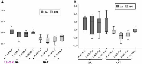
Figure 2