Electrospun Poly(lactide-co-glycolide)/Nanotube Composite ...
Upload
independentCategory
view
1download
0
This article appeared in a journal published by Elsevier. The attachedcopy is furnished to the author for internal non-commercial researchand education use, including for instruction at the authors institution
and sharing with colleagues.
Other uses, including reproduction and distribution, or selling orlicensing copies, or posting to personal, institutional or third party
websites are prohibited.
In most cases authors are permitted to post their version of thearticle (e.g. in Word or Tex form) to their personal website orinstitutional repository. Authors requiring further information
regarding Elsevier’s archiving and manuscript policies areencouraged to visit:
http://www.elsevier.com/copyright

Author's personal copy
Multifunctional hydroxyapatite and poly(D,L-lactide-co-glycolide) nanoparticles forthe local delivery of cholecalciferol
Nenad Ignjatović a, Vuk Uskoković b, Zorica Ajduković c, Dragan Uskoković a,⁎a Centre for Fine Particle Processing and Nanotechnologies, Institute of Technical Sciences of the Serbian Academy of Sciences and Arts, Knez Mihailova 35/4, 11000 Belgrade, Serbiab Therapeutic Micro and Nanotechnology Laboratory, Department of Bioengineering and Therapeutic Sciences, University of California, San Francisco, CA, USAc Clinic of Stomatology, Department of Prosthodontics, Faculty of Medicine, University of Niš, Niš, Serbia
a b s t r a c ta r t i c l e i n f o
Article history:Received 25 August 2012Received in revised form 5 October 2012Accepted 15 November 2012Available online 24 November 2012
Keywords:Multifunctional nano materialsCholecalciferolHApPLGAOsteogenesis
Cholecalciferol, vitamin D3, plays an important role in bone metabolism by regulating extracellular levels of cal-cium. Presented here is a study on the effects of the local delivery of cholecalciferol (D3) using nanoparticulatecarriers composed of hydroxyapatite (HAp) and poly(D,L-lactide-co-glycolide) (PLGA). Multifunctionalnanoparticulate HAp-based powders were prepared for the purpose of: (a) either fast or sustained, local deliv-ery of cholecalciferol, and (b) the secondary, osteoconductive and defect-filling effect of the carrier itself. Twotypes of HAp-based powders with particles of narrowly dispersed sizes in the nano range were prepared andtested in this study: HAp nanoparticles as direct cholecalciferol delivery agents and HAp nanoparticles coatedwith cholecalciferol-loaded poly(D,L)-lactide-co-glycolide (HAp/D3/PLGA).Satisfying biocompatibility of particulate systems, when incubated in contact with MC3T3-E1 osteoblasticcells in vitro, was observed for HAp/D3/PLGA and pure HAp. In contrast, an extensively fast release ofcholecalciferol from the system comprising HAp nanoparticles coated with cholecalciferol (HAp/D3) triggerednecrosis of the osteoblastic cells in vitro. Artificial defects induced in the osteoporotic bone of the rat mandiblewere successfully reconstructed following implantation of cholecalciferol-coated HAp nanoparticles as well asthose comprising HAp nanoparticles coated with cholecalciferol-loaded PLGA (HAp/D3/PLGA). The greatestlevels of enhanced angiogenesis, vascularization, osteogenesis and bone structure differentiation were achievedupon the implementation of HAp/D3/PLGA systems.
© 2012 Elsevier B.V. All rights reserved.
1. Introduction
Synthetic calcium phosphates, particularly calcium hydroxyapatite(HAp), are suitable candidates for a broad range of applications inbiomedicine, from bone reconstruction to cancer therapies [1,2].Bioresorbable polymers with comparatively slow degradation timesin physiological conditions are, on the other hand, viable carriers ofhydrophobic drugs, difficult to deliver by oral means, and are thereforethe focus of study of numerous research groups in the drug deliveryfield [3,4].
The design of particulate platforms for controlled drug delivery is ofparticular interest for the biomedical community [5]. The current at-tempts to engineer efficient local and targeted drug release agentsare mainly focused on nanoparticulate systems [6,7]. An interest inthe applications of nanoparticles of calcium phosphates as carriers intargeted and controlled drug delivery and tissue engineering is rapidlygrowing [8]. HAp nanoparticles have so far been used as carriers fordifferent pharmaceutically active components in controlled drug andgene delivery [9,10]. At the same time, a significant improvement of
the processes of bone defect reconstruction has been achieved owingto the use of nanoparticulate systems based on HAp [9].
Multifunctional nanoparticulate systems based on HAp coatedwith drug-loaded bioresorbable polymers make up a separate groupof controlled drug delivery systems in bone tissue engineering. Whilecomparatively slow bioresorption of the polymer enables sustaineddrug release, HAp particles may act as active fillers of the bony defect.One such concept has been applied in the design of HAp nanoparticlesfor the local delivery of tigecycline in the treatment of osteomyelitis[10]. Sonochemical homogenous precipitation has been used for thecontrolled synthesis of multifunctional core-shell nanospheres com-posed of a bioresorbable polymer and HAp for the controlled deliveryof clindamycin [11].
New strategies in the synthesis of multifunctional nanoparticlesmay lead to creation of orally deliverable, injectable or surgicallyimplantable platforms as carriers of both diagnostic and therapeuticagents [12]. Precipitation techniques and membrane dialysis havehelped create nanoparticulate systems for combined targeted andcontrolled drug delivery, allowing for their simultaneous diagnosticand therapeutic efficacy [13,14]. In this study, we have used precipi-tation chemistry to prepare multifunctional nanoparticulate systemsfor: (a) both rapid and sustained local delivery of cholecalciferol;
Materials Science and Engineering C 33 (2013) 943–950
⁎ Corresponding author.E-mail address: [email protected] (D. Uskoković).
0928-4931/$ – see front matter © 2012 Elsevier B.V. All rights reserved.http://dx.doi.org/10.1016/j.msec.2012.11.026
Contents lists available at SciVerse ScienceDirect
Materials Science and Engineering C
j ourna l homepage: www.e lsev ie r .com/ locate /msec

Author's personal copy
and (b) the secondary osteoconductive effect of the defect-filling HApcarrier itself.
Cholecalciferol, an important regulator of concentrations of calciumand phosphate ions in blood serum, plays a critical role in skeletalhomeostasis by controlling bone metabolism and regulating osteoblastdifferentiation, proliferation, apoptosis and expression of specific boneproteins and growth factors in the newly forming bone tissue [15,16].Two types of nanoparticulate powders with controlled particle shapes,sizes and properties were prepared in this study: HAp particles asnano-carriers of cholecalciferol (HAp/D3), and HAp nanoparticles coat-ed with cholecalciferol-loaded poly(D,L)-lactide-co-glycolide (HAp/D3/PLGA). Their synthesis was complemented with physicochemical char-acterization of structure and properties as well as the evaluation oftheir effect on osteoblastic cells and the regeneration of artificiallymade bone defects in vivo.
2. Experimental
2.1. Synthesis of materials
Aqueous calcium nitrate (Ca(NO3)2) solution (150 ml; 26.6 wt. %)was added to the solution of ammonium phosphate ((NH4)3PO4)(7 ml H3PO4+165 ml NH4OH+228 ml H2O) at 50 °C over the peri-od of 60 minutes, while stirring at the rate of 100 rpm. The solutionwas then subjected to heat treatment for 60 minutes at 100 °C[9,17]. The resulting gel was dried at room temperature in a vacuumdrier for 72 h, after which the final product — HAp powder — wasobtained.
Cholecalciferol (crystalline 99+%, Acros Organics) dissolved inacetone (0.4 wt.%) was mixed with the HAp gel in the 2:8 massratio while stirring with a magnetic stirrer at 700 rpm. Distilledwater was added drop-wise to the mixture of cholecalciferol andHAp, while stirring at 21,000 rpm (Ultra-Turrax T25, IKA, Germany).The resulting mixture was then centrifuged at 5000 rpm and 5 °Cfor 1 h (Hettich Universal 320R). The obtained powder was subjectedto frozen vacuum drying at−10 to−60 °C, under pressures between0.37 and 0.1 mbar over the periods between 1 and 8 h (Christ Alpha1-2/LD Plus). The obtained product was the powder composed ofcholecalciferol-coated HAp nanoparticles (HAp/D3). PLGA (50:50,Sigma, USA) and cholecalciferol dissolved in acetone were mixedwith the HAp gel in the mass ratio of 3:2:8, respectively. The obtainedmixture of cholecalciferol, PLGA and the HAp gel was slowly pouredinto 0.1 vol% water solution of poloxamer 188 (polyethylene-polypropylene glycol) at the speed of 21,000 rpm for one hour. Theobtained mixture was then centrifuged at 500 rpm and 5 °C for 1 h,and the obtained powder was subjected to frozen vacuum drying at−10 to −60 °C, and pressures from 0.37 mbar to 0.1 mbar for 1 to8 h. The obtained product was the powder composed of HApnanoparticles coated with cholecalciferol-loaded poly(D,L)-lactide-co-glycolide (HAp/D3/PLGA).
2.2. Characterization of the products
Roentgen structural analysis (XRD) was performed on a PhilipsPW-1050 diffractometer with Ni-filtered CuKα radiation. The scanningstep was 0.02°. Infrared spectroscopy (FTIR) was done on a BomemMB 100 (Hartmann & Braun) spectrometer using the KBr techniquein the spectral range from 400 to 4000 cm−1. The spectral resolutionwas 4 cm−1. The field-emission scanning electron microscopy(FE-SEM) measurements were performed on a SUPRA 35 VP CarlZeiss microscope. Electrokinetic parameters of the suspensions ofsynthesized particles were analyzed using a Zeta-Sizer Nano (MalvernInstruments Ltd.) in distilled water and pH 6.5. The particle size distri-bution (PSD) was measured on 10 mg/ml of powders dispersed inwater using a Mastersizer 2000 (Malvern Instruments Ltd.) and aHydroS dispersion unit for liquid dispersants.
2.3. Cell culture
Mouse calvarial preosteoblastic cell line, MC3T3-E1 subclone 4,was purchased from American Tissue Culture Collection (ATCC, Rock-ville, MD). MC3T3-E1 cells were cultured in Alpha Minimum EssentialMedium (α-MEM; Gibco) supplemented with 10% fetal bovine serum(FBS, Invitrogen) and no ascorbic acid (AA). The mediumwas replacedevery 48 h, and the cultures were incubated at 37 °C in a humidifiedatmosphere containing 5% CO2. Every 7 days, the cells were detachedfrom the surface of the 75 cm2 cell culture flask (Greiner Bio-One)using 0.25 wt.% trypsin, washed, centrifuged (1000 rpm×3 min),resuspended in 10 ml of the cell culture medium and subcultured in1:7 volume ratio. Cell passages 6–12 were used for the experimentsreported hereby. The cultures were regularly examined under anoptical microscope to monitor growth and possible contamination.
MC3T3-E1 cells were seeded on glass cover slips placed in 24 wellplates and 500 μl of α-MEM supplemented with 10% fetal bovineserum (FBS, Invitrogen) and no AA at the density of 6×10⁴ cells perwell. After 5 days of incubation, nearly confluent cells were treatedwith α-MEM containing 50 μg/ml AA as the mineralization inductor.At the same time, 2 mg/cm2 of HAp, HAp/D3 and HAp/D3/PLGAwere added to the cells, separately. The subsequent incubation lastedfor 7 days, during which α-MEM supplemented with 50 μg/ml AAwas replenished every 48 h. After the given period, one portionof cells was fixed for 15 min in 3.7 wt.% paraformaldehyde at roomtemperature and then stained for f-actin and nucleus usingphalloidin-tetramethylrhodamine (AlexaFluor 555, Invitrogen) and4′,6-diamidino-2-phenylindole dihydrochloride nuclear counterstain(DAPI, Invitrogen), following a previously described protocol [18].
The cover slips containing the fixed and stained cells were mountedonto glass slides using hard set vectashield and nail hardener and weresubsequently imaged under a confocal laser scanning microscope —
C1si (UCSF Nikon Imaging Center) at 100× magnification in oil.
2.4. In vivo tests
Bone regeneration tests were conducted on 36 rats, Wisterspecies. The animals were introduced to the experiment whenthey reached the age of six to eight weeks. They were divided intotwo groups: one group (9 animals) was the control group and theother one was the experimental group (27 animals). The experimen-tal group was treated with glucocorticoids in order to induce osteopo-rosis. The animals were given glucocorticoids–metilprednisoloneNa-succinate (Lemod-Solu, Hemofarm,Vršac, Srbija) dosed 2 mg perkg of body mass and dexamethasone-Na-phosphate (Dexason, ICNGalenika, Belgrade, Serbia) dosed 0.2 mg per kg of body mass. Thesesubstances were applied intramuscularly and alternately in thecourse of 12 weeks, so that each substance was applied every otherday. During those 12 weeks, the control group of animals receivedsaline intramuscularly on a daily basis. After the 12 week period theanimals in the control group did not exhibit any defects, whereasthe animals from the experimental group had defects introduced onthe left side of the mandible alveolar bone, in the region betweenthe medial line and foramen mentale. The defect was created using asterile steel borer, 1.6 mm in diameter and 1.8 mm in length. Theanimals had been prepared for this intervention by the application ofdiazepam (Bensendin, ICN Galenika, Belgrade, Serbia) and were subse-quently anesthetized with ketamine hydrochloride USP (Ketalar,Rotexmedica Gmbh, Trittau, Germany). The experimental group wasdivided into three subgroups with nine animals each. Rats in the firstgroup had HAp powder implanted into the defect; rats in the secondgroup had HAp/D3 powder implanted into the defect; rats in thethird group had HAp/D3/PLGA powder implanted into the defect. Thepowders delivered to of all three experimental subgroups had previ-ously been mixed with saline (0.9% NaCl solution) in the 2:1 ratio.The animals were sacrificed 6 and 24 weeks after the intervention
944 N. Ignjatović et al. / Materials Science and Engineering C 33 (2013) 943–950

Author's personal copy
because previous studies have shown that the best results can beexpected in this period [9]. After the mentioned time interval,decalcified samples of the alveolar bonewere taken, and after dehydra-tion in a series of alcohol, paraffin blocks were made, out of which wetook fragments with 10 μm in width, coloured according to H&Emethod for pathohistological analysis [9]. The procedures involvingexperimental animals were done in accordance with the Guidelinesfor Work with Experimental Animals adopted by the Ethics Committeeof the Faculty of Medicine, University of Niš, Serbia (No 01-890-3/2011).
3. Results and discussion
Fig. 1 shows the diffractograms of PLGA, HAp, cholecalciferol, HAp/D3 and HAp/D3/PLGA. No diffraction peaks are visible on thediffractogram of PLGA, as expected due to the amorphous nature ofthis polymer. The most intensive peaks of HAp (International Centerfor Diffraction Data, JCPDS file No. 09-432) are found at 31.8° (2 1 1),32.2° (1 1 2), 32.9° (3 0 0), 25.9° (0 0 2) and 49.5° (2 1 3). It is likelythat the given phase is calcium-deficient HAp, as in accordance withthe previously published results [17,19]. The mean crystallite size (D)of the powders calculated from the half-width (β1/2) of the XRD reflec-tion of the (002) plane (2θ=25.9°) using the Scherrer equation was12.5 nm. The degree of crystallinity of HAp could be facilely deter-mined from its XRD pattern [20]. Similar in appearance to that obtainedby different researchers [17,19,21], the diffractogram shown in Fig. 1corresponds mainly to poorly crystalline HAp. This result was expectedbearing in mind that the HAp gel was synthesized by precipitation at100 °C and pH 12; HAp synthesis at this temperature leads to poorlycrystalline HAp [21]. Hand-in-handwith poor crystallinity of HAp pow-ders typically goes a reduction in stoichiometric Ca/P ratios towardsvalues lower than 1.67, as observed by others [22]. Thus obtained poor-ly crystalline form of HAp bears resemblance to bone apatite and can behypothesized to present a more osteogenic crystalline form comparedto highly structured apatite obtainable by annealing at elevated tem-peratures. The high crystalline form of HAp, distinguished by narrowand sharp diffraction peaks, was obtained using a similar synthesis
procedure, but involving also additional calcination at high tempera-tures [17,21,23].
The diffractogram of cholecalciferol with the most intensive peak at18.2° suggests its existence in a crystalline form and is in agreementwith the XRD research reported elsewhere [24]. The diffractogram ofHAp/D3 powder exhibits the diffraction peaks typical for the twophases separately, confirming their mutual presence in it. Compara-tively broader diffraction peaks discernable in the XRD pattern ofHAp/D3/PLGA indicate that the structure of the organic component inthe composite sample exhibits a lesser degree of ordering comparedto its pure crystalline state.
The electrokinetic parameters of materials in aqueous suspensionindicate their stability in suspended state and thus suggest the possi-bility for their therapeutic application by injection using water as thedispersion medium. Generally, zeta potential (ZP) values of 0 to ±15 mV indicate sols prone to flocculation and aggregation, whereasabsolute ZP values higher than 15 mV are characteristic for stableand aggregation-resistant particles [25,26]. Table 1 shows the valuesof ZP, mobility and conductivity of particles in the aqueous medium.Fig. 2 shows the distribution of the ZP values for the synthesizedparticles of HAp, HAp/D3 and HAp/D3/PLGA in distilled water at25 °C and pH 6.5. ZP values are in the range of −7.5 mV for HAp to−23.6 mV for HAp/D3 to −33.1 mV for HAp/D3/PLGA. Electropho-retic mobility and conductivity values also follow the decreasingtrend in the sequence HAp>HAp/D3>HAp/D3/PLGA and confirmthe different electrodynamic nature of the particle surfaces. The pro-cess of coating of HAp nanoparticles as well as their subjugation toencapsulation of hydrophobic drugs can be confirmed by followingthe corresponding changes in electrokinetic parameters, particularlyZP [27]. The results shown in Table 1 indicate a progressive increasein the absolute values of ZP as the core HAp particles are firstcoated with cholecalciferol and then with cholecalciferol-loadedpoly(D,L)-lactide-co-glycolide. Whereas ZP of HAp particles is com-paratively low, in the order of 0 to −10 mV [28], both cholecalciferoland poly(D,L)-lactide-co-glycolide are typified by significantly morenegative ZP values, approaching −40 mV at the physiological pHfor the latter [29]. To further confirm this observation, FT-IR andFE-SEM examination were performed next.
FT-IR spectra of cholecalciferol, HAp, HAp/D3 and HAp/D3/PLGA, aswell as the magnified spectrum in Region A are shown in Fig. 3. TheFT-IR spectrum of HAp is defined by a doublet with maxima at 1035and 1092 cm−1 and by a triplet with most pronounced maxima at561 and 603 cm−1, all of which are due to vibrations of the phosphategroup. The bands with maxima at 632 and 3567 cm−1 are caused bythe vibrations of the hydroxyl group in the crystal lattice of HAp [30].Cholecalciferol is typified by the CH3 asymmetric stretching modeand the CH2 symmetric stretching mode at 2943 and 2875 cm−1, re-spectively [31,32], the two bands that are observable in our spectrumof the vitamin too. The spectrum of HAp/D3 exhibits the most intensivebands of both HAp and cholecalciferol, indicating that the synthesizedmaterial is composed of HAp and cholecalciferol, as in accordancewith the XRD results. The appearance of new bands is also noticed, es-pecially in the region marked by A. In comparison with the X-raydiffractograms of HAp/D3/PLGA, its FT-IR spectrum shows the specificbands of PLGA (the most significant one being the stretching C=O
Fig. 1. XRD diagrams of HAp, HAp/D3 and HAp/D3/PLGA.
Table 1Zeta potential, electrophoretic mobility and conductivity of HAp, HAp/D3 and HAp/D3/PLGA particles (ξ—zeta potential; ue—mobility; K-conductivity).
Sample name Temperature [°C] ξ [mV] ue [μmcm/Vs] K [10−3 mS/cm] pH
HAp 25.0 −7.5 −0.58 19.2 6.5HAp/D3 25.0 −21.6 −1.6 10.4 6.5HAp/D3/PLGA 25.0 −33.4 −2.6 5.2 6.5
945N. Ignjatović et al. / Materials Science and Engineering C 33 (2013) 943–950

Author's personal copy
vibrations at around 1760 cm−1 [33]), as well as the presence of thebands of HAp and cholecalciferol.
Phosphate groups in calcium phosphates can form hydrogen bondsin contact with vitamins from the group D [32]. The appearance of newandweak bands in spectra of HAp/D3 and HAp/D3/PLGA, in the range of
3650–3690 cm−1, may indicate the formation of the surface P-O-Hgroups originating from the protonation of the surface PO4
3− groupsof HAp [34]. The surface PO4
3− groups from HAp are prone to forminghydrogen bonds in the close presence of hydroxyl groups [35], beingthe basic mechanism bywhich hydrogen bonds between HAp and cho-lecalciferol or PLGA form. Fig. 3b shows the magnified spectra of thematerials in the region 3620–3750 cm−1 where the appearanceof new bands reflecting the formation of hydrogen bonds betweenHAp and other constituents thatmake up the particles can be observed.The weak bands at 3689 cm−1, 3675 cm−1 and 3648 cm−1 areassigned to hydrogen bonding to the surface P-OH groups of HAp.The mechanism for the formation of hydrogen bonds in HAp/D3/PLGAis more complex because the protonation of the surface groups ofHAp most likely does not originate solely from cholecalciferol, butalso from PLGA. The appearance of new and weak bands in HAp/poly-lactide nanocomposites in the range of 3650–3690 cm−1 hasthus been explained by the formation of hydrogen bonds betweenthe polymer and HAp [36]. The structure of cholecalciferol(9,10-Secocholesta-5,7,10(19)-trien-3beta-ol) suggests the existenceof spatially interconnected ―H and ―OH groups, while the structureof PLGA has spatially connected O and ―H groups in the polymerchain available to participate in the formation of hydrogen bondswith the surface groups of HAp.
FE-SEM images of HAp, HAp/D3 and HAp/D3/PLGA are shown inFig. 4. The particles of HAp display a rod-like morphology (Fig. 4a).After the coating of HAp with cholecalciferol (Fig. 4b), more roundparticle shapes were obtained compared to pure HAp (Fig. 4a).HAp/D3/PLGA particles had spherical morphologies too (Fig. 4c).Additional processing in the centrifugal field of the suspension ofnanoparticles of HAp coated with PLGA has previously led to the for-mation of spherical morphologies [37,38]; accordingly, since we usedcentrifugal processing too in the processing method reported herein,spherical morphologies were obtained as well.
Aqueous suspensions of the particles of HAp, HAp/D3 and HAp/D3/PLGA were analyzed with the aim of establishing their size distribu-tions. Fig. 5 shows the size distributions for the suspended particlesof HAp, HAp/D3 and HAp/D3/PLGA powders in distilled water. Theparticle sizes for all the analyzed powders range from 30 nm to1 μm. HAp particles (Fig. 5a) have d10=35 nm; d50=65 nm andd90=124 nm. HAp/D3 system has a distribution with the parame-ters d10=36 nm, d50=68 nm and d90=143 nm, and HAp/D3/PLGA d10=37 nm, d50=71 nm and d90=168 nm (Fig. 5b–c). Ageneral trend towards increasing the average particle size (d50)with the addition of a new component can be noticed; hence, the se-quence HAp>HAp/D3>HAp/D3/PLGA with respect to d50, d10 andd90. The largest particles of 1.5 μm were detected in HAp/D3/PLGAand comprised 0.01% of the overall particle number. The smallestones, measuring 22 nm in size, were detected in HAp (0.5% of thetotal particle count).
A consistent increase in the particle size (HAp>HAp/D3>HAp/D3/PLGA) is expected as the result of the coating process. The coatingof HAp particles with cholecalciferol thus leads to an increasein their size, while coating of HAp particles with a polymer inwhich the vitamin was immobilized leads to a further increase insize. The procedure for coating HAp particles with cholecalciferol(or cholecalciferol-loaded PLGA) takes place by adding a non-solventto induce the precipitation of cholecalciferol (or cholecalciferol-loadedPLGA) with the existing HAp particles acting as sedimentation centres.In our earlier research, we examined the possibility of coating themicroparticles of HAp with PLGA by the solvent/non-solvent methodin which HAp particles were the sedimentation centres for PLGA[23]. For the abovementioned reasons, the size increase of particlesafter coating HAp with cholecalciferol or the polymer is expected. Thethickness of the coated layer chiefly depends on the mass proportionof cholecalciferol and PLGA relative to the quantity of the initialHAp particles.
Fig. 2. Zeta potential distribution for HAp, HAp/D3 and HAp/D3/PLGA particles.
Fig. 3. FT-IR spectra of cholecalciferol, HAp, HAp/D3 and HAp/D3/PLGA (a), magnifiedspectra in Region A (b).
946 N. Ignjatović et al. / Materials Science and Engineering C 33 (2013) 943–950

Author's personal copy
Fig. 6 displays the confocal optical images of osteoblastic cellsfollowing incubation with the three types of particles prepared andanalyzed in this study, fluorescently counterstained for nucleus(blue) and f-actin cytoskeletal microfilaments (red). It is apparentthat while the viability of cells incubated in the presence of either
pure HAp or HAp/D3/PLGA does not show any diminishment northe healthy morphology of cells seems to be lost when comparedto the control, cells incubated with HAp/D3 appear morphologicallydeformed, necrotic and exhibiting a significantly lower cell densityin comparison with the control sample. An extensive amount of cho-lecalciferol released into the cell medium has thus been shown to
Fig. 4. SEM micrographs of HAp (a), HAp/D3 (b), HAp/D3/PLGA (c).
Fig. 5. Particle size distributions of HAp (a), HAp/D3 (b), HAp/D3/PLGA (c).
947N. Ignjatović et al. / Materials Science and Engineering C 33 (2013) 943–950

Author's personal copy
have a drastic effect on reducing the cell viability. A possible expla-nation for this effect may relate to the already observed abilityof calcitriol, the bioactive form of vitamin D3 derivative, to force the re-lease of RANKL, a tumor necrosis factor cytokine, which stops the boneformation process and activates osteoclasts, the bone-resorbing cells[39].
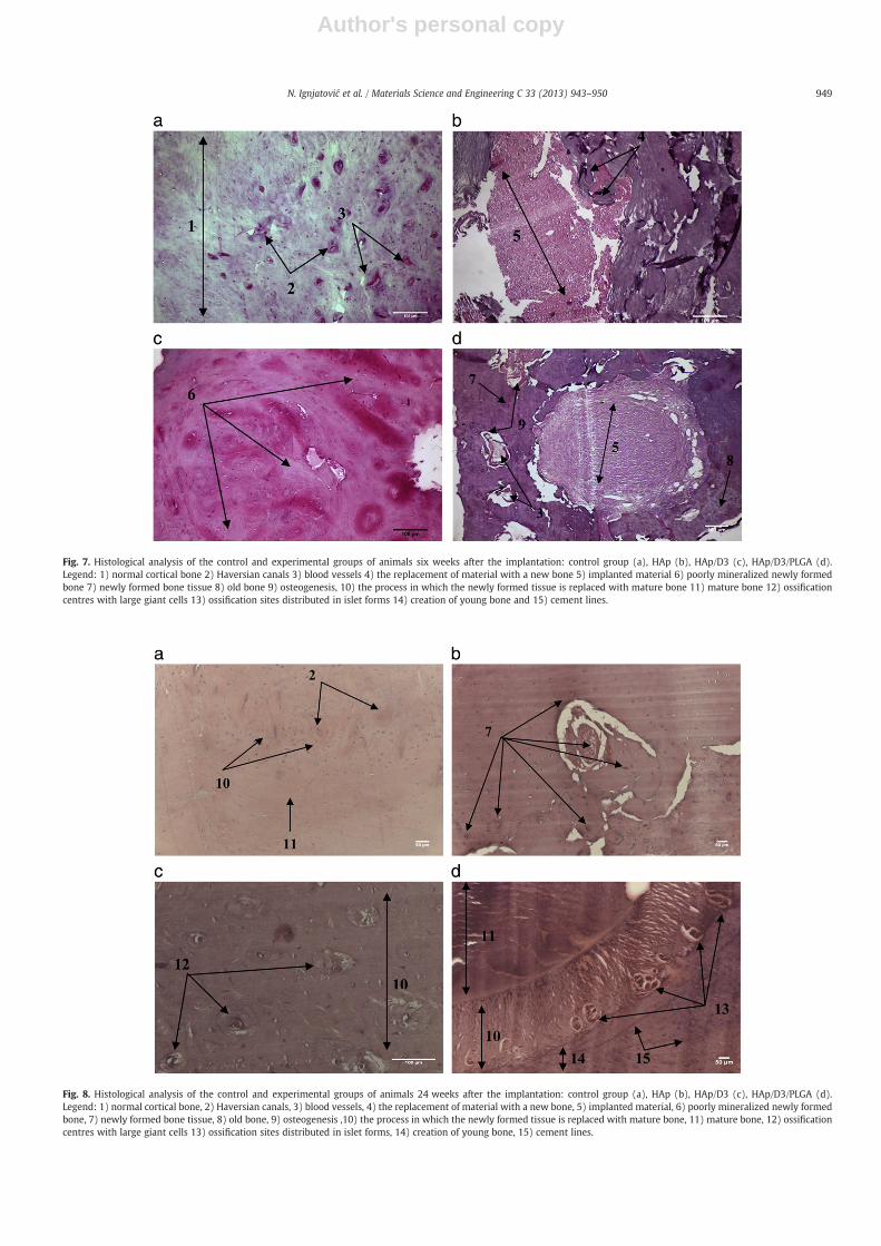
Having a role in mitosis and differentiation of osteoblasts, it wasnatural to expect that cholecalciferol would have an effect on osteogen-esis. So far, no results of in vivo analyses related to multifunctionalnanoparticulate systems for local delivery of the vitamins from the Dgroup could be found in literature. Figs. 7 and 8 show histopathologicalimages of the mandible bone 6 and 24 weeks after the implantationof different materials has been carried out, respectively. Fig. 7a of thecontrol group, in which no defect has been made, shows mature boneas well as blood vessels and Haversian canals. The new bone withremains of ossification centres can be seen in Fig. 7b. Fig. 7c showsnumerous ossification centres with giant cells which are not distribut-ed in a palisade, but scattered in a disordered manner. Specific forms ofossification distributed in islet-like formations, the creation of youngbone, i.e., accelerated activity of osteogenesis cells, cement lines andmature bone can be seen in Fig. 7d.
Compared to the cortical bone of the control group 24 weeks afterthe implantation, healing in the presence of any of the materials
synthesized within this study seems to be facilitated. After the defectshave been filled with HAp, the resorption of the implanted materialand its replacement by the newly formed bone are observed (Fig. 7b).After the implantation of the HAp/D3, patches of poorly mineralizednew hard tissue become visible too (Fig. 7c).
Six weeks after the reconstruction of the defect with HAp/D3/PLGA(Fig. 8d), newly formed bone around the defect was visible as well ascement lines and the blood supply. In the centre of the defect, a pieceof the implanted material with ray-like zones where the material wasreplaced with the newly formed, poorly mineralized bone tissuecan be observed. Intensive angiogenesis is present, as well as vascu-larization and osteogenesis. Vascularization is essential for successfulregeneration of bone, as confirmed in other studies [40].
Twenty-four weeks after the reconstruction of the bone defect, theformation of new bone tissue has additionally improved after the appli-cation of all types of materials. After the reconstruction with HAp/D3
(Fig. 8c), numerous ossification centres were visible, along with thegiant cells. The transition of young bone tissue into mature tissue is ob-servable in all the reconstructed defects. Following the period of inten-sive angiogenesis and vascularization (Fig. 7d), the most intensivedifferentiation and osteogenesis were seen when HAp/D3/PLGA wasused as the filler (Fig. 8d). In the central area of the image, a broadline of young bone tissue transforming into mature tissue is visible,
Fig. 6. Confocal optical micrographs of osteoblastic cells incubated with no particles (a), with HAp (b), with HAp/D3 (c), or with HAp/D3/PLGA (d), fluorescently stained for nucleus(blue) and cytoskeletal f-actin (red) following 4 days of incubation. The size of each image is 300×300 μm.
948 N. Ignjatović et al. / Materials Science and Engineering C 33 (2013) 943–950
Author's personal copy
Fig. 7. Histological analysis of the control and experimental groups of animals six weeks after the implantation: control group (a), HAp (b), HAp/D3 (c), HAp/D3/PLGA (d).Legend: 1) normal cortical bone 2) Haversian canals 3) blood vessels 4) the replacement of material with a new bone 5) implanted material 6) poorly mineralized newly formedbone 7) newly formed bone tissue 8) old bone 9) osteogenesis, 10) the process in which the newly formed tissue is replaced with mature bone 11) mature bone 12) ossificationcentres with large giant cells 13) ossification sites distributed in islet forms 14) creation of young bone and 15) cement lines.
Fig. 8. Histological analysis of the control and experimental groups of animals 24 weeks after the implantation: control group (a), HAp (b), HAp/D3 (c), HAp/D3/PLGA (d).Legend: 1) normal cortical bone, 2) Haversian canals, 3) blood vessels, 4) the replacement of material with a new bone, 5) implanted material, 6) poorly mineralized newly formedbone, 7) newly formed bone tissue, 8) old bone, 9) osteogenesis ,10) the process in which the newly formed tissue is replaced with mature bone, 11) mature bone, 12) ossificationcentres with large giant cells 13) ossification sites distributed in islet forms, 14) creation of young bone, 15) cement lines.
949N. Ignjatović et al. / Materials Science and Engineering C 33 (2013) 943–950

Author's personal copy
interspersed with several islet-like ossification centres. Moving fromright to left in Fig. 8d, represented is the transition from young boneto mature bone.
To become bioactive, cholecalciferol has to be hydroxylated twice:first in the liver and then by the convoluted tubule cells of the kidney.Its direct delivery to cells in its raw, naturally found form and in overlyhigh fluxes apparently has a negative effect on osteoblastic cells whichundergo necrosis under such conditions. The positive effects on theorthodontic stability of teeth in the treatment of parodontopathyhas been noted previously upon the local delivery of calcitriol, that is,1,25-dihydroxycholecalciferol, the bioactive form of cholecalciferol[41,42]. In those studies, the activation effect of vitamin D on mono-cytes was shown, as well as the stimulating effect on cell-mediatedimmunity with the suppression of lymphocytic proliferation [41]. Thelocal use of calcitriol significantly increased the activity of osteoblasts,which resulted in an improved recovery of alveolar bone [42]. In thefuture studies, we will extend our investigation of the local deliveryof vitamin D family of compounds to calcitriol.
4. Conclusion
Development of a multifunctional nanoparticulate material forsimultaneous (a) acceleration of natural bone regeneration and(b) sustained drug release, composed of hydroxyapatite and poly-(DL-lactide-co-glycolide), presented the subject of this study. In thiswork, we prepared two different osteoconductive carriers for thelocal delivery of cholecalciferol: pulse (HAp/D3) and sustained (HAp/D3/PLGA). The fast delivery was achieved by desorption of the drugfrom the HAp particle surface, while the slow delivery was conditionedby the rather slow degradation of PLGA in physiological conditions.Although the former way of delivery had a highly negative effect on os-teoblastic cells in vitro, it did not hinder the healing of osteoporoticbone in vivo. An important feature of these nanoparticulate carriers istheir multifunctionality. Namely, after the drug has been delivered,HAp could take the role of the defect filler that is to be graduallysubstituted with the new bone.
Acknowledgements
The authors acknowledge Dr. Vladimir Srdić of the Faculty ofTechnical Sciences, University of Novi Sad for his assistance in zeta po-tential measurements, Dr. Saša Drmanić of the Faculty of Technologyand Metallurgy, University of Belgrade, for his help in IR measure-ments, and Dr. S. Škapin of Jozef Stefan Institute, Slovenia, for theFE-SEM imaging. The research presented in this paper was supportedby the Ministry of Education and Science of the Republic of Serbia,under project no. III45004 and the US National Health Institute grantK99-DE021416.
References
[1] L. Hench, J. Am. Ceram. Soc. 74 (1991) 1487–1510.[2] M. Vallet-Regí, E. Ruiz-Hernández, Adv. Mater. 23 (2011) 5177–5218.[3] H. Naderi, M. Matin, A. Bahrami, J. Biomater. Appl. 26 (2011) 383–417.[4] R. Gref, Y. Minamitake, M. Peracchia, V. Trubetskoy, V. Torchilin, R. Langer,
Science 263 (1994) 1600–1603.[5] N. Kolishetti, Sh. Dhar, P. Valencia, L. Lin, R. Karnik, S. Lippard, R. Langer, O.
Farokhzad, Proc. Natl. Acad. Sci. U. S. A. 107 (2010) 17939–17944.[6] D. Depan, R.D.K. Misra, Mater. Sci. Eng. C Mater. Biol. Appl. 32 (2012) 1704–1709.[7] Z. Zhang, G. Huang, J. Nanotechnol. (2012), http://dx.doi.org/10.1155/2012/748909.[8] S. Bose, S. Tarafder, Acta Biomater. 8 (2012) 1401–1421.[9] N. Ignjatović, Z. Ajduković, V. Savić, D. Uskoković, J. Biomed. Mater. Res. B Appl.
Biomater. 94 (2010) 108–117.[10] N. Ignjatović, P. Ninkov, R. Sabetrasekh, D. Uskoković, J. Mater. Sci. Mater. Med. 21
(2010) 231–239.[11] M. Vukomanović, S.D. Škapin, I. Poljanšek, E. Žagar, B. Kralj, N. Ignjatović, D.
Uskoković, Colloids Surf. B Biointerfaces 82 (2011) 414–421.[12] S. Parveen, R. Misra, S. Sahoo, Nanomedicine 8 (2012) 147–166.[13] X. Li, Y. Qian, T. Liu, X. Hu, G. Zhang, Y. You, Sh. Liu, Biomaterials 32 (2011)
6595–6605.[14] K. Kim, J. Kim, H. Park, Y.-S. Kim, K. Park, H. Nam, S. Lee, J. Park, R.-W. Park, I.-S.
Kim, K. Choi, S. Kim, K. Park, I. Kwon, J. Control. Release 146 (2010) 219–227.[15] M. van Driel, H. Pols, J. van Leeuwen, Curr. Pharm. Des. 10 (2004) 2535–2555.[16] G. Jones, Am. J. Clin. Nutr. 88 (2008) 582S–586S.[17] N.L. Ignjatović, C.Z. Liu, J.T. Czernuszka, D.P. Uskoković, Acta Biomater. 3 (2007)
927–935.[18] V.Uskoković, P. Lee, L.Walsh, K. Fischer, T. Desai, Biomaterials 33 (2012) 1663–1672.[19] E. Caroline Victoria, F.D. Gnanam, Trends Biomater. Artif. Organs 16 (2002)
12–14.[20] L. Keller, W. Dollase, J. Biomed. Mater. Res. 49 (2000) 244–249.[21] P. Kumta, C. Sfeir, D. Lee, D. Olton, D. Choi, Acta Biomater. 1 (2005) 65–83.[22] R. LeGeros, S. Lin, R. Rohanizadeh, D. Mijares, J. LeGeros, J. Mater. Sci. Mater. Med.
14 (2003) 201–209.[23] N. Ignjatovic, Z. Ajdukovic, D. Uskokovic, J. Mater. Sci. Mater. Med. 16 (2005) 621–626.[24] X. Fei, J. Heyang, Z. Yaping, G. Xinqiu, Chin. J. Chem. Eng. 19 (2011) 1039–1046.[25] R. Xu, Particuology 6 (2008) 112–115.[26] V. Uskoković, Dynamic light scattering and microelectrophoresis: main prospects and
limitations, J. Dispers. Sci. Technol. (2012), http://dx.doi.org/10.1080/01932691.2011.625523.
[27] Q. Xu, Y. Tanaka, J. Czernuszka, Biomaterials 28 (2007) 2687–2694.[28] V. Uskoković, D. Uskoković, J. Biomed. Mater. Res. B Appl. Biomater. 96B (2011)
152–191.[29] Y. Patil, J. Panyam, Int. J. Pharm. 367 (2009) 195–203.[30] N. Ignjatović, D. Uskoković, Spectroscopy 18 (2004) 553–565.[31] W. Morris, J. Wilkie, S. Jones, L. Friedman, Anal. Chem. 34 (1962) 381–384.[32] N. Toyran, F. Severcan, Spectroscopy 16 (2002) 399–408.[33] T. Paragkumar, D. Edith, S. Jean-Luc, Appl. Surf. Sci. 253 (2006) 2758–2764.[34] T. Ishikawa, M. Wakamura, S. Kondo, Langmuir 5 (1989) 144–147.[35] L. Borum-Nicholas, O. Wilson Jr., Biomaterials 24 (2003) 3671–3679.[36] Sh. Zhou, X. Zheng, X. Yu, J. Wang, J. Weng, X. Li, B. Feng, M. Yin, Chem. Mater. 19
(2007) 247–253.[37] M. Vukomanović, S.D. Škapin, B. Jančar, T. Maksin, N. Ignjatović, V. Uskoković, D.
Uskoković, Colloids Surf. B Biointerfaces 82 (2011) 404–413.[38] M. Vukomanović, T. Zavašnik-Bergant, I. Bračko, V. Radmilović, S. Davor Škapin, N.
Ignjatović, D. Uskoković, Colloids Surf. B Biointerfaces 87 (2011) 226–235.[39] R. Bringhurst, M. Demay, S. Krane, H. Kronenberg, in: A. Fauci, E. Braunwald, D.
Kasper, S. Hauser, D. Longo, J. Jameson, J. Loscalzo (Eds.), Harrison's Principlesof Internal Medicine, 17th edition, McGraw-Hill, New York, 2008, (Chapter 346).
[40] A. Das, E. Botchwey, Tissue Eng. Part B Rev. 17 (2011) 403–414.[41] Y. Tachi, H. Shimpuku, Y. Nosaka, T. Kawamura, M. Shinohara, M. Ueda, H. Imai, K.
Ohura, Life Sci. 73 (2003) 3313–3321.[42] M. Kawakami, T. Takano-Yamamoto, J. Bone Miner. Metab. 22 (2004) 541–546.
950 N. Ignjatović et al. / Materials Science and Engineering C 33 (2013) 943–950
Copyright © 2022 FDOKUMEN