Structure of fumarate reductase from Wolinella succinogenes at 2.2 Å resolution

Release Characteristics and Osteogenic Activity of RecombinantHuman Bone Morphogenetic Protein-2 grafted to Novel Self-Assembled Poly(lactide-co-glycolide fumarate) Nanoparticles
Angel E Mercado, Junyu Ma, Xuezhong He, and Esmaiel Jabbari, PhDUniversity of South Carolina
AbstractFunctionalized biodegradable nanoparticles (NPs) provide reactive groups and large surface area forgrafting Recombinant human bone morphogenetic protein-2 (rhBMP-2) to reduce protein diffusionand maintain sufficient concentration for recruitment and differentiation of osteoprogenitor cells.The objective of this work was to investigate release characteristics and osteogenic activity ofrhBMP-2, grafted to biodegradable NPs based on succinimide-terminated poly(lactide fumarate)(PLAF-NHS) and poly(lactide-co-glycolide fumarate) (PLGF-NHS) macromers. The release ofrhBMP-2 from the NPs, measured by enzyme-linked immunosorbent assay, was linear with time inthe first two weeks, and 24.70±1.30% and 48.7±0.7% of the protein grafted to PLGF-NHS and PLAF-NHS NPs, respectively, was released in the enzymatically active conformation after completedegradation/erosion of the NPs. After 14 days of incubation with bone marrow stromal (BMS) cells,rhBMP-2 grafted to PLAF-NHS and PLGF-NHS NPs was as effective in inducing mineralization asthe native rhBMP-2 that was directly added to the cell culture media. At any incubation time,rhBMP-2 grafted to PLAF had the highest expression of osteopontin (OP) and osteocalcin (OC),followed by rhBMP-2 grafted to PLGF and rhBMP-2 directly added to media. Higher OP and OCexpression for BMP-gPLAF and BMP-gPLGF groups may be related to other factors in the cascadeof osteogenesis, such as differentiation of BMS cells to the vasculogenic lineage and formation of avascularized/mineralized marix.
1. IntroductionThere are approximately 0.5 million fractures in the US annually that require bone graftprocedures to ensure rapid skeletal repair [1]. Bone morphogenetic proteins (BMPs) play amajor role in initiating the cascade of chemotaxis, differentiation of marrow stromal cells, andbone regeneration [2]. In particular, recombinant human bone morphogenetic protein-2(rhBMP-2) is a potent differentiation factor that is capable of inducing bone formationfollowing implantation and is used clinically for spinal fusion [3]. BMP signaling is highlyregulated in-vivo [4]. Therefore, 4–5 orders of magnitude higher than the amount foundendogenously (1 mg/ml for rhBMP-2) have to be loaded in the graft to induce bone formation[5]. High doses, coupled with diffusion of rhBMP-2 away from the intended site ofregeneration, cause adverse effects such as bone overgrowth and immunological reactions[6]. Furthermore, the rate at which rhBMP-2 is released from the carrier can affect the efficacyof bone induction [7]. The optimum release profile for rhBMP-2 is not known, but sustained
Publisher's Disclaimer: This is a PDF file of an unedited manuscript that has been accepted for publication. As a service to our customerswe are providing this early version of the manuscript. The manuscript will undergo copyediting, typesetting, and review of the resultingproof before it is published in its final citable form. Please note that during the production process errors may be discovered which couldaffect the content, and all legal disclaimers that apply to the journal pertain.
NIH Public AccessAuthor ManuscriptJ Control Release. Author manuscript; available in PMC 2010 December 3.
Published in final edited form as:J Control Release. 2009 December 3; 140(2): 148–156. doi:10.1016/j.jconrel.2009.08.009.
NIH
-PA Author Manuscript
NIH
-PA Author Manuscript
NIH
-PA Author Manuscript

release of rhBMP-2 in-vivo over 4 weeks induced bone formation to a higher extent comparedto the same amount released in 3 days [7].
A composite poly(dl-lactic-co-glycolic acid)/calcium phosphate (CaP) cement has been usedas a matrix for sustained release of rhBMP-2, but due to interaction of the protein with CaP, alarge fraction of rhBMP-2 (50–75%) was not released from the matrix after 28 days [8]. Inanother study, titanium discs coated with rhBMP-2 incorporated poly(D,L-lactide) (PDLLA)did not induce ectopic bone formation when implanted in sheep muscle, possibly due to thevery slow release or deactivation during the coating process [9]. Hydrogel microparticles (MPs)based on dextran, functionalized with carboxylate, sulfate and benzylamide groups, were usedas a carrier for rhBMP-2 but >60% of the protein was released from the MPs in the first 24 hfollowed by a very slow (insufficient) release rate for 4 weeks [10]. Gelatin MPs crosslinkedwith glutaraldehyde were also used as a carrier for rhBMP-2, but <25% of the encapsulatedprotein was released after 28 days in collagenase-free media [11]. Glycidyl methacrylateddextran/gelatin MPs can slowly release rhBMP-2 over 4 weeks, but these MPs are limited bytheir low loading capacity (rhBMP-2 is loaded by swelling of the MPs) [12]. When rhBMP-2was encapsulated in PLGA NPs, burst release was >50% for low molecular weight (MW)PLGA while <20% of the protein was released after 8 weeks with high MW PLGA [13].Encapsulation in micro/nano particles and fibers has been used to reduce diffusion of rhBMP-2away from the application site and to reduce its in-vivo enzymatic degradation [12–16].Although encapsulated rhBMP-2 is shown to enhance mineralization and bone formation[17], a large fraction of the protein is deactivated in the process of emulsification in organicsolvents and solidification with isopropanol, and the release profile is not optimal [18,19].Therefore, relatively high doses have to be loaded in micro/nano particles which affect thesafety profile of rhBMP-2 in clinical applications [6,20]. Functionalized nanoparticles (NPs)provide large surface area and reactive groups for grafting proteins to the NPs. Graftingreactions can be carried out in aqueous media, thus reducing protein deactivation due exposureto organic solvents.
Our laboratory has developed novel poly(lactide-co-glycolide fumarate) (PLGF), and poly(lactide-co-ethylene oxide fumarate) (PLEOF) unsaturated macromers that self-assemble toform biodegradable NPs [21]. In the process of NPs formation, biodegradable PLEOFmacromer acts as a surfactant to stabilize the NPs. NPs ranging 50–500 nm in size can beproduced by varying the ratio of PLEOF to PLGF in the blend. The macromers can befunctionalized with succinimide groups for grafting proteins to the NPs in aqueous media[22]. The objective of this work was to investigate the release characteristics and osteogenicactivity of rhBMP-2, grafted to self-assembled biodegradable NPs. In this work, PLGFmacromer is reacted with N,N′-disuccinimidyl carbonate (DSC) to produce succinimide-terminated PLGF-NHS macromer. Next, PLGF-NHS and PLEOF blend macromers are self-assembled by dialysis to produce NPs. Then, rhBMP-2 is grafted to the NPs in aqueous solutionby the reaction between amine groups of the protein with PLGF-NHS succinimide groups onthe NPs. Release characteristics of rhBMP-2 from the NPs was measured by Enzyme-LinkedImmunosorbent Assay (ELISA). Osteogenic activity of the grafted rhBMP-2 was determinedby incubation with bone marrow stromal cells.
2. Methods and materials2.1. Materials
L-lactide (LA; >99.5% purity) and glycolide (GL; >99.0% purity) monomers were obtainedfrom Ortec (Easley, SC) and Boehringer Chemicals (Ingelheim, Germany), respectively. LAand GL monomers were dried under vacuum at 40°C for at least 12 h before the reaction. Poly(ethylene glycol) (PEG, nominal molecular weight 3.4 kDa), triethylamine (TEA), tin (II) 2-ethylhexanoate (TOC), and dimethylsulfoxide (DMSO) were purchased from Aldrich (Sigma-
Mercado et al. Page 2
J Control Release. Author manuscript; available in PMC 2010 December 3.
NIH
-PA Author Manuscript
NIH
-PA Author Manuscript
NIH
-PA Author Manuscript

Aldrich, St. Louis, MO). PEG was dried by azeotropic distillation from toluene. Fumarylchloride (FC) was obtained from Aldrich and distilled at 161°C prior to use. Diethylene glycolinitiator (DEG; >99% purity) was purchased from Fisher (Pittsburgh, PA). N,N′-disuccinimidylcarbonate (DSC) was obtained from Novabiochem (EMD Biosciences, San Diego, CA).Dichloromethane (DCM), dimethylformamide (DMF), tetrahydrofuran (THF), diethyl ether,and hexane were purchased from Acros (Fairfield, OH). DCM was dried by distillation overcalcium hydride. Spectro/Por dialysis tube (molecular weight cut-off 3.5 kDa) was purchasedfrom Spectrum Laboratories (Rancho Dominquez, CA). All solvents (except for DCM) wereused as received. Ethylenediaminetetraacetic acid disodium salt (EDTA), penicillin,streptomycin, Alizarin red, and paraformaldehyde were purchased from Sigma (St. Louis,MO). Dulbecco’s phosphate-buffered saline (PBS) and Dulbecco’s Modified Eagle’s Medium(DMEM; 4.5 g/L glucose with L-glutamine and without sodium pyruvate) were obtained fromGIBCO BRL (Grand Island, NY). Trypsin and fetal bovine serum (FBS, screened forcompatibility with rat bone marrow stromal cells) were obtained from Invitrogen (Carlsbad,CA) and Atlas Biologicals (Fort Collins, CO), respectively. Quant-it PicoGreen dsDNAreagent kit was obtained from Molecular Probes (Eugene, OR). QuantiChrom calcium assayand QuantiChrom alkaline phosphatase assay kits were purchased from Bioassay Systems(Hayward, CA). rhBMP-2 solution (100 μL, 1.5 mg/mL in rhBMP-2 buffer) was generouslydonated by Medtronic (Minneapolis, MN). Concentration of rhBMP-2 was measured byELISA using the BMP Quantikine kit (R&D Systems, Minneapolis, MN).
2.2. PLGF and PLEOF Macromer synthesis and succinimide terminationUltra-low molecular weight poly(lactide) (ULMW-PLA) and poly(lactide-co-glycolide)(ULMW-PLGA) were synthesized by ring-opening polymerization of LA (100%) and LA+GL(50% LA and 50% GL) monomers, respectively, as previously described [23]. , andPDI of ULMW-PLA macromer was 1450 Da, 1730 Da, and 1.2, respectively, and those ofULMW-PLGA was 1660 Da, 2150 Da, and 1.3. Next, PLAF or PLGF was synthesized bycondensation polymerization of ULMW-PLA or PLGA with FC, as previously described[21,24]. Similarly, PLEOF was synthesized by reacting ULMW-PLA and PEG with FC [25–27]. The molar ratios of FC:(PEG+PLA) and TEA:(PEG+PLA) were 0.9:1.0 and 1.8:1.0,respectively. Succinimide-terminated PLAF-NHS/PLGF-NHS macromers were produced byreacting the hydroxyl end-groups of PLAF/PLGF macromers with DSC, as previouslydescribed [28]. Briefly, 800 mg PLAF or PLGF and 26 mg DSC were mixed in 15 ml DMFin a reaction flask. After purging with nitrogen, 40 μL TEA was added while stirring, and thereaction was allowed to continue for 8 h at ambient conditions. The synthesized macromerswere characterized by 1H-NMR and GPC [23,27].
2.3. Characterization of the macromersThe chemical structure of the synthesized macromers was characterized by a VarianMercury-300 1H-NMR (Palo Alto, CA). The macromer was dissolved in deuterated chloroform(Aldrich, 99.8% deuterated; 50 mg/mL) and 1% v/v trimethylsilane (TMS; Aldrich) was usedas the internal standard. The molecular weight distribution of the macromers was measured byGPC [23]. Measurements were carried out with a Waters 717 Plus Autosampler GPC system(Waters, Milford, MA). The columns consisted of a styragel HT guard column (7.8 mm × 300mm; Waters) in series with a styragel HR 4E column (7.8 mm × 300 mm, Waters) heated to37°C. The Empower software (Waters) was used for determination of , and PDI. Thesample (20 μl; 10 mg/mL in THF) was eluted at 1 mL/min. Monodisperse polystyrene standards(PDI<1.1; Waters) were used to construct the calibration curve.
Mercado et al. Page 3
J Control Release. Author manuscript; available in PMC 2010 December 3.
NIH
-PA Author Manuscript
NIH
-PA Author Manuscript
NIH
-PA Author Manuscript

2.4. Nanoparticle formation and rhBMP-2 graftingMixtures of PLAF–NHS (or PLGF–NHS) and PLEOF macromers in DMF/DMSO solventmixture were self-assembled into NPs by dialysis against PBS buffer, as shown in Figure 1.Briefly, 45 mg PLAF–NHS (or PLGF–NHS) and 5 mg 90/10 PLEOF, dissolved in a mixtureof 1 mL DMF and 8 mL DMSO, were loaded in the dialysis tube (molecular cutoff: 3.5 kDa)and dialyzed against sterile PBS. The macromer solution was dialyzed for 24 h with changeof dialysis buffer every 2–4 h until DMSO and DMF were removed. Next, the NPs suspensionwas collected from the dialysis tube and freeze-dried to obtain a free flowing powder. Forgrafting, 2 ml of the suspension (10 mg NPs) was centrifuged at 18,350 rcf (15,000 rpm) for10 min, the supernatant was decanted, and the precipitated NPs were re-suspended in 0.5 mlPBS by sonication for 1 min with a 3-mm probe Ultrasonic Processor (Cole-Parmer, VernonHills, IL). Next, 0.5 ml rhBMP-2 in PBS solution (400 ng/mL) was added to the suspension,and the protein was allowed to react with succinimide terminated NPs under ambient conditionsfor 12 h (see Figure 1). After reaction, the suspension was dialyzed against PBS to remove theby-product, N-hydroxy succinimide.
2.5. Characterization of NPsMorphology and size distribution of the NPs was examined using a JSM-5400 scanningelectron microscope (JOEL, Japan) at an accelerating voltage of 20 KeV. Freeze-dried NPswere placed on a graphite surface and coated with gold using an Ion Sputter Coater (JOEL,JFC-1100) at 20 mA for 1 min. Size distribution of the NPs was measured by dynamic lightscattering with a NICOMP Submicron Particle Sizer (Model 370, NICOMP, Santa Barbara,CA). 500 μl of the diluted suspension was added to a culture tube and placed in the instrumentcell holder. The scattered light intensity was inverted to size distribution by inverse Laplacetransform using the NICOMP CW370 software.
2.6. Degradation kinetics of NPsThe NPs suspension was centrifuged at 18,350 rcf for 10 min, the supernatant was decantedto remove the unassembled macromers, and the NPs were re-suspended in PBS. Fordegradation experiments, 50 mg NPs were suspended in 1 ml PBS and the suspensions wereincubated at 37°C until complete degradation (no mass remaining or no NPs detected by lightscattering). At each time point, size distribution of the NPs was measured by dynamic lightscattering. Next, samples were freeze-dried and mass of the dried powder was measured. Thefraction of mass remaining was determined by dividing the dried mass at time t by the initialmass.
2.7. Grafting efficiency and release kinetics of rhBMP-2-loaded NPsSince molecular weight cutoff of the dialysis membrane was lower than rhBMP-2 molecularweight (12 kDa for rhBMP-2 versus 3500 Da membrane cutoff molecular weight; [29]),ungrafted rhBMP-2 was not removed by dialysis. To determine grafting efficiency, the dialyzedsuspension was centrifuged at 18,350 rcf for 10 min, and the enzymatically active concentrationof rhBMP-2 in the supernatant was measured by ELISA using the BMP Quantikine kitaccording to manufacturer’s instructions. After the addition of chromogen and assay stopsolution, the absorbance was measured with a plate reader (Synergy HT, Bio-Tek) at twowavelengths of 450 and 570 nm. The values at 570 were subtracted from those at 450 nm tocorrect for background absorbance. The corrected absorbance values were related toconcentration using a calibration curve constructed from the absorbance of solutions withknown rhBMP-2 concentration. Grafting efficiency was determined by dividing the measuredamount of rhBMP-2 by the initial amount in the grafting reaction. For determination of releasecharacteristics, the precipitate was resuspended and incubated in 1 ml PBS at 37°C with orbitalshaking. At each time point, suspension was centrifuged at 18350 rcf for 10 min, supernatant
Mercado et al. Page 4
J Control Release. Author manuscript; available in PMC 2010 December 3.
NIH
-PA Author Manuscript
NIH
-PA Author Manuscript
NIH
-PA Author Manuscript
was removed and stored in microvials for analysis. The fraction of released rhBMP-2 wasdetermined by dividing the measured amount of rhBMP-2 at each time point to the total amountat time zero (after grafting).
2.8. Bone marrow stromal cell isolationBMS cells were isolated from the bone marrow of young adult male Wistar rats according toestablished protocols [27,30]. Cell isolations were performed under a protocol approved by theInstitutional Animal Care and Use Committee of the University of South Carolina. Afterflushing the bone marrow, cell suspensions were centrifuged at 200×g for 5 min, the cell pelletswere re-suspended in primary media (DMEM supplemented with 10% FBS, 100 units/mLpenicillin (PEN), 100 μg/mL streptomycin (SP), 50 μg/mL gentamicin sulfate (GS), and 250ng/mL fungizone (FZ)) and maintained in a humidified 5% CO2 incubator at 37°C. Cultureswere replaced with fresh media at 3 and 7 days to remove haematopoietic and other unattachedcells. After 10 days, cells were detached from the flasks with 0.05% trypsin-0.53 mM EDTAand used for in-vitro osteogenic experiments.
2.9. In vitro osteogenic activity of BMS cells incubated with rhBMP-2 grafted NPsThe procedure for determination of osteogenic activity of rhBMP-2 grafted to NPs isdemonstrated schematically in Figure 2. BMS cells (Figure 2b) isolated from rats (Figure 2a),were seeded in 24-well plates at a density of 5×10⁴ cells/mL in primary media. After 24 h forcell attachment (time zero), media was replaced with standard osteogenic media (primarymedia supplemented with 100 nM dexamethasone (DEX), 50μg/mL ascorbic acid (AA), 10mMβ-glycerophosphate (βGP)) with the addition of time-released rhBMP-2 from a suspension of200 ng/mL rhBMP-2 grafted NPs (Figure 2c). rhBMP-2 (200 ng) was grafted to the NPs (10mg) in 1 mL PBS as described in section 2.6. Suspension was centrifuged at 18,350 rcf,supernatant was removed, and the precipitate was resuspended in osteogenic media (Figure2e) and incubated for 24 h (Figure 2f). After incubation, suspension was centrifuged (Figure2g), and the supernatant was added to the seeded BMS cells (time zero; Figure 2c). Theprecipitate (figure 2h) was resuspended in osteogenic media and incubated until the next timepoint. The time-released rhBMP-2 from the NPs was added to BMS cell cultures at time points4, 7, 11, 14, and 18 days (the time points for refreshment of the media). NPs were not directlyadded to cell cultures because refreshment of media every 3–4 days interfered with the releaseprofile of rhBMP-2 from the NPs which would have introduced uncertainty in the total amountof rhBMP-2 released. 200 ng/mL rhBMP-2 directly added to BMS cell cultures at time zeroin osteogenic media was used as the positive control, without refreshment at each media change[31]. This group simulated dipping a scaffold in rhBMP-2 solution, followed by implantationin a defect, where rhBMP-2 can not be refreshed after implantation. The same amount ofrhBMP-2 grafted to PLGF NPs incubated in osteogenic media was used to simulate dipping ascaffold in rhBMP-2 grafted NPs suspension, assuming that the NPs are retained at the defectby NPs slow migration and larger size (compared to the protein). BMS cells cultured in primary(Control) and osteogenic (OM) media were used as control groups. At each time point (4, 7,14, and 21 days; Figure 2d)), cultures were washed with PBS, cells were lysed with 0.4 mLlysis buffer (10mM tris, 2% triton) for 1 h. After centrifugation, supernatant was used fordetermination of DNA content, ALPase activity, and calcium content.
2.10. Measurement of DNA content, ALPase activity, and calcium contentThe double stranded DNA (dsDNA) content of the samples was measured using a Quant-itPicoGreen assay according to manufacturer’s instructions. The fluorescence of the solutionwas measured with a Synergy HT plate reader at emission and excitation wavelength of 485and 528 nm, respectively. Measured intensities were correlated to cell numbers using acalibration curve constructed with BMS cells of known concentration ranging from zero to
Mercado et al. Page 5
J Control Release. Author manuscript; available in PMC 2010 December 3.
NIH
-PA Author Manuscript
NIH
-PA Author Manuscript
NIH
-PA Author Manuscript

4×10⁴ cells/mL. ALPase activity was assessed using QuantiChrom ALPase assay kit accordingto manufacture’s instructions. A 10 μL aliquot of the cell lysate was added to 190 μL of reagentsolution containing 10 mM p-nitrophenyl phosphate and 5 mM magnesium acetate andabsorbance was recorded at time zero and again after 4 min. ALPase activity was calculatedusing the equation [(At=4−At=0)/(Acalibrator −AddH2O)×808] expressed as IU/L. The absorbancewas measured on a plate reader at 405 nm. The measured ALPase activity was normalized toDNA content. Calcium content was measured using QuantiChrom calcium assay kit accordingto manufacturer’s instructions. The absorbance was measured on a plate reader at 612 nm.Measured intensities were correlated to equivalent amounts of Ca2+ using a calibration curveconstructed with calcium chloride solutions of known concentration ranging from zero to 200μg/mL.
2.11. mRNA analysisTotal cellular RNA was isolated from the cells using TRIzol (Invitrogen, Carlsbad, CA) plusRNeasy Mini-Kit (Qiagen, Valencia, CA) according to the manufacturer’s instructions. Thequalitative and quantitative analysis of the RNA samples was performed with NanoDrop 2000(Thermo Scientific, Waltham, MA). 1μg of the extracted total RNA was subjected to cDNAconversion using the Reverse Transcription System (Promega, Madison, WI). Primers for real-time PCR analysis were designed and selected using the Primer3 web-based software asdescribed [24,32]. The annealing temperatures were optimized by classical PCR and agarosegel electrophoresis [24,32]. Real-time PCR (RT-qPCR) was performed to analyze thedifferential expression of osteopontin, osteocalcin, and osteonectin genes with SYBR greenRealMasterMix (Eppendorf, Hamburg, Germany) using Bio-Rad iCycler machine (Bio-Rad,Hercules, CA). The following forward and reverse primers were employed: Osteonectin:forward 5′-ACA AGC TCC ACC TGG ACT ACA and reverse 5′-TCT TCT TCA CAC GCAGTT T; Osteopontin: forward 5′-GAC GGC CGA GGT GAT AGC TT and reverse 5′-CATGGC TGG TCT TCC CGT TGC; Osteocalcin: forward 5′-AAA GCC CAG CGA CTC T andreverse 5′-CTA AAC GGT GGT GCC ATA GAT; and ARBP: forward 5′-CGA CCT GGAAGT CCA ACT AC and reverse 5′-ATC TGC TGC ATC TGC TTG [24,32]. Quantificationof gene expression was based on the crossing-point threshold (CT) value for each sample[33]. The expression of ARBP (acidic ribosomal phosphoproteins; house-keeping) gene wasused as the reference and the fold difference in gene expression was normalized to that at timezero.
2.12. Statistical analysisData are expressed as means ± standard deviation. All experiments were done in triplicate.Significant differences between groups were evaluated using a two-tailed student t-test. A valueof p<0.05 was considered statistically significant.
3. Results3.1. Macromer characterization
The assignment of chemical shifts in the NMR spectrum of PLAF was described previously[23,24]. Incorporation of succinimide group to PLAF chain ends was confirmed by thechemical shift at 2.9 ppm in the NMR spectrum and presence of absorption bands at 2900 and1780 cm−1 due to methyl and carbonyl vibrations in the FTIR spectra. The average number ofsuccinimide end-groups per chain, obtained from the NMR chemical shifts, was 1.4 for PLAF–NHS and PLGF–NHS macromers. , and PDI of the synthesized PLAF were 4.5 kDa,8.6 kDa, and 1.9, respectively; those of PLGF were 5.2 kDa, 10.9 kDa, and 2.1; and those ofPLEOF were 9.7 kDa, 15.4 kDa, and 1.6. Attachment of succinimide end-groups to PLAF
Mercado et al. Page 6
J Control Release. Author manuscript; available in PMC 2010 December 3.
NIH
-PA Author Manuscript
NIH
-PA Author Manuscript
NIH
-PA Author Manuscript

macromer increased from 4.5 to 4.7 kDa and slightly increased from 8.61 to 8.63 kDa.PDI decreased slightly from 1.91 to 1.83. Similar results were obtained for PLGF-NHS.
3.2. Particle size distribution and degradationThe change in size distribution of PLAF-NHS and PLGF-NHS NPs with incubation time isshown in Figures 3(a) and 3(b), respectively. NPs had spherical geometry (see insets in Figures3(a) and (b)). Average size and breadth of the distribution increased with incubation time forPLAF-NHS and PLGF-NHS NPs. The increase in NPs size with incubation could be explainedby the swelling of amphiphilic PLEOF macromer of the NPs, as shown in Figure 1.
Average size of the PLAF-NHS and PLGF-NHS NPs with incubation is compared with thoseof PLAF and PLGF NPs (without attachment of succinimide end-group) in Figure 4(a). Ingeneral, size of PLGF or PLGF-NHS NPs was significantly less than that of PLAF or PLAF-NHS NPs for all degradation times. Size of PLAF and PLGF NPs was relatively constant withincubation while those of PLAF-NHS and PLGF-NHS increased in the first two weeksfollowed by a decrease after 3 weeks. These results indicate that NPs self-assembled withsuccinimide-terminated macromers had higher hydrophilicity compared to those of PLAF andPLGF which resulted in significant swelling of the NPs in the first two weeks.
Mass loss of PLAF-NHS and PLGF-NHS NPs with incubation time is compared with thoseof PLAF and PLGF NPs in Figure 4(b). Mass loss was nearly linear in the first four weeks ofincubation in which >80% of the mass loss occurred. PLAF and PLGF NPs degradedcompletely in 7 and 5 weeks, respectively, and 80% and 90% of their mass loss occurred inthe first 4 weeks. Succinimide modification of PLGF macromer did not significantly changethe mass loss profile of PLGF-NHS NPs, but complete degradation time of PLAF-NHS NPsdecreased from 7 to 5 weeks. Succinimide termination slightly increased hydrophilicity of themacromers which reduced degradation time of the more hydrophobic PLAF NPs but had littleeffect on degradation of PLGF NPs.
3.3. Release characteristics of rhBMP-2Average size of PLAF-NHS NPs increased from 242±67 to 251±78 nm after grafting withrhBMP-2 while that of PLGF-NHS NPs increased from 195±42 to 199±72 nm. This wasconsistent with size of the protein in the native conformation [34]. Grafting efficiency wasrelatively high at 97.0±0.5% for PLAF-NHS and 97.5±0.4% for PLGF-NHS NPs, which wasconsistent with high grafting efficiencies reported with heparin-modified particles [12]. Therelease characteristics of rhBMP-2 from PLAF-NHS and PLGF-NHS NPs are shown in Figure5. Protein stability with time was tested by incubating 100 μg/mL rhBMP-2 in PBS with 5 wt% PLEOF. The relative enzymatic activity after incubation for 1, 15, and 30 days was 100±8,102±8, and 103±8%, respectively, which demonstrated that incubation time did not affectprotein stability. Release of active rhBMP-2 from PLGF-NHS NPs was linear in the first 5days followed by a slower release rate period from day 5 to 15. PLAF-NHS NPs displayedlinear release of rhBMP-2 in the first 15 days followed by a slower release rate period from 15to 28 days. Nearly 25% (24.70±1.30%) and 50% (48.7±0.7%) of the grafted rhBMP-2 wasreleased in enzymatically active conformation after complete degradation of PLGF-NHS andPLAF-NHS NPs, respectively.
3.4. Osteogenic activity of released rhBMP-2Experimental groups included BMS cells cultured in osteogenic media supplemented withtime-released rhBMP-2 from grafted PLAF-NHS (BMP-gPLAF) and PLGF-NHS (BMP-gPLGF) NPs; rhBMP-2 directly added to BMS cell cultures in osteogenic media (BMP);without rhBMP-2 in osteogenic media (OM); and without rhBMP-2 in primary media(Control). The DNA content of BMS cells with incubation time for the five groups is shown
Mercado et al. Page 7
J Control Release. Author manuscript; available in PMC 2010 December 3.
NIH
-PA Author Manuscript
NIH
-PA Author Manuscript
NIH
-PA Author Manuscript
in Figure 6(a). The DNA content of Control group (BMS cells cultured in primary media inthe absence of osteogenic factors) showed a significant increase from day 4 to 7 as the cellsproliferated, followed by a decrease from day 7 to 14, with no further change after 21 days.Relatively high seeding density (5×10⁴ cells/cm2) was used because cell-cell contact plays acritical role in differentiation of adherent BMS cells [35]. Consequently, the DNA content ofControl group decreased from day 7 to 14 as the seeded cells became highly confluent, ceasedto proliferate, and eventually a fraction of the cells underwent apoptosis. At day 4, there wasno statistical difference between the DNA content of the four groups; however, the DNAcontent of control and OM groups was significantly higher than BMP, BMP-gPLAF, and BMP-gPLGF groups after 7 and 14 days. At day 14, DNA content of BMP group was statisticallylower (173±12 ng/mL) than those of BMP-gPLAF (353±32 ng/mL) and BMP-gPLGF (363±46 ng/mL) groups. At day 21, DNA content of BMP, BMP-gPLAF, and BMP-gPLGF groupswas similar to that of OM but significantly lower (indicated by a star) than that of Controlgroup. This is consistent with previous results showing that the addition of osteogenic factorsand rhBMP-2 to culture media reduces proliferation of progenitor BMS cells and enhancestheir differentiation to the osteogenic lineage. For example, Marolt and collaborators observedthat DNA content of silk scaffolds, initially seeded with equal cell numbers, was 0.0015±0.0003% of the scaffold weight after incubation for 36 days in osteogenic media versus 0.008±0.002% in primary media [36].
ALPase activity of BMS cells with incubation time for the five groups is shown in Figure 6(b). ALPase activity of OM group statistically increased in days 7 and 14 while that of Controldid not increase significantly for any of the time points. ALPase activity of BMP-gPLAF andBMP-gPLGF groups statistically increased (indicated by one star with respect to Control) fromday 4 to 7 and then returned to baseline level after 14 and 21 days, while that of BMP groupincreased statistically from day 4 to 7, remained at the elevated level at day 14, and then returnedto baseline level after 21 days. At day 7, ALPase activity of BMP-gPLAF and BMP groupswas significantly higher than those of OM and control groups while ALPase activity of BMP-gPLGF group was significantly higher than Control but not OM. Overall, rhBMP-2 grafted toPLAF NPs showed higher ALPase activity compared to PLGF NPs.
Calcium content of BMS cells with incubation time for the five groups is shown in Figure 6(c). At day 4, there was statistically significant increase in calcium content of BMP-gPLAF(3.1±0.5 mg/dl), BMP-gPLGF (2.4±0.3 mg/dl), and BMP (8.2±0.5 mg/dl) groups with respectto Control (1.3±0.3 mg/dl) and OM (0.7±0.3 mg/dl) groups (indicated by one and two stars,respectively), and calcium content of BMP group was significantly higher than those of BMP-gPLAF and BMP-gPLGF (indicated by three stars). Calcium content of BMP-gPLAF, BMP-gPLGF, and BMP groups showed a statistically significant 6.9-, 2.5-, and 19.5-fold increase,respectively, from day 4 to 7 and 6.6-, 7.8-, and 2.7-fold increase from day 7 to 14. The groupwith BMP directly added to the culture media showed early increase in calcium content in day7 (19.5-fold increase from day 4 to 7), but the three BMP groups ultimately showed similarlevels of mineralization after 14 days. After 21 days, the three BMP groups and OM group hadsimilar levels of calcium content. It is interesting to note that the OM group did not showincrease in calcium content at days 7 and 14 but its calcium content increased to the level ofBMP groups after 21 days. The calcium contents demonstrate that rhBMP-2 directly added orgrafted to NPs, accelerates (as opposed to increase) mineralization, which is consistent within-vivo results that rhBMP-2 soaked collagen sponge implanted in lumbar spine produces signsof fusion as early as eight weeks [37].
The expression level of osteogenic markers OP, OC, and ON with incubation time is shownin Figures 6(d), 6(e), and 6(f), respectively. OP and OC expression levels increased while thatof ON decreased with incubation time. At each time point, BMP-gPLAF group hadsignificantly higher expression of OP and OC, followed by BMP-gPLGF and BMP groups. At
Mercado et al. Page 8
J Control Release. Author manuscript; available in PMC 2010 December 3.
NIH
-PA Author Manuscript
NIH
-PA Author Manuscript
NIH
-PA Author Manuscript

each time point, OP and OC expression of BMP-gPLAF was significantly higher than that ofBMP-gPLGF, consistent with higher cumulative release of rhBMP-2 from BMP-gPLAF NPs,as shown in Figure 5. After 21 days, OP expression of Control, OM, BMP, BMP-gPLGF, andBMP-gPLAF was 2, 6.5, 6.8, 13, and 29, respectively, and OC expression was 12, 68, 67, 83,and 150. ON expression decreased with incubation as shown in Figure 6(f), and BMP-gPLAFhad the lowest ON expression (0.01 fold), followed by BMP-gPLGF (0.09 fold) after 21 days.The increase in OP and OC expression for BMP groups is consistent with the findings of Huand collaborators [38].
4. DiscussionThe unique property of these NPs is that the grafted rhBMP-2 is released concurrent withdegradation/erosion of the matrix (compare mass loss in Figure 4(b) with rhBMP-2 release inFigure 5). While the release of model drugs from high molecular weight (40–60 kDa) PLA andPLGA systems is by diffusion through a porous matrix [39–41], release from PLAF-NHS andPLGF-NHS NPs is dominated by matrix erosion [22]. NPs self-assembled from the relativelyshort PLGF-NHS chains (compared to PLGA) degrade primarily by swelling and erosion. Theamphiphilic PLEOF chains provide the driving force for NPs swelling in aqueous media,followed by reduction in chain overlap density and finally erosion of the swollen layer. As theswollen layer erodes, a new layer is exposed, and the process of swelling and erosion is repeateduntil the NP is completely degraded. This process results in linear mass loss of the NPs andlinear rhBMP-2 release with incubation time in the first two weeks. Furthermore, the highdensity of chain ends in low molecular weight PLGF-NHS macromers increases the extent ofrhBMP-2 grafting to the NPs. For example, grafting density of heparin to PLGA NPs increasedby 29-fold when PLGA molecular weight was reduced from 53 to 15 kDa [42]. Cell viabilityof BMS cells (4×10⁴ cells/cm2) incubated with 150 mg/mL PLAF NPs (15 times higher thanthe concentration used for in-vitro osteogenic experiments) was 95±9, 94±2, and 95±3% after1, 2, and 3 days of incubation, respectively. Furthermore, PLGA and PEG (the building blocksof PLGF and PLEOF) are FDA approved for certain clinical applications, and fumaric acidoccurs naturally in the Kreb’s cycle. PLGA degrades to lactic acid and glycolic acid and PEGwith molecular weights <4 kDa is excreted by the kidneys. Therefore, PLGF NPs are a viablecarrier for sustained in-vivo delivery of rhBMP-2.
Theoretical models and molecular dynamic simulations predict that stability of proteinstethered/grafted to a substrate has enthalpic as well as entropic contributions [43,44]. Whilethe reduction in entropy of unfolding in the tethered state has a stabilizing effect, the energeticinteraction of tethered protein with the substrate can adversely affect stability. rhBMP-2 graftedto the less polar PLAF-NHS has less energetic interaction with the substrate, resulting in higherstability and higher fraction of the released rhBMP-2 in the active conformation. On the otherhand, rhBMP-2 grafted to relatively (compared to PLAF-NHS) more polar PLGF-NHSinteracts more strongly with the substrate, resulting in lower stability and significantly lowerfraction of the released rhBMP-2 in the active conformation. Chung and collaboratorsconjugated rhBMP-2 to heparin-functionalized PLGA (90 kDa MW) NPs in a fibrin gel [16].When rhBMP-2 was directly added to fibrin gel, >50% active rhBMP-2 (measured by ELISA)was released from the gel after 2 weeks, but only 22% rhBMP-2 was released when conjugatedto PLGA NPs. Our results demonstrate that 37% rhBMP-2 can be released in enzymaticallyactive conformation by grafting to NPs produced from succinimide-terminated low molecularweight PLAF.
The higher OP and OC expression for BMP-gPLAF and BMP-gPLGF groups with sustainedrelease of rhBMP-2 may be related to other factors in the cascade of osteogenesis andmineralization. For example, we have previously shown that differentiation of BMS cellsseeded in collagen type-I tubes to vasculogenic lineage and formation of capillary-like
Mercado et al. Page 9
J Control Release. Author manuscript; available in PMC 2010 December 3.
NIH
-PA Author Manuscript
NIH
-PA Author Manuscript
NIH
-PA Author Manuscript

structures is related to higher expression of osteopontin [32]. It has also been shown that murineaortic endothelial (MAE) cells transfected with fibroblast growth factor-2 (FGF-2) geneoverexpress OP compared to parental cells, and the cross-talk between OP and FGF-2 triggersvasculogenesis [45]. Hirama and collaborators reported that OP-overexpressing neuroblastomacells contribute to vasculogenesis and tumor growth and the tumorogenic activity correlatedwith the amount of secreted OP [46]. Furthermore, Hamada and collaborators havedemonstrated that a peptide from osteopontin (SVVYGLR) has as potent vasculogenic activityon endothelial cells seeded on collagen gels as vascular endothelial growth factor (VEGF)[47].
Since rhBMP-2 is a potent factor for osteogenic differentiation of progenitor BMS cells,diffusion of rhBMP-2 away from the intended site can cause bone overgrowth [5]. In a practicalsituation like spine fusion or segmental fractures, rhBMP-2 grafted NPs will be suspended ina natural or synthetic hydrogel precursor solution, and a porous scaffold (the hard phase withlong-term degradation time to provide structural stability to the regenerating region) will bedipped in the precursor solution and allowed to crosslink to suspend the NPs in the pore volumeof the scaffold. The hydrogel phase immobilizes the NPs and localizes its delivery to thescaffold while the NPs provide a sustained dose of rhBMP-2 with time. rhBMP-2 grafted todegradable PLAF-NHS/PLGF-NHS NPs not only provides a sustained delivery system forrecruitment and differentiation of osteoprogenitor cells, but it has the potential to reducediffusion of the protein away from the regeneration site.
5. ConclusionsRelease of active rhBMP-2 from PLGF-NHS and PLAF-NHS NPs was linear in the first 5 and15 days, respectively, followed by a slower release rate period. Nearly 25% (24.70±1.30%)and 50% (48.7±0.7%) of the grafted rhBMP-2 was released in enzymatically activeconformation after complete degradation of PLGF-NHS and PLAF-NHS NPs, respectively.The extent of mineralization of BMS cells as a function of time for rhBMP-2 grafted to PLAF-NHS, PLGF-NHS NPs, and rhBMP-2 directly added to the culture media showed a statisticallysignificant 6.9-, 2.5-, and 19.5-fold increase, respectively, from day 4 to 7 compared to thecontrol group and 6.6-, 7.8-, and 2.7-fold increase from day 7 to 14. For any of the time points,BMP-gPLAF group had significantly higher expression of OP and OC, followed by BMP-gPLGF and BMP groups.
AcknowledgmentsThis work was supported by research grants to E. Jabbari from the National Science Foundation (under Grant No.CBET-0756394), The National Institutes of Health (under grant number R03-DE19180), and the National FootballLeague Charities. The authors also thank Dr. Erin L. Connolly and Dr. Zhihuan Gao (Department of BiologicalSciences at USC) for the use and for assistance with RT-qPCR machine.
References1. Yaszemski, MJ.; Oldham, JB.; Lu, L.; Currier, BL. Clinical needs for bone tissue engineering
technology. Em Squared; Toronto: 2000.2. Wozney JM. Overview of bone morphogenetic proteins. Spine 2002;27(16):S2–S8. [PubMed:
12205411]3. Robinson Y, Heyde CE, Tschoke SK, Mont MA, Seyler TM, Ulrich SD. Evidence supporting the use
of bone morphogenetic proteins for spinal fusion surgery. Exp Rev Med Dev 2008;5(1):75–84.4. Hillger F, Herr G, Rudolph R, Schwarz E. Biophysical comparison of BMP-2, ProBMP-2, and the free
pro-peptide reveals stabilization of the pro-peptide by the mature growth factor. J Biol Chem 2005;280(15):14974–14980. [PubMed: 15695507]
Mercado et al. Page 10
J Control Release. Author manuscript; available in PMC 2010 December 3.
NIH
-PA Author Manuscript
NIH
-PA Author Manuscript
NIH
-PA Author Manuscript
5. McKay B, Sandhu HS. Use of recombinant human bone morphogenetic protein-2 in spinal fusionapplications. Spine 2002;27(16):S66–S85. [PubMed: 12205423]
6. Shields LBE, Raque GH, Glassman SD, Campbell M, Vitaz T, Harpring J, Shields CB. Adverse effectsassociated with high-dose recombinant human bone morphogenetic protein-2 use in anterior cervicalspine fusion. Spine 2006;31(5):542–547. [PubMed: 16508549]
7. Jeon O, Song SJ, Yang HS, Bhang SH, Kang SW, Sung MA, Lee JH, Kim BS. Long-term deliveryenhances in vivo osteogenic efficacy of bone morphogenetic protein-2 compared to short-termdelivery. Biochem Biophys Res Comm 2008;369(2):774–780. [PubMed: 18313401]
8. Ruhe PQ, Boerman OC, Russel FGM, Spauwen PHM, Mikos AG, Jansen JA. Controlled release ofrhBMP-2 loaded poly(DL-lactic-co-glycolic acid)/calcium phosphate cement composites in vivo. JContr Rel 2005;106(1–2):162–171.
9. Wildemann B, Kandziora F, Krummrey G, Palasdies N, Haas NP, Raschke M, Schmidmaier G. Localand controlled release of growth factors (combination of IGF-I and TGF-beta I, and BMP-2 alone)from a polylactide coating of titanium implants does not lead to ectopic bone formation in sheepmuscle. J Contr Rel 2004;95(2):249–256.
10. Maire M, Chaubet F, Mary P, Blanchat C, Meunier A, Logeart-Avramoglou D. Bovine BMPosteoinductive potential enhanced by functionalized dextran-derived hydrogels. Biomaterials2005;26(24):5085–5092. [PubMed: 15769544]
11. Patel ZS, Yamamoto M, Ueda H, Tabata Y, Mikos AG. Biodegradable gelatin microparticles asdelivery systems for the controlled release of bone morphogenetic protein-2. Acta Biomater 2008;4(5):1126–1138. [PubMed: 18474452]
12. Chen FM, Zhao YM, Sun HH, Jin T, Wang QT, Zhou W, Wu ZF, Jin Y. Novel glycidyl methacrylateddextran (Dex-GMA)/gelatin hydrogel scaffolds containing microspheres loaded with bonemorphogenetic proteins: Formulation and characteristics. J Contr Rel 2007;118(1):65–77.
13. Wei G, Jin Q, Giannobile WV, Ma PX. The enhancement of osteogenesis by nano-fibrous scaffoldsincorporating rhBMP-7 nanospheres. Biomaterials 2007;28:2087–2096. [PubMed: 17239946]
14. Lin H, Zhao Y, Sun W, Chen B, Zhang J, Zhao W, Xiao Z, Dai J. The effect of crosslinking heparinto demineralized bone matrix on mechanical strength and specific binding to human bonemorphogenetic protein-2. Biomaterials 2008;29:1189–1197. [PubMed: 18083224]
15. Fu YC, Nie H, Ho ML, Wang CK, Wang CH. Optimized bone regeneration based on sustained releasefrom three-dimensional fibrous PLGA/HAp composite scaffolds loaded with BMP-2. BiotechnolBioeng 2008;99(4):996–1006. [PubMed: 17879301]
16. Chung YI, Ahn KM, Jeon SH, Lee SY, Lee JH, Tae G. Enhanced bone regeneration with BMP-2loaded functional nanoparticle-hydrogel complex. J Contr Rel 2007;121(1–2):91–99.
17. Schrier JA, Fink BF, Rodgers JB, Vasconez HC, DeLuca PP. Effect of a freeze-dried CMC/PLGAmicrosphere matrix of rhBMP-2 on bone healing. AAPS PharmSciTech 2001;2(3):E18. [PubMed:14727877]
18. Jabbari, E.; Florschutz, AV.; Petersen, LG.; Liu, N.; Lu, L.; Currier, BL.; Yaszemski, MJ. Releasecharacteristics of recombinant human bone morphogenic protein-2 from PLGA microspheresembedded in a poly(propylene fumarate) porous scaffold. Transactions of 7th World BiomaterialsCongress; Sidney, Australia. 2004. p. 512
19. Wei Q, Wei W, Lai B, Wang LY, Wang YX, Su ZG, Ma GH. Uniform-sized PLA nanoparticles:Preparation by premix membrane emulsification. Int J Pharmaceut 2008;359(1–2):294–297.
20. Meikle MC. On the transplantation, regeneration and induction of bone: The path to bonemorphogenetic proteins and other skeletal growth factors. Surgeon - Journal of the Royal College ofSurgeons of Edinburgh and Ireland 2007;5(4):232–243.
21. He X, Ma J, Mercado AE, Xu W, Jabbari E. Cytotoxicity of Paclitaxel in biodegradable self-assembledcore-shell poly(lactide-co-glycolide ethylene oxide fumarate) nanoparticles. Pharm Res 2008;25(7):1552–1562. [PubMed: 18196205]
22. Mercado AE, He X, Xu W, Jabbari E. The release characteristics of a model protein from self-assembled succinimide-terminated poly(lactide-co-glycolide ethylene oxide fumarate)nanoparticles. Nanotechnology 2008;19(32):325609.
Mercado et al. Page 11
J Control Release. Author manuscript; available in PMC 2010 December 3.
NIH
-PA Author Manuscript
NIH
-PA Author Manuscript
NIH
-PA Author Manuscript

23. Jabbari E, He X. Synthesis and characterization of bioresorbable in situ crosslinkable ultra lowmolecular weight poly(lactide) macromer. J Mater Sci Mater Med 2008;19:311–318. [PubMed:17597374]
24. Jabbari E, He X, Valarmathi M, Sarvestani AS, Xu W. Material properties and bone marrow stromalcells response to in situ crosslinkable rgd-functionlized lactide-co-glycolide scaffolds. J BiomedMater Res 2009;89A(1):124–137.
25. Sarvestani A, He X, Jabbari E. Viscoelastic characterization and modeling of gelation kinetics ofinjectable in situ cross-linkable poly(lactide-co-ethylene oxide-co-fumarate) hydrogels.Biomacromolecules 2007;8:406–415. [PubMed: 17253761]
26. Sarvestani A, Xu W, He X, Jabbari E. Gelation and degradation characteristics of in situ photo-crosslinked poly(L-lactid-co-ethylene oxide-co-fumarate) hydrogels. Polymer 2007;48:7113–7120.
27. He X, Jabbari E. Material properties and cytocompatibility of injectable MMP degradable poly(lactideethylene oxide fumarate) hydrogel as a carrier for marrow stromal cells. Biomacromolecules2007;8:780–792. [PubMed: 17295540]
28. Morpurgo M, Bayer EA, Wilchek M. N-hydroxysuccinimide carbonates and carbamates are usefulreactive reagents for coupling ligands to lysines on proteins. J Biochem Biophys Methods1999;38:17–28. [PubMed: 10078870]
29. Lindholm, S. Advances in skeletal reconstruction using bone morphogenetic proteins. University ofOulu; Finland: 2002.
30. He X, Ma J, Jabbari E. Effect of Grafting RGD and BMP-2 Protein-Derived Peptides to a HydrogelSubstrate on Osteogenic Differentiation of Marrow Stromal Cells. Langmuir 2008;24(21):12508–12516. [PubMed: 18837524]
31. Vehof JWM, De Ruijter AE, Spauwen PHM, Jansen JA. Influence of rhBMP-2 on rat bone marrowstromal cells cultured on titanium fiber mesh. Tissue Eng 2001;7(4):373–383. [PubMed: 11506727]
32. Henderson JA, He XZ, Jabbari E. Concurrent differentiation of marrow stromal cells to osteogenicand vasculogenic lineages. Macromol Biosci 2008;8(6):499–507. [PubMed: 17941111]
33. Pfaffl MW. A new mathematical model for relative quantification in real-time RT-PCR. NucleicAcids Res 2001;29(9):e45. [PubMed: 11328886]
34. Scheufler C, Sebald W, Hülsmeyer M. Crystal Structure of Human Bone Morphogenetic Protein-2at 2.7 Å Resolution. J Mol Biol 1999;287:103–115. [PubMed: 10074410]
35. Huss R, Hoy CA, Deeg HJ. Contact- and growth factor-dependent survival in a canine marrow-derivedstromal cell line. Blood 1995;85(9):2414–2421. [PubMed: 7537113]
36. Marolt D, Augst A, Freed LE, Vepari C, Fajardo R, Patel N, Gray M, Farley M, Kaplan D, Vunjak-Novakovic G. Bone and cartilage tissue constructs grown using human bone marrow stromal cells,silk scaffolds and rotating bioreactors. Biomaterials 2006;27(36):6138–6149. [PubMed: 16895736]
37. Hecht BP, Fischgrund JS, Herkowitz HN, Penman L, Toth JM, Shirkhoda A. The use of recombinanthuman bone morphogenetic protein 2 (rhBMP-2) to promote spinal fusion in a nonhuman primateanterior interbody fusion model. Spine 1999;24(7):629–636. [PubMed: 10209790]
38. Hu ZM, Peel SAF, Ho SKC, Sandor GKB, Clokie CML. Role of bovine bone morphogenetic proteinsin bone matrix protein and osteoblast-related gene expression during rat bone marrow stromal celldifferentiation. J Craniofac Surg 2005;16(6):1006–1014. [PubMed: 16327548]
39. Klose D, Siepmann F, Elkharraz K, Krenzlin S, Siepmann J. How porosity and size affect the drugrelease mechanisms from PLIGA-based microparticles. Int J Pharmaceut 2006;314(2):198–206.
40. Messaritaki A, Black SJ, van der Walle CF, Rigby SP. NMR and confocal microscopy studies of themechanisms of burst drug release from PLGA microspheres. J Contr Rel 2005;108(2–3):271–281.
41. Siepmann J, Elkharraz K, Siepmann F, Klose D. How autocatalysis accelerates drug release fromPLGA-based microparticles: A quantitative treatment. Biomacromolecules 2005;6(4):2312–2319.[PubMed: 16004477]
42. Jeon O, Kang SW, Lim HW, Chung JH, Kim BS. Long-term and zero-order release of basic fibroblastgrowth factor from heparin-conjugated poly(L-lactide-co-glycolide) nanospheres and fibrin gel.Biomaterials 2006;27(8):1598–1607. [PubMed: 16146647]
43. Knotts TA, Rathore N, de Pabloz JJ. An entropic perspective of protein stability on surfaces. BiophysJ 2008;94(11):4473–4483. [PubMed: 18326646]
Mercado et al. Page 12
J Control Release. Author manuscript; available in PMC 2010 December 3.
NIH
-PA Author Manuscript
NIH
-PA Author Manuscript
NIH
-PA Author Manuscript

44. Friedel M, Baumketner A, Shea JE. Stability of a protein tethered to a surface. J Chem Phys 2007;126(9):1–12.
45. Leali D, Dell’Era P, Stabile H, Sennino B, Chambers AF, Naldini A, Sozzani S, Nico B, Ribatti D,Presta M. Osteopontin (Eta-1) and fibroblast growth factor-2 cross-talk in angiogenesis. J Immunol2003;171(2):1085–1093. [PubMed: 12847283]
46. Hirama M, Takahashi F, Takahashi K, Akutagawa S, Shimizu K, Soma S, Shimanuki Y, Nishio K,Fukuchi Y. Osteopontin overproduced by tumor cells acts as a potent angiogenic factor contributingto tumor growth. Cancer Lett 2003;198(1):107–117. [PubMed: 12893437]
47. Hamada Y, Egusa H, Kaneda Y, Hirata I, Kawaguchi N, Hirao T, Matsumoto T, Yao M, Daito K,Suzuki M. Synthetic osteopontin-derived peptide SVVYGLR can induce neovascularization inartificial bone marrow scaffold bioniaterials. Dental Mater J 2007;26(4):487–492.
Mercado et al. Page 13
J Control Release. Author manuscript; available in PMC 2010 December 3.
NIH
-PA Author Manuscript
NIH
-PA Author Manuscript
NIH
-PA Author Manuscript

Figure 1.Schematic diagram for self-assembly of PLGF-NHS and PLEOF blend to produce NPs.
Mercado et al. Page 14
J Control Release. Author manuscript; available in PMC 2010 December 3.
NIH
-PA Author Manuscript
NIH
-PA Author Manuscript
NIH
-PA Author Manuscript
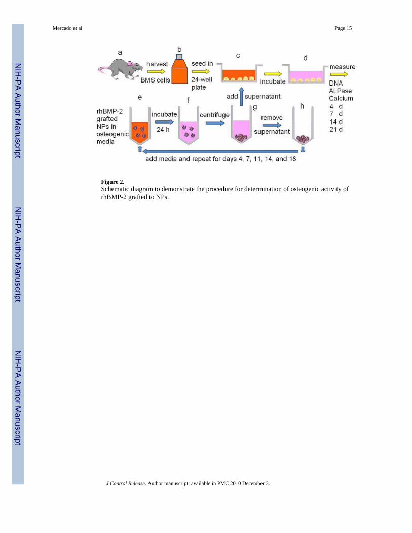
Figure 2.Schematic diagram to demonstrate the procedure for determination of osteogenic activity ofrhBMP-2 grafted to NPs.
Mercado et al. Page 15
J Control Release. Author manuscript; available in PMC 2010 December 3.
NIH
-PA Author Manuscript
NIH
-PA Author Manuscript
NIH
-PA Author Manuscript
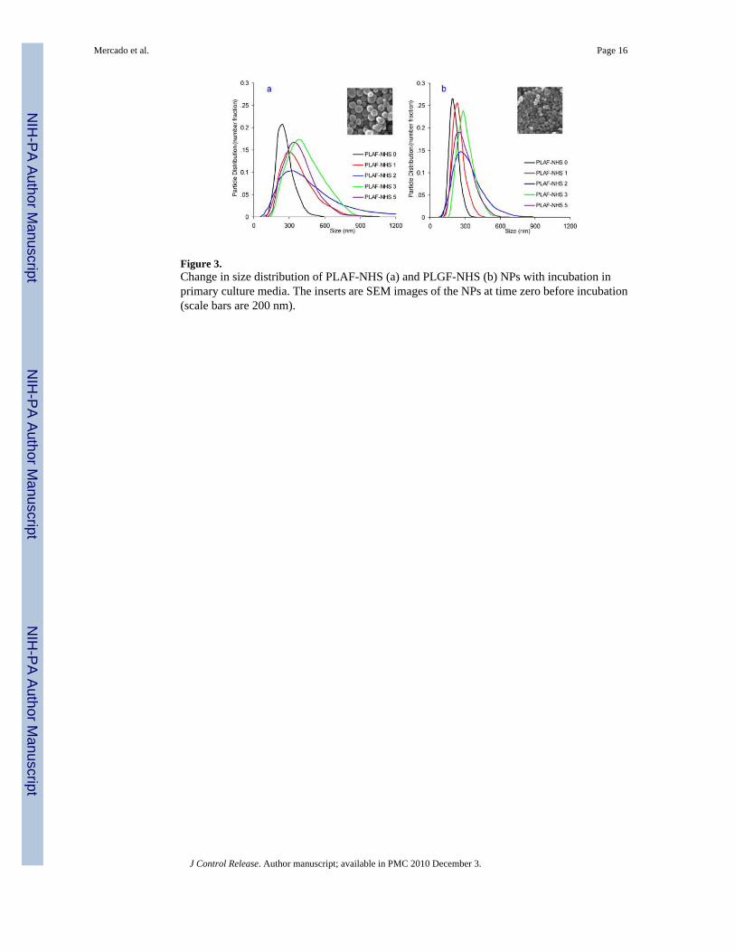
Figure 3.Change in size distribution of PLAF-NHS (a) and PLGF-NHS (b) NPs with incubation inprimary culture media. The inserts are SEM images of the NPs at time zero before incubation(scale bars are 200 nm).
Mercado et al. Page 16
J Control Release. Author manuscript; available in PMC 2010 December 3.
NIH
-PA Author Manuscript
NIH
-PA Author Manuscript
NIH
-PA Author Manuscript
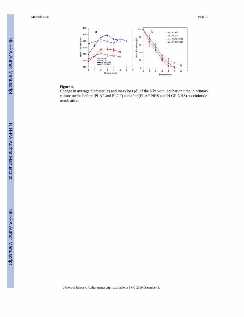
Figure 4.Change in average diameter (c) and mass loss (d) of the NPs with incubation time in primaryculture media before (PLAF and PLGF) and after (PLAF-NHS and PLGF-NHS) succinimide-termination.
Mercado et al. Page 17
J Control Release. Author manuscript; available in PMC 2010 December 3.
NIH
-PA Author Manuscript
NIH
-PA Author Manuscript
NIH
-PA Author Manuscript
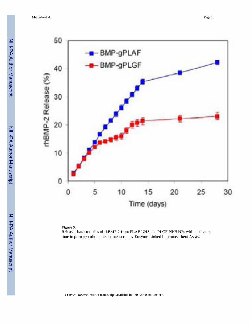
Figure 5.Release characteristics of rhBMP-2 from PLAF-NHS and PLGF-NHS NPs with incubationtime in primary culture media, measured by Enzyme-Linked Immunosorbent Assay.
Mercado et al. Page 18
J Control Release. Author manuscript; available in PMC 2010 December 3.
NIH
-PA Author Manuscript
NIH
-PA Author Manuscript
NIH
-PA Author Manuscript

Figure 6.DNA content (a), ALPase activity (b), calcium content (c), mRNA expression levels (as folddifference) of osteopontin (d; OP), osteocalcin (e, OC), and osteonectin (f, ON) genes of BMScells as a function of incubation time cultured in osteogenic media supplemented with thereleased rhBMP-2 from the NPs. One star in (a–c) indicates statistically significant differencebetween test group and Control; two stars between test group and OM; three stars betweengrafted-BMP groups and BMP. Error bars correspond to means ± 1 SD for n = 3. The mRNAexpression levels are the mean from three scaffolds. The standard error for mRNA level was±2 fold difference.
Mercado et al. Page 19
J Control Release. Author manuscript; available in PMC 2010 December 3.
NIH
-PA Author Manuscript
NIH
-PA Author Manuscript
NIH
-PA Author Manuscript
Copyright © 2022 FDOKUMEN