Skin absorption studies of octyl-methoxycinnamate loaded poly(D,L-lactide) nanoparticles: Estimation...
11
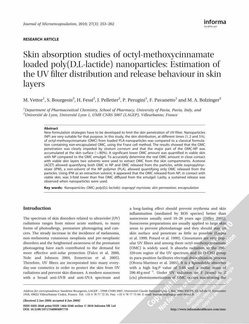
Journal of Microencapsulation, 2010; 27(3): 253–262 RESEARCH ARTICLE Skin absorption studies of octyl-methoxycinnamate loaded poly(D,L-lactide) nanoparticles: Estimation of the UV filter distribution and release behaviour in skin layers M. Vettor 1 , S. Bourgeois 2 , H. Fessi 2 , J. Pelletier 2 , P. Perugini 1 , F. Pavanetto 1 and M. A. Bolzinger 2 1 Department of Pharmaceutical Chemistry, School of Pharmacy, University of Pavia, Pavia, Italy, and 2 Universite ´ de Lyon, Universite ´ Lyon 1, UMR CNRS 5007 (LAGEP), Villeurbanne, France Abstract New formulation strategies have to be developed to limit the skin penetration of UV-filter. Nanoparticles (NP) are very suitable for that purpose. In this study, the skin distribution, at different times (1, 2 and 3 h), of octyl-methoxycinnamate (OMC) from loaded PLA-nanoparticles was compared to a classical formula- tion containing non-encapsulated OMC, using the Franz cell method. The results showed that the OMC penetration was clearly impeded by stratum corneum and that the major part of the OMC-NP was accumulated at the skin surface (480%). A significant lower OMC amount was quantified in viable skin with NP compared to the OMC emulgel. To accurately determine the real OMC amount in close contact with viable skin layers two solvents were used to extract OMC from the skin compartments. Acetone (ACET) allowed quantifying both OMC in NP and OMC released from the particles, while isopropylmyr- istate (IPM), a non-solvent of the NP polymer (PLA), allowed quantifying only OMC released from the particles. Using IPM as an extraction solvent, it appeared that the OMC released from NP, in contact with viable skin, was 3-fold lower than free OMC diffused from the emulgel. Lastly, a sustained release was observed when nanoparticles were used. Key words: Nanoparticles; OMC; poly(D,L-lactide); isopropyl myristate; skin permeation; encapsulation Introduction The spectrum of skin disorders related to ultraviolet (UV) radiations ranges from minor acute sunburn, to many forms of photoallergy, premature photoageing and can- cers. The steady increase in the incidence of melanoma, non-melanoma cutaneous neoplasia and pre-neoplastic disorders and the heightened awareness of the premature photoageing have each contributed to the demand for more effective anti-solar protection (Falco et al. 2000, Nole and Johnson 2004, Ermertcan et al. 2005). Therefore, UV filters are incorporated into many every- day-use cosmetics in order to protect the skin from UV radiations and prevent skin diseases. A modern sunscreen with a broad anti-UVB and anti-UVA spectrum and a long-lasting effect should prevent erythema and skin inflammation (mediated by ROS species) better than sunscreens usually used 10–20 years ago (Diffey 2005). Sunscreens preparations are usually applied to large skin areas to prevent photodamage and they should stay on skin surface and penetrate as little as possible (Gupta et al. 1999, Potard et al. 1999). Cinnamates are very pop- ular UV filters and among them octyl-methoxycinnamate (OMC) is widely used. It absorbs radiation in the 290– 320 nm region of the UV spectrum and its O-CH3 group in para-position facilitates electron delocalization process (Olvera-Martinez et al. 2005). It is a liposoluble absorber with a high log P value of 5.68 and a molar mass of 290.40 g mol 1 . Under UV radiation an E (trans) to Z (cis) photoisomerization of OMC occurs inactivating the Address for correspondence: Sandrine Bourgeois, LAGEP – UMR CNRS 5007, Universite ´ Claude Bernard Lyon 1, Bat. 308G ESCPE, 43, bd du 11 November 1918, 69622 Villeurbanne Cedex, France. Tel: þ33 4 78 77 72 35. Fax: þ33 4 78 77 75 84. E-mail: [email protected] (Received 2 Jun 2009; accepted 8 Jun 2009) ISSN 0265-2048 print/ISSN 1464-5246 online ß 2010 Informa UK Ltd DOI: 10.3109/10717540903097770 http://www.informahealthcare.com/mnc (Received 2 Jun 2009; accepted 8 Jun 2009) ISSN 0265-2048 print/ISSN 1464-5246 online ß 2010 Informa UK Ltd DOI: 10.3109/10717540903097770 http://www.informahealthcare.com/mnc
Transcript of Skin absorption studies of octyl-methoxycinnamate loaded poly(D,L-lactide) nanoparticles: Estimation...
Journal of Microencapsulation, 2010; 27(3): 253–262
RESEARCH ARTICLE
Skin absorption studies of octyl-methoxycinnamateloaded poly(D,L-lactide) nanoparticles: Estimation ofthe UV filter distribution and release behaviour in skinlayers
M. Vettor1, S. Bourgeois2, H. Fessi2, J. Pelletier2, P. Perugini1, F. Pavanetto1 and M. A. Bolzinger2
1Department of Pharmaceutical Chemistry, School of Pharmacy, University of Pavia, Pavia, Italy, and2Universite de Lyon, Universite Lyon 1, UMR CNRS 5007 (LAGEP), Villeurbanne, France
AbstractNew formulation strategies have to be developed to limit the skin penetration of UV-filter. Nanoparticles(NP) are very suitable for that purpose. In this study, the skin distribution, at different times (1, 2 and 3 h),of octyl-methoxycinnamate (OMC) from loaded PLA-nanoparticles was compared to a classical formula-tion containing non-encapsulated OMC, using the Franz cell method. The results showed that the OMCpenetration was clearly impeded by stratum corneum and that the major part of the OMC-NP wasaccumulated at the skin surface (480%). A significant lower OMC amount was quantified in viable skinwith NP compared to the OMC emulgel. To accurately determine the real OMC amount in close contactwith viable skin layers two solvents were used to extract OMC from the skin compartments. Acetone(ACET) allowed quantifying both OMC in NP and OMC released from the particles, while isopropylmyr-istate (IPM), a non-solvent of the NP polymer (PLA), allowed quantifying only OMC released from theparticles. Using IPM as an extraction solvent, it appeared that the OMC released from NP, in contact withviable skin, was 3-fold lower than free OMC diffused from the emulgel. Lastly, a sustained release wasobserved when nanoparticles were used.
Key words: Nanoparticles; OMC; poly(D,L-lactide); isopropyl myristate; skin permeation; encapsulation
Introduction
The spectrum of skin disorders related to ultraviolet (UV)
radiations ranges from minor acute sunburn, to many
forms of photoallergy, premature photoageing and can-
cers. The steady increase in the incidence of melanoma,
non-melanoma cutaneous neoplasia and pre-neoplastic
disorders and the heightened awareness of the premature
photoageing have each contributed to the demand for
more effective anti-solar protection (Falco et al. 2000,
Nole and Johnson 2004, Ermertcan et al. 2005).
Therefore, UV filters are incorporated into many every-
day-use cosmetics in order to protect the skin from UV
radiations and prevent skin diseases. A modern sunscreen
with a broad anti-UVB and anti-UVA spectrum and
a long-lasting effect should prevent erythema and skin
inflammation (mediated by ROS species) better than
sunscreens usually used 10–20 years ago (Diffey 2005).
Sunscreens preparations are usually applied to large skin
areas to prevent photodamage and they should stay on
skin surface and penetrate as little as possible (Gupta
et al. 1999, Potard et al. 1999). Cinnamates are very pop-
ular UV filters and among them octyl-methoxycinnamate
(OMC) is widely used. It absorbs radiation in the 290–
320 nm region of the UV spectrum and its O-CH3 group
in para-position facilitates electron delocalization process
(Olvera-Martinez et al. 2005). It is a liposoluble absorber
with a high log P value of 5.68 and a molar mass of
290.40 g mol�1. Under UV radiation an E (trans) to Z
(cis) photoisomerization of OMC occurs inactivating the
Address for correspondence: Sandrine Bourgeois, LAGEP – UMR CNRS 5007, Universite Claude Bernard Lyon 1, Bat. 308G ESCPE, 43, bd du 11 November1918, 69622 Villeurbanne Cedex, France. Tel: þ33 4 78 77 72 35. Fax: þ33 4 78 77 75 84. E-mail: [email protected]
(Received 2 Jun 2009; accepted 8 Jun 2009)
ISSN 0265-2048 print/ISSN 1464-5246 online � 2010 Informa UK LtdDOI: 10.3109/10717540903097770 http://www.informahealthcare.com/mnc
(Received 2 Jun 2009; accepted 8 Jun 2009)
ISSN 0265-2048 print/ISSN 1464-5246 online � 2010 Informa UK LtdDOI: 10.3109/10717540903097770 http://www.informahealthcare.com/mnc
filter (Pattanaargson and Limphong 2001, Serpone et al.
2002). Percutaneous absorption of OMC from a wide range
of formulations was monitored in vitro using the Franz cell
method. According to its high log P value OMC does not
permeate very quickly through the skin from solution but
in formulations containing silicone emulsifiers, sucrose
esters or glycols and alcohol, a higher penetration effi-
ciency is achieved (Godwin et al. 2002, Montenegro et al.
2004, Calderilla-Fajardo et al. 2006, Klinubol et al. 2008).
In this context, some attempts have been made to increase
the stability and efficacy of sunscreen products and
decrease their local intolerance by using new formulation
strategies such as nanoparticles (NP), microparticles,
cyclodextrins or liposomes (Jimenez et al. 2004b, Olvera-
Martinez et al. 2005, Ramon et al. 2005, Molinari et al.
2006, Tursilli et al. 2007). For example, some authors
have demonstrated that the encapsulation was an effective
way to avoid OMC photodegradation in formulations
(Perugini et al. 2002, Anumansirikul et al. 2008, Weiss-
Angeli et al. 2008) and to slow down its skin penetration
leading to an SPF enhancement and a decrease of systemic
absorption (Alvarez-Roman et al. 2001, Klinubol et al.
2008, Vettor et al. 2008). Moreover, recent investigations
have indicated that nanoparticles of lipidic type (Solid
Lipid Nanoparticles or SLN) could increase the efficacy
of sunscreens by providing a good epidermal targeting
effect (Lombardi Borgia et al. 2005, Chen et al. 2006).
However, only few research teams have studied the diffu-
sion into the skin of cosmetic actives from polymeric
nanoparticles. Some of the most widely used polymers in
the NP formulation are poly-�-caprolactone (PCL), poly-
lactic acid (PLA), polyglycolic acid and their co-polymer,
poly(lactide-co-glycolide) (PLGA), which are known for
their good biocompatibility and resorbability through nat-
ural pathways. If their potential for drug delivery has been
extensively explored for oral or parenteral routes of
administration their potential for dermal applications is
rather unexplored (Brannon-Peppas & Blanchette 2004,
Luengo et al. 2006).
Alvarez Roman et al. (2004c) showed that polymeric
nanoparticles accumulated in stratum corneum, skin fur-
rows and hair follicles. Consequently, these authors
observed that OMC-loaded nanoparticles preparations
resulted in a significantly better protection against
UV-induced erythema than a simple OMC-gel because
they remained where they were intended to act. This effec-
tiveness was also attributed to the large NP-specific area
facilitating skin coverage and high substantivity (Alvarez-
Roman et al. 2001). Moreover, Jimenez et al. (2004a), who
studied the permeation of octyl-methoxycinnamate from
OMC-loaded poly-"-caprolactone nanoparticles by the
Franz cell method, demonstrated that the penetration of
OMC in the skin was 3–4-times lower than from a conven-
tional emulsion. However, in their study the OMC amount
found in each skin layer was over-estimated because
NP-shells were completely solved by acetonitrile during
skin extraction and the total OMC amounts encapsulated
or not were both quantified. Under these conditions the
total OMC amount contained in NP and released in skin
are considered as a whole.
Therefore, the objective of the present study was to
evaluate percutaneous absorption of OMC released from
polymeric nanoparticles. For this purpose, the evaluation
of OMC distribution in skin compartments from OMC-
loaded PLA nanoparticles formulated in an emulsion gel
(OMC-NP emulgel) comparatively to a non-encapsulated-
OMC emulsion gel (OMC-emulgel) was performed. Both
formulations contained 5% OMC. The classical Franz cell
method was firstly applied and OMC amounts in each skin
strata (stratum corneum (SC), epidermis (E), dermis (D), in
the receptor fluid (RF) and at the skin surface (SS) (unpe-
netrated dose)) were determined after 1, 2 and 3 h expo-
sure time. Secondly, the skin distribution of OMC released
from NP in each skin layer was estimated using a validated
new method to extract non-encapsulated OMC from skin
layers in order to evaluate the real amount of OMC in close
contact with viable skin cells. This method, based on dif-
ferential polymer solubility, was used to leverage the
released OMC in skin from that retained in the
nanoparticles.
Materials and methods
Chemicals
OMC (Parsol�MCX) was supplied by Hoffmann-Laroche
Ltd (Basel, Switzerland). Poly (D,L-lactide) (PLA)
Resomer�R 202 H, was obtained from Boehringer
Ingelheim (Ingelheim, Germany). Isopropylmyristate
(Crodamol�, IPM) was a gift from Croda Chemical
Europe Ltd (East Yorkshire, UK). Phosphate buffered
saline tablets, pH 7.4, were purchased from Sigma
Aldrich (St. Quentin-Fallavier, France). Butyl glucoside,
polyacrylamide/C13-14 isoparaffin/laureth-7 (Sepigel�
305) and the mixture of paraben and phenoxyethanol
(Sepicide� HB) used in the emulgel formulation were a
generous gift from Seppic (Paris, France). Methanol, acet-
onitrile, tetrahydrofuran (HPLC grade) and acetone ana-
lytical grade were purchased from Carlo Erba (Chaussee
en Vexin, France).
OMC formulations for skin permeation studies
OMC-loaded poly-D,L-lactic acid (PLA) nanoparticles
were prepared by a salting out technique as described in
a previous study (Vettor et al. 2008). Briefly, polymer and
254 M. Vettor et al.
sunscreen (1 : 1 w/w) were dissolved in acetone as organic
phase; a viscous solution was prepared separately by dis-
solving CaCl2 (40% w/w) in 3% (w/w) polyvinyl alcohol
aqueous solution. Addition of the aqueous solution to
the organic phase under vigorous stirring at 14 000 rpm
(IKA Ultraturrax T25) gave a w/o emulsion that was con-
verted to an o/w emulsion by further addition of the aque-
ous phase portion (final organic phase:aqueous phase,
1 : 2 w/w). Pure water was then added to the emulsion to
induce diffusion of the organic solvent from the polymer
organic solution resulting in a nanoparticle dispersion.
The purification and concentration of nanoparticles were
performed by ultrafiltration with a 300 000 NMWL poly-
etheresulphone membrane (Millipore, Vimodrone, Italy).
NP were characterized by their particle size, morphol-
ogy and their OMC loading. Particle size analysis was per-
formed by light diffraction method using a Mastersizer�
2000 (Malvern Instruments, UK). NP suspension was
diluted into dispersion unit and analysed under stirring
at 2500 rpm at room temperature. Three analyses were
performed for each sample. Morphological characteristics
of nanoparticles were studied by Atomic Force Microscopy
(AFM). Measurements were performed by an AutoProbe
CP Research scanning microscope (Thermo Microscope,
Sunnyvale, USA). The analyses were performed in air and
under constant applied force conditions (contact mode)
and theoretical spring constant (k¼ 0.9 N m).
Nanoparticles were fixed on a silicon support, which had
been glued onto a steel disc to enable magnetic fixation
under the scanning tip. Images were processed and ana-
lysed using the Image Processing Data Analysis 2.0 soft-
ware provided by Thermo Microscope (Sunnyvale, USA).
For OMC loading determination, NP were isolated by
centrifugation and dissolved with acetone. Then, the
amount of OMC encapsulated was quantified by HPLC
as described later.
The sunscreen agent, free or encapsulated in nanopar-
ticles, was incorporated in an o/w emulsion gel formulated
with Sepigel�305 (3%) and isopropylmyristate (10%)
(Perugini et al. 2002). Emulsion-gels were prepared at
room temperature by dispersion of the polymeric gelling
agent (Sepigel� 305) in the oil phase (isopropylmyristate)
before addition of the aqueous phase. Two sunscreen for-
mulations containing 5% OMC were obtained: an OMC-
free emulsion-gel (OMC-emulgel) and an OMC-NP emul-
sion-gel (OMC-NP emulgel).
Franz cell preparation
The SCCP (Scientific Committee on Consumer Products)
guidelines were followed (SCCP 2006). The percutaneous
penetration studies were performed with a modified Franz
cell method (2.54 cm2, 11 mL receptor fluid). Flanks of pig
skin (22 kg, Landras pigs) were chosen as cutaneous
model, because they share essential permeation charac-
teristics with human skin, especially for lipophilic com-
pounds (Bronaugh et al 1982, 1986). Full-thickness pig
female or male skins were obtained from the Laboratoire
de Physiologie (Universite de Lyon, France). The skin was
cleaned up with tap water. The bristles were cut with an
electric cutter. The skin surface was shortly washed with
1% sodium dodecyl sulphate aqueous solution and rinsed
with tap water and distilled water and lastly blotted very
well with soft household paper. Integrity of skin samples
was examined by a physical method, Transepidermal
Water Loss (TEWL) (Skin Station, La Licorne, Meylan,
France) (Schaefer & Loth 1996). The TEWL was measured
for 1 min (8.04� 0.05 g h�1 m�2). Skin samples with TEWL
values415 g h�1 m�2 were discarded. The thickness of
each skin piece (1.5� 0.05 mm mean thickness) was mea-
sured with a micrometer (Mitutoyo, Japan). The skin discs
were stored flat at �20�C until use (not more than 3
months) (Harrison et al. 1984, Schaefer and Loth 1996,
OECD 2004). On the day of use, the cleaned skin discs
were thawed, mounted in Franz cells and placed in a ther-
mostated water bath at 37�C. The dermal side was exposed
to the receptor fluid and the stratum corneum was exposed
to the air (non-occlusive conditions). The receptor com-
partment was filled with a phosphate buffered saline solu-
tion (0.01 M, pH 7.4) containing 4% of butylglucoside. The
solubility of OMC in the receptor fluid was checked to
ensure that sink conditions were applied. Cells were stir-
red with a magnetic bar during the experiment and
2 mg cm�2 of each formulation (corresponding to 100mg
OMC cm�2) were deposited on the skin surface.
After 1, 2 and 3 h the receptor fluid was collected, fil-
tered through a 0.45 mm nylon membrane and analysed by
HPLC. After removing the emulsion on the top of the skin
(skin surface), the amounts of OMC located in the stratum
corneum were evaluated by the tape stripping method
(Clarys et al. 2001, Weigmann et al. 2001, Jimenez et al.
2004a). For this purpose, stratum corneum was removed
by 19 successive tape strippings (Gupta et al. 1999,
Benech-Kieffer et al. 2000) using D-Squame� (diameter
22 mm) (Monaderm, Monaco). The first strip was dis-
carded and treated separately because it could allow
determining the remains of a substance after some time
of application (Jimenez et al. 2004a). The 18 other strips
were pooled three-by-three in a vessel and the OMC was
extracted from strips with an adequate solvent.
After eliminating stratum corneum from skin samples,
viable epidermis was separated from dermis with dis-
section after being immersed in water at 60�C for 45 s.
Previous studies have shown that water could not
extract OMC during the epidermal/dermal separation
(Weigmann et al. 2001).
Skin absorption studies of octyl-methoxycinnamate loaded by poly(D,L-lactide) nanoparticles 255
To summarize, the amount of OMC was measured in
five locations at each time:
. skin surface called SS (residual amount),
. stratum corneum (SC),
. viable epidermis (VE),
. dermis (D), and
. receptor fluid (RF).
Six replicates were carried out for each experiment.
Two extractive methods of OMC in skin sheets were
applied to discriminate OMC released from the nanopar-
ticles from that still encapsulated in nanoparticles. Both
methods and the validation are described in the following
sections.
Extraction of OMC with acetone: ‘ACET method’
This first protocol, considered as the ‘classical method’,
involved the use of acetone as extraction solvent for
OMC. Acetone led to the dissolution of the nanoparticle
PLA shell.
OMC formulation remaining at the skin surface (SS)
was removed with a spatula and dissolved in 10 mL of
acetone. As mentioned previously, the skin was then
stripped (19 strips). The first one was discarded and
pooled with the SS amount. The others were pooled
three-by-three in small bottles and immersed in 2 mL ace-
tone under stirring for OMC extraction. The viable epider-
mis and dermis were separated and each of them cut in
small pieces. OMC amounts located in the viable epider-
mis and the dermis were respectively extracted with 1 mL
and 2 mL of acetone by using a vortex mixer for 1 min and
ultrasounds for 20 min. The solvent was then filtered
through a 0.45 mm nylon membrane and injected for
HPLC analysis.
This method, called ‘ACET method’, using acetone as
extraction solvent gave the total amount of OMC (released
and encapsulated) accumulated in each skin
compartment.
Extraction of OMC with IPM: ‘IPM method’
The second method, called ‘IPM method’, used IPM as
extraction solvent and allowed only quantifying the
amount of OMC released from NP, in the different skin
layers. Briefly, OMC formulation remaining at the skin sur-
face (SS) was removed with a spatula and then dissolved
in 10 mL of IPM. The skin was stripped. The first strip was
discarded and the 18 following strips were pooled three-
by-three and placed in small vessels; 1 mL of IPM was
added in each vessel and the samples were stirred for
10 min for OMC extraction. In order to separate
nanoparticles and strips from released OMC, samples
were centrifuged (15 min at 14 000 rpm). Then the super-
natant was diluted, filtrated and analysed by HPLC. It cor-
responds to OMC released amount from NP in the strips.
After separation the viable epidermis and the dermis
were respectively mixed with 0.5 and 1 mL IPM during
10 min under magnetic stirring. OMC dissolved in IPM
was separated from nanoparticles and skin residues by
centrifugation for 15 min at 14 000 rpm. The supernatant
was diluted, filtered and OMC was quantified by HPLC.
Validation of the IPM method
In order to control that IPM may extract OMC from
formulations and different skin layers as well as more clas-
sically used solvents such as acetone, two experiments
were conducted. OMC was first assayed in the OMC emul-
gel by using IPM or ACET as extractive solvents. Secondly,
OMC was quantified in skin layers either by acetone or
by IPM after having applied OMC emulgel. Furthermore,
in order to control that IPM couldn’t extract OMC from
nanoparticles, the OMC content in nanoparticles was
determined after incubation of OMC-loaded NP in IPM.
Dosage of OMC in the OMC-emulgel using IPM vs
acetone. Five milligrams of OMC-emulgel was mixed
with 20 mL of acetone or IPM and then vortexed for
1 min and ultrasonicated for 20 min. After filtration
(0.45 mm) the solution was diluted and injected. The
experiment was performed in triplicate. The new method
was considered as validated if the OMC percentage
obtained with IPM was not significantly different from
the percentage obtained with acetone (Student t-test,
p5 0.05).
Measurement of OMC amounts in each skin layer
extracted with acetone or IPM after skin absorption
study of the OMC-emulgel. The validation of the extrac-
tion method using IPM was performed in order to evaluate
the ability of IPM to extract OMC present in each skin
compartment such as acetone. For this purpose, skin per-
meation studies were conducted over 3 h with non-encap-
sulated OMC (OMC-emulgel), as previously described.
The OMC extraction was then performed using either
acetone or IPM as solvent as described earlier. The com-
parison of the OMC amount retrieved in each skin layer
with both solvents was performed. This experiment was
conducted on Franz cells (n¼ 6) with each protocol. The
new method was considered as validated if results
obtained with IPM were not significantly different from
those obtained with acetone.
256 M. Vettor et al.
Estimation of IPM ability to extract OMC from NP. Ten
millilitres of IPM were added to 5 mg of OMC-NP suspen-
sion corresponding to the OMC amount applied on skin
samples for permeation studies (100mg cm�2 of OMC).
After 10 min of incubation in IPM, the NP were separated
from IPM by centrifugation (15 min at 14 000 rpm) and the
OMC amount retrieved in the supernatant was evaluated
by HPLC. This experiment was conducted in triplicate.
HPLC method for the OMC quantification
A HPLC analytical method was applied to quantify OMC in
the formulations and in the skin compartments (Perugini
et al. 2002). The HPLC system (Waters, USA) consisted
of a solvent delivery pump (Waters 515) equipped with a
10ml autosampler injector (Waters 717). Separation was
performed at 308 nm on a 5 mm X-Terra MS C18 (250�
4.6 mm i.d.), thermostated at 20�C and eluted isocratically
at a flow rate of 1.0 mL min�1, with methanol-acetonitrile-
tetrahydrofuran-water (65 : 10 : 10 : 15, v/v). The retention
time was 10.3 min. UV filter concentrations were deter-
mined from calibration curves measured with OMC
(0.4–40 mg mL�1) dissolved either in acetone or IPM.
The detection limit for OMC quantification was evaluated
at 0.2 fg.
Statistical analysis
The penetration results were expressed as the percentage
of the OMC applied dose. Each measurement was
repeated 6-fold. Depending on experiments, statistical
analysis was performed with either ANOVA test or
Student’s t-test. A p5 0.05 was considered statistically
significant.
Results and discussion
Nanoparticles characterization
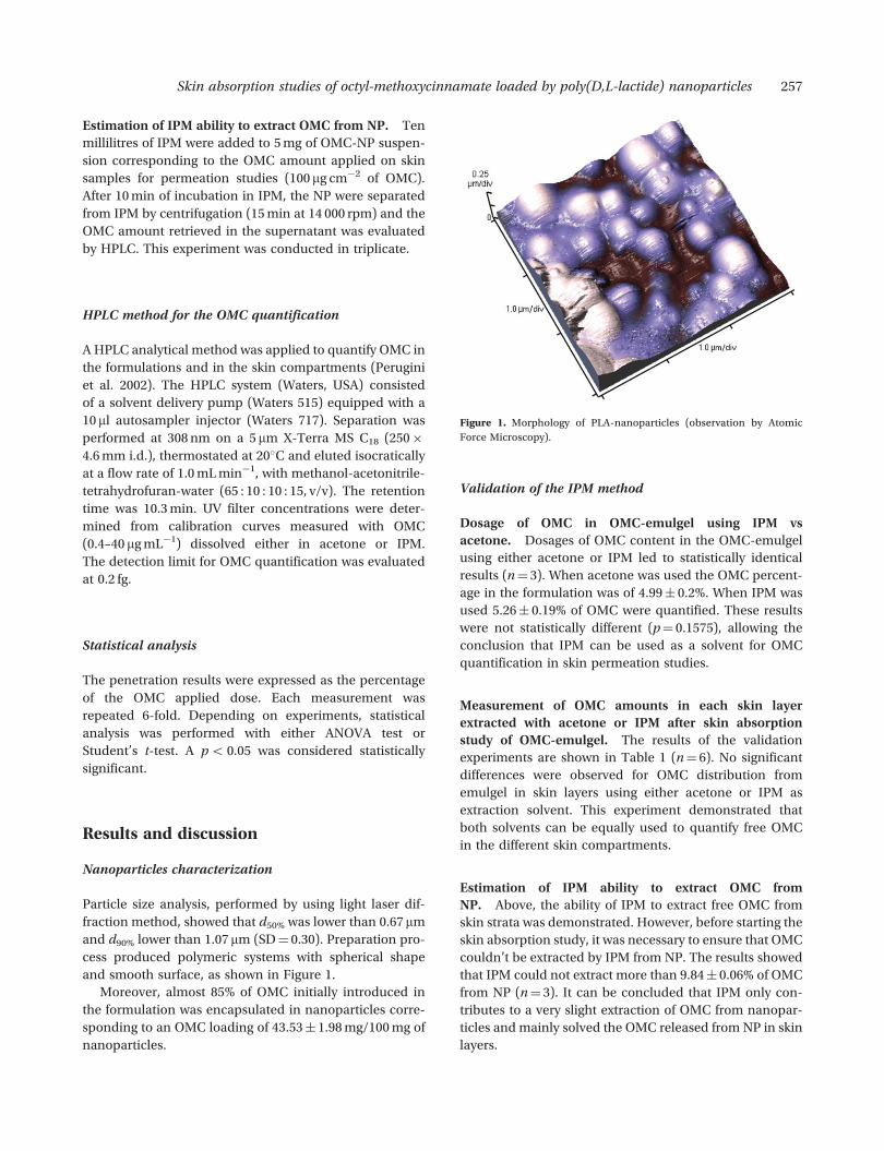
Particle size analysis, performed by using light laser dif-
fraction method, showed that d50% was lower than 0.67 mm
and d90% lower than 1.07 mm (SD¼ 0.30). Preparation pro-
cess produced polymeric systems with spherical shape
and smooth surface, as shown in Figure 1.
Moreover, almost 85% of OMC initially introduced in
the formulation was encapsulated in nanoparticles corre-
sponding to an OMC loading of 43.53� 1.98 mg/100 mg of
nanoparticles.
Validation of the IPM method
Dosage of OMC in OMC-emulgel using IPM vs
acetone. Dosages of OMC content in the OMC-emulgel
using either acetone or IPM led to statistically identical
results (n¼ 3). When acetone was used the OMC percent-
age in the formulation was of 4.99� 0.2%. When IPM was
used 5.26� 0.19% of OMC were quantified. These results
were not statistically different (p¼ 0.1575), allowing the
conclusion that IPM can be used as a solvent for OMC
quantification in skin permeation studies.
Measurement of OMC amounts in each skin layer
extracted with acetone or IPM after skin absorption
study of OMC-emulgel. The results of the validation
experiments are shown in Table 1 (n¼ 6). No significant
differences were observed for OMC distribution from
emulgel in skin layers using either acetone or IPM as
extraction solvent. This experiment demonstrated that
both solvents can be equally used to quantify free OMC
in the different skin compartments.
Estimation of IPM ability to extract OMC from
NP. Above, the ability of IPM to extract free OMC from
skin strata was demonstrated. However, before starting the
skin absorption study, it was necessary to ensure that OMC
couldn’t be extracted by IPM from NP. The results showed
that IPM could not extract more than 9.84� 0.06% of OMC
from NP (n¼ 3). It can be concluded that IPM only con-
tributes to a very slight extraction of OMC from nanopar-
ticles and mainly solved the OMC released from NP in skin
layers.
Figure 1. Morphology of PLA-nanoparticles (observation by Atomic
Force Microscopy).
Skin absorption studies of octyl-methoxycinnamate loaded by poly(D,L-lactide) nanoparticles 257
Conditions used in this experiment are close to that
used for OMC extraction at the skin surface in permeation
studies. These correspond to the highest dilution rate
(OMC/IPM) used for OMC extraction in skin permeation
experiments, which could maximize the possible OMC
extraction from NP.
To conclude this section acetone was used to quantify
the total amount of OMC (free and encapsulated), whereas
IPM was only used to investigate free OMC outside the NP
and released in skin strata.
Total penetration and distribution of OMC in skin
layers over time for the OMC-emulgel and OMC-NP
emulgel formulations (ACET method)
The results reported in Table 2 show the in vitro distribu-
tion of OMC in skin compartments for both formulations
using either ‘ACET’ or ‘IPM’ methods. Comparison
between OMC-emulgel and OMC-NP emulgel gave
useful informations on the cutaneous uptake of OMC in
the skin depending on both formulations.
First, it should be noticed that no OMC was recovered
in the receiving compartment at each time for both for-
mulations. Anumansirikul et al. (2008) found some OMC
amounts in receptor fluids from OMC solutions, but the
skin model was baby mouse skin (more permeable to
active substances than pig skin). In normal skin this
result can be explained by the high lipophilicity of OMC
(log P¼ 5.68), preventing diffusion in the skin deeper
layers.
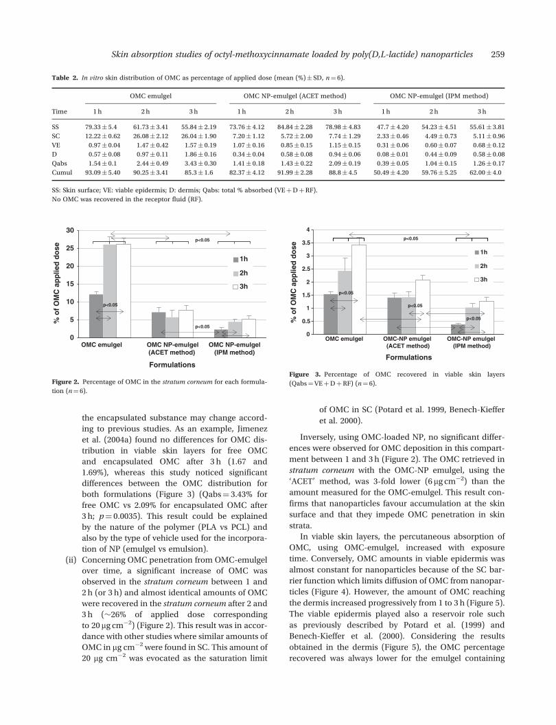
When applied encapsulated in NP, the major part of
OMC was retained at the skin surface (SS) over time.
Indeed, after 2 and 3 h, �85% and 80% of the applied
OMC were, respectively, recovered on the skin surface
with NP. These amounts were much higher than that
obtained when OMC emulgel was applied (62% and
56%, respectively, after 2 and 3 h). This result demon-
strated the nanoparticle accumulation at the skin surface
which has been previously described by Alvarez-Roman
et al. (2004a, c), who studied the distribution of fluorescent
nanoparticles in the skin by confocal microscopy.
Nanoparticles cover the skin surface and accumulate in
furrows and appendages independently of their size
(Alvarez-Roman et al. 2004c, Kohli & Alpar 2004). For
SLN it was demonstrated that nanoparticle aggregation
phenomena on the skin surface may occur, leading to a
film formation, to a better skin coverage and consequently
to the improvement of sunscreen efficiency (Wissing &
Muller 2002, Jimenez et al. 2004a). In the case of polymeric
particles, this study didn’t demonstrate a film formation,
but the accumulation on the surface may favour the
UV-filter efficiency.
High amounts of OMC were also detected in stratum
corneum (between 12–26% for OMC-emulgel and from
5–8% for OMC-NP emulgel with ACET method
(Figure 2). This result agrees with previous studies showing
that the affinity of active substances for stratum corneum
and their accumulation were mainly governed by the lipo-
philicity of the applied agents and their log P value
(Hagedorn-Leweke & Lippold 1998). OMC, which is a lipo-
philic substance (log P¼ 5.68) accumulated on the top of
the skin and in stratum corneum (Benech-Kieffer et al.
2000). More than 80% of OMC concentrated in these two
compartments after 3 h exposure time. The main differ-
ence between the OMC-emulgel and the OMC-NP emulgel
was OMC distribution between these two compartments.
In the case of NP, the percentage of accumulated OMC was
10-fold higher on the top of the skin than in the SC. This
value dropped to 2-fold with OMC-emulgel.
Consequently, OMC amounts in viable skin layers
(Qabs¼VEþDþRF) were superior for OMC-emulgel
compared to OMC-NP emulgel (�3.5% vs �2% after 3 h)
(Table 2 and Figure 3) because stratum corneum may play
a role of reservoir as previously noticed by Potard et al.
(1999). This result firstly confirmed the higher affinity of
OMC for the lipophilic skin layers and secondly that the
transport of NP was clearly impeded by the stratum cor-
neum. This result was in the same range as that described
by Jimenez et al. (2004a), who studied the skin penetration
of OMC-loaded poly-"-caprolactone (PCL) nanoparticles
suspended in o/w emulsions in the laboratory. They also
showed that the skin penetration of OMC from their nano-
particles was clearly limited by the stratum corneum since
only 8.3% of the OMC applied dose was retrieved in stra-
tum corneum after 3 h with the NC o/w emulsion. With the
non-encapsulated OMC formulated in an o/w emulsion
this amount rose to �40.5%.
Two additional suggestions may be done concerning
this experiment:
(i) If a consensus exists concerning the accumulation
of NP at the skin surface the distribution of
Table 1. Comparison of OMC skin absorption studies at 3 h conducted
with OMC emulgel using either ACET method or IPM method.
ACET method
(% of OMC
applied dose)
IPM method
(% of OMC
applied dose)
p-value
SS 55.84� 2.19 58.32� 2.67 0.4565
SC 26.04� 1.90 25.92� 5.06 0.9828
VE 1.83� 0.13 2.13� 0.07 0.1175
D 1.86� 0.16 1.71� 0.16 0.5200
Qabs 3.43� 0.30 3.98� 0.25 0.1798
Total recovery 89.00� 1.60 92.06� 5.00
SS: Skin surface; SC: stratum corneum; VE: viable epidermis; D: dermis;
Qabs: total % absorbed (VEþDþRF).
No OMC was recovered in the receptor fluid (RF).
258 M. Vettor et al.
the encapsulated substance may change accord-
ing to previous studies. As an example, Jimenez
et al. (2004a) found no differences for OMC dis-
tribution in viable skin layers for free OMC
and encapsulated OMC after 3 h (1.67 and
1.69%), whereas this study noticed significant
differences between the OMC distribution for
both formulations (Figure 3) (Qabs¼ 3.43% for
free OMC vs 2.09% for encapsulated OMC after
3 h; p¼ 0.0035). This result could be explained
by the nature of the polymer (PLA vs PCL) and
also by the type of vehicle used for the incorpora-
tion of NP (emulgel vs emulsion).
(ii) Concerning OMC penetration from OMC-emulgel
over time, a significant increase of OMC was
observed in the stratum corneum between 1 and
2 h (or 3 h) and almost identical amounts of OMC
were recovered in the stratum corneum after 2 and
3 h (�26% of applied dose corresponding
to 20 mg cm�2) (Figure 2). This result was in accor-
dance with other studies where similar amounts of
OMC in mg cm�2 were found in SC. This amount of
20 mg cm�2 was evocated as the saturation limit
of OMC in SC (Potard et al. 1999, Benech-Kieffer
et al. 2000).
Inversely, using OMC-loaded NP, no significant differ-
ences were observed for OMC deposition in this compart-
ment between 1 and 3 h (Figure 2). The OMC retrieved in
stratum corneum with the OMC-NP emulgel, using the
‘ACET’ method, was 3-fold lower (6 mg cm�2) than the
amount measured for the OMC-emulgel. This result con-
firms that nanoparticles favour accumulation at the skin
surface and that they impede OMC penetration in skin
strata.
In viable skin layers, the percutaneous absorption of
OMC, using OMC-emulgel, increased with exposure
time. Conversely, OMC amounts in viable epidermis was
almost constant for nanoparticles because of the SC bar-
rier function which limits diffusion of OMC from nanopar-
ticles (Figure 4). However, the amount of OMC reaching
the dermis increased progressively from 1 to 3 h (Figure 5).
The viable epidermis played also a reservoir role such
as previously described by Potard et al. (1999) and
Benech-Kieffer et al. (2000). Considering the results
obtained in the dermis (Figure 5), the OMC percentage
recovered was always lower for the emulgel containing
Table 2. In vitro skin distribution of OMC as percentage of applied dose (mean (%)� SD, n¼ 6).
OMC emulgel OMC NP-emulgel (ACET method) OMC NP-emulgel (IPM method)
Time 1 h 2 h 3 h 1 h 2 h 3 h 1 h 2 h 3 h
SS 79.33� 5.4 61.73� 3.41 55.84� 2.19 73.76� 4.12 84.84� 2.28 78.98� 4.83 47.7� 4.20 54.23� 4.51 55.61� 3.81
SC 12.22� 0.62 26.08� 2.12 26.04� 1.90 7.20� 1.12 5.72� 2.00 7.74� 1.29 2.33� 0.46 4.49� 0.73 5.11� 0.96
VE 0.97� 0.04 1.47� 0.42 1.57� 0.19 1.07� 0.16 0.85� 0.15 1.15� 0.15 0.31� 0.06 0.60� 0.07 0.68� 0.12
D 0.57� 0.08 0.97� 0.11 1.86� 0.16 0.34� 0.04 0.58� 0.08 0.94� 0.06 0.08� 0.01 0.44� 0.09 0.58� 0.08
Qabs 1.54� 0.1 2.44� 0.49 3.43� 0.30 1.41� 0.18 1.43� 0.22 2.09� 0.19 0.39� 0.05 1.04� 0.15 1.26� 0.17
Cumul 93.09� 5.40 90.25� 3.41 85.3� 1.6 82.37� 4.12 91.99� 2.28 88.8� 4.5 50.49� 4.20 59.76� 5.25 62.00� 4.0
SS: Skin surface; VE: viable epidermis; D: dermis; Qabs: total % absorbed (VEþDþRF).
No OMC was recovered in the receptor fluid (RF).
0
5
10
15
20
25
30
OMC NP-emulgel(ACET method)
OMC NP-emulgel(IPM method)
Formulations
% o
f O
MC
ap
plie
d d
ose
1h
2h
3h
p<0.05
p<0.05
p<0.05
OMC emulgel
Figure 2. Percentage of OMC in the stratum corneum for each formula-
tion (n¼ 6).
0
0.5
1
1.5
2
2.5
3
3.5
4
OMC-NP emulgel (ACET method)
OMC-NP emulgel (IPM method)
Formulations
% o
f O
MC
ap
plie
d d
ose
1h
2h
3h
p<0.05
p<0.05
p<0.05
p<0.05
OMC emulgel
Figure 3. Percentage of OMC recovered in viable skin layers
(Qabs¼VEþDþRF) (n¼ 6).
Skin absorption studies of octyl-methoxycinnamate loaded by poly(D,L-lactide) nanoparticles 259
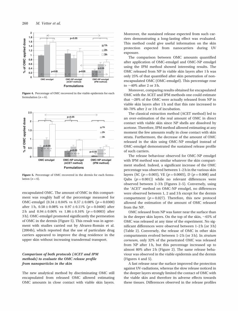
encapsulated OMC. The amount of OMC in this compart-
ment was roughly half of the percentage measured for
OMC-emulgel (0.34� 0.04% vs 0.57� 0.08% (p¼ 0.0300)
after 1 h, 0.58� 0.08% vs 0.97� 0.11% (p¼ 0.0400) after
2 h and 0.94� 0.06% vs 1.86� 0.16% (p¼ 0.0003) after
3 h). OMC-emulgel promoted significantly the permeation
of OMC in the dermis (Figure 5). This result was in agree-
ment with studies carried out by Alvarez-Roman et al.
(2004b), which reported that the use of particulate drug
carriers appeared to improve the drug residence in the
upper skin without increasing transdermal transport.
Comparison of both protocols (ACET and IPM
methods) to evaluate the OMC release profile
from nanoparticles in the skin
The new analytical method by discriminating OMC still
encapsulated from released OMC allowed estimating
OMC amounts in close contact with viable skin layers.
Moreover, the sustained release expected from such car-
riers demonstrating a long-lasting effect was evaluated.
This method could give useful information on the skin
protection expected from nanocarriers during UV
exposure.
The comparison between OMC amounts quantified
after application of OMC-emulgel and OMC-NP emulgel
using the IPM method showed interesting results. The
OMC released from NP in viable skin layers after 1 h was
only 25% of that quantified after skin penetration of non-
encapsulated OMC (OMC-emulgel). This percentage rose
to �40% after 2 or 3 h.
Moreover, comparing results obtained for encapsulated
OMC with the ACET and IPM methods one could estimate
that �28% of the OMC were actually released from NP in
viable skin layers after 1 h and that this rate increased to
60–70% after 2 or 3 h of incubation.
The classical extraction method (ACET method) led to
an over-estimation of the real amount of OMC in direct
contact with viable skin since NP shells are dissolved by
acetone. Therefore, IPM method allowed estimating at any
moment the few amounts really in close contact with skin
strata. Furthermore, the decrease of the amount of OMC
released in the skin using OMC-NP emulgel instead of
OMC-emulgel demonstrated the sustained release profile
of such carriers.
The release behaviour observed for OMC-NP emulgel
with IPM method was similar whatever the skin compart-
ment studied. Indeed, a significant increase of the OMC
percentage was observed between 1–2 h in the various skin
layers (SC (p¼ 0.005), VE (p¼ 0.0005), D (p¼ 0.008) and
Qabs (p¼ 0.001)) while no relevant differences were
observed between 2–3 h (Figures 2–5). Conversely, using
the ‘ACET’ method on OMC-NP emulgel, no differences
were observed between 1, 2 and 3 h except for the dermis
compartment (p¼ 0.027). Therefore, this new protocol
allowed the estimation of the amount of OMC released
from the NP.
OMC released from NP was faster near the surface than
in the deeper skin layers. On the top of the skin, �65% of
OMC was released at any time of the experiment. No sig-
nificant differences were observed between 1–2 h (or 3 h)
(Table 2). Conversely, the release of OMC in other skin
compartments evolved between 1–2 h (or 3 h). In stratum
corneum, only 32% of the penetrated OMC was released
from NP after 1 h, but this percentage increased up to
almost 80% after 2 h (Figure 2). The same release beha-
viour was observed in the viable epidermis and the dermis
(Figures 4 and 5).
A fast release near the surface improved the protection
against UV-radiations, whereas the slow release noticed in
the deeper layers strongly limited the contact of OMC with
the viable skin and therefore its adverse effects towards
these tissues. Differences observed in the release profiles
0
0.2
0.4
0.6
0.8
1
1.2
1.4
1.6
1.8
2
OMC-NP emulgel(ACET method)
OMC-NP emulgel(IPM method)
Formulations
1h
2h
3h
% o
f O
MC
ap
plie
d d
ose
p<0.05
50.0<p 50.0<pp<0.05
OMC emulgel
Figure 4. Percentage of OMC recovered in the viable epidermis for each
formulation (n¼ 6).
0
0.5
1
1.5
2
2.5
OMC-NP emulgel(ACET method)
OMC-NP emulgel(IPM method)
Formulations
1h
2h
3h
% o
f O
MC
ap
plie
d d
ose
p<0.05
p<0.05
p<0.05
p<0.05
OMC emulgel
Figure 5. Percentage of OMC recovered in the dermis for each formu-
lation (n¼ 6).
260 M. Vettor et al.

during the 1st h could be attributed to the local environ-
ment at the skin surface which is roughly different from
the inner layers and could lead to nanoparticle
degradation.
Several factors such as the polymer nature and its con-
centration could also influence the release profile of OMC
from NP. The key parameters controlling the release rate
of drugs in vivo through the eroded nanoparticles is still
under debate: the hydrophobicity and the surface charge
of the polymer coating as well as its crystallinity or its
molecular weight have been evoked (Edlund &
Albertsson 2001). Because PLA is faster hydrolysed com-
pared to PCL, it will be more suitable for skin protection
under sun exposure.
Conclusion
The effectiveness of a sunscreen is correlated with its affin-
ity to the stratum corneum. To be efficient UV filters must
remain in the outermost regions of the skin. Polymeric
nanoparticles preferentially stayed at the skin surface
where OMC was intended to act. Consequently nanopar-
ticles limited OMC penetration in viable skin layers. The
new analytical method allowed estimating the OMC
amount directly in close contact with skin strata and
gave useful information on OMC release.
In conclusion, nanoencapsulation is an optimal strat-
egy to improve the safety of the OMC reducing the trans-
dermal penetration and the contact with skin layers and to
increase its efficacy by maintaining a high level of the UV
filter in the stratum corneum.
Declaration of interest: The authors report no conflicts of
interest. The authors alone are responsible for the content
and writing of the paper.
References
Alvarez-Roman R, Barre G, Guy RH, Fessi H. 2001. Biodegradable polymernanocapsules containing a sunscreen agent: preparation and photopro-tection. Eur J Pharm Biopharm 52: 191–195.
Alvarez-Roman R, Naik A, Kalia YN, Fessi H, Guy RH. 2004a. Visualizationof skin penetration using confocal laser scanning microscopy. Eur JPharm Biopharm 58: 301–316.
Alvarez-Roman R, Naik A, Kalia YN, Guy RH, Fessi H. 2004b.Enhancement of topical delivery from biodegradable nanoparticles.Pharm Res 21: 1818–1825.
Alvarez-Roman R, Naik A, Kalia YN, Guy RH, Fessi H. 2004c. Skin pene-tration and distribution of polymeric nanoparticles. J Contr Rel 99:53–62.
Anumansirikul N, Wittayasuporn M, Klinubol P, Tachaprutinun A,Wanichwecharungruang SP. 2008. UV-screening chitosan nanocontai-ners: increasing the photostability of encapsulated materials and con-trolled release. Nanotechnology 19 (20): 205101.
Benech-Kieffer F, Wegrich P, Schwarzenbach R, Klecak G, Weber T,Leclaire J, Schaefer H. 2000. Percutaneous absorption of sunscreens
in vitro: interspecies comparison, skin models and reproducibilityaspects. Skin Pharmacol Appl Skin Physiol 13: 324–335.
Brannon-Peppas L, Blanchette JO. 2004. Nanoparticle and targetedsystems for cancer therapy. Adv Drug Deliv Rev 56: 1649–1659.
Bronaugh RL, Stewart RF, Congdon ER. 1982. Methods for in vitro percu-taneous absorption studies. II. Animal models for human skin. ToxicolAppl Pharmacol 62: 481–488.
Bronaugh RL, Stewart RF, Simon M. 1986. Methods for in vitro percuta-neous absorption studies. VII: Use of excised human skin. J Pharm Sci75: 1094–1097.
Calderilla-Fajardo SB, Cazares-Delgadillo J, Villalobos-Garcia R,Quintanar-Guerrero D, Ganem-Quintanar A, Robles R. 2006.Influence of sucrose esters on the in vivo percutaneous penetration ofoctyl methoxycinnamate formulated in nanocapsules, nanoemulsion,and emulsion. Drug Dev Ind Pharm 32: 107–113.
Chen H, Chang X, Du D, Liu W, Liu J, Weng T, Yang Y, Xu H, Yang X. 2006.Podophyllotoxin-loaded solid lipid nanoparticles for epidermal target-ing. J Contr Rel 110: 296–306.
Clarys P, Gabard B, Lambrecht R, Barel A, Bieli E, Ludi S. 2001. There is noinfluence of a temperature rise on in vivo adsorption of UV filters intothe stratum corneum. J Dermatol Sci 27: 77–81.
Diffey BL. 2005. Sunscreens and melanoma: the future looks bright.Br J Dermatol 153: 378–381.
Edlund U, Albertsson A-C. 2001. Degradable polymer microspheres forcontrolled drug delivery. In: A-C Albertsson, editor. Degradable alipha-tic polyesters, Vol. 157, Berlin, Heidelberg: Springer. pp. 67–112.
Ermertcan AT, Ozturkcan S, Dinc G, Yurtman D, Pala T, Sahin MT. 2005.Sunscreen use and sun protection practices in students and personnelof Celal Bayar University. Photodermatol Photoimmunol Photomed 21:191–197.
Falco OB, Plewig G, Wolff HH, Burgdorf WHC. 2000. Diseases caused byenvironmental exposure or trauma. In: OB Falco, G Plewig, HH Wolffand WHC Burgdorf, editors. Dermatology, New York: Springer-Verlag.pp. 521–569.
Godwin DA, Kim NH, Felton LA. 2002. Influence of Transcutol CG onthe skin accumulation and transdermal permeation of ultraviolet absor-bers. Eur J Pharm Biopharm 53: 23–27.
Gupta VK, Zatz JL, Rerek M. 1999. Percutaneous absorption of sunscreensthrough micro-yucatan pig skin in vitro. Pharm Res 16: 1602–1607.
Hagedorn-Leweke U, Lippold BC. 1998. Accumulation of sunscreens andother compounds in keratinous substrates. Eur J Pharm Biopharm 46:215–221.
Harrison SM, Barry BW, Dugard PH. 1984. Effects of freezing on humanskin permeability. J Pharm Pharmacol 36: 261–262.
Jimenez MM, Pelletier J, Bobin MF, Martini MC. 2004a. Influence ofencapsulation on the in vitro percutaneous absorption of octyl methox-ycinnamate. Int J Pharm 272: 45–55.
Jimenez MM, Pelletier J, Bobin MF, Martini MC, Fessi H. 2004b. Poly-epsilon-caprolactone nanocapsules containing octyl methoxycinna-mate: preparation and characterization. Pharm Dev Technol 9:329–339.
Klinubol P, Asawanonda P, Wanichwecharungruang SP. 2008.Transdermal penetration of UV filters. Skin Pharmacol Physiol 21:23–29.
Kohli AK, Alpar HO. 2004. Potential use of nanoparticles for transcuta-neous vaccine delivery: effect of particle size and charge. Int J Pharm275: 13–17.
Lombardi Borgia S, Regehly M, Sivaramakrishnan R, Mehnert W,Korting HC, Danker K, Roder B, Kramer KD, Schafer-Korting M. 2005.Lipid nanoparticles for skin penetration enhancement-correlation todrug localization within the particle matrix as determined by fluores-cence and parelectric spectroscopy. J Contr Rel 110: 151–163.
Luengo J, Weiss B, Schneider M, Ehlers A, Stracke F, Konig K, Kostka KH,Lehr CM, Schaefer UF. 2006. Influence of nanoencapsulation on humanskin transport of flufenamic acid. Skin Pharmacol Physiol 19: 190–197.
Molinari A, Tursilli R, Casolari A, Maldotti A, Scalia S. 2006. Influence ofcomplexation with cyclodextrins on photo-induced free radical produc-tion by the common sunscreen agents octyl-dimethylaminobenzoateand octyl-methoxycinnamate. Pharmazie 61: 41–45.
Montenegro L, Paolino D, Puglisi G. 2004. Effects of silicone emulsifiers onin vitro skin permeation of sunscreens from cosmetic emulsions.J Cosmetic Sci 55: 509–518.
Skin absorption studies of octyl-methoxycinnamate loaded by poly(D,L-lactide) nanoparticles 261
Nole G, Johnson AW. 2004. An analysis of cumulative lifetime solar ultra-violet radiation exposure and the benefits of daily sun protection.Dermatol Ther 17 (Suppl 1): 57–62.
OECD. 2004. Guidance document for the conduct of skin absorption stu-dies: OECD, Paris, France.
Olvera-Martinez BI, Cazares-Delgadillo J, Calderilla-Fajardo SB,Villalobos-Garcia R, Ganem-Quintanar A, Quintanar-Guerrero D.2005. Preparation of polymeric nanocapsules containing octyl methox-ycinnamate by the emulsification-diffusion technique: penetrationacross the stratum corneum. J Pharm Sci 94: 1552–1559.
Pattanaargson S, Limphong P. 2001. Stability of octyl methoxycinnamateand identification of its photo-degradation product. Int J Cosmetic Sci23: 153–160.
Perugini P, Simeoni S, Scalia S, Genta I, Modena T, Conti B, Pavanetto F.2002. Effect of nanoparticle encapsulation on the photostability of thesunscreen agent, 2-ethylhexyl-p-methoxycinnamate. Int J Pharm 246:37–45.
Potard G, Laugel C, Baillet A, Schaefer H, Marty JP. 1999. QuantitativeHPLC analysis of sunscreens and caffeine during in vitro percutaneouspenetration studies. Int J Pharm 189: 249–260.
Ramon E, Alonso C, Coderch L, De La Maza A, Lopez O, Parra JL,Notario J. 2005. Liposomes as alternative vehicles for sun filter formula-tions. Drug Deliv 12: 83–88.
SCCP. 2006. Basic criteria for the in vitro assessment of dermal absorptionof cosmetic ingredients: SCCP. SCCP guidelines are published by theEuropean Commission.
Schaefer UF, Loth H. 1996. An ex-vivo model for the study of drug pene-tration into human skin. Pharm Res 13: 366.
Serpone N, Salinaro A, Emeline AV, Horikoshi S, Hidaka H, Zhao J. 2002.An in vitro systematic spectroscopic examination of thephotostabilities of a random set of commercial sunscreen lotions andtheir chemical UVB/UVA active agents. Photochem Photobiol Sci 1:970–981.
Tursilli R, Piel G, Delattre L, Scalia S. 2007. Solid lipid microparticlescontaining the sunscreen agent, octyl-dimethylaminobenzoate: effectof the vehicle. Eur J Pharm Biopharm 66: 483–487.
Vettor M, Perugini P, Scalia S, Conti B, Genta I, Modena T, Pavanetto F.2008. Poly(D,L-lactide) nanoencapsulation to reduce photoinactivationof a sunscreen agent. Int J Cosmetic Sci 30: 219–227.
Weigmann HJ, Lademann J, Schanzer S, Lindemann U, von Pelchrzim R,Schaefer H, Sterry W, Shah V. 2001. Correlation of the local distributionof topically applied substances inside the stratum corneum determinedby tape-stripping to differences in bioavailability. Skin Pharmacol ApplSkin Physiol 14 (Suppl 1): 98–102.
Weiss-Angeli V, Poletto FS, Zancan LR, Baldasso F, Pohlmann AR,Guterres SS. 2008. Nanocapsules of octyl methoxycinnamatecontaining quercetin delayed the photodegradation of bothcomponents under ultraviolet A radiation. J Biomed Nanotechnol 4:80–89.
Wissing SA, Muller RH. 2002. Solid lipid nanoparticles as carrier for sunsc-reens: in vitro release and in vivo skin penetration. J Contr Rel 81:225–233.
262 M. Vettor et al.

Copyright of Journal of Microencapsulation is the property of Taylor & Francis Ltd and its content may not be
copied or emailed to multiple sites or posted to a listserv without the copyright holder's express written
permission. However, users may print, download, or email articles for individual use.