Uncomplicated Skin and Soft Tissue Infections
410
-
Upload
independent -
Category
Documents
-
view
0 -
download
0
Transcript of Uncomplicated Skin and Soft Tissue Infections


Infectious Disease
Series EditorV.S. Georgiev, National Institute of Health,Bethesda, MD
For other titles published in this series, go towww.springer.com/series/7646

Arch G. Mainous III · Claire PomeroyEditors
Management ofAntimicrobials in InfectiousDiseases
Impact of Antibiotic Resistance
Second Edition

EditorsArch G. Mainous IIIDepartment of Family MedicineMedical University of South Carolina295 Calhoun St.Charleston, SC [email protected]
Claire PomeroyDepartment of Medical Microbiology &
ImmunologyUniversity of CaliforniaDavis School of MedicineDavis CA [email protected]
ISBN 978-1-60327-238-4 e-ISBN 978-1-60327-239-1DOI 10.1007/978-1-60327-239-1Springer New York Dordrecht Heidelberg London
Library of Congress Control Number: 2009941650
© Springer Science+Business Media, LLC 2001, 2010All rights reserved. This work may not be translated or copied in whole or in part without the writtenpermission of the publisher (Humana Press, c/o Springer Science+Business Media, LLC, 233 SpringStreet, New York, NY 10013, USA), except for brief excerpts in connection with reviews or scholarlyanalysis. Use in connection with any form of information storage and retrieval, electronic adaptation,computer software, or by similar or dissimilar methodology now known or hereafter developed isforbidden.The use in this publication of trade names, trademarks, service marks, and similar terms, even if they arenot identified as such, is not to be taken as an expression of opinion as to whether or not they are subjectto proprietary rights.
Printed on acid-free paper
Humana Press is part of Springer Science+Business Media (www.springer.com)

To my wife, Amy.
To my husband, Bill.

Preface
Since the first edition of Management of Antimicrobials in Infectious Disease:Impact of Antibiotic Resistance was published in 2001, a myriad of new or increaseddisease management issues with implications for antibiotic resistance have appearedon the landscape and many continue to challenge us. Consequently, this book hasbeen updated substantially.
The book is intended as a resource to provide practical guidelines for generalistphysicians and mid-level practitioners, as well as infectious disease specialists. Wedesigned this book to serve as a resource for evidence-based advice about antimicro-bial treatment of infectious diseases encountered in both the hospital and outpatientsettings.
Our goal is to facilitate an understanding of commonly encountered infectiouspathogens as well as rational approaches to the management of associated diseases.Optimal antimicrobial use is essential in this era of escalating antibiotic resistanceand an understanding of appropriate use of antimicrobials, particularly in light ofresistant pathogens, is necessary for clinicians engaged in front-line care.
The book focuses on the importance of appropriate diagnosis and treatment ofinfectious diseases with an emphasis on special aspects of treatment necessitated bythe growing problem of antibiotic resistance.
Our book is arranged with chapters focusing on pathogens, followed by chaptersfocusing on clinical conditions. This strategy was undertaken to provide the cliniciantwo different, yet complementary ways of understanding and managing a clinicalproblem. In addition, areas such as strategies to promote appropriate antimicrobialuse and future trends in treatment and antimicrobial resistance are included to morefully explicate the message of appropriate use of antimicrobials.
It is our hope that this book will disseminate practical knowledge that willimprove the quality of medical care and help in addressing the ongoing threat ofantimicrobial resistance.
Arch G. Mainous III, PhDClaire Pomeroy, MD, MBA
vii

Acknowledgements
The authors thank Kathleen C. MacColl, Tara M. Hogue, Antionette J. Caruso, andErica V. Whitney for outstanding support of the contributing authors.
ix

Contents
Antibiotic Resistance and Implications for the Appropriate Useof Antimicrobial Agents . . . . . . . . . . . . . . . . . . . . . . . . . . . 1Meredith Deutscher and Cindy Friedman
Part I Significant Pathogens
Antimicrobial Resistance Among Epidemiologically ImportantGram-Positive Bacteria . . . . . . . . . . . . . . . . . . . . . . . . . . . 33Cassandra D. Salgado
Gram-Negative Bacteria . . . . . . . . . . . . . . . . . . . . . . . . . . . 45Craig A. Martin
Tuberculosis . . . . . . . . . . . . . . . . . . . . . . . . . . . . . . . . . 61Vidya Sundareshan and Martin E. Evans
Community-Acquired Viral Infections . . . . . . . . . . . . . . . . . . . 79Chris Parsons
Influenza . . . . . . . . . . . . . . . . . . . . . . . . . . . . . . . . . . . 109Christian Sandrock
Invasive Fungal Infections . . . . . . . . . . . . . . . . . . . . . . . . . . 127Javeed Siddiqui
Part II Evidence-Based Management of Infectious Diseases
Upper Respiratory Infections and Acute Bronchitis . . . . . . . . . . . 151Arch G. Mainous III and William J. Hueston
Pneumonia . . . . . . . . . . . . . . . . . . . . . . . . . . . . . . . . . . 169Hien H. Nguyen
Urinary Tract Infections . . . . . . . . . . . . . . . . . . . . . . . . . . . 183Dimitri M. Drekonja and James R. Johnson
xi
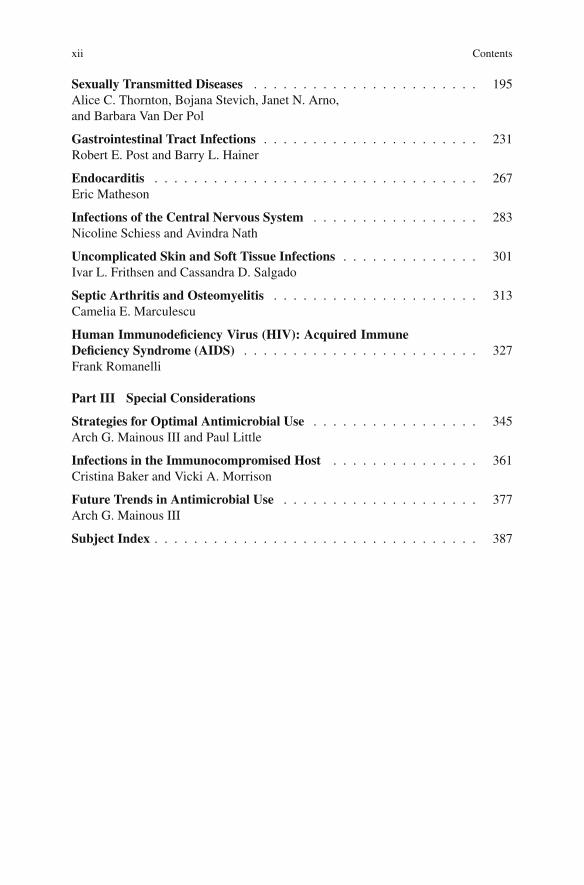
xii Contents
Sexually Transmitted Diseases . . . . . . . . . . . . . . . . . . . . . . . 195Alice C. Thornton, Bojana Stevich, Janet N. Arno,and Barbara Van Der Pol
Gastrointestinal Tract Infections . . . . . . . . . . . . . . . . . . . . . . 231Robert E. Post and Barry L. Hainer
Endocarditis . . . . . . . . . . . . . . . . . . . . . . . . . . . . . . . . . 267Eric Matheson
Infections of the Central Nervous System . . . . . . . . . . . . . . . . . 283Nicoline Schiess and Avindra Nath
Uncomplicated Skin and Soft Tissue Infections . . . . . . . . . . . . . . 301Ivar L. Frithsen and Cassandra D. Salgado
Septic Arthritis and Osteomyelitis . . . . . . . . . . . . . . . . . . . . . 313Camelia E. Marculescu
Human Immunodeficiency Virus (HIV): Acquired ImmuneDeficiency Syndrome (AIDS) . . . . . . . . . . . . . . . . . . . . . . . . 327Frank Romanelli
Part III Special Considerations
Strategies for Optimal Antimicrobial Use . . . . . . . . . . . . . . . . . 345Arch G. Mainous III and Paul Little
Infections in the Immunocompromised Host . . . . . . . . . . . . . . . 361Cristina Baker and Vicki A. Morrison
Future Trends in Antimicrobial Use . . . . . . . . . . . . . . . . . . . . 377Arch G. Mainous III
Subject Index . . . . . . . . . . . . . . . . . . . . . . . . . . . . . . . . . 387

Contributors
Janet N. Arno Indiana University School of Medicine, Indianapolis, IN, USA
Cristina Baker Staff Physician, Infectious Diseases, Hennepin County MedicalCenter, University of Minnesota, Minneapolis, MN, USA
Meredith Deutscher Division of Bacterial Diseases, Respiratory Diseases Branch,Centers for Disease Control and Prevention, National Center for Immunizations andRespiratory Diseases, Atlanta, GA, USA
Dimitri M. Drekonja Minneapolis Veterans Affairs Medical Center; Departmentof Medicine, University of Minnesota, Minneapolis, MN, USA
Martin E. Evans Division of Infectious Diseases, Department of InternalMedicine, University of Kentucky School of Medicine; Lexington Veterans AffairsMedical Center, Lexington, KY, USA
Cindy Friedman Division of Bacterial Diseases, Respiratory Diseases Branch,Centers for Disease Control and Prevention, National Center for Immunizations andRespiratory Diseases, Atlanta, GA, USA
Ivar L. Frithsen Department of Family Medicine, Medical University of SouthCarolina, Charleston, SC, USA
Barry L. Hainer Department of Family Medicine, Medical University of SouthCarolina, Charleston, SC, US
William J. Hueston Department of Family Medicine, Medical University of SouthCarolina, Charleston, SC, USA
James R. Johnson Minneapolis Veterans Affairs Medical Center; Department ofMedicine, University of Minnesota, Minneapolis, MN, USA
Paul Little Community Clinical Sciences, University of Southampton, UK
Arch G. Mainous III Department of Family Medicine, Medical University of SouthCarolina, Charleston, SC, USA
xiii

xiv Contributors
Camelia E. Marculescu Division of Infectious Diseases, Department of InternalMedicine, Medical University of South Carolina, Charleston, SC, USA
Craig A. Martin Department of Pharmacy Practice and Science, UK HealthCare,University of Kentucky; UK Chandler Hospital, Pharmacy Services H110,Lexington, KY, USA
Eric Matheson Department of Family Medicine, Medical University of SouthCarolina, Charleston, SC, USA
Vicki A. Morrison Staff Physician, Hematology/Oncology & Infectious Disease,Veterans Affairs Medical Center, University of Minnesota, Minneapolis, MN, USA
Avindra Nath Director Division of Neuroimmunology and Neurological infections,Departments of Neurology and Neuroscience, Baltimore, MD, USA
Hien H. Nguyen Division of Infectious Diseases, Division of Pulmonary CriticalCare Medicine, Department of Internal Medicine, Davis Health System, Universityof California, Sacramento, CA, USA
Chris Parsons Division of Infectious Diseases, Department of Internal Medicine,Medical University of South Carolina, Charleston, SC, USA
Robert E. Post Department of Family Medicine, Medical University of SouthCarolina, Charleston, SC, USA
Frank Romanelli Clinical Specialist in HIV/AIDS, College of Pharmacy,University of Kentucky, Lexington, KY, USA
Cassandra D. Salgado Division of Infectious Diseases, Department of Medicine,Medical University of South Carolina, Charleston, SC, USA
Christian Sandrock University of California Davis Health System, Sacramento,CA, USA
Nicoline Schiess Division of Neuroimmunology and Neuroinfectious Disease,Johns Hopkins Department of Neurology, Baltimore, MD, USA
Javeed Siddiqui Division of Infectious and Immunologic Diseases, School ofMedicine, University of California Davis, Sacramento, CA, USA
Bojana Stevich Division of Pharmacy, The University of Texas M.D. AndersonCancer Center, Houston, TX, USA
Vidya Sundareshan Division of Infectious Diseases, Department of InternalMedicine, University of Kentucky School of Medicine, Lexington, KY, USA
Alice C. Thornton Division of Infectious Diseases, Department of InternalMedicine, University of Kentucky School of Medicine, Lexington, KY, USA
Barbara Van Der Pol Indiana University School of Medicine Marion CountyHealth Department, Indianapolis, IN, USA

Antibiotic Resistance and Implicationsfor the Appropriate Use of Antimicrobial Agents
Meredith Deutscher and Cindy Friedman
1 Introduction
Antimicrobial resistance is a major public health threat associated with increasedmorbidity and mortality as well as enormous healthcare costs that are attributed tolonger hospital stays, which require multiple antimicrobial therapies. After recog-nizing antimicrobial resistance as a phenomenon and the need for a response, theInstitute of Medicine published a report in 1998, Antimicrobial Resistance: Issuesand Options [1]. The report asserted that antimicrobial resistance was accumulatingand accelerating while the tools for combating it were decreasing. It was estimatedthat antimicrobial resistance generated a minimum of $4 billion to $5 billion in coststo U.S. society and individuals annually. Some of the key areas delineated as prioritiesby the Institute of Medicine report were the following: establishment of surveillancefor antimicrobial resistance, understanding the use of antibiotics in food production,prolonging antibiotic effectiveness through educational programs and guidelines forappropriate antibiotic use, developing new products, and regulatory interventions.
In this chapter, we review the epidemiology of resistant pathogens, resis-tance mechanisms, and methods to measure and monitor antimicrobial resistance.Factors contributing to the development of antimicrobial resistance and measuresto control resistance are also discussed. Since these factors differ for community-acquired pathogens and hospital-acquired pathogens, the two groups are consideredseparately; the main focus is on community-acquired pathogens.
While antibiotic resistance is the focus of this chapter, antiviral resistance is alsoan important public health issue. For example, antiviral resistance to influenza andHIV has become a problem on a worldwide scale. While discussion of antiviralresistance is beyond the scope of this chapter, tools such as vaccines and diagno-sis of nonbacterial pathogens are important components to controlling antibioticresistance [2–4].
M. Deutscher (B)Division of Bacterial Diseases, Respiratory Diseases Branch, Centers for Disease Control andPrevention, National Center for Immunizations and Respiratory Diseases, Atlanta, GA, USAe-mail: [email protected]
1A.G. Mainous III, C. Pomeroy (eds.), Management of Antimicrobials in InfectiousDiseases, Infectious Disease, DOI 10.1007/978-1-60327-239-1_1,C© Springer Science+Business Media, LLC 2001, 2010

2 M. Deutscher and C. Friedman
2 Background and Epidemiology of Resistant Pathogens
The number of pathogens exhibiting antimicrobial resistance is increasing. Inrecent years, methicillin-resistant Staphylococcus aureus, vancomycin-resistantEnterococcus, multidrug-resistant tuberculosis, and amantadine/rimantadine-resistant and oseltamivir-resistant influenza virus have all emerged as public healththreats [5, 6]. The frequency and level of antimicrobial resistance and the result-ing implications on morbidity and mortality ultimately determine the public healthimpact of resistant pathogens.
2.1 Level of Antimicrobial Resistance
Almost every pathogen has acquired resistance to a therapeutic agent. To be consid-ered a public health burden, resistant pathogens must cause frequent and/or severeinfections, be managed with antimicrobial therapy as the standard of care, andhave few alternative drugs available for treatment. Several resistant pathogens havecreated a significant public health burden (Table 1).
Streptococcus pneumoniae is an example of a pathogen that meets these crite-ria. In 2006, national estimates of invasive pneumococcal disease in the UnitedStates were 41,400 cases and 5,000 deaths. Meningitis accounted for 6.1% ofcases, and bacteremia without focus accounted for 23.0% [7]. The case/fatalityratio may be higher than 25% for certain high-risk groups with bacteremia andmeningitis despite appropriate treatment [8, 9]. In 1994, drug-resistant S. pneu-moniae became a nationally notifiable disease in the United States, when sporadicreports of increasing infection and the active surveillance data from the Centersfor Disease Control and Prevention (CDC) documented an increase in antibiotic-resistant isolates [10]. In 2006, antibiotic susceptibility testing of isolates fromCDC’s Active Bacterial Core surveillance (ABCs) continued to show a large per-centage of resistant isolates; 25.6% were nonsusceptible to penicillin, 21.6% werenonsusceptible to erythromycin, and 22.7% were nonsusceptible to trimethoprim/sulfamethoxazole [7].
Chloroquine-resistant malaria is a problem of public health importance on aglobal scale. Forty-one percent of the world’s population live in areas where malariais transmitted [11]. Each year, 350–500 million cases of malaria occur, and approx-imately one million people die of this infection [12]. Plasmodium falciparumresistance to chloroquine has been confirmed in almost all areas with P. falciparummalaria; exceptions are the Dominican Republic, Haiti, Central America west ofthe Panama Canal, Egypt, and some other countries in the Middle East [13]. Insome regions, as many as 90% of the parasites may be resistant to chloroquine [14].Major contributors to the development of parasitic resistance have been the limitednumber of available antimalarial drugs and inadequate dosing for malaria treatment[15, 16].

Antibiotic Resistance and Implications for the Appropriate Use of Antimicrobial Agents 3
Tabl
e1
Exa
mpl
esof
antim
icro
bial
-res
ista
ntm
icro
orga
nism
sof
publ
iche
alth
impo
rtan
ce
Mic
roor
gani
smM
echa
nism
sof
resi
stan
cePe
rcen
tres
ista
nce
inU
nite
dSt
ates
aC
DC
estim
ated
annu
alre
sist
ant
infe
ctio
nsin
Uni
ted
Stat
esR
efer
ence
s
Gra
m-p
osit
ive
bact
eria
Peni
cilli
n-no
nsus
cept
ible
Stre
ptoc
occu
spn
eum
onia
ebTa
rget
alte
ratio
n:PB
P25
.6%
10,6
00[7
,31]
Mac
rolid
e-re
sist
antS
trep
toco
ccus
pneu
mon
iaec
Targ
etal
tera
tion:
ribo
som
alm
ethy
lase
sA
ctiv
eef
flux
21.6
%8,
940
[7,1
13]
Van
com
ycin
-res
ista
ntE
nter
ococ
cus
(nos
ocom
ial)
dTa
rget
alte
ratio
n:ce
llw
all
Bac
teri
alre
gula
tory
syst
emal
tera
tion
33%
No
estim
ate
[114
,115
]
Met
hici
llin-
resi
stan
tSt
aphy
loco
ccus
aure
us(n
osoc
omia
l)
Targ
etal
tera
tion:
PBP
56%
No
estim
ate
[31,
115–
117]
Van
com
ycin
-int
erm
edia
teSt
aphy
loco
ccus
aure
use
Unk
now
nFe
wre
port
edca
ses
inw
orld
<10
[118
]
Gra
m-n
egat
ive
bact
eria
Fluo
roqu
inol
one-
resi
stan
tN
eiss
eria
gono
rrho
eaef,
gTa
rget
alte
ratio
n:D
NA
gyra
sean
dto
pois
omer
ase
15.1
%31
,900
[119
–124
]
Thi
rd-g
ener
atio
nce
phal
ospo
rin-
resi
stan
tK
lebs
iell
apn
eum
onia
e(n
osoc
omia
l)
Ant
ibio
ticm
odifi
catio
n:E
SBL
s24
%N
oes
timat
e[3
9,11
5,11
6]
Imip
enem
-res
ista
ntP
seud
omon
asae
rugi
nosa
Dec
reas
edpe
rmea
bilit
y24
%N
oes
timat
e[3
4,11
5,11
6,12
5,12
6]A
cid-
fast
bact
eria
INH
-an
dri
fam
pin-
resi
stan
tM
ycob
acte
rium
tube
rcul
osis
Targ
etal
tera
tion
orin
crea
sed
targ
etpr
oduc
tion
0.9%
nopr
ior
TB
4.2%
with
prio
rT
B11
6M
DR
case
s[1
7,12
7]

4 M. Deutscher and C. Friedman
Tabl
e1
(con
tinue
d)
Mic
roor
gani
smM
echa
nism
sof
resi
stan
cePe
rcen
tres
ista
nce
inU
nite
dSt
ates
aC
DC
estim
ated
annu
alre
sist
ant
infe
ctio
nsin
Uni
ted
Stat
esR
efer
ence
s
Viru
ses
Zid
ovud
ine-
resi
stan
thum
anim
mun
odefi
cien
cyvi
rush
Targ
etal
tera
tion:
vira
lrev
erse
tran
scri
ptas
e1–
2%in
new
case
s40
0–80
0i[5
1,12
8–13
1]
Am
anta
dine
/rim
anta
dine
-res
ista
ntin
fluen
zaM
odifi
edst
ruct
ural
prot
ein
(M2
prot
ein)
99.8
%j
10.6
%k
5–20
%of
popu
latio
nin
fect
edw
ithin
fluen
zaan
nual
ly[6
]
Ose
ltam
ivir
-res
ista
ntin
fluen
zal
Enz
yme
mod
ifica
tion:
mut
atio
nin
neur
amin
idas
e0%
j 11%
k
0%m
5–20
%of
popu
latio
nin
fect
edw
ithin
fluen
zaan
nual
ly[6
]
Acy
clov
ir-r
esis
tant
herp
essi
mpl
exvi
rusn
Inhi
bitio
nof
drug
activ
atio
n:m
utat
ion
invi
ralt
hym
idin
eki
nase
0.18
%(S
TD
clin
ic)
5.3%
(HIV
)N
oes
timat
e[1
32–1
34]
Fun
giFl
ucon
azol
e-re
sist
antC
andi
dasp
p.In
crea
sed
drug
efflu
xTa
rget
alte
ratio
n:cy
toch
rom
eP4
50In
crea
sed
targ
etpr
oduc
tion
Dec
reas
edce
llula
rpe
rmea
bilit
y
10.3
%o
4.0%
pN
oes
timat
e[1
35,1
36]
Para
site
sC
hlor
oqui
ne-r
esis
tant
Pla
smod
ium
falc
ipar
umIn
crea
sed
drug
efflu
xW
ides
prea
dw
orld
wid
e–
US
dise
ase
refle
cts
regi
onof
impo
rtat
ion
No
estim
ate
[12–
16,3
7]

Antibiotic Resistance and Implications for the Appropriate Use of Antimicrobial Agents 5
Abb
revi
atio
ns:E
SBL
,ext
ende
d-sp
ectr
umβ
-lac
tam
ase;
INH
,iso
niaz
id;P
BP,
peni
cilli
n-bi
ndin
gpr
otei
n;T
B,t
uber
culo
sis.
Mul
tidru
gre
sist
ance
isde
fined
asre
sist
ance
toth
ree
orm
ore
clas
ses
ofan
timic
robi
aldr
ugs
exce
ptin
the
case
ofM
.tub
ercu
losi
s,in
whi
chit
isde
fined
asre
sist
ance
toIN
Han
dri
fam
pin.
a Yea
rsva
rybe
twee
n20
00an
d20
07;s
eere
fere
nce
for
exac
tyea
r.bM
any
are
also
mul
tidru
gre
sist
ant.
Alth
ough
notl
iste
dhe
re,r
esis
tanc
eto
trim
etho
prim
-sul
fam
etho
xazo
leis
also
impo
rtan
t.c E
ryth
rom
ycin
.dE
nter
ococ
ciar
ein
trin
sica
llyre
sist
ant
toβ
-lac
tam
s,am
inog
lyco
side
s,cl
inda
myc
in,
fluor
oqui
nolo
nes,
and
trim
etho
prim
-sul
fam
etho
xazo
lean
dre
adily
acqu
ire
resi
stan
ceto
high
conc
entr
atio
nsof
β-l
acta
ms,
high
conc
entr
atio
nsof
amin
ogly
cosi
des,
tetr
acyc
line,
eryt
hrom
ycin
,fluo
roqu
inol
ones
,rif
ampi
n,ch
lora
mph
enic
ol,f
usid
icac
id,n
itrof
uran
toin
,in
addi
tion
toth
egl
ycop
eptid
esva
ncom
ycin
and
teic
opla
nin.
e Alth
ough
we
have
liste
don
lyva
ncom
ycin
,the
sest
aphy
loco
ccia
rem
ultid
rug
resi
stan
t.f C
ipro
floxa
cin.
gA
lthou
ghon
lyflu
oroq
uino
lone
sar
elis
ted,
quin
olon
es-r
esis
tant
N.g
onor
rhoe
aeis
also
peni
cilli
n-an
dte
trac
yclin
e-re
sist
ant.
hPr
imar
ym
utat
ions
asso
ciat
edw
ithzi
dovu
dine
and
othe
rnu
cleo
side
reve
rse
tran
scri
ptas
ein
hibi
tors
.i B
ased
onan
estim
ate
of40
,000
new
infe
ctio
nspe
rye
ar.
j Influ
enza
A(H
3N2)
.kIn
fluen
zaA
(H1N
1).
l Am
anta
dine
/rim
anta
dine
has
noac
tivity
agai
nst
influ
enza
Bvi
ruse
s.O
selta
miv
iran
dza
nam
ivir
have
activ
ityag
ains
tbo
thin
fluen
zaA
and
Bvi
ruse
s.C
DC
reco
mm
ends
the
use
ofos
elta
miv
iran
dza
nam
ivir
for
the
trea
tmen
tor
prev
entio
nof
influ
enza
.U
seof
aman
tadi
neor
rim
an-
tadi
neis
not
curr
ently
reco
mm
ende
ddu
eto
high
leve
lsof
resi
stan
cein
influ
enza
A(H
3N2)
viru
ses.
Gui
danc
eon
influ
enza
antiv
iral
use
can
befo
und
atht
tp://
ww
w.c
dc.g
ov/m
mw
r/pr
evie
w/m
mw
rhtm
l/rr5
606a
1.ht
m.
Add
ition
alin
form
atio
nof
antiv
iral
resi
stan
ceca
nbe
foun
dat
http
://w
ww
.cdc
.gov
/flu/
abou
t.qa.
antiv
iral
resi
stan
ce.h
tm.
mIn
fluen
zaB
.nA
cycl
ovir
requ
ires
activ
atio
nby
phos
phor
ylat
ion
byvi
ral
enzy
mes
–th
ymid
ine
kina
sein
the
case
ofhe
rpes
sim
plex
.C
ellu
lar
enzy
mes
com
plet
eth
eph
osph
oryl
atio
n.T
hen
the
drug
sta
rget
vira
lD
NA
poly
mer
ase
and
prev
ent
elon
gatio
nof
vira
lD
NA
bybe
ing
pref
eren
tially
inco
rpor
ated
into
the
elon
gatin
gvi
ralD
NA
chai
n,th
uste
rmin
atin
gfu
rthe
rvi
ralD
NA
repl
icat
ion.
oC
andi
dagl
abra
ta.A
mon
gN
orth
Am
eric
anis
olat
esco
llect
edin
the
AR
TE
MIS
Glo
balA
ntif
unga
lSur
veill
ance
Prog
ram
,200
1–20
02.
pC
andi
dapa
raps
ilos
is.A
mon
gU
Sis
olat
esco
llect
edin
the
AR
TE
MIS
Glo
balA
ntif
unga
lSur
veill
ance
Prog
ram
,200
1–20
05.

6 M. Deutscher and C. Friedman
The public health implications of tuberculosis have become more pronounced asthe pathogen has become increasingly resistant to antimicrobial therapy. In 2006,approximately 14,000 tuberculosis cases were diagnosed in the United States [17].Twelve percent of Mycobacterium tuberculosis strains in the United States are resis-tant to at least one drug [18]. A large global survey conducted from 1999 to 2002by the World Health Organization (WHO) and the International Union AgainstTuberculosis and Lung Disease documents that the problem of resistance is world-wide; resistant M. tuberculosis has been identified in approximately 70 countriesand regions. The median prevalence of resistance to any of the first-line antituber-culosis drugs in new cases of tuberculosis that were identified in 76 countries andgeographical settings was 10.2% (range 0.0–57.1). The median prevalence of mul-tidrug resistance in new cases was 1.0% (range 0.0–14.2) [18]. In the United States,interventions targeting multidrug-resistant tuberculosis (MDR-TB) have had someeffect; MDR-TB declined from 2.8 to 1.1% of total tuberculosis cases from 1993to 1998 [19]. The proportion of MDR-TB cases stabilized and, in 2005, were 1.2%[20].
Gonorrhea, the second most commonly reported notifiable disease in the UnitedStates, is another example of an infection where antimicrobial resistance is ofgreat concern. Approximately 14% of isolates tested in 2006 were resistant to flu-oroquinolones, which was up from 2.2% in 2002 [21, 22]. Consequently, CDCrecommends that treatment now be limited to a single class of antibiotics, thecephalosporins.
2.2 Implications for Morbidity and Mortality
Antimicrobial resistance results in increased morbidity and mortality. In one studythat assessed mortality rate in patients with hospital-acquired S. aureus bacteremia,the difference between the mortality rates of methicillin-resistant and methicillin-susceptible S. aureus was 22.2% [22]. An investigation evaluating patients withMDR-TB showed that only 85% of those with infection resistant to rifampin andisoniazid responded to alternative treatments and eventually had negative sputumcultures, compared with a 98–99% cure rate for drug-susceptible disease [23, 24].Among patients with resistant tuberculosis that resulted in treatment failure, 40%died [24]. The risk of death following diagnosis with HIV was found to be threefoldhigher among individuals diagnosed with multidrug-resistant HIV when comparedwith the overall group of HIV-infected individuals [25]. Resistant pneumococ-cal meningitis has been associated with persistently infected cerebrospinal fluid[26–28].
Clinicians often prescribe alternate empiric treatment based on knowledge ofpotential resistance or change treatment based on lack of clinical response, whichcould lead to an increase in mortality resulting from treatment with an ineffec-tive or less effective drug; how much the case of suboptimal alternative treatmentmay contribute to the impact of resistance on mortality is unknown. A cause of

Antibiotic Resistance and Implications for the Appropriate Use of Antimicrobial Agents 7
increased morbidity and mortality might be that medical management is compli-cated by drug resistance. Treatment with an antimicrobial to which an organism isresistant can result in treatment failure; thus, resistant infections may result in moreserious disease [29].
3 Cell Physiology and Genetics of Antimicrobial Drug Resistance
3.1 Cellular Physiologic Mechanisms
Three physiologic mechanisms cause most antimicrobial resistance: modification ordestruction of the antibiotic by enzymes, alteration of the antibiotic target sites, andchanges in antibiotic uptake or efflux by the microorganism. Each organism mayuse one or more of these strategies (Table 1) [30–33].
An example of a bacterium that depends on antibiotic modification is S. aureus,which produces the enzyme β-lactamase. This enzyme cleaves β-lactam rings,resulting in inactivation of β-lactam-based antibiotics [31, 33].
Resistance by target site alteration occurs when the antibiotic can reach itsusual target but is unable to act because of a change in that target. For penicillinto act against streptococci, the drug depends on binding to the target penicillin-binding proteins (PBPs). Because penicillin-resistant S. pneumoniae produces adifferent PBP with low affinity for penicillin, it is able to evade the drug’s effects[31, 33].
As a result of reduced permeability of their outer layer, bacteria can havedecreased uptake of antibiotic. Pseudomonas aeruginosa and Escherichia coli havean outer membrane with low permeability to antibiotics [34]. Because antibioticsmust be able to penetrate the cell by means of bacterial porins, the diffusion rate ofthe drugs is altered by changes in these porin channels. Loss of the porin requiredfor cellular entry of the antibiotic imipenem causes P. aeruginosa to developimipenem resistance [34]. Alternatively, cellular exiting of drug may be enhanced.Tetracycline resistance for a number of bacteria, including many enterobacteriaceae,some staphylococci, and some streptococci, results from active export of the antibi-otic out of the bacterial cell (drug efflux) [31, 35]. Increased drug efflux is also amechanism of chloroquine resistance in P. falciparum [15, 36, 37].
3.2 Genetic Basis for Resistance
There are three main types of genetic changes that lead to antimicrobial resistance:chromosomal mutations of common resistance genes, acquisition of resistancegenes carried on plasmids and other exchangeable genetic segments, and inducibleexpression of existing genes [31, 33, 34, 38, 39]. Each type of genetic variation hasimplications on a population level for surveillance and on an individual patient levelfor clinical management (Table 2).

8 M. Deutscher and C. Friedman
Table 2 Genetic mechanisms of antimicrobial resistance with public health and clinicalimplications
Genetic changesExamples ofpathogens
Surveillanceimplications Clinical implications
Chromosomalmutations –accumulated andsingle mutations
MycobacteriumtuberculosisHIVPlasmodiumfalciparumStaphylococcusaureus
Nonsusceptibilityprevalence willchange gradually,independent ofother drugresistances
Can test for specificdrug andmicroorganismcombinations, expectstability ofsusceptibility overshort term, selectivepressure over time inan individual patientis important
Plasmid and othergene segmentswhich areexchanged amongmicroorganisms(transposons,integrons, phagegenes)
Klebsiella(extended-spectrumβ-lactamases)Staphylococcusaureus
Nonsusceptibilityprevalence canchange suddenly,often with severaldrug resistanceslinked together
Anticipate co-resistance
Inducible expression EnterobacterVancomycin-resistantEnterococcus
Surveillance is notuseful becauseresistancedevelops duringtherapy
Anticipatemid-treatment failuredespite initialsusceptibility ofisolate
3.3 Chromosomal Mutations
Chromosomal mutations in common resistance genes can be spontaneous or can becomplex, accumulated mutations (Table 2). For example, M. tuberculosis acquiresresistance when chromosomal mutations alter the bacterial antibiotic target siteor cause the bacteria to overproduce the target [40]. MDR-TB develops whenmutations in individual chromosomal genes accumulate; the likelihood of a M.tuberculosis mutant being simultaneously resistant to two or more drugs is the prod-uct of individual probabilities of a single mutation [40]. Thus for the purposes ofsurveillance for antimicrobial resistance, organisms such as M. tuberculosis willgradually change their susceptibility patterns, and the development of resistance toeach drug is independent of the existing drug resistances (Table 2).
Chromosomal mutations hold implications for clinicians choosing treatment forindividual patients. Clinicians can expect microorganisms that typically acquirechromosomal mutations to have stable resistance patterns in the short term; yet,selective pressures in an individual patient will be very important over the longterm. This relative stability means that clinicians can test for resistance in a specificmicroorganism and tailor antimicrobial therapy accordingly (Table 2). Because theprobability of a multiply-resistant organism developing in one patient is the product

Antibiotic Resistance and Implications for the Appropriate Use of Antimicrobial Agents 9
of the probabilities of developing each resistance individually, a high load of theorganism in the infected person is needed for multiple resistance to develop, andtreatment with multiple drugs may prevent the emergence of resistance [40].
Plasmids and other exchangeable segments of genes such as transposons, genecassettes, integrons, and phage genes are more rapidly disseminated than are chro-mosomal mutations. Transposons are segments of DNA that have a repeat of aninsertion sequence element at each end and can migrate from one plasmid to anotherwithin the same bacterium, to a bacterial chromosome, or to a bacteriophage. Genecassettes are a family of discrete mobile genetic elements that each contain anantibiotic resistance gene and are dependent upon integrons for integration in chro-mosomes [41]. Integrons are receptor elements on the chromosome that provide thesite into which the gene cassette is integrated and provide the enzyme for integration[41].
One example of the role of these exchangeable gene segments is the plasmid-encoded extended-spectrum β-lactamases (ESBLs) in gram-negative organisms.ESBLs confer resistance to ampicillin, carbenicillin, ticarcillin, and the extended-spectrum cephalosporins. Their broad activity arises from amino acid substitutionsthat alter the configuration around the active site of the β-lactamase enzyme andthus increase the enzyme affinity for broad-spectrum β-lactam antibiotics [42].
The rapid exchangeability of plasmids or other exchangeable gene segmentshas several implications: (1) surveillance systems need the ability to detect suddenchanges in resistance patterns in a community, (2) resistances may be easily trans-ferred between bacterial species, and (3) resistances to several different drugs maytravel together. The clinician treating an individual patient must expect resistanceto multiple drugs when resistance to one drug occurs. This co-resistance prob-lem should always be anticipated, particularly when one resistance is known to becarried on an exchangeable element (Table 2). For example, lower respiratory tractpneumococcal isolates recovered from patients younger than 18 years of age inthe United States have been shown to have elevated rates of resistance to peni-cillin, azithromycin, and trimethoprim-sulfamethoxazole, and penicillin resistancecorrelated with co-resistance to these other antimicrobial agents [43]. Although S.pneumoniae co-resistances are not plasmid-borne, they are complex gene mosaicsthat appear to be tightly linked like those on plasmids.
3.4 Inducible Mechanisms
Inducible mechanisms cause resistance that arises during treatment with a givenantimicrobial agent. For example, treatment of influenza A with rimantadine reg-ularly results in the rapid emergence of resistant virus in the affected patient [44,45]. Several enterobacteriaceae possess a cephalosporinase that is not normallyexpressed, but certain cephalosporins will trigger expression of high concentrationsof the enzyme [46]. Effective surveillance for these inducible mechanisms is prob-lematic because they are not expressed phenotypically at baseline. For pathogens

10 M. Deutscher and C. Friedman
known to have inducible mechanisms of resistance, the clinician must be preparedfor mid-treatment failure despite initial sensitivity of the isolate.
4 Measuring Antimicrobial Resistance
4.1 Laboratory Testing
Laboratory testing for antimicrobial resistance is generally done using phenotypicassays, although for an increasing number of cases, genotype-based assays canprovide rapid information [47, 48].
Phenotypic assays are based on in vitro inhibition of growth of a microorganismin the presence of an antimicrobial. These assays are used for organisms that canbe cultured on artificial media – bacteria on agar or broth media and viruses incell culture. For bacteria, disk diffusion or broth–agar dilution methods are used todetermine the minimum inhibitory concentration (MIC) [47, 48]. The MIC is theminimum concentration of antimicrobial that will inhibit growth of the organism invitro. For viruses, drug susceptibility is expressed as the drug concentration that isrequired to inhibit viral replication by 50% (IC[50]) [49, 50].
Genotypic assays test for the presence of resistance genes that confer pheno-typic resistance. Although they are indirect, genotypic analyses are important fororganisms that are difficult to grow in culture; some viruses such as hepatitis B andC, papillomaviruses, and Norwalk-like virus cannot be cultivated at present. Whilesome assays need to be checked to make certain that genotype and clinical pheno-type correlate, genotypic assays are particularly advantageous for viruses becausein comparison to viral culture, which can take a week or more, many genotypictests are relatively quick to perform [49]. Types of assays include sequencing ofthe microorganism’s genome, restriction fragment length polymorphism assays, andline probe assays [49, 51].
Whereas phenotypic testing by disk diffusion for common bacterial resistancerequires basic technology and resources, many of the other resistance testing tech-niques are complex. A laboratory may be constrained by the limits of technologicalresources available and also by the limits of testing technology.
In the United States, the Clinical and Laboratory Standards Institute (CLSI)defines antimicrobial susceptibility for most pathogens of clinical interest. Resistantisolates are those organisms that are not inhibited by the usually achievable con-centrations of antimicrobials. Intermediate resistant isolates are those organismswith MICs that approach typically attainable blood and tissue concentrations ofantimicrobial drugs and for which response rates may be lower than for suscep-tible isolates. Susceptible isolates are those organisms for which an infection dueto the pathogen may be treated with the usual dosage of the antimicrobial drug[52]. Nonsusceptible refers to the combined categories of full and intermediate resis-tance. For MICs to be meaningfully interpreted, CLSI takes into account multiplefactors when defining the breakpoints for susceptibility for a given antimicrobialagent. These factors include in vitro activity, safety and tolerability of the drug,
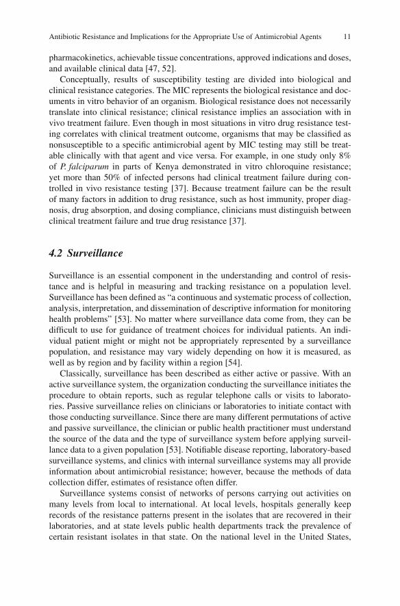
Antibiotic Resistance and Implications for the Appropriate Use of Antimicrobial Agents 11
pharmacokinetics, achievable tissue concentrations, approved indications and doses,and available clinical data [47, 52].
Conceptually, results of susceptibility testing are divided into biological andclinical resistance categories. The MIC represents the biological resistance and doc-uments in vitro behavior of an organism. Biological resistance does not necessarilytranslate into clinical resistance; clinical resistance implies an association with invivo treatment failure. Even though in most situations in vitro drug resistance test-ing correlates with clinical treatment outcome, organisms that may be classified asnonsusceptible to a specific antimicrobial agent by MIC testing may still be treat-able clinically with that agent and vice versa. For example, in one study only 8%of P. falciparum in parts of Kenya demonstrated in vitro chloroquine resistance;yet more than 50% of infected persons had clinical treatment failure during con-trolled in vivo resistance testing [37]. Because treatment failure can be the resultof many factors in addition to drug resistance, such as host immunity, proper diag-nosis, drug absorption, and dosing compliance, clinicians must distinguish betweenclinical treatment failure and true drug resistance [37].
4.2 Surveillance
Surveillance is an essential component in the understanding and control of resis-tance and is helpful in measuring and tracking resistance on a population level.Surveillance has been defined as “a continuous and systematic process of collection,analysis, interpretation, and dissemination of descriptive information for monitoringhealth problems” [53]. No matter where surveillance data come from, they can bedifficult to use for guidance of treatment choices for individual patients. An indi-vidual patient might or might not be appropriately represented by a surveillancepopulation, and resistance may vary widely depending on how it is measured, aswell as by region and by facility within a region [54].
Classically, surveillance has been described as either active or passive. With anactive surveillance system, the organization conducting the surveillance initiates theprocedure to obtain reports, such as regular telephone calls or visits to laborato-ries. Passive surveillance relies on clinicians or laboratories to initiate contact withthose conducting surveillance. Since there are many different permutations of activeand passive surveillance, the clinician or public health practitioner must understandthe source of the data and the type of surveillance system before applying surveil-lance data to a given population [53]. Notifiable disease reporting, laboratory-basedsurveillance systems, and clinics with internal surveillance systems may all provideinformation about antimicrobial resistance; however, because the methods of datacollection differ, estimates of resistance often differ.
Surveillance systems consist of networks of persons carrying out activities onmany levels from local to international. At local levels, hospitals generally keeprecords of the resistance patterns present in the isolates that are recovered in theirlaboratories, and at state levels public health departments track the prevalence ofcertain resistant isolates in that state. On the national level in the United States,
12 M. Deutscher and C. Friedman
CDC has several active surveillance systems that collect population-based resistancedata from laboratories around the country for certain bacteria.
Active Bacterial Core surveillance (ABCs) is an example of an active surveil-lance system that detects the emergence of antimicrobial resistance. ABCs, acomponent of CDC’s Emerging Infections Programs (EIP) network, is an activelaboratory- and population-based surveillance system for several invasive bacterialpathogens. For each case of invasive disease in the surveillance population, a casereport with demographic information is completed and bacterial isolates are sent toCDC and other reference laboratories for evaluation. ABCs data have been used totrack disease trends, such as emerging fluoroquinolone resistance in pneumococcaldisease among nursing home residents and decreasing macrolide resistance in pneu-mococcal disease after the introduction of the pneumococcal conjugate vaccine [55,56]. Data have also been used to track the emergence of serogroup Y meningococ-cal disease. Based on data collected by ABCs, a program to assist state and localhealth departments with surveillance for MRSA and drug-resistant S. pneumoniaehas been developed.
Heightened surveillance for a particular outcome can be useful for public healthgoals and for good patient care. Only a handful of vancomycin-intermediate andvancomycin-resistant S. aureus (VISA, VRSA) infections have occurred through-out the world. Because of the severity of disease with VISA or VRSA, lack ofavailable treatment options, and the potential for emergence of a fully resistantstrain, guidelines for heightened passive surveillance were developed. These guide-lines encourage enhanced reporting for VISA and VRSA infections so that they canbe identified rapidly, interventions can occur, and risk factors for infection can bedetermined [30, 57].
There are many barriers to population-based surveillance and use of the result-ing data [58]. At the most basic level, the laboratory performing the initial isolationmust have adequate resources and measurement capacity, and laboratories in a givensurveillance area must use standardized methods and reporting systems and be ableto communicate. There is also a lack of available data correlating local antimi-crobial utilization rates with resistance rates; the data would be useful for under-standing the drivers of resistance and monitoring interventions. Well-constructedpopulation-based active surveillance has the potential to provide accurate, repre-sentative, and timely information on changing rates and patterns of antimicrobialresistance.
5 Factors Promoting Antimicrobial Resistance and Measuresto Control Its Spread
5.1 Community-Acquired Pathogens
Antimicrobial exposure is the main factor promoting selective pressure and increas-ing the development of antimicrobial resistance in the community. Crowding and

Antibiotic Resistance and Implications for the Appropriate Use of Antimicrobial Agents 13
Table 3 Types of resistant pathogens and the implications for control measures
Community-acquired Hospital-acquired
Definition Resistant pathogen acquired in thecommunity
Resistant pathogen acquired inhospital
Example Drug-resistant Streptococcuspneumoniae
Vancomycin-resistantEnterococcus
Factorsassociatedwith increasedlikelihood ofresistance
Recent (previous 3 months)antibiotic use, community withhigh resistance rates, day care,schools, military
Parenteral antibiotics, prolongedantibiotics, empirical antibiotictherapy, surgical prophylaxis,poor infection control practices
Audience foreducation
Community medical practitioners,public
Hospital personnel
Control measures Appropriate antibiotic use byclinician, public education,improved diagnostic techniques,hand washing, vaccines, greateruse of animal vaccines
Formulary controls, good infectioncontrol precautions (handwashing), cohorting, laboratorysurveillance for resistance
other factors also contribute to the spread of resistant organisms (Table 3) [30,59–61]. While measures to curb the development of resistance among community-acquired pathogens are different for different pathogens, they all rely on somecombination of the following tools: (1) vaccines or other measures to preventdisease, (2) development of new antimicrobials to treat resistant infections, and(3) education promoting judicious use of antimicrobial agents to slow or haltdevelopment of antimicrobial resistance.
An example of a community-acquired pathogen in which exposure to antimi-crobials promotes resistance is S. pneumoniae [62]. S. pneumoniae is a frequentcause of outpatient respiratory infections, including otitis media, pneumonia, andsinusitis. The strongest risk for developing an infection with drug-resistant S. pneu-moniae (DRSP) is prior use of antibiotics, in particular during the three previousmonths [63, 64]. Other risk factors for DRSP infection relate either directly orindirectly to antibiotic exposure. These risk factors include young age, white race,higher income, suburban residence, and day care attendance [63, 65–68]. Day careattendance has been an important risk factor, probably because the environmentpresents a combination of frequent antibiotic usage with crowding and close con-tact of a large number of small children who share respiratory and other secretions[65, 67, 68].
Vaccines can prevent disease caused by resistant pathogens. The current 7-valentpneumococcal conjugate vaccine not only reduces carriage of penicillin-susceptibleand penicillin-nonsusceptible strains of S. pneumoniae, but also reduces rates ofinvasive disease due to penicillin-nonsusceptible strains. Routine use of this pneu-mococcal conjugate vaccine to prevent pediatric disease has proved to be a valuabletool in controlling pneumococcal resistance [69, 70]. After vaccine introduction, therate of resistant invasive pneumococcal disease decreased substantially; between

14 M. Deutscher and C. Friedman
1999 and 2004, rates of penicillin-nonsusceptible invasive disease due to vaccinestrains decreased by 87% [70].
Emerging resistance continues to drive the need for the development of newantimicrobials. Unfortunately, there have been few new antimicrobials developedin recent years. Reasons for the decline of antimicrobial development are generallyrelated to cost. New drug development is estimated to cost $400–800 million perapproved agent [71]. Antimicrobials, which are typically used for a short duration,have a lower rate of financial return when compared with drugs that are used to treatchronic illnesses [72]. As a result, pharmaceutical companies may be more likely topromote research and development for drugs used to treat chronic conditions such ashypertension and diabetes. The lack of available guidance from the U.S. Food andDrug Administration (FDA) regarding the types of studies and evidence the FDAconsiders to be acceptable to demonstrate the safety and efficacy of new drugs hasbeen described as a deterrent to antimicrobial development by pharmaceutical andbiotechnology companies [73–75].
Antimicrobials have been overused for acute upper respiratory illnesses (ARIs).Five specific ARIs (upper respiratory tract infections, bronchitis, otitis media,sinusitis, and pharyngitis) account for the majority of ambulatory antibiotic pre-scriptions [76]. In 1998, there were approximately 84 million ambulatory officevisits for ARI, resulting in 45 million antibiotic prescriptions in the United States[77]. An estimated 55% of these antibiotics were used for infections that wereunlikely to be bacterial in origin [77]. Because ARIs constitute such a largeamount of unnecessary antibiotic use, and because antibiotic use has been associ-ated with carriage of resistant pneumococci and invasive disease, efforts to decreaseantimicrobial resistance have focused on judicious use of antimicrobial agents foroutpatient ARIs [64, 78–80].
Antibiotic use in children and adolescents, which increased dramatically duringthe 1980s, began to decline in the 1990s [76]. Still, the numbers of antibiotics pre-scribed remained high; for example, an estimated 108 antibiotic courses for otitismedia per 100 children under age 5 years were prescribed during 1996–1997 [81].Efforts to discover why physicians continued to prescribe antibiotics inappropriatelydespite guidance stating otherwise were conducted in the late 1990s. Focus groupswith parents, pediatricians, and family physicians have highlighted differences inphysician and parent perceptions about antimicrobial use [82]. Parents state thatthey want a clear explanation when a provider decides not to prescribe an antibi-otic. Parents also state that they believe that antibiotics are indicated if their childhas green nasal discharge. A third reason that patients seek antibiotics is for a needto return to work, school, or day care quickly. There is a perception that antibi-otics speed recovery from upper respiratory tract infections. Physicians indicate thatantibiotic prescribing could be safely reduced, but feel pressured to prescribe antibi-otics because of diagnostic uncertainty surrounding ARI diagnosis (i.e., no test tohelp them determine that the infection is viral) and because the short amount oftime they have with each patient does not allow time to explain the rationale for notprescribing an antibiotic.

Antibiotic Resistance and Implications for the Appropriate Use of Antimicrobial Agents 15
A variety of educational interventions address the problem of antibiotic overuse.Educational interventions aimed at consumers include fact sheets and brochures forparents of young children. These provide answers to commonly asked questionsabout using antibiotics for upper respiratory infections. Posters designed for use indoctors’ offices, clinics, and other healthcare facilities raise awareness about appro-priate antibiotic use for upper respiratory infections in children (Fig. 1). Educationalinterventions aimed at physicians include the following: imitation “prescription
Fig. 1 Example of poster targeted at consumers to raise awareness about appropriate antibioticuse

16 M. Deutscher and C. Friedman
pads” to provide physicians with written recommendations regarding symptomaticmanagement of viral respiratory illness (Fig. 2); practice-based small-group meet-ings to educate physicians about appropriate antibiotic use; detailing sheets onappropriate use of antibiotics similar to ones used by pharmaceutical companiespromoting new antibiotics; and practice guideline development [80, 83].
Several studies have evaluated the impact of both physician and patient inter-ventions (Table 4). In general, the interventions that work best were the ones thatwere multifaceted and targeted both consumers and providers. One study that evalu-ated the impact of these consumer and physician multifaceted interventions found asubstantial decrease in antibiotic prescription rates for adults diagnosed with bron-chitis, from 74% to 48% [84]. Another found a decrease of 19% in solid antibioticprescriptions per clinician and 11% in liquid antibiotic prescriptions per clinician[42].
To address the growing problem of antimicrobial resistance, in September 2003CDC launched Get Smart: Know When Antibiotics Work, a multifaceted nationalpublic service and media outreach campaign to educate consumers and providersabout appropriate antibiotic use. The objectives of this campaign are threefold: (1)promote appropriate antibiotic prescribing practices among medical providers, (2)decrease demand for antibiotics for viral upper respiratory tract infections by con-sumers, and (3) increase adherence to prescribed antibiotics [85]. The campaignprovided a kit which included public service announcements and media outreachtools to help state and county partners implement the campaign locally. Partnershipdevelopment with nonprofit and for-profit entities has helped to expand the reach ofthe Get Smart program.
Subsequent phases of the program were designed to reach key audiences, includ-ing Spanish-speaking and American Indian/Alaska Native populations. The GetSmart program has sponsored the development of educational curricula for med-ical students and residents and continuing education for healthcare providers andallied health professionals. The Get Smart program maintains a website that featurescampaign information and resources [85].
So has any progress been made in reducing antimicrobial prescribing rates inthe community? National prescribing data from the National Ambulatory MedicalCare Survey and the National Hospital Ambulatory Medical Care Survey haveshown a 20% decline in antibiotic prescribing from 1995/1996 and 2004/2005 forthe five ARI (pharyngitis, otitis media, sinusitis, URI, and acute bronchitis) out-patient diagnoses [86]. While overall use of antimicrobials in ambulatory patientshas decreased, use of expensive broad-spectrum antimicrobial agents has risen(Fig. 3) [87].
5.2 Hospital-Acquired Pathogens
Antimicrobial resistance within the hospital setting is addressed by preventingthe development of resistance in individual patients with previously suscepti-ble infections and controlling the spread of nosocomial pathogens [32]. CDC’s

Antibiotic Resistance and Implications for the Appropriate Use of Antimicrobial Agents 17
Fig. 2 Example of “prescription pad” with written recommendations regarding symptomaticmanagement

18 M. Deutscher and C. Friedman
Tabl
e4
Sum
mar
yof
publ
ishe
dco
ntro
lled
tria
lspr
omot
ing
appr
opri
ate
use
ofan
tibio
tics
[137
]
Pres
crip
tion
rate
decl
ine
Loc
atio
nof
stud
y(y
ear)
Setti
ngPr
ovid
ered
ucat
ion
Patie
nted
ucat
ion
Publ
iced
ucat
ion
Scop
eIn
terv
entio
nC
ontr
ol
Den
ver
[84]
(199
7–19
98)
HM
OPr
escr
ibin
gra
tefe
edba
ck,
smal
l-gr
oup
pres
enta
tions
,and
prac
tice
tips
for
with
hold
ing
antib
iotic
s(f
ull
inte
rven
tion
site
son
ly)
Offi
ce-b
ased
educ
atio
nal
mat
eria
lsin
clud
ing
post
ers
for
exam
room
san
dpa
tient
info
rmat
ion
shee
ts(l
imite
dan
dfu
llin
terv
entio
nsi
tes)
Hou
seho
ldm
ailin
gto
patie
nts
incl
udin
gpa
mph
lets
,re
frig
erat
orm
agne
ts,a
ndle
tter
from
clin
icdi
rect
or(f
ulli
nter
vent
ion
site
son
ly)
No
addi
tiona
leff
orts
aim
edat
gene
ral
publ
ic
Adu
ltbr
onch
itis
only
35%
(for
full
inte
rven
-tio
nsi
te)
3%
Bos
ton/
Seat
tle[1
38]
(199
7–19
98)
HM
OSm
all-
grou
pof
fice
pres
enta
tions
led
bype
diat
rici
an“p
eer
lead
ers,
”pr
escr
ibin
gra
tefe
edba
ck
Bro
chur
esm
aile
dto
patie
nts
atho
me;
post
ers
and
pam
phle
tsin
wai
ting
room
san
dex
amro
oms
No
addi
tiona
leff
orts
aim
edat
gene
ral
publ
ic
Res
pira
tory
cond
ition
sam
ong
child
ren
less
than
6ye
ars
old
18.6
%(3
–<36
mon
thol
d)15
%(3
6–<
72m
onth
old)
11.5
%(3
–<36
mon
thol
d)9.
8%(3
6–<
72m
onth
old)

Antibiotic Resistance and Implications for the Appropriate Use of Antimicrobial Agents 19
Tabl
e4
(con
tinue
d)
Pres
crip
tion
rate
decl
ine
Loc
atio
nof
stud
y(y
ear)
Setti
ngPr
ovid
ered
ucat
ion
Patie
nted
ucat
ion
Publ
iced
ucat
ion
Scop
eIn
terv
entio
nC
ontr
ol
Tenn
esse
e[1
39]
(199
7–19
98)
Met
ropo
litan
area
,m
edic
aid-
man
aged
care
enro
llees
Lec
ture
sto
targ
eted
prov
ider
sin
mul
tiple
setti
ngs
(pri
mar
yca
recl
inic
s,gr
and
roun
ds,h
ospi
tal
staf
fm
eetin
gs);
guid
elin
edi
stri
butio
n;ar
ticle
sin
coun
tyhe
alth
jour
nal
Bro
chur
esdi
stri
bute
dto
pare
nts
ofne
wbo
rns,
child
ren
inda
yca
rean
dK
–3rd
grad
e;pa
tient
educ
atio
nm
ater
ials
dist
ribu
ted
tota
rget
edpr
ovid
ers
Bro
chur
esdi
stri
bute
dto
hosp
itals
,clin
ics,
dent
ists
,per
sons
rece
ivin
gflu
vacc
ine,
and
phar
mac
ycl
ient
s;T
V,r
adio
,and
new
spap
erco
vera
ge;p
ublic
serv
ice
anno
unce
men
ts
Res
pira
tory
cond
ition
sam
ong
child
ren
less
than
15ye
ars
old
19%
8%
Wis
cons
iu[4
2](1
999)
Rur
al com
mun
ities
Gra
ndro
unds
pres
enta
tions
;pr
actic
e-ba
sed
smal
l-gr
oup
mee
tings
led
byph
ysic
ian
educ
ator
s;gu
idel
ine
dist
ribu
tion;
CD
Cfa
ctsh
eets
,pat
ient
educ
atio
nm
ater
ials
,vi
ralp
resc
ript
ion
pad
Info
rmat
ion
and
educ
atio
nal
mat
eria
lspr
esen
ted
tocl
inic
sby
proj
ect
nurs
es;“
cold
kits
”pr
ovid
edto
clin
ics
for
dist
ribu
tion
toad
ults
and
adol
esce
nts
Edu
catio
nfo
rch
ildca
repr
ovid
ers,
publ
iche
alth
agen
cies
,par
ent
grou
ps,a
ndco
mm
unity
orga
niza
tions
;ed
ucat
iona
lm
ater
ials
(bro
chur
esan
dpo
ster
s)di
stri
bute
dto
clin
ics,
phar
mac
ies,
child
care
faci
litie
s,an
dsc
hool
s
Res
pira
tory
cond
ition
sam
ong
child
ren
11% (l
iqui
ds)a
19%
(sol
ids)
b
(+12
%)
(liq
uids
)8%
(sol
ids)

20 M. Deutscher and C. Friedman
Tabl
e4
(con
tinue
d)
Pres
crip
tion
rate
decl
ine
Loc
atio
nof
stud
y(y
ear)
Setti
ngPr
ovid
ered
ucat
ion
Patie
nted
ucat
ion
Publ
iced
ucat
ion
Scop
eIn
terv
entio
nC
ontr
ol
Ala
ska
[140
](1
998–
2000
)
Rur
alvi
llage
sW
orks
hops
for
com
mun
ityhe
alth
aide
san
dph
ysic
ians
Pres
enta
tions
atvi
llage
-wid
em
eetin
gs;h
ealth
new
slet
ters
mai
led
toho
mes
;hi
gh-s
choo
led
ucat
ion;
heal
thfa
irs
Res
pira
tory
cond
ition
sam
ong
child
ren
and
adul
ts
31%
9.5%
aL
iqui
dan
tibio
tics.
bSo
lidan
tibio
tics.

Antibiotic Resistance and Implications for the Appropriate Use of Antimicrobial Agents 21
0
5
10
15
20
25
1993–94
1995–96
1997–98
1999–2000
2001–02
2003–04
2005–06
Prescriptions per 1,000 ambulatory care visits
NOTE: All trends shown are significant (p < .01). Quinolone estimates are for persons >=15 years of age. SOURCE: CDC/NCHS, NAMCS/NHAMCS, 1993–2006.
Amoxicillin/clavulanate potassium
Aziththromycin/clarithromycin
Quinolones
0
10
20
30
40
50
1993–94
1995–96
1997–98
1999–2000
2001–02
2003–04
2005–06
Prescriptions per 1,000 ambulatory care visits
NOTE: All trends shown are significant (p < .01).SOURCE: CDC/NCHS, NAMCS/NHAMCS, 1993–2006.
Amoxicillin/ampicillin
Erythromycins
Cephalosporins
0
4
8
12
16
1993–1994
1995–96
1997–98
1999–2000
2001–02
2003–04
2005–06
Prescriptions per 1,000 ambulatory care visits
NOTE: 1 (p < .01).SOURCE: CDC/NCHS, NAMCS/NHAMCS, 1993–2006.
Trimethoprim-sulfamethoxazle1
Other penicillins1
Tetracyclines
Fig. 3 Antibiotic prescribing in the ambulatory care setting, 1993–2004

22 M. Deutscher and C. Friedman
campaign to prevent antimicrobial-resistant infections in healthcare settings setforth measures for control of resistance in hospital-acquired pathogens whichinclude (1) preventing infection, (2) diagnosing and treating infections effec-tively, (3) appropriate antimicrobial prescribing practices, and (4) preventingtransmission [88].
Measures to prevent infection among hospitalized patients include the follow-ing: hand hygiene practices, proper catheter insertion and care, and use of specificperioperative measures to decrease the risk of surgical site infections.
Effective diagnosis and treatment of infection will also help control antimi-crobial resistance within the hospital setting. Availability of rapid, sensitive, andspecific diagnostic tests helps assure that the most appropriate and narrowestspectrum antimicrobial agent is used. Clinicians should base empiric treatmentof hospitalized patients with probable nosocomial infections on hospital surveil-lance antibiograms. Infectious diseases experts should be consulted to addressmanagement of complicated infections.
Antimicrobial resistance develops in response to the heavy use of antimicrobialagents in hospitals, and resistance to many drugs has been closely correlated withprevious use of that drug. An example of this is vancomycin-resistant Enterococcus(VRE); patients who have had exposure to vancomycin are more apt to developinfections with VRE [89]. VRE was first reported in 1986 and is thought to beassociated with the use of orally administered vancomycin for treating antibiotic-associated diarrhea in hospitals. Another example is methicillin-resistant S. aureus(MRSA), a pathogen in which resistance is associated with the use of semisyn-thetic penicillins. Nosocomial infections caused by MRSA have been increasing:2% of staphylococcal infections in U.S. intensive care units were MRSA in 1974,22% in 1995, and 64% in 2004 [90]. Resistance of gram-negative organismsto extended-spectrum β-lactam antibiotics is also on the rise. In 1997, amongKlebsiella pneumoniae strains isolated in the United States, resistance rates to cef-tazidime and other third-generation cephalosporins were 6.6%, 9.7%, 5.4%, and3.6% for bloodstream, pneumonia, wound, and urinary tract infections, respectively[91]. In 2003, 20.6% of all K. pneumoniae isolates from National NosocomialInfection Surveillance System intensive care units were resistant to thesedrugs [92].
Appropriate antimicrobial prescribing practices is another tool needed to con-trol resistance in hospital-acquired pathogens. Reducing antimicrobial use in orderto decrease antimicrobial resistance in the hospital setting is difficult, and strate-gies are still being developed to address this problem. Instituting antimicrobialpractice guidelines or protocols is one strategy hospitals use to decrease inappro-priate antimicrobial prescribing [93]. Use of practice guidelines has been associatedwith stable antimicrobial susceptibility patterns for both gram-positive and gram-negative bacteria [94, 95]. Re-evaluating whether prolonged antimicrobial therapyis actually necessary is another strategy used to decrease antimicrobial resistance.Patients in the intensive care unit who receive long courses of antibiotic therapy are

Antibiotic Resistance and Implications for the Appropriate Use of Antimicrobial Agents 23
at increased risk of developing infection with a resistant pathogen [96, 97]. Clinicaltrials assessing duration of antibiotic treatment on treatment effectiveness found thata shorter course of antibiotics is acceptable for patients with ventilator-associatedpneumonia who do not have bacteremia [97–99].
Formulary restrictions and preauthorization requirements for specific agents arealso used to control inappropriate antimicrobial use [100]. Implementation of theseprograms has led to short-term increased susceptibilities among some gram-negativepathogens [101, 102]. The Infectious Diseases Society of America (IDSA) andSociety for Healthcare Epidemiology of America (SHEA) guidelines for developinginstitutional programs to enhance antimicrobial stewardship note that “formularyrestriction and preauthorization requirements can lead to immediate and significantreductions in antimicrobial use and cost” [100].
Hospital clinicians should be educated about the appropriate use of antimi-crobials. Good communication between pharmacists, infection control profes-sionals, and clinicians may help facilitate this. However, educational interven-tions can be a challenging way to induce behavior change. Constraints ontime and persistent acceptance of long-held beliefs are difficult obstacles forany educational program to overcome [103]. Face-to-face educational interven-tions have been used to modify suboptimal practices of hospital clinicians.Computer interactions are another educational strategy used to decrease inap-propriate prescribing of antimicrobials in the hospital setting. Use of com-puterized order entry to follow antimicrobial prescribing practices within aninstitution may help determine which antimicrobial prescribing practices needto be addressed [104]. Computer-interaction systems have been successful inguiding and monitoring antimicrobial prescribing and have decreased the pre-valence of multidrug-resistant organisms in certain patient care units [95, 105, 106].
Preventing transmission, another tool in controlling antimicrobial resistance inhospitals, depends on good infection control practices. Spread occurs becausepatients in hospitals are in close proximity to each other, and there are many oppor-tunities for the transmission of resistant organisms. Transmission can occur byrespiratory droplets on the hands of healthcare personnel and visitors, as well as onequipment that has been insufficiently cleaned [107, 108]. Guidelines for prevent-ing transmission of infections in the healthcare setting, published by The HealthcareInfection Control Practices Advisory Committee in 2007, specifically address infec-tion control practices to prevent and control healthcare associated infections. Theguidelines include recommendations for the following practices: hand hygiene,personal protective equipment, respiratory hygiene and cough etiquette, patientplacement, patient care equipment and instruments/devices, care of the environ-ment, textiles, and laundry, safe injection practices, lumbar punctures, and workersafety [109]. Infection control professionals have successfully provided educationand feedback to housekeeping staff to improve cleaning of contaminated environ-mental surfaces [110]. Direct educational interventions to improve hand hygienehave also been successful [111, 112].

24 M. Deutscher and C. Friedman
Key Points
• Antimicrobial resistance is widespread and growing in scope. Few resistantinfections are completely untreatable, but many are associated with increasedmorbidity and mortality.
• Efforts to decrease inappropriate use of antimicrobial agents and decrease theantimicrobial pressure that drives natural selection for resistance hold promise forprolonging the life span of currently available antimicrobial agents. Surveillanceprograms to monitor and track resistance patterns are necessary to determine howbest to focus these efforts to control resistance.
• Measures to control resistance depend on the type of setting. Key measuresfor community-acquired pathogens are vaccines, development of new antimi-crobials, and education promoting judicious use of antimicrobial agent. Forhospital-acquired pathogens, key measures include preventing infection, effec-tive diagnosis and treatment, appropriate antimicrobial prescribing practices, andpreventing transmission.
References
1. Forum on Emerging Infections (1998). Antimicrobial Resistance: Issues and Options,Workshop report. Washington, DC: National Academy Press, Division of Health SciencesPolicy Institute of Medicine
2. Bridges CB, Thompson WW, Meltzer MI et al. (2000). Effectiveness and cost-benefit ofinfluenza vaccination of healthy working adults: A randomized controlled trial. J Am MedAssoc 284(13):1655–1663
3. Poehling KA, Zhu Y, Tang YW, Edwards K (2006). Accuracy and impact of a point-of-carerapid influenza test in young children with respiratory illnesses. Arch Pediatr Adolesc Med160(7):713–718
4. Sharma V, Dowd MD, Slaughter AJ, Simon SD (2002). Effect of rapid diagnosis of influenzavirus type a on the emergency department management of febrile infants and toddlers. ArchPediatr Adolesc Med 156(1):41–43
5. Deyde VM, Xu X, Bright RA et al. (2007). Surveillance of resistance to adamantanes amonginfluenza A(H3N2) and A(H1N1) viruses isolated worldwide. J Infect Dis 196(2):249–257
6. CDC. Influenza weekly report. (Accessed on May 20, 2008 http://www.cdc.gov/flu/weekly/index.htm)
7. Centers for Disease Control and Prevention (2007). Active Bacterial Core Surveillance(ABCs) report (2007) Emerging Infections Program Network, Streptococcus pneumoniae,2006. (Accessed on Mar 21, 2008 http://www.cdc.gov/ncidod/dbmd/abcs/reports.htm.)
8. CDC (1996). Defining the public health impact of drug-resistant Streptococcus pneumoniae:Report of a working group. MMWR 45:1–20
9. CDC (1997). Prevention of pneumococcal disease: Recommendations of the AdvisoryCommittee on Immunization Practices (ACIP). MMWR 46:1–25
10. Council of State and Territorial Epidemiologists (1994). National surveillance for drug-resistant Streptococcus pneumoniae (DRSP) invasive disease. CSTE Position Statement1994–10
11. World Health Organization (1997). World malaria situation in 1994, part 1. Wkly EpidemiolRec 72:269–276

Antibiotic Resistance and Implications for the Appropriate Use of Antimicrobial Agents 25
12. Guinovart C, Navia MM, Tanner M et al. (2006). Malaria: Burden of disease. Curr Mol Med6:137–140
13. CDC (2008). Health Information for International Travel 2008. Atlanta, GA: Centers forDisease Control, Department of Health and Human Services
14. Bjorkman A, Phillips-Howard PA (1990). The epidemiology of drug-resistant malaria. TransR Soc Trop Med Hyg 84:177–180
15. Krogstad DJ (1996). Malaria as a reemerging disease. Epidemiol Rev 18:77–8916. Olliaro P, Cattani J, Wirth D (1996). Malaria, the submerged disease. J Am Med Assoc
275:230–23317. CDC (2007). Reported Tuberculosis in the United States, 2006. Atlanta, GA: US Department
of Health and Human Services, Centers for Disease Control and Prevention18. Aziz MA, Wright A, Lazlo A et al. (2006). Epidemiology of antituberculosis drug resistance
(the Global Project on Anti-tuberculosis Drug Resistance Surveillance): An updated anal-ysis: WHO/International Union Against Tuberculosis and Lung Disease Global Project onAnti-tuberculosis Drug Resistance Surveillance
19. CDC (1998). Progress toward the elimination of tuberculosis – United States, 1998. MMWR48:732–736
20. CDC (2007). Trends in tuberculosis incidence – United States, 2006. MMWR 56:245–25021. CDC (2008). Sexually Transmitted Diseases Surveillance 2006 Supplement, Gonococcal
Isolate Surveillance Project (GISP) Annual Report 200622. Wang FD, Chen YY, Chen TL et al. (2008). Risk factors and mortality in patients with
nosocomial Staphylococcus aureus bacteremia. Am J Infect Control 36(2):118–12223. Goble M, Iseman MD, Madsen LA et al. (1993). Treatment of 171 patients with pulmonary
tuberculosis resistant to isoniazid and rifampin. N Engl J Med 328:527–53324. Chan ED, Laurel V, Strand MJ et al. (2004). Treatment and outcome analysis of 205 patients
with multidrug-resistant tuberculosis. Am J Respir Crit Care Med 169:1103–110925. Grover D, Copas A, Green H et al. (2008). What is the risk of mortality following diagnosis
of multidrug-resistant HIV-1? J Antimicrob Chemother 61(3):705–71326. Kleinman MB, Weinberg GA, Reynolds JK, Allen SD (1993). Meningitis with beta-lactam
resistant Streptococcus pneumoniae: The need for early repeat lumbar puncture. PediatrInfect Dis J 12:782–784
27. Lonks JR, Durkin MR, Meyerhoff AN et al. (1995). Meningitis due to ceftriaxone-resistantStreptococcus pneumoniae. N Engl J Med 332:893–894
28. Catalan MI, Fernandez JM, Vazquez A et al. (1994). Failure of cefotaxime in the treat-ment of meningitis due to relatively resistant Streptococcus pneumoniae. Clin Infect Dis 18:766–769
29. Friedland IR, McCracken GH (1994). Management of infections caused by antibiotic-resistant Streptococcus pneumoniae. N Engl J Med 331:377–382
30. Tenover FC, Hughes JM (1996). The challenges of emerging infectious diseases:Development and spread of multiply-resistant bacterial pathogens. J Am Med Assoc275:300–304
31. Jacoby GA, Archer GL (1991). New mechanisms of bacterial resistance to antimicrobialagents. N Engl J Med 324:601–612
32. Shlaes DM, Gerding DN, John JF et al. (1997). Society for Healthcare Epidemiology ofAmerica and Infectious Diseases Society of America Joint Committee on the Preventionof Antimicrobial Resistance: Guidelines for the prevention of antimicrobial resistance inhospitals. Clin Infect Dis 25:584–599
33. Hawkey PM (1998). The origins and molecular basis of antibiotic resistance. Br Med J317:657–660
34. Nikaido H (1994). Prevention of drug access to bacterial targets: Permeability barriers andactive efflux. Science 264:382–388
35. Levy SB (1992). Active efflux mechanisms for antimicrobial resistance. Antimicrob AgentsChemother 36:695–703

26 M. Deutscher and C. Friedman
36. White NJ (1992). Antimalarial drug resistance: The pace quickens. J Antimicrob Chemother30:571–585
37. Barat LM, Bloland PB (1997). Drug resistance among malaria and other parasites. InfectDis Clin North Am 11:969–987
38. Davies J (1994). Inactivation of antibiotics and the dissemination of resistance genes.Science 264:375–382
39. Gold HS (1996). Antimicrobial drug resistance. N Engl J Med 335:1445–145340. Rattan A, Kalia A, Ahmad N (1998). Multidrug resistant Mycobacterium tuberculosis:
Molecular perspectives. Emerg Infect Dis 4:195–20941. Hall RM (1997). Mobile gene cassettes and integrons: Moving antibiotic resistance genes in
gram-negative bacteria. Ciba Foundation Symposium, 192–20242. Belongia EA, Sullivan BJ, Chyou PH et al. (2001). A community intervention trial to
promote judicious antibiotic use and reduce penicillin-resistant Streptococcus pneumoniaecarriage in children. Pediatrics 108(3):575–583
43. Karlowsky JA, Thornsberry C, Jones ME et al. (2003). Factors associated with rela-tive rates of antimicrobial resistance among Streptococcus pneumoniae in the UnitedStates: results from the TRUST surveillance program (1998–2002). Clin Infect Dis 36:963–970
44. Hayden FG, Belshe RB, Clover RD et al. (1989). Emergence and apparent transmission ofrimantadine-resistant influenza A virus in families. N Engl J Med 321:1696–1702
45. Hall CB, Dolin R, Gala CL et al. (1987). Children with influenza A infection: Treatmentwith rimantadine. Pediatrics 80:275–282
46. Neu HC (1992). The crisis in antibiotic resistance. Science 257:1064–107347. Jorgenson JH (1997). Laboratory issues in the detection and reporting of antibacterial
resistance. Infect Dis Clin North Am 11:785–80248. US Congress Office of Technology Assessment (1995). Impacts of Antibiotic Resistant
Bacteria. Washington, DC: US Government Printing Office, OTA-H-2949. Ballard AL, Cane PA, Pillay D (1998). HIV drug resistance: Genotypic assays and their
possible applications. Sex Transm Infect 74:243–24850. Hirsch MS, Conway B, D’Aquila RT et al. (1998). Antiretroviral drug resistance testing in
adults with HIV infection. J Am Med Assoc 279:1984–199151. Pillay D (1998). Emergence and control of resistance to antiviral drugs in resistance in herpes
viruses, hepatitis B viruses, and HIV. Commun Dis Public Health 1:5–1352. Dowell SF, Butler JC, Giebink GS et al. (1999). Acute otitis media: Management and surveil-
lance in an era of pneumococcal resistance – a report from the Drug-resistant Streptococcuspneumoniae Therapeutic Working Group. Pediatr Infect Dis J 18:1–9
53. Buehler JW (1998). Surveillance. In: Rothman KJ, Greenland S, (eds) ModernEpidemiology, 2nd ed. Philadelphia, PA: Lippincott-Raven
54. Centers for Disease Control and Prevention (1998). Active Bacterial Core surveillance(ABCs) report, Emerging Infections Program Network, Streptococcus pneumoniae, 1998.(Accessed on Jan 3, 2008 http://www.cdc.gov/ncidod/dbmd/abcs)
55. Stephens DS, Zughaier SM, Whitney CG et al. (2005). Incidence of macrolide resistance inStreptococcus pneumoniae after introduction of the pneumococcal conjugate vaccine. Lancet365(9462):855–863
56. Pletz MW, Shergill AP, McGee L et al. (2006). Prevalence of first-step mutants amonglevofloxacin-susceptible invasive isolates of Streptococcus pneumoniae in the United States.Antimicrob Agents Chemother 50(4):1561–1563
57. Hageman JC, Patel JB, Carey RC, Tenover FC, Mcdonald LC (2006). Investigation andcontrol of vancomycin-intermediate and -resistant Staphylococcus aureus: A guide for healthdepartments and infection control personnel (2006). Atlanta, GA. (Accessed on Feb 12, 2008http://www.cdc.gov/ncidod/dhqp/ar_visavrsa_prevention.html)
58. Fidler DR (1998). Legal issues associated with antimicrobial drug resistance. Emerg InfectDis 4:169–177

Antibiotic Resistance and Implications for the Appropriate Use of Antimicrobial Agents 27
59. Seppala H, Klaukka T, Vuopio-Varkila J et al. (1997). The effect of changes in the consump-tion of macrolide antibiotics on erythromycin resistance in group A streptococci in Finland.N Engl J Med 337:441–446
60. Dowell SF, Whitney CG, Schwartz B (1997). Regional Characteristics of Drug-ResistantRespiratory Pathogens. Series: New Challenges in Respiratory Tract Infections andCausative Pathogens. Part II: Diagnostic Perspectives and Antibiotic-Resistant Bacteria inLower Respiratory Tract Infections. Cohen MS, (ed) Am J Med Continuing Education Series9–16
61. Wainberg MA, Friedland G (1998). Public health implications of antiretroviral therapy andHIV drug resistance. J Am Med Assoc 279:1977–1983
62. Chen DK, McGeer A, De Azavedo JC et al. (1999). Decreased susceptibilityof Streptococcus pneumoniae to fluoroquinolones in Canada. N Engl J Med 341:233–239
63. Levine OS, Farley M, Harrison L et al. (1999). Risk factors for invasive pneumococcal dis-ease in children: A population-based case-control study in North America. Pediatrics 103:(Accessed on Nov 19, 2007 http://www.pediatrics.org/cgi/content/full/103-3/e28)
64. Dowell SF, Schwartz B (1997). Resistant pneumococci: Protecting patients through judi-cious use of antibiotics. Am Fam Physician 55:1647–1654
65. American Academy of Pediatrics (1997). Children in Out-of-Home Child Care. In: PeterG (eds) Red Book: Report of the Committee on Infectious Diseases, 24th ed. Elk GroveVillage, IL: American Academy of Pediatrics
66. American Academy of Pediatrics (1997). School Health. In: Peter G (eds) 1997 Red BookReport of the Committee on Infectious Diseases, 24th ed. Elk Grove Village, IL: AmericanAcademy of Pediatrics
67. Grossman LB (2006). Infection Control in the Child Care Center and Preschool, 7th ed.Silverchair: Charlottesville, VA
68. American Public Health Association and American Academy of Pediatrics (1992). Caringfor Our Children. National Health and Safety Performance Standards: Guidelines for Out-of-Home Child Care Programs. Washington, DC: The Association
69. Whitney CG, Pilishvili T, Farley M et al. (2006). Effectiveness of seven-valent pneumococ-cal conjugate vaccine against invasive pneumococcal diseases; a matched case-control study.Lancet 368:1495–1502
70. Kyaw MH, Lynfield R, Schaffner W et al. (2006). Effect of introduction of the pneu-mococcal conjugate vaccine on drug resistant Streptococcus pneumoniae. N Engl J Med354(14):1455–1463
71. DiMassa JA, Hansen RW, Grabowski HG (2003). The price of innovation: New estimates ofdrug development costs. J Health Econ 2003(22):151–185
72. Projan SJ (2004). Why is big pharma getting out of antibacterial drug discovery? Curr OpinMicrobiol 6:427–430
73. Blaser MJ, Bartlett JG (2006). Letter to FDA Commissioner Andrew C. vonEschenbach, MD. (Accessed on Oct 16, 2007 http://www.idsociety.org/WorkArea/downloadasset.aspx?id=3384)
74. Boucher HW (2008). Open public forum. In: FDA Anti-Infective Drugs AdvisoryCommittee: Joint with Drug Safety and Risk Management Advisory Committee. SilverSpring, MD
75. Spellberg B, Guidos R, Gilbert D et al. (2008). The epidemic of antibiotic-resistant infec-tions: A call to action for the medical community from the Infectious Diseases Society ofAmerica. Clin Infect Dis 46:155–164
76. McCaig LF, Hughes JM (1995). Trends in antimicrobial drug prescribing among office-based physicians in the United States. J Am Med Assoc 273:214–219
77. Gonzales R, Malone DC, Maelli JH et al. (2001). Excessive antibiotic use for acuterespiratory infections in the United States. Clin Infect Dis 33:757–762
28 M. Deutscher and C. Friedman
78. Arnold KE, Leggiadro RJ, Breiman RF et al. (1999). Risk factors for carriage of drug-resistant Streptococcus pneumoniae among children in Memphis, Tennessee. J Pediatr128:757–764
79. Hofmann J, Cetron MS, Farley M et al. (1995). The prevalence of drug-resistantStreptococcus pneumoniae in Atlanta. N Engl J Med 333:481–486
80. Dowell SF, Marcy SM, Phillips WR et al. (1998). Principles of judicious use of antimicrobialagents for pediatric upper respiratory tract infections. Pediatrics 101:163–165
81. US Department of Health and Human Services (2000). Healthy People 2010, 2nd ed.With Understanding and Improving Health and Objectives for Improving Health, 2 vols.Washington, DC: US Government Printing Office
82. Barden LS, Dowell SF, Schwartz B, Lackey C (1998). Current attitudes regarding use ofantimicrobial agents: results from physicians’ and parents’ focus group discussions. ClinPediatr 37(11):665–671
83. Gonzales R, Bartlett JG, Besser RE et al. (2001). Principles of appropriate antibiotic usefor treatment of acute respiratory tract infections in adults: Background, specific aims, andmethods. Ann Intern Med 134:479–486
84. Gonzales R, Steiner JF, Lum A, Barrett PHJ (1999). Decrease in antibiotic use in ambulatorypractice: Impact of a multidimensional intervention on the treatment of uncomplicated acutebronchitis in adults. J Am Med Assoc 281:1512–1519
85. Get Smart: Know When Antibiotics Work Campaign Information (Accessed on Nov 15,2007 http://www.cdc.gov/drugresistance/community/campaign_info.htm)
86. McCaig LF. Centers for Disease Control and Prevention, National Coordinating Center forHealth Information and Service, National Center for Health Statistics, Division of HealthCare Statistics. Personal communication
87. Steinman MA, Gonzales R, Linder J et al. (2003). Changing use of antibiotics in community-based outpatient practice 1991–1999. Ann Intern Med 138:525–533
88. Campaign to prevent antimicrobial resistance in healthcare settings. (Accessed on Dec 26,2007 http://www.cdc.gov/drugresistance/healthcare/overview.htm)
89. CDC (1994). Recommendations for preventing the spread of vancomycin resistance:Recommendations of the Hospital Infection Control Practices Advisory Committee(HICPAC). MMWR 44:1–13
90. Klevens RM, Edwards JR, Tenover FC et al. (2006). National Nosocomial InfectionsSurveillance System. Changes in the epidemiology of methicillin-resistant Staphylococcusaureus in intensive care units in the US hospitals, 1992–2003. Clin Infect Dis 42(3):389–391
91. Jones RN (2001). Resistance patterns among nosocomial pathogens: Trends over the pastfew years. Chest 119(2 Suppl):397S–404S
92. NNIS System (2003). National Nosocomial Infections Surveillance (NNIS) System Report,data summary from January 1992 through June 2003. Am J Infect Control 31(8): 481–498
93. Kollef MH (2006). Is antibiotic cycling the answer to preventing the emergence of bacterialresistance in the intensive care unit? Clin Infect Dis 43(Suppl 2):S82–S88
94. Pestotnik SL, Classen DC, Evans RS et al. (1996). Implementing antibiotic practice guide-lines through computer-assisted decision support: Clinical and financial outcomes. AnnIntern Med 124:884–890
95. Evans RS, Pestotnik SL, Classen DC et al. (1998). A computer-assisted managementprogram for antibiotics and other antiinfective agents. N Engl J Med 338:232–238
96. Dennesen PJW, van der Ven AJ, Kessels AGH et al. (2001). Resolution of infectious param-eters after antimicrobial therapy in patients with ventilator-associated pneumonia. Am JRespir Crit Care Med 163:1371–1375
97. Ibrahim EH, Ward S, Sherman G et al. (2001). Experience with a clinical guideline for thetreatment of ventilator-associated pneumonia. Crit Care Med 29:1109–1115
98. Chastre J, Wolff M, Fagon JY et al. (2003). Comparison of a 15 vs. 8 days of antibiotictherapy for ventilator-associated pneumonia in adults: A randomized trial. J Am Med Assoc290:2588–2598

Antibiotic Resistance and Implications for the Appropriate Use of Antimicrobial Agents 29
99. Micek ST, Lloyd AE, Ritchie DJ et al. (2005). Pseudomonas aeruginosa bloodstreaminfection: Importance of appropriate initial antimicrobial treatment. Antimicrob AgentsChemother 49:1306–1311
100. Dellit TH, Owens RC, McGowan JE et al. (2007). Infectious Diseases Society of Americaand the Society for Healthcare Epidemiology of America guidelines for developing aninstitutional program to enhance antimicrobial stewardship. Clin Infect Dis 44:159–177
101. White AC, Atmar RL, Wilson J et al. (1997). Effects of requiring prior authorization forselected antimicrobials: Expenditures, susceptibilities, and clinical outcomes. Clin InfectDis 25:230–239
102. Bamberger DM, Dahl SL (1992). Impact of voluntary vs. enforced compliance of third-generation cephalosporin use in a teaching hospital. Arch Intern Med 152:554–557
103. Goldmann DA, Weinstein RA, Wenzel RP et al. (1996). Strategies to prevent and control theemergence of spread of antimicrobial-resistant microorganisms in hospitals: A challenge tohospital leadership. J Am Med Assoc 275:234–240
104. Evans RS, Larsen RA, Burke JP et al. (1986). Computer surveillance of hospital-acquiredinfections and antibiotic use. J Am Med Assoc 256:1001–1007
105. Avorn J, Solomon DH (2000). Cultural and economic factors that (mis)shape antibiotic use:The nonpharmacologic basis of therapeutics. Arch Intern Med 133:128–135
106. Schiff GD, Rucker TD (1998). Computer prescribing. Building up electronic infrastructurefor better medication usage. J Am Med Assoc 279:1024–1029
107. Gamer J (1996). Hospital Infection Control Practices Advisory Committee. Guidelines forisolation precautions in hospitals. Am J Infect Control 24:24–52
108. CDC (1988). Guidelines for handwashing and hospital environmental control. MMWR37:377–382
109. Siegel JD, Rhinehart E, Jackson M et al. (2007). 2007 guideline for isolation precautions:Preventing transmission of infectious agents in healthcare settings
110. Eckstein BC, Adams DA, Rao A et al. (2007). Reduction of Clostridium difficile andvancomycin-resistant Enterococcus contamination of environmental surfaces after an inter-vention to improve cleaning methods. BMC Infect Dis 7:61
111. Hussein R, Khakoo R, Hobbs G (2007). Hand hygiene practices in adult versus pediatricintensive care units at a university hospital before and after intervention. Scand J Infect Dis39(6–7):566–570
112. Thomas M, Gillespie W, Krauss J et al. (2005). Focus group data as a tool in assessingeffectiveness of a hand hygiene campaign. Am J Infect Control 33(6):368–373
113. Shortridge VD, Doern GV, Brueggemann AB et al. (1999). Prevalence of macrolide resis-tance mechanisms in Streptococcus pneumoniae isolates from a multicenter antibioticresistance surveillance study conducted in the United States in 1994–1995. Clin Infect Dis29(5):1186–1188
114. Moellering RC (1998). Vancomycin-resistant enterococci. Clin Infect Dis 26:1196–1199115. CDC. Percent of Resistant Pathogens among Central Line-Associated Blood Stream
Infection (CLABSI), Ventilator-Associated Pneumonia (VAP), Catheter-Associated UTI(CAUTI) Healthcare-Acquired Infections; National Healthcare Safety Network (NHSN)January 2006–October 2007. In: Centers for Disease Control and Prevention
116. Fridkin SK, Gaynes RP (1999). Antimicrobial resistance in intensive care units. Clin ChestMed 20:303–316
117. Mulligan ME, Murray-Leisure KA, Ribner BS, Standiford HC, John JF, Korvick JA (1993).Methicillin resistant Staphylococcus aureus: A consensus review of the microbiology, patho-genesis, and epidemiology with implications for prevention and management. Am J Med94:313–328
118. Smith TL, Pearson ML, Wilcox KR et al. (1999). Emergence of vancomycin resistance inStaphylococcus aureus. N Engl J Med 340:493–501
119. CDC (2007). Sexually Transmitted Diseases Surveillance 2005 Supplement, GonococcalIsolate Surveillance Project (GISP) Annual Report 2005. Atlanta, GA: US Department ofHealth and Human Services, Centers for Disease Control and Prevention

30 M. Deutscher and C. Friedman
120. Sexually transmitted disease surveillance 2005 supplement: Gonococcal Isolate SurveillanceProject (GISP) annual report, 2005. Centers for Disease Control and Prevention,US Department of Health and Human Services, 2007. (Accessed on Apr 21, 2008http://www.cdc.gov/std/GISP2005/default.htm)
121. Ross JDC (1998). Fluoroquinolone resistance in gonorrhea: How, where and so what. Int JSTD AIDS 9:318–322
122. Erbelding E, Quinn TC (1997). The impact of antimicrobial resistance on the treatment ofsexually transmitted diseases. Infect Dis Clin North Am 11:889–903
123. Ison CA (1996). Antimicrobial agents and gonorrhoea: Therapeutic choice, resistance, andsusceptibility testing. Genitourin Med 72:253–257
124. Division of STD Prevention. Sexually transmitted disease surveillance of 1997 supplement:Gonococcal isolate surveillance project (GISP) annual report-1997. Atlanta: US Departmentof Health and Human Services, Public Health Service, Centers for Disease Control andPrevention; http://www.cdc.gov/ncidod/dastlr/gcdir/resist/gisp.html
125. Quinn JP, Dudek EJ, DiVincenzo CA et al. (1986). Emergence of resistance to imipenemduring therapy for Pseudomonas aeruginosa. J Infect Dis 154:289–294
126. Hancock REW (1998). Resistance mechanisms in Pseudomonas aeruginosa and othernonfermentative gram-negative bacteria. Clin Infect Dis 27(Suppl 1):S93–S99
127. CDC (2008). Trends in tuberculosis – United States, 2007. MMWR 57:281–285128. Hodinka RL (1997). What clinicians need to know about antiviral drugs and viral resistance.
Infect Dis Clin North Am 11:945–967129. Little S, Daar E, Keiser P et al. (1999). The spectrum and frequency of reduced antiretroviral
drug susceptibility with primary HIV infection in the United States [Abstract 121]. AntivirTher 4(Suppl 1):86
130. Wegner S, Brodine S, Mascola J et al. (1999). High frequency of antiretroviral drug resis-tance in HIV-1 from recently infected therapy-naive individuals [Abstract 119]. Antivir Ther4(Suppl 1):85
131. Weinstock H, Respess R, Heneine W et al. (2000). Prevalence of mutations associated withantiretroviral drug resistance among HIV-1 seroconverters in the United States, 1993–1998.J Infect Dis 182(1):330–333
132. Reyes M, Shaik NS, Graber JM et al. (2003). Task Force on Herpes Simplex VirusResistance. Acyclovir-resistant genital herpes among persons attending sexually transmitteddisease and human immunodeficiency virus clinics. Arch Intern Med 163(1):76–80
133. Laufer DS, Starr SE (1995). Resistance to antivirals. Pediatr Clin North Am 42:583–599134. Reyes M, Subedar N, Graber JM et al. (1998). Acyclovir-Resistant Herpes Simplex Virus
Pilot Surveillance System. San Diego, CA: International Society for Antiviral Resistance135. Pfaller MA, Messer SA, Boyken L et al. (2004). Geographic variation in the susceptibilities
of invasive isolates of Candida glabrata to seven systemically active antifungal agents: Aglobal assessment from the ARTEMIS Antifungal Surveillance Program conducted in 2001and 2002. J Clin Microbiol 42:3142–3146
136. Pfaller MA, Diekema DJ, Gibbs DL et al. (2008). Geographic and temporal trends inisolation and antifungal susceptibility of Candida parapsilosis: A global assessment fromthe ARTEMIS DISK Antifungal Surveillance Program, 2001 to 2005. J Clin Microbiol46:842–849
137. Weissman J, Besser RE (2004). Promoting appropriate antibiotic use for pediatric patients:A social ecological framework. Semin Pediatr Infect Dis 15:41–51
138. Finkelstein JA, Davis RL, Dowell SF et al. (2001). Reducing antibiotic use in children: Arandomized trial in 12 practices. Pediatrics 108(1):1–7
139. Perz JF, Craig AS, Coffey CS et al. (2002). Changes in antibiotic prescribing for childrenafter a community-wide campaign. J Am Med Assoc 287:3103–3109
140. Hennessy TW, Petersen KM, Bruden D et al. (2002). Changes in antibiotic-prescribingpractices and carriage of penicillin-resistant Streptococcus pneumoniae: A controlled inter-vention trial in rural Alaska. Clin Infect Dis 34:1543–1550

Part ISignificant Pathogens

Antimicrobial Resistance AmongEpidemiologically Important Gram-PositiveBacteria
Cassandra D. Salgado
1 Introduction
The emergence of antimicrobial resistance among clinically relevant bacteria hasresulted in profound changes in the approach to treatment of infections causedby these pathogens. This chapter will focus on three epidemiologically impor-tant gram-positive bacteria: Streptococcus pneumoniae, Staphylococcus aureus,and Enterococcus species. Common infections due to these organisms, commonresistance mechanisms, and available treatment options will be reviewed.
2 Streptococcus pneumoniae
The pneumococcus, S. pneumoniae, is a gram-positive bacterium that replicates inchains when grown in a liquid medium. In the microbiology laboratory, pneumo-cocci have been traditionally identified by four standard reactions: (1) α-hemolysisof blood agar media; (2) catalase negativity; (3) susceptibility to optochin, and (4)solubility in bile salts. Several characteristics of the organism allow it to produceinfection in a susceptible host; however, it is the outer polysaccharide capsule thathas received the greatest amount of study and description. The capsule protects theorganism against phagocytosis and is responsible for the virulence characteristic ofthe strain. This capsular antigen provokes a type-specific protective immunity (ant-icapsular antibodies), which has served as the basis for the serotype identificationsystem of the organism. There are currently 90 different serotypes that have beenidentified; however, serotypes 6, 14, 18, 19, and 23 are the most prevalent, account-ing for the majority of disease worldwide [1]. The cell wall is covalently boundto the capsule and is composed primarily of glycopeptides. It is the antigens con-tained within the cell wall that cause the profound inflammatory reaction among
C.D. Salgado (B)Division of Infectious Diseases, Medical University of South Carolina, Charleston,SC, USAe-mail: [email protected]
33A.G. Mainous III, C. Pomeroy (eds.), Management of Antimicrobials in InfectiousDiseases, Infectious Disease, DOI 10.1007/978-1-60327-239-1_2,C© Springer Science+Business Media, LLC 2001, 2010

34 C.D. Salgado
infected hosts. Other components of the cell wall are responsible for attachmentof the organism and then subsequent entry into activated host cells. Activated hostcells up-regulate platelet-activating receptors on their surface. Phosphorylcholine, akey component in the pneumococcal cell wall, is responsible for inserting the bac-teria into these receptors, later to be taken into the cell by an endocytic vacuole.Additionally, most clinically relevant pneumococcal isolates produce an impor-tant virulence factor, pneumolysin, which is an effective cytotoxin responsible forinjuring neutrophils, endothelial cells, and alveolar epithelia.
S. pneumoniae is a common colonizing organism of the nasopharynx in humans.When cultured at any given single point in time, the prevalence varies by age group,with pneumococci present in 5–10% of adults and 20–40% of children. Infantsbecome colonized with S. pneumoniae, on average, at the age of 6 months, andthe initial serotype appears to persist for a mean of 4 months. Adults colonizedwith individual serotypes have been shown to harbor them for shorter periods oftime, usually 2–4 weeks [2]. The worldwide rate of invasive pneumococcal disease,defined as isolation of the organism from a normally sterile body site, has beenreported as 15 per 100,000 persons, per year [3], and is more common among thevery young (less than 2 years of age) and the elderly (more than 65 years of age).Fortunately, recent data from the United States have suggested that the incidenceof pneumococcal disease is decreasing, perhaps as a result of the use of the proteinconjugate vaccine in children [4].
Because the organism may be present in the nasopharynx, S. pneumoniae hasbeen recognized as a common cause of pneumonia, sinusitis, and otitis media. Theseinfections likely occur via direct spread and invasion. Less frequently, S. pneumo hasbeen reported as a cause of meningitis, endocarditis, peritonitis, or bone and jointinfections. These infections likely occur via hematogenous spread of the organismfrom transient or persistent bacteremia.
Over the last 40 years, S. pneumo has developed resistance to a variety ofantibiotics, including that to β-lactams, macrolides, tetracyclines, trimethoprim-sulfamethoxazole, and fluoroquinolones. Factors which increase a patient’s risk forhaving an antibiotic-resistant strain of S. pneumo have been described as previ-ous exposure to antibiotics; exposure to day care or pre-school; stay in a nursinghome or other long-term care facility; and having a history of a recent respiratoryinfection (including viral infections). Of all the classes of antibiotic resistance, themost clinically relevant and most studied among S. pneumo isolates is that towardpenicillin.
Penicillin inhibits S. pneumo by binding to proteins on the cell wall. Thesepenicillin-binding proteins (PBPs) are enzymes needed for synthesis of peptidogly-can. When these PBPs become altered, they have much less affinity for penicillin(and often other β-lactams). Traditionally, resistance to penicillin has been describedas concentration-dependent. This definition has been based upon achievable drugconcentrations in the cerebral spinal fluid (CSF); however, it has been determinedthat achievable drug concentrations in the CSF are often much lower than what canbe achieved in the plasma, inner-ear fluid, or alveolar fluid. Thus, it is important torealize this when considering whether or not an isolate is resistant and may depend

Antimicrobial Resistance Among Epidemiologically Important Gram-Positive Bacteria 35
upon where the infection is located. In the microbiology laboratory, S. pneumo isdefined as susceptible to penicillin when the MIC <0.06 μg/mL; intermediatelyresistant to penicillin when the MIC is 0.10–1.0 μg/mL; and highly resistant topenicillin when the MIC >2.0 μg/mL. In the United States, approximately 60%of S. pneumo are susceptible to penicillin, 20% have intermediate resistance, and20% are highly resistant, but this percentage may vary depending on the region [5].Children are colonized and infected with more resistant strains compared to adultsand in general, invasive isolates tend to be more susceptible than those that causeotitis media. Fortunately, in the United States, use of the 7-valent protein conjugatepneumococcal vaccine has resulted in an 80% reduction in invasive disease and a>95% decrease in invasive S. pneumo isolates which are covered by the vaccine[4]. However, not entirely unexpected, there has been an increase among strainsthat are not covered in the vaccine (type 6 (non-B), 19 (non-F), 35, 11, and 15)and, unfortunately, these strains have demonstrated resistance to antibiotics, as well.Resistance to cephalosporins follows a similar concept and susceptibility to ceftri-axone, a common third-generation cephalosporin used for treatment of S. pneumoinfections, is defined in the microbiology lab as follows: susceptible if the MIC<1.0 μg/mL; intermediately resistant if the MIC = 2.0 μg/mL; and resistant if theMIC >4.0 μg/mL.
Also important to consider when making treatment decisions is that in the UnitedStates, almost 30% of S. pneumo isolates are resistant to macrolides, but this variesdramatically depending on the region; up to 10% are resistant to clindamycin; lessthan 5% are resistant to fluoroquinolones (although this may be higher among long-term care facility residents); 20% are resistant to tetracyclines; and almost one-thirdare resistant to trimethoprim-sulfamethoxazole [5, 6].
Because the achievable drug concentration differs depending on the body site,therapeutic decisions may differ, depending on the site infected. For Example, totreat all but the most resistant S. pneumo isolates, recommended first-line antibi-otic therapy for otitis media and sinusitis has been higher dose amoxicillin given at90 mg/kg per day, divided into twice or thrice daily doses. If treatment failure occursor the patient has a penicillin allergy, one might consider a macrolide antibiotic, andif one suspects cross-resistance to penicillin as well as the macrolide class of antibi-otics, alternatives such as clindamycin or a third-generation cephalosporin should beconsidered. For pneumonia and bacteremia, there is debate regarding whether or notinfection due to penicillin-resistant strains is associated with a worse outcome whencompared to infection due to susceptible strains. Some studies have shown that theelderly and those with underlying co-morbid conditions do worse when sufferingfrom pneumonia due to a β-lactam-resistant S. pneumo and, thus, many recommenda β-lactam plus a macrolide for patients presenting with community-acquired pneu-monia where S. pneumo is a consideration [7]. For bacteremia in a normal host,most experts recommend cefuroxime, cefotaxime, or ceftriaxone at standard doses,as the plasma levels achievable typically exceed the desired MIC. Meningitis canbe associated with extremely poor outcomes when not treated appropriately and theachievable drug concentration in the CSF is lower than that achievable in plasmaor alveoli. Thus, the treatment recommendations for meningitis suspected to be due
36 C.D. Salgado
to S. pneumo in an area where isolates exist with intermediate or high resistance topenicillin and/or ceftriaxone (or the patient has risk factors for an antibiotic-resistantstrain) are for higher dose third-generation cephalosporins such as cefotaxime 2 gIV q4h or ceftriaxone 2 g IV q12h, plus vancomycin. Of note, vancomycin does notpenetrate the blood–brain barrier well, so, once susceptibilities return, if treatmentcan be continued with a β-lactam, this is desirable.
Unlike many bacteria that cause significant disease, there are vaccines availablefor prevention of S. pneumo infection. The 7-valent pneumococcal conjugate vac-cine (Prevnar R©), released in 2000, is recommended for children under the age of2 years. Pneumococcal vaccines for the prevention of disease among children andadults who are 2 years and older are the Pneumovax R© and the Pnu-Immune R©.These vaccines are 23-valent polysaccharides currently recommended for use inall adults who are older than 65 years and for persons who are 2 years and olderand at high risk for disease (e.g., sickle cell disease, HIV infection, or otherimmunocompromising condition).
3 Staphylococcus aureus
S. aureus, a member of the Micrococcaceae, are named after their ability to growin grape-like clusters in solid media. In the microbiology laboratory, staphylo-cocci are characterized by a positive catalase test and identified as S. aureus by apositive coagulase test (indicating the presence of coagulase enzymes). Several vir-ulence factors have been described which may contribute to S. aureus’ ability tocause clinical disease in a susceptible host. These include toxins which act on cellmembranes; exotoxins such as toxic shock syndrome toxin and enterotoxins; leuko-cidin, which mediates the destruction of phagocytes; and catalases, coagulases, andhyaluronidases, which promote invasion and survival in tissue. Of particular inter-est is the Panton Valentine leukocidin (PVL) gene, which encodes for release of acytotoxin responsible for tissue necrosis and leukocyte destruction. The presence ofthis gene has been associated with infections of greater severity [8]. S. aureus is acommon colonizer of human skin and mucosa. It preferentially colonizes the ante-rior nares, particularly in adults, and at any given time 10–40% of the populationis transiently colonized with the organism. A small proportion may become chron-ically colonized with S. aureus and, as such, is often at increased risk for clinicaldisease.
The discovery of penicillin proved to be extremely valuable in the treatment ofinfections due to staphylococci; however, resistance to this agent developed rapidly.Currently, susceptibility rates to penicillin are in single digits and thus, use of syn-thetic penicillins has become commonplace since the development of methicillin inthe 1960s. Unfortunately, development of methicillin resistance among S. aureuswas detected within months of its release. Resistance to methicillin develops onceS. aureus acquires a large mobile genetic element, the staphylococcal cassette chro-mosome mec (SCCmec). Within this cassette the genes ccrA and ccrB mediatemobilization, and the mecA gene mediates β-lactam resistance. Specifically, mecA

Antimicrobial Resistance Among Epidemiologically Important Gram-Positive Bacteria 37
encodes for an altered penicillin-binding protein, PBP2a. This protein, when foundon the surface of the bacteria, has little affinity for β-lactam antibiotics and con-fers resistance to the entire class. Additionally, the mecA gene is often flankedby IS431, an insertion sequence that acts as a “collector” for additional antibiotic-resistance genes, thus promoting multidrug-resistant strains of MRSA. At least fivedifferent varieties of the SCCmec have been described (types I–V), based largelyupon their corresponding mecA genes. Types I–III are large in size and tend tocarry multiple antibiotic-resistance encoding genes. Types IV and V are smaller insize, are likely more mobile, and contain fewer antibiotic-resistance encoding genes.S. aureus resistant to methicillin has been defined in the laboratory as isolates withan MIC of > 16 mg/L to methicillin or an MIC of > 4 mg/L to oxacillin; how-ever, more rapid and often more dependable methods for laboratory identificationof methicillin resistance are those that actually detect the mecA gene, or the productof the gene, PBP2a.
MRSA has traditionally been considered an organism acquired within the health-care system, but over the past several years, an increasing number of reportshave described MRSA occurring among patients without this history. This emerg-ing epidemiology has led many to describe MRSA as either healthcare-associatedor community-associated, and the prevalence, resistance patterns, and clinicalsyndromes associated with the organism depend on this classification.
3.1 Healthcare-Associated MRSA
MRSA may be defined as healthcare-associated using a time-based definition suchas being isolated from a patient after at least 72 h of admission to a healthcare facil-ity. Additionally, healthcare-associated strains of MRSA tend to harbor the SCCmectypes I, II, or III and are therefore usually multidrug-resistant. MRSA amonghospitalized patients continues to be isolated with increased frequency and is animportant cause of hospital-acquired infections. These infections include pneumo-nia (including ventilator-associated pneumonia), device-associated infections suchas central-line associated bacteremia, and surgical site infections. According to datacollected from US hospitals, the proportion of hospital-acquired infections due toS. aureus that were resistant to methicillin has continued to increase over the pasttwo decades, and in 2004 approached 60% among ICU patients [9]. Experts suggestthat evolutionary changes in the microorganism, combined with ineffective (or non-compliance with) infection-control measures and selective pressure from antibioticuse have all likely contributed to the continued rise of MRSA.
Risk factors for acquisition of healthcare-associated MRSA include prolongedhospital stay, particularly those in ICUs; exposure to and prolonged use of antibi-otics; presence of severe underlying illness; receipt of invasive Procedures or foreignbodies; and being in close proximity to other MRSA-colonized or infected patients.Spread of MRSA in hospitals is thought to be largely due to patient-to-patienttransmission from the contaminated equipment and hands of healthcare providers.MRSA is important because when patients develop infection due to the organism,

38 C.D. Salgado
they suffer increased morbidity, mortality, and greater hospital costs than thosewho develop infection due to susceptible strains of the organism [10, 11]. A meta-analysis of studies of patients with S. aureus bacteremia reported that those withMRSA died almost twice as often, compared to those with methicillin-susceptibleS. aureus [10], and another study of patients with S. aureus surgical site infectionsfound that those with MRSA died more than three times as often, compared tothose with methicillin-susceptible S. aureus, and had a 1.9-fold increase in hospitalcosts [11].
4 Community-Associated MRSA
MRSA may also be defined as community-associated, using the time-basedapproach described above, such as if the organism is isolated from a patient at thetime of admission or within 48–72 h of admission to the hospital. Additionally,one may classify MRSA as community-associated if it is isolated from a patientpresenting in the outpatient clinical setting or emergency department. Community-associated strains of MRSA usually harbor SCCmec IV and, less often, SCCmec V,and thus may retain some susceptibility to other classes of antibiotics. Also of note,MRSA isolates of community origin are more likely to possess the gene responsi-ble for encoding PVL, a recognized virulence factor, and are often identified as theUSA 300 clone when subjected to pulsed-field gel electrophoresis. Recent studieshave described the frequency with which CA-MRSA has been occurring, as wellas important characteristics among individuals with infections due to the organism[12, 13]. A study of patients with MRSA infections from Baltimore, Atlanta, andMinnesota reported that from 2001 through 2002, 8–20% of the MRSA isolateswere classified as community-associated. The annual incidence of CA-MRSA dis-ease was significantly higher among those less than 2 years old, and sometimesamong blacks. Furthermore, the majority of infections (77%) were skin or soft tis-sue, but 6% were invasive in nature (e.g., bacteremias) and almost a quarter ofpatients with CA-MRSA infections required hospitalization. Also of interest wasthat among patients with CA-MRSA, many had contact with the healthcare system,such as visiting their physician within the previous year, or receiving antimicro-bial therapy [12]. Another recent study documented that MRSA was the single mostcommon identifiable cause of skin and soft-tissue infections among patients present-ing to emergency departments in 11 US cities. The overall prevalence of MRSA was59% and, among those, 97% were the USA 300 clone and 98% harbored SCCmecIV and were PVL positive [13]. Even though the majority of infections due to CA-MRSA have involved skin and soft tissue, the organism may also be a cause ofmore invasive infections, such as necrotizing fasciitis, bacteremia, or necrotizingpneumonia.
An important study was recently published and was the first to documentthe incidence and characteristics of invasive MRSA infections in nine US cities[14]. This population-based active case-finding study reported that, in 2005, morethan 94,000 invasive MRSA infections occurred, for an estimated incidence rate

Antimicrobial Resistance Among Epidemiologically Important Gram-Positive Bacteria 39
of 31.8 per 100,000 persons. These infections were associated with more than18,000 deaths, for an estimated mortality rate of 6.3 per 100,000 persons. Therewas geographic variability, but in general most invasive MRSA infections werehealthcare-associated; 58.4% were healthcare-associated community-onset infec-tions and 26.6% were healthcare-associated hospital-onset infections. Only 13.7%of invasive MRSA infections were community-associated. Additionally, molecularanalysis provided evidence that strains of community origin do cause a measurableamount of hospital-onset disease and in fact, 16% of invasive hospital-onset MRSAinfections were due to the USA 300 clone [14].
Treatment for serious infections due to MRSA had been somewhat limited toagents such as vancomycin; however, newer agents with activity against the organ-ism are now available. Vancomycin therapy requires an intravenous route, as wellas monitoring of blood levels in order to assure adequate dosing and to avoid poten-tial toxicity. Daptomycin, also given intravenously, is indicated for use in MRSA-and MSSA-complicated skin infections (at 4 mg/kg daily dose) and bacteremia,including right-sided endocarditis (at 6 mg/kg daily dose). Daptomycin levels donot need to be monitored; however, patients receiving the drug should be followedfor the onset of muscle pain or weakness and weekly CPK levels should be mea-sured. Another recently released intravenous agent with activity against MRSA istygecycline. Regarding MRSA, this agent has clinical indications only for treatmentof complicated skin and skin-structure infections at an initial dose of 100 mg, fol-lowed by 50 mg every 12 h. Linezolid, which can be given orally, has demonstratedpathogen-eradication rates and clinical efficacy comparable to that of vancomycinfor commonly encountered infections, including skin and soft-tissue infections [15],and it may be associated with a more favorable outcome when used for treatment ofMRSA nosocomial pneumonia (including ventilator-associated pneumonia) [16].Additionally, older agents such as trimethoprim-sulfamethoxazole, clindamycin,and often tetracyclines may have activity and are often used for outpatient man-agement of CA-MRSA infections, particularly those involving the skin and softtissue.
5 Vancomycin-Intermediate Staphylococcus aureusand Vancomycin-Resistant Staphylococcus aureus
The past decade has seen first the emergence of S. aureus with intermediateresistance to and later frank resistance to vancomycin. Vancomycin-intermediateS. aureus (VISA) is defined in the microbiology lab as having an MIC toward van-comycin of 8–16 mg/L and has been described as a cause of infection primarilyamong patients on hemodialysis receiving long courses of vancomycin for MRSAinfections [17]. The mechanism of resistance has been described as due to cell-wallthickening, which may cause the large vancomycin molecule to become trapped andunable to reach its functional targets. Vancomycin-resistant S. aureus (VRSA) isdefined in the microbiology lab as having an MIC toward vancomycin of >64 mg/L.The mechanism of resistance for this extremely worrisome pathogen is acquisition

40 C.D. Salgado
of the vanA gene from vancomycin-resistant Enterococcus (VRE). Emergence ofthis organism has originated in areas where MRSA and VRE have co-existed, andat least seven cases have been described in the United States [18]. Fortunately, theseVISA and VRSA strains have retained susceptibility to many alternative antibioticagents, but their mere existence highlights the importance of controlling their spreadin healthcare facilities.
6 Enterococcus Species
Enterococcus, a resident normal flora of the gastrointestinal and genitourinary tract,was once classified as a group D streptococcus; however, advancements in nucleicacid analysis revealed that enterococci were not closely related to streptococci anda new genus was proposed [19]. Enterococci are facultative anaerobic organismsthat grow at extreme temperatures and hydrolyze esculin in the presence of bile.Once thought to be of insignificant consequence to humans, Enterococcus is now thesecond- to third-most common cause of nosocomial infections in US hospitals andtwo species, Enterococcus faecalis and E. faecium, cause 90% of these infections.
Enterococci exhibit intrinsic resistance of varying degrees to many antibioticstraditionally used to treat infections due to gram-positive pathogens. Enterococciare much less susceptible to β-lactams than streptococci. For Example, E. faecalis’MIC toward ampicillin is 1 μg/mL, and its MIC toward penicillin and piperacillinis 2 μg/mL. Additionally, the cephalosporins are essentially ineffective againstenterococci. Intrinsic resistance to β-lactams results from the reduced affinity ofthe penicillin-binding protein of the organism. Also of note, even if enterococciare susceptible to penicillins, if they are exposed to this class of antibiotics, theymay develop tolerance to the drug’s killing effect [19]. Acquired resistance amongenterococci is an additional concern, particularly toward the aminoglycosides (strep-tomycin and gentamicin) and toward the glycopeptide, vancomycin. The mostcommon and best described mechanism for enterococci to become resistant to van-comycin is by acquisition of the vanA gene cluster found on the transposable geneticelement Tn1546. When enterococci possess this gene cluster and are exposed to van-comycin, they produce the enzymes necessary to cross-link peptides with alteredterminal sequences (D-Ala-D-lactate instead of D-Ala-D-Ala). These altered siteshave much less affinity for binding glycopeptides.
Unfortunately, just as we have seen the continued increase in other antibiotic-resistant organisms, VRE has followed suit. Data from US hospitals suggest thatthe prevalence of enterococci causing nosocomial infections that were vancomycin-resistant has continued to increase over the past decade and now has surpassed30% among ICU patients [9]. Emergence of the organism in the healthcare systemhas been facilitated by the overuse of broad-spectrum antibiotics. Patients acquirecolonization or infection and subsequently contaminate their hospital environmentwith this hardy organism. Just as is the case for other resistant organisms, suchas MRSA, spread from patient to patient occurs almost always by contaminatedhands and equipment of healthcare workers. Risk factors for VRE acquisition have

Antimicrobial Resistance Among Epidemiologically Important Gram-Positive Bacteria 41
been described and include the presence of an underlying co-morbid condition suchas diabetes, renal failure, or malignancy; lengthy hospital stay; receipt of broad-spectrum antibiotics such as cephalosporins and vancomycin; having indwellingdevices; and being in close proximity to another VRE-colonized or infected patient.VRE colonization increases the risk of VRE infection. The most common site ofinfection has been described as the urinary tract (cystitis, pyelonephritis, prostatitis),but more invasive infections occur, such as bloodstream infections and endocarditis.
The continued increase in VRE is concerning because patients who developinfection with VRE suffer increased morbidity, mortality, and greater hospital coststhan those who develop infection caused by vancomycin-susceptible Enterococcus(VSE).
A large retrospective-matched case–control study comparing patients with VREbloodstream infections to those with VSE bloodstream infections reported that thosewith VRE had significantly greater mortality (RR 2.13, 95% CI 1.05–4.37); greaterlength of stay (RR 1.73, 95% CI 1.43–2.10); greater mean cost (RR 1.40, 95% CI1.26–1.59); greater need for surgery (RR 2.74, 95% CI 1.52–4.92); greater need forICU admission (RR 3.47, 95% CI 1.75–6.85); and greater need to be discharged toa long-term care facility (RR 2.01, 95% CI 1.34–3.02) [20]. Additionally, data frommultiple studies comparing patients with VRE bloodstream infection to those withVSE bloodstream infection suggest that VRE bacteremia has been associated withhigher rates of recurrent BSI (16.9% vs. 3.7%, p<0.0001); higher crude case fatalityrates (RR=2.57, 95% CI = 2.27–2.91); higher mortality due to bacteremia (RR =1.79, 95% CI = 1.28–2.50); and greater hospital costs of $27,000 per episode ofbloodstream infection (p = 0.04) [21].
The treatment of infections due to vancomycin-resistant strains of Enterococcusis a challenge because there are a limited number of agents available with suffi-cient activity against the organism and fewer agents have actually been studied inclinical trials. Quinupristin-dalfopristin has activity against E. faecium, but not moststrains of E. faecalis. This, accompanied by the fact that it has significant side effectsand must be given through a central venous catheter, significantly limits its use.Linezolid has activity against VRE [22], but this drug is bacteriostatic and thus mustbe used with caution among patients with bacteremia or endocarditis where bacte-ricidal therapy is preferred. Linezolid is also associated with myelosuppression andthus is not typically considered a good choice for infections where long-term therapy(i.e., greater than 2 weeks) is needed. Daptomycin is bactericidal against VRE andmay be considered for these more invasive infections; however, there are no compar-ative clinical studies specifically directed toward its use against VRE. Additionally,some strains of E. faecium have higher MICs toward daptomycin. Tigecycline hasin vitro activity against VRE and has been studied for complicated skin and soft-tissue infections and intra-abdominal infections where VRE was isolated, but thereis no formal indication for use of this drug for VRE infections [23]. Thus, eachpatient must be approached individually and much must be taken into considera-tion, such as type of infection and ability to remove foreign bodies or drain areas ofinfection, as well as underlying host factors. For patients with VRE infections withMICs to ampicillin ranging between 16 and 64 μg/mL, high doses of the drug may

42 C.D. Salgado
be used (24 g a day divided q4h) plus gentamicin or streptomycin for severe inva-sive infections such as endocarditis. For invasive disease due to ampicillin-resistantVRE, off-label use of daptomycin or tigecycline should be considered. For endo-carditis, these agents should be used in combination with another agent, basedupon susceptibilities. These normally would include gentamicin or streptomycin;however, in vitro data support the use of doxycycline, rifampin, imipenem, or afluoroquinolone.
7 Conclusion
Antibiotic resistance among gram-positive organisms continues to be a growingconcern. Patients who acquire invasive infections due to these pathogens often staysicker longer, have excess costs associated with their care, and more importantly,have increased risk of mortality. Treatment options are often limited for infectionsdue to resistant gram positives and thus, efforts expended for control are needed.
Reducing the use of unnecessary antibiotics, particularly in the outpatient setting,coupled with vaccination efforts, will be important if the emergence of resistantS. pneumo is to be halted. Additionally, guidelines exist regarding control ofantibiotic-resistant organisms, such as MRSA and VRE, and include reducing emer-gence of the organism by antibiotic control or effective stewardship, reducingpatient-to-patient spread by reducing contamination of the environment (disinfec-tion, terminal cleaning, dedicated pt equipment), and reducing contamination ofthe healthcare worker (hand hygiene, gowns, and gloves). Additionally, healthcare-acquired infections can be effectively controlled by closely following institutionalinfection-control measures, as well as published prevention guidelines for cen-tral venous catheter-associated bloodstream infections and ventilator-associatedpneumonia.
References
1. Lee C, Banks SD, Li JP (1999). Virulence, immunity and vaccine related Streptococcuspneumoniae. Crit Rev Microbiol 18:89–114
2. Ekdahl K, Ahlinder I, Hansson HB et al. (1997). Duration of nasopharyngeal car-riage of penicillin-resistant Streptococcus pneumoniae: Experiences from South SwedishPneumococcal Intervention Project. Clin Infect Dis 25:1113–1117
3. Fedson DS, Musher DM (2003). Pneumococcal vaccine. In: Plotkin SA, Orenstein WB (eds)Vaccines. Philadelphia: WB Saunders
4. Whitney CG, Farley MM, Hadler J, Harrison LH (2003). Decline in invasive pneumococcaldisease after the Introduction of protein-polysaccharide conjugate vaccine. N Engl J Med348:1737–1746
5. Whitney CG, Farley MM, Hadler J et al. (2000). Increasing prevalence of multidrug-resistantStreptococcus pneumoniae in the United States. N Engl J Med 343:1917–1924
6. Doern GV, Richter SS, Miller A et al. (2005). Antimicrobial resistance among Streptococcuspneumoniae in the United States: Have we begun to turn the corner on resistance to certainantimicrobial classes? Clin Infect Dis 41:139–148

Antimicrobial Resistance Among Epidemiologically Important Gram-Positive Bacteria 43
7. Waterer GW, Somes GW, Wunderink RG (2001). Monotherapy may be suboptimal for severebacteremic pneumococcal pneumonia. Arch Intern Med 161:1837–1842
8. Francis JS, Lopatin U et al. (2005). Severe community-onset pneumonia in healthy adultscaused by methicillin-resistant Staphylococcus aureus carrying the Panton Valentine leuko-cidin genes. Clin Infect Dis 40:100–107
9. National Nosocomial Infection Surveillance (NNIS) System (2004). National NosocomialInfections Surveillance (NNIS) System Report, data summary from January 1992 throughJune 2004, issued October 2004. Am J Infect Control 32:470–485
10. Cosgrove SE, Sakoulas G, Perencevich EN et al. (2003). Comparison of mortality associatedwith methicillin-resistant and methicillin-susceptible Staphylococcus aureus bacteremia: Ameta-analysis. Clin Infect Dis 36:53–59
11. Engemann JJ, Carmeli Y, Cosgrove SE et al. (2003). Adverse clinical and economic outcomesattributable to methicillin resistance among patients with Staphylococcus aureus surgical siteinfection. Clin Infect Dis 36:592–598
12. Fridkin SK, Hageman JC, Morrison M et al. (2005). Methicillin-resistant Staphylococcusaureus disease in three communities. N Engl J Med 352:1436–1444
13. Moran GJ, Krishnadasan A, Gorwitz RJ et al. (2006). Methicillin-resistant S. aureus infectionsamong patients in the emergency department. N Engl J Med 355:666–674
14. Klevens RM, Morrison MA, Nadle J et al. (2007). Invasive methicillin-resistantStaphylococcus aureus infections in the United States. J Am Med Assoc 298:1763–1771
15. Sharpe JN, Shively EH, Polk HC, Jr. (2005). Clinical and economic outcomes of oral linezolidversus intravenous vancomycin in the treatment of MRSA-complicated, lower-extremity skinand soft-tissue infections caused by methicillin-resistant Staphylococcus aureus. Am J Surg189(4):425–428
16. Kollef MH, Rello J, Cammarata SK et al. (2004). Clinical cure and survival in gram-positive ventilator-associated pneumonia: Retrospective analysis of two double-blind studiescomparing linezolid with vancomycin. Intensive Care Med 30(3):388–394
17. Rice LB (2006). Antimicrobial resistance in gram-positive bacteria. Am J Med 119:S11–S1918. Sievert DM, Rudrik JT, Patel JB et al. (2008). Vancomycin-resistant Staphylococcus aureus
in the United States, 2002–2006. Clin Infect Dis 46:668–67419. Murray BE (2000). Vancomycin-resistant enterococcal infections. N Engl J Med 342:710–72120. Carmeli Y et al. (2002). Health and economic outcomes of vancomycin-resistant enterococci.
Arch Intern Med 162:2223–223821. Salgado CD, Farr BM (2003). Outcomes associated with vancomycin-resistant enterococci:
A meta-analysis. Infect Control Hosp Epidemiol 24:690–69822. Moellering RC (2003). Linezolid: The first oxazolidinone antimicrobial. Ann Intern Med
138:135–14223. Bradford PA (2004). Tigecycline: A first in class glycylcycline. Clin Microbiol Newsl 26:
163–168

Gram-Negative Bacteria
Craig A. Martin
1 Introduction
Gram-negative bacteria are important causes of infection in institutionalized as wellas community-dwelling patients. Urinary tract infections (UTI), otitis media (OM),sexually transmitted diseases (STD), acute and chronic bronchitis, and community-acquired pneumonia (CAP) are among the infections often caused by gram-negativeorganisms in ambulatory settings. Gram-negative pathogens commonly seen inprimary care include Escherichia coli (UTI); Haemophilus influenzae (CAP andbronchitis); Klebsiella pneumoniae (CAP and UTI); Neisseria gonorrhoeae (gonor-rhea), and Neisseria meningitidis (meningitis). Each of these pathogens presentsthe clinician with unique challenges, including awareness and management ofantimicrobial resistance. Historically, healthcare providers have considered bacte-rial resistance a hospital dilemma with little impact on ambulatory patients. Withoutquestion, inpatient facilities are areas of concentration for resistance, but severalfactors have forced practitioners in all settings to become familiar with these issues.Increasing antimicrobial use in the community, a relative shortage of new antimicro-bials with novel mechanisms of action, and increases in outpatient therapy for manydiseases have forced resistance issues into the ambulatory setting. Proper diagnosis,responsible antimicrobial use, and familiarity with local resistance patterns can helpequip the clinician with the tools needed to treat patients adequately, while avoidingantimicrobial collateral damage.
The cell wall of gram-negative bacteria differs significantly from its gram-positive counterpart. The peptidoglycan layer, while thick in gram-positives, is thinin gram-negatives and lies beneath a bilayer consisting of lipopolysaccharide andother macromolecules. Due to the lipophilicity of the bilayer, water-soluble sub-stances (such as antimicrobials) are unable to diffuse into the cell. Instead, theseagents must gain access to their sites of action through water-filled porins. These
C.A. Martin (B)Department of Pharmacy Practice and Science, UK HealthCare, University of Kentucky,Lexington, KY, USAe-mail: [email protected]
45A.G. Mainous III, C. Pomeroy (eds.), Management of Antimicrobials in InfectiousDiseases, Infectious Disease, DOI 10.1007/978-1-60327-239-1_3,C© Springer Science+Business Media, LLC 2001, 2010

46 C.A. Martin
Table 1 Examples of resistance mechanisms in gram-negative bacteria
General resistance category Specific Examples
Production of enzymes that alterantimicrobial structure
Beta-lactamases; aminoglycoside-modifyingenzymes
Cell wall impermeability Porin channel deletions mediating imipenem resistance;Enterobacteriaceae resistance to penicillin
Target-site modification Changes in DNA gyrase and topoisomerase IV(fluoroquinolone resistance); methylation of ribosome(macrolide and clindamycin resistance)
Efflux pump mediated eliminationof drug from cell
Fluoroquinolone resistance in E. coli
selective channels allow certain ions and molecules to gain entry into the cell andare relatively discriminating. If an antimicrobial agent is unable to gain entry intothe cell through this route, it will be ineffective in eradicating the organism.
Over the past several years, resistance to antimicrobial agents commonly used totreat gram-negative pathogens has increased dramatically. Examples of resistancemechanisms common to gram-negative bacteria are listed in Table 1. Pseudomonasaeruginosa, Acinetobacter baumannii, and K. pneumoniae are among the mostcommon of the so-called multidrug-resistant (MDR) bacteria. Excessive use ofantimicrobials, infection control indiscretions, and plasmid-mediated resistancegene transfer all play important roles in this emerging threat. Reports of gram-negative pathogens resistant to all – or nearly all – commonly used antimicrobialagents have become increasingly commonplace, both in the United States andabroad. As a true example of the amazing genetic adaptability of these organisms, P.aeruginosa often combines multiple mechanisms to result in broad-spectrum resis-tance [1]. In what has been described as a public health crisis, perilously few novelantimicrobial agents are in development for the treatment of MDR gram-negativepathogens [2]. Efforts to preserve the current antimicrobial arsenal are crucial tofuture success in treating gram-negative infections.
2 Beta-Lactam Antibiotics: Activity and Resistance
The gram-negative activity of penicillin agents typically increases as one goes frompenicillin to ampicillin to ticarcillin to piperacillin (Table 2). Generally speak-ing, the gram-negative spectrum of penicillin G is limited to Neisseria spp. Eventhen, beta-lactamase production in N. gonorrhoeae has become common, limitingthe usefulness of penicillin G for the treatment of gonorrhea. The addition of anamino group (such as with ampicillin and amoxicillin) allows entry through theporin channels and expands the gram-negative spectrum to include efficacy againstsuch pathogens as E. coli, Proteus mirabilis, and H. influenzae, among others. Itshould be noted that this enhanced gram-negative spectrum does not include specific

Gram-Negative Bacteria 47
Table 2 Penicillin agent subtypes and activity
Subtype Representative(s)Gram-negative spectrumexamples Notes
Penicillin Penicillin G Neisseria gonorrhoeae;Neisseria meningitidis
Beta-lactamase (–)strains only
Aminopenicillin Amoxicillin;ampicillin
As above + Escherichiacoli; Proteus mirabilis;Haemophilus influenzae
Beta-lactamase (–)strains only
Carboxypenicillin Ticarcillin As above + Pseudomonasaeruginosa; Enterobacterspp.; Acinetobacter spp.;Serratia spp.; otherEnterobacteriaceae
Improved stability tocertainbeta-lactamases
Ureidopenicillin Piperacillin As above As aboveBeta-lactamase
inhibitorsAmoxicillin/
clavulanate;ampicillin/sulbactam;ticarcillin/clavulanate;piperacillin/tazobactam
Activity of parentcompound (includingmany beta-lactamaseproducers) + anaerobes(Bacteroides fragilis andothers)
Addition ofbeta-lactamaseinhibitor may restoreactivity of parentcompound in the faceof beta-lactamaseproduction
strains that produce beta-lactamases, as amoxicillin and ampicillin have no protec-tion against such enzymes. The carboxypenicillin ticarcillin is afforded an expandedgram-negative spectrum, owing its relative stability to the beta-lactamases producedby P. aeruginosa and Enterobacter spp. Piperacillin, a ureidopenicillin, also hassignificant activity against such pathogens [3].
Like penicillins, cephalosporins vary significantly in their gram-negative activ-ity. First-generation agents such as cefazolin are active against important pathogenslike E. coli and K. pneumoniae. This spectrum includes those isolates that pro-duce narrow-spectrum beta-lactamases. Third-generation agents, including cef-tazidime, cefotaxime, and ceftriaxone, and the fourth-generation agent cefepimeexhibit significantly greater potency against these pathogens. In addition, theseextended-spectrum cephalosporins possess activity against Enterobacter spp.;Citrobacter spp.; Serratia spp.; Salmonella spp.; Shigella spp.; and Acinetobacterspp. Ceftazidime and cefepime also have activity against P. aeruginosa. Mutationalresistance is common in these pathogens and is discussed later [4].
Aztreonam is currently the only marketed agent from the class of drugs called themonobactams – a subgroup of beta-lactam antimicrobials. The spectrum of activityof aztreonam is limited to aerobic gram-negative bacteria and includes nearly allclinically relevant bacteria of this classification. Aztreonam is most often used forpatients in whom beta-lactam therapy is desired, but who have serious allergies toother beta-lactam drugs. The risk of cross-sensitivity between aztreonam and otherbeta-lactams is negligible [5].
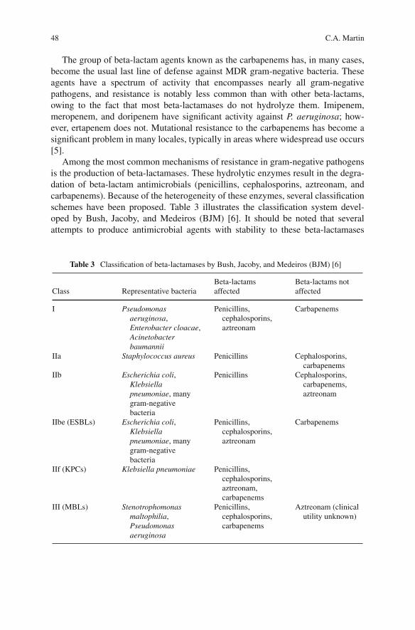
48 C.A. Martin
The group of beta-lactam agents known as the carbapenems has, in many cases,become the usual last line of defense against MDR gram-negative bacteria. Theseagents have a spectrum of activity that encompasses nearly all gram-negativepathogens, and resistance is notably less common than with other beta-lactams,owing to the fact that most beta-lactamases do not hydrolyze them. Imipenem,meropenem, and doripenem have significant activity against P. aeruginosa; how-ever, ertapenem does not. Mutational resistance to the carbapenems has become asignificant problem in many locales, typically in areas where widespread use occurs[5].
Among the most common mechanisms of resistance in gram-negative pathogensis the production of beta-lactamases. These hydrolytic enzymes result in the degra-dation of beta-lactam antimicrobials (penicillins, cephalosporins, aztreonam, andcarbapenems). Because of the heterogeneity of these enzymes, several classificationschemes have been proposed. Table 3 illustrates the classification system devel-oped by Bush, Jacoby, and Medeiros (BJM) [6]. It should be noted that severalattempts to produce antimicrobial agents with stability to these beta-lactamases
Table 3 Classification of beta-lactamases by Bush, Jacoby, and Medeiros (BJM) [6]
Class Representative bacteriaBeta-lactamsaffected
Beta-lactams notaffected
I Pseudomonasaeruginosa,Enterobacter cloacae,Acinetobacterbaumannii
Penicillins,cephalosporins,aztreonam
Carbapenems
IIa Staphylococcus aureus Penicillins Cephalosporins,carbapenems
IIb Escherichia coli,Klebsiellapneumoniae, manygram-negativebacteria
Penicillins Cephalosporins,carbapenems,aztreonam
IIbe (ESBLs) Escherichia coli,Klebsiellapneumoniae, manygram-negativebacteria
Penicillins,cephalosporins,aztreonam
Carbapenems
IIf (KPCs) Klebsiella pneumoniae Penicillins,cephalosporins,aztreonam,carbapenems
III (MBLs) Stenotrophomonasmaltophilia,Pseudomonasaeruginosa
Penicillins,cephalosporins,carbapenems
Aztreonam (clinicalutility unknown)

Gram-Negative Bacteria 49
have been undertaken, with some success. While the early penicillins – penicillinand ampicillin – do not exhibit stability to beta-lactamases, the extended-spectrumpenicillins; beta-lactamase inhibitor combination agents; cephalosporins; carbapen-ems; and aztreonam all possess varying degrees of stability to beta-lactamases, acharacteristic that is highly dependent on the specific beta-lactamase in question.
Despite advances in drug discovery for agents active against beta-lactamase-producing gram-negative bacteria, pinpoint mutations in genes mediating beta-lactamase production can extend the activity of the parent enzyme and havegiven rise to extended-spectrum beta-lactamases (ESBLs). The ESBLs were iden-tified and have proliferated in the time period following the Introduction ofoxyiminocephalosporins (e.g., ceftazidime, cefotaxime, and ceftriaxone). Theseenzymes are most commonly seen in E. coli and Klebsiella spp., but have recentlyproliferated in other gram-negative bacteria as well. In clinical settings where oxy-iminocephalosporins are heavily utilized, ESBL-producing gram-negative bacteriabecome commonplace. Attempts to control ESBL production by decreasing theuse of these agents are often successful but may carry other consequences, a phe-nomenon known as “squeezing the resistance balloon” [7]. The key to controllingESBL production is prevention through prudent antimicrobial use, especially ofoxyiminocephalosporins.
Laboratory identification of ESBL-producing organisms is difficult, again dueto the heterogeneity of the enzymes in question. Most clinical isolates producingESBLs will initially test “resistant” to multiple beta-lactams, including third-generation cephalosporins. In some, however, the bacteria may not have a minimuminhibitory concentration (MIC) to third-generation cephalosporins high enoughto garner this classification. Given the inability of Clinical Laboratory StandardsInstitute (CLSI; formerly NCCLS) breakpoints to predict efficacy in “suscepti-ble” ESBL producers, identification of these bacteria is of paramount importance.CLSI has introduced a screening method to identify and confirm suspected ESBLsin isolates with elevated MICs, but the method is currently only validated andapproved for E. coli, Klebsiella spp., and P. mirabilis. Clinical microbiologylaboratories have little guidance on the identification of ESBLs in other gram-negative bacteria [8]. One method for detecting ESBLs (E-test R©) is depicted inFig. 1.
According to CLSI, when Enterobacteriaceae-harboring ESBLs are identified,the laboratory must report the isolate as “resistant” to all penicillins, cephalosporins,and aztreonam. This is an important point, as not all ESBL-producing bacteria willtest as overtly resistant to all of these agents. In these cases, however, the appearanceof in vitro susceptibility is false and fails to predict reliably in vivo success [9]. Beta-lactamase inhibitor combinations (ampicillin/sulbactam, amoxicillin/clavulanate,piperacillin/tazobactam, and ticarcillin/clavulanate) may have varying degrees ofin vitro activity, but their clinical utility in patients infected with these organismsremains questionable. Thus, these agents cannot be routinely recommended forESBL-producing bacteria. Carbapenems (imipenem, meropenem, ertapenem, anddoripenem) typically become the drugs of choice, as fluoroquinolone co-resistance

50 C.A. Martin
Fig. 1 Example of the E-test R© method to confirming ESBL production in gram negative bacteria.The addition of the beta-lactamase inhibitor (lower portion of each strip) to the beta-lactam (upperportion) restores much of the activity of the drug, as noted by the larger zones of inhibition. Photocourtesy of Dr. Julia Ribes, University of Kentucky
is common and aminoglycosides are not often recommended for monotherapy.A newer glycylcycline agent, tigecycline, may prove to have some benefit in thisarea, but the exact place in therapy for this agent is still being elucidated.
Some Enterobacteriaceae (e.g., Enterobacter spp.), as well as P. aeruginosa andAcinetobacter spp., have long been known to produce a beta-lactamase (BJM class I,Table 3) mediated by the gene called ampC. Under normal circumstances, this beta-lactamase is subject to tight control by accessory genes and is mostly produced asa protective mechanism for the bacteria when exposed to beta-lactam agents. Underspecial circumstances – through mutation in those accessory genes – the produc-tion of this beta-lactamase is unabated. Some beta-lactam agents may preferentiallyselect these resistant mutants through their activity against the susceptible majority.As the resistant minority proliferates, overall beta-lactamase production increases,and the risk for treatment-emergent resistance follows. The risk of clinical failure inEnterobacter spp. bacteremia upon treatment with a third-generation cephalosporinis as high as 20–30% [10]. This phenomenon has been called “ampC hyperpro-duction,” as well as “stable derepression.” The resulting high level of enzyme
Gram-Negative Bacteria 51
production mediates resistance to penicillins, most cephalosporins, and aztreonam.Cefepime may continue to demonstrate in vitro activity, but its clinical utility inthese cases remains largely unproven. Notably, the beta-lactamase inhibitors (clavu-lanate, sulbactam, and tazobactam) have no significant inhibitory activity againstthese enzymes. As with ESBLs, the drugs of choice often become carbapenems,due to the large database of clinical utility with these agents. Other agents, such asfluoroquinolones, aminoglycosides, and tigecycline, may also provide some benefitdepending on individual susceptibilities [8].
Metallo-beta-lactamases (MBLs) have emerged in recent years and representan especially concerning resistance trend. These molecules are so named becausethey rely on a high concentration of zinc within the enzyme to interact with thebeta-lactam (most other beta-lactamases rely on high serine concentrations). Ithas been known for quite sometime that certain gram-negative bacteria – mostnotably Stenotrophomonas maltophilia – are common producers of these enzymes,which are included in BJM class III (Table 3). The most concerning aspect ofthese enzymes is their broad spectrum of hydrolysis, which includes penicillins,cephalosporins, and notably the carbapenems. For this reason, MBLs are alsoclassified as “carbapenemases.” Organisms producing these enzymes have prolif-erated in some geographic areas, typically those in which carbapenem consumptionis high. They have also become more common in organisms other than thosethat have historically produced MBLs. P. aeruginosa, A. baumannii, K. pneumo-niae, and Enterobacter spp. are among the organisms found to produce MBLs[10]. Given the broad scope of resistance that accompanies this mechanism ofresistance, antimicrobial therapy with an agent outside the beta-lactam family isrequired.
Fairly recently, researchers and clinicians have noted an increase in organ-isms that produce beta-lactamases known as KPCs (Klebsiella pneumoniaecarbapenemase), which are members of BJM class II (Table 3). First discov-ered in K. pneumoniae in 1996, these beta-lactamases are plasmid-mediated andconfer resistance to penicillins, cephalosporins, and carbapenems. Quickly afterthe first isolation of KPC-1 in K. pneumoniae, KPC-2 and KPC-3 – amino acidvariants of KPC-1 – were detected in the United States and in other parts of theworld. Currently, K. pneumoniae is the predominant producer of KPC enzymes, butEnterobacter spp., E. coli, and Salmonella spp. have also been found to harbor KPCenzymes.
As with MBLs, the proliferation of KPCs seems to cluster in areas of highcarbapenem usage. The best Example of this is the New York metropolitan area,where carbapenem use has often been necessitated by outbreaks of ESBL-producingK. pneumoniae. In response to this increased carbapenem use, K. pneumoniaethat produce KPC-2 have become common. In the setting of infection with thistype of MDR organism, treatment options are limited. Tigecycline, polymyxin B,polymyxin E (colistin), and other non-beta-lactam agents often become the drugs ofchoice, but each has its limitations.

52 C.A. Martin
3 Fluoroquinolones: Activity and Resistance
Fluoroquinolones have become commonly used agents in ambulatory settings fora broad range of infections including UTIs, pneumonia and other respiratoryinfections, and gastrointestinal infections, among others. Their rapidly expand-ing use in the community is due to a variety of factors. They are relatively safecompounds with high bioavailability, making oral dosing for moderate infectionsreasonable. While they have been shown to be effective for a multitude of indi-cations, their increasing use has been associated with increasing resistance inkey pathogens. Fluoroquinolones have varying degrees of gram-negative activ-ity. Ciprofloxacin and levofloxacin exhibit the broadest activity, including mostEnterobacteriaceae, Acinetobacter spp., and P. aeruginosa, among others. The clin-ician should observe caution when using fluoroquinolones to treat infections causedby P. aeruginosa, as resistance occurs frequently. For this reason, if P. aeruginosais the suspected or confirmed pathogen in a serious infection (other than UTI) it isprudent to avoid monotherapy with a fluoroquinolone, and only use these agentsas part of a combination. Urinary tract infection may represent an exception tothis rule for ciprofloxacin and levofloxacin, given their ability to concentrate inurine. Moxifloxacin possesses moderate gram-negative activity, with a spectrumthat includes most Enterobacteriaceae, but not P. aeruginosa. Gemifloxacin hassignificantly less gram-negative activity than its counterparts.
Resistance to fluoroquinolones in gram-negative bacteria has been increasingrecently. Alterations in DNA gyrase and topoisomerase IV – the fluoroquinolonetargets – constitute much of this mutational resistance. The presence of multidrugefflux pumps – pumps that remove the drug before its antimicrobial action can beachieved, which is seen especially in P. aeruginosa – has emerged as a significantcontributor to fluoroquinolone resistance as well.
4 Aminoglycosides: Activity and Resistance
Aminoglycosides remain among the most commonly used antimicrobial agents forthe treatment of infections caused by gram-negative pathogens, typically in com-bination with other agents. Gentamicin, tobramycin, and amikacin are the agentstypically used for the treatment of gram-negative infections. Their spectrum of activ-ity includes Enterobacteriaceae, Acinetobacter spp., and P. aeruginosa. They exhibitconcentration-dependent bactericidal action by binding to the 30S ribosomal subunitand preventing bacterial protein synthesis.
Intrinsic aminoglycoside resistance occurs mostly via drug exclusion from thecell. Anaerobic bacteria are intrinsically resistant to aminoglycosides through thismechanism. Several other mechanisms of resistance have been identified, includ-ing multidrug efflux pumps, target-site modification at the level of the ribosome,and enzymes that destroy the aminoglycoside molecule (aminoglycoside-modifyingenzymes). Clinically, gentamicin and tobramycin are more often affected byaminoglycoside-modifying enzymes than amikacin.
Gram-Negative Bacteria 53
5 Gram-Negative Organisms
5.1 Escherichia coli
E. coli is among the most common gram-negative pathogens encountered in clinicalpractice. A part of the normal human gastrointestinal flora, E. coli is the primarycause of urinary tract infections (UTIs) in the outpatient setting. Infections causedby this pathogen, however, range from uncomplicated UTIs to hospital-acquiredpneumonia and sepsis. For this reason, antimicrobial resistance in E. coli is ofsignificant import in both the ambulatory and hospitalized patient.
Inexpensive and generally effective, sulfamethoxazole/trimethoprim(SMZ/TMP) is a mainstay of therapy for UTIs in the ambulatory setting.Historically, resistance to SMZ/TMP in E. coli has remained fairly low. In recentyears, however, resistance has been increasing (near or above 20% in some locales),and clinical failures have been reported [11]. As a result, clinicians are faced witha difficult quandary – whether or not to continue using SMZ/TMP empiricallyfor the treatment of UTIs. On one hand, clinical failure rates, even in the settingof up to 20% resistance, remain relatively low. On the other hand, investigatorshave suggested that when resistance rates exceed 20%, SMZ/TMP becomesless cost-effective than fluoroquinolone therapy because of the increased rate oftreatment failure and resource utilization. These recommendations do not take intoaccount increasing rates of fluoroquinolone resistance.
E. coli is a common producer of beta-lactamases, most often those of BJMfunctional group II. The typical beta-lactamase produced by E. coli clinical iso-lates is a narrow-spectrum enzyme (TEM-1) with hydrolytic activity limited tothe unprotected penicillin agents (e.g., ampicillin, amoxicillin, etc.). This enzymeis plasmid-mediated and, therefore, can be transferred to or from other bacteriathrough gene transfer. In an isolate without beta-lactamase production, the inclu-sion of an amino group on the penicillin core affords ampicillin and amoxicillin– the so-called aminopenicillins – significant antimicrobial activity against E. coli.In the presence of beta-lactamase, however, these agents are rendered ineffective.The addition of a beta-lactamase inhibitor to amoxicillin (clavulanate), ampicillin(sulbactam), ticarcillin (clavulanate), or piperacillin (tazobactam) restores the activ-ity of the agent. Other beta-lactam agents, such as cephalosporins, aztreonam,and carbapenems, retain significant activity against E. coli strains that producenarrow-spectrum beta-lactamases such as TEM-1 and SHV-1.
As with some other gram-negative bacteria, pinpoint mutations in genes medi-ating beta-lactamase production can give rise to ESBLs. In fact, E. coli is amongthe most common ESBL-producing bacterium in clinical practice. In this setting,the clinician is advised to avoid all penicillins, cephalosporins, and aztreonam.Depending on the susceptibility to other agents outside the beta-lactam family, aswell as the site of infection, carbapenems may be the therapy of choice.
Resistance to fluoroquinolones among clinical strains of E. coli has been onthe rise in recent years. In 2004, according to the National Nosocomial InfectionsSurveillance (NNIS) project, nearly 10% of E. coli strains isolated from hospital-ized patients were resistant to the fluoroquinolones [12]. In strains that also harbor

54 C.A. Martin
ESBLs, resistance rates are even higher and approach 50% in some reports [9].Most commonly, this resistance is due either to efflux pumps or target-site mod-ification. Ciprofloxacin and levofloxacin, along with some older fluoroquinolonessuch as ofloxacin and norfloxacin, are commonly used to treat complicated anduncomplicated UTIs, where E. coli is a common pathogen. As mentioned previ-ously, clinicians are often faced with the choice of SMZ/TMP or a fluoroquinolone,for empiric treatment of UTIs. Fluoroquinolone resistance in E. coli makes this deci-sion significantly more difficult. Culture and susceptibility data are important forchoosing appropriate antimicrobial therapy.
5.2 Klebsiella pneumoniae
K. pneumoniae is a commonly encountered pathogen in clinical practice. In thecommunity, it is associated with urinary tract infections and, less commonly,community-acquired pneumonia. In the hospital setting, K. pneumoniae causesseveral diverse infections, including hospital-acquired and ventilator-associatedpneumonia; bacteremia; urinary tract infections; and intra-abdominal infections,among others. This broad scope of pathogenicity, along with its ability to harborresistance mechanisms – either intrinsically or acquired – makes K. pneumoniae aformidable pathogen.
In contrast to E. coli, which may or may not produce a beta-lactamase, K. pneu-moniae is a constant producer of SHV-1 (BJM class IIb). While ampicillin andamoxicillin could be expected to penetrate the cell wall of K. pneumoniae, theyhave no protection against beta-lactamases, precluding therapy with these agents.Even piperacillin and ticarcillin are subject to some hydrolysis by this enzyme.The addition of a beta-lactamase inhibitor (e.g., piperacillin/tazobactam, ticar-cillin/clavulanate, ampicillin/sulbactam, or amoxicillin/clavulanate) should restorethe efficacy of the penicillin agent, provided no ESBLs or other resistance mech-anisms are present. Extended-spectrum cephalosporins (cefotaxime, ceftriaxone,ceftazidime, cefepime, etc.), aztreonam, and carbapenems are also afforded stabilityto the SHV-1 enzyme and would therefore be reasonable treatment options for thistype of isolate. Other non-beta-lactam agents can also be considered.
As with E. coli, emergence of ESBL-producing K. pneumoniae has increasedalong with the increasing use of oxyiminocephalosporins. According to the NationalNosocomial Infections Surveillance (NNIS) system, K. pneumoniae resistance toceftazidime in US intensive care units increased approximately 600% between 1988and 2004 (this resistance can be seen as a surrogate for the presence of ESBLs) [12].Given the multidrug resistance profile of ESBL-producing gram-negative bacteria, areasonable assumption can be made that this increase has also occurred with respectto penicillins and aztreonam.
Fluoroquinolone resistance in K. pneumoniae is similar to that in E. coli and otherEnterobacteriaceae, albeit at a low rate of occurrence. Resistance most commonlyoccurs as a mutation of DNA gyrase, with gyrA being the specific gene affected.Topoisomerase IV mutation (also a binding site alteration) also occurs, but is moreoften associated with gram-positive resistance than gram-negative resistance. Efflux
Gram-Negative Bacteria 55
pumps are also known to mediate fluoroquinolone resistance in K. pneumoniae.Any one of these mutations may be enough to mediate resistance to the fluoro-quinolones, but the combination of multiple mutations often Results in high-levelresistance.
5.3 Pseudomonas aeruginosa
P. aeruginosa is the most common gram-negative bacteria causing hospital-acquiredinfections in critically ill patients. Even without the presence of mutational resis-tance, P. aeruginosa is inherently resistant to many agents, either through beta-lactamase production or through exclusion of antimicrobial agents from the cell[1]. Mutational resistance has been increasing in frequency as well. In 2004,the proportion of P. aeruginosa isolates resistant to ceftazidime and imipenemapproached 25% [12]. Imipenem – along with the other antipseudomonal carbapen-ems, meropenem and doripenem – is often seen as the last line of therapy forresistant gram-negative infections, especially those caused by P. aeruginosa. In thesetting of carbapenem resistance, clinicians may be forced to use polymyxin B orcolistin – agents once thought of as too toxic for systemic use. Tigecycline can bea treatment option for other MDR gram-negative pathogens, but it exhibits poor invitro activity against P. aeruginosa.
Beta-lactam resistance in P. aeruginosa can be due to a variety of mechanisms.First and foremost, P. aeruginosa is a producer of beta-lactamases, most often BJMclass I (Table 3). Wild-type strains produce these enzymes in an inducible fashion. Inthis state, ticarcillin and piperacillin are not significantly affected. As mentioned pre-viously, however, once these ampC-mediated enzymes become stably derepressed,they confer resistance to all penicillins, cephalosporins, and aztreonam and are notaffected by beta-lactamase inhibitors. Other beta-lactamases have also been foundin P. aeruginosa. Class II (including ESBLs) and class III (carbapenemases) havebeen reported and are increasing in frequency [1].
Antimicrobial efflux pumps are becoming more frequent in clinical isolates ofP. aeruginosa. These pumps remove the antimicrobial from the cell before antimi-crobial action can occur. Like cell wall impermeability, efflux pumps may be eitherdrug-specific or have action on several agents. P. aeruginosa has been shown to har-bor several efflux pumps, affecting antimicrobials ranging from fluoroquinolonesand aminoglycosides to penicillins, cephalosporins, aztreonam, and carbapenems.In fact, upregulation of these pumps affects most beta-lactams. It should be notedthat imipenem is the beta-lactam least affected by efflux-mediated resistance [1].
As mentioned previously, antimicrobials are reliant upon porin channels in thegram-negative cell wall in order to access their respective sites of action. One chan-nel, OprD, is particularly subject to mutational downregulation. The loss of thisporin channel significantly reduces the antibacterial effect of imipenem, as its entryinto the cell is solely dependent on this route. Downregulation of OprD is a commonmechanism of imipenem resistance in P. aeruginosa. Meropenem and doripenemcan gain entry into the cell through alternative routes and are, therefore, less affectedby OprD loss.

56 C.A. Martin
Throughout the 1990s and 2000s, P. aeruginosa became increasingly resistantto fluoroquinolones. As these agents have become more commonly used in clin-ical practice, resistance has followed [14]. In fact, 30–40% of clinical strains arenow resistant to ciprofloxacin and levofloxacin, the only fluoroquinolones withsignificant P. aeruginosa activity. This resistance can be due to alterations inDNA gyrase and topoisomerase IV or to efflux pumps. Even those strains thatremain susceptible are unlikely to be eradicated with either of these agents asmonotherapy, as resistance often emerges during treatment. For these reasons,many clinicians will employ the combination of an antipseudomonal beta-lactamand an aminoglycoside for serious pseudomonal infections, reserving fluoro-quinolones as an aminoglycoside substitute in patients who may be at risk fornephrotoxicity.
5.4 Acinetobacter baumannii
A. baumannii is a significant and emerging nosocomial pathogen, as well as aformidable cause of infections in soldiers who have been deployed in the MiddleEast. While the overall proportion of infections caused by A. baumannii remainslower than P. aeruginosa, its propensity for resistance is concerning. From 1986 to2004, resistance in ICUs to ceftazidime increased in this pathogen from approx-imately 25 to nearly 70% [13]. Several outbreaks have been reported in whichresistance to almost every available antimicrobial was seen [15]. Like P. aeruginosa,Acinetobacter produces an inducible BJM class I beta-lactamase that can becomestably derepressed through mutation. This type of resistance negates the penicillins,cephalosporins, and aztreonam. Because co-resistance with fluoroquinolones andaminoglycosides is common, carbapenems become the agents most often used inthis setting. Several epidemiological studies have been published outlining the effectof carbapenem overuse on the MDR profile of A. baumannii [7, 16]. In areaswhere carbapenem use is high, resistance to all beta-lactams can be seen, due todiverse beta-lactamase production (class I, II, or III), porin channel downregula-tion, efflux pumps, or a combination of these. Resistance to fluoroquinolones andaminoglycosides occurs through similar mechanisms as those seen in P. aerugi-nosa, with fluoroquinolone resistance occurring frequently. Unlike P. aeruginosa,ampicillin/sulbactam may provide a therapeutic alternative for MDR A. baumanniiisolates.
Tigecycline, the first agent in a new class called glycylcyclines (derivatives ofminocycline) may provide some therapeutic utility for MDR Acinetobacter infec-tions. The spectrum of tigecycline is broad, but importantly, it does not include P.aeruginosa. Many MDR Enterobacteriaceae will be susceptible to tigecycline, and itcan be considered a treatment option in those cases. In vitro data suggest that mostA. baumannii strains can be inhibited with clinically achievable concentrations of

Gram-Negative Bacteria 57
tigecycline, although resistance has been described. It should be noted that tigecy-cline does not have an FDA-approved indication for the treatment of A. baumanniiinfections [17].
6 Role of the Primary Care Provider in ManagingGram-Negative Infections
Primary care providers are in a unique position to improve the use of antimicrobialagents in the community and, subsequently, to help avoid unnecessary antimicrobialresistance in all settings. Indiscriminate use of antimicrobials has serious conse-quences, many of which have had, and will continue to have, a direct effect onpatient care. For Example, a large study found that approximately 70% of patientsseen in outpatient clinics for chief complaint of “sore throat” received a prescriptionfor antibiotics [18]. Given that Streptococcus pyogenes is the only major bacte-rial cause of pharyngitis, that it causes approximately 20% of cases (with mostbeing viral), and that a rapid diagnostic test is available, far fewer patients shouldreceive antimicrobial therapy. In addition, 70% of patients who received an antibi-otic received a non-recommended agent (mostly fluoroquinolones and macrolides).While streptococcal pharyngitis is obviously not a gram-negative infection, indis-criminate use of fluoroquinolones and macrolides, among other agents, certainlyhas an untoward effect on gram-negative pathogens such as E. coli, K. pneumonia,H. influenzae, and others. In certain geographical areas, empiric fluoroquinolonetherapy for STD is no longer recommended due to resistance. The proper use ofantimicrobials for any given disease requires knowledge of the likely pathogens,likely resistance issues, and proper diagnosis. Microbiologic studies should be usedwhenever possible to tailor therapy. Proper culture and susceptibility techniques canallow the clinician to narrow therapy if appropriate or to alter initial therapy if resis-tance is detected. The correct antimicrobial regimen for any infection is the onethat eradicates the pathogen and has as little effect as possible on non-pathogeniccommensal organisms.
7 Lack of New Agents in the Development Pipeline
While several new agents have recently been marketed for gram-positive infections,the outlook for the treatment of MDR gram-negative organisms is considerablybleaker. Mostly due to the huge costs, many pharmaceutical companies are shy-ing away from the development of novel antimicrobial agents. Even those that havebeen recently marketed, such as tigecycline, have limitations in their activity. Inthe case of tigecycline, that limitation is the lack of efficacy P. aeruginosa. In2006, the Infectious Diseases Society of America (IDSA) advocated a legislative

58 C.A. Martin
response to this public health crisis [2]. Until such actions come to fruition, clin-icians have a responsibility to practice antimicrobial restraint when possible. Thereader is referred to the IDSA’s guidelines on Antimicrobial Stewardship for a morein-depth review of this topic [19].
Key Points
• Resistance in gram-negative pathogens is increasing and may be due to a varietyof mechanisms.
• Beta-lactamase production, especially those of the broad-spectrum variety,presents unique challenges and should provide the impetus for the developmentof compounds that escape this resistance mechanism.
• Detection of extended-spectrum beta-lactamases (ESBLs) in clinical micro-biology laboratories is technically difficult and may be subject to reportingerror.
• Carbapenems are typically viewed as the drugs of choice for ESBL-producinggram-negative pathogens.
• Empiric therapy for serious infections should be broad spectrum and take intoaccount local resistance patterns. The likelihood of a successful outcome isincreased dramatically if empiric therapy is appropriate and initiated promptly.
• Few novel antimicrobial agents for MDR gram-negative pathogens are in thedevelopment pipeline. Antimicrobial stewardship should help preserve the cur-rent armamentarium.
References
1. Livermore DM (2002). Multiple mechanisms of antimicrobial resistance in Pseudomonasaeruginosa: Our worst nightmare? Clin Infect Dis 34(5):634–640
2. Talbot G, Bradley J, Edwards JJ et al. (2006). Bad bugs need drugs: An update on the devel-opment pipeline from the Antimicrobial Availability Task Force of the Infectious DiseasesSociety of America. Clin Infect Dis 42(5):657–668
3. Chambers HF (2005). Penicillins. In: Mandell GL, Bennett JE, Dolin R, (eds) Principles andPractice of Infectious Diseases, 6th ed. Elsevier, Philadelphia, 281–293
4. Andes DR, Craig WA (2005). Cephalosporins. In: Mandell GL, Bennett JE, Dolin R, (eds)Principles and Practice of Infectious Diseases. Elsevier, Philadelphia, 294–311
5. Chambers HF (2005). Other Beta-Lactam Antibiotics. In: Mandell GL, Bennett JE, Dolin R,(eds) Principles and Practice of Infectious Diseases. Elsevier, Philadelphia, 311–318
6. Bush K, Jacoby G, Medeiros A (1995). A functional classification scheme for beta-lactamasesand its correlation with molecular structure. Antimicrob Agents Chemother 39(6):1211–1233
7. Rahal JJ, Urban C, Segal-Maurer S (2002). Nosocomial antibiotic resistance in multiple gram-negative species: Experience at one hospital with squeezing the resistance balloon at multiplesites. Clin Infect Dis 34(4):499–503
8. Yang K, Guglielmo BJ (2007). Diagnosis and treatment of extended-spectrum and AmpC{beta}-lactamase-producing organisms. Ann Pharmacother 41(9):1427–1435
9. Paterson DL (2006). Resistance in gram-negative bacteria: Enterobacteriaceae. Am J InfectControl 34(5 Suppl 1):S20–S28
Gram-Negative Bacteria 59
10. Livermore DM, Woodford N (2006). The [beta]-lactamase threat in Enterobacteriaceae,Pseudomonas and Acinetobacter. Trends Microbiol 14(9):413–420
11. Erb A, Stürmer T, Marre R et al. (2007). Prevalence of antibiotic resistance in Escherichiacoli: overview of geographical, temporal, and methodological variations. Eur J Clin MicrobiolInfect Dis 26(2):83–90
12. System NNIS (2004). National Nosocomial Infections Surveillance (NNIS) System Report,data summary from January 1992 through June 2004. Am J Infect Control 32(8):470–485
13. Gaynes R, Edwards J (2005). Overview of nosocomial infections caused by gram-negativebacilli. Clin Infect Dis 41:848–854
14. Neuhauser M, Weinstein R, Rydman R et al. (2003). antibiotic resistance among gram-negative bacilli in US intensive care units: Implications for fluoroquinolone use. J Am MedAssoc 289:885–888
15. Richet H, Fournier PE (2006). Nosocomial infections caused by Acinetobacter baumannii: Amajor threat worldwide. Infect Control Hosp Epidemiol 27:645–646
16. Manikal VM, Landman D, Saurina G et al. (2000). Endemic carbapenem-resistantAcinetobacter species in Brooklyn, New York: Citywide prevalence, interinstitutional spread,and relation to antibiotic usage. Clin Infect Dis 31:101–106
17. Slover CM, Rodvold KA, Danziger LH (2007). Tigecycline: A novel broad-spectrumantimicrobial. Ann Pharmacother 41(6):965–972
18. Linder JA, Stafford RS (2001). Antibiotic treatment of adults with sore throat by communityprimary care physicians: A national survey, 1989–1999. J Am Med Assoc 286:1181–1186
19. Dellit T, Owens R, McGowan JJ et al. (2007). Infectious Diseases Society of America and theSociety for Healthcare Epidemiology of America guidelines for developing an institutionalprogram to enhance antimicrobial stewardship. Clin Infect Dis 44:159–177

Tuberculosis
Vidya Sundareshan and Martin E. Evans
1 Epidemiology
In 2007, it was estimated that 2.2 billion persons, or one-third of the world’s popu-lation, was infected with Mycobacterium tuberculosis [1] and the organism caused1.6 million deaths. A decade after identifying TB as a global emergency, it stillremains the second leading infectious cause of death among adults [2]. The mor-tality rate has steadily declined since it peaked at 400 deaths/100,000 persons in1750, when the disease occurred in association with migration to cities during theIndustrial Revolution. When streptomycin was introduced in 1946, the mortalityrate was 33/100,000. By 2005, the mortality in the United States had declined to<1/100,000 [3]. Global mortality rates, however, currently average 24/100,000, withthe highest rates in India (29/100,000) [1]. The AIDS epidemic is responsible for therise of TB cases in many parts of the world, and co-infection with both HIV and TBcontributes significantly to TB-related mortality.
Today, the expected cure rate for disease caused by drug-susceptible M. tuber-culosis approaches 100% [4], but the emergence of antibiotic resistance hascomplicated the management of TB and increased the chance of treatment fail-ure. The cure rate for infections caused by bacteria resistant to isoniazid (INH)or rifampin (RIF) (termed multidrug-resistant tuberculosis, or MDR-TB) is only80–95%. The prognosis of MDR-TB is even worse in patients co-infected withHIV, with mortality rates up to 66% during treatment [5]. An emerging threatis from extensively resistant tuberculosis (XDR-TB), defined as resistance toINH, RIF, any fluoroquinolone, and at least one second-line injectable drug(amikacin, kanamycin, or capreomycin) [6]. XDR-TB renders some patients practi-cally untreatable with currently available drugs. In one series from KwaZulu-Natal,South Africa, the mortality rate was 98% among patients infected with XDR-TB.
M.E. Evans (B)Lexington Veterans Affairs Medical Center and University of Kentucky School of Medicine,Lexington, KY, USAe-mail: [email protected]
61A.G. Mainous III, C. Pomeroy (eds.), Management of Antimicrobials in InfectiousDiseases, Infectious Disease, DOI 10.1007/978-1-60327-239-1_4,C© Springer Science+Business Media, LLC 2001, 2010

62 V. Sundareshan and M.E. Evans
The median survival was 16 days from the time of the first sputum collec-tion. In the United States, the mortality rate may be up to 12% in patients withXDR-TB [7].
Drug resistance can be primary or acquired. In the former, the patient is infectedwith a resistant organism even though the patient has never received therapy. Inthe latter, the patient’s initially sensitive organism becomes resistant after exposureto antimicrobials. During 2005 in the United States, the overall prevalence of pri-mary resistance to INH was 7.3% (4.1% among US-born persons and 9.8% amongforeign-born persons) and the overall prevalence of acquired resistance to INH was14.6% (9.1% in US-born persons and 19.7% in foreign-born persons). In the sameyear, the overall prevalence of primary MDR-TB in the United States was 1% (0.4%in US-born and 1.4% in foreign-born persons). MDR-TB in previously treated caseswas 4.7% (1.3% in US-born and 7.9% in foreign-born persons) [3]. The prevalenceof XDR-TB in the United States in 2006 was approximately 0.02% (17 cases). Morethan 80% had no prior history of TB and therefore could be considered to haveprimary XDR–TB [7].
Worldwide, from 2002 to 2007, the median prevalence of primary resistance toINH was estimated to be 10.3% (ranging from 0% in certain countries in westernEurope to 42.4% in Tashkent, Uzbekistan), and the median prevalence of acquiredresistance to INH was 27.7% (ranging from 0 to 81.2% in Tashkent, Uzbekistan) [8].The worldwide prevalence of MDR-TB among all TB cases in 2006 was estimatedto be 5.3%. The prevalence of primary MDR-TB was 2.9% and the prevalence ofacquired MDR-TB was 15.3%, with the highest in Tashkent, Uzbekistan (60%) [8].The overall global prevalence of XDR-TB in 2006 was estimated to be 2% [9].XDR data available from developed countries indicate it ranges from 0 up to 33.3%(in Ireland and Slovenia). Data on primary and acquired XDR-TB are not availablefrom all countries, but approximately half of the 53 XDR-TB cases reported fromKwazulu-Natal in South Africa had never received prior TB treatment.
2 Drug Resistance
Drug resistance in M. tuberculosis is caused by spontaneous mutations in the chro-mosome. Mutations associated with RIF and fluoroquinolone resistance occur ata frequency of approximately 10–8, and mutations leading to INH, streptomycin,ethambutol (EMB), kanamycin, or p-aminosalicylic acid (PAS) resistance occur ata frequency of approximately 10–6. Mutations leading to resistance to ethionamide,capreomycin, cycloserine, or thiacetazone occur at a frequency of approximately10–3 [10].
These resistance frequencies are important because resistance to a combinationof antibiotics, such as RIF plus INH, would be expected to occur at the multiplicandof the two resistance frequencies (i.e., 10–8 × 10⁻⁶ or 10–14). Because tuberculouscavities often contain 109 organisms [11], patients with cavitary TB may carry upto 1,000 organisms with primary resistance to INH and 10 organisms with primaryresistance to RIF before receiving therapy. Because few, if any, bacteria are usually

Tuberculosis 63
resistant to both drugs, however, the probability of cure using both drugs is highsince INH would kill RIF-resistant bacteria and RIF would kill INH-resistant bac-teria. If, however, the patient were infected with INH-resistant M. tuberculosis, themajority of organisms in the cavity would be resistant to INH, and a regimen ofINH and RIF would, in effect, be single-drug therapy with RIF alone. RIF resis-tance would develop among the INH-resistant population at a frequency of 10–8.The patient might improve initially while RIF kills the majority of the bacteria, butthen strains resistant to both drugs would emerge, and the patient would relapse.
Clinicians’ prescribing behavior can lead to drug resistance. In 1977, Byrd andco-workers assessed the management of TB by non-pulmonary physicians and con-cluded that 73% of the patients had been treated inappropriately [12]. The mostcommon errors were the use of inadequate or excessive drug doses and the use ofa single drug to treat bacteriologically proven disease. In 1993, similar problemswere noted in an analysis of the previous management of patients with MDR-TBreferred to the National Jewish Medical and Research Center in Denver. In thisstudy, common errors leading to MDR-TB included (1) failure to obtain suscepti-bility testing; (2) failure to start an adequate initial regimen; (3) failure to modifythe regimen when the susceptibility of the organism changed; and (4) failure to usedirectly observed therapy (DOT) [13]. In a survey of tuberculosis management prac-tices in Kentucky in 1999, investigators found that TB was diagnosed by culture inonly 66% of patients (thus, no susceptibility data were obtained in the remainingpatients); 12 different regimens were used to treat the patients; monitoring of bacte-riologic cure was appropriate in less than 65% of cases; and DOT was used in only38% of patients [14]. Reports from other locales have documented similar problems.
3 Treatment of Drug-Susceptible TB Disease
3.1 General
The treatment of TB is important for the patient and for the community. Any privateprovider undertaking the responsibility of treating a patient with TB must assume apublic health function not only to prescribe an appropriate regimen but also to man-age drug side effects and ensure adherence until treatment is completed. For thisreason, it is often best for the practitioner to partner with the local health depart-ment to provide care for the patient. Local health departments can often providemedications, laboratory, and radiology support to the patient without charge andcan provide outreach personnel who can visit the home and ensure directly observedtherapy. The local health department often has resources to help maximize adher-ence to therapy including social service support; treatment incentives and enablers;housing assistance; referral for treatment of substance abuse; and coordination oftuberculosis services with those of other providers.
Several regimens have been recommended for the treatment of drug-susceptiblepulmonary TB (Table 1) using first-line agents (Tables 2 and 3) [15]. Each regimen

64 V. Sundareshan and M.E. Evans
Tabl
e1
Dru
gre
gim
ens
for
cultu
re-p
ositi
vepu
lmon
ary
TB
caus
edby
adr
ug-s
usce
ptib
leor
gani
sm[1
5]
Initi
alph
ase
Con
tinua
tion
phas
e
Reg
imen
Dru
gsIn
terv
alan
ddo
ses‡
(min
imal
dura
tion)
Reg
imen
Dru
gsIn
terv
alan
ddo
ses‡,
§(m
inim
aldu
ratio
n)R
ange
ofto
tald
oses
(min
imal
dura
tion)
1IN
HR
IFPZ
AE
MB
7da
ys/w
eek
for
56do
ses
(8w
eeks
)or
5da
ys/w
eek
for
40do
ses
(8w
eeks
)¶
1aIN
H/R
IF7
days
aw
eek
for
126
dose
s(1
8w
eeks
)¶
or5
days
/wee
kfo
r90
dose
s(1
8w
eeks
)18
2–13
0(2
6w
eeks
)
1bIN
H/R
IFTw
ice
wee
kly
for
36do
ses
(18
wee
ks)#
92–7
6(2
6w
eeks
)1c
∗∗IN
H/R
PTO
nce
wee
kly
for
18do
ses
(18
wee
ks)
74–5
8(2
6w
eeks
)2
INH
RIF
PZA
EM
B
7da
ys/w
eek
for
14do
ses
(2w
eeks
),th
enth
rice
wee
kly
for
12do
ses
(6w
eeks
)or
5da
ys/w
eek
for
10do
ses
(2w
eeks
)¶ ,the
nth
rice
wee
kly
for
12do
ses
(6w
eeks
)
2aIN
H/R
IFTw
ice
wee
kly
for
36do
ses
(18
wee
ks)#
62–5
8(2
6w
eeks
)
2b∗∗
INH
/RPT
Onc
ew
eekl
yfo
r18
dose
s(1
8w
eeks
)44
–10
(26
wee
ks)
(con
tinue
d)

Tuberculosis 65
Tabl
e1
(con
tinue
d)
Initi
alph
ase
Con
tinua
tion
phas
e
Reg
imen
Dru
gsIn
terv
alan
ddo
ses‡
(min
imal
dura
tion)
Reg
imen
Dru
gsIn
terv
alan
ddo
ses‡,
§(m
inim
aldu
ratio
n)R
ange
ofto
tald
oses
(min
imal
dura
tion)
3IN
HR
IFPZ
AE
MB
Thr
eetim
esa
wee
kfo
r24
dose
s(8
wee
ks)
3aIN
H/R
IFT
hree
times
wee
kly
for
54do
ses
(18
wee
ks)
78(2
6w
eeks
)
4IN
HR
IFE
MB
7da
ys/w
eek
for
56do
ses
(8w
eeks
)or
5da
ys/w
eek
for
40do
ses
(8w
eeks
)¶
4aIN
H/R
IF7
days
aw
eek
for
217
dose
s(3
1w
eeks
)or
5da
ys/w
eek
for
155
dose
s(3
1w
eeks
)¶27
3–19
5(3
9w
eeks
)
4bIN
H/R
IFTw
ice
wee
kly
for
62do
ses
(31
wee
ks)
118–
102
(39
wee
ks)
Defi
nitio
nof
abbr
evia
tions
:EM
B,e
tham
buto
l;IN
H,i
soni
azid
;PZ
A,p
yraz
inam
ide;
RIF
,rif
ampi
n;R
PT,r
ifap
entin
e.‡
Whe
nD
OT
isus
ed,d
rugs
may
begi
ven
5da
ys/w
eek
and
the
nece
ssar
ynu
mbe
rof
dose
sad
just
edac
cord
ingl
y.A
lthou
ghth
ere
are
nost
udie
sth
atco
mpa
refiv
ew
ithse
ven
daily
dose
s,ex
tens
ive
expe
rien
cein
dica
tes
this
wou
ldbe
anef
fect
ive
prac
tice.
§Pa
tient
sw
ithca
vita
tion
onin
itial
ches
tra
diog
raph
and
posi
tive
cultu
res
atco
mpl
etio
nof
2m
onth
sof
ther
apy
shou
ldre
ceiv
e7-
mon
th(3
1w
eeks
,eith
er21
7do
ses
{dai
ly}
or62
dose
s{t
hric
ew
eekl
y})
cont
inua
tion
phas
e.¶
Five
-day
-a-w
eek
adm
inis
trat
ion
isal
way
sgi
ven
byD
OT.
#N
otre
com
men
ded
for
HIV
-inf
ecte
dpa
tient
sw
ithC
D4
cell
coun
ts<
100
cells
/μl.
∗∗O
ptio
ns1c
and
2bsh
ould
beus
edon
lyin
HIV
-neg
ativ
epa
tient
sw
hoha
vene
gativ
esp
utum
smea
rsat
the
time
ofco
mpl
etio
nof
2m
onth
sof
ther
apy
and
who
dono
thav
eca
vita
tion
onin
itial
ches
trad
iogr
aph.
For
patie
nts
star
ted
onth
isre
gim
enan
dfo
und
toha
vepo
sitiv
ecu
lture
from
the
spec
imen
obta
ined
at2
mon
ths
ofbe
ing
onth
isre
gim
en,t
reat
men
tsho
uld
beex
tend
edan
extr
a3
mon
ths.

66 V. Sundareshan and M.E. Evans
Tabl
e2
Dos
age
reco
mm
enda
tions
for
the
initi
altr
eatm
ento
fdr
ug-s
usce
ptib
leT
Bam
ong
child
ren
and
adul
ts[1
5]
Dos
age
Dai
ly2
times
/wee
k3
times
/wee
kD
rugs
Chi
ldre
nA
dults
Chi
ldre
nA
dults
Chi
ldre
nA
dults
Ison
iazi
d10
–15
mg/
kg5
mg/
kg20
–30
mg/
kg15
mg/
kgN
otre
com
men
ded
15m
g/kg
Max
300
mg
Max
300
mg
Max
900
mg
Max
900
mg
Not
reco
mm
ende
dM
ax90
0m
gR
ifam
pin
10–2
0m
g/kg
10m
g/kg
10–2
0m
g/kg
10m
g/kg
Not
reco
mm
ende
d10
mg/
kgM
ax60
0m
gM
ax60
0m
gM
ax60
0m
gM
ax60
0m
gN
otre
com
men
ded
Max
600
mg
Pyra
zina
mid
e∗15
–30
mg/
kg15
–25
mg/
kgM
ax2
gM
ax2
gE
tham
buto
l∗15
–20
mg/
kg15
–20
mg/
kgM
ax1
gM
ax1.
6g
∗ See
[15]
for
inte
rmitt
entd
osin
gba
sed
onbo
dyw
eigh
t.

Tuberculosis 67
Table 3 Drugs used for tuberculosis [15]
Antimicrobial Usual dose Major side effects (1–10%)
First-line drugsIsoniazid 300 mg orally daily Gastrointestinal, hepatitis,
peripheral neuropathyRifampin 600 mg orally daily Gastrointestinal, discoloration
of urine, tears, etc., druginteractions
Rifabutin 300 mg orally daily Same as rifampinRifapentine 600 mg orally each week Same as rifampinPyrazinamide 15–25 mg/kg/day Malaise, gastrointestinal,
arthralgia, myalgiaEthambutol 15–20 mg/kg/day Optic neuritis, peripheral
neuropathy, nausea/vomiting
Second-line drugsLevofloxacin 500–1,000 mg/day Headache, gastrointestinal, rashGatifloxacin 400 mg/day Headache, gastrointestinal, rashMoxifloxacin 400 mg/day Headache, gastrointestinal, rashStreptomycin 15 mg/kg/day intramuscularly Deafness, vertigo, renal
dysfunctionAmikacin 15 mg/kg/day intravenously or
intramuscularlyDeafness, vertigo, renal
dysfunctionKanamycin 15 mg/kg/day intravenously or
intramuscularlyDeafness, vertigo, renal
dysfunctionCapreomycin 15 mg/kg/day intravenously or
intramuscularlyDeafness, vertigo, renal
dysfunctionCycloserine 500 mg orally each morning,
250 mg orally each eveningCentral nervous system,
congestive heart failure,rash, hepatitis, tremor
p-Aminosalicylate(PAS) (Paser R©
granules)
4 g packet mixed in orangejuice or apple juice twicedaily
Gastrointestinal upset
Ethionamide 250 mg orally each morning,500 mg orally each evening
Central nervous system,Stevens–Johnson,gastrointestinal, SLEsyndrome
has an initial phase of 2 months, followed by a choice of several options for a con-tinuation phase of 4 or 7 months. In all regimens INH, RIF, pyrazinamide (PZA),and EMB are begun initially. INH and RIF are the most active agents. Relapse ratesare 0–4% with INH- and RIF-susceptible M. tuberculosis; 2–7% with INH-resistantbacteria; and up to 72% if the bacteria are RIF resistant [16]. The inclusion of PZAis essential for the rapid sterilization of cavities, but the ability of PZA to preventthe emergence of resistance is low. For this reason, the fourth drug, EMB, is rec-ommended initially. EMB can be stopped as soon as the laboratory confirms thatthe isolate is susceptible to INH and RIF. PZA should be continued for 2 months,at which time the use of the other agents and the duration of therapy should be

68 V. Sundareshan and M.E. Evans
assessed based on the presence of cavitary lung disease on the initial chest radio-graph and/or persistently positive smears and cultures for acid-fast bacilli (Fig. 1).Probably the simplest continuation phase includes rifapentine (PriftinTM, HoechstMarion Roussel Kansas City, MO). This drug has a half-life of over 13 h, whichallows it to be administered once each week along with INH in HIV seronegativepatients.
Fig. 1 Treatment algorithm for tuberculosis. Patients in whom tuberculosis is proved or stronglysuspected should have treatment initiated with isoniazid, rifampin, pyrazinamide, and ethambu-tol for the initial 2 months. A repeat smear and culture should be performed when 2 months oftreatment has been completed. If cavities were seen on the initial chest radiograph or the acid-fastsmear is positive at the completion of 2 months of treatment, the continuation phase of treatmentshould consist of isoniazid and rifampin daily or twice weekly for 4 months to complete a total of 6months of treatment. If cavitation was present on the initial chest radiograph and the culture at thetime of completion of 2 months of therapy is positive, the continuation phase should be lengthenedto 7 months (total of 9 months of treatment). If the patient has HIV infection and the CD4 cellcount is <100/μl, the continuation phase should consist of daily or three times weekly isoniazidand rifampin. In HIV-uninfected patients having no cavitation on chest radiograph and negativeacid-fast smears at completion of 2 months of treatment, the continuation phase may consist ofeither once weekly isoniazid and rifapentine, or daily or thrice-weekly isoniazid and rifampin, tocomplete a total of 6 months (bottom). Patients receiving isoniazid and rifapentine, and whose 2-month cultures are positive, should have treatment extended by an additional 3 months (total of 9months) [15]∗EMB may be discontinued when results of drug susceptibility testing indicate no drug resistance.†PZA may be discontinued after it has been taken for 2 months (56 doses).‡RPT should not be used in HIV-infected patients with tuberculosis or in patients with extrapul-monary tuberculosis.§Therapy should be extended to 9 months if 2-month culture is positive.CXR, chest radiograph; EMB, ethambutol; INH, isoniazid; PZA, pyrazinamide; RIF, rifampin;RPT, rifapentine.

Tuberculosis 69
The resolution of cough, fever, and weight loss are important clinical signs ofsuccess. The patient’s sputum should be cultured on a monthly basis until two con-secutive negative sputum cultures have been documented. Conversion should occurwithin 4 months if the regimen is successfully going to treat the patient’s disease[15].
Clinicians should be aware of, and know how to manage, side effects of anti-tuberculous drugs. Ideally, all TB drugs are given at one time to increase compliancewith DOT, but this may initially cause gastrointestinal symptoms for the first fewweeks of therapy. Ingestion of the drugs with food may moderately decrease theabsorption of anti-tuberculosis drugs but may ameliorate gastrointestinal symptoms.
INH can cause a peripheral neuropathy, although this side effect is rare (lessthan 0.2%). Pyridoxine (vitamin B6) at a dose of 50 mg per day should be givenwith INH in persons with conditions that predispose them to neuropathy, suchas diabetes; alcoholism; nutritional deficiency; HIV infection; renal failure; preg-nancy; and breastfeeding, to prevent this side effect. Color vision and acuity mustbe assessed periodically if the patient is taking EMB, which can cause opticneuritis.
It is important to monitor the serum aspartate aminotransferase (AST) levelmonthly for patients on potentially hepatotoxic drugs such as RIF, INH, and PZA.All TB therapy should be held if AST levels are more than three times the upper limitof normal in the presence of symptoms (anorexia, nausea, vomiting, right upperquadrant pain, jaundice) or more than five times the upper limit of normal in theabsence of symptoms. The patient may be left without TB therapy for a short timewhile AST levels normalize, or alternative agents can be prescribed after consulta-tion with an expert. Once AST levels decrease to less than two times the upper limitof normal and symptoms significantly improve, the medications can be restarted insequential fashion with a new drug added every 2–3 days (EMB, then PZA, thenRMP, then INH) with careful monitoring of symptoms and AST levels.
Healthcare providers should also be aware of drug–drug interactions, which areespecially common with the rifamycins.
3.2 Directly Observed Therapy (DOT) and CombinationAntimicrobial Preparations
Patient non-compliance with prescribed therapy for TB is of concern because it isa frequent cause of treatment failure and the most common cause for the emer-gence of drug-resistant TB. In 1991, only 77% of TB patients in the United Statescompleted the prescribed course of antibiotics. In 1993, the Advisory Council forthe Elimination of Tuberculosis of the Centers for Disease Control and Prevention(CDC) recommended that DOT be considered for all patients in areas that do notachieve a 90% completion rate of therapy. The World Health Organization currentlyrecommends DOTS (directly observed treatment – short course) for all TB patientsworldwide [15]. DOT is designed to help the patient take all doses of medications

70 V. Sundareshan and M.E. Evans
and prevent the emergence of resistance. In DOT, a healthcare worker watches thepatient swallow the medicines. The advantages of DOT are that patients receive theirmedicines, can be monitored frequently for side effects, and can have all drugs dis-continued if they fail to appear for therapy. Erratic self-administration is avoided,and if the patient relapses, the organism is more likely to remain sensitive to theinitial antibiotic regimen.
DOT has been successful in improving compliance and decreasing TB case rates.Control of an epidemic of TB in New York City in recent years has been attributedlargely to the use of DOT. To conserve on personnel resources, DOT should utilizeintermittent therapies with twice- or thrice-weekly dosing regimens if possible.
The use of fixed-dose combinations of anti-tuberculous drugs also has been pro-posed as one method to improve compliance and prevent drug resistance [17].RifamateTM (Hoechst Marion Roussel), containing 150 mg INH and 300 mg RIFper tablet, and RifaterTM (Hoechst Marion Roussel), containing 50 mg INH, 120 mgRIF, and 300 mg PZA per tablet, are currently the only two fixed-dose combinationproducts on the US market. The idea is that patients taking fixed-dose combina-tions would not be able to stop a single agent without stopping therapy completely.Thus, selection of organisms resistant to a single antibiotic is unlikely. However,because multiple tablets need to be ingested to achieve the necessary dose, thereis the possibility that a patient may not take the full complement of pills andthus be under-dosed. This might allow the emergence of resistance to all agentssimultaneously.
When difficulties arise with anti-tuberculous therapy, the treatment regimens mayneed to be adjusted. This is difficult with fixed-dose combinations. It is easier toidentify the drug responsible for adverse reactions if drugs are being administeredindividually. If the primary problem is drug toxicity, it is safe to change or add asingle agent as long as the patient is being treated with at least two drugs to whichthe mycobacterium is still sensitive. It is imperative, however, never to add a sin-gle agent to a regimen where the patient is not improving or worsening clinically.Clinical failure implies that the organism is resistant to most, if not all, agents beingused. If only a single new agent is added, the probability that the organism willbecome resistant to the new agent is high.
3.3 Considerations for Patients Co-infected with HIV
The immunosuppression due to human immunodeficiency virus (HIV) infectionimpairs the host response to infection with TB, and TB accelerates the progres-sion of HIV disease. It is estimated that 13 million persons are co-infected withHIV and TB worldwide. Concurrent HIV infection is estimated to confer more thana 100-fold increased risk for development of active TB compared to HIV-negativepersons. Those with advanced HIV infection are at most risk of developing activeTB infection.
Treatment of TB in HIV-infected persons can be very complex for a numberof reasons. First, drug resistance may be more common in HIV-infected patients

Tuberculosis 71
than in uninfected persons. In one report, resistance rates among HIV-infected andnon-infected patients were 11.3 and 5.5% for INH; 8.9 and 1.6% for RIF; 5.1 and1.8% for PZA; and 6.2 and 1.3% for combined INH and RIF (MDR), respectively[18]. Second, interactions are common between the rifamycins and drugs used totreat HIV infection. Alternative drugs or changes in dosing must be consideredwhen attempting to treat both infections simultaneously. This is because the drugsused to treat TB and HIV have differing effects on the cytochrome P450 (CYP450)system and the resulting drug metabolism. Protease inhibitors, for instance, inhibitthe CYP450 system and decrease the metabolism of other drugs. Among the non-nucleoside reverse transcriptase inhibitors, delavirdine inhibits CYP450 enzymes,nevirapine induces the enzymes, and efavirenz has both effects. Rifamycins, onthe other hand, uniformly induce CYP450 enzymes and increase the metabolismof other drugs. The relative effect is RIF > rifapentine > rifabutin. Dosing inother drugs used in combination with the rifamycins often must be adjusted.Guidelines published by the CDC (http://www.cdc.gov/tb/) should be reviewed forspecific drug–drug interactions. For these reasons, it is strongly recommended thatpatients with TB–HIV co-infection be managed by providers experienced in themanagement of both diseases.
Some patients receiving therapy for HIV and TB infection at the same time expe-rience a reaction characterized by fevers, lymphadenopathy, worsening chest radio-graphs (e.g., miliary infiltrates, pleural effusions) or worsening cutaneous lesions orsigns of peritonitis. This is termed the “paradoxical reaction” or immune reconsti-tution inflammatory syndrome (IRIS) and is thought to be due to restoration of theimmune system by antiretroviral therapy. The incidence of these reactions rangesfrom 7 to 36%. It usually begins within 15 days of starting antiretroviral therapy, isassociated with decreases in viral load, and may last from 1 to 7 months [15].
4 Treatment of Drug-Resistant TB Disease
Drug resistance should be suspected in a patient (1) who has had prior therapy; (2)from an area where drug resistance is prevalent; (3) when sputum smears remainpositive after 2 months of therapy; (4) when sputum cultures remain positive after4 months of therapy; and (5) when there is unexplained worsening of the patient’schest radiograph or clinical course. Furthermore, a drug should be considered ofdubious efficacy if testing reveals that it was the only drug in a prescribed regimento which the bacterium is sensitive and the drug had been used for more than 1month.
Drug resistance also may emerge if the anti-tuberculous drugs are poorlyabsorbed. This may occur in persons who have undergone ileal bypass surgery orgastrectomy, in patients with D-xylose malabsorption, and in HIV-infected patientswith diarrhea. In some cases, drug levels at the site of the mycobacterial infectionmay be far below those in serum. This may occur in patients with thick-walledTB cavities, in tissues which are heavily fibrosed, or in areas with extensivecalcification.

72 V. Sundareshan and M.E. Evans
When INH, RIF, PZA, or EMB cannot be used because of drug resistance,second-line agents must be employed. In general, these agents (Table 3) are lessdesirable than first-line agents because they are more toxic and there is less clinicalexperience with their efficacy. For this reason, it is important to seek consultationwith an expert when dealing with drug-resistant TB.
Among second-line agents, cycloserine is usually well tolerated, but can havesubstantial central nervous system toxicity. This may be manifested as impairedmentation, psychoses, suicidal ideation, or seizures. The addition of pyridoxine(50–100 mg/day) may be of some benefit. PAS is usually well tolerated whengiven in the form of PaserTM granules (Jacobus Pharmaceutical Co., Inc., Princeton,NJ). In this formulation, the drug is adsorbed to methylcellulose beads. Patientsmay be concerned about the appearance of methylcellulose beads in their stools.The drug is not well absorbed unless it is given in an acid medium such asorange juice, cranberry juice, or applesauce. Ethionamide is the most poorly tol-erated antibiotic, especially if it is given with PAS. Gastrointestinal side effectsincluding nausea, vomiting, diarrhea, and a metallic taste are commonly reported.The injectable drugs streptomycin, amikacin, and kanamycin are valuable adjunctsto any re-treatment regimen. Their principle toxicities affect the kidney, vestibu-lar, and auditory systems. Serum creatinine and drug levels should be monitoredcarefully as well as monthly audiometry and clinical examinations of vestibularfunction.
Recommended regimens for treatment of resistant TB should be based on bacte-rial susceptibility testing and are listed in Table 4. The following principles shouldbe observed in designing a re-treatment regimen in addition to those recommendedfor the treatment of drug-susceptible TB (see below):
Table 4 Potential regimens for management of patients with drug-resistant tuberculosis [15]
Pattern of drug resistance Suggested treatmentDuration oftreatment
INH (±SM) RIF, PZA, EMB (FQ may strengthenregimen for extensive diseases)
6
INH and RIF (±SM) FQ, PZA, EMB, IA ± alternative agent 18–24INH, RIF (±SM), and EMB
or PZAFQ (EMB or PZA if active), IA and two
alternative agents24
RIF INH, EMB, FQ supplemented with PZAfor the first 2 months (an IA may beincluded for patients with extensivedisease in the first 2–3 months)
12–18
Definitions of abbreviations: EMB, ethambutol; IA, injectable agent; INH, isoniazid; PZA,pyrazinamide; RIF, rifampin; SM, streptomycin; FQ, fluoroquinolone; most experience involvesofloxacin, levofloxacin, or ciprofloxacin, injectable agents may include aminoglycosides (strepto-mycin, amikacin, kanamycin) or the polypeptide capreomycin; alternative agents= ethionamide,cycloserine, p-aminosalicylic acid, clarithromycin, amoxicillin-clavulanate, linezolid.

Tuberculosis 73
1. Never add a single drug to a failing regimen.2. Re-treatment should not be begun until reliable susceptibility data are available
from the laboratory.3. Consider prior treatments. Regard with suspicion any drug that has been used for
more than 1 month in a regimen where it was the only active agent. Such a drugmay have diminished activity regardless of in vitro susceptibility testing data.
4. A re-treatment regimen should contain at least three drugs, including aninjectable one. Regimens employing four to six agents provide better results inpatients with MDR organisms in whom there is resistance to first-line agents inaddition to INH and RIF.
5. Consider the relative in vitro efficacy of antibiotics against M. tubercu-losis. Among second-line drugs, ethionamide > amikacin > kanamycin =capreomycin > cycloserine=PAS. Among the fluoroquinolones, levofloxacin >gatifloxacin=moxifloxacin.
6. There is no point in continuing drugs that have failed clinically and/or to whichthe organism is resistant in vitro.
7. Therapy should always be initiated in the hospital. This allows for monitoring ofcompliance and side effects.
8. DOT is essential in treatment of drug-resistant TB. Do not use intermittentregimens for drug-resistant TB.
9. Consider monitoring serum drug peak and trough levels since the pharma-cokinetics of second-line anti-tuberculous drugs is often unpredictable. This isespecially true in AIDS patients.
The success of therapy should be gauged as it is with drug-susceptible tuber-culosis. Patients should improve clinically and cultures should become negative.Thereafter smear and culture should be obtained every 2 months for the next2 years. Parenteral agents are usually continued for 6 months and oral drugs fora total of 18–24 months after the sputum culture becomes negative. Drug levelsshould be followed in patients with AIDS, malabsorption, a history of gastroin-testinal surgery, reduced renal function, or if the patient has not improved. Levelsare especially important with cycloserine because of the narrow therapeutic-to-toxicmargin.
Traditional indications for surgery in TB have been massive hemoptysis, bron-chopleural fistulae, bronchial stenosis, or trapped lung. Recently the addition ofsurgery to the management of MDR-TB has been recommended, but there are norandomized studies to support definitely its benefit [15]. If surgery is to be done, itshould be postponed, if possible, until the patient has had at least 3 months of medi-cal therapy in an attempt to clear the sputum of mycobacteria. Long-term antibiotictherapy is still required after surgery to achieve high cure rates [19]. Chances ofpositive outcome with surgery are much higher in patients with localized disease,good nutritional status, good medical treatment after surgery, and the availability ofan experienced surgeon [20].

74 V. Sundareshan and M.E. Evans
5 Preventive Regimens for TB Disease (Treatment of Latent TBInfection)
Persons exposed, but without active TB disease, represent a reservoir of infection.The lifetime risk for progression from latent TB infection (LTBI) to active TB isapproximately 5% in the first year of infection, and 5% for the remainder of thelifetime of an individual not infected with HIV. In HIV-infected individuals, therisk of progression to active disease approaches 10% per year [21]. Nursing homeresidents are also at increased risk of reactivation due to immunodeficiency relatedto aging, malnutrition, co-morbid illnesses, and use of immunosuppressive medica-tions. In the past, persons previously infected with M. tuberculosis were thought tobe relatively immune to re-infection. It is now known that re-infection can occur,especially if the patient has AIDS [21]. In addition, patients receiving treatmentfor drug-susceptible infection may also become infected with additional organismsresistant to the drugs being used. This is usually seen in severely immunocompro-mised patients such as those with AIDS, but may occur in other patients as well[21].
Persons to be screened for preventive therapy with the tuberculin skin test includethose with or at risk for HIV infection; close contacts of persons with activeTB; recent immigrants from countries with high rates of TB infection; homelesspersons; healthcare workers; residents or employees of long-term care facilities; andother persons with medical conditions which make them more susceptible to devel-oping active disease. These include persons with diabetes mellitus; chronic renalfailure; leukemia or lymphoma; carcinomas of the head, neck, and lung; jejunoilealbypass; gastrectomy; silicosis; or diseases resulting in ≥10% weight loss [21]. Alsoat risk are patients treated with anti-TNF-α antibodies (infliximab (Remicade R©)or adalumimab (Humira R©)) or the soluble TNF receptor, eternacept (Enbrel R©).The necessity for preventive therapy is based on risk group and skin test reactivity(Table 5).
An alternative to the tuberculin skin test is a commercially available whole bloodassay for interferon-γ (INF-γ) (QuantiFERON R©-TB Gold, Cellistis, Inc., Valencia,CA). This ELISA test measures INF-γ elaborated from memory T cells of indi-viduals previously exposed to M. tuberculosis. The test appears to have increasedspecificity for diagnosis of LTBI (86–99%) compared to the tuberculin skin test,but like the skin test, it cannot be used to distinguish active TB infection fromLTBI. The CDC suggests that the QuantiFERON R©-TB Gold test may be used inany circumstance in which the tuberculin skin test is currently used [22].
In most cases, INH is the drug of choice for the treatment of LTBI [21].Generally, the drug is given at a dose of 300 mg daily or 900 mg of INH twiceweekly for 9 months. Alternative regimens include 4 months of RIF (600 mg/day),and a 2-month regimen of RIF (600 mg/day) and PZA (20 mg/kg/day). The latteris generally not recommended any longer because of unacceptable hepatotoxicity[23].

Tuberculosis 75
Table 5 Candidates for preventive therapy [21]
Tuberculin skin test with > 5 mm induration• HIV-infected persons, or persons with high-risk behaviors• Recent contacts of a person newly diagnosed with infectious tuberculosis• Persons with chest radiographs showing fibrotic lesions likely to represent old TB• Persons with organ transplants or other immunosuppressive medical conditions (receiving the
equivalent of prednisone ≥15 mg/day for 1 month or more)
Tuberculin skin test with > 10 mm induration• Recent immigrants (within past 5 years) from high-prevalence countries• Injection drug users• Residents and employees of high-risk congregate settings (e.g., prisons, jails, nursing homes,
homeless shelters)• Mycobacteriology laboratory personnel• Persons with clinical conditions that place them at high risk (see text above)• Children <4 years of age, or infants, children, or adolescents exposed to adults at high risk
Tuberculin skin test with > 15 mm induration• Persons with no risk factors for TB
Regimens for the preventive therapy of presumed resistant TB are not well estab-lished. INH is probably of little value for persons exposed to INH-resistant TB. The4-month RIF regimen (see above) may be appropriate in this setting. For preventivetherapy of persons exposed to MDR-TB, the CDC recommends first categorizingpatients who have been exposed to persons with MDR-TB according to their risk ofdeveloping active disease. Persons at highest risk include those with known HIVinfection, persons with risk factors for HIV infection but unknown HIV status,and persons with other conditions known to cause severe immunosuppression [21].Persons in these categories should receive preventive therapy with at least two drugs.A combination of PZA (25–30 mg/kg/day) and EMB (15–25 mg/kg/day), or PZAand levofloxacin (500–1,000 mg daily) has been recommended [21]. The antibioticsshould be given for 12 months. Two options are recommended for persons not in theabove risk groups who have been exposed to MDR-TB. The first option is no pre-ventive therapy. In this instance, patients should be followed clinically and treatedif they develop disease based on the susceptibility of the isolates recovered. Thesecond is to treat with PZA and EMB or PZA and levofloxacin, as above.
Key Points
• If TB is suspected, always obtain sputum to get an isolate.• Do antimicrobial susceptibility testing on the isolate to guide therapy.• Begin empiric treatment with four drugs.• Use directly observed therapy (DOT).

76 V. Sundareshan and M.E. Evans
• Manage drug-susceptible TB in close coordination with the local healthdepartment.
• Manage HIV–TB co-infection and drug-resistant TB in consultation with anexpert.
References
1. Henry J (2007). Kaiser Family Foundation, globalhealthfacts.org (Accessed athttp://globalhealthfacts.org/.)
2. Frieden TR, Sterling TR, Munsiff SS et al. (2003). Tuberculosis. Lancet 362:887–8993. Centers for Disease Control and Prevention (2005). Reported tuberculosis in the
United States, 2005: Mycobacterium tuberculosis (Accessed at http://www.cdc.gov/tb/surv/surv2005/PDF/TBSurvFULLReport.pdf.)
4. Iseman MD, Cohn DL, Sbarbaro JA (1993). Directly observed treatment of tuberculosis. Wecan’t afford not to try it. N Engl J Med 328:576–578
5. Mitnick CD, Castro KG, Harrington M et al. (2007). Randomized trials to optimize treatmentof multidrug-resistant tuberculosis. PLoS Med 4:e292
6. Centers for Disease Control and Prevention (2006). Revised definition of extensively drug-resistant tuberculosis. Morb Mortal Wkly Rep 55:1176
7. Centers for Disease Control and Prevention (2007). Extensively drug-resistant tuberculosis –United States, 1993–2006. Morb Mortal Wkly Rep 56:250–253
8. World Health Organization (2008). Anti-tuberculosis drug resistance in the world (Accessedat http://www.who.int/tb/publications/2008/drs_report4_26feb08.pdf)
9. Centers for Disease Control and Prevention (2006). Emergence of Mycobacterium tuberculo-sis with extensive resistance to second-line drugs–worldwide, 2000–2004. Morb Mortal WklyRep 55:301–305
10. Shimao T (1987). Drug resistance in tuberculosis control. Tubercle 68:5–1011. Bloch AB, Simone PM, McCray E et al. (1996). Preventing multidrug-resistant tuberculosis.
J Am Med Assoc 275:487–48912. Byrd RB, Horn BR, Solomon DA et al. (1977). Treatment of tuberculosis by the nonpul-
monary physician. Ann Intern Med 86:799–80213. Mahmoudi A, Iseman MD (1993). Pitfalls in the care of patients with tuberculosis. Common
errors and their association with the acquisition of drug resistance. J Am Med Assoc 270:65–68
14. Evans ME, Perkins DJ, Simmons GD (1999). Tuberculosis management practices inKentucky: Comparison with national guidelines. South Med J 92:375–379
15. Centers for Disease Control and Prevention (2003). Treatment of tuberculosis. Morb MortalWkly Rep 52(RR-11):1–77
16. Cohn DL (1995). Treatment of multidrug-resistant tuberculosis. J Hosp Infect 30(Suppl):322–328
17. Moulding T, Dutt AK, Reichman LB (1995). Fixed-dose combinations of antituberculousmedications to prevent drug resistance. Ann Intern Med 122:951–954
18. Centers for Disease Control and Prevention (1998). Prevention and treatment of tuberculo-sis among patients infected with human immunodeficiency virus: Principles of therapy andrevised recommendations. Morb Mortal Wkly Rep 47(RR-20):1–58
19. Pomerantz M, Brown JM (1997). Surgery in the treatment of multidrug-resistant tuberculosis.Clin Chest Med 18:123–130
20. Kim HJ, Kang CH, Kim YT et al. (2006). Prognostic factors for surgical resection in patientswith multidrug-resistant tuberculosis. Eur Respir J 28(3):576–580
21. Centers for Disease Control and Prevention (2000). Targeted tuberculin testing and treatmentof latent tuberculosis infection. Am J Respir Crit Care Med 161:S221–S247
Tuberculosis 77
22. Centers for Disease Control and Prevention (2005). Guidelines for using QuantiFERON-TBGold test for detecting Mycobacterium tuberculosis infection, United States. Morb MortalWkly Rep 54(RR-15):49–55
23. Centers for Disease Control and Prevention (2003). Update: Adverse event data and revisedAmerican Thoracic Society/Centers for Disease Control and Prevention recommendationsagainst the use of rifampin and pyrazinamide for treatment of latent tuberculosis infection –United States, 2003. Morb Mortal Wkly Rep 52:735–740

Community-Acquired Viral Infections
Chris Parsons
1 Influenza Virus
1.1 Virology
Influenza is an RNA virus of the orthomyxovirus family. Influenza A viruses areclassified into subtypes based on two surface antigens: hemagglutinin (H) andneuraminidase (N). Genetic evolution resulting in alterations in H or N proteinsaccounts for the diversity and adaptability of these viruses. Antigenic shift refersto the emergence of influenza viruses encoding novel H or N antigens, possibly theresult of genetic reassortment, although the precise origin of pandemic influenzastrains is unknown. Antigenic shifts resulted in the global pandemics of 1918, 1957,1968, and 1977. Antigenic drift refers to the emergence of influenza viruses encod-ing novel H or N antigens as the result of point mutations in the genes for H and/orN, and results in less extensive and severe influenza outbreaks than antigenic shift.Antigenic characteristics of circulating strains provide the basis for selecting thevirus strains used in each year’s influenza vaccine. Infection or vaccination targetingprevious strains typically does not result in protective immunity to strains resultingfrom antigenic drift. Major epidemics of respiratory disease are caused by influenzavirus strains not represented in that year’s vaccine [1–3].
1.2 Epidemiology
Each year, approximately 10–20% of United States residents develop clinicallysignificant infections caused by influenza. Previous pandemics caused the great-est morbidity and mortality among younger, more susceptible members of the
C. Parsons (B)Division of Infectious Diseases, Department of Internal Medicine, Medical University of SouthCarolina, Charleston, SC, USAe-mail: [email protected]
79A.G. Mainous III, C. Pomeroy (eds.), Management of Antimicrobials in InfectiousDiseases, Infectious Disease, DOI 10.1007/978-1-60327-239-1_5,C© Springer Science+Business Media, LLC 2001, 2010

80 C. Parsons
population. Recently, 80–90% of influenza-related deaths have occurred in indi-viduals greater than 65 years of age, especially when influenza A type H3N2 haspredominated. Between 1972 and 1992, influenza infections claimed the lives ofapproximately 21,000 individuals annually. It is the fifth leading cause of deathin individuals over 65, and the most common infectious cause of death in theUnited States. Rates of disease are increased in individuals 65 years of age orolder and in those with underlying health problems, including cardiovascular dis-ease, diabetes, and renal disease. These high-risk groups are more likely to requirehospitalization due to secondary complications such as bacterial pneumonia, wors-ening of chronic respiratory or cardiac disease, and primary influenza pneumonia[1, 2, 4].
1.3 Clinical Syndromes
Influenza is transmitted from person to person by virus-laden aerosolized particlesreleased by infected persons during coughing, sneezing, and talking. The abruptonset (1–2 days) of fever, myalgia, sore throat, and a nonproductive cough char-acterize influenza infection. Unlike other common respiratory illnesses, infectionwith influenza may cause easy fatiguability lasting for several days. The symptomsvary based on age, as children commonly present with cough, rhinorrhea, and croup,while adults present with cough, myalgia, sore throat, and headache. Despite the rel-atively common presence of severe sore throat, oropharyngeal findings on physicalexamination are unusual. Cervical adenopathy may be present, especially in youngerpatients. The elderly most commonly complain of cough alone, or in combinationwith headache [1–3].
Complications of influenza infection include primary influenza pneumonia, sec-ondary bacterial pneumonia, and myositis. Primary influenza pneumonia occurswhen influenza virus infection directly involves the lung, typically producing amore severe respiratory syndrome. Clinical suspicion for primary influenza pneu-monia should be raised when symptoms persist and increase instead of resolvingin a patient with acute influenza. High fever, dyspnea, and even progression tocyanosis can be seen. Exacerbation of fever and respiratory symptoms after ini-tial improvement in the symptoms of acute influenza may signify the onset ofsecondary bacterial pneumonia, an important complication of influenza infection.This is most often caused by Streptococcus pyogenes, Staphylococcus aureus,and Haemophilus influenzae [5]. Myositis and rhabdomyolysis have been reportedmost frequently during influenza infections in children. Although myalgias are aprominent feature of most cases of influenza, true myositis is uncommon. Thehallmark of myositis is extreme tenderness of the affected muscles, most com-monly in the legs. In the most severe cases, swelling and bogginess of the musclesmay be noted. Markedly elevated serum creatine phosphokinase (CK) concentra-tions are seen, and myoglobinuria with associated renal failure has been reported[6, 7].

Community-Acquired Viral Infections 81
1.4 Diagnosis
During the initial evaluation of patients with symptoms suggestive of influenza,several other infectious agents must be considered in the differential diagnosisincluding RSV, parainfluenza virus, adenoviruses, enteroviruses, Mycoplasma andChlamydia species, and Streptococcus pyogenes. Diagnostic testing for influenzashould be considered when patients exhibit suggestive signs and symptoms dur-ing the appropriate season of the year. Although viral culture is the gold standardfor laboratory diagnosis, it takes 48–72 h to confirm cytopathic effects of thevirus in tissue culture [8]. Rapid viral diagnostic tests employing immunologi-cal and molecular techniques are becoming increasingly available. The sensitivityand specificity of rapid influenza-antigen tests have been compared to the refer-ence standard of viral culture and range from 72 to 95% (sensitivity) from 76to 84% (specificity) [9]. Another factor that influences sensitivity is the timingof the test in relationship to clinical symptoms. Viral shedding peaks at 24–48 h of illness and then rapidly declines, and viral replication is rarely detectablein the respiratory tract after 5–10 days [10]. PCR-based testing is more sensi-tive than culture and should be used if possible in place of the rapid antigentest.
1.5 Prevention
Resolution of influenza infection typically occurs within 5–7 days. Supportivetherapies include bed rest, oral hydration, and acetaminophen to reduce fever.Immunoprophylaxis with inactivated (formalin-killed) vaccine is recommended forcertain high-risk groups, and antiviral agents may shorten the disease course anddecrease secondary complications [11].
Vaccine: The influenza vaccine contains three virus strains that are predictedto circulate in the United States during the upcoming influenza season. The vac-cine usually contains two influenza A strains and one influenza B strain madefrom highly purified, inactivated, egg-grown viruses. Following vaccination, thepatient develops antibodies to hemagglutinin. Maximal antibody titers occur within2–4 weeks and decline within 10 months. The elderly and those with under-lying chronic medical conditions may have diminished antibody responses andremain susceptible to influenza infections, although vaccination can still preventlower respiratory-tract involvement or other secondary complications in this set-ting. Vaccination in closed settings (i.e., nursing homes and chronic care facilities)Results in “herd immunity” and, if done prior to the influenza season, can decreasethe number of hospitalizations and secondary complications in this population(Table 1) [1].
Influenza vaccination is recommended for (1) persons aged 50 years and older;(2) residents of nursing homes or chronic care facilities; (3) individuals who

82 C. Parsons
Effectiveness of influenza vaccination
Population% Illnessprevented
% Hospitalizationsand pneumoniaprevented
% Deathprevented
Healthy <65 70–9065 years and older not in a
chronic care facility30–70
65 years and older in achronic care facility
30–40 50–60 80
References [1].
have underlying medical conditions, including pulmonary, cardiovascular, renal,hepatic or metabolic disorders (including diabetes), asplenia, immune suppres-sion, or neurologic conditions that may compromise respiratory secretions; (4)individuals 6 months to 18 years of age who receive long-term aspirin therapyand have an increased risk for developing Reyes syndrome after being infectedwith influenza virus; (5) women who will be pregnant during the influenza sea-son; and (6) employees of hospitals, outpatient settings, nursing homes, and otherchronic care facilities that care for high-risk patients [12]. Travelers who have notpreviously been vaccinated during the appropriate season may also benefit fromvaccination.
Although side effects may be noted in a small minority of recipients followingvaccination, the vaccine does not cause influenza-related illness. Known side effectsof the vaccine include
1. Soreness at the injection site.2. Systemic symptoms: Fever, malaise, and myalgia are more common in those that
have never been exposed to the antigens in the vaccine (typically children). Thereaction usually begins 6–12 h after the vaccination and lasts for 1–2 days.
3. Hypersensitivity reaction: This includes hives, angioedema, allergic asthma, andsystemic anaphylaxis. These reactions are probably due to hypersensitivity toa component of the vaccine (i.e., residual egg products). Therefore, individualswith allergies to egg products should not be vaccinated.
4. Guillain–Barre Syndrome (GBS): One to two cases of GBS occur per 1 mil-lion doses of the influenza vaccine. It is unclear whether the vaccine itself causesGBS, although patients diagnosed with GBS appear to be at no increased risk forcomplications from the vaccine [13, 14]. It may be prudent to withhold vaccina-tion from patients who develop GBS within 6 weeks of receiving the influenzavaccine.
Due to the decline of antibody titers and the antigenic variation observed yearto year, individuals should receive the vaccine annually. The optimal time for

Community-Acquired Viral Infections 83
vaccination is October to mid-November. The activity of influenza in the UnitedStates usually peaks between late December and early March; therefore, vaccina-tion in high-risk groups should not be administered earlier since antibody levelsmay decline within a few months of vaccination.
Influenza vaccine dosing recommendations are as follows:
Age group Vaccine Dose Number of dosesa Route
6–35 months Split virus only 0.25 cc 1 or 2 Intramuscular3–8 years Split virus only 0.50 cc 1 or 2 Intramuscular9–12 years Split virus only 0.50 cc 1 Intramuscular> 12 years Split or whole virus 0.50 cc 1 Intramuscular
aFor children under 9 years of age who are receiving the vaccine for the first time, two dosesshould be administered 1 month apart.
An intranasal, live-attenuated, cold-adapted, trivalent vaccine may represent aneffective and convenient approach in children. Efficacy in individuals 1–5 years ofage ranged from 86 to 100% [15, 16].
Chemoprophylaxis: Oseltamivir and zanamivir, two inhibitors of the viral neu-raminidase, are currently the only drugs recommended for the prevention ofinfluenza in the United States. This recommendation is based upon the efficacyof these drugs and the lower rates of resistance compared to the adamantanes. Ina meta-analysis of seven prevention trials, prophylactic therapy with oseltamiviror zanamivir was associated with a relative reduction in the odds of develop-ing influenza of 70–90%, depending upon the population studied and the strategyadopted [17]. The recommended prophylactic dose of oseltamivir is 75 mg oncedaily. The recommended prophylactic dose of zanamivir is 10 mg (two inhalations)once daily. The usual duration of therapy varies with the indication. Prophylacticantiviral therapy should begin within 2 days after exposure to an infected indi-vidual and should be given for 7–10 days [18]. During community outbreaks,prophylaxis in unvaccinated individuals may be given throughout the period ofpeak influenza activity or throughout the entire influenza season (usually 6–8 weeks). The development of immunity following vaccination takes about 2weeks; as a result, prophylactic therapy should be considered for the followingpersons:
1. High-risk persons during an outbreak of influenza A: These individuals shouldstill be vaccinated, but the development of protective antibodies can take up to2 weeks. If these individuals have significant risk of secondary complications,chemoprophylaxis should be considered. Antiviral therapy does not interferewith antibody response to the vaccine. If the patient is a child receiving the vac-cine for the first time, extended chemoprophylaxis should be considered (i.e.,continue for 2 weeks after the second vaccine injection).
84 C. Parsons
2. Individuals who have close contact with patients diagnosed with influenza. Thisincludes family members or healthcare workers previously unvaccinated whocome in close contact with patients with influenza.
3. Immune deficiency: Individuals who are expected to have an inadequate anti-body response and are at high-risk for secondary complications should be offeredchemoprophylaxis throughout the peak influenza period.
4. Persons with contraindications for vaccination: High-risk individuals with severeanaphylactic hypersensitivity to egg protein or other vaccine component shouldbe considered for chemoprophylaxis.
1.6 Treatment
Neuraminidase inhibitors: Zanamivir is a sialic acid analog that selectively inhibitsviral surface neuraminidase, an enzyme essential for viral replication, in bothinfluenza A and B. The drug has demonstrated efficacy in patients when therapyis initiated within 48 h of the signs and symptoms of infection. Treatment bene-fits include a decrease in the time to clinical resolution by 1–1.5 days. No effect wasseen in patients who presented later in the disease process or presented without fever.Zanamivir was well tolerated except in patients with underlying chronic pulmonarydisease, where the drug may cause bronchospasm and/or decline in lung function.No studies have compared zanamivir with the adamantanes, amantadine or rimanta-dine. Zanamivir is dosed 10 mg (two oral inhalations at 5 mg/inhalation) every 12 h,for 5 days. The most common complaints during therapy included headache, nausea,vomiting, diarrhea, bronchitis, cough, sinusitis, dizziness, and ear and nasal symp-toms. These adverse events occurred with similar frequencies in patients receivingzanamivir or placebo and are difficult to distinguish from the symptoms of influenza[19–21].
The prodrug oseltamivir (GS4104), the first FDA-approved oral neuraminidaseinhibitor, has been approved for the treatment of influenza types A and B. Dosedat 75 mg, twice a day for 5 days, oseltamivir indications include adults with acuteuncomplicated illness and those who present within 48 h of symptom onset. Whengiven to individuals between the ages of 18 and 65, a 1- to 2-day decrease intime to clinical improvement was observed compared to the placebo group, andthe drug reduced both illness severity and the incidence of secondary complicationsof influenza. Major adverse events included nausea and vomiting in less than 1% ofstudy patients discontinuing the medication. Typically, these events resolved afterthe first few doses without discontinuation of the medication and are minimizedwhen the drug is taken with food [1, 22, 23].
Adamantanes: Multiple studies have also shown that treatment with amanta-dine or rimantadine is effective in reducing the severity and duration of symp-toms related to influenza A, if given within 48 h of illness. Drug-resistant viruscan emerge after treatment of children or adults and then transmitted to familycontacts.

Community-Acquired Viral Infections 85
Amantadine and rimantadine dosing recommendations:
Population Type of TX Amantadine Rimantadine
Child ≤45 kg or age1–9 years old
Treatment 5 mg/kg/day up to150 mg in 2 divideddoses
Not approved fortreatment
Prophylaxis 5 mg/kg/day up to150 mg in 2 divideddoses
5 mg/kg/day up to150 mg in 2divided doses
Age 10–13 years old Treatment 100 mg bid Not approved fortreatment
Prophylaxis 100 mg bid 100 mg bidAge 14–64 years old Treatment 100 mg bid 100 mg bid
Prophylaxis 100 mg bid 100 mg bidAge 65 years old Treatment ≤ 100 mg/day 100 or 200 mg/day
Prophylaxis ≤ 100 mg/day 100 or 200 mg/day
Renal impairment
CreatinineclearancemL/min/1.73 m2 Dose
CreatinineclearancemL/min/1.73 m2 Dose
30–50 200 mg on day onethen 100 mg/qd
≥10 No change
15–29 200 mg on day onethen 100 mg/qod
≤10 100 mg/day
≤15 200 mg every 7 daysLiver dysfunction No change 100 mg/day in
severe casesSeizure Close
observationClose observation
References [1, 2].
The associated effects seen with these medications are usually mild, cease afterdiscontinuation, or diminish or disappear after the first week of therapy, despitecontinuation.
The following are the side effects when young healthy adults are dosed with200 mg/day:
1. Amantadine:
a. Gastrointestinal: nausea and anorexiab. CNS: nervousness, anxiety, difficulty concentrating, and lightheadedness.
2. Rimantadine:
a. Gastrointestinal: nausea and anorexiab. CNS: nervousness, anxiety, difficulty concentrating, and lightheadedness.

86 C. Parsons
More severe side effects have been associated with high plasma concentra-tions of drug in patients with renal insufficiency and the elderly on 200 mg/daydosing, patients with seizure disorders, and certain psychiatric disorders. Thesemore serious side effects include seizures, agitation, hallucinations, delirium, andmarked behavioral changes. Usually a decrease in dose results in fewer of thesetoxicities [1].
1.7 Antiviral Resistance
Compared to the adamantanes, neuraminidase inhibitors have been less likely topromote the onset of viral resistance, with rates of resistance in influenza A virusesapproximating 1–5% in clinical trials and less in ambulatory care settings prior to2007 [24]. The Centers for Disease Control and Prevention reports an 8% rate ofresistance of H1N1 influenza viruses to oseltamivir in the United States in the 2007–2008 season [25]. The H1N1 mutation responsible for the oseltamivir resistancedescribed above causes >400-fold reduced susceptibility of H1N1 influenza virus tooseltamivir, but does not cause resistance to zanamivir [26].
2 Herpes Simplex Viruses
2.1 Virology
Herpes simplex viruses (HSV) are enveloped double-stranded DNA viruses. HSVI and II have the capacity to invade and replicate in the central nervous system,establish latent infection, and recur in the presence of humoral and cell-mediatedimmunity. HSV establishes latent infection following entry into sensory nerve end-ings during primary infection. The virus is then transported to the nuclei of sensoryganglia where, in the majority of patients, it remains for the life of the individual.Reactivation often follows local or systemic stimuli, including physical or emotionalstress, fever, exposure to ultraviolet light, tissue damage, or immunosuppression.The spectrum of HSV disease includes primary and recurrent infections of mucousmembranes (i.e., gingivostomatitis, herpes labialis, and genital HSV), keratocon-junctivitis, neonatal herpes, visceral HSV infections in immunocompromised hosts,and HSV encephalitis [27, 28].
Greater than one third of the world’s population has recurrent HSV infection.The frequency of HSV I infections is influenced by geographic location, socioe-conomic status, race, sex, and age. Individuals in developing countries and lowersocioeconomic populations have evidence of seroconversion earlier in life, withapproximately one third of children under the age of five having serologic evidenceof HSV I infection. The prevalence of HSV I infection increases to 70–80% byearly adolescence. Middle socioeconomic populations demonstrate a 20% serocon-version rate in children over 5 years of age that increases to 40–60% by the secondand third decades of life. University students have a 5–10% annual incidence of
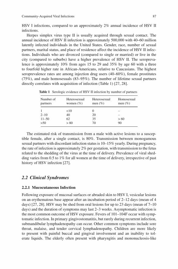
Community-Acquired Viral Infections 87
HSV I infections, compared to an approximately 2% annual incidence of HSV IIinfections.
Herpes simplex virus type II is usually acquired through sexual contact. Theannual incidence of HSV II infection is approximately 500,000 with 40–60 millionlatently infected individuals in the United States. Gender, race, number of sexualpartners, marital status, and place of residence affect the incidence of HSV II infec-tions. Individuals who are divorced (compared to single or married) or live in thecity (compared to suburbs) have a higher prevalence of HSV II. The seropreva-lence is approximately 10% from ages 15 to 29 and 35% by age 60 with a threeto fourfold higher rate in African-Americans, relative to Caucasians. The highestseroprevalence rates are among injection drug users (40–60%), female prostitutes(75%), and male homosexuals (83–95%). The number of lifetime sexual partnersdirectly correlates with acquisition of infection (Table 1) [27, 28].
Table 1 Serologic evidence of HSV II infection by number of partners
Number ofpartners
Heterosexualwomen (%)
Heterosexualmen (%)
Homosexualmen (%)
1 <10 0 –2–10 40 20 –11–50 62 35 > 60>50 > 80 70 90
The estimated risk of transmission from a male with active lesions to a suscep-tible female, after a single contact, is 80%. Transmission between monogamoussexual partners with discordant infection status is 10–15% yearly. During pregnancy,the rate of infection is approximately 2% per gestation, with transmission to the fetusrelated to the shedding of the virus at the time of delivery. Prevalence of viral shed-ding varies from 0.5 to 1% for all women at the time of delivery, irrespective of pasthistory of HSV infection [27].
2.2 Clinical Syndromes
2.2.1 Mucocutaneous Infection
Following exposure of mucosal surfaces or abraded skin to HSV I, vesicular lesionson an erythematous base appear after an incubation period of 2–12 days (mean of 4days) [27, 28]. HSV may be shed from oral lesions for up to 23 days (mean of 7–10days) and the duration of symptoms may last 2–3 weeks. Asymptomatic infection isthe most common outcome of HSV exposure. Fevers of 101–104F occur with symp-tomatic infection. In primary gingivostomatitis, but rarely during recurrent infection,submandibular lymphadenopathy can occur. Other common symptoms include sorethroat, malaise, and tender cervical lymphadenopathy. Children are more likelyto present with painful buccal and gingival involvement and an inability to tol-erate liquids. The elderly often present with pharyngitis and mononucleosis-like

88 C. Parsons
symptoms. During the initial evaluation, the physician must consider herpangina(usually due to coxsackieviruses), candida infections, Epstein–Barr virus, Stevens–Johnson syndrome (especially if the patient is on medications), and lesions inducedby chemotherapy or radiation therapy.
Recurrent infections are usually preceded by a prodrome of pain, burning, tin-gling, or itching approximately 6 h prior to the eruption of vesicles. In recurrentoral-labial disease, the vesicles are typically found on the vermilion border of thelips. Vesicles last an average of 48 h before progressing to a pustular or ulcer-ative and crusting stage. Lesions last for 72–96 h, with complete resolution in8–10 days.
Primary HSV II genital infection typically presents as macules and papules thatprogress to vesicles, pustules, and ulcers. Virus is shed for 11–12 days and thelesions heal in approximately 3 weeks. Systemic complaints of fever, dysuria, local-ized inguinal lymphadenopathy, and malaise occur in about 70% of cases. Mostinfections go unrecognized but symptomatic infections are more severe in women.Symptoms of primary HSV II infection are attenuated in the presence of antibodiesto HSV I. Genital herpes caused by HSV I is less severe clinically and less likely torecur. In women, lesions appear on the vulva and are typically bilateral, but they canalso involve the buttocks, perineum, vagina, and cervix. Primary infection can resultin a urinary retention syndrome in 10–15% of the cases, and aseptic meningitisin up to 25% of the cases. Men typically present with vesicular lesions superim-posed on an erythematous base on the glans penis or the penile shaft. As in females,the thigh, buttocks, and perineum may be involved. Other complications occurringin both groups include sacral radiculomyelitis, neuralgias, and meningoencephali-tis. Proctitis is more common in homosexual men following primary HSV IIinfection.
Recurrent infection is typically preceded by a prodrome often described as a localirritation. Three to five vesicles appear on the shaft of the penis in men, while womentypically present with vulvar irritation. The duration of symptoms is typically 8–10days with viral shedding lasting 2–5 days. Neurologic and systemic complicationsare uncommon in recurrent disease, but paresthesia and dysesthesias may occur. Thefrequency of recurrences varies among individuals, but the more severe the primaryinfection, the more likely and frequent are the recurrences. One-third of patientswill experience eight or nine episodes per year, one-third will have four to seven,and one-third will have two to three. Transmission of the virus may occur in bothsymptomatic and asymptomatic recurrences. Most HSV II-infected individuals areunaware of the infection and the risk to their partners.
Other disorders seen with herpes viruses include eczema herpeticum occurringin patients with underlying atopic dermatitis, and herpes gladitorium. Both HSV Iand II can trigger erythema multiforme.
2.2.2 Neonatal Disease
The incidence of neonatal HSV infection ranges from one case per 2,000–5,000deliveries per year, resulting in 1,500–2,200 cases annually in the United States
Community-Acquired Viral Infections 89
[27–29]. The transmission from mother to fetus is influenced by several factors.During primary maternal infection, the transmission rate is 30–50%, compared with3% during recurrent infection. The presence of maternal antibodies may reduce theseverity and the likelihood of transmission. Rupture of membranes for more than6 h can result in ascending infection of the neonate via the cervix. Inoculation ofnewborns may also occur following inoculation at the site of fetal scalp monitors.
About 75–80% of infections are acquired through intrapartum contact withinfected genital secretions of the mother. The remainder of vertical transmissionsare due to postnatal acquisition and, rarely, in utero infections. Neonates infectedin utero present with skin vesicles or scarring, ocular disease, and microencephalyor hydranencephaly. Due to the high mortality, these neonates should be diagnosedand treated within the first 24 h. Other neonatal HSV infections include: those local-ized to the skin, eye, and mouth; encephalitis with or without skin involvement; anddisseminated infection involving CNS, lungs, liver, adrenal glands, skin, eye, and/ormouth. Relatives and hospital personnel with orolabial herpes may also spread HSVto newborns.
2.2.3 Keratoconjunctivitis
HSV is the most common cause of corneal opacification and infection-related visionloss in the industrialized world. HSV I is the major agent beyond the neonatal period[27, 28, 30, 31]. The incidence of new and recurrent episodes is 20.7 per 100,000person-years. Primary infection is often asymptomatic and results in latent infectionin the trigeminal or other sensory ganglia. Recurrent viral shedding can be unilat-eral or bilateral and may be associated with preauricular adenopathy, photophobia,tearing, eyelid edema, chemosis, or the pathognomonic branching dendritic lesions.Recurrent episodes are typically unilateral and occur with similar frequency to thatof herpes labialis. Infection may involve either superficial (eyelids, conjunctiva, orcorneal surface) or deep (cornea or anterior uvea) structures. Infection of the deeperstructures is more serious and may cause permanent visual loss. Active disease withthe initial episode of keratitis averages 2–3 weeks, while recurrent episodes lastapproximately 1 month. In 90% of cases, visual acuity of 20/40 or better will bemaintained [17, 20, 21].
2.2.4 HIV and HSV
Genital herpes with HSV II has been linked to both the acquisition and transmis-sion of HIV-1 and represents the most frequent sexually transmitted disease amongHIV-1 seropositive individuals [27]. Since the prevalence of HSV antibodies amonginjection drug users and men who have sex with men is high when HIV infectionis detected, few cases of primary HSV infection occur in HIV-infected adults oradolescents. Reactivation of HSV in HIV-infected individuals is more frequent thanin immunocompetent individuals and may result in multiple sites of shedding aswell as chronic, persistent mucocutaneous disease. HSV infection in this setting can

90 C. Parsons
present as large ulcerative lesions involving any area of the body, although the gen-ital and perirectal regions are the most common. Less frequently, HSV may resultin CNS infection, bronchitis, pneumonitis, and disseminated disease. The severeforms of HSV infection, including mucocutaneous ulcers lasting ≥1 month, bron-chitis, pneumonitis, or esophagitis are more likely to occur in those with CD4 counts≤100 cells/mm3 and may be the first clinical manifestation of HIV infection.
2.2.5 CNS Infections
HSV accounts for 10–20% of all cases of viral encephalitis and is the most com-mon cause of fatal sporadic encephalitis in the United States [27, 32]. The incidenceof this disease is three per 100,000 cases and, beyond the neonatal period, is typi-cally caused by HSV I. HSV II may cause aseptic meningitis in adults, and thesepatients present without the mental status changes seen with encephalitis. Treatedappropriately, HSV encephalitis still incurs a mortality of approximately 30%, whilemortality in untreated patients exceeds 70%, with <3% of survivors regaining nor-mal neurologic function. Patients typically present within 7 days of symptom onset.About 90% of these patients present with fever and focal neurologic findings,including hemiparesis; dysphasia; aphasia; ataxia; or focal seizures. Patients mayalso have altered consciousness or unusual behavior. Although no pathognomonicsigns exist, HSV encephalitis should be suspected in those with a progressively dete-riorating level of consciousness in association with fever, an abnormal CSF profile,and focal neurological findings in the absence of other etiologies.
2.2.6 HSV in Pregnancy
Approximately 1,500–2,200 cases of neonatal herpes occur each year in the UnitedStates, most often due to HSV II. Due to the high morbidity and mortality inuntreated cases of neonatal infections, reduction in perinatal transmission is a majortarget in preventative care. Approximately 2% of susceptible women acquire HSVinfection during pregnancy, which can lead to an increased risk of preterm labor,intrauterine growth retardation, and spontaneous abortion. These infections occurduring any phase of pregnancy. Those patients with the highest risk of acquiring theinfection are young, single women with a history of previous sexually transmitteddiseases. Vertical transmission and pregnancy-related morbidity for women infectedearly in their pregnancy is uncommon [33]. Acquisition of the disease near the timeof delivery, however, results in vertical transmission and complications [33]. Table 2reflects the rates of maternal–fetal transmission of HSV based on maternal infection[28, 29, 33].
2.3 Treatment
No cure for HSV exists, but steps can be taken to prevent transmission, and antiviraltherapies reduce viral shedding and healing time. The main issue concerning genitalHSV transmission, however, is preventative care. Individuals exhibiting high-risk

Community-Acquired Viral Infections 91
Table 2 Rate of maternal – fetal transmission
Maternal infection Fetal infection rate (%)
Primary infection with active lesions at delivery 50Asymptomatic primary infection 33Recurrent infection with active lesions at delivery 3–4Asymptomatic recurrent infection 0.04
behaviors and those with current infection, even if asymptomatic, should be edu-cated on the proper use of condoms and behavioral modification to decrease the riskof spreading and acquiring HSV.
Acyclovir is a synthetic acyclic purine nucleoside analog, and its derivativeswith greater oral bioavailability include the prodrug valacyclovir (converted to acy-clovir) and famciclovir (converted to penciclovir). The major adverse effect fromthese medications is alteration of renal function caused by crystallization of thedrug in renal tubules, resulting in reversible elevation of serum creatinine and, lesscommonly, acute tubular necrosis. The risk of renal dysfunction is substantiallyincreased in individuals who are dehydrated or have underlying renal insufficiency.Thrombotic thrombocytopenic purpura–hemolytic uremic syndrome has been asso-ciated with high-dose valacyclovir therapy in HIV-infected and transplant patients,although a cause–effect relationship is unproven.
For mucocutaneous infections, therapy shortens the duration of viral sheddingand length of time to healing while reducing associated pain, if initiated within 24 hof onset. These effects are more demonstrable for primary HSV infection. Even withearly initiation of therapy, the frequency of recurrences is not affected. Treatment ofasymptomatic individuals will also reduce shedding and HSV transmission to unin-fected partners, although this effect is lost with discontinuation of the medication.Table 3 describes the therapeutic options for HSV infections.
Suppressive therapy should be considered in individuals with more than sixepisodes per year, or in those with severe cases as with HIV infection. Therapycan be initiated in adherent patients at the start of symptom prodrome to decreasethe length of time to healing. The FDA has approved therapy for 12 months withacyclovir, although studies have demonstrated efficacy with no cumulative toxicityover 5 years. Patients on suppressive therapy should have dose adjustments or tri-als of discontinuation of suppressive therapy every 12 months to assess the need forcontinued therapy. It may be safe to discontinue suppressive therapy in HIV-infectedpatients once their CD4 count rises above 350 cells/mm3.
2.4 Pregnancy
Since the greatest risk of vertical transmission occurs near the time of delivery,patients should be educated on abstinence and the use of condoms during thethird trimester – especially if the HSV status of the partner is unknown. The FDA

92 C. Parsons
Table 3 Treatment of genital HSV infections
Infection Acyclovir Valacyclovir Famciclovir
Initialepisode
200 mg PO 5×/day × 5–10days
5 mg/kg iv q8h × 5 days5% ointment topically
q6h × 7 daysRecurrent
episode200 mg PO 5×/day × 5
days400 mg PO tid × 5 days800 mg PO bid × 5 days
1,000 mg PO bid× 5–10 days
250 mg PO tid × 5–10days
Suppression 200 mg PO tid 500 mg PO bid× 5 days
125 mg PO bid × 5 days
Immunocompro-mised
400 mg PO bid 250 mg PO bid500 mg PO qd
250 mg PO bid
Initialepisode
200–400 mg PO 5×/day ×10 days
5 mg/kg iv q8h × 7–10days (or up to 14 days inHIV-infected patients)
Recurrentepisode
200–400 mg PO 5×/day ×5–10 days
500 mg PO bid× 10–14 days
125–250 mg PO bid ×10–14 days
Suppression 200 mg PO tid400 mg PO bid
500 mg PO bid× 5–10 days
125–250 mg PO bid ×10–14 days
HSVencephali-tis
10 mg/kg iv q8h × 10–14days
500 mg PO qd500 mg PO bid
125–250 mg PO bid
NeonatalHSV
10 mg/kg iv q8h × 10–14days
∗ ∗
HSV inpregnancy
See special cases below ∗ ∗
HSVresistance
See special cases below
∗Signifies that these drugs are not used in these clinical settings.References [27, 32, 34, 35].
has categorized current antiviral medications as class C (acyclovir) and class B(valacyclovir and famciclovir) for use in pregnancy. Acyclovir crosses the placenta,concentrates in the amniotic fluid and breast milk, and achieves therapeutic levelsin the fetus, when given by the oral or intravenous route to the mother. Studies havedemonstrated no increase in fetal complications even when acyclovir is given in thefirst trimester. Guidelines on the treatment of pregnant women remain controversialamong experts in the field [33, 36, 41].
For treatment of a primary infection in the first and second trimester, stan-dard doses for genital infections should be employed. Suppressive therapy overthe last 4 weeks of pregnancy may prevent recurrence at term, thereby decreas-ing the need for Cesarean section. Termination of pregnancy is not recommended
Community-Acquired Viral Infections 93
for women who become pregnant while receiving antiviral therapy. If the primaryinfection occurs in the third trimester, Cesarean section should be considered inall patients, especially if symptoms occur within 4 weeks of delivery. If Cesareansection is contraindicated or the membranes ruptured more than 4–6 h prior toa Cesarean section, antiviral therapy of mother and newborn may be indicated.In all patients that are considered high risk for vertical transmission, proceduresthat could damage the newborn’s skin and create a portal of entry for infection(e.g., scalp electrodes, fetal blood sampling, and instrumental delivery) should beminimized.
For treatment of a recurrent infection, normal vaginal delivery is indicated ifgenital lesions are not apparent at the time of delivery. Cultures during late gestationto predict viral shedding at term or cultures at the time of delivery are not indicated.If the patient presents at delivery with lesions, the risk of HSV transmission to thenewborn must be assessed on an individual basis. The available evidence suggeststhat the risks of vaginal delivery for the fetus are small and must be weighed againstthe risks to the mother of a Cesarean section.
2.5 Resistance in Immunocompromised Patients
The prevalence of acyclovir-resistant HSV infection in immunocompromisedpatients with HIV infection or stem-cell transplantation is approximately 5%. Viralresistance is promoted by the degree of immunosuppression of the patient and byprolonged use of acyclovir, although resistance emerging during chronic suppressivetherapy is relatively uncommon. Cross-resistance for valacyclovir and famciclovirwould be expected in this setting, as the mechanisms of resistance are similar forthese agents [42]. If the patient has not responded to initial therapy within 14 days,resistance should be suspected. The virus may be susceptible, but higher doses ofmedication or changing from acyclovir to one of the newer prodrugs with betterbioavailability may be needed. Benefits from increasing acyclovir to 800 mg orally,five times a day or dosing acyclovir as a continuous-infusion at 1.5–2.0 mg/kg/hruntil the lesions have crusted have been demonstrated.
If these regimens fail, antiviral-susceptibility testing may be indicated and con-sideration given for therapy with foscarnet or cidofovir. Foscarnet is consideredfirst-line therapy in acyclovir-resistant infections. The major side effects are nephro-toxicity and mineral and electrolyte abnormalities, but these typically resolve withdiscontinuation. Foscarnet is dosed at 40 mg/kg intravenously every 8 h and shouldbe dosed based on creatinine clearance. Therapy is continued until the lesions havecrusted and complete re-epithelialization has occurred (usually after 2–3 weeks).Cidofovir is currently approved for the treatment of cytomegalovirus disease andshould only be considered in individuals who have failed high-dose acyclovir andfoscarnet. The major adverse event is renal tubular toxicity. In order to diminishthis nephrotoxicity, both hydration and probenecid dosed pre- and post-infusion arerecommended. Cidofovir is dosed at 3–5 mg/kg intravenously weekly for 2–4 weeks. Once lesions have resolved, recurrent infections are typically caused

94 C. Parsons
by acyclovir-susceptible HSV; therefore, therapy with acyclovir or related agentsshould be initiated as first-line therapy in this setting [35, 43, 44].
3 Varicella Zoster Virus
Like the HSV, Varicella-zoster virus (VZV) is an enveloped, double-stranded, DNAvirus. Primary exposure to VZV results in varicella (chickenpox), a usually benign,highly contagious infection of children. Reactivation of latent VZV results in herpeszoster (shingles), an illness most commonly seen in adults.
3.1 Epidemiology
VZV is spread person-to-person by direct contact or aerosolization from skin lesionsor respiratory-tract secretions, and the mucosa of the upper respiratory tract andconjunctiva are the main portals of entry. VZV is transmissible to susceptible hostsfrom the source individual from approximately 2 days prior to symptom onset untilskin lesions begin to crust over. Of the four million cases of varicella per year, 33%occur in preschool children (1–4 years of age) and 44% occur in school age children(5–9 years of age). The secondary attack rate is 90% among susceptible individualsin the same household. These secondary varicella cases within the family are usuallymore severe than the primary cases, likely due to the greater intensity of exposure.Only 5% of varicella infections are subclinical (i.e., without rash).
A history of VZV infection is a reliable marker for immunity with a posi-tive history 97–99% predictive of serologic immunity. Approximately 70–90% ofindividuals with a negative history of VZV are also seropositive.
Preventative measures are important for the control of nosocomial transmissionof VZV. After exposure to VZV, susceptible employees can serve as vectors withtransmission to patients. VZV has been reported in hospital employees withoutdirect contact with patients having active lesions but exposed to air from the patient’sroom [45].
Herpes zoster is more common in adults and the immunocompromised, 75% ofcases occur in patients over the age of 45. Immunocompetent children, adolescents,and young adults may develop herpes zoster, although in adolescents and youngadults, this should prompt concern for HIV infection in patients with risk factors forHIV. Acute acquisition of VZV is not associated with herpes zoster.
3.2 Clinical Syndromes
3.2.1 Varicella
Varicella in children is a self-limited disease of 4–5 days duration and incursfever, malaise and a generalized, pruritic, vesicular rash that starts on the face andscalp then spreads to the trunk and extremities. The incubation period is 10–21

Community-Acquired Viral Infections 95
days. Successive crops of vesicular lesions appear over 2–4 days and eventuallybecome purulent and crust over. Thus, the simultaneous presence of different stagesof vesicular lesions is characteristic of varicella. Complications from varicellainclude infection with beta-hemolytic Group A streptococci, pneumonia, menin-goencephalitis, cerebellar ataxia, and hepatitis. Reyes syndrome associated withaspirin use during VZV infection is now considered uncommon [46]. Adolescentsand adults are at greater risk for complications, particularly varicella pneumonia,and exhibit a higher mortality rate.
Although perinatal infection is uncommon given maternal immunity to VZV,intrauterine VZV infection may result in fetal varicella syndrome (low birth weight,cutaneous scarring, limb hypoplasia, microencephaly, cortical atrophy, chorioretini-tis) if maternal infection occurs during the first half of the pregnancy. The incidenceof fetal varicella syndrome with VZV infection in weeks 1–12 is 0.4% and weeks13–20 is 2%. Varicella infection of the mother 5 days before to 2 days after deliverymay result in severe varicella in 17–30% of newborns with a 31% risk of death ifuntreated. Passive immunization with varicella immune globulin (VZIG) is effectivein reducing mortality [47, 48].
3.2.2 Herpes Zoster
After primary infection, latent VZV persists within the sensory dorsal root gan-glia. Herpes zoster presents as a unilateral vesicular rash distributed over one tothree dermatomal segments, although it is most often limited to a single dermatomein immunocompetent patients. The characteristic rash consists of closely croppedsmall vesicles, often referred to as “dew drops on a rose petal.” Vesicles typicallycrust over within 10 days and completely heal within 1 month. The most com-mon complication of herpes zoster is pain, referred to as postherpetic neuralgia.Postherpetic neuralgia most often affects those over 50 years of age and can besevere, lasting for weeks to months. Pain may also precede the appearance of therash [49].
Although less common, VZV infection, with or without the typical rash, maycause more severe clinical manifestations. CNS syndromes caused by VZV includemeningitis, radiculitis, meningoencephalitis, and stroke syndromes. When thetrigeminal nerve is involved, especially the ophthalmic branch (zoster ophthalmi-cus), complications include dendritic keratitis, anterior uveitis, iridocyclitis, andpanophthalmitis. Zoster oticus (involvement of the vestibular nerve) may result inhearing compromise and brain-stem involvement. One form of this disease, RamseyHunt Syndrome, is characterized by vesicles located in the auditory canal withcoincident facial nerve paralysis [50, 51].
In the immunocompromised host (HIV-infected or organ transplant recipient),reactivation of VZV may result in a disseminated infection with a generalizederuption along with central nervous system, pulmonary, hepatic, and pancreaticinvolvement. Disseminated zoster in these hosts, and thus the need for more dili-gent evaluation and monitoring, is signified by involvement of more than onedermatome.

96 C. Parsons
3.3 Diagnosis
The clinical features of acute varicella infection in children, and the unilateral,dermatomal rash seen in herpes zoster, are sufficiently characteristic of these ill-nesses that diagnostic testing is usually not necessary. However, herpes simplex,measles, and other causes of vesicular eruptions may mimic herpes zoster. Althoughviral culture is the traditional gold standard for identifying VZV from vesiclefluid, more recently developed PCR assays are more sensitive. In addition, PCRand intrathecal serologic tests can be useful in diagnosing CNS VZV infections[50, 51].
3.4 Antiviral Therapy
Acyclovir, when given within 24 h of onset of varicella, decreases the number oflesions and the formation of new lesions, the duration of fever, and the severity ofcutaneous and systemic signs and symptoms. Acyclovir does not, however, decreasetransmission or reduce absence from school and is not recommended for the rou-tine treatment of healthy children. However, children at risk for complications ofvaricella should receive acyclovir, including those with cutaneous or cardiopul-monary disorders, and those on chronic corticosteroids [52]. Since varicella isa more severe disease in adolescents, adults, and immunocompromised children,treatment with acyclovir is recommended [53, 54]. Prophylaxis with acyclovir canprevent secondary cases among close contacts. In a placebo-controlled trial, 16%of ACV treated patients developed varicella, compared with 100% of controls [55].Treatment must begin early because in the immunocompetent host, viral replicationis undetectable 72 h after the rash appears.
Acyclovir is also useful in the treatment of herpes zoster. If given within 72 h ofthe appearance of rash, it will accelerate the rate of healing, reduce severity of dis-ease, and diminish the incidence and severity of postherpetic neuralgia. Acyclovir isespecially useful in treating those over the age of 50 who have a greater incidence ofpostherpetic neuralgia [56]. While steroids do not reduce the incidence of posther-petic neuralgia, the addition of prednisone to acyclovir during acute primary herpeszoster decreases the duration of pain and the return to daily activities [57].
Valacyclovir, a prodrug of acyclovir with better absorption and higher serumlevels, is also approved for the treatment of herpes zoster [58]. Famciclovir, a pro-drug of penciclovir, is effective in treating varicella and herpes zoster [59]. Theadvantage of valacyclovir and famciclovir is convenience: both are dosed 3 times aday instead of 5 times per day with acyclovir. Famciclovir and valacyclovir maydecrease the duration of postherpetic neuralgia but not its incidence in elderlypatients.
Since therapeutic levels after oral administration are unreliable, intravenousacyclovir is recommended for severe VZV infections in the setting of dissemi-nated disease, transplant recipients, and HIV-infected patients. Adequate hydration

Community-Acquired Viral Infections 97
must be maintained during intravenous acyclovir administration to prevent ACVprecipitation in the renal tubules resulting in acute renal failure.
In summary, acyclovir is recommended for high-risk children and adults withvaricella. Valacyclovir or famciclovir are recommended in the initial treatment ofzoster, given their superior pharmacokinetic profiles.
Disease Valacyclovir Famciclovir Acyclovir
VaricellaImmunocompetent Host
20 mg/kg (maximum800 mg) 4 timesdaily for 5 days
VaricellaImmunocompromised Host
10 mg/kgintravenouslyevery 8 h for 7–10days
Herpes ZosterImmunocompetent Hosts
1 g 3 times dailyfor 7 days
500 mg 3 timesdaily for 7 days
800 mg 5 times dailyfor 7 days
Herpes ZosterImmuno compromised Host
10 mg/kgintravenouslyevery 8 h for 7days
Herpes ZosterImmuno compromised HostAcyclovir-resistant virus
Foscarnet40 mg/kgintravenously 3times daily for7–14 days
Resistance to these three drugs is uncommon, but when resistance is present, fos-carnet, which acts by directly inhibiting the viral DNA polymerase, may be useful.The major toxicities of foscarnet are renal dysfunction and electrolyte imbalance.
3.5 Prevention
3.5.1 Post-exposure Prophylaxis
Varicella Zoster Immune Globulin (VZIG)
If given to exposed persons, particularly immunocompromised children, within96 h of exposure to someone with varicella, VZIG may prevent or amelioratevaricella infection. Protection lasts for 3 weeks with a single dose of VZIG.Unfortunately, VZIG is nearly depleted due to the discontinuation of its productionby the manufacturer [60].
VariZIG
VariZIG is a purified human immune globulin preparation made from plasmacontaining high levels of anti-varicella antibodies. This product is lyophilized

98 C. Parsons
and must be reconstituted for intramuscular injection. VariZIG is currentlyrecommended for immunosuppressed patients and pregnant women with docu-mented exposure to persons with varicella, who lack either a history of VZV vacci-nation or detectable VZV IgG antibodies. VariZIG should be administered with 96 hof exposure. An expanded access program is available through the manufacturer(800-843-7477) [61].
IVIG
If VZIG or VariZIG cannot be administered, IVIG (400 mg/kg 1-time dose) shouldbe considered, although no data currently exist for its utility in this setting.
3.5.2 Vaccination
A live, attenuated VZV vaccine has been developed and proven effective in pre-venting both varicella and herpes zoster. Unlike wild-type VZV, the vaccine straincauses a subclinical infection, leading to immunity that is 70–90% effective in pre-venting the symptoms of varicella. Transmission of the vaccine strain to others israre, but it is advisable for the vaccinee to avoid close contact with those at riskfor severe complications of varicella. The vaccine is also effective in preventing ormodifying varicella infection if given within 3 days of exposure to persons withvaricella. Herpes zoster and the severity of illness, including the duration of pain,are also reduced in vaccinees [60].
The Advisory Committee on Immunization (ACIP) has recommended that allchildren entering child care facilities and elementary schools receive the VZV vac-cine if they lack evidence of varicella immunity (physician diagnosis of varicella,reliable history, or serologic evidence) [60]. The first dose is recommended at age12–15 months, with a second vaccine recommended for 4–6 years of age. Since therisk of severe varicella is high in adolescents and adults, the vaccine is also recom-mended for persons over 13 years of age lacking evidence of prior immunity. Twodoses should be given 4–8 weeks apart in this age group.
The vaccine should not be given to patients with cellular immunodeficienciesbecause of the risk of severe vaccine-associated varicella, but VZV vaccine can begiven to patients with humoral immunodeficiencies and asymptomatic or mildlysymptomatic (age-specific CD4+ T-lymphocyte percentage of ≥25%) HIV-infectedchildren. Pregnant women should not receive the vaccine because of concerns offetal varicella syndrome and the increased risk of severe varicella in late pregnancy.Routine, universal VZV vaccination will decrease the booster effect from exposureto wild-type virus, and it is clear that vaccinated persons >5 years removed from vac-cination are at greater risk for acquiring VZV and for suffering more severe disease[62]. Therefore, it is unclear whether vaccinees with no history of VZV exposurewill require additional booster vaccinations to prevent severe varicella infectionslater in life, although vaccination every 5 years may eventually be warranted.

Community-Acquired Viral Infections 99
4 Respiratory Syncytial Virus
4.1 Virology and Epidemiology
RSV is a single-stranded RNA virus and member of the Paramyxoviridae family.Subtypes A and B are often both present in outbreaks, although subtype A is associ-ated with more severe disease. RSV infections are distributed worldwide and are theleading cause of lower respiratory-tract infections (50–90% of bronchiolitis casesare due to RSV) in infants and young children. In the United States, RSV infec-tions generally begin in November, peak from January to March, then continuethrough April to mid-May. Approximately 120,000 children are hospitalized and4,500 deaths occur annually in the United States due to the complications of RSV.
About 50–69% of all children develop primary RSV infection by the age of12 months with 15–22% having lower respiratory involvement. One-half to 2%will be hospitalized with a mortality of 0.5–3.5%. By 2 years of age, greater than95% have been infected with RSV. Immunity following infection is short-lived, andre-infection occurs throughout life. Re-infection rates in preschool aged childrenrange from 40 to 70%, with approximately 20% recurrences in school-aged children,adolescents, and adults.
RSV is highly infectious and easily transmitted from person to person by closecontact. The primary modes of transmission include direct contact with large secre-tion droplets (small particle aerosol is not a significant mode of transmission) andself-inoculation of the eyes and nose with hands made infectious by touching con-taminated objects. For Example, RSV can be isolated from countertops more than6 h after contact with an infected source such as nasal secretions. Other infectioussources include rubber gloves (90 min), gowns (30 min), and hands (25 min). Goodhand washing and proper disposal practices are, therefore, important since infectedindividuals shed the virus for 1–21 days (mean of 6.7 days), even if asymptomatic[63, 64].
Increased disease severity is associated with low socioeconomic status, ethnic-ity, male gender, young age, body mass of less than 5 kg, prematurity, chroniclung disease, congenital heart disease (especially in association with pulmonaryhypertension), and T-cell immunodeficiency. Increased risk of acquiring the dis-ease occurs in crowded conditions (two or more individuals sharing a bedroom),school age siblings, multiple births, lack of breast feeding, passive smoke exposure,day-care attendance, and birth within 6 months prior to an anticipated RSV season[63, 65].
4.2 Disease Pathogenesis
The average incubation period of RSV is 5 days. Symptoms can range from cold-likesymptoms to severe bronchiolitis or pneumonia. Symptomatic re-infection typicallypresents with milder illness. The hallmark of infection – involvement of the smallintrapulmonary airways – results in the classic clinical syndrome of bronchiolitis.

100 C. Parsons
Lower respiratory-tract infections are due to transfer of the virus from the upperrespiratory tract and results in sloughing of the bronchiolar epithelium, hypersecre-tion of mucus, peribronchiolar mononuclear infiltration, and submucosal edema.The plugs of mucus and cellular debris lead to partial or complete airway obstruc-tion, especially in the small lumens of infants. Following infection, immunity toRSV is transient and imperfect. Recurrent upper respiratory infections are probablydue to the transient nature of immunity of IgA. In contrast, lower respiratory-tractresistance appears to be more durable [63, 65].
4.3 Neonatal Infection
Newborns typically acquire RSV via contact with visitors and health care person-nel. The clinical variation and rare clinical occurrence of lower respiratory-tractinvolvement in individuals less than 3 weeks of age is probably due to the pres-ence of maternally derived neutralizing antibodies. Signs of upper respiratorytract infections occur in less than 50% of infected neonates and are highly vari-able and nonspecific. These clinical signs include poor feeding, lethargy, andirritability.
4.4 Infection in Infants
Infants younger than 1 year who have low RSV antibody titers and are not breast-fed have an increased risk of developing lower respiratory tract disease in the first5 months of life. One of the early manifestations in this age group following RSVinfection is apnea. This occurs more readily in infants less than 6 weeks of age,those who are born prematurely, and those with low arterial oxygenation satura-tions. Mechanical ventilation, if required, is necessary for approximately 48 h, withpostextubation apnea being uncommon.
Infants with severe disease and in need of more intensive monitoring can be iden-tified with six independent clinical and laboratory findings: (1) oxygen saturation ofless than 95%, determined by pulse oximetry; (2) atelectasis on chest radiograph;this occurs in 50% of hospitalized patients and may be the only radiographic findingin 15% of the cases; diffuse interstitial pneumonitis, hyperexpansion, and subseg-mental consolidation, typically in the right upper or middle lobe (25% of cases),also occur; (3) respiratory rate greater than or equal to 70 breaths per min; (4) ges-tational age less than 34 weeks; (5) age less than 3 months; and (6) “Ill” or “toxic”appearance. If hospitalization is required, the length of stay is typically 4–7 days,with full recovery at about 2 weeks. Major complications include respiratory failure,apnea, and secondary bacterial infections. Long-term complications are minimal,with recurrent episodes of wheezing being the major clinical sequelae. Recurrencesdiminish after the first few years, with no increased risk for airway hyper-reactivityor pulmonary function abnormalities by the ages of 8–12.

Community-Acquired Viral Infections 101
4.5 Childhood Infection
Initial infections with RSV are typically symptomatic and range from a mild cold-like illness to severe bronchiolitis or pneumonia. These latter syndromes occur in30–70% of cases following the initial exposure to RSV and can be difficult to dif-ferentiate, but the classic signs of bronchiolitis are wheezing and hyperexpansion ofthe lung. Typically, children have fever ranging from 38 to 40◦C during the first 2–4days of the illness, nasal discharge, pharyngitis, and cough. Hoarseness and laryn-gitis are uncommon. By the time these children present to their local health carefacility, lower respiratory tract symptoms are more prominent. Signs and symptomsare based on the severity of disease and can include increased cough, increasedrespiratory rate of up to 80 breaths per min with substernal and intercostal retrac-tions during inspiration, a prolonged expiratory phase, hypoxemia typically withoutcyanosis, hyperexpanded and hyperresonance in the chest, and intermittent rales andwheezes.
4.6 Infection in Adults and the Elderly
Infection with RSV after the age of 2 most commonly manifests as an upper respira-tory tract illness or tracheobronchitis. Typically, symptoms include nasal congestionand cough with a more severe and prolonged course as compared to “colds” causedby other respiratory viruses. RSV appears to be an increasing cause of respira-tory disease in this population, especially those in nursing care facilities. Duringoutbreaks, the attack rate ranges from 10 to 40% and accounts for 5–27% of allrespiratory-tract infections in long-term care facilities. Individuals over the age of60 present with mild nasal congestion, but fever, anorexia, pneumonia, or bronchitismay develop [66, 68].
4.7 Diagnosis
A diagnosis of RSV pulmonary syndrome should be suspected during the appro-priate winter season when involving children under 1 year of age, the elderly, orimmunocompromised adults, and if bronchiolitis is present. The standard test fordiagnosing RSV is viral growth in culture, using nasal wash or nasopharyngealswab samples for inoculation. Rapid antigen testing and PCR methodologies arelargely supplanting culture for diagnosis given their more rapid turnaround timeand increased sensitivity in some settings [69–71].
4.8 Treatment
The mainstays of therapy consist of respiratory support, nutrition, and hydra-tion. Bronchodilators (beta-agonists and epinephrine) and anti-inflammatory agents

102 C. Parsons
(cromolyn sodium and budesonide) have demonstrated some clinical utility, but fur-ther studies are required to confirm these findings [63, 65]. The use of antivirals inthe treatment of RSV infections also remains controversial.
Ribavirin is the only antiviral agent licensed for the treatment of RSV infec-tions. Ribavirin is dosed at 6 g/300 cc of water over 18 h or 6 g/100 cc ofwater over 2 h, 3 times a day. Early clinical trials of aerosolized ribavirin ther-apy suggested some therapeutic benefit. However, interpretation of the results iscomplicated by the investigators’ use of distilled water, a known bronchoconstric-tor, as the placebo treatment. A similar study, when conducted using aerosolizedsaline as the placebo, found no clinical benefit from ribavirin therapy [72]. Cohortstudies also failed to demonstrate an improved clinical outcome with ribavirin ther-apy [73, 74]. The Committee on Infectious Diseases of the American Academyof Pediatrics has changed its recommendations on the use of ribavirin to “may beconsidered” in selected infants and young children at high risk for serious RSVdisease [75]; however, most clinicians do not use aerosolized ribavirin because ofthe limited clinical benefit, cost, and difficulty in administration. In addition, rib-avirin should not be used for pregnant women, given its teratogenicity in animalmodels.
4.9 Prevention
Palivizumab is an FDA-approved humanized monoclonal antibody directed againstan RSV surface glycoprotein. Initial studies of palivizumab dosed at 15 mg/kg IMevery month for 5 months during the peak RSV season demonstrated an over-all reduction in RSV-related hospitalizations by 55%, with significant reductionsin infection of premature infants with chronic lung disease (39%) and prematureinfants without chronic lung disease (78%). Other significant reductions includednumber of hospital days (42%), days of oxygen requirement (40%), and incidenceof ICU care (57%) [76, 77]. Advantages to the use of palivizumab include intramus-cular route of administration, a delay in dosing other vaccines is not required, andpalivizumab is not a blood product, thus, has no risk of transmitting blood-bornepathogens. Given more recent data for the use of palivizumab in high-risk pedi-atric patients, it is recommended for the following groups: children under 2 withbronchopulmonary dysplasia, children under 2 who have hemodynamically signif-icant congenital heart disease, and children under 1 born <28 weeks gestation, orunder 6 months born 29–32 weeks gestation with risk factors for RSV acquisition[77–79].
Interrupting transmission at health care facilities is necessary to prevent thespread of infection. Special precautions should be advocated in RSV infectedpatients during the peak RSV season and, especially, when a hospital outbreakdevelops. The table below lists infection control guidelines.

Community-Acquired Viral Infections 103
General control measures to prevent nosocomial RSV transmission:
• Educate hospital staff about RSV epidemiology, modes of transmission, and means ofprevention.
• Use contact and droplet isolation for RSV-positive patients including gloves and gown.• Maintain good hand washing Procedures following any contact with RSV-infected patients
or fomites, even if gloves are used.• Limit visitors. Do not allow visitors who have symptoms of respiratory infection to visit
uninfected pediatric, immunosuppressed, or cardiac patients.• Restrict staff with upper respiratory symptoms from patients at high risk for complications
from RSV infection.
Control measures during RSV outbreaks:
• Avoid elective admissions for high-risk patients.• Admit young children with symptoms of viral upper respiratory infections to single rooms.• Cohort patients with RSV infection.• Cohort staff to infected or uninfected patients.
Ref. [64, 69].
Key Points
• Illnesses caused by influenza, HSV, VZV, and RSV are commonly seen inoutpatient settings and incur substantial morbidity and mortality.
• Prevention is important to control these viral illnesses. Influenza and varicella areeffectively managed through vaccination. HSV transmission can be preventedthrough behavioral changes, education, and occasionally medical treatment. Inhigh-risk individuals, RSV infection can be reduced by immunoprophylaxis.
• Antiviral agents are used effectively to decrease morbidity and mortality for theseillnesses. Although resistance to older antiviral agents has been seen, the adventof newer therapies for these infections has reduced concern for the effect ofresistance on the outcomes of these infections.
References
1. Fiore AE, Shay DK, Haber P et al. (2007). Prevention and control of influenza.Recommendations of the Advisory Committee on Immunization Practices (ACIP), 2007.MMWR Recomm Rep 56:1
2. Zimmerman RK, Ruben FL, Ahwesh ER (1997). Influenza, influenza vaccine, and amanta-dine/rimantadine. J Fam Pract 45(2):107–122
3. Cox NJ, Fukuda K (1998). Influenza. Infect Dis Clin North Am 12(1):27–384. Simonsen L, Clarke MJ, Schonberger LB et al. (1998). Pandemic versus epidemic influenza
mortality: A pattern of changing age distribution. J Infect Dis 78(1):53–605. Schwarzmann SW, Adler JL, Sullivan RJ, Jr. et al. (1971). Bacterial pneumonia during the
Hong Kong influenza epidemic of 1968–1969. Arch Intern Med 127:10376. Dell KM, Schulman SL (1997). Rhabdomyolysis and acute renal failure in a child with
influenza A infection. Pediatr Nephrol 11:363
104 C. Parsons
7. Cunningham E, Kohli R, Venuto RC (1979). Influenza-associated myoglobinuric renal failure.JAMA 242:2428
8. Treanor JJ (2005). Influenza Virus. In: Mandell GL, Bennett JE, Dolin R (eds) Principles andPractice of Infectious Diseases, 6th ed. Philadelphia, PA: Churchill Livingstone, 2060
9. Rodriguez WJ, Schwartz RH, Thorne MM (2002). Evaluation of diagnostic tests for influenzain a pediatric practice. Pediatr Infect Dis J 21:193
10. Hayden FG, Fritz R, Lobo MC et al. (1998). Local and systemic cytokine responses duringexperimental human influenza A virus infection. Relation to symptom formation and hostdefense. J Clin Invest 101:643
11. Mossad SB (1999). Underused options for preventing and treating influenza. Cleve Clin J Med66(1):19–23
12. Fiore AE, Shay DK, Haber P et al. (2007). Prevention and Control of Influenza.Recommendations of the Advisory Committee on Immunization Practices (ACIP), 2007.MMWR Recomm Rep 56:1
13. Jacobs BC, Rothbarth PH, van der Meche FG et al. (1998). The spectrum ofantecedent infections in Guillain-Barre syndrome:a case-control study. Neurology 51(4):1110–1115
14. Lasky T, Terracciano GJ, Magder L et al. (1998). The Guillain-Barre syndrome and the 1992–1993 and 1993–1994 influenza vaccines. N Eng J Med 339(25):1797–1802
15. Belshe RB, Mendelman PM, Treanor J et al. (1998). The efficacy of live attenuated, cold-adapted, trivalent, intranasal influenzavirus vaccine in children. N Eng J Med 338(20):1405–1412
16. Treanor JJ, Mattison HR, Dumyati G et al. (1992). Protective efficacy of combined liveintranasal and inactivated influenza A virus vaccines in the elderly. Ann Intern Med117(8):625–633
17. Cooper NJ, Sutton AJ, Abrams KR et al. (2003). Effectiveness of neuraminidase inhibitorsin treatment and prevention of influenza A and B: Systematic review and meta-analyses ofrandomised controlled trials. BMJ 326:1235
18. Welliver R, Monto AS, Carewicz O et al. (2001). Effectiveness of oseltamivir in preventinginfluenza in household contacts: A randomized controlled trial. JAMA 285:748
19. The MIST (Management of Influenza in the Southern Hemisphere Trialists) Study Group(1998). Randomised trial of efficacy and safety of inhaled zanamivir in treatment of influenzaA and B virus infections. Lancet 352(9144):1877–1881
20. Waghorn SL, Goa KL (1998). Zanamivir. Drugs 55(5):721–725; Discussion 725–72721. Hayden FG, Osterhaus AD, Treanor JJ et al. (1997). Efficacy and safety of the neuraminidase
inhibitor zanamivir in the treatment of influenzavirus infections. GG167 Influenza StudyGroup. N Eng J Med 337(13):874–880
22. Treanor JJ, Hayden FG, Vrooman PS et al. (2000). Efficacy and safety of the oral neu-raminidase inhibitor oseltamivir in treating acute influenza: A randomized controlled trial.JAMA 283:1016
23. Nicholson KG, Aoki FY, Osterhaus ADME et al. (2000). Efficacy and safety of oseltamivir intreatment of acute influenza: A randomised controlled trial. Lancet 355:1845
24. Hayden FG, Pavia AT (2006). Antiviral management of seasonal and pandemic influenza. JInfect Dis 194(Suppl 2):S119
25. http://www.cdc.gov/flu/weekly/index.htm26. Mishin VP, Hayden FG, Gubareva LV (2005). Susceptibilities of antiviral-resistant influenza
viruses to novel neuraminidase inhibitors. Antimicrob Agents Chemother 49:451527. Whitley RJ, Kimberlin DW, Roizman B (1998). Herpes simplex viruses. Clin Infect Dis
26(3):541–553; quiz 554–55528. Riley LE (1998). Herpes simplex virus. Semin Perinatol 22(4):284–29229. Jacobs RF (1998). Neonatal herpes simplex virus infections. Semin Perinatol 22(1):64–7130. Herpetic Eye Disease Study Group (1998). Acyclovir for the prevention of recurrent herpes
simplex virus eye disease. N Eng J Med 339(5):300–306

Community-Acquired Viral Infections 105
31. Jabs DA (1998). Acyclovir for recurrent herpes simplex virus ocular disease. N Eng J Med339(5):340–341
32. Levitz RE (1998). Herpes simplex encephalitis: A review. Heart Lung 27(3):209–21233. Brown ZA, Selke S, Zeh J et al. (1997). The acquisition of herpes simplex virus during
pregnancy. N Eng J Med 337(8):509–51534. Sacks SL (1999). Improving the management of genital herpes. Hosp Pract (Off Ed) 34(2):41–
49; quiz 13935. Pottage J, Kessler H (1999). Herpes Simplex Virus Infections. In: Dolin R, Masur H, Saag M
(eds) AIDS Therapy. Philadelphia, PA: Churchill Livingstone, 491–49936. Baker DA (1998). Antiviral therapy for genital herpes in nonpregnant and pregnant women.
Int J Fertil Womens Med 43(5):243–24837. Smith JR, Cowan FM, Munday P (1998). The management of herpes simplex virus infection
in pregnancy. Br J Obstet Gynaecol 105(3):255–26038. Scott LL, Alexander J (1998). Cost-effectiveness of acyclovir suppression to prevent recurrent
genital herpes in term pregnancy. Am J Perinatol 15(1):57–6239. Scott LL, Sanchez PJ, Jackson GL et al. (1996). Acyclovir suppression to prevent cesarean
delivery after first-episode genital herpes. Obstet Gynecol 87(1):69–7340. Brocklehurst P, Kinghorn G, Carney O et al. (1998). A randomised placebo controlled trial of
suppressive acyclovir in late pregnancy in women with recurrent genital herpes infection. BrJ Obstet Gynaecol 105(3):275–280
41. Whitley RJ, Kimberlin DW (1997). Treatment of viral infections during pregnancy and theneonatal period. Clin Perinatol 24(1):267–283
42. Gaudreau A, Hill E, Balfour HH, Jr. et al. (1998). Phenotypic and genotypic characterizationof acyclovir-resistant herpes simplex viruses from immunocompromised patients. J Infect Dis178(2):297–303
43. Cassady KA, Whitley RJ (1997). New therapeutic approaches to the alphaherpesvirusinfections. J Antimicrob Chemother 39(2):119–128
44. Wald A (1999). New therapies and prevention strategies for genital herpes. Clin Infect Dis28(Suppl 1):S4–S13
45. Josephson A, Gombert ME (1998). Airborne transmission of nosocomial varicella fromlocalized zoster. J Infect Dis 158(1):238–241
46. Belay ED, Bresee JS, Holman RC et al. (1999). Reyes syndrome in the United States from1981 through 1997. N Eng J Med 340(18):1377–1382
47. Enders G, Miller E, Cradock-Watson J et al. (1994). Consequences of varicella and herpeszoster in pregnancy: Prospective study of 1739 cases. Lancet 343(8912):1548–1551
48. Pastuszak AL, Levy M, Schick B et al. (1994). Outcome after maternal varicella infection inthe first 20 weeks of pregnancy. N Eng J Med 330(13):901–905
49. Opstelten W, Mauritz JW, de Wit NJ et al. (2002). Herpes zoster and postherpestic neuralgia:Incidence and risk indicators using a general practice research database. Fam Pract 19(5):471–475
50. Gregoire SM, van Pesch V, Goffette S et al. (2006). Polymerase chain reaction analysis andoligoclonal antibody in the cerebrospinal fluid from 34 patients with varicella-zoster infectionof the nervous system. J Neurol Neurosurg Psychiatry 77(8):938–942
51. Grote V, von Kries R, Rosenfeld E et al. (2007). Immunocompetent children accountfor the majority of complications in childhood herpes zoster. J Infect Dis 196(10):1455–1458
52. American Academy of Pediatrics Committee on Infectious Diseases (1993). The use of oralacyclovir in otherwise healthy children with varicella. Pediatrics 91(3):674–676
53. Balfour HH, Jr., Rotbart HA, Feldman S et al. (1992). Acyclovir treatment of varicella inotherwise healthy adolescents. The Collaborative Acyclovir Varicella Study Group. J Pediatr120(4 Pt 1):627–633
54. Wallace MR, Bowler WA, Murray NB et al. (1992). Treatment of adult varicella with oralacyclovir. A randomized, placebo-controlled trial. Ann Intern Med 117(5):358–363

106 C. Parsons
55. Asano Y, Yoshikawa T, Suga S et al. (1993). Postexposure prophylaxis of varicella in familycontact by oral acyclovir. Pediatrics 92(2):219–222
56. Wood MJ, Kay R, Dworkin RH et al. (1996). Oral acyclovir therapy accelerates pain resolu-tion in patients with herpes zoster: A meta-analysis of placebo-controlled trials. Clin InfectDis 22(2):341–347
57. Whitley RJ, Weiss H, Gnann JW, Jr. et al. (1996). Acyclovir with and without prednisone forthe treatment of herpes zoster. A randomized, placebo-controlled trial. The National Instituteof Allergy and Infectious Diseases Collaborative Antiviral Study Group. Ann Intern Med125(5):376–383
58. Beutner KR, Friedman DJ, Forszpaniak C et al. (1995). Valaciclovir compared with acy-clovir for improved therapy for herpes zoster in immunocompetent adults. Antimicrob AgentsChemother 39(7):1546–1553
59. Tyring S, Barbarash RA, Nahlik JE et al. (1995). Famciclovir for the treatment of acute her-pes zoster: Effects on acute disease and postherpetic neuralgia. A randomized, double-blind,placebo-controlled trial. Collaborative Famciclovir Herpes Zoster Study Group. Ann InternMed 123(2):89–96
60. Centers for Disease Control and Prevention (2007). Prevention of varicella: Recommendationsof the Advisory Committee on Immunization Practices (ACIP). MMWR 56(RR-4):1–40
61. Centers for Disease Control and Prevention (2006). A new product (VariZIG) for postexposureprophylaxis of varicella available under an investigational new drug application expandedaccess protocol. MMWR 55:209
62. Chaves SS, Garquillo P, Zhang JX et al. (2007). Loss of vaccine-induced immunity to varicellaover time. N Eng J Med 356:1121
63. Darville T, Yamauchi T (1998). Respiratory syncytial virus. Pediatr Rev 19(2):55–6164. McCarthy CA, Hall CB (1998). Recent approaches to the management and prevention of
respiratory syncytial virus infection. Curr Clin Top Infect Dis 18:1–1865. Sandritter TL, Kraus DM (1997). Respiratory syncytial virus-immunoglobulin intravenous
(RSV-IGIV) for respiratory syncytial viral infections: Part I. J Pediatr Health Care 11(6):284–291; quiz 292–293
66. Mlinaric-Galinovic G, Falsey AR, Walsh EE (1996). Respiratory syncytial virus infection inthe elderly. Eur J Clin Microbiol Infect Dis 15(10):777–781
67. Falsey AR (1998). Respiratory syncytial virus infection in older persons. Vaccine16(18):1775–1778
68. Han LL, Alexander JP, Anderson LJ (1999). Respiratory syncytial virus pneumonia amongthe elderly: An assessment of disease burden. J Infect Dis 179(1):25–30
69. Ahluwalia G, Embree J, McNicol P et al. (1987). Comparison of nasopharyngeal aspirate andnasopharyngeal swab specimens for RSV diagnosis by cell culture, indirect immunofluores-cence assay, and enzyme-linked immunosorbent assay. J Clin Microbiol 25:763
70. Englund JA, Piedra PA, Jewell A et al. (1996). Rapid diagnosis of RSV infections inimmunocompromised adults. J Clin Microbiol 34:1649
71. Casiano-Colon AE, Hulbert BB, Mayer TK et al. (2003). Lack of sensitivity of rapid antigentests for the diagnosis RSV infection in adults. J Clin Microbiol 28:169
72. Meert KL, Sarnaik AP, Gelmini MJ et al. (1994). Aerosolized ribavirin in mechanically ven-tilated children with respiratory syncytial virus lower respiratory tract disease: A prospective,double-blind, randomized trial. Crit Care Med 22(4):566–572
73. Wheeler JG, Wofford J, Turner RB (1993). Historical cohort evaluation of ribavirin efficacyin respiratory syncytial virus infection. Pediatr Infect Dis J 12(3):209–213
74. Moler FW, Steinhart CM, Ohmit SE et al. (1996). Effectiveness of ribavirin in otherwise wellinfants with respiratory syncytial virus-associated respiratory failure. Pediatric Critical StudyGroup. J Pediatr 128(3):422–428
75. American Academy of Pediatrics Committee on Infectious Diseases (1996). Reassessmentof the indications for ribavirin therapy in respiratory syncytial virus infections. Pediatrics97(1):137–140
Community-Acquired Viral Infections 107
76. The IMpact-RSV Study Group (1998). Palivizumab, a humanized respiratory syncytial virusmonoclonal antibody, reduces hospitalization from respiratory syncytial virus infection inhigh-risk infants. Pediatrics 102(3 Pt 1):531–537
77. Meissner HC, Long SS (2003). Revised indications for the use of Palivizumab and RSVimmune globulin intravenous for the prevention of RSV infections. Pediatrics 112:1447
78. Feltes TF, Cabalka AK, Meissner HC et al. (2003). Palivizumab prophylaxis reduces hospi-talization due to RSV in young children with hemodynamically significant congenital heartdisease. J Pediatr 143:532
79. Sorrentino M, Powers T (2000). Effectiveness of Palivizumab: Evaluation of outcomes fromthe 1998 to 1999 RSV season. The Palivizumab Outcomes Study Group. Pediatr Infect Dis J19:1068

Influenza
Christian Sandrock
1 Introduction
Influenza is a dynamic, widespread virus that causes disease in a broad range ofhosts with far-reaching public health effects. The influenza virus belongs to thegenera Orthomyxoviridae and causes an acute respiratory tract illness in childrenand adults. Influenza undergoes frequent and extensive genetic changes, allowingfor a yearly seasonal pattern of recurrent infections along with the development ofnovel subtypes that have the potential to create larger pandemics. More recently,avian subtypes have begun to cause disease in humans who have had direct con-tact with infected birds. Unfortunately, resistance to antiviral agents has emergedover the last few years. Influenza is one of the most influential viruses in history,and will undoubtedly continue to affect public health and challenge clinicians andpolicymakers in the future.
2 Basic Virology
2.1 Viral Structure
Influenza A, B, and C are the most important genera of the Orthomyxoviridae family.Influenza A is responsible for human pandemic outbreaks and seasonal epidemics,and Influenza B is responsible for increasing numbers of cases of seasonal disease.Influenza C causes rare human disease, mostly in children. Influenza viruses areenveloped, single-stranded RNA viruses with a segmented genome. The eight RNAsegments of the genome encode for 11 viral proteins, including the polymeraseproteins (PB1, PB2), matrix proteins (M1, M2), and the surface glycoproteinshemagglutinin (HA) and neuroaminidase (NA) (Table 1). Influenza A viruses areclassified into subtypes on the basis of the antigenic properties of the HA and NA
C. Sandrock (B)University of California Davis Health System, Sacramento, CA, USAe-mail: [email protected]
109A.G. Mainous III, C. Pomeroy (eds.), Management of Antimicrobials in InfectiousDiseases, Infectious Disease, DOI 10.1007/978-1-60327-239-1_6,C© Springer Science+Business Media, LLC 2001, 2010

110 C. Sandrock
glycoproteins expressed on the surface of the virion. To date, 16 HA and 9 NA sub-types have been identified and are found in 144 different combinations (e.g., H1N1,H3N2, H5N1) (Table 1) [1, 2].
Table 1 Characteristics of influenza viruses
Influenza A Influenza B Influenza C
Genetic structure 8 segments 8 segments 7 segmentsViral proteins 10 total 11 total 9 totalUnique viral protein M2 NB HEFAntigenic
determinantsHemagglutinin (HA) and
neuroaminidase (NA)HA and NA HA and NA
Genetic change Antigenic shift and drift Antigenic drift Antigenic driftHost range Avians, humans swine,
marine mammals, horsesHumans Humans and
swineHuman
epidemiologyPandemics and seasonal
epidemicsSeasonal epidemics No seasonality
The HA glycoprotein mediates attachment and entry of the virus by binding tosialic acid receptors on the cell surface. The binding affinity of the HA to the hostsialic acid allows for the host specificity of Influenza A. Once inside the cell, viralreplication begins immediately with the cessation of host cell protein synthesis. TheNA glycoprotein allows the spread of the virus by cleaving the glycosidic linkages tosialic acid on the host cells and the surface of the virus. The virus is then spread viabodily fluids or through direct contact with secretions. Viral synthesis and releaseoccurs for approximately 12–24 h, and then eventual cell death occurs due to ces-sation of host protein synthesis and cell-mediated apoptosis driven by the influenzainfection [2].
2.2 Genetic Changes
Influenza A viruses are highly variable, due to molecular changes in the RNA seg-ments that occur through a number of mechanisms; the most important are pointmutation (antigenic drift) and RNA segment reassortment (antigenic shift). Likeother RNA viruses, the Influenza A viruses lack proofreading ability and are there-fore subject to point mutations. These individual mutations in the viral genomecause minor changes in the antigenic character of the virus, with amino acid changesin HA and NA of principal importance. This drift occurs as the virus travels andreplicates through a susceptible population. Subsequent disease caused by a newstrain created from antigenic drift is less severe than with a novel strain, as par-tial immunity within the population often remains, but the extent of the seasonalepidemic varies yearly [2–4].
Reassortment occurs when a host cell is infected with two or more Influenza Aviruses and leads to the creation of a novel subtype containing a new HA or NAthat is immunologically distinct from that of the previous circulating strains. The

Influenza 111
three major pandemics that have occurred in the last century (1918 H1N1, 1957H2N2, and 1968 H3N2) have been associated with reassortment. As Influenza A ishost-specific, a less specific host, or “mixing vessel,” that is more susceptible to allsubtypes is required. Pigs, which possess receptors for both the human and aviansubtypes, are a traditional “mixing vessel,” but any species, including humans andwildlife, can potentially play a similar role. In the 1957 (H2N2) and 1968 (H3N2)pandemics, a pig was implicated as the intermediate host for viral “mixing” of twostrains. Recent infections of humans with avian subtypes of Influenza A (e.g., H5N1)have heightened concerns that humans can function as a “mixing vessel” after directinfection from birds [2–4].
2.3 Virulence Features
Severe clinical findings occur with certain subtypes, for example, the 1918 H1N1and some avian subtypes. The virulence factors responsible for severe disease out-breaks are becoming better understood. First, the binding location may determinethe extent of local inflammation and severity of disease. Human virus subtypes H1,H2, and H3 bind to α-2,6 linkages found in human upper respiratory epithelium.Humans also have a smaller number of α-2,3 linkages, mostly located in their lowerrespiratory tract and conjunctivae; these are the preferred binding sites of the aviansubtypes. Thus, binding of an avian subtype is more likely to occur in the lowerrespiratory tract and thus presents as pneumonia rather than as upper respiratorysymptoms (Table 2) [5]. Second, local and systemic cytokine release varies withsubtypes. In a mouse model of an engineered 1918 H1N1, significantly higher lev-els of specific cytokines, most notably IL-2, IL-6, and TNF were found locally andsystemically [1, 5]. Additionally, the NS1 protein, which antagonizes host cell inter-feron, appears to have had increased activity. These characteristics of the deadly1918 virus have also been found with the most recent severe human infections withinfluenza caused by the avian subtype H5N1 [1]. Finally, influenza viral replicationis usually limited largely to the respiratory tract, but in some subtypes (e.g., H5N1)viremia has been detected, adding to the burden of disease [5].
Table 2 Characteristics and pathogenicity of Influenza A viruses
Viral features
HA subtypes 16NA subtypes 9Predominant human subtypes H1, H2, H3Avian subtypes H1–H16Highly pathogenic avian influenza (HPAI) subtypes H5 and H7Conversion to high pathogenicity Basic amino acid insertion in HAAvian sialic acid-galactose linkages α-2,3 linkagesHuman sialic acid-galactose linkages α-2,6 linkages
H or HA, hemagglutinin; HPAI, highly pathogenic avian influenza; NA, neuraminidase.

112 C. Sandrock
2.4 Pathogenicity Characteristics of Avian Influenza Viruses
In avian subtypes of Influenza A, the viruses are also characterized by theirpathogenicity. Highly pathogenic avian influenza (HPAI) is defined by the WorldOrganization for Animal Health (OIE) as any influenza that causes severe diseaseor death in domestic poultry. HPAI viruses, with very few exceptions, are of theH5 or H7 subtype, but not all H5 and H7 subtypes are HPAI viruses. The potentialpathogenicity of H5 and H7 subtypes can be evaluated by sequencing the HA gene,since pathogenicity is associated with the presence of multiple basic amino acidsat the HA cleavage site (Table 2). A change from a low pathogenic H5 or H7 sub-type to a highly pathogenic form may occur upon introduction into poultry and isthought to occur primarily as a result of insertion of basic amino acids in the HAcleavage site. Molecular studies have shown that the 1918 human pandemic H1N1subtype originated as a low pathogenic avian virus. In contrast, current human casesof H5N1 worldwide are the result of a highly pathogenic avian influenza virus.
2.5 Host Immune Responses to Influenza Virus
Immunity to influenza occurs through a number of steps. Initially, a large cytokineresponse occurs, characterized predominantly by IL-2, IL-6, and interferon gammaproduction. This leads to extensive local inflammation with neutrophils andmacrophages infiltrating the sub-epithelium of the respiratory tract. Particularly withavian subtypes, this leads to a hemophagocytic syndrome and severe diffuse alveolardamage, causing the clinical findings of severe pneumonia and respiratory failure.Within the alveolar macrophages and pneumocytes, MHC I upregulation leads toantigen presentation of the hemagglutinin and other subcapsular proteins. This even-tually leads to natural killer cell destruction of infected cells and the development ofneutralizing antibodies (largely against HA) by day 14 of infection. When comparedto human subtypes H1 and H3, infection with avian subtype H5N1 appears to infectlower respiratory tract cells, inhibit NK cell function, deplete lymphocytes, inhibitMHC class I upregulation, and inhibit cell apoptosis [2].
3 Epidemiology
3.1 Seasonal Influenza
Influenza follows a seasonal pattern with outbreaks occurring yearly in the wintermonths. Influenza A viruses predominate, with a H3N2 subtype currently makingup the majority of isolates. Influenza B viruses are isolated more frequently late inthe season and are more likely to be found in children [2]. Influenza B viruses arethe most commonly reported influenza type in Europe, while both Influenza A andInfluenza B viruses predominate in Asia and the southern hemisphere. Outside of the

Influenza 113
winter season, influenza occurs infrequently. In temperate tropical regions, influenzacan occur at lower levels throughout the year. Furthermore, summertime outbreaksof influenza have occurred on cruise ships or in individuals returning from areas ofincreased activity [2, 4]. Influenza A outbreaks typically begin abruptly, they peakover a 2–3 week period, and they last for 2–3 months. The earliest indication ofinfluenza activity is an increase in febrile respiratory illnesses, usually in childrenand then adults.
The factors that determine the extent and severity of outbreaks vary yearly. Thesusceptibility of the population, as determined by the prevalence of antibodies to cir-culating virus, clearly plays a major role. Additionally, the virulence of the seasonalstrains differ each year, and thus their efficiency of transmission or their ability tocause symptomatic infection will vary as well.
Influenza B outbreaks are generally less extensive and are associated with lesssevere disease when compared to Influenza A outbreaks. Influenza B has beenreported most frequently in schools, military camps, and chronic care facilities. Aswith Influenza A, population immunity and the intrinsic virulence of the virus willaffect severity in a given season. Influenza C causes very sporadic disease, mainlyin children, without seasonality [2, 4].
3.2 Avian Subtypes
Over the past two decades, the incidence of avian subtypes of Influenza A directlyinfecting humans has dramatically increased. Land use changes, cultural practices,and worldwide poultry outbreaks have contributed to this increase. Initially, sporadiccases of avian influenza in humans occurred in both the United States and UnitedKingdom with subtype H7N7 (Table 3). Only a few cases of self-limiting conjunc-tivitis were reported in patients in each country, at the time of a poultry outbreak.The patients had each had contact with a sick bird without the appropriate protec-tive equipment. Then, in 1997, a large-scale HPAI H5N1 outbreak occurred amongpoultry in Hong Kong farms and markets, and direct human disease occurred, with18 documented human cases and 6 fatalities. Two subsequent poultry outbreaksin Hong Kong in 1999 and 2003 with HPAI H5N1 occurred without human casesuntil 2003, when two members of a family in Hong Kong contracted HPAI H5N1.In December of 2003, HPAI H5N1 surfaced in poultry in Korea and China, and from2003 to 2006 the poultry outbreak spread worldwide. Human cases of HPAI H5N1followed the poultry outbreak, with over 300 cases and 170 deaths to date [1, 6].
Although HPAI H5N1 is the largest outbreak, other notable avian subtypes havecaused human disease in recent years. In 2003 in the Netherlands, a large poultryoutbreak of H7N7 subtype resulted in 89 cases of influenza disease among poultryworkers and a veterinarian; there was one death (the veterinarian). Two cases ofconjunctivitis caused by subtype H7N3, a low pathogenicity strain, occurred amongpoultry workers during a Canadian outbreak in 2004. Finally, in 1999 and again in2003, Influenza A subtype H9N2 caused a mild, self-limiting respiratory infection

114 C. Sandrock
in children during a poultry outbreak. Clearly, HPAI H5N1 is the largest and mostsignificant poultry and human avian influenza outbreak recorded, but other subtypeshave caused significant diseases as well (Table 3) [6].
Table 3 Influenza subtypesassociated with humaninfection and disease
Influenza subtype
Seasonal subtypesH1N1H2N2H3N2
Avian subtypesH5N1H7N7H7N3H9N2
Human cases of avian influenza have been mostly acquired by direct transmis-sion from infected birds to humans. In each of the outlined outbreaks, the humanshave consumed undercooked or raw poultry products or they have had direct contactwith infected poultry or contaminated fomites. All birds have been ill or dead, andtransmission has only been documented from domesticated poultry or waterfowl.No transmission to humans from wild birds has been reported. Thus far, human-to-human transmission has occurred in a few cases and with poor efficiency. Duringthe 1997 HPAI H5N1 Hong Kong outbreak, one household contact of an ill poul-try worker became infected. Health-care worker studies at the time also showed a3.7% seroprevalance rate for H5 antibodies among the workers caring for the illduring the outbreak (0.5% in controls). During the 2003 worldwide HPAI H5N1outbreak, two family clusters were suspected, and in 2004 one case of daughter tomother transmission was confirmed in Thailand. An additional family cluster fromIndonesia with eight infections from a single source was also suspected in 2007 [1].Three subsequent studies of health-care workers in Thailand, Vietnam, and Chinaall showed not nosocomial transmission. Finally, in 2003 during the H7N7 outbreakin the Netherlands, three cases of household contacts with minimal disease werereported [1, 6].
3.3 Pandemic and Novel Strains
Novel strains of influenza develop through reassortment or antigenic drift. If thenovel strain has little immunity in the general population, if it causes disease, and itis transmittable from human to human, then the likelihood for a pandemic is high.The advent of a pandemic may initially be noted by severe cases, particularly inyoung, lower risk adults, and an elevated mortality. The number of cases may beexceedingly high and also may occur outside of the winter season. Additionally, theexcess mortality related to influenza may be higher in the first or second season. For

Influenza 115
example, in North America, the majority of influenza-related deaths in 1968/1969and 1969/1970 occurred during the first pandemic season [2, 4].
4 Clinical Features
4.1 Seasonal Influenza in Adults
Infection by influenza begins by inhalation of virus-containing respiratory secre-tions from an infected person through small-particle aerosols or through directcontact with the virus. Respiratory secretions of infected persons that contain largeamounts of virus are transmitted through sneezing, coughing, and talking. Viralspread and survival increases with lower temperatures and humidity. After initialinfection, viral replication appears to occur only in the respiratory tract [7].
Influenza infections have a broad spectrum of presentation, ranging from afebrilerespiratory illnesses similar to the common cold to illnesses with systemic signsand symptoms. After an incubation period of 1–2 days, influenza often begins withthe abrupt onset of fever, headache, myalgias, and malaise. These symptoms areaccompanied by respiratory tract illness with cough and sore throat. A few physicalfindings may be noted in uncomplicated influenza. Fever, flushing, and mild cer-vical lymphadenopathy may be present and are more frequent in younger patients.The respiratory examination is often normal, but on occasion, rales may be present.Uncomplicated influenza usually improves over 2–5 days, although the illness maylast for 1 week or more. Some patients have persistent symptoms of weakness oreasy fatigability, referred to as postinfluenza asthenia, which last for several weeks.
In certain individuals, particularly those at high risk, complications may occur[7]. Individuals at high risk for complications include those with renal dis-ease, underlying pulmonary disease, and cardiovascular disease. Patients receivingimmunosuppressive agents as well as those in chronic care facilities are also atrisk. Pneumonia is the most common complication and can either be a primaryor secondary pneumonia. Primary influenza pneumonia occurs when influenzadirectly infects the lower respiratory tract, with the disease usually being relativelysevere. Those with cardiac and underlying pulmonary disease, such as obstructivelung disease, are the most susceptible to primary pneumonia. Secondary pneu-monia, which is more common than primary pneumonia, occurs when bacteriainvade and infect the lower respiratory tract after loss of the upper respiratorytract cilia’s natural clearance mechanism. The most common complicating bacterialpathogens are Pneumococcus, Haemophilis influenzae, and Staphylococcus aureus.Over the past few years, methicilin-resistant Staphylococcus aureus has become anincreasing factor. Clinically, patients with a secondary bacterial pneumonia oftenimprove from their initial influenza and then subsequently worsen, as the secondarybacterial pneumonia worsens. Bacterial pneumonia following an influenza infec-tion accounts for over 25% of all influenza-related deaths. Other complicationsof influenza include myositis, rhabdomylitis, encephalitis, transverse myelitis, andGuillain–Barré syndrome [2, 7].

116 C. Sandrock
As with complications from pneumonia, influenza mortality is highest in thosewith underlying comorbid disease or at the extremes of age. The elderly, particularlythose in chronic care facilities, constitute the majority of deaths, followed by thoseover age 50 with underlying illnesses. Children under 1 year of age are the mostvulnerable of the pediatric group [7].
4.2 Seasonal Influenza in Children
The incidence of influenza remains high in children, with 25–40% of school-agedchildren becoming infected each year. Uncomplicated influenza in children canpresent as in adults, but there is often more variability. Conjunctivitis, prolongedfever, oropharyngeal injection, nasal symptoms, and cervical adenopathy are morecommon. Additionally, respiratory symptoms, including shortness of breath, cough,and wheezing are increased. The clinical course for uncomplicated influenza inchildren is similar to adults, with symptoms usually resolving in 2–5 days [8].
As with adults, childhood complications from influenza usually occur in thosewith underlying medical conditions such as heart disease, congenital pulmonarydisease (cystic fibrosis), hemoglobinopathies, and congenital metabolic abnormal-ities. Complications of influenza infection in children include pneumonia (bothprimary and secondary), otitis media, myositis, rhabdomyelitis, myocarditis, andneurological complications. Otitis media can occur in up to 50% of children withinfluenza. Secondary pneumonia can occur in healthy children as well as childrenwith underlying conditions, and Pneumococcus and Staphylococcus aureus are themost common bacterial agents. Primary pneumonia is more common in children andcan often be severe. The CDC began following severe pediatric cases of influenzain 2003, most of them manifested as pneumonia (23% were bacterial in nature).Half of these severe cases were in previously healthy children, and mortality washighest amongst those under age 6 months. Additionally, hospitalizations are higheramong children when compared to adults, particularly for children under 6 monthsas well [8].
4.3 Avian Subtypes of Influenza A in Humans
The clinical manifestations of avian influenza in humans have ranged from mild con-junctivitis to severe pneumonia with multi-organ system failure. The median age ofthe cases in the 1997 HPAI H5N1 outbreak was 17.2 years, and, in the 2003–2006Southeast Asian cases, it was 16 years (range 2 months to 90 years). The incubationperiod ranged from 2 to 8 days after contact with sick or dead birds. The predom-inant clinical findings appear to vary with each Influenza A subtype. For example,in the 2003 Netherlands outbreak (H7N7), where 92% of patients (82 of 89) pre-sented with conjunctivitis and a minority had respiratory symptoms. However, withHPAI in Hong Kong in 1997, and in Southeast Asia currently, pneumonia progress-ing to multi-organ failure, ARDS, and death was the predominant finding. Reye’ssyndrome, pulmonary hemorrhage, and severe nausea, vomiting, and diarrhea can

Influenza 117
occur in complicated cases. The clinical course of patients with HPAI H5N1 is rapid,with 68% of patients developing ARDS and multi-organ failure within 6 days ofdisease onset. The case fatality rate has ranged from 67 to 80%, depending on theseries. Once patients reach the critical care unit, however, the mortality is 90%. Theaverage time of death from disease onset is 9 to 10 days.
5 Diagnosis
The diagnosis of influenza ranges from recognition of clinical findings to viralisolation and subtype analysis. During a seasonal outbreak of influenza, clinicaldiagnosis by experienced clinicians has a relatively high degree of certainty. Thefindings of fever, cough, sore throat, and malaise have a positive predictive valueof 79%. However, this predictive value is best in young adults, as the elderly andthose with chronic underlying diseases may not present with traditional symptoms[7]. Additionally, sporadic cases of influenza (outside of the yearly season) are verydifficult to differentiate from other respiratory illnesses. Thus, clinical findings aresupportive, particularly in a seasonal outbreak, but cannot completely include orexclude the diagnosis of influenza [2, 9].
Therefore, a laboratory confirmation of influenza is the most reliable. Viralculture is the laboratory gold standard and is performed with nasal or oropharyn-geal washes or with lower respiratory samples, such as bronchoalveolar lavage.Viral culture also allows for further subtype analysis if needed, but it does take48–72 h and may not be well suited for immediate needs or initiation of medicationadministration. Rapid tests can be performed by immunofluorescence or enzymeimmunoassays and have a sensitivity and specificity over 90% during seasonal out-breaks. Polymerase chain reaction-based assays are helpful for rapid diagnosis ofnew or avian subtypes and can additionally detect very low viral levels, which maybe helpful after the initial 48 h of peak viral shedding. Finally, serology is helpfulfor retrospective analysis of exposure to certain strains or subtypes and is performedby complement fixation or enzyme-linked immunoabsorbant assay [9].
6 Treatment
Two classes of antiviral drugs, neuraminidase inhibitors and M2 inhibitors (adaman-tanes), are used for both the treatment and prevention of influenza. Due to resistanceand side effects with the adamantanes, the neuraminidase inhibitors have become themainstay of influenza antiviral treatment and prophylaxis [10].
6.1 Antivirals: Adamantanes
Amantadine and rimantadine are the two main drugs in the class of adamantanes(Table 4). These drugs target the M2 protein of Influenza A, which forms a protein

118 C. Sandrock
Table 4 Summary of antivirals for the treatment and prophylaxis of influenza
AdamantanesNeuraminidaseinhibitors
Amantidine Rimantidine Oseltamivir Zanamivir
Spectrum Influenza A Influenza A Influenza A and B Influenza A and BAdministration Oral Oral Oral InhalationTreatment dose 100 mg BID 200 mg QD 75 mg BID 10 mg BIDProphylaxis dose 100 mg QD 100 mg QD 75 mg QD 10 mg QDSide effects Nausea,
vomiting,CNS
Nausea,vomiting,CNS
Nausea, vomiting,psychiatric(children)
Bronchospasm,cough
Dose reduction Renal dys-function
Elderly None None
Generic Yes Yes No NoPediatric
indicationYes (4–
8 mg/kg/d)Yes
(5 mg/kg/d)Over 1 year
(1 mg/kg/d) fortreatment andprophylaxis
Over age 8 (10 mgBID) fortreatment, overage 5 forprophylaxis
Pregnancyschedule
C C C C
Resistance inseasonalstrains
Over 90% Over 90% 1–5% 1%
BID, twice daily; CNS, central nervous system; QD, once daily.
channel in the viral membrane that is essential for efficient viral replication. Byblocking this ion channel, the virus is unable to fuse with the host cell mem-brane and thus replication is stopped. The adamantanes only have activity againstInfluenza A, as Influenza B lacks the M2 protein. Initial studies performed in youngadults showed that both amantadine and rimantadine decreased the length of symp-toms of influenza by approximately 24 h [10]. Additionally, the severity of feverand malaise was also decreased during the study. There was no effect on overallmortality or the development of complicated influenza in initial studies.
Both amantadine and rimantadine reach peak serum levels approximately 1–2 hafter initial dose. Serum concentration is slightly higher with rimantadine, whichhas a half-life elimination of approximately 36 h. In the elderly, amantadine reacheshigher serum levels and has a longer half-life of 16 h when compared to youngadults. Rimantidine is metabolized and excreted by the liver and thus must bedose-adjusted in hepatic dysfunction. Amantadine is excreted unmetabolized in theurine, and if the creatinine clearance is below 50 ml/min, the dose must be reduced[10, 11].
The adamantanes are well tolerated by young adults [11, 12]. However, espe-cially in the elderly, central nervous system side effects are common. These includeanxiety, insomnia, confusion, and hallucinations and are more prominent with

Influenza 119
amantadine. Increased seizure activity in those with a prior history has occurred.Finally, anticholinergic side effects of dry mouth and worsening glaucoma havealso been reported. In initial studies with amantadine, the discontinuation rate was13–17% (4–8% with placebo), largely based on the neurological side effects. Bothamantadine and rimantadine have teratogenic effects in rat animal models and thusare not used in pregnancy [11].
6.2 Antivirals: Neuroaminidase Inhibitors
Oseltamivir and zanamivir are the two neuraminidase inhibitors currently used forprophylaxis and treatment of influenza (Table 4). The neuraminidase inhibitors pre-vent the release of virions from the host cell by acting as sialic acid analogs. Bybinding to sialic acid, they prevent hemagglutinin from binding to uninfected cellsas well as inhibit sialic acid cleavage of progeny virions, reducing further spread.Due to their mechanism of action as a sialic acid analog, neuraminidase inhibitorsare active against both Influenza A and Influenza B viruses [2, 10].
For treatment, Zanamivir is given by dry powder inhaler at 10 mg twice daily.Approximately 13% of the dose is deposited in the distal tracheal and bronchi whilethe remaining 78% remains in the oropharynx. Zanamivir concentrations remain1,000 times the viral IC50 at 12 h after dosing. The small amount of drug thatenters the systemic system is excreted renally. However, due to the low levels,no adjustment for renal dysfunction is required. In clinical studies with zanamiviradministered twice daily for 5 days, the median time to alleviation of influenzasymptoms was 1 day shorter compared to placebo [12, 13]. This occurred for bothInfluenza A and B but was dependent on time of administration. Benefit was seenwhen zanamivir was given within 30 h of the onset of symptoms. No benefit wasdemonstrated if administered after 30 h. Studies in immunocompromised hosts andin patients with severe cases of influenza were not performed, so in those settingswhere viral shedding and disease may last beyond 48 h, zanamivir may have sometheoretical benefit. In high-risk patients, the development of influenza complica-tions, especially asthma and bacterial pneumonia, was also diminished. The majorside effect of zanamivir was asthma and bronchospasm and thus the drug should beused with caution in those with underlying lung disease.
Oseltamivir has good bioavailability and is administered as either a capsule orpowder (for liquid suspension) at 75–150 mg twice daily. Bioavailability is 80%with a wide body of distribution, and a 100 mg does yields serum levels of 250 ng/mlwith a half-life of 8 h. The drug is renally excreted and a dose reduction is recom-mended for a creatinine clearance less than 30 ml/min. In multiple clinical trials,oseltamivir reduced the median duration of symptoms of influenza (fever, cough,malaise) by 1–2 days when given within 24 h of onset of symptoms. The reductionof symptoms appeared to be greater in laboratory-confirmed Influenza A when com-pared to Influenza B. Complications, including pneumonia, bronchitis, sinusitis, andotitis media, were lowered by as much as 50% in adults and children. Antibiotic

120 C. Sandrock
use also was reduced in the oseltamivir group. As with zanamivir, oseltamivir iswell tolerated, with only 15% of patients reporting nausea. In the past few years,particularly in pediatric cases from Japan, delirium and self-injury, including sui-cide, have been reported [8, 10]. Oseltamivir is pregnancy category C and there islimited data on pregnant patients; however, in a rabbit model, minor skeletal changeshave been reported [12, 13].
6.3 Antiviral Resistance
Influenza resistance to the adamantanes occurs rapidly and is currently widespread,limiting their use in treatment and prophylaxis. In fact, the CDC currently does notrecommend the use of the adamantanes unless susceptibility is known, such as ina focal outbreak. Resistance to both amantadine and rimantadine occurs througha single point mutation, leading to high-level resistance that confers to the entireclass. Initially, resistance sometimes developed during therapy, starting as early asday 2–3 of drug administration. However, most recently, the CDC reported that92% of Influenza A (H3N2) isolates in 2006 were resistant to adamantanes [10,11]. All of the resistant isolates had a substitution at codon 31 of the M2 protein.This widespread resistance has developed from adamantane overuse in the clinicalsettings as well as use in poultry outbreaks over the years, thus nearly rendering thisclass useless [14, 15].
Resistance in neuraminidase inhibitors appears to arise much more slowly andcurrently is significantly less, about 1–5% of isolates (Table 4) [15]. Resistanceoccurs with amino acid substitutions at either the NA or HA site. With NA muta-tions, a substitution of glutamine to glycine at position 119 in Influenza A and117 in Influenza B results in decreased binding affinity at the NA active site.Cross-resistance is variable with this mutation, as resistance to oseltamivir wasnot conferred to zanamivir; when conferred, it has led to tenfold increase in theinhibitory concentration. Additionally, this mutation has yielded decreased viralinfectivity and binding ability that was subsequently demonstrated in animal models[15]. Changes in the HA at or near the sialic acid binding site alter the viral bindingand decrease the viral dependence on the NA. This change confers class resistance,as HA mutations to oseltamivir during treatment lead to a tenfold increase in theinhibitory concentrations of zanamivir. The development of resistance appears tobe related to prolonged viral replication and shedding. In a few cases in high-risk patients (bone marrow transplant), therapy with oseltamivir led to resistanceafter receipt of the drug for 2 weeks, with two NA mutations and an M2 muta-tion conferring a 1,000-fold increase in zanamivir inhibitor concentrations. Aftertherapy was terminated, the NA mutation disappeared but the M2 change persisted.Thus, therapy with prolonged viral shedding in certain hosts (e.g., immunocompro-mised) may lead to multiple mutations at the NA and HA sites, increasing resistance.However, thus far, there is no evidence that these mutations persist in the community[14, 15].

Influenza 121
7 Prevention
7.1 Vaccine Basics
Influenza vaccination is the cornerstone of influenza prevention. Vaccine devel-opment, delivery, and administration to select groups can significantly impact thescope and severity of the influenza outbreak each season. Vaccine design andpreparation are followed by targeted vaccination of high-risk groups and children,resulting in increased population immunity and a lower burden of disease. Humanstrains of Influenza A (H1N1, H3N2) and Influenza B circulate each year, andimmunity develops from antibodies directed against the HA and NA surface gly-coproteins. However, these glycoproteins vary each year (antigenic drift), makingyearly vaccination paramount [16, 17].
As Influenza A and B undergo antigenic drift and shift, new variants emerge eachyear that are able to evade host immunity developed from vaccination or infectionin prior years. Thus, worldwide surveillance of current strains is essential in order toestimate the most accurate HA and NA that will emerge the following year. Both theWorld Health Organization and the CDC estimate the strains available for vaccinedevelopment. Two major types of vaccines currently exist: an inactivated compound(TIV) and a live attenuated virus (LAIV). Both types of vaccines are trivalent, inthat they contain the HA and NA for two Influenza A strains (H1N1, H3N2) and oneInfluenza B strain. The inactivated vaccine contains either whole virus or subvirionproducts, which are designed to ensure the presentation of a relevant HA and NAto the immune system. Inactivated influenza vaccine is given intramuscularly. Thelive attenuated virus is different as it is developed from a cold-adapted attenuatedvirus that contains the reassorted HA and NA of the selected strains. The LIV isadministered by nasal mist spray and subsequently leads to replication of the virusin the distal upper respiratory tract (cold adaptation area). Thus, immunity developsthrough infection and subsequent antigen presentation [16, 17].
Vaccine manufacture takes approximately 6–9 months, so selection and devel-opment of influenza strains must begin in advance of the yearly influenza season.Therefore, vaccine mismatch can occur, reducing the efficacy of the vaccine andaffecting the number and severity of influenza cases throughout the community.Additionally, developing a vaccine 6–9 months in advance means that policymakersmust anticipate yearly usage, which can lead to shortages or extensive surpluses,depending on the year [16–18].
7.2 Vaccination in Adults
Vaccine efficacy depends largely on how closely the strain selected for the vaccinecompares to the circulating strain during the yearly influenza season. Additionally,efficacy varies for the TIV and LAIV vaccines and according to the age of the indi-vidual and the presence or absence of underlying medical conditions. In a year whenthe vaccine and circulating strains are well matched, vaccine efficacy in healthy

122 C. Sandrock
adults can be up to 89% for Influenza A and 88% for Influenza B with the TIV. Inyears of a single strain mismatch, efficacy can drop to 50%. For the LAIV, reduc-tions in febrile illness, respiratory illness, and missed workdays were significantlylower despite a mismatch between vaccine and circulating strains [16]. However,in a TIV-LAIV comparative trial, vaccine efficacy against serologically confirmedInfluenza A was 74% for both TIV and LAIV, but TIV provided significantly bet-ter protection than LAIV for Influenza B (80% vs. 40%) [16, 18]. In a second trialconducted over 5 years, adults with Influenza A had 74% protection with TIV [16,18]. With LAIV, 85% efficacy was noted with H1N1 subtype, but only 58% withH3N2. Both trials reported significant decreases in missed workdays, antibiotic use,and development of febrile illnesses, in addition to reducing serologically confirmedinfluenza [16, 18].
In high-risk groups, vaccination has shown additional benefits. In the elderly,vaccination efficacy has been reported to be as low as 50% [19], with outbreaksoccurring in long-term care facilities among vaccinated individuals. However,vaccination is associated with significantly lower rates of hospitalization, pneu-monia, and respiratory tract disease among these high-risk individuals. Mortalityreduction in the elderly was not demonstrated in an individual study, but a meta-analysis suggested a slight reduction in the vaccination group [19]. Other at-riskgroups, including patients with HIV infection or other chronic underlying dis-eases, receiving, immunosuppression, or experiencing pregnancy, have not beenwell studied in randomized trials and vaccine efficacy is more difficult to deter-mine. However, due to their elevated risk for complicated influenza, vaccination isrecommended in persons with these indications [19].
Adverse reactions vary for both the TIV and LAIV. Because the TIV is eggdeveloped, allergy and anaphylaxis to egg protein can develop. Local reaction siteinflammation and infection can also occur. An association between Guillain–Barrésyndrome and influenza vaccination has been documented but was more preva-lent with older preparations and with influenza infection itself [16]. The LAIV isalso well tolerated, but due to its intranasal administration, rhinorrhea and nasalcongestion are the most common side effects reported. In initial trials, wheezing,particularly in children under 4 months of age, was reported [16, 18]. Individualswith moderate to severe asthma were excluded from these studies.
The Advisory Committee on Immunization Practices at the CDC (Table 5) devel-ops lists of targeted individuals for vaccination. Currently, these higher risk groupsinclude those greater than 50 years of age, residents of a chronic care facility ornursing home, health-care providers, individuals with a chronic illness (pulmonary(including asthma), cardiovascular (excluding hypertension), renal, hepatic, hema-tologic, or metabolic disorders (including diabetes mellitus), or immunosuppression(including immunosuppression caused by medications or HIV infection)), and thosewho are pregnant. The LAIV is indicated for individuals aged 24 months to 49 yearsand should not be given to those patients with a chronic illness, pregnancy, historyof Guillain–Barré syndrome, or immunosuppression. Thus, for individuals outsideof the LAIV age group or with any contraindications, the TIV is the only form ofvaccine indicated [18, 20].

Influenza 123
Table 5 Targeted groups for influenza vaccination [18, 19]
Indication for vaccination
Adults Children
• Age > 50 years• Resident of chronic or long-term
facility (e.g., nursing home)
• Chronic illness (e.g., asthma,cardiovascular, renal, hepatic,hematologic, metabolic disorders(diabetes))
• Immunosuppression (e.g., HIV,transplantation, medications)
• Chronic neurologic disease (e.g.,cognitive dysfunction, spinal cord injuries,seizure disorders, neuromusculardisorders)
• Pregnancy during season• Health-care workers
• Contacts of chronically ill
• Age 6–59 months• Chronic and congenital pulmonary and
cardiovascular disease (e.g., cysticfibrosis)
• Children on long-term aspirin therapy
• Children with difficulty controlling oralsecretions (e.g., cognitive illness)
• Metabolic and renal disorders
• Children in household of high-risk adult• Healthy school children desiring
vaccination
7.3 Vaccination in Children
As for adults, vaccination efficacy in children is directly correlated with closeness inthe match between the vaccine and seasonal strain [8, 18]. In studies with TIV, chil-dren over the age of 2 years had 59–65% protection against laboratory-confirmedinfluenza. In children under 36 months of age, efficacy appeared directly related toa history of prior influenza infection, as those with antibodies to Influenza A or Bprior to vaccination developed a more robust response [16]. In addition to reducedrates of influenza infection, lower rates of otitis media and asthma exacerbation werealso noted. Finally, children’s immunity appeared to confer protection to other non-vaccinated household members, with an over 40% reduction in household membersexperiencing a febrile illness [21].
LAIV has efficacy rates of 30–93% depending on vaccine match, but with aclose match, efficacy in children was around 93% [16]. Interestingly, protectionin the second year after vaccination was 87%, even when the vaccine strain didnot match, suggesting some increased protection against mismatched strains inchildren. As with TIV, cases of influenza acquired after vaccination were signifi-cantly milder. Multiple studies comparing LAIV to TIV in children show a 55%greater efficacy of LAIV than TIV, as assessed by laboratory-confirmed cases ofInfluenza A. Additional increased efficacy with LAIV in those with asthma (35%)and chronic respiratory infections (55%) was also noted. Finally, where vaccinemismatch occurred, LAIV was 35% efficacious while TIV provided no protection

124 C. Sandrock
[16, 18]. For Influenza B, the efficacy of LAIV and TIV was similar in these studies[38, 16, 18, 21].
The dosing for LAIV and TIV is different for children than for adults. The TIVdose is 0.5 ml intramuscularly and LAIV is 0.2 ml (0.1 ml per nostril). However,in children under 9 years of age, two doses of vaccine are recommended in the firstyear of vaccination, with only one dose needed in the years thereafter. Two dosesprovided optimal protection and efficacy (up to 93%); the first dose should be givena few months before the season, with the second dose 2–3 months after the first [8,16, 18, 21].
The adverse events for TIV and LAIV are similar for children and adults.With TIV, local site reactions, including swelling, erythema, and pain can occur.Additionally, individuals with an egg allergy can develop anaphylaxis. Rash canalso occur, and rarely seizures have been reported. Asthma exacerbations have notbeen associated with TIV. With LAIV, asthma exacerbations were initially thoughtto be associated, but post hoc analysis showed that the rates of asthma exacer-bation in those with a history of mild asthma were similar in the two groups.Wheezing did initially appear to be increased among children aged 6–24 monthsin the LAIV group, along with hospitalization rates. However, further analysissuggested that medically significant wheezing and hospitalization were similarbetween the two groups. Overall, LAIV may increase the likelihood of wheezingbut clinically significant asthma or hospital admission was not noted in the studies[8, 16, 18, 21, 22].
7.4 Antiviral Prophylaxis
Studies have been performed with both the adamantane and the neuraminidaseclasses of antivirals [10, 12]. Prior to the recent development of widespread resis-tance, the adamantanes provided adequate prophylaxis with a 50% reduction inlaboratory-confirmed influenza and a 70% reduction in influenza-like illness whenadministered for 3–7 weeks. However, with the advent of widespread resistance,along with the efficacy and reduced side effects of the neuraminidase inhibitors,adamantanes are no longer recommended for prophylaxis. In certain situations,adamantanes may be indicated, such as in response to an outbreak in a chronic carefacility where the virus is known to be susceptible to the drugs.
Both oseltamivir and zanamivir have been studied extensively for prophylaxis(Table 4). Oseltamivir has been approved for once daily use as prevention and hasbeen studied in household contacts, nursing homes, and the general community.When given Oseltamivir at 75 mg daily for 7 weeks, healthy, unvaccinated adultshad 75% fewer cases of influenza as compared to those given placebo. In studiesevaluating household contacts of known influenza cases, a risk reduction of 59% wasseen. Finally, during an outbreak in nursing home residents who were vaccinated,oseltamivir markedly reduced the rate of influenza development. Zanamivir, whencompared to placebo, has been shown to decrease rates of laboratory-confirmedinfluenza (2% vs. 6%) when given to young, unvaccinated adults. In household

Influenza 125
contacts, influenza significantly reduced the rates of influenza (4% vs. 16%) whencompared to placebo. The drug was well tolerated in these studies and no emergenceof resistance was noted. Thus, antiviral prophylaxis with neuraminidase inhibitorsis indicated for seasonal influenza in patients unable to receive or respond effec-tively to vaccination, household contacts, and contacts in high-risk groups duringan outbreak [10, 12, 13].
In regard to pandemic influenza, little is known about the efficacy of antiviralprophylaxis. Because a pandemic will come in multiple waves affecting differentpopulations, targeted prophylaxis will be difficult. Strain type, resistance, and vir-ulence may all effect susceptibility, and the necessary length of prophylaxis willbe unknown. Thus, the need for large amounts of neuraminidase inhibitors willbe a limiting factor in providing protection and prophylaxis to all members ofthe community. Targeted prophylaxis algorithms and plans will need to be usedinstead to lessen the burden of disease and focus on the highest risk members of thecommunity [17].
Key Points
• Influenza A and B cause a yearly influenza outbreak with variable respiratorydisease ranging from mild to severe. Complications and death are highest in theelderly, children under 6 months, and high-risk individuals.
• Due to genetic changes, new strains of influenza emerge constantly, thus creatingthe possibility of a pandemic. Additional changes in the last two decades includethe increased infection in humans by avian subtypes with high morbidity andmortality.
• Treatment of influenza is supportive, and if given early, antiviral treatment canreduce symptom time and complications. Due to recent emergence of widespreadresistance to adamantanes, only the neuraminidase inhibitor class is currentlyrecommended for treatment.
• Vaccination against influenza is the mainstay for prevention, with the elderly,children, and high-risk groups recommended for vaccination. Both inactivatedand live attenuated virus vaccines are available. Prophylaxis with neuraminidaseinhibitors can provide protection in high-risk groups.
References
1. Sandrock C, Kelly T (2007). Clinical review: Update of avian influenza A infections inhumans. Crit Care 11:209
2. Hessen MT (2007). In the clinic. Influenza. Ann Intern Med 147:ITC10-1–ITC10-163. Ohmit SE, Victor JC, Rotthoff JR et al. (2006). Prevention of antigenically drifted influenza
by inactivated and live attenuated vaccines. N Engl J Med 355:2513–25224. Whitley RJ, Bartlett J, Hayden FG et al. (2006). Seasonal and pandemic influenza:
Recommendations for preparedness in the United States. J Infect Dis 194(Suppl 2):S155–S161

126 C. Sandrock
5. Shinya K, Ebina M, Yamada S et al. (2006). Avian flu: Influenza virus receptors in the humanairway. Nature 440:435–436
6. Beigel JH, Farrar J, Han AM et al. (2005). Avian influenza A (H5N1) infection in humans.N Engl J Med 353:1374–1385
7. Monto AS, Gravenstein S, Elliott M et al. (2000). Clinical signs and symptoms predictinginfluenza infection. Arch Intern Med 160:3243–3247
8. Westcar S, Chantler T (2007). Influenza: An overview with a paediatric focus. CommunityPract 80:37–39
9. Ellis JS, Zambon MC (2002). Molecular diagnosis of influenza. Rev Med Virol 12:375–38910. Antiviral drugs for influenza (2007). Med Lett Drugs Ther 49:85–8611. Alves Galvao MG, Rocha Crispino Santos MA et al. (2008). Amantadine and rimantadine for
influenza A in children and the elderly. Cochrane Database Syst Rev (1):CD00274512. Monto AS (2006). Antivirals for influenza in healthy adults. Lancet 367:1571–1572; (author
reply 3)13. Whitley RJ (2007). The role of oseltamivir in the treatment and prevention of influenza in
children. Expert Opin Drug Metab Toxicol 3:755–76714. Halloran ME, Hayden FG, Yang Y et al. (2007). Antiviral effects on influenza viral trans-
mission and pathogenicity: Observations from household-based trials. Am J Epidemiol165:212–221
15. Monto AS, McKimm-Breschkin JL, Macken C et al. (2006). Detection of influenza virusesresistant to neuraminidase inhibitors in global surveillance during the first 3 years of their use.Antimicrob Agents Chemother 50:2395–2402
16. Influenza vaccine 2007–2008 (2007). Med Lett Drugs Ther 49:81–8317. Mossad SB (2007). Influenza update 2007–2008: Vaccine advances, pandemic preparation.
Cleve Clin J Med 74:889–89418. Campos-Outcalt D (2008). Vaccine update: New CDC recommendations from 2007. J Fam
Pract 57:181–18419. Bader MS (2007). Immunization for the elderly. Am J Med Sci 334:481–48620. Bridges CB, Harper SA, Fukuda K et al. (2003). Prevention and control of influenza.
Recommendations of the Advisory Committee on Immunization Practices (ACIP). MMWRRecomm Rep 52:1–34; quiz CE1-4
21. Whitley RJ, Monto AS (2006). Prevention and treatment of influenza in high-risk groups:Children, pregnant women, immunocompromised hosts, and nursing home residents. J InfectDis 194(Suppl 2):S133–S138
22. Tosh PK, Boyce TG, Poland GA (2008). Flu myths: Dispelling the myths associated with liveattenuated influenza vaccine. Mayo Clin Proc 83:77–84

Invasive Fungal Infections
Javeed Siddiqui
1 Introduction
Over the last 30 years, invasive fungal infections have been an increasing cause ofmorbidity and mortality in immunocompromised and immunocompetent patients.Currently, invasive candidiasis, specifically candidemia, represents the fourth mostcommon nosocomial bloodstream infection [1]. Furthermore, major transplant cen-ters are dealing with an increase in invasive aspergillosis in both allogeneic andautologous hematopoietic stem cell transplant patients.
A fundamental principle explaining why invasive fungal diseases occur is theconcept that “nature abhors a vacuum.” As we treat more patients with broaderspectrum antibacterial agents, something has to fill the void – this tends to be eitherresistant bacteria or fungi. Given the current epidemiology, we can no longer expectthat invasive fungal infections will occur only in significantly immunocompromisedpatients. Under the appropriate circumstances, any of our ICU, oncology, or post-transplantation patients could be at risk for invasive fungal infections.
In addition, there has been a clear shift in the species of Candida associatedwith invasive disease. No longer can we assume that all candidial infections aredue to Candida albicans, as 50% or more of invasive candidiasis is now due tonon-albicans species [2].
Advances in diagnostics beyond the traditional culture-based methodologiesallow for more sophisticated identification and speciation of fungi. Laboratoriesnow employ peptide nucleic acid fluorescent in situ hybridization (PNA-FISH)techniques for Candida species. A double-sandwich enzyme-linked immunosorbentassay for the detection of galactomannan and a PCR-based methodology is availableto detect Aspergillus.
J. Siddiqui (B)Division of Infectious and Immunologic Diseases, School of Medicine, University of CaliforniaDavis, Sacramento, CA, USAe-mail: [email protected]
127A.G. Mainous III, C. Pomeroy (eds.), Management of Antimicrobials in InfectiousDiseases, Infectious Disease, DOI 10.1007/978-1-60327-239-1_7,C© Springer Science+Business Media, LLC 2001, 2010

128 J. Siddiqui
With regard to treatment, it is reasonable to say that we are now in a “goldenage” of antifungal therapy. With the development of extended spectrum triazoles andechinocandins, we have truly changed the treatment paradigm for invasive candidi-asis, aspergillosis, and other fungal infections. The recent approval of posaconazolenow offers clinicians an additional option to treat invasive zygomycosis. Thus,the medical management of invasive mycosis is a dynamic field with increasingdiagnostic and treatment options.
1.1 Fungi of Importance
Even though there are over a hundred thousand different species of yeasts andmoulds occurring in the environment, the majority of medically relevant inva-sive mycoses involve a select number of organisms. The medically importantyeasts include Candida species and Cryptococcus species. The medically rele-vant moulds include Aspergillus species, Alternaria species, Bipolaris species andCurvularia species (causing phaeohyphomycosis), Fusarium spp., Pseudallescheriaboydii (Scedosporium apiospermum), and Penicillium marneffei (causing hyalohy-phomycosis),and the Zygomycetes, which include Mucor species, Rhizopus species,Rhizomucor species and Absidia species. In addition, the dimorphic fungi includethe prominent organisms responsible for endemic mycosis, i.e., Coccidioidesimmitus, Histoplasma capsulatum, Blastomyces dermatitidis, and Cryptococcusneoformans.
1.2 Differences Between Bacteria and Fungi
A key taxonomy issue is the difference between bacteria and fungi. Bacteria areprokaryocytes and fungi are eukaryocytes. Bacteria are unicellular microorganismswhose shape is determined by their cell wall. The difference in their classification,gram positive or gram negative, is based on the staining properties of the individualcell walls. Microbiologically, bacteria grow best in a neutral pH (6.8–7.2) environ-ment and often require limited assistance in terms of specialized media [3]. Fungiare nonmotile eukaryotic microorganisms that contain a nucleus, produce filamen-tous and branched structures, and have a cell wall that contains chitin or cellulose.Fungi prefer to grow in an acidic environment, pH (4.5–6.0), and require specificmedia and incubation at 25–30◦C for optimal growth [3].
To further understand fungal taxonomy, it is essential to understand that fungioccur in two basic growth states:
• A unicellular or yeast form that is defined morphologically as a single-celledfungus that reproduces by simple budding to form a blastoconidia. Colonies aredescribed as moist or mucoid. Yeast are classified as basidiomycetes such asCryptococcus species or as ascomycetes such as Candida species.

Invasive Fungal Infections 129
• Moulds are large, complex multicellular organisms characterized by the produc-tion of hyphae with unique morphological characteristics that can assist theiridentification. Often there is confusion between the two common forms of theword – mold or mould. The word “mould” is derived from the Norse word mean-ing fuzzy, whereas the term “mold” is from the French derivative meaning a formor shape, such as a “plaster mold.” From a medical viewpoint, the appropriatespelling is mould.
2 Candidial Infections
2.1 Taxonomic Classification
Kingdom: Fungi; Phylum: Ascomycota; Subphylum: Ascomycotina; Class:Ascomycetes; Order: Saccharomycetales; Family: Saccharomycetaceae; Genus:Candida
Candida species, which are yeasts, are the most common fungi involved in inva-sive human disease. These organisms occur abundantly in the human gastrointestinaltract and vagina. Candida species can be isolated in stool and from various mucusmembranes where they exist as a commensal species. Although over 150 vari-ous species of Candida have been identified, the common species associated withmedical mycosis are listed in Table 1 [4].
Table 1 Species commonlycausing invasive candidiasis Species name
C. albicansC. glabrataC. tropicalisC. parapsilosisC. kruseiC. lusitaniaC. dubliniensisC. guilliermondii
2.2 Epidemiology
Traditionally, physicians were concerned primarily with C. albicans, because it wasthe most common cause of candidiasis in humans. Due to myriad epidemiologicalfactors, we can no longer regard invasive candidiasis as essentially always due toC. albicans. Today, standard of care dictates identifying Candida to the level ofspeciation. A clinician must be aware of which precise species of Candida isresponsible for the infection, as various Candida species have differing antifungalresistance patterns.

130 J. Siddiqui
2.2.1 Changing Epidemiology
Non-albicans Candida species increasingly cause nosocomial bloodstream infec-tions. Data from the National Nosocomial Infections Surveillance System (NNIS)reveal that non-albicans species were responsible for only 24% of nosocomial fun-gal infections involving Candida species in the 1980s [4]. Within a decade, however,non-albicans species accounted for 46% of bloodstream infections due to Candidaspecies, as shown by data from the SENTRY Antimicrobial Surveillance Program(1997–2000) [5]. Numerous other epidemiological surveys have documented theshift from predominately C. albicans to non-albicans species (Table 2) [2, 6].
Table 2 Candida species causing bloodstream infections
Percent of infections reported (years studied)
Wingard et al.[18].
Rex et al.[19].
Nguyen et al.[20].
Pfalleret al.[5, 6].
(1952–1992) (1989–1993) (1990–1994) (1992–1998)
Candida speciesC. albicans 54 56 52 52C. glabrata 8 13 16 18C. parapsilosis 7 10 11 15C. krusei 4 2 4 2Candida spp 2 2 2 2
The shift in Candida toward non-albicans species is likely due at least in part toselective pressures imposed by the increased utilization of antifungal agents suchas azoles. Previous exposure to azole derivatives such as fluconazole increases theprobability of C. glabrata and C. krusei occurrence; absence of previous azole expo-sure is associated with the presence of C. albicans and C. tropicalis. In addition,other factors undoubtedly contribute: C. glabrata infections are associated with pro-longed hospitalization, prior broad-spectrum antibacterial exposure, prior exposureto fluconazole, and often mixed fungal infection; occur predominantly nosocomi-ally; and have a predilection for immunocompromised and/or debilitated hosts.C. glabrata patients typically have a longer duration of hospitalization compared tocontrols. C. parapsilosis infection is associated with use of central venous catheters,hyperalimentation, nosocomial infection, receipt of a BMT or solid organ transplant,and admission to a neonatal care unit. In one study, one third of candidemia casesin patients >70 years of age were due to C. glabrata, thus bringing forth the con-cept that increasing age may result in changing colonization. This, coupled withthe above-mentioned changes in medical practice, could account for the shift inepidemiology toward C. glabrata [6].
The changing landscape of Candida species infection stresses the importance ofclinicians knowing the specific epidemiology for the intensive care units (ICUs) andmedical centers in which they work. Substantial variations can occur even between

Invasive Fungal Infections 131
ICUs in the same medical center; each ICU may have its own micro-climate withregard to Candida species (Table 3) [8].
Table 3 Difference in Candida bloodstream infections among intensive care units [8]
C. albicans C. glabrata C. parapsilosis C. krusei C. tropicalis
SICU (%) 33 52 7 8 0MICU (%) 46 24 7 2 21CICU (%) 55 22 3 0 19NSICU (%) 65 35 0 0 0PICU (%) 65 28 6 1 0NICU 40% 10% 46% 0% 1%
2.2.2 Patients at Risk for Developing Invasive Candidiasis
The patients classically at risk for invasive candidiasis are those receiving broad-spectrum antibacterials, experiencing immune suppression, and/or receiving intra-venous total parenteral nutrition. The definition of immune suppression can bebroad, including those patients on primary immune-suppressive agents and thosewho are intrinsically immunosuppressed secondary to diabetes mellitus, severeburns, or premature birth. Predisposing factors for invasive Candida infectionsinclude underlying conditions, immune deficiencies, and iatrogenic conditions.Examples of underlying conditions are the following: leukemia, lymphoma, bonemarrow transplant, organ transplant, graft-versus-host disease, AIDS, primaryimmune disorders, gastrointestinal disease, diabetes mellitus, severe burns, prema-ture birth, and intravenous drug abuse. Granulocytopenia represents the primaryimmune deficiency leading to invasive candidiasis, followed by alterations in T-cell-mediated immunity occurring in patients with HIV/AIDS. Finally, iatrogenic factorsinclude chemotherapy, immunosuppressive agents for stem cell or solid organtransplantation, broad-spectrum antibiotics, high-dose corticosteroids, indwellingcatheters, receipt of peritoneal dialysis, hemodialysis, prolonged hospitalization,and parenteral nutrition. Identifying patients who may have an increased risk ofdeveloping invasive candidiasis can aid the clinician in choosing optimal diagnostictechniques and possibly an accelerated initiation of appropriate therapy.
2.3 Diagnosis
Traditionally, the primary method of diagnosing candidemia has been the standardblood culture or the fungal blood culture. Despite its convenience and ease of use, itprovides disappointing results, with a false-negative rate of 30–50% [7]. Recently,a new “non-culture” method has become commercially available that may offer anenhanced benefit over the routine blood culture.
The enzymatic assay for (1, 3)-beta-D glucan (Fungitell, Associates of Cape Cod,Inc., East Falmouth, MA), a specific component of the fungal cell wall, has demon-strated adequate sensitivity and specificity in recent clinical trials. Detection of

132 J. Siddiqui
(1, 3)-beta-D glucan in such sterile fluid and blood can provide a more rapiddiagnosis with an enhanced sensitivity and specificity of 70 and 87%, respectively.
Diagnosis requires not only identifying Candida but also properly speciating theorganism. As such, traditional methodology is time consuming, first requiring theidentification of Candida, then an additional 24 h to perform the germ tube, andpossibly an additional 72 h for speciation. Again, recent advancements in speciationtechniques with in situ hybridization and new agar materials are likely to assist theclinician in timely diagnosis.
3 Aspergillosis
3.1 Taxonomic Classification
Kingdom: Fungi; Phylum: Ascomycota; Class: Eurotiomycetes; Order: Eurotiales;Family: Trichocomaceae; Genus: Aspergillus
Aspergillus species are filamentous, ubiquitous fungi found abundantly in soiland plant debris. The genus Aspergillus includes over 185 identified species, withapproximately 20 species known to cause disease in humans. The common speciesassociated with medical mycosis include the following: A. flavus, A. terreus, andA. fumigatus.
3.2 Pathophysiology
Aspergillus species can cause an array of human diseases. The key predisposingfactor for invasive aspergillosis is immunosuppression, specifically granulocytope-nia. Typically, when we are exposed to blown dust, we inhale inactive conidia ofAspergillus species, which due to their size of 3–5 microns can lodge in the lowerrespiratory tract. The conidia then can swell and shed their outer coat. At this stage,normal macrophage function can block further progression of Aspergillus speciesto prevent invasive disease. However, if a patient has compromised macrophagefunction, then the conidia can germinate into hyphae. Again, the body’s neutrophilscan block this phase of disease progression; however, if there is a lack of neu-trophils, the hyphae invade the lung parenchyma and systemic vessels, therebycompleting the final stage of invasive disease. Primary pulmonary disease causedby Aspergillus traditionally manifests as invasive pneumonia or as allergic bron-chopulmonary aspergillosis. Although they have a common site of infection, the twodiseases represent opposite ends of the spectrum. Invasive aspergillosis is rapidlyprogressive, often fatal, and usually occurs in patients with profound and sustainedgranulocytopenia. On the other hand, allergic bronchopulmonary aspergillosis is ahypersensitivity reaction to A. fumigatus colonization of the tracheobronchial tree,which usually occurs in patients with asthma and/or cystic fibrosis and does not rep-resent invasive disease. Despite the fact that Aspergillus species have a predilectionfor angioinvasion, it is rare to have positive blood cultures.

Invasive Fungal Infections 133
3.3 Diagnosis
Historically, invasive aspergillosis had mortality rates on average of 60% [8].Diagnosis has been difficult, with cultures requiring long incubation times and yield-ing limited results. New methodologies of diagnosis may have a substantial impacton the overall management of invasive aspergillosis.
3.3.1 Galactomannan
Galactomannan is a polysaccharide component of the cell wall of Aspergillusspecies that is released into the circulation during invasive aspergillosis. The galac-tomannan antigen test is a non-culture based diagnostic technique detecting thepresence of a specific fungal antigen in sterile body fluid. The commercially avail-able galactomannan assay, Platelia Aspergillus EIA (Bio-Rad Laboratories), is aone-stage immunoenzymatic sandwich microplate assay that utilizes rat monoclonalantibody EBA-2 to detect circulating galactomannan antigen. In contrast to tradi-tional culture-based methodologies, results from the galactomannan assay can beavailable in 3 h. Galactomannan antigen can be detected on average 7 days prior tothe development of clinical signs in 65.2% of patients studied [9]. The assay is bestsuited as a screening test for patients at high risk of developing invasive aspergillo-sis. Three serial assays, every 48–72 h, should be obtained in order to maximizethe potential benefits of the test. A positive result supports the diagnosis of invasiveaspergillosis.
3.3.2 Diagnosis of Allergic Bronchopulmonary Aspergillosis (ABPA)
Clinical and laboratory diagnostic criteria for ABPA include the following: (1)clinical deterioration, including cough, wheeze, increased sputum production,diminished exercise tolerance, or diminished pulmonary function; (2) total serumIgE level greater than 1,000 IU/ml or greater than twofold rise from baseline;(3) positive serology results for Aspergillus species (Aspergillus precipitins orAspergillus-specific IgG or IgE); and (4) new infiltrates on chest radiographs ora computed tomography (CT) scans. In addition, treatment for ABPA is also rec-ommended in patients with cystic fibrosis who have new radiographic findings andsymptoms and a change in baseline IgE level to greater than 500 IU/ml.
3.3.3 Imaging Studies
To evaluate a patient for invasive aspergillosis, a chest radiograph is usually theinitial imaging study. However, often a chest radiograph is non-diagnostic or abnor-mal, offering limited insight into the disease process. As such, a CT scan of thechest, or more appropriately a high-resolution computed tomography scan (HRCT),can frequently provide important information about the patient’s disease.
HRCT can identify lung pathology including solitary or multiple nodules, cav-itary lesions, or alveolar infiltrates, with the characteristic halo sign (an area of

134 J. Siddiqui
ground-glass infiltrate surrounding nodular densities). Additional features of inva-sive aspergillosis include a crescent of air surrounding nodules, which are indicativeof a cavitation, or a wedge-shaped, pleural-based, and cavitary lesion that is consis-tent with pulmonary infarction. As such, many clinicians believe that it is reasonableto forego the chest radiograph and proceed directly to HRCT in patients suspectedof having Aspergillus pneumonia.
In contrast, imaging studies of patients with ABPA demonstrate variable man-ifestations, ranging from fleeting pulmonary infiltrates to mucoid impaction tocentral bronchiectasis. Again, HRCT of the chest may further elucidate areas ofbronchiectasis or lobulated infiltrates.
4 Coccidioides immitis and Coccidioides posadasii
4.1 Taxonomic Classification
Kingdom: Fungi; Phylum: Ascomycota; Class: Euascomycetes; Order: Onygenales;Family: Onygenaceae; Genus: Coccidioides
Coccidioides immitis and C. posadasii are dimorphic fungi found in soil of theWestern Hemisphere from latitudes 40◦ north to 40◦ south. C. immitis is foundpredominately in California’s San Joaquin Valley whereas C. posadasii occurs inthe desert southwest of the United States, Mexico, and South America. However,the two species coexist in the desert southwest of the United States and Mexico.Although the two species are morphologically identical, they are genetically andepidemiologically diverse. The organisms are referred to as dimorphic fungi dueto their morphological variability. The organisms grow in the mould form in soil,forming arthroconidia within the hyphae. The arthroconidia break off when thewind blows or when the soil is disrupted by humans, by animals, or by machines.Once released into the atmosphere, the arthroconidia are inhaled and the host isthen subsequently infected. Once lodged in the respiratory tract, the arthroconidiaundergo morphologic changes and develop into a spherule. For the next 48–72 h,the spherules then undergo repeated internal divisions until they are filled to capac-ity with thousands of endospores. Once they have reached a critical mass, thespherules rupture, releasing the endospores to continue to propagate the infectiousprocess.
4.2 Epidemiology
In the United States, an estimated 100,000 patients develop coccidioidomyco-sis annually. In the Central California region known as the San Joaquin Valley,prevalence studies have estimated that approximately 24% of the population hasbeen infected with C. immitis [10]. Of the individuals infected, approximately60% are asymptomatic. In symptomatic individuals, 90% will experience flu-likeillness that resolves without specific treatment. The remaining 10% of patients

Invasive Fungal Infections 135
develop severe chronic pulmonary or extra-pulmonary disease [10]. Predisposingrisk factors for invasive disease include male gender, an immunocompromisedstate, pregnancy, diabetes mellitus, older age group, and African, Asian, or Filipinoancestry. It is possible that there may be a genetic predisposition to developingsymptomatic coccidioidomycosis. The HLA class II DRB1∗1301 allele marks agenetic predisposition to developing disseminated disease. This HLA haplotypemay interfere with coccidioidal antigen presentation to T cells, thereby interferingwith the body’s immune response to control the organism and to prevent diseaseprogression.
4.3 Diagnosis
The diagnosis of coccidioidal infection is best made through either serum or cerebralspinal fluid serology. The immunodiffusion IgM antibody appears early in infectionand is usually detected as a precipitating antibody. Within weeks to months of a pri-mary infection, the complement fixation antibody, corresponding to the IgG level,is measurable. When present, this titer can then be followed as a marker of diseaseprogression or successful treatment. For example, an increasing complement fix-ation titer in the presence of therapy would indicate inadequate dosing or patientnoncompliance and disease progression.
When necessary, culture and histopathological straining remain valuable optionsfor the diagnosis of invasive coccidioidal infection. When submitting a specimento the microbiology laboratory, clinicians should inform the staff that they suspectcoccidioidal infection, as a number of laboratory personnel through the years havebeen iatrogenically exposed.
5 Histoplasma Capsulatum
5.1 Taxonomic Classification
Kingdom: Fungi; Phylum: Ascomycota; Class: Ascomycetes; Order: Onygenales;Family: Onygenaceae; Genus: Histoplasma; Specific descriptor: capsulatum.
5.2 Epidemiology
Histoplasmosis is a disease caused by the dimorphic endemic fungi Histoplasmacapsulatum var. capsulatum. H. capsulatum is found in soils throughout the world.In the United States, the fungus is endemic in central and eastern states along the St.Lawrence, Ohio, Mississippi, and the Rio Grande rivers. The fungus grows best insoils that have a higher nitrogen content, specifically those enriched with bird or batdroppings. Histoplasma is a thermally dimorphic fungi existing as a mycelium inthe environment but changing to a yeast once in the body and exposed to 37˚C. The

136 J. Siddiqui
organisms enter the host as a microconidia via inhalation, lodging in the alveolarspace and then budding into a yeast cell.
In immunocompetent individuals, histoplasmosis can be asymptomatic or canpresent as a mild, flu-like respiratory illness accompanied by fever, cough, malaise,shortness of breath, myalgias and arthalgias. In immunocompromised patients,histoplasmosis can result in disseminated disease with fungemia and end organdamage, including manifestation of hepatosplenomegaly.
5.3 Diagnosis
Diagnosis of invasive histoplasmosis can be effectively made using serum and urineantigen detection methodologies. In patients with disseminated disease, the urineantigen has been accurate in 90% of cases. Blood cultures and an examination ofthe peripheral smear may also provide valuable clinical clues in these patients.
6 Blastomyces Dermatitidis
6.1 Taxonomic Classification
Kingdom: Fungi; Phylum Ascomycota; Class: Ascomycetes; Subclass: Incertaesedis; Order: Incertae sedis; Family: Incertae sedis; Genus: Blastomyces.
6.2 Epidemiology
Blastomyces is an endemic, dimorphic fungi found in the Mississippi and Ohioriver valleys and the northern regions of the United States. It is dimorphic becauseit is able to exist in a mould-like state at 25◦C and a yeast-like state at 37◦C.Humans are infected by inhaling the mould organisms following disruption of soil.Once lodged in the pulmonary tree, the organisms undergo a phase transition tothe pathogenic yeast form. This morphological transition is an essential featureof the pathogenicity of Blastomyces. The organism can then multiply within thelung parenchyma and disseminate throughout the body. Primarily disseminated dis-ease occurs in immunocompromised individuals, whereas localized disease tendsto occur in immunocompetent patients. Pulmonary blastomycosis most commonlyinvolves the upper and middle lobes, with a clinical presentation ranging fromasymptomatic disease to mild pneumonia to acute respiratory distress syndrome[11]. Patients may present with fever, cough, myalgias, pleuritic chest pain, andoccasionally with hemoptysis.
In contrast, cutaneous blastomycosis results from direct inoculation of the organ-ism into the skin. Clinical presentation of cutaneous disease ranges from nodularlesions, which may begin as small pustules, to chronic non-healing ulcerations.

Invasive Fungal Infections 137
6.3 Diagnosis
Diagnosis can be established through direct examination of tissue or via a urineantigen test. Microscopic examination of tissue reveals the presence of the charac-teristic broad-based budding yeast. The commercially available urine antigen testdemonstrates a high degree of sensitivity. Some case reports have indicated the util-ity of the urine antigen test as tool to monitor treatment response in patients withdisseminated disease [11].
7 Cryptococcus Neoformans
7.1 Taxonomic Classification
Kingdom: Fungi; Phylum: Basidiomycota; Class: Urediniomycetes; Order:Sporidiales; Family: Sporidiobolaceae; Genus: Cryptococcus; Species: C. gattii,C. neoformans var. neoformans, C. neoformans var. grubii.
7.2 Epidemiology
Cryptococcus neoformans is a fungi that grows like a yeast and replicates by bud-ding. Under optimal conditions C. neoformans can grow as a filamentous fungus.When existing as a yeast, it has a distinctive polysaccharide capsule. The speciesis composed of three variants (v.): C. neoformans v. gattii, v. grubii, and v. neofor-mans. The variants C. neoformans v. grubii and v. neoformans have a worldwidedistribution and are often found in soil that has been contaminated by bird excre-ment. C. neoformans v. gattii is found mostly in the tropics, but it has also beenconfirmed on southern Vancouver Island on the southwestern coast of Canada [12].
Disease presentations associated with Cryptococcus include pneumonia,fungemia, and central nervous system disease. Older immunocompetent patientsmay present with a chronic pneumonia. The incidence of disease secondaryto Cryptococcus increased with the emergence of the HIV/AIDS epidemic.As patients’ T-cell functions deteriorate, they become susceptible to invasiveCryptococcus infection. HIV/AIDS patients develop pneumonia with fungemia andsubsequently central nervous system disease. Often, cryptococcus meningitis is anAIDS-presenting illness.
7.3 Diagnosis
Invasive Cyptococcus can be identified by direct examination of cerebral spinalfluid, sputa, or bronchoalveolar lavage. Specimens are stained with an Indiaink preparation. Under high power, the India ink preparation shows a thickpolysaccharide capsule surrounding the yeast. When performed by an experienced

138 J. Siddiqui
microbiologist, India ink methodologies can be used to diagnose disease in 25–50% of patients with cryptococcal meningitis. C. neoformans can be cultured usingstandard fungal culture media, Sabouraud dextrose agar. Rapid diagnosis is basedon serological testing using a latex agglutination test to detect the cryptococ-cal polysaccharide in serum and CSF; this test is extremely sensitive in patientswith meningitis [12]. In addition, serologic testing can be an effective and use-ful tool in ongoing management of the disease. By following the decrease orrise in serologic titers, clinicians can quantitatively monitor the effectiveness oftreatment.
8 Treatment Strategies and Options
We are currently in the “golden age” of antifungal therapy. No longer are we lim-ited to the use of one or two medications with incomplete efficacy and wide rangingtoxicities. Lipid formulations of polyenes, echinocandins, and extended spectrumtriazoles are now available and have dramatically expanded the antifungal arma-mentarium. As clinicians, we need to demonstrate good antimicrobial stewardshipand carefully choose treatments that best fit with the appropriate spectrum of activ-ity, have a favorable resistance profile, and pose limited risk of toxicities for thepatient.
8.1 Azoles
This class of antifungal agents works by inhibiting the cytochrome P450 14a-demethylase pathway. Azoles are fungistatic drugs that are available in both oraland intravenous formulations with the exception of posaconazole, which is onlyavailable in an oral suspension. The most commonly used azoles are: flucona-zole, itraconazole, voriconazole, and posaconazole. Dosing for these agents mustbe adjusted based on the patient’s creatinine clearance. Their primary toxicities aresecondary to hepatic inflammation.
8.1.1 Fluconazole
Fluconazole is the first triazole that was highly effective and tolerable; it is availablein both oral and intravenous formulations. Fluconazole is effective in the treat-ment of Candida species, with the exception of C. krusei, and is effective againstCryptococcus species and Coccidioides species. Fluconazole has a wide dose rang-ing scheme from 200 to 1,600 mg/day, with the appropriate dosing dependent onthe organism and the site of infection. Of note, the oral formulation of fluconazolehas approximately 97% bioavailability.

Invasive Fungal Infections 139
8.1.2 Voriconazole
Voriconazole is an extended spectrum triazole; the others extended spectrum tria-zoles are posaconazole and itraconazole. (Fluconazole and ketoconazole are consid-ered traditional triazoles as they have a limited spectrum of activity.) Voriconazolecomes in both intravenous and oral preparations. For intravenous voriconazole, thestandard loading dose is 6 mg/kg q12h × 2 doses, followed by a maintenancedose of 4 mg/kg q12h. For patients taking an oral dose who are ≥40 kg, the loadingdose is 400 mg q12h × 2 doses. For patients taking an oral dose who are <40 kg, theloading dose is 200 mg q12h × 2 doses and the maintenance dose is 100 mg q12h.
In vitro, voriconazole demonstrates activity against Candida species, Aspergillus,the phaeohyphomycoses: Alternaria species, Bipolaris species, and Curvulariaspecies and the hyalohyphomycoses: Fusarium spp. Pseudoallescheria boydii(Scedosporium apiospermum) and Penicillium marneffei. Voriconazole does notdemonstrate activity against the Zygomycetes species.
Limitations of voriconazole are its extensive cytochrome P450 interaction,specifically 2C19, 2C9, and 3A4. Patients with invasive fungal infections notresponding to therapy had low voriconazole blood levels in one study, which empha-sizes the need to monitor voriconazole levels in patients regardless of the route ofadministration [13]. The primary toxicity of voriconazole is photopsia, the presenceof perceived flashes of light, which occur within 2 h of administration. It has beendetermined that photopsia does not result in long-term effects or toxicities.
8.1.3 Posaconazole
The newest of the extended spectrum triazoles to be available is posaconazole. Thestandard dosing of posaconazole is 200 mg three times a day when used for prophy-laxis and 400 mg two times a day when used for treatment. Posaconazole is onlyavailable in an oral suspension and should be administered with food or a nutritionalsupplement. In vitro, posaconazole demonstrates activity against Candida species,Aspergillus species, and the Zygomycetes species.
Randomized clinical trials have demonstrated posaconazole to be an effec-tive option for prophylaxis of fungal infection in patients with acute leukemiaundergoing chemotherapy [14].
8.1.4 Itraconazole
Itraconazole is a triazole antifungal with activity against most species of Candida,Aspergillus, Coccidioides, Histoplasma, and Blastomyces. Its mechanism of actionis identical to the other triazoles in its inhibition of the fungal cytochrome P450oxidase-mediated synthesis of ergosterol. Itraconazole is approximately 99% pro-tein bound and thus may not have effective central nervous system concentration andthereby should not routinely be used for the treatment of meningitis. Itraconazoleis dosed 200–400 mg daily. The drug displays nonlinear pharmacokinetics; as

140 J. Siddiqui
such, slight dose modifications may result in substantial increases in drug con-centration. Itraconazole is available in a capsule formulation, liquid suspension,and intravenous formulation. A key issue with the capsule formulation is gastricacid that is needed to facilitate absorption of the drug. Itraconazole capsules maybe taken with food, orange juice, or a cola drink to enhance gastric acid produc-tion. However, despite these measures, issues with erratic absorption and varyingdegrees of bioavailability have complicated the use of itraconazole capsules. Theseabsorption issues are of much less concern with the oral suspension. Itraconazoleoral suspension has improved absorption and is not significantly affected by gastricacid. Of note, the oral suspension contains cyclodextrin, which can be associatedwith an osmotic diarrhea. The intravenous formulation of itraconazole also containscyclodextrin and is thereby not indicated in patients with a creatinine clearance lessthan 30 ml/min. In addition to the hepatic toxicities found with all triazole anti-fungals, itraconazole has displayed negative inotropic effects and should be used incaution in those patients with underlying cardiac disease.
8.2 Polyene Antifungals
The polyene antifungals are agents composed of alternating conjugated doublebonds that constitute a part of their macrolide ring structure. The agents within thisclass include nystatin, amphotericin B, and pimaricin. These agents interact withergosterol in fungal cell membranes to form porin channels through the membrane,resulting in a loss of cellular content and cellular death.
8.2.1 Amphotericin B
Initially developed in the 1960s, amphotericin B has been the mainstay of antifun-gal therapy. Amphotericin B occurs in three primary preparations: amphotericin Bdeoxycholate, amphotericin B lipid complex, and amphotericin B liposome.
The primary toxicities associated with amphotericin B are renal and systemic[14]. Upon infusion, amphotericin B results in an acute phase cytokinemia dueto increased levels of TNF-alpha, IL-6, IL-1, and prostaglandin E [15]. Patientscan develop fever, chills, nausea, vomiting, and headache. These infusion-relatedtoxicities can be decreased by premedicating with acetaminophen and/or diphen-hydramine. The rate-limiting effects of amphotericin B are its renal toxicities.Amphotericin B results in a decrease in glomerular filtration due to constrictionof the afferent arterioles, and continued use results in a type 1 renal tubular acidosis[16]. The impact and extent of decreased renal blood flow can be attenuated by pre-hydration, if clinically feasible, and renal tubular acidosis should be managed withreplacement of magnesium and potassium [16]. Although the lipid formulationsof amphotericin B have less toxicity, clinicians must monitor all patients receivingamphotericin B closely for evidence of nephrotoxicity.
Amphotericin B can be used in the treatment of invasive candidiasis, aspergillo-sis, zygomycosis, and endemic fungal infections (Table 4).

Invasive Fungal Infections 141
Table 4 Amphotericin Bdosages for candidalinfections
Preparation Dose
Amphotericin B deoxycholate 0.5–1 mg/kgAmphotericin B lipid complex 5 mg/kgAmphotericin B liposome 3–5 mg/kg
8.3 Echinocandins
Echinocandins are a new class of antifungals that demonstrate fungicidal activitywith a low toxicity profile. In contrast to polyene or triazoles, which both work atthe level of the cell membrane, echinocandins offer a novel mechanism of action byinhibiting (1,3)-beta-D glucan, a component of the fungal cell wall. Currently, threemembers of the echinocandin class are commercially available, caspofungin, mica-fungin, and anidulafungin. They have not demonstrated rate-limiting toxicities, butisolated case reports have indicated possible elevation of hepatic enzymes. Dosingfor the three agents is listed in Table 5.
Table 5 Echinocandins dosages
Drug Loading dose Maintenance dose
Caspofungin 70 mg IV (1 dose) 50 mg IV dailyAnidulafungin 200 mg IV (1 dose) 100 mg IV dailyMicafungin N/A 100–150 mg IV daily
8.4 Treatment of Invasive Candidiasis
There are several options for the treatment of invasive candidiasis: triazoles(fluconazole), polyenes (amphotericin B), and echinocandins (caspofungin, mica-fungin, anidulafungin). Although all these agents have activity against C. albicans,their utility in the non-albicans species is varied. Each drug has its strengths andweakness and in order to optimize the likelihood of success, the clinician mustweigh the balance among those qualities.
8.4.1 Fluconazole
Traditional dosing of fluconazole for most Candida infections has been a single200 mg loading dose followed by 100 mg daily maintenance dose. But for can-didemia, this may be a suboptimal dosing scheme. For candidemia, many clinicianselect to elevate the dose to a 400 mg loading dose followed by 200 mg daily mainte-nance dose. However this does not apply for C. glabrata. C. glabrata’s susceptibilityto fluconazole is dependent upon the dose, an attribute known as “susceptible dosedependent.” As C. glabrata has an average fluconazole MIC of 32 ug/ml, a loading

142 J. Siddiqui
dose of 800 mg followed by a maintenance dose of 400 mg per day should producesteady levels of 40 ug/ml, sufficient for effective treatment.
Complicating the treatment paradigm is data that some C. glabrata may be devel-oping absolute resistance to fluconazole with a MIC > 64 ug/ml [17]. Accordingly,clinicians must be aware of their own hospital’s resistance data and may choose todefer the use of fluconazole until resistance data on a specific isolate is available. Ofnote, C. krusei is intrinsically resistant to fluconazole and thus this drug should notbe considered as a treatment option.
8.4.2 Amphotericin B and Candidiasis
In the case of candidial infection, amphotericin B has long been considered the “goldstandard.” In vitro, amphotericin B has activity against most species of Candida.The notable exceptions are C. lusitaniae, which is intrinsically resistant to ampho-tericin B, and C. guilliermondii, which has been shown to develop resistance toamphotericin B and empirically should not be considered susceptible. Although C.glabrata was once thought to be uniformly susceptible to amphotericin B, one studyfound that 25% of the isolates demonstrated resistance to amphotericin B [18]. Thetreatment of C. glabrata can be challenging and should be customized based on thehospitals local resistance data.
8.4.3 Echinocandins and Candidiasis
Echinocandins possess activity against all species of Candida, including C. kru-sei and C. glabrata. In vitro data indicates that the echinocandins demonstratefungicidal activity against all Candida species. Caspofungin, micafungin, andanidulafungin have all been evaluated in randomized, blinded, noninferiority clini-cal trials for the treatment of candidemia. Based on the results of the above trials,expanding clinical experience, the medication’s fungicidal activity and minimaltoxicity; echinocandins represent an effective first line treatment option.
8.5 Treatment of Aspergillosis
With the expansion of antifungal agents, several new treatment strategies haveemerged for patients with invasive aspergillosis.
8.5.1 Amphotericin B
For over 45 years, amphotericin B deoxycholate has been the mainstay of treat-ment for the management of invasive aspergillosis. The limitations have been itsside effect profile and less than adequate response rates. A revolution in the appli-cation of amphotericin B occurred with the advent of lipid-based formulations. Thelipid preparations allow for increased drug exposure and a reduced toxicity profile.

Invasive Fungal Infections 143
Dosing of the lipid preparations for the treatment of invasive Aspergillus species canvary: ambisome is given at 5–10 mg/kg per day and abelcet at 5–7 mg/kg per day.
8.5.2 Itraconazole
In clinical trials, itraconazole has demonstrated approximately 50% efficacy in thetreatment of invasive aspergillosis [19].
8.5.3 Voriconazole
Few clinical trials have reshaped the treatment landscape as much as the “GlobalComparative Aspergillosis Study,” which evaluated the efficacy of voriconazole[20]. This study is the largest prospective, randomized, open-label, comparativetrial of primary therapy for invasive aspergillosis to date. Conducted in 95 centersworldwide, the study compared voriconazole (6 mg/kg q12h day 1, then 4 mg/kgq12h) to amphotericin B deoxycholate (1.0–1.5 mg/kg/day). A significantly higherproportion of patients in the voriconazole arm had a successful response – 53%,compared with 32% in the amphotericin B arm after 12 weeks. Successful responserequired complete or partial resolution of all attributable symptoms, signs, andradiographic abnormalities present at baseline. Given the above data, voriconazole isnow regarded by many clinicians as the drug of choice for the treatment of invasiveaspergillosis.
8.5.4 Posaconazole
Posaconazole, an extended spectrum triazole, is the latest agent to be approved forthe treatment of invasive fungal infections (IFI). Posaconazole is approved for pro-phylaxis in immunocompromised patients for the prevention of invasive Aspergillusand Candida infections. Two randomized clinical trials have demonstrated the effi-cacy of posaconazole (200 mg PO tid) in preventing invasive fungal infections ascompared to standard triazole therapy (fluconazole or itraconazole) in immuno-compromised patients with neutropenia [21, 22]. Proven or probable IFI werediagnosed in 2% of patients receiving posaconazole in comparison to 8% in thecomparison group [21, 22]. The majority of breakthrough fungal infections in thefluconazole/itraconazole group were due to aspergillosis [21].
Posaconazole has also been investigated as salvage therapy for aspergillosis[23]. In these patients, the overall response to posaconazole therapy (800 mg/daydivided doses) was 42%; compared to 26% in patients on “standard of care” ther-apy. The clear role of posaconazole in aspergillosis needs to be further defined, butits approval offers an enhancement to our overall antifungal armamentarium.
8.5.5 Echinocandins
The echinocandins offer a new option for the treatment of invasive aspergillosis.Caspofungin is the only echinocandin approved for the treatment of refractory

144 J. Siddiqui
or intolerant invasive aspergillosis. In one clinical trial involving patients failinginitial therapy for invasive aspergillosis, caspofungin demonstrated an approxi-mately 45% clinical response [24]. Limited clinical data are available regarding theuse of the remaining echinocandins, micafungin and anidulafungin, for aspergillo-sis. Mechanistically, the echinocandins impact the apical segments of Aspergillusspecies, thus conveying a “static” effect. Animal model studies have suggested thatAspergillus species remain viable despite exposure to an echinocandin. As such, thequestion arises whether echinocandins are appropriate as monotherapy for invasiveaspergillosis but clearly opens the door for the concept of combinations antifungaltherapy.
8.5.6 Combination Antifungal Therapy
Few concepts in infectious diseases have polarized physicians as much as the con-cept of combination therapy in the management of invasive aspergillosis. To datethere has not been a randomized clinical trial to determine the efficacy of combi-nation vs. monotherapy in invasive aspergillosis. Despite this shortfall, several invitro and animal model studies have suggested benefits of combining antifungalagents that work at different sites within the organisms. As previously discussed,the echinocandins’ site of action is the fungal cell wall, the triazoles function atthe cell membrane level, and the polyenes also work at the cell membrane level.The basic principle of combination antifungal therapy is to combine agents withcomplementary sites of actions. As such, combining an azole and a polyene maynot provide an enhanced benefit over monotherapy, because both agents functionat the level of the cell membrane. In contrast, combining an echinocandin witheither a polyene or a triazole attacks the organism at both the cell wall and thecell membrane, respectively. Among the most impressive studies of combinationantifungal therapy was the salvage therapy trial published by Marr and colleagues[25]. This study focused on 47 patients with proven or probable invasive aspergillo-sis who were receiving voriconazole for salvage therapy. Subsequently, the salvageregimen was then augmented to the combination of caspofungin and voricona-zole. Patients receiving the combination of caspofungin and voriconazole displayeda greater 3 months survival from both time of diagnosis and time of therapy.In addition, the probability of death was lower in patients receiving combinationantifungal therapy in comparison to the monotherapy cohort. Further studies areneeded to clearly elucidate the role of combination antifungal therapy for invasiveaspergillosis.
8.6 Treatment of Coccidioidal Infection
Prior to the late 1980s, polyenes were the mainstays of treatment for invasive coc-cidioidal infection. With the advent of fluconazole, the management of coccidioidalinfection has been revolutionized. In utilizing fluconazole, the starting dose is basedon the site of infection. In the case of primary pulmonary coccidioidal infection,most physicians initiate fluconazole therapy at 200–400 mg per day. In comparison,coccidioidal meningitis would likely mandate a minimum of 800 mg of fluconazole

Invasive Fungal Infections 145
per day. The key to fluconazole therapy is evaluation of subsequent complementfixation titers to monitor treatment success or failure. In the realm of coccidioidalinfection, fluconazole demonstrates a wide therapeutic window ranging from 200 to1,600 mg/day in the setting of a normal creatinine clearance.
Amphotericin B deoxycholate continues to have a role in the treatment ofcoccidioidal infection. Experienced clinicians remain steadfast that in the pres-ence of coccidioidal meningitis, the intrathecal administration of amphotericin Bdeoxycholate offers a therapeutic advantage. In addition, anecdotal case presenta-tions have been reported suggesting an advantage of intrathecal amphotericin B inpatients who develop central nervous system vasculitis secondary to coccidioidalmeningitis.
Finally, posaconazole may have an emerging role in the treatment of patientswith disseminated coccidioidal infection. A recently published open labeled clini-cal trial administered posaconazole, 800 mg/d, in divided doses, to patients knownto be refractory to previous antifungal therapy [26]. Most of the patients wererefractory to regimens including amphotericin B with or without the presence ofan azole. At the conclusion of the trial, 73% of patients had demonstrated a success-ful response. Further studies need to be conducted in order to clearly define the roleof posaconazole in the management of coccidioidal infection.
8.7 Treatment of Histoplasmosis
The polyenes, i.e., amphotericin B, have been the mainstay of therapy in patientswith disseminated disease. Standard doses of deoxycholate and lipid formula-tion may be used; however, higher doses, such as 7.5–10 mg/kg/day, should beconsidered in patients with central nervous system disease.
Itraconazole has been considered for mild primary pulmonary disease, chronicpulmonary disease, and as consolidation phase therapy in patients with disseminateddisease.
8.8 Treatment of Cryptococcus Infection
Cryptococcal pulmonary disease can be successfully treated with fluconazolemonotherapy. Dosing ranges from 200 to 800 mg per day. Duration of therapy isvaried based on clinical and serologic response. In the presence of CNS disease,immunocompromised patients should be treated with a combination of antifun-gal medications, amphotericin B 0.7–1.0 (mg/kg)/day, and oral flucytosine 100(mg/kg)/day for 2 weeks followed by fluconazole 400–800 mg/day. Lipid-basedamphotericin B preparations may be utilized (e.g., Ambisome 7.5–10 mg/kg/day).
It is important to note that the above data is based on studies conducted inimmuno-compromised patients, specifically patients with HIV/AIDS. However, nonHIV/AIDS currently follow a similar treatment paradigm but may require a longerduration of treatment.

146 J. Siddiqui
8.9 Treatment of Blastomycosis
Multiple treatment options exist for the management of blastomycosis. Antifungaloptions include amphotericin B preparations, itraconazole, and fluconazole.Although case reports have indicated the effectiveness of each of the above statedoptions, there is no data from randomized clinical trials to differentiate effectivenessof the various agents. For patients with disseminated disease, clinicians often optfor the polyenes followed by the triazoles for consolidative treatment. In contrast,cutaneous blastomycosis is primarily treated with oral triazoles.
Key Points
• There are several new treatment options for the management of invasive fungalinfections. Clinicians must choose the medication which best suits the patient,the disease state, and the spectrum of drug activity.
• There has been a significant change in the epidemiology of invasive candidi-asis; no longer is C. albicans the primary species associated with invasivedisease.
• Candida speciation and drug susceptibility testing are becoming standard of care.Non-albicans species demonstrate variable resistance to both azole and polyeneantifungals.
• In the management of invasive aspergillosis, combination therapy offers an inter-esting treatment option; however, its primary utilization needs to be validated bya randomized clinical trial.
References
1. Edmond MB, Wallace SE, McClish DK et al. (1999). Nosocomial bloodstream infections inUnited States hospitals: A three-year analysis. Clin Infect Dis 29:239–244
2. Forrest GN, Mankes K, Jabra-Rizk MA et al. (2006). Peptide nucleic acid fluorescence insitu hybridization-based identification of Candida albicans and its impact on mortality andantifungal therapy costs. J Clin Microbiol 44:3381–3383
3. Ryan KJ, Ray CG (eds) (2004). Sherris medical microbiology: An introduction to infectiousdiseases, 4th ed. New York: McGraw Hill
4. Beck-Sagué CM, Jarvis WR (1993). Secular trends in the epidemiology of nosocomial fun-gal infections in the United States, 1980–1990. National Nosocomial Infections SurveillanceSystem. J Infect Dis 167:1247–1251
5. Pfaller MA, Diekema DJ, Jones RN et al.; SENTRY Participants Group (2002). Trendsin antifungal susceptibility of Candida spp. isolated from pediatric and adult patients withbloodstream infections: SENTRY Antimicrobial Surveillance Program, 1997 to 2000. J ClinMicrobiol 40:852–856
6. Pfaller MA, Jones RN, Doern GV et al. (1999). International surveillance of blood streaminfections due to Candida species in the European SENTRY program: Species distributionand antifungal susceptibility including the investigational triazole and echinocandin agents.Diagn Microbiol Infect Dis 35:19–25

Invasive Fungal Infections 147
7. Diekema DJ, Messer SA, Brueggemann AB et al. (2002). Epidemiology of candidemia:Three-year results from the emerging infections and the epidemiology of lowa organismsstudy. J Clin Microbiol 40:1298–1302
8. Ristow KL, Schreckenberger PC, Danziger LH (2002). Epidemiology of Candida blood-stream infections in the intensive care unit. In: Program and abstracts of Focus on FungalInfections 12, Phoenix, AZ, March 20–22, 2002. Alpharetta, GA: Imedex R© 7 abstract
9. Eilard T (1987). Isolation of fungi in blood cultures. A review of fungal infections in thewestern part of Sweden 1970–1982. Scand J Infect Dis 19:145–156
10. Lin SJ, Schranz J, Teutsch SM (2001). Aspergillosis case-fatality rate: Systematic review ofthe literature. Clin Infect Dis 32:358–366
11. Laine L, Dretler RH, Conteas CN et al. (1992). Fluconazole compared with ketoconazolefor the treatment of Candida esophagitis in AIDS: A randomized trial. Ann Intern Med 117:655–660
12. Louie L, Ng S, Hajjeh R et al. (1999). Influence of host genetics on the severity ofcoccidioidomycosis. Emerg Infect Dis 5:672–680
13. Chapman SW, Bradsher RW, Campbell GD (2000). Practice guidelines for the managementof patients with blastomycosis. Clin Infect Dis 30:679–683
14. MacDougall L, Kidd SE, Galanis E et al. (2007). Spread of Cryptococcus gattii in BritishColumbia, Canada and detection in the Pacific Northwest, USA. Emerg Infect Dis 13:42–50
15. Pascual AA, Bolay S, Marchietti O (2006). Documentation of low voriconazole blood levelsfollowed by dose adjustment in patients with invasive fungal infections not responding totherapy (abstract M-1304). Program and abstracts of the 46th Interscience Conference onAntimicrobial Agents and Chemotherapy; September 27–30, 2006. San Francisco, California
16. Khoo SH, Bond J, Denning DW (1994). Administering amphotericin B – a practical approach.J Antimicrob Chemother 33:203–213
17. Ellis ME, Al-Hokail AA, Clink HM et al. (1992). Double-blind randomized study of the effectof infusion rates on toxicity of amphotericin B. Antimicrob Agents Chemother 36:172–179
18. Wingard JR, Kubilis P, Lee L et al. (1999). Clinical significance of nephrotoxicity in patientstreated with amphotericin B for suspected or proven aspergillosis. Clin Infect Dis 29:1402–1407
19. Rex JH, Rinaldi MG, Pfaller MA (1995). Resistance of Candida species to fluconazole.Antimicrob Agents Chemother 39:1–8
20. Nguyen MH, Peacock JE, Jr., Morris AJ et al. (1996). The changing face of candidemia:Emergence of non-Candida albicans species and antifungal resistance. Am J Med 100:617–623
21. Cordonnier C, Bresnik M, Ebrahimi R (2007). Liposomal amphotericin B (AmBisome R©)efficacy in confirmed invasive aspergillosis and other filamentous fungal infections inimmunocompromised hosts: A pooled analysis. Mycoses 50:205–209
22. Herbrecht R, Denning DW, Patterson TF et al. (2002). Voriconazole versus amphotericin Bfor primary therapy of invasive aspergillosis. N Engl J Med 347:408–415
23. Marr KA (2008). Primary antifungal prophylaxis in hematopoietic stem cell transplantrecipients: Clinical implications of recent studies. Curr Opin Infect Dis 21:409–414
24. Cornely OA, Maertens J, Winston DJ et al. (2007). Posaconazole vs. fluconazole or itracona-zole prophylaxis in patients with neutropenia. N Engl J Med 356:348–359
25. Walsh TJ, Raad I, Patterson TF et al. (2007). Treatment of invasive aspergillosis withposaconazole in patients who are refractory to or intolerant of conventional therapy: Anexternally controlled trial. Clin Infect Dis 44:2–12
26. Maertens J, Raad I, Petrikkos G et al. (2004). Efficacy and safety of caspofungin for treat-ment of invasive aspergillosis in patients refractory to or intolerant of conventional antifungaltherapy. Clin Infect Dis 39:1563–1571
27. Marr KA, Boeckh M, Carter RA et al. (2004). Combination antifungal therapy for invasiveaspergillosis. Clin Infect Dis 39:797–802
28. Stevens DA, Rendon A, Gaona-Flores V et al. (2007). Posaconazole therapy for chronicrefractory coccidioidomycosis. Chest 132:952–958

Part IIEvidence-Based Management
of Infectious Diseases

Upper Respiratory Infections and AcuteBronchitis
Arch G. Mainous III and William J. Hueston
1 Introduction
Upper respiratory infections include the following: uncomplicated upper respira-tory infections also known as the “common cold,” acute otitis media, pharyngi-tis/tonsillitis, and acute sinusitis. These conditions, along with acute bronchitis, arevery common illnesses that are commonly seen in outpatient settings and are widelytreated with antibiotics. In fact, these conditions are the primary indications for out-patient antibiotic prescriptions. These conditions tend to have overlapping clinicalcharacteristics yet evidence regarding the utility of antimicrobial treatments variesacross conditions.
2 Uncomplicated Upper Respiratory Infection/Common Cold
2.1 Clinical Description
Uncomplicated upper respiratory infections (URIs) are characterized by rhinorrhea,nasal congestion, sneezing, sore or “scratchy” throat, and cough [1]. The incubationperiod varies between 48 and 72 h. While a low-grade fever in some cases is present,in adults, temperature elevation is rare. Early symptoms may be minimal and limitedto malaise and nasal symptoms. The nasal discharge is initially clear and watery.There is a subsequent transition period where the nasal discharge becomes viscous,opaque, and discolored (white, yellow, green) [2]. The color of the secretions is notpredictive of a bacterial infection. The clinical presentation is similar in both adultsand children. The episode tends to be self-limited. The median duration of a cold is 1week, with most patients improving by the 10th day; however, lingering symptomsmay last up to 2 weeks.
A.G. Mainous III (B)Department of Family Medicine, Medical University of South Carolina, Charleston, SC, USAe-mail: [email protected]
151A.G. Mainous III, C. Pomeroy (eds.), Management of Antimicrobials in InfectiousDiseases, Infectious Disease, DOI 10.1007/978-1-60327-239-1_8,C© Springer Science+Business Media, LLC 2001, 2010
152 A.G. Mainous III and W.J. Hueston
2.2 Epidemiology
URIs, or the “common cold,” are exactly as the name implies – common. URIs areconsistently one of the five most common diagnoses in ambulatory physician officevisits [3, 4]. Adults have two to four URIs annually, and children in day care haveas many as six or seven [5, 6].
The significant costs of URIs can be conceptualized as both direct and indirectcosts. The direct costs of URIs include the costs associated with the substantialnumber of office visits. URIs account for more than 36 million physician office visitsa year [7]. In addition, microbiologic and laboratory diagnostic tests are sometimesperformed but are of dubious clinical value and, therefore, contribute unnecessarilyto the cost of URIs [8]. The total economic impact of non-influenza-related URIshas been estimated to approach $40 billion annually (direct costs, $17 billion peryear; and indirect costs, $22.5 billion per year) in the United States [9].
Indirect costs for URIs include productivity losses related to lost workdays foradults who are sick as well as adults who have to deal with sick children. Otherindirect costs that are many times overlooked are the impact of URIs on missedopportunities to immunize young children. Although the interpretation of guide-lines by the American Academy of Pediatrics and the Advisory Committee onImmunization Practices, particularly for fever and moderate illness, rests with theclinician [10, 11], a large proportion of children are not immunized on schedule dueto visits for URIs [12]. This finding also suggests additional visits for immuniza-tions thereby requiring additional direct costs and indirect costs inherent in takingchildren to the physician’s office.
The mechanisms of transmission suggest that URIs can be spread through contactwith inanimate surfaces [13] and hand-to-hand contact [14]. URIs have a seasonalvariation with an increased prevalence in the United States between Septemberand March. It is unclear why this variation exists, although it may be related toincreased crowding of indoor populations in the colder months. Temperature isnot the key to seasonal variation without the presence of a pathogen. Evidencefrom Antarctica showed that spacious well-ventilated rooms reduced transmissionof URIs as compared to crowded poorly ventilated rooms regardless of temperature[15].
2.3 Etiology
Viruses have been shown to be the major pathogens in URIs [16]. One studyestablished viral etiology in 69% of URIs [17]. Rhinoviruses were found in52% of the patients by viral culture or PCR assay. Coronaviruses were the sec-ond most common group of causative agents, followed by influenza A or Bvirus. Identified bacterial pathogens were Chlamydia pneumoniae, Haemophilusinfluenzae, Streptococcus pneumoniae, and Mycoplasma pneumoniae. None of thepatients had beta-hemolytic group A Streptococcus. In terms of bacterial pathogens,infections without evidence of a viral infection occurred in only 0.05% of the cases.

Upper Respiratory Infections and Acute Bronchitis 153
2.4 Treatment
A variety of studies in the 1990s showed a high rate of prescribing antibioticsfor URIs [18, 19]. More recent data have indicated a drop in the prescribing ofantibiotics; however, the use of antibiotics is still far from optimal [20]. Controlledtrials of antimicrobial treatment of URIs have consistently demonstrated no benefit[21, 22]. In eight trials of antimicrobial treatment of URIs, six found no differencebetween the groups either in terms of improvement or in terms of complications.Complications tend to be minimal and occur at a rate of 10–15%. One trial foundsome slight benefit in decreasing the presence of purulent rhinitis [23]. Anotherfound a decrease in rhinorrhea at day 5 but no difference between the groups at day10 [24]. Similarly, an additional trial attempted to isolate “bacterial colds” for whichantibiotics might be effective treatments [25]. Although there was some indicationof patient improvement at day 5, the differences were gone by day 10. It is importantto remember that the normal presentation of a URI is a week to 10 days.
Few successful treatments have been identified. Vitamin C, zinc gluconate, andEchinacea have all shown mixed results [26, 27]. Antihistamines, with a few excep-tions, have not been shown to be effective treatments [28]. The most effectivesymptomatic treatments are over-the-counter decongestants [9].
3 Acute Sinusitis
3.1 Clinical Description
Acute sinusitis has considerable overlap with URIs in its constellation of signs andsymptoms. One half to two thirds of patients with sinus symptoms seen in primarycare are unlikely to have sinusitis [29]. In 300 patients who presented with a URI,19% had radiographic evidence of maxillary sinusitis but had no symptoms of sinusinfection [25]. URIs are often precursors of sinusitis, and, at some point, symptomsfrom each condition may overlap. Sinus inflammation from a URI, without bacte-rial infection, is also common. In a series of 60 children undergoing computerizedtomography (CT) for non-sinus-related diagnoses, 47% had evidence sinus inflam-mation with no clinical signs of sinusitis and with complete resolution followingtheir viral illness [30].
Acute sinusitis tends to start with a URI that leads to sinus ostial obstruction. Thesigns and symptoms that increase the likelihood that the patient has acute sinusitisare a “double sickening” phenomenon whereby the patient seems to improve follow-ing the URI and then deteriorates, exhibiting symptoms such as maxillary toothache,purulent nasal discharge, poor response to decongestants, and a history of discolorednasal discharge [31, 32]. Other authors have stressed that the symptoms need to per-sist longer than 1 week to distinguish sinusitis from a URI [33]. It should be pointedout that the commonly used sign of facial pain or swelling has low sensitivity foracute sinusitis [32].

154 A.G. Mainous III and W.J. Hueston
3.2 Epidemiology
Since sinusitis is most often a complication of upper respiratory viral infections, itfollows the same seasonal pattern as colds. This pattern produces a winter peak withmore cases seen than those exposed to upper respiratory tract infections.
In children seen in a large health system, sinusitis is frequently found as a comor-bidity with otitis media. Nearly half of all children with sinusitis also had otitismedia [34]. Children are also more likely to have posterior ethmoidal and sphenoidinflammation, while adults have mainly maxillary and anterior ethmoidal sinusitis[35]. Some medical conditions may increase the risk for sinusitis; these includecystic fibrosis, asthma, immunosuppression, and allergic rhinitis [36]. Cigarettesmoking may also increase the risk of bacterial sinusitis during a cold because ofreduced mucociliary clearance.
3.3 Etiology
Sinus inflammation can be caused by viral, fungal, and bacterial infections as wellas allergies. The majority of acute sinusitis is caused by viral infection. As indicatedabove, many cases of the common cold have concomitant sinus inflammation. Theinflammation associated with viral infections clears without additional therapy.
Bacterial superinfection of URIs is rare and occurs in only 0.5–1% of colds.Studies examining the treatment of sinusitis confirm that response rates to antibi-otics are either small [37]. When sinusitis is confirmed by a CT scan, response ratesto antibiotics are improved [38].
Cultures of material obtained from patients with sinusitis show that themost prevalent organisms are Strep. pneumoniae and, especially in smokers,H. influenzae. These two organisms are present in 70% of cases of bacterial acutesinusitis [39]. When antibiotics are used for the treatment of bacterial sinusitis, theselection of antibiotics should include sufficient coverage of these two organisms.
Fungal sinusitis are very rare and usually occur in immunosuppressed individualsor those with diabetes mellitus [39].
3.4 Treatment
Antibiotics are commonly prescribed for adult patients who present with complaintsthat are consistent with acute sinusitis. The effectiveness of antibiotics is unclear.Three recent placebo-controlled, double-blind, randomized trials in general practicesettings have yielded mixed results [37, 38, 40]. Two of these trials showed no bene-ficial effect of antibiotics [37, 40]; the third trial, however, demonstrated a significanteffect of penicillin and amoxicillin [38]. The trial showing an effect used more strin-gent enrollment criteria than the other two; the criteria in the trial are more consistentwith those used in daily practice by primary care physicians. These data suggest thatpatients with more severe signs and symptoms may benefit from an antibiotic. If an

Upper Respiratory Infections and Acute Bronchitis 155
antibiotic is to be used, some evidence with trimethoprim/sulfamethoxazole sug-gests that short-duration treatment (e.g., 3 days) is as effective as longer treatment[41]. Further, narrow-spectrum agents seem as effective as broad-spectrum agents[42].
In patients with severe signs and symptoms, antibiotics have some utility intreating acute sinusitis. If antibiotics are to be used, then short-course therapy withnarrow-spectrum agents is recommended. The key to the judicious use of antibioticsis to first make an accurate diagnosis of sinusitis rather than overtreating URIs.
4 Acute Otitis Media
4.1 Clinical Description
The evaluation and management of otitis media has been subject to a wide vari-ance in approaches. The variation in management of otitis media is typified in anexamination of the management of otitis media in nine countries in the mid-1980s[43]. In this study, antibiotics were used over a wide range (31–98%) of episodesof otitis media with similar variation in the types of antibiotics used and durationof therapy. To help bring some consensus to the process, the American Academy ofPediatrics issued a guideline for the evaluation and treatment of otitis media in 2004[44]. While the guideline suffers from a lack of definitive evidence in several areasof care, the recommendations are an effective tool for bringing some clarity to anissue that has suffered from a wide variation in management strategies.
The AAP guideline recommends that the diagnosis of otitis media requires threeessential components: an acute onset of illness, presence of a middle ear effusion,and signs and symptoms of middle ear inflammation. Middle ear effusion is evidentin children with bulging of the tympanic membrane, reduced or absent mobility ofthe membrane with pneumatic otoscopy, an air–fluid level behind the membrane,and pain in the effected side. Inflammatory signs noted in the report include ery-thema of the tympanic membrane along with pain on that side. In consideringall these factors, the combination of reduced mobility, erythema, and a bulgingtympanic membrane is the best predictor of otitis media.
The most essential step in managing otitis media is assuring that the diagnosis iscorrect. Otitis media may be overdiagnosed, especially in younger children, whichcomplicates the evaluation of treatment effectiveness. Studies show that a physi-cian’s certainty about the diagnosis of otitis media is dependent on the patient’s age.In a multinational study, it was found that physicians were certain of the diagnosisin only 58% of children under the age of 1 [43]. This increased to 66% in thosebetween 1 year and 30 months of age and up to 73% in those over 30 months of age.
4.2 Epidemiology
Historically, acute otitis media has been one of the most common pediatricconditions seen in primary care. However, since the introduction of vaccines

156 A.G. Mainous III and W.J. Hueston
against common respiratory pathogens, there is evidence that the frequency of thisproblem has decreased considerably. In the Netherlands, visits to general practition-ers for otitis media with effusion in children under the age of 2 fell by 66% between1995 and 2003 [45]. A similar decrease in visits for acute otitis in children has beenreported in a large health system in the United States [46], with a smaller reduc-tion in visits noted in the emergency department setting [47]. The introduction ofH. influenzae B vaccine in the 1980s, followed by universal childhood immuniza-tion with conjugated Strep. pneumoniae, may be responsible for the reduction inotitis media cases encountered.
Since otitis media is a complication of an upper respiratory infection, it has a peakincidence in the winter when colds are most likely to occur. Unlike sinusitis, whichis more likely to affect adults, otitis media is predominantly a disease of youngerchildren with a peak incidence between 6 and 36 months of age [48]. Otitis mediaoccurs with varying frequency in children. In a large population study, it was foundthat during the first 3 years of life about a third of children never had otitis media andanother third had one or two episodes, while the remaining third had three or moreepisodes.
Otitis media occurs more often in males, children in lower socioeconomicgroups, and in certain ethnic groups such as native Americans. Because of dif-ferences in the mechanics of the posterior pharynx and Eustachian tube, childrenborn with craniofacial congenital abnormalities such as cleft lip/palate and thosewith Trisomy 21 also are more likely to have otitis media as a complication of acold.
4.3 Etiology
Otitis media arises from Eustachian tube dysfunction that accompanies URIs orallergic rhinitis. Inflammation of the Eustachian tube and middle ear results intube occlusion and fluid accumulation in the middle ear space. Eustachian tubeobstruction is more common in younger children because of less cartilage supportof the tube making collapse more likely. The Eustachian tube obstruction not onlycauses entrapment of existing fluid but also produces a negative pressure in the mid-dle ear that results in additional fluid accumulation that characterizes serous otitismedia. Contamination of this fluid with bacteria results in acute suppurative otitismedia.
Suppurative otitis media is most often caused by the same organisms thatresult in sinusitis. Studies of middle ear aspirates suggest that Strep. pneu-monia is the most common bacterial cause of otitis media and is found inabout 40% of effusions. H. influenzae accounts for approximately another 20%.B. catarrhalis and Staphylococcus aureus each make up fewer than 10% of cases.In neonates, gram-negative species also should be considered as potential etiologicagents.

Upper Respiratory Infections and Acute Bronchitis 157
Otitis media also may result from noninfectious obstruction of the Eustachiantube. Allergic rhinitis, as noted above, is one such mechanism. Other causes includeenlargement of the adenoids and posterior pharyngeal tumors.
4.4 Treatment
Treatment recommendations from the AAP/AAFP guidelines for the managementof acute otitis media suggest that observation rather the initial use of antibiotics isappropriate depending on the child’s overall health, age, severity of illness, and like-lihood that they can follow-up if necessary. For healthy children over the age of 2,antibiotics are recommended only if the child is severely ill; if the child is mildly illor if the diagnosis is uncertain, then observation is acceptable. For children youngerthan this, antibiotics are recommended for a certain diagnosis of otitis media andfor those under age 6 months where the diagnosis is uncertain. Antibiotics are notrecommended for use in healthy children between 6 months and 2 years who havean uncertain diagnosis (AAP Subcommittee). If patients who are observed fail toimprove in 48–72 h, then antibiotic therapy is recommended.
Based on the AAP/AAFP guidelines, routine observation or “wait and see pro-tocol (WASP)” as an alternative to universal antibiotic use has been evaluated inemergency room setting. A randomized trial of the WASP approach compared withroutine antibiotics showed that antibiotic use was reduced from 62 to 13% with nodifferences in prolonged fever, ear pain, or unscheduled subsequent visit for the earinfection [49]. Despite evidence that the WASP or observation period is effective,primary care physicians have been slow to adopt this in practice [50].
When antibiotics are selected for the management of acute suppurative otitismedia, selection of an agent should provide coverage for the two most commonorganisms, the AAP/AAFP recommends initial treatment with amoxicillin at a doseof 80–90 mg/kg per day. Second, the duration of antibiotic treatment is unclear. Ina meta-analysis of trials that compared short-duration antibiotic therapy with thetraditional 10 day course, no benefit was found of using longer courses of treat-ment; however, methodologic problems may complicate the interpretation of theseresults [51]. In their guidelines, the AAP/AAFP recommends that a 5–7 day courseof antibiotics should be sufficient for treatment.
In addition to short-course therapy, a single intramuscular dose therapy of cef-triaxone has been shown to be equally beneficial to longer courses of amoxicillin[52], cefaclor [53], or trimethoprim–sulfamethoxazole [54] for the treatment ofacute suppurative otitis media. Where antibiotic resistance to S. pneumonia is highor where patient compliance is an issue, ceftriaxone may be a viable alternative.In addition, some studies have evaluated the use of a single dose of azithromycin(30 mg/kg) for treatment of uncomplicated otitis media. In a review of thesestudies, the overall success rate was 82% [55]. Macrolide resistance to S. pneumo-niae was the largest impediment to success. Based on this, it was suggested that

158 A.G. Mainous III and W.J. Hueston
single-dose azithromycin may be an alternative in areas with resistance toS. pneumoniae is uncommon.
The primary concern in the treatment of otitis media is a primary treatment fail-ure (i.e., persistent illness or an early recurrence of disease following initial therapyof a new otitis episode) [56]. A meta-analysis of 33 randomized trials supportsinitial antibiotic use demonstrated no significant differences in failure rates whencomparing “standard” or first-line (penicillin, amox/ampicillin, erythromycin, andsulfamethoxazole) and “extended-spectrum” or second-line antibiotics or with dura-tion of therapy. The only factor that appears to be consistently linked to a higherlikelihood of a primary treatment failure is a child’s age [57, 58], with childrenyounger than 2 years of age having treatment failures in 26–37.5% of cases. Forolder children, treatment failures occur in 2–19% of episodes [57, 58].
Also of concern is how to manage a new case of otitis media when a previ-ous treatment failure has occurred. In a study that examined failure rates in newinfections for children who had a previous treatment failure, there was no benefitof starting therapy with an extended-spectrum agent compared to “first-line” drugs.Thus it appears that in a case of previous treatment failure, new cases should bemanaged with narrow-spectrum agents such as amoxicillin or TMP–SMX [59].
The use of second-line antibiotics when a first-line agent will suffice createstwo problems. First, in most cases the use of broad-spectrum drugs adds significantexpense to therapy. Others have reported that use of second-line agents compared toamoxicillin or SMX–TMP adds 16% to the overall cost of the episode [56]. Sincethe results of this study show comparable failure rates for first- and second-lineantibiotics, there appears to be no justification for this additional cost.
Second, the injudicious use of broad-spectrum antibiotics may increase thepotential for future development of antibiotic resistance. The overuse of antibioticshas been proposed as one reason for the observed growth in antibiotic resistancereported in common childhood organisms such as S. pneumoniae. Otitis media isa condition in which antibiotics are frequently prescribed for children and wherebroad-spectrum antibiotics may be used unnecessarily. Limiting the use of broad-spectrum drugs to situations in which they are beneficial (i.e., managing the resistantcase of otitis) may help reduce further development of drug resistance in children.
5 Tonsillitis/Pharyngitis
5.1 Clinical Description
Sore throat is a common reason that patients consult with a physician. Most of theseare viral infections related to upper respiratory infections, but about 15–30% aresecondary to infection with group A beta-hemolytic streptococcus. The primary roleof the physician is to differentiate streptococcal pharyngitis from viral illnesses.
Since most patients with sore throats probably do not visit their doctor, it is diffi-cult to state with any certainty how often sore throats occur in healthy populations.However, pharyngitis is one of the most common diagnoses for physician office

Upper Respiratory Infections and Acute Bronchitis 159
visits. Estimates from 2005 suggest that more than 11 million visits in the UnitedStates each year are for pharyngitis [7]. Frequently antibiotics are prescribed forthese conditions without evidence of a bacterial etiology.
5.2 Epidemiology
Both viral and group A streptococcal pharyngitis have peak occurrences in the win-ter and early spring. Streptococcal infection, in particular, can be recognized inepidemic patterns frequently affecting groups that spend considerable time togetherin close quarters such as day cares, schools, and places of employment. Strep throatalso is related to patient age. While infection in the very young (< 1 year old) isuncommon, the peak occurrence for strep throat is between 5 and 15 years of agewith diminished risk over the age of 20.
5.3 Etiology
The most common causes of pharyngitis are respiratory viruses. Adenovirus and therhinoviruses account for about 80% of cases of sore throat in children that are seenby physician [60, 61]. Coxackievirus, herpesvirus, and Epstein–Barr virus can causetonsillitis but are less common that adenovirus [62]. Adenovirus, coxackievirus, andEpstein–Barr virus can cause exudative pharyngitis that can mimic the appearanceof streptococcal infection. While exudative tonsillitis is thought to be a hallmarkof group A streptococcal infection, this sign is actually present more often fromadenovirus than streptococcus. It is important to identify group A streptococcalinfections because trials of antibiotics in undifferentiated sore throat populationsshow little benefit [63].
Group A beta-hemolytic streptococcus can cause an acute tonsillopharyngitis andmay colonize the oropharynx without symptoms. The asymptomatic carrier rate ofgroup A strep ranges from about 10 to 30% of healthy children, a rate that nearlymatches the true infection rate [64, 65]. This means that in testing for group Astreptococcus, positive tests are just as likely to occur from carriers of group Astrep who have a concomitant virus as those actually infected with the organism. Incontract to group A streptococcal tonsillopharyngitis, treatment of the carrier stateis not necessary and does not reduce symptoms or reduce complications [66].
The reasons for antibiotic treatment of beta-hemolytic group A streptococcalpharyngitis are to alleviate symptoms, reduce the spread of disease, and reduce therisk of suppurative and nonsuppurative complications. Although some authors havesuggested that antibiotics are not justified to reduce the risk of rheumatic fever, acomplication of beta-hemolytic group A streptococcal pharyngitis, the AmericanHeart Association in 2009 still recommends antibiotic treatment [67, 68].
Differentiating group A streptococcal pharyngitis from viral disease is the mostvexing problem in the management of acute sore throat. The clinical impressionof the treating physician has been shown to be fairly inaccurate at making this
160 A.G. Mainous III and W.J. Hueston
differentiation [69–71]. A clinical prediction rule for presence of strep throat that hassome utility uses the presence of tonsillar exudate, pharyngeal exudate, or exposureto strep throat infection and the absence of tender anterior cervical nodes, tonsillarenlargement, or exudate. No individual element of history-taking or physical exam-ination is accurate enough by itself to rule in or rule out strep throat [72]. Anotherdilemma in identifying group A strep in patients with pharyngitis is the sensitiv-ity of rapid group A antibody kits compared to a throat culture. Many studies haveshown that a rapid test is less sensitive than the culture for identifying the presenceof group A strep. The rapid tests have a high degree of specificity, but their sen-sitivity in clinical practice can be unacceptably low. Sensitivities for the rapid testcompared to a standard blood agar culture vary considerably but are generally inthe range of 60–70%. Studies also have demonstrated that in circumstances whenthe colony counts are low, rapid tests are more likely to miss the presence of groupA streptococcus. However, when the seroconversion of ASO titers is used as thegold standard for infection, rapid tests perform very well [69]. It is likely that rapidtests miss patients who have a small number of organisms and who are likely to becolonized instead of infected. Thus, rapid testing may be more specific in identify-ing patients with actual strep-related disease than cultures, which also identify thosewho are likely to be carriers. This comparison suggests that follow-up throat cul-tures are not necessary and may actually confuse treatment decisions. Rapid streptesting without culture also has been shown to be the most cost-effective approachto managing acute pharyngitis [73].
As indicated above, reports regarding the role of Chlamydia and Mycoplasmaindicate that these two organisms also may be associated with acute pharyngi-tis. However, there have been few treatment trials that demonstrate any benefit oftreating non-group A streptococcus with antibiotics that would treat either of theseorganisms. In a study using erythromycin to treat non-group A strep pharyngitis[74], patients who received placebo had the same speed of symptom resolution asthose treated with active antibiotics.
5.4 Treatment
5.4.1 Group A Beta-Hemolytic Streptococcal Tonsillopharyngitis
Once group A streptococcus has been implicated in the infection, the choice ofantibiotic is controversial. With only scant evidence that treatment reduces thesymptomatic period and a low risk of complications from untreated group A strepto-coccal pharyngitis, some investigators suggest that antibiotic treatment carries morerisks than not treating and encourages future health seeking and antibiotic expecta-tions for future sore throats [75]. However, formal decision analyses suggest that incases of moderate probability of strep throat (40–85%) with symptom duration of 2days or less, rapid strep testing and treatment is beneficial [76].
Selection of an appropriate antibiotic and duration of therapy are important con-siderations in treating strep pharyngitis. Penicillin V resistance in group A strep

Upper Respiratory Infections and Acute Bronchitis 161
as well as erythromycin resistance has led to investigations of other drugs formanagement of strep throat. Since streptococcal pharyngitis is a self-limited prob-lem even without antibiotic therapy, much of this resistance has been based onpositive throat cultures following the termination of treatment. This may be mis-leading since colonized patients may continue to harbor streptococcus even aftertherapy.
When drug failure rates are examined with penicillin, cultures remain positive in11–45% of treated patients [77, 78]. However, single-dose therapy with amoxicillinat 40 mg/kg/day for 10 days appears to be very successful resulting in excellentclinical responses and low rates (5–10%) of posttreatment carrier rates [77, 78].Treatment with other agents such as azithromycin and clarithromycin produces nobetter results than amoxicillin or penicillin V [79–81]; however, these treatmentsamount to a much greater expense.
Attempts at “short-course” therapy have been studied with azithromycin [82].Both short-course treatment with azithromycin and 10 days of cefaclor have exactlythe same clinical cure rates (86%) by day 3 of therapy. However, patients treatedwith cefaclor were less likely to become recolonized with group A strep overthe next 45 days than those treated with the short course of azithromycin (20%vs. 55%). Since the significance of rapid recolonization is still unclear, short-course therapy with azithromycin or other antibiotics still requires additionalinvestigation.
5.4.2 Group A Streptococcal Carriers
While the carrier rate does not require treatment [66], some clinicians attempt toeradicate those colonized by group A strep to prevent spread to other family mem-bers and close contacts. A regimen of intramuscular penicillin V plus oral rifampinhas been shown to reverse the carrier status in 93% of patients treated [83]. Therehave been no studies performed more recently that have explored whether thisregimen remains effective with increased group A strep resistance to penicillin.
5.4.3 Non-group A Streptococcal Pharyngitis
Despite evidence that Chlamydia and Mycoplasma may be associated with acutepharyngitis, there have been no studies that have shown a benefit from treatment ofpatients with non-group A streptococcal pharyngitis with antibiotics: studies withpenicillin [69], which would not be expected to cover these agents, and macrolides[74], which would have not shown any significant improvement over placebo. Untilspecific tests that can rapidly identify these organisms are developed which wouldallow for targeted treatment and studies can demonstrate that treatment reducessymptoms and complications, indiscriminate antibiotic therapy for non-group Astrep pharyngitis should be avoided.

162 A.G. Mainous III and W.J. Hueston
6 Acute Bronchitis
6.1 Clinical Description
Acute bronchitis is an inflammatory condition of the tracheobronchial tree usuallyassociated with a generalized respiratory infection. Cough begins early in the courseof the illness and is the most prominent feature of the condition. An initially drycough may later result in sputum production which characteristically changes fromclear to discolored in the later stages of the illness. The cough may last for a signifi-cant time. Although the duration of the condition is variable, one study showed that50% of patients had a cough for more than 3 weeks and 25% had a cough for morethan 4 weeks [84].
Patients with acute bronchitis usually have a viral respiratory infection withtransient inflammatory changes that produce sputum and symptoms of airwayobstruction. Acute bronchitis is essentially a diagnosis of exclusion. The historyshould include information on cigarette use, exposure to environmental toxins, aswell as medication history (e.g., use of angiotensin-converting enzyme inhibitors).The chronicity of the cough should be established to distinguish acute bronchitisfrom chronic bronchitis since they have different treatments.
Both acute bronchitis and pneumonia can present with fever, constitutional symp-toms, and a productive cough. While patients with pneumonia often have rales, thisfinding is neither sensitive nor specific for the illness. When pneumonia is suspectedon the basis of a presence of a high fever, constitutional symptoms, severe dyspnea,and certain physical findings or risk factors, a chest radiograph should be obtainedto confirm the diagnosis.
Asthma and allergic bronchospastic disorders can mimic the productive coughof acute bronchitis. When obstructive symptoms are not obvious, mild asthma maybe diagnosed as acute bronchitis. Further, since respiratory infections can triggerbronchospasm in asthma, patients with asthma that occurs only in the presence ofrespiratory infections resemble patients with acute bronchitis.
Asthma should be considered in patients with repetitive episodes of acute bron-chitis. Patients who repeatedly present with cough and wheezing can be given fullspirometric testing with bronchodilation or provocative testing with a methacholinechallenge test to help differentiate asthma from recurrent bronchitis.
Finally, nonpulmonary causes of cough should enter the differential diagnosis.In older patients, congestive heart failure may cause cough, shortness of breath, andwheezing. Reflux esophagitis with chronic aspiration can cause bronchial inflam-mation with cough and wheezing. Bronchogenic tumors may produce a cough andobstructive symptoms.
6.2 Epidemiology
Acute bronchitis in the otherwise healthy adult is one of the most common medicalproblems encountered in primary care [4, 7]. The prevalence of acute bronchitispeaks in the winter and is much less common in the summer.

Upper Respiratory Infections and Acute Bronchitis 163
6.3 Etiology
Viral infection is considered the primary cause of most episodes of acute bronchitis.A wide variety of viruses have been shown as causes of acute bronchitis includ-ing influenza, rhinovirus, adenovirus, coronavirus, parainfluenza, and respiratorysyncytial virus [85]. Nonviral pathogens including Mycoplasma pneumoniae andC. pneumoniae (TWAR) have also been identified as causes [86, 87].
The etiologic role of bacteria like H. influenzae and S. pneumoniae in acutebronchitis is unclear since these bacteria are common upper respiratory tractflora. Sputum cultures for acute bronchitis are therefore difficult to evaluate sinceit is unclear whether the sputum has been contaminated by pathogens in thenasopharynx.
6.4 Evaluation
Usually, laboratory and imaging tests are not needed in the diagnosis of acute bron-chitis. However, a new test under consideration might be helpful in differentiatingviral acute bronchitis from more serious bacterial infections such as pneumonia. Bymeasuring procalcitonin, a precursor to the hormone calcitonin, Christ-Crain andcolleagues have been able to distinguish patients at high risk for bacterial infections(those with higher procalcitonin levels) from those with low risk for bacterial infec-tion. Evaluation of this method in the emergency department has led to reductionsin antibiotic prescribing without any differences in clinical outcomes for patientspresenting with acute cough syndromes [88]. While a point-of-care version of thetest for procalcitonin has been developed that can be done quickly in a physician’soffice, the test is still expensive and has not been evaluated outside the emergencydepartment.
6.5 Treatment
Antibiotic treatment for acute bronchitis is quite common with evidence indicatingthat 60–75% of adults visiting a doctor for acute bronchitis receiving an antibiotic[19, 89]. Clinical trials of the effectiveness of antibiotics in treating acute bronchi-tis have had mixed results. One reason for the lack of consensus is that in each ofthe nine trials, different antibiotics were used as well as different outcomes. In aneffort to quantitatively review the data, two different meta-analyses were recentlyconducted [90, 91]. In the Fahey et al., meta-analysis resolution of cough wasnot affected by antibiotic treatment and neither was clinical improvement at re-examination. Importantly, the side-effects of antibiotics were more common in theantibiotic groups compared to placebo. The Smucny et al., meta-analysis concludedthat antibiotics may be modestly effective for a minority of patients with acute bron-chitis, although it is unclear which subgroups might benefit. The conclusion of bothmeta-analyses was that the benefits or antibiotics are marginal and are not useful forthe general group of patients with acute bronchitis.

164 A.G. Mainous III and W.J. Hueston
Recent data from clinical trials suggest that bronchodilators may provide effec-tive symptomatic relief to patients with acute bronchitis [92, 93]. Treatment withbronchodilators demonstrated significant relief of symptoms including faster reso-lution of cough, as well as return to work. One study evaluated the effect of albuterolin a population of patients with undifferentiated cough and found no beneficial effect[94]. Since a variety of conditions present with cough, there may have been somemisclassification in generalizing this to acute bronchitis.
Key Points
• Upper respiratory infections and acute bronchitis are common illnesses that areaccount for a large proportion of total outpatient healthcare utilization as well asnearly 75% of prescribed outpatient antibiotics.
• Evidence does not support the use of antibiotics for the common cold, acute bron-chitis, initial cases of otitis media with effusion, and non-group A streptococcalpharyngitis. These conditions are self-limited and currently are optimally treatedwith symptomatic medicines.
• Although the data are mixed regarding the utility of antibiotic treatment foracute sinusitis, otitis media, and group A streptococcal pharyngitis, antibioticsmay have some benefit. Short-course therapy with narrow-spectrum antibioticsappropriate for the likely pathogen is recommended.
References
1. Gwaltney JM, Jr., Hendley JO, Simon G et al. (1967). Rhinovirus infections in an industrialpopulation. II. Characteristics of illness and antibody response. JAMA 202:494
2. Gohd RS (1954). The common cold. N Engl J Med 250:687–6913. Schappert SM (1994). National Ambulatory Medical Care Survey: 1991 Summary. Vital
Health Stat 13(116). Hyattsville, MD: National Center for Health Statistics4. Hing E, Cherry DK, Woodwell DA (2005). National Ambulatory Medical Care Survey: 2003
Summary. Advance Data from Vital and Health Statistics, no. 365. Hyattsville, MD: NationalCenter for Health Statistics
5. Croughan-Minihane MS, Petitti DB, Rodnick JE, Eliaser G (1993). Clinical trial examiningeffectiveness of three cough syrups. J Am Board Fam Pract 6:109–115
6. Sperber SJ, Levine PA, Sorrentino JV, Riker DK et al. (1989). Ineffectiveness of recombinantinterferon-beta serine nasal drops for prophylaxis of natural colds. J Infect Dis 160:700–705
7. Cherry DK, Woodwell DA, Rechtsteiner EA (2007). National Ambulatory Medical CareSurvey: 2005 Summary. Advance Data from Vital and Health Statistics, no. 387. Hyattsville,MD: National Center for Health Statistics
8. Carroll K, Reimer L (1996). Microbiology and laboratory diagnosis of upper respiratory tractinfections. Clin Infect Dis 23:442–448
9. Fendrick AM, Monto AS, Nightengale B, Sarnes M (2003). The economic burden of non-influenza-related viral respiratory tract infection in the United States. Arch Intern Med163:487–494
10. Committee on Infectious Diseases (1994). Report of the Committee of Infectious Diseases,23rd ed. Elk Grove, IL: American Academy of Pediatrics

Upper Respiratory Infections and Acute Bronchitis 165
11. Centers for Disease Control and Prevention (1994). General recommendations on immu-nization: Recommendations of the Advisory Committee on Immunization Practices (ACIP).MMWR 43:1–38
12. Holt E, Guyer B, Hughart N et al. (1996). The contribution of missed opportunities tochildhood underimmunization in Baltimore. Pediatrics 97:474–480
13. Sattar SA, Jacobsen H, Springthorpe VS et al. (1993). Chemical disinfection to interrupttransfer of rhinovirus type 14 from environmental surfaces to hands. Appl Environ Microbiol59:1579–1585
14. Ansari SA, Springthorpe VS, Sattar SA et al. (1991). Potential role of hands in the spreadof respiratory viral infections: Studies with human parainfluenza virus 3 and rhinovirus 14. JClin Microbiol 29:2115–2119
15. Warshauer DM, Dick EC, Mandel AD et al. (1989). Rhinovirus infections in an isolatedantarctic station. Transmission of the viruses and susceptibility of the population. Am JEpidemiol 129:319–340
16. Jackson GG, Muldoon RL (1973). Viruses causing common respiratory infections in man.J Infect Dis 127:328–355
17. Makela MJ, Puhakka T, Ruuskanen O et al. (1998). Viruses and bacteria in the etiology of thecommon cold. J Clin Microbiol 36:539–542
18. Mainous AGIII, Hueston WJ, Clark JR (1996). Antibiotics and upper respiratory infection:Do some folks think there is a cure for the common cold? J Fam Pract 42:357–361
19. Gonzales R, Steiner JF, Sande MA (1997). Antibiotic prescribing for adults with colds,upper respiratory tract infections, and bronchitis by ambulatory care physicians. JAMA 278:901–904
20. Vanderweil SG, Pelletier AJ, Hamedani AG et al. (2007). Declining antibiotic prescriptionsfor upper respiratory infections, 1993–2004. Acad Emerg Med 14:366–369
21. Hardy LM, Traisman HS (1956). Antibiotics and chemotherapeutic agents in the treatment ofuncomplicated respiratory infections in children. J Pediatr 48:146–156
22. Lexomboon U, Duangmani C, Kusalasai V et al. (1971). Evaluation of orally adminis-tered antibiotics for treatment of upper respiratory infections in Thai children. J Pediatr 78:772–778
23. Taylor B, Abbott GD, Kerr MM et al. (1977). Amoxycillin and co-trimoxazole in presumedviral respiratory infections of childhood: Placebo-controlled trial. Br Med J 2:552–554
24. Stott NC, West RR (1976). Randomised controlled trial of antibiotics in patients with coughand purulent sputum. Br Med J 2:556–559
25. Kaiser L, Lew D, Hirschel B et al. (1996). Effects of antibiotic treatment in the sub-set of common-cold patients who have bacteria in nasopharyngeal secretions. Lancet 347:1507–1510
26. Douglas RM, Hemila H, Chalrker E et al. (2007). Vitamin C for preventing and treating thecommon cold. Cochrane Database Syst Rev (3):CD000980
27. Arroll B (2005). Non-antibiotic treatments for upper-respiratory tract infections (commoncold). Respir Med 99:1477–1484
28. Gwaltney JM, Jr., Park J, Paul RA et al. (1996). Randomized controlled tiral of clemastinefumarate for treatment of experimental rhinovirus colds. Clin Infect Dis 22:656–662
29. Holleman DR, Jr., Williams JW, Jr., Simel DL (1995). Usual care and outcomes in patientswith sinus complaints and normal results of sinus roentgenography. Arch Fam Med 4:246–251
30. Manning SC, Biavati MJ, Phillips DL (1996). Correlation of clinical sinusitis signs andsymptoms to imaging findings in pediatric patients. Int J Pediatr Otorhinolaryngol 37:65–74
31. Lindbaek M, Hjortdahl P, Johnsen ULH (1996). Use of symptoms, signs and blood tests todiagnose acute sinus infections in primary care: Comparison with computed tomography. FamMed 28:183–186
32. Williams JW, Jr., Simel DL (1993). Does this patient have sinusitis? Diagnosing acute sinusitisby history and physical examination. JAMA 270:1242–1246
33. Shapiro GG, Rahelefsky GS (1992). Introduction and definition of sinusitis. J Allergy ClinImmunol 90:417–418

166 A.G. Mainous III and W.J. Hueston
34. Aitkin M, Taylor JA (1998). Prevalence of clinical sinusitis in young children followed up byprimary care pediatricians. Arch Pediatr Adolesc Med 152:244–248
35. Gordts F, Clement PA, Destryker A et al. (1997). Prevalence of sinusitis signs on MRI in anon-ENT paediatric population. Rhinology 35:154–157
36. Henriksson G, Westrin KM, Kumlien J et al. (1996). A 13-year report on childhood sinusi-tis: Clinical presentations, predisposing factors and possible means of prevention. Rhinology34:171–175
37 Van Bucham FL, Knottnerus JA, Schrijnemaekers VJJ et al. (1997). Primary-case-based ran-domised placebo-controlled trial of antibiotic treatment in acute maxillary sinusitis. Lancet349:683–687
38. Lindbaek M, Hjortdahl P, Johnsen U (1996). Randomised, double blind, placebo controlledtrial of penicillin V and amoxycillin in treatment of acute sinus infection in adults. BMJ313:325–329
39. Evans KL (1994). Diagnosis and management of sinusitis. Lancet 309:1415–142240. Stalman W, van Essen GA, van der Graaf Y (1997). The end of antibiotic treatment in adults
with acute sinusitis-like complaints in general practice? A placebo-controlled double-blindrandomized doxycycline trial. Br J Gen Pract 47:794–799
41. Williams JW, Jr., Holleman DR, Jr., Samsa GP et al. (1995). Randomized controlled trialof 3 vs 10 days of trimethoprim/sulfamethoxazole for acute maxillary sinusitis. JAMA 273:1015–1021
42. De Bock GH, Dekker FW, Stolt J et al. (1997). Antimicrobial treatment in acute maxillarysinusitis: A meta-analysis. J Clin Epidemiol 50:881–890
43. Froom J, Culpepper L, Grob P, Bartelds A et al. (1990). Diagnosis and antibiotic treatment ofacute otitis media: Report from International Primary Care Network. BMJ 300:528–586
44. American Academy of Pediatrics Subcommittee on Management of Acute Otitis Media(2004). Diagnosis and management of acute otitis media. Pediatrics 113:1451–1465
45. Plasschaert AI, Rovers MM, Schilder AG et al. (2006). Trends in doctor consultations, antibi-otic prescription and specialist referrals for otitis media in children: 1995–2003. Pediatrics117:1879–1886
46. Sox CM, Finkelstein JA, Yin R et al. (2008). Trends in otitis media treatment and relapse.Pediatrics 121:674–679
47. Fisher T, Singer AJ, Lee C, Thode HC, Jr. (2007). National trends in emergency depart-ment antibiotic prescribing in children with acute otitis media, 1996–2005. Acad Emerg Med14:1172–1175
48. Bluestone CD (1982). Otitis media in children: To treat or not to treat? N Engl J Med306:1399–1404
49. Spiro DM, Tay KY, Arnold DH et al. (2006). Wait-and-see prescription for the treatment ofacute otitis media: A randomized controlled trial. JAMA 206:1235–1241
50. Vernacchio L, Vezina RM, Mitchell AA (2007). Management of acute otitis media byprimary care physicians: Trends since the release of the 2004 American Academy ofPediatrics/American Academy of Family Physicians clinical practice guidelines. Pediatrics120:281–287
51. Kozyrskyi A, Hildes-Ripstein GE, Longstaffe SEA et al. (1998). Treatment of acute otitismedia with a shortened course of antibiotics. JAMA 279:1736–1742
52. Green SM, Rothrock SG (1993). Single-dose intramuscular ceftriaxone for acute otitis mediain children. Pediatrics 91:23–30
53. Chamberlain JM, Boenning DA, Waisman Y et al. (1994). Single-dose ceftiraxone versus 10days of cefaclor for otitis media. Clin Pediatr 33:642–646
54. Barnett ED, Teele DS, Klein JO et al. (1997). Comparison of ceftriaxone and trimethoprim-sulfamethoxasole for acute otitis media. Pediatrics 99:23–28
55. Soley CA, Arguedas A (2005). Single-dose azithromycin for the treatment of children withacute otitis media. Expert Rev Anti Infect Ther 3(5):707–717
56. Kaplan B, Wandstrat TL, Cunningham JR (1997). Overall cost in the treatment of otitis media.Pediatr Infect Dis J 16(2 suppl):S9–S11

Upper Respiratory Infections and Acute Bronchitis 167
57. Hathaway TJ, Katz HP, Dershewitz RA, Marx TJ (1994). Acute otitis media: Who needs posttreatment follow-up? Pediatrics 94:143–147
58. Puczynski MS, Stankiewicz JA, Cunningham DG et al. (1985). Follow-up visit after acuteotitis media. Br J Clin Pract 39(4):132–135
59. Hueston WJ, Ornstein S, Jenkins RG et al. (1999). Treatment of recurrent otitis media after aprevious treatment failure: Which antibiotics work best? J Fam Pract 48:43–46
60. Valkenburg HA, Havorkorn MJ, Goslings WRO et al. (1971). Streptococcal pharynfitis in thegeneral population. J Infect Dis 124:348–358
61. Siegel AC, Johnson EE, Stollerman GH (1961). Controlled studies of streptococcal pharyngi-tis in a pediatric population. N Engl J Med 565:559–571
62. Richardson MA (1999). Sore throat, tonsillitis, and adenoiditis. Otolaryngol Clin North Am83:75–83
63. Little P, Williamson I, Warner G et al. (1997). Open randomised trial of prescribing strategiesin managing sore throat. BMJ 314:722–727
64. Reed BD, Huck W, Lutz L et al. (1987). Prevalence of chlamydia trachomatis and mycoplasmapneumonia in children with and without pharyngitis. J Fam Pract 26:387–392
65. Kaplan EL, Top FH, Dudding BA et al. (1971). Diagnosis of streptococcal pharyngitis:Differentiation of active infection from the carrier state in the symptomatic child. J InfectDis 123:490–501
66. Randolph MF, Gerber MA, DeMeo KK, Wright L (1985). Effect of antibiotic therapy on theclinical course of streptococcal pharyngitis. J Pediatr 106:870–875
67. Petersen I, Johnson AM, Islam A et al. (2007). Protective effect of antibiotics against seriouscomplications of common respiratory tract infections: Retrospective cohort study with the UKGeneral Practice Research Database. BMJ 335:982
68. Gerber MA, Baltimore RS, Eaton CB, Gewitz M, Rowley AH, Shulman ST, Taubert KA(2009). Prevention of rheumatic fever and diagnosis and treatment of acute streptococcalpharyngitis. Circulation 119:1541–1551
69. Dagnelie CF, Bartelink ML, van der Graaf Y et al. (1998). Towards a better diagnosis ofthroat infections (with group A beta-hemolytic stretococcus) in general practice. Br J GenPract 48:959–962
70. McIsaac WJ, White D, Tannenbaum D, Low DE (1998). A clinical score to reduce unneces-sary antibiotic use in patient with sore throat. CMAJ 158:75–83
71. Dobbs F (1996). A scoring system for predicting group A streptococcal throat infection. Br JGen Pract 46:461–464
72. Ebell MH, Smith MA, Barry HC et al. (2000). The rational clinical examination. Does thispatient have strep throat? JAMA 284:2912–2918
73. Webb KH (1998). Does culture confirmation of high-sensitivity rapid streptococcal tests makesense? A medical decision analysis. Pediatrics 101:E2
74. Petersen K, Phillips RS, Soukup J et al. (1997). The effect of erythromycin on resolution ofsymptoms among adults with pharyngitis not caused by group A streptococcus. J Gen InternMed 12:95–101
75. Little P, Gould C, Williamson I et al. (1997). Reattendance and cmplications in a randomisedtrial of prescribing strategies for sore throat: The medicalising effect of prescribing antibiotics.BMJ 315:350–352
76. Dippel DWJ, Touw-Otten F, Habema JDF (1992). Management of children with acutepharyngitis: A decision analysis. J Fam Pract 34:149–159
77. Feder HM, Gerber MA, Randolph MF et al. (1999). Once daily therapy for streptococcalpharyngitis with amoxicillin. Pediatrics 103:47–51
78. Gopicharnd I, Williams GD, Medendorp SV et al. (1998). Randomized, single-blinded com-parative study of the efficacy of amoxicillin (40 mg/kg/day) versus standard-dose penicillin Vin the treatment of group A streptococcal pharyngitis in children. Clin Pediatr 37:341–346
79. Kearsley NL, Campbell A, Sanderson AA, Weir RD et al. (1997). Comparison of clar-ithromycin suspension and amoxycillin syrup for the treatment of children with pharyngitisand/or tonsillitis. Br J Clin Pract 51:133–137

168 A.G. Mainous III and W.J. Hueston
80. O’Doherty B (1996). Azithromycin versus penicillin V in the treatment of paediatric patientswith acute streptococcal pharyngitis/tonsillitis. Paediatric Azithromycin Study Group. Eur JClin Microbiol Infect Dis 15:718–724
81. Schaad UB, Heynen G (1996). Evaluation of the efficacy, safety and toleration of azithromycinvs. penicillin V in the treatment of acute streptococcal pharyngitis in children: Results ofa multicenter, open comparative study. The Swiss Tonsillopharyngitis Study Group. PediatrInfect Dis J 15:791–795
82. Cremer J, Wallrauch C, Milatovic D et al. (1998). Azithromucin versus cefaclor in the treat-ment of pediatrci patient with acute group A beta-hemolytic streptococcal tonsillopharyngitis.Eur J Clin Microbiol Infect Dis 17:235–239
83. Tanz RR, Shulman ST, Barthel MJ et al. (1985). Penicillin plus rifampin eradicates pharyngealcarriage of group A streptococci. J Pediatr 106:876–880
84. Williamson HA (1984). A randomized controlled trial of doxycycline in the treatment of acutebronchitis. J Fam Pract 19:481–486
85. Tyrrell DAJ (1965). Common Colds and Related Diseases. Baltimore: Williams & Wilkins86. Mogabgab WJ (1968). Mycoplasma pneumoniae and adenovirus respiratory illnesses in
military and university personnel. Am Rev Respir Dis 97:345–35887. Falck G, Heyman L, Gnarpe J et al. (1994). Chlamydia pneumoniae (TWAR): A common
agent in acute bronchitis. Scand J Infect Dis 26:179–18788. Christ-Crain M, Jaccard-Stolz D, Bingisser R et al. (2004). Effect of procalcitonin-guided
treatment on antibiotic use and outcome in lower respiratory tract infections: Cluster-randomized, single-blinded intervention trial. Lancet 363:600–607
89. Mainous AGIII, Zoorob RJ, Hueston WJ (1996). Current management of acute bronchitis inambulatory care: The use of antibiotics and bronchodilators. Arch Fam Med 5:79–83
90. Smucny JJ, Becker LA, Glazier RH, McIsaac W (1998). Are antibiotics effective treatmentfor q acute bronchitis? A meta-analysis. J Fam Pract 47:453–460
91. Fahey T, Stocks N, Thomas T (1998). Quantitative systematic review of randomised controlledtrials comparing antibiotic with placebo for acute cough in adults. BMJ 316:906–910
92. Melbye H, Aasebo U, Straume B (1991). Symptomatic effect of inhaled fenoterol in acutebronchitis: A placebo-controlled double-blind study. Fam Pract 8:216–222
93. Hueston WJ (1994). Albuterol delivered by metered-dose inhaler to treat acute bronchitis. JFam Pract 39:437–440
94. Littenberg B, Wheeler M, Smith DS (1996). A randomized controlled trial of oral albuterol inacute cough. J Fam Pract 42:49–53

Pneumonia
Hien H. Nguyen
1 Introduction
The management of pneumonia remains one of the most dynamic areas of medicine.Due to its high prevalence and mortality rate, changing antimicrobial resistance pat-terns, and evolving treatment algorithms, pneumonia deservedly takes its place asSir William Osler noted over a century ago as the “Captain of the Men of Death”[1]. While the diagnosis and management of pneumonia will continue to evolve asmore data refine our knowledge, a number of regulatory bodies such as the Centerfor Medicare and Medicaid Services and the Joint Commission on Accreditationof Healthcare Organizations have chosen pneumonia as an indicator for qualitycare. Current knowledge of evidence-based practice will be important not only forsuccessful patient outcomes but also as a measured indicator of the level of qualityprovided by the clinician and healthcare organization.
Pneumonia can be caused by a wide array of bacterial, viral, and fungaletiologies, and it can affect any age group. This chapter will focus on the treat-ment of adults with bacterial community-acquired (CAP) and healthcare-associatedpneumonia (HCAP).
2 Community-Acquired Pneumonia (CAP)
2.1 Epidemiology
In the United States, pneumonia, along with influenza, is the eighth leading cause ofdeath with approximately 60,000 deaths annually [2]. However, the overall burdenof this disease is not clearly captured by this lone statistic. There are approximately
H.H. Nguyen (B)Division of Infectious Diseases, Division of Pulmonary Critical Care Medicine, Department ofInternal Medicine, Davis Health System, University of California, Sacramento, CA, USAe-mail: hien.nguyen@[email protected]
169A.G. Mainous III, C. Pomeroy (eds.), Management of Antimicrobials in InfectiousDiseases, Infectious Disease, DOI 10.1007/978-1-60327-239-1_9,C© Springer Science+Business Media, LLC 2001, 2010

170 H.H. Nguyen
4 million cases of pneumonia each year with a quarter of cases resulting in hos-pitalization and totaling more than $9 billion a year in costs [3]. In the populationover the age of 65, it is estimated that over 900,000 cases of pneumonia occur annu-ally and that 1 of every 20 persons over the age of 85 will have a new episode ofpneumonia each year [4].
Despite its prevalence and the ample amounts of research conducted on this con-dition, outcomes related to pneumonia have not improved since the development ofpenicillin [5]. In fact, adjusted and unadjusted mortality rates may have increased.This is due, in part, to the increased proportion of the population over age 65 withunderlying medical conditions and to other host factors that may affect mortality.The recognition that the status of the host plays a significant role in the outcome ofthis infection has influenced the management of CAP significantly. This has, in part,led to the development of several prognostic scores based on host factors associatedwith mortality (see Section 2.4 below).
2.2 Etiology
A good understanding of the pathogens that may cause pneumonia forms thefoundation for understanding the guidelines for diagnostic tests and empiricantimicrobial use (Fig. 1) [6]. The most common bacteria causing CAP can bedivided into two general categories: typical and atypical. Typical organisms includeStreptococcus pneumophila, Moraxella catarrhalis, and Haemophilus influenzae.Atypical organisms include Mycoplasma pneumoniae, Chlamydophila pneumoniae,
48%
7%2%
19%
24%
S. pneumoniae
H. influenzae
M. catarrhalis
Viral
Atypicals
Fig. 1 The common etiologies of community-acquired pneumonia

Pneumonia 171
Table 1 Common etiologiesof severecommunity-acquiredpneumonia requiringadmission to the intensivecare unit
Patient type Etiology
Outpatient S. pneumoniaeM. pneumoniaeC. pneumoniaeRespiratory viruses
Inpatient (non-ICU) Outpatient etiologies, plus:Legionella speciesAspiration
Inpatient (ICU) S. pneumoniaeS. aureusLegionella speciesGram-negative bacilliH. influenzae
and Legionella pneumoniae. Generally, atypical organisms are more difficult todiagnose by gram stain or standard bacterial culture (although S. pneumoniae canalso be difficult to culture), and, from a practical standpoint, typical and atypicalorganisms entail treatment with different classes of antibiotics. Typical organismsoften respond to beta-lactam antibiotics, while atypical organisms generally requireuse of macrolide or fluoroquinolone antibiotics.
The severity of illness, although not reliable, may also be an indicator of thetype of bacteria causing illness (Table 1) [7]. With the exception of Legionella spp.,atypical organisms generally cause less severe disease while infections with typi-cal organisms are more often responsible for patients hospitalized for pneumonia.Severe infections resulting in admission to an intensive care unit may also broadenthe potential list of bacteria to consider, including Staphylococcus aureus (especiallyduring an influenza outbreak). In immunocompromised patients, specific defects inimmunity predispose to infection with a broad array of additional organisms.
2.3 Diagnosis
Prompt and accurate diagnosis of pneumonia is extremely important. Typically,without proper treatment, the outcome of infection is evident 6–10 days after thestart of illness, with a case-fatality rate of approximately 40%. However, with treat-ment, the rate is about 5%. The diagnosis of pneumonia can be difficult to make dueto the frequency of acute respiratory symptoms as a reason for visiting a physician inthe office. In fact, respiratory symptoms accounted for approximately 3% (approxi-mately 27 million visits) of all office visits in 2004, with only a small proportion dueto pneumonia [8]. While appropriate treatment of pneumonia is important, overuti-lization of antimicrobials for acute respiratory symptoms (which are often due toviral infections) can lead to increased antibiotic resistance among common respira-tory pathogens. Therefore, making the appropriate diagnosis is not only importantfor good patient care but also to avoid unnecessary use of antimicrobials.

172 H.H. Nguyen
The diagnosis is based on clinical signs and symptoms as well as laboratoryand imaging tests. Typical presentations of pneumonia start with a sudden single,shaking chill followed by sustained fever and a productive cough. However, clinicalsigns and symptoms of pneumonia can vary and are often subtle, particularly in theelderly. In fact, early on, at the onset of symptoms, there may be only fever, pleuriticchest pain, and a nonproductive cough. Generally, 12–24 h later, with intravenousfluid hydration, a clear infiltrate on chest radiograph and physical exam findings ofconsolidation are present. Cough tends to be the most common respiratory symp-tom and is present in over 80% of cases. Fever, while common in the young, canbe absent in the elderly or seriously ill. Other respiratory symptoms may includedyspnea and hemoptysis. While clinical signs and symptoms may appear obvious,it is typically the nonrespiratory symptoms that are more concerning because theymay mask or delay the diagnosis. Marked confusion or altered mentation may be themost prominent sign of pneumonia in the elderly and is more common in Legionellasp. and psittacosis pneumonias. Acute abdominal pain may be the most prominentsign in lower lobe pneumonias and Legionella sp. infections.
The physical examination may show diminished breath sounds, rales, wheezes,increased tactile fremitus, dullness to percussion, egophony, or whispering pecto-riloquy (transmission of whispered or spoken voice). These signs all relate to thereplacement of air in the lungs with exudate. Unfortunately, the sensitivity of thesephysical exam findings is poor, ranging from 47 to 69% in one study [9], anda diagnosis should also include laboratory and imaging studies, with or withoutconfirmatory microbiologic studies.
2.3.1 Diagnostic Tests
Diagnostic testing should usually include a leukocyte count and a chest radio-graph. Neutrophilia or a left shift usually indicates a bacterial infection, althoughin immunocompromised or elderly patients, neutrophilia may not be present. Chestradiographs are helpful not only in confirming the replacement of air in the lungswith exudate or evidence of inflammation but also in revealing other evidence of apulmonary process such as a parapneumonic effusion, abscess, or cavity. Cavitarylung findings may also lead to consideration of less common etiologic bacte-ria such as S. aureus, Klebsiella spp., other gram-negative bacteria, or a mixedaerobic/anaerobic infection. Additionally, patients should be screened with pulseoximetry, which may be helpful in suggesting the presence of a pulmonary processin patients without obvious signs of pneumonia.
Patients with CAP should have cultures obtained to confirm a specific pathogenif they are critically ill or when circumstances occur that may alter appropriatechoices of empiric antimicrobial management. While a routine diagnostic evalu-ation with blood or sputum cultures is discouraged in most patients because ofthe low yield and possible delays in therapy secondary to obtaining these cul-tures before antimicrobial initiation, these cultures may be useful for individualpatients, especially those who are seriously ill or have underlying immunode-ficiencies, and give valuable epidemiologic information on pathogen frequency

Pneumonia 173
and antimicrobial resistance patterns. Treatment should never be delayed for thesetests.
Additionally, urinary antigen testing (UAT) may be helpful in making a microbi-ologic diagnosis for S. pneumoniae and L. pneumoniae, serotype 1. The advantagesof S. pneumoniae UAT are its rapidity (approximately 15 min) and the ability todetect this pathogen after antimicrobial therapy has been started. In this scenario, amicrobiologic etiology can be confirmed and antimicrobial therapy may potentiallybe narrowed. UAT for Legionella is useful because of the difficulty in culturingthis organism in standard sputum cultures. The clinical indication for use of theLegionella test is in determining whether an environmental exposure caused aninfection. Identification of Legionella may prevent exposure of other susceptiblepatients. Recommendations for use of additional microbiologic tests are listed inTable 2 [7].
Table 2 Recommendations for additional diagnostic microbiology tests for community-acquiredpneumonia
Additional diagnostic testing Clinical indication
Legionella urinary antigen ICU admissionFailure of outpatient antibiotic therapyActive alcohol abuseRecent travel (within past 2 weeks)Pleural effusion
Pneumococcal urinary antigen Above indicationsLeukopeniaChronic severe liver diseaseAsplenia (anatomic or functional)
Endotracheal aspirate ICU admissionFungal and tuberculosis cultures Cavitary infiltratesThoracentesis and pleural fluid cultures Pleural effusion
2.4 Management and Treatment
The management of CAP requires three major components: determination of theneed for hospitalization and intensive care unit admission, timing and choice ofempiric therapy, and other supportive measures that reflect high-quality care.
Determination of the site of care is based on the initial assessment of pneu-monia severity. Because this decision impacts overall management approaches, itis a crucial step in CAP management. The advantages of treating patients at lowrisk of death in the outpatient setting include resuming normal activity sooner,lower risk of thromboembolic events and other nosocomial complications, lowercost, and greater patient satisfaction. However, physicians, in the past, have tendedto overestimate clinical severity [10], which has resulted in the development of a

174 H.H. Nguyen
number of objective scoring systems to help assess severity and need for hospital-ization. It should be noted that these scoring systems were never meant to replaceclinical judgment and should only be used as a tool to augment the decision-makingprocess.
The pneumonia severity index (PSI) was based on derivation and validationcohorts totaling more than 50,000 patients with CAP [11]. The PSI uses host factorsand initial clinical signs and symptoms to stratify patients into five mortality riskclasses (Table 3). On the basis of overall mortality in the classes, it is suggested thatpatients in classes 1 and 2 can be treated as outpatients and classes 4 and 5 shouldbe admitted to the hospital. Risk class 3 should be closely observed and potentiallyadmitted for a short hospitalization. This prognostic model method of risk stratifi-cation is based on very low mortality rates of 0.1–0.4%, 0.6–0.7%, and 0.9–2.8% inclasses 1–3, respectively.
Table 3 Pneumonia severityindex scoring system PSI risk class Mortality rate (%)
Class I 0.1Class II (score ≤ 70) 0.6Class III (score 71–90) 0.9Class IV (score 91–130) 9.3Class V (score >130) 27
Another scoring system, based on severity of illness, was developed by theBritish Thoracic Society and dubbed the CURB-65 score (Table 4). The score isbased on five criteria: confusion (based on a specific test for disorientation), bloodurea nitrogen level (>20 mg/dL), respiratory rate (≥30 breaths per minute), bloodpressure (systolic <90 mmHg or diastolic ≤60 mmHg), and age ≥65 years. Thescore gives 1 point for fulfilling each criteria. Scores of 0–1 can be treated as outpa-tients with 30 day mortality rates of 0.7 and 2.1%. Scores of 2 should be admittedand scores of 3 or greater generally required ICU care [12]. A simplified version,which does not require measuring a BUN level, may be useful in the primary carepractitioner’s office [13]. While both scores offer assistance in determining the need
Table 4 CURB-65 scoring system
CURB – 65 criteria Definition
C = confusion New disorientation in person, place, or timeU = urea BUN > 20 mg/dLR = respiratory rate RR ≥ 30/minB = blood pressure Systolic BP < 90 or diastolic BP ≤ 60 mm Hg65 = age Age ≥ 65

Pneumonia 175
for admission, the CURB-65 score may be more direct in assessing the need forhospitalization.
The time to first antibiotic dose for empiric therapy has been incorporated into theJoint Commission and the Center for Medicare and Medicaid Services’ core mea-sures, which may reflect the overall quality of patient care for CAP. While it seemsreasonable that the first dose of antibiotics should be given as soon as possible afterthe diagnosis is considered likely, there continues to be some controversy over theresultant circumstances of imposing a deadline as a measure of quality for the firstdose of antibiotics. The time to the first dose of antibiotics that CMS and the JointCommission consider a quality measure for pneumonia therapy is 4 h after presen-tation. An initial study suggested that 30 day mortality increased when antibioticswere given 8 h after presentation [14]; however, a subsequent study, which lim-ited the cohort to patients with criteria of a suspected diagnosis of pneumonia atadmission, radiographic evidence of pneumonia at admission, and a principal diag-nosis of pneumonia suggests that delays up to 4 h may increase mortality [15]. Itshould also be noted that these data were collected from patients aged 65 years orabove.
The controversy over an imposed deadline as a measure of quality is grounded inthe observation that pneumonia can be difficult to diagnose in the elderly, especiallywhen presenting with altered mental status. As a result, many feel that the 4 h ruleis not a good measure of quality [16]. Additionally the treatment of patients in theemergency room can be adversely affected when health systems try to meet qualityindicators either by giving everyone with respiratory symptoms a dose of antibi-otics to ensure they meet the deadline or by triaging potentially less ill patientswith respiratory complaints ahead of more ill patients in order to quickly assess forpneumonia. The IDSA/ATS guidelines recommend that the first dose is given in theemergency department, without reliance on a time deadline [7].
Empiric treatment of CAP is based on the treatment of the most common causesof pneumonia as discussed above, and the choice of therapy is also considered anindicator of quality (Table 5) [7].
While the selection of an appropriate antimicrobial agent is an important stepin the management of CAP, the decision on whether or not to prescribe antimicro-bials is equally important. With growing bacterial resistance rates, it is importantthat primary care practitioners continue to judiciously prescribe antimicrobialswhen indicated. Of note, bronchitis and sinusitis are generally secondary to viralpathogens and do not require the use of antibacterials. The amount of use ofantibiotics in hospitals has been tracked and directly correlated with increasedbacterial resistance rates. Similarly, when the community use of antibiotics byprimary care practitioners has been tracked, the amount of antibiotic use in thecommunity impacts rates of resistance in hospitals as well [17]. These points rein-force the notion that antimicrobials must be used judiciously in all healthcaresettings.

176 H.H. Nguyen
Table 5 Recommendations for empiric treatment of community-acquired pneumonia
Patient status Treatment
Outpatient (previously healthywithout history of antimicrobialsin the last 3 months)
MacrolideorDoxycycline
Outpatient (comorbidities such aschronic heart, lung, liver or renaldisease, diabetes, alcoholism,malignancy, asplenia,immunosuppressed, or historyof antimicrobials in the last3 months)
Respiratory fluoroquinoloneorBeta lactam plus macrolide
Inpatient (non-ICU) Respiratory fluoroquinoloneorBeta lactam plus macrolide
Inpatient (ICU) Beta lactamplusEither azithromycin or respiratory fluoroquinolone
Inpatient (with concern ofPseudomonas)
Antipneumococcal, antipseudomonal beta lactam(piperacillin–tazobactam, cefepime, imipenem,or meropenem) plus ciprofloxacin or levofloxacin
orAbove beta lactam plus either aminoglycoside or
azithromycinorAbove beta lactam plus azithromycin and
aminoglycosideInpatient (with concern of
methicillin-resistantStaphylococcus aureus)
Add vancomycin or linezolid to regimen
3 Hospital-Acquired Pneumonia (HAP) andHealthcare-Associated Pneumonia (HCAP)
3.1 Epidemiology
By definition, hospital-acquired pneumonia (HAP) is pneumonia that was notpresent on admission and was diagnosed at least 48 h after hospitalization. While thesigns and symptoms of CAP and HAP are similar, the underlying reason for makinga distinction between CAP and HAP lies in the higher risk for multidrug-resistantinfections in HAP, which dramatically changes the choice of empiric antibiotics.
Additionally, with a growing number of chronically ill elderly patients, the cat-egory of healthcare-associated infections has also emerged. Healthcare-associatedinfections are defined as infections that arise from contact with a healthcare set-ting, most commonly skilled nursing facilities, home health settings, dialysis clinics,

Pneumonia 177
and recent hospitalizations (within 90 days). While healthcare-associated infec-tions have in the past been categorized as a “community” infection due to theonset occurring outside of the hospital, there is growing recognition that the typesof organisms causing these infections are similar to those that patients acquire ina hospital setting. Since the treatment and management of healthcare-associatedpneumonia (HCAP) is similar to HAP, treatment recommendations are generallysimilar. Ventilator-associated pneumonia (VAP) is considered as a form of HAP inthis discussion.
The management and prevention of HAP has received special attention fromhealth plans, which look at nosocomial infections as potentially preventable condi-tions. Thus, payors like the Centers of Medicare and Medicaid Services are consid-ering denial of payments for prolonged hospital stays due to these hospital-acquiredcomplications. Additionally, many states are considering or have implemented pub-lic reporting of these infections with the hope that it will stir efforts to prevent theseinfections. While HAP is not among the first set of nosocomial infections to undergothese kinds of regulatory pressures to improve patient outcomes, regulations relatedto HAP are expected.
HAP is second only to urinary tract infections as the most common nosoco-mial infection and is associated with the highest morbidity and mortality [18]. Thedevelopment of this infection increases hospital stays on average 7–9 days and isassociated with a cost of over $40,000 per patient. Due to its impact on patientoutcomes, clinicians tend to overprescribe antibiotics for suspected but unprovenVAP. Additionally, by accounting for more than half of the antibacterials pre-scribed in the ICU, HAP is likely the single most important infection responsiblefor overutilization of antibacterials in hospitals.
3.2 Etiology and Prevention
Critically ill patients are colonized by bacteria found in the intensive care unit(ICU). The longer patients spend in the ICU, the higher the likelihood of coloniza-tion. These colonizing bacteria subsequently are the microorganisms responsiblefor HAP (Table 6) [18]. Healthcare workers can act as transient carriers, transfer-ring and spreading bacteria in the ICU from one patient to the next. Hand hygienemay prevent the spread and colonization of multidrug-resistant bacteria within theICU. With regard to nosocomial pneumonia, the oral pharynx becomes colonizedwith bacteria within the ICU, and it is thought that microaspiration of secretionsof the mouth and GI tract causes VAP. Ironically, when older ventilation systems,like the iron lung, were used, less ventilator-associated pneumonia was seen thanwith our modern version of mechanical ventilation. The plastic tube that is insertedinto the airways essentially bypasses the body’s normal defense mechanisms thatprevent infection of the lungs.
Prevention of HAP can occur with three different steps: First, methods can beemployed to prevent use of mechanical ventilation. The use of noninvasive ventila-tion has been shown to prevent VAP and other nosocomial infections. Essentially,

178 H.H. Nguyen
Table 6 Common etiologies of hospital-acquired pneumonia
Patient status Pathogens
Hospital acquiredEarly onset (within 7 days of admission)Without previous antimicrobial exposure
S. pneumoniaeH. influenzaeS. aureusSusceptible enteric gram-negative bacilli (E. coli,
K. pneumoniae, Enterobacter species,Proteus species, Serratia marcescens)
Hospital acquiredLate onsetPrevious antimicrobial exposure
Pathogens above, plus:P. aeruginosaK. pneumoniae (extended-spectrum beta
lactamase)Acinetobacter speciesMethicillin-resistant S. aureusLegionella pneumophila
placement of an endotracheal tube can be avoided and the associated infectionscan be prevented. Analogously, the sooner the patient can be extubated or weanedfrom the ventilator, the sooner normal defense mechanisms can prevent infec-tion. Protocols in the ICU for ventilator weaning may be effective for aggressivelyencouraging extubation. Daily breaks in sedation are recommended to assess readi-ness for extubation. Second, we can employ methods to prevent the colonizationof the oropharynx with drug-resistant bacteria. The use of hand hygiene helps pre-vent the colonization with multidrug-resistant bacteria. In addition, aggressive oralhygiene with the use of antiseptics to prevent colonization may also decrease therisk of VAP. Finally, the prevention of microaspiration may prevent VAP; with thehead of the bed at an angle of 30–45◦, it is more difficult for contents of the stom-ach and associated bacteria to be aspirated. While the effectiveness of each of theseinterventions alone may have only a minimal impact in an individual patient, the useof these interventions within hospital systems as a “bundle” of practices has beenshown to effectively reduce rates of VAP [19].
3.3 Diagnosis
Although the general signs and symptoms of HAP are similar to CAP, the speci-ficity of clinical and radiographic findings in critically ill patients is poor [18]. Ingeneral, the presence of a radiographic infiltrate along with fever, leukocytosis, orpurulent sputum should lead to more diagnostic testing for HAP. There are no clini-cal parameters that will help define a microbiologic etiology and a lower respiratorytract culture such as an endotracheal aspirate, a bronchiolar lavage, or a protectedspecimen brush should be performed. Even in this circumstance, however, organ-isms isolated in culture may simply represent colonization. As a result, HAP isgenerally still a clinical diagnosis.

Pneumonia 179
3.4 Treatment
The choice of empiric therapy for VAP may have an impact on mortality. Delaysin the initiation of antimicrobials that have adequate activity against subsequentlyisolated bacteria may double the overall mortality rate [20]. While adequate antimi-crobial therapy is the emphasized recommended course of action for critically illpatients, it is unclear if increased mortality is seen in hospitalized non-ICU patients.The major limitation to the use of broad-spectrum antimicrobials is that it maylead to overutilization of antibiotics and consequent increased bacterial resistancepatterns in hospitals. Some investigators have suggested that the overuse of antimi-crobials may lead to increased morbidity as well [21]. One method of helpingto balance over- and underutilization of broad-spectrum empiric antimicrobials isawareness of hospital- and unit-specific antibiograms. Specifically, examining VAP-specific antibiograms will allow creation of hospital guidelines for managementof VAP in situations that require less broad-spectrum antibiotics, like early-onsetVAP without prior antimicrobial exposure, and broader spectrum antibiotics, likelate-onset VAP with prior antimicrobial exposure [22].
Table 7 Modified clinical pulmonary infection score
Criteria Points
Clinical pulmonary infection score day #1
Temperature Tm > 39◦C = 2 pointsTm = 38.5–38.9◦C = 1 pointTm = 36–38.4◦C = 0 pointsTm < 36◦C = 2 points
White blood cell count(WBC)
WBC > 11,000 = 1 pointWBC 4–11,000 = 0 pointsWBC < 4,000 = 1 pointBands > 50% = add 1 point
Chest radiograph Localized infiltrate = 2 pointsDiffuse or patchy infiltrate = 1 pointNo infiltrate = 0 points
Tracheal secretions Purulent secretions = 2 pointsNonpurulent secretions = 1 pointNo secretions = 0 points
Oxygenation PaO2/FiO2 ≤ 240 and no ARDS∗ = 2 pointsPaO2/FiO2 > 240 = 0 points
Clinical pulmonary infection score day #3 (recalculate above plus below)
Chest radiographprogression
Progression of infiltrate = 2 pointsNo change = 0 points
Culture of tracheal aspirate Pathogenic bacteria in moderate or heavy growth = 1 pointPathogenic bacteria in rare or light growth = 0 pointsGram stain matches culture of pathogenic bacteria = add 1 point
∗ARDS, acute respiratory distress syndrome (defined as ratio < 200, pulmonary arterial wedgepressure < 18, acute bilateral infiltrates).

180 H.H. Nguyen
An important component of the use of broad-spectrum antimicrobials is the de-escalation of therapy. There are generally two components of de-escalation thatwill improve utilization of antibiotics. The first component of de-escalation is tai-loring the choice of therapy based on sputum culture results. Commonly, whencultures return on treatment day 2 or 3, a less broad-spectrum choice of therapycan replace the initial regimen. Another component of therapy relates to discontin-uation of antimicrobial therapy. Using a modified version of the clinical pulmonaryinfection score (CPIS) (Table 7), Singh and colleagues have demonstrated that lowscores on day 1 and 3 of treatment represent a patient population that may haveantimicrobials safely discontinued for VAP, resulting in lower ICU stay, less drugresistance, and less cost [23]. Additionally, the duration of treatment for VAP hasundergone a significant decrease as well from 2 weeks duration to 1 week for typicalcases [24].
One disturbing trend in the management of multidrug-resistant bacteria foundin the hospital relates to the lack of new classes of antimicrobials to treat growingbacterial resistance. Given that the research and development of new antibacterialagents generally takes over 10 years before availability of the drug, we must makeconcerted efforts not only to prevent HAP infections but also to prevent or slowdown the rates of rising bacterial resistance [25].
4 Conclusions
Pneumonia remains an infection that has a great impact on medicine and health. Asa result, the use of antimicrobials for the management of pneumonia in all of itsvarious forms remains a challenge.
Key Points
• The management of CAP continues to evolve and has become a measure of thequality of care provided by physicians and hospitals.
• Respiratory viral illnesses should not receive antibacterials as this practice wouldincrease rates of antibiotic resistance.
• Newer diagnostic tests are now available and recommended in making anetiologic diagnosis of CAP.
• The prevention of HAP is also considered a measure of quality and attentionshould be devoted to the implementation of “bundles” to prevent HAP.
• The diagnosis of VAP continues to be difficult to determine, necessitating empiricuse of antibacterials for management of VAP.
• For severe HAP, due to the increased risk of multidrug-resistant bacteria, broad-spectrum antibacterial therapy should be initiated while awaiting cultures.
• De-escalation of therapy is essential in preserving antibacterial efficacy againstmultidrug-resistant bacteria.

Pneumonia 181
References
1. Osler W (1901). The Principles and Practice of Medicine, 4th ed. New York: Appleton2. Heron M (2007). Deaths: Leading Causes for 2004. Hyattsville, MD: National Center for
Health Statistics3. Halm EA, Teirstein AS (2002). Clinical practice. Management of community-acquired
pneumonia. N Engl J Med 347:2039–20454. Jackson ML, Neuzil KM, Thompson WW et al. (2004). The burden of community-acquired
pneumonia in seniors: Results of a population-based study. Clin Infect Dis 39:1642–16505. Feikin DR, Schuchat A, Kolczak M et al. (2000). Mortality from invasive pneumococ-
cal pneumonia in the era of antibiotic resistance, 1995–1997. Am J Public Health 90:223–229
6. Lim WS, Macfarlane JT, Boswell TC et al. (2001). Study of community acquired pneumoniaaetiology (SCAPA) in adults admitted to hospital: Implications for management guidelines.Thorax 56:296–301
7. Mandell LA, Wunderink RG, Anzueto A et al. (2007). Infectious Diseases Society ofAmerica/American Thoracic Society consensus guidelines on the management of community-acquired pneumonia in adults. Clin Infect Dis 44(Suppl 2):S27–S72
8. Hing E, Cherry DK, Woodwell DA (2006). National Ambulatory Medical Care Survey: 2004summary. Adv Data (374):1–33
9. Wipf JE, Lipsky BA, Hirschmann JV et al. (1999). Diagnosing pneumonia by physicalexamination: Relevant or relic? Arch Intern Med 159:1082–1087
10. Fine MJ, Hough LJ, Medsger AR et al. (1997). The hospital admission decision forpatients with community-acquired pneumonia. Results from the pneumonia Patient OutcomesResearch Team cohort study. Arch Intern Med 157:36–44
11. Fine MJ, Auble TE, Yealy DM et al. (1997). A prediction rule to identify low-risk patientswith community-acquired pneumonia. N Engl J Med 336:243–250
12. Lim WS, van der Eerden MM, Laing R et al. (2003). Defining community acquired pneumoniaseverity on presentation to hospital: An international derivation and validation study. Thorax58:377–382
13. Capelastegui A, Espana PP, Quintana JM et al. (2006). Validation of a predictive rule for themanagement of community-acquired pneumonia. Eur Respir J 27:151–157
14. Meehan TP, Fine MJ, Krumholz HM et al. (1997). Quality of care, process, and outcomes inelderly patients with pneumonia. JAMA 278:2080–2084
15. Houck PM, Bratzler DW, Nsa W, Ma A, Bartlett JG (2004). Timing of antibiotic administra-tion and outcomes for Medicare patients hospitalized with community-acquired pneumonia.Arch Intern Med 164:637–644
16. Metersky ML, Sweeney TA, Getzow MB et al. (2006). Antibiotic timing and diagnostic uncer-tainty in Medicare patients with pneumonia: Is it reasonable to expect all patients to receiveantibiotics within 4 hours? Chest 130:16–21
17. MacDougall C, Powell JP, Johnson CK et al. (2005). Hospital and community fluoroquinoloneuse and resistance in Staphylococcus aureus and Escherichia coli in 17 US hospitals. ClinInfect Dis 41:435–440
18. American Thoracic Society; Infectious Diseases Society of America (2005). Guidelinesfor the management of adults with hospital-acquired, ventilator-associated, and healthcare-associated pneumonia. Am J Respir Crit Care Med 171:388–416
19. Babcock HM, Zack JE, Garrison T et al. (2004). An educational intervention to reduceventilator-associated pneumonia in an integrated health system: A comparison of effects.Chest 125:2224–2231
20. Rello J, Gallego M, Mariscal D et al. (1997). The value of routine microbial investigation inventilator-associated pneumonia. Am J Respir Crit Care Med 156:196–200
21. Yu VL, Singh N (2004). Excessive antimicrobial usage causes measurable harm to patientswith suspected ventilator-associated pneumonia. Intensive Care Med 30:735–738

182 H.H. Nguyen
22. Beardsley JR, Williamson JC, Johnson JW et al. (2006). Using local microbiologic datato develop institution-specific guidelines for the treatment of hospital-acquired pneumonia.Chest 130:787–793
23. Singh N, Rogers P, Atwood CW et al. (2000). Short-course empiric antibiotic therapyfor patients with pulmonary infiltrates in the intensive care unit. A proposed solution forindiscriminate antibiotic prescription. Am J Respir Crit Care Med 162:505–511
24. Chastre J, Wolff M, Fagon JY et al. (2003). Comparison of 8 vs 15 days of antibiotic therapyfor ventilator-associated pneumonia in adults: A randomized trial. JAMA 290:2588–2598
25. Spellberg B, Powers JH, Brass EP et al. (2004). Trends in antimicrobial drug development:Implications for the future. Clin Infect Dis 38:1279–1286

Urinary Tract Infections
Dimitri M. Drekonja and James R. Johnson
1 Introduction
Urinary tract infection (UTI) is one of the most commonly diagnosed infections, inboth primary care clinics and urgent care or emergency departments [1]. The costsassociated with UTI stem from a combination of lost productivity, due to time spentseeking medical care, costs of diagnostic testing, costs of antimicrobial therapy, andcosts associated with hospitalization, as is sometimes indicated for more seriousforms of UTI such as pyelonephritis [2]. An evidenced-based approach, combinedwith prudent clinical judgment, can limit costs from each of these sources [3].
This chapter will focus on specific strategies that can be used when diagnosingand treating different types of UTI and will touch briefly upon the definition, pre-sentation, and epidemiology of each of the relevant clinical entities [1, 4]. One keyprinciple is that outside of a few narrowly defined patient groups, the diagnosis ofUTI should not be pursued in asymptomatic individuals. Adherence to this singleprinciple has the potential to significantly reduce costs by limiting diagnostic testing,antibiotic use, selection of resistant organisms, and drug-related adverse events.
2 Cystitis
2.1 Definition, Presentation, and Epidemiology
Cystitis, defined as infection or inflammation of the urinary bladder (and oftenaccompanied by urethral inflammation), is among the most commonly diagnosedinfectious diseases [1]. It is much more prevalent among females than males; thisdiscussion will be limited to cystitis among females. Females at greatest risk forcystitis include those who are sexually active or are postmenopausal [5]. Symptomsof cystitis include dysuria, urinary frequency, suprapubic pain, and occasionally
D.M. Drekonja (B)Minneapolis Veterans Affairs Medical Center, Department of Medicine, University of Minnesota,Minneapolis, MN, USAe-mail: [email protected]
183A.G. Mainous III, C. Pomeroy (eds.), Management of Antimicrobials in InfectiousDiseases, Infectious Disease, DOI 10.1007/978-1-60327-239-1_10,C© Springer Science+Business Media, LLC 2001, 2010

184 D.M. Drekonja and J.R. Johnson
back pain [5]. Fever is generally absent, and if present suggests upper urinary tractinvolvement (pyelonephritis). The combination of dysuria and frequency, combinedwith absence of vaginal discharge or irritation, has a positive predictive value forUTI of greater than 90% [6]. The presence of a vaginal discharge or irritation lowersthis value substantially and should prompt consideration of other entities, includ-ing urethritis due to herpes simplex virus, Neisseria gonorrhoeae, and Chlamydiatrachomatis, and vaginitis due to Candida and Trichomonas species [1, 4].
The distribution of microorganisms that cause cystitis is highly predictable: 75–90% of cases are due to Escherichia coli, 5–10% to Staphylococcus saprophyticus,and the remainder to other Gram-negative bacilli (e.g., Proteus, Klebsiella, andEnterobacter) and Enterococcus species [1, 4]. Knowledge of local susceptibilityprofiles (particularly for E. coli) can be combined with this predictable spectrum oforganisms to guide the selection of empiric therapy for patients in whom cystitisis strongly suspected, thereby limiting inconvenience as well as costs of diag-nostic testing to the patient. Risk factors for infection with E. coli resistant totrimethoprim/sulfamethoxazole (TMP–SMX) should be kept in mind when consid-ering empiric therapy; these include recent use of TMP–SMX, foreign travel, andHispanic or Asian descent [4].
2.2 Diagnosis
Women who have previously experienced cystitis can reliably predict a subsequentepisode based on symptoms alone [7]. For such patients, telephone-based protocols(usually involving a nurse with a standardized questionnaire) can provide effectivemanagement while significantly reducing diagnostic testing and time spent by thepatient and clinician. Patient-initiated therapy at the onset of symptoms is also effec-tive, especially for women with recurrent episodes of cystitis. For women with afirst episode of UTI or those in whom the diagnosis of UTI is uncertain, analysis forpyuria with either a rapid test for leukocyte esterase or a urine microscopy may beindicated. In the setting of typical UTI symptoms, a positive dipstick or microscopytest result has a high positive predictive value and provides sufficient evidence todiagnose UTI and initiate therapy [4]. Urine culture is generally not indicated foran initial episode of uncomplicated cystitis (i.e., cystitis occurring in the absence ofhost factors that predispose a patient to UTI, make UTI more difficult to treat, orshift the spectrum of causative microorganisms; as discussed below). However, ifthere is a history of recurrent UTI or recent antimicrobial therapy, a culture may bebeneficial to screen for antimicrobial resistance. Post-therapy testing of any sort isnot useful in the absence of clinical symptoms and should be discouraged [1].
2.3 Therapy
Knowledge of local antimicrobial resistance patterns is essential for selecting appro-priate therapy. In settings where the prevalence of TMP–SMX resistance in E. coliis known to be <20%, TMP–SMX is the preferred first-line agent for uncomplicated

Urinary Tract Infections 185
Table 1 Antimicrobial agents, dose, dosing interval, and duration of treatment for uncomplicatedcystitis in women
Drug and dose Dosing interval Duration Comments
Trimethoprim–sulfamethoxazole160/800 mg
q12 h 3 days 1st line, if local TMP–SMXresistance prevalence < 20%
Ciprofloxacin 250 mg q12 h 3 days Alternative if TMP–SMXresistance prevalence >20%available in genericformulation, thus preferredover other fluoroquinolonesfor cost reasons
Nitrofurantoinmonohydrate/macrocrystals100 mg
q12 h 5 days Alternative if local TMP–SMXor fluoroquinolone resistanceprevalence > 20% orTMP–SMX allergy
Fosfomycin 3 g Single dose NA 3rd-line; resistance uncommonAmoxicillin–clavulanate
250/125 or 500/125 mgq8 h (250/125 mg)
to q12 h(500/125 mg)
7 days Alternative with TMP–SMXand fluoroquinoloneresistance
Amoxicillin, 250 or 500 mg q8 h (250 mg) toq12 h (500 mg)
7 days Useful in pregnancy and forenterococcal UTI
cystitis (Table 1) [1]. In the absence of conditions that classify a UTI as complicated(see below), 3 days of therapy has been shown to be as effective as 7 days, with theadvantages of decreased costs and fewer adverse effects. Single-dose therapy offersthe appeal of decreasing costs even further; however, single-dose treatment is asso-ciated with failure rates higher than those observed with 3- and 7-day therapy andis not recommended. In areas where the E. coli resistance prevalence to TMP–SMXis known to exceed 20% or if the patient has risk factors for TMP–SMX resistance,alternative agents to which susceptibility is more likely should be used for empirictherapy [1]. Options include 3-day therapy with a fluoroquinolone or, alternatively,5 days of nitrofurantoin (Table 1) [8, 9]. Because of the value of fluoroquinolones’ intreating pyelonephritis and other serious infections due to Gram-negative bacilli, flu-oroquinolones should be used sparingly for uncomplicated cystitis. Nitrofurantoinhas remained active against most uropathogens even after decades of use, and whengiven for 5 days for cystitis has similar efficacy to 7 days of TMP–SMX [8]. Anotheroption is single-dose fosfomycin; however, success rates are lower than the optionsdiscussed above, making this a third-line agent [10].
3 Pyelonephritis
3.1 Definition, Presentation, and Epidemiology
Pyelonephritis implies infection of the kidney and the renal pelvis. The classic mani-festations are flank pain and tenderness accompanied by fever. A wide range of other

186 D.M. Drekonja and J.R. Johnson
signs and symptoms may be present, ranging from dysuria, nausea, and malaise tosevere sepsis and septic shock. In patients with symptoms suggestive of cystitis, butwho also have indicators of a more systemic disease, the diagnosis of pyelonephri-tis (or its analog, febrile UTI) should be considered [4]. The causative organismsof pyelonephritis are similar to those in cystitis, except that S. saprophyticus isless common. E. coli remains the most frequently isolated pathogen, while otherGram-negative bacilli are less common. Among the Gram-positive organisms, ente-rococci and S. aureus occasionally cause pyelonephritis but are quite uncommon inthe absence of an indwelling urinary device [1, 4].
3.2 Diagnosis
The diagnosis of pyelonephritis should be made clinically, based primarily on his-tory and physical examination, with initial supporting laboratory evidence froma urinalysis or Gram’s stain demonstrating pyuria or bacteriuria [4]. In addition,because of the severity of illness, a pretreatment urine culture should be obtained forall patients suspected of having pyelonephritis [1]. A Gram’s stain of unspun urinecan be helpful in selecting an initial antimicrobial regimen, although in patients withless than 103 organisms/mL of urine, this test may be falsely negative and should notbe used to rule out pyelonephritis [11]. Blood cultures are positive in a substantialpercentage of patients with pyelonephritis; however, they provide little additionalinformation beyond what is obtained from a pre-therapy urine culture and can beomitted from the routine evaluation for pyelonephritis unless another diagnosis forwhich they are relevant is being considered [4].
Additional laboratory testing or imaging should be ordered only if the resultswould aid in patient management. For example, measurement of serum creatinineto estimate renal function can facilitate appropriate dosing of antimicrobials. In con-trast, routine imaging of the kidneys and collecting system with either computerizedtomography (CT) or ultrasonography contributes substantially to the cost of treat-ing pyelonephritis, with little evidence of clinical benefit in most patients [3, 12].In case of diagnostic uncertainty, contrast-enhanced CT is the preferred imagingmodality since it has high sensitivity for detecting pyelonephritis and other poten-tial causes of the patient’s clinical manifestations. CT is also the test of choice toevaluate for complications such as a perinephric or intra-renal abscess, so shouldbe considered if the patient’s condition has not improved appreciably after 48 h oftreatment with an appropriate antimicrobial agent [12]. Post-therapy urine cultureshave no established clinical value, so should not be done routinely.
3.3 Therapy
The treatment of pyelonephritis should include consideration of the appropriateantimicrobial regimen and duration of therapy. Outpatient therapy in appropriatepatients is a potentially safe and cost-effective approach. Substantial published

Urinary Tract Infections 187
experience indicates that patients with pyelonephritis, who have mild-to-moderatesymptoms, can be treated successfully as outpatients with oral antimicrobials, withor without an initial dose of parenteral therapy and with intravenous volume reple-tion in the Emergency Department [4]. Indications for hospital admission includehypotension, nausea, vomiting, and tachycardia unresponsive to initial fluid admin-istration as well as inability of the patient to obtain timely medical care if symptomslater worsen. Patients who lack these features can be considered candidates foroutpatient therapy, whereas those exhibiting any of these features (or who haveother clinical manifestations suggestive of more severe disease) may need extendedobservation to ensure clinical improvement prior to discharge from the EmergencyDepartment with oral therapy or may require hospital admission [1].
For most patients with mild-to-moderate pyelonephritis, oral antibiotics can beused from the outset, provided there is no recent history of UTI with an organismresistant to all oral options and if vomiting or nausea does not threaten the reliabilityof the oral route. In contrast, patients who are sufficiently ill to warrant hospitaliza-tion should generally be initiated on intravenous therapy followed by conversionto an oral regimen once symptoms have improved sufficiently that oral therapy isfeasible (i.e., nausea and vomiting are resolved or controlled, and hypotension andtachycardia have been corrected). Waiting for final urine culture and susceptibilityresults is unnecessary before choosing an oral regimen, if the patient is clinicallyready for oral antimicrobials [1, 4].
Empiric therapy, whether intravenous or oral, should be selected based on thelikely causative microorganisms and local susceptibility profile, if known. Becausecommunity-acquired pyelonephritis is usually caused by E. coli [1], suitable empirictherapy options include oral and intravenous fluoroquinolones, intravenous third-generation cephalosporins, and aminoglycosides (Table 2) [4]. In most of the UnitedStates, the prevalence of TMP–SMX resistance among uropathogens is high enoughthat TMP–SMX is inappropriate for empiric therapy for pyelonephritis. However,if testing shows that the isolated organism is susceptible, TMP–SMX is an excel-lent option for completion of therapy [13]. Patients with recent urologic surgery,indwelling foreign material (i.e., ureteral stents, urinary catheter, or percutaneousnephrostomy tubes), or other complicating conditions have an increased likeli-hood of resistant uropathogens such as Pseudomonas and enterococci [1]. For suchpatients a carbapenem or beta-lactam/beta-lactamase inhibitor combination mayhave a role for initial therapy, especially if the urine Gram’s stain suggests ente-rococci. An aminoglycoside (plus ampicillin, if Enterococcus is suspected) offerssimilarly broad Gram-negative coverage, but lower costs, compared with neweragents such as carbapenems.
In some regions of the United States, resistance among uropathogens to car-bapenems, fluoroquinolones, and beta-lactam antibiotics is becoming increasinglyprevalent, whereas resistance to aminoglycosides has remained uncommon [14].Certainly, the aminoglycosides’ favorable resistance profile, high achievable drugconcentrations in renal tissue, and comparatively low cost must be weighed againsttheir nephro- and ototoxicity potential [1]. However, because aminoglycoside tox-icity typically develops only after several days of therapy, and given that most

188 D.M. Drekonja and J.R. Johnson
Table 2 Antimicrobial agent, dose, dosing interval, route of administration, and duration oftreatment for uncomplicated pyelonephritis
Drug and doseDosinginterval
Route ofadministration
Durationof therapy Comments
Ciprofloxacin 400 mg q12 h Intravenous Variablea Available in genericformulation
Ciprofloxacin 500 mg q12 h Oral 7 days Equivalent to intravenousroute if functioning GItract; available in genericformulation
Ceftriaxone 1 g q24 h Intravenous Variablea Cost-effective option forinitial therapy
Gentamicin3–5 mg/kgb
q24 h Intravenous Variablea Initial therapy (oftencombined withampicillin); favorableresistance profile;anti-Pseudomonasactivity; alternative ifpenicillin orcephalosporin allergy
Ampicillin 1 gc q6 h Intravenous Variablea Combined with gentamicinfor enterococcal activity
Trimethoprim–sulfamethoxazole160/800 mg
q8 h–q12 h Intravenous/oral 10–14days
Cost-effective option iforganism sensitive; notsuitable for empirictherapy in most locationsdue to resistanceconcerns
Amoxicillin 875 mg q12 h Oral 10–14days
Useful in pregnancy and forenterococcal UTI; notsuitable for empirictherapy
Amoxicillin/clavulanate875/125 mg
q12 h Oral 10–14days
Useful in pregnancy and forenterococcal UTI; manyE. coli are resistant
Aztreonam 1 g q8 h–q12 h Intravenous Variablea Alternative for penicillin orcephalosporin allergy
Cefepime 1 g q8 h–q12 h Intravenous Variablea Anti-Pseudomonas activityImipenem/cilastatin
500 mgq8 h Intravenous Variablea Initial therapy (for
complicated or severedisease); available as ageneric formulation in2009
Pipercillin/tazobactam3.375 g
q6 h Intravenous Variablea Initial therapy (complicatedor severe infection)
aMost patients can be transitioned from an intravenous regimen to an oral regimen, see text fordetails.
bCan combine with ampicillin if suspicion for enterococci is high.cCombine with gentamicin if used for empiric therapy.

Urinary Tract Infections 189
patients can be switched to a non-aminoglycoside regimen before this point, toxicityconcerns can often be avoided.
Duration of antibiotic therapy should be determined by the following: balancebetween clinical effectiveness, the risk of adverse events, and the emergence ofresistance. Fluoroquinolones can be effective with short courses of therapy com-pared to other agents [1]. Levofloxacin 750 mg daily for 5 days or ciprofloxacin500 mg twice daily for 7 days usually provide excellent clinical efficacy in treatingpyelonephritis [4]. Whether the favorable results observed with 5 days of high-doselevofloxacin can be extrapolated to a similar regimen with ciprofloxacin is unknown.The levofloxacin regimen’s once-daily dosing and slightly shorter overall durationmust be weighed against its generally greater cost per dose, since ciprofloxacin isnow available in the United States as a generic formulation. Non-fluoroquinolone-based therapy for pyelonephritis should be given for a longer duration, generally10–14 days, and this time may require adjustment based on individual patientcharacteristics (Table 2) [4].
4 Complicated UTI
The designation complicated UTI implies the presence of host characteristics thatmake UTI more likely, reduce the effectiveness of therapy, or predispose towardless virulent, but often more antimicrobial-resistant, causative microorganisms [4].Such host characteristics include specific medical conditions (most importantlydiabetes mellitus) and functional and anatomical abnormalities of the urinary tract.Male sex is often regarded as a criterion for complicated UTI; however, a subset ofmen with cystitis (generally, those who are younger and otherwise healthy) appearto respond well to the shorter courses of therapy which are traditionally reserved for
uncomplicated cystitis in women [1]. Of note, UTI caused by a resistant organismis not per se classified as complicated because therapy with an agent to which theorganism is susceptible will generally produce clinical success without the need toextend therapy.
The approach to diagnosing and initiating empiric treatment of complicated UTI,whether cystitis or pyelonephritis, is similar to that for uncomplicated UTI, with twoimportant caveats. First, treatment of complicated cystitis may be more effectivewhen guided by pre-therapy urine cultures, although this has not been adequatelystudied. Second, the initial antimicrobial regimen for complicated UTI should bebroader spectrum than for uncomplicated infections, since the range of etiologicagents is wider and antimicrobial resistance is more likely [5]. For cystitis, ini-tial therapy often entails using a fluoroquinolone as a first-line agent, whereas forpyelonephritis an initial regimen with anti-pseudomonal activity is reasonable. Costsavings may be achievable by appropriately changing therapy to the most econom-ical agent that has activity against the isolated pathogen (once culture results areavailable) and by using oral agents in the outpatient setting when feasible.

190 D.M. Drekonja and J.R. Johnson
5 Recurrent UTI
5.1 Definition and Epidemiology
Recurrent UTI is commonly defined as two episodes of UTI which occur within6 months [15]. Up to 20–30% of women experience recurrent UTI at some point intheir lifetime [7]. Among premenopausal women, risk factors for recurrence includeuse of spermicide-based contraception, female gender, and being a nonsecretor ofblood group substances [15], whereas risk factors among postmenopausal womeninclude decreased level of estrogen, incontinence, presence of a cystocele, and post-voiding residual urine [15]. The microbiology of recurrent UTI depends heavily onthe presence or absence of underlying complicating conditions (as discussed abovefor complicated UTI) [4].
5.2 Prevention
Several inexpensive approaches can reduce the frequency of recurrent UTI. Theseinclude avoidance of spermicide-based contraception methods, use of topical estro-gen therapy in postmenopausal women to help restore a lactobacillus-dominatedpremenopausal vaginal flora, antimicrobial prophylaxis (whether continuous, inter-mittent, or post-coital), and patient-initiated antimicrobial therapy for UTI [15]. Inaddition, daily intake of cranberry products may be an effective non-pharmacologicaid for decreasing the frequency of symptomatic UTI [16]. However, in a recentCochrane Database systematic review of cranberry ingestion to decrease UTIs,dropout rates in the reviewed trials were high, the beneficial effect was weakerin elderly subjects, and the total number of included subjects was relatively small(n = 1,049) [16]. Therefore, the reliability and applicability of these findings mustbe interpreted with caution. For patients with recurrent UTI, investigation for anunderlying structural abnormality has been traditionally recommended; however,in otherwise healthy women, such testing is generally unhelpful unless the recur-rent infections are of unusual severity, occur shortly after completing an appropriatecourse of therapy, or are caused by seemingly identical microorganisms.
6 Catheter-Associated UTI
6.1 Definition, Presentation, and Epidemiology
Management of catheter-associated UTI (CAUTI) can be challenging. The vastmajority of clinical episodes that are labeled as CAUTI are actually catheter-associated bacteriuria/funguria (CAB/F) occurring in the absence of symptoms orother clinical manifestations of infection, which therefore should not be diagnosedor treated [17, 18]. Bacterial (and fungal) colonization of indwelling catheters,including urethral and suprapubic catheters and percutaneous nephrostomy tubes,

Urinary Tract Infections 191
occurs in a time-dependent manner, such that by 1 month after catheter insertionvirtually all catheterized patients will have a positive urine culture (i.e., will haveCAB/F) [18]. A minority of patients with CAB/F will develop clinical manifesta-tions, which can range from suprapubic discomfort to overt urosepsis. The clinicalsignificance of CAB/F can be difficult to determine if the patient has sensory impair-ment, if the patient has altered mental status, or if the patient has difficulty incommunicating symptoms.
6.2 Prevention
Prevention of CAB/F, and by extension CAUTI, is best approached by limiting theuse of indwelling drainage devices. It is important that clinicians maintain strictadherence to the principle of not obtaining a urine culture in the absence of clini-cal manifestations that could plausibly be attributed to UTI in these patients. Thepresence of urine sediment, increased urine concentration, and malodor are not bythemselves indicative of CAUTI (or CAB/F) and should not be used as a rationalefor obtaining a urine culture [4].
Methods to limit indwelling drainage devices include automated stop orders forcatheters, daily reminders to clinicians that a catheter is in place, and daily check-lists that include assessment of the need for all indwelling devices. Alternatives toindwelling catheters include intermittent catheterization, external (condom or bag)catheters in suitable patients, and absorbent pads/garments. Antimicrobial-coatedcatheters have been demonstrated to reduce CAB/F in short-term catheterizedpatients but not to prevent symptomatic CAUTI or to reduce costs, antimicrobialuse, or length of hospital stay [18].
6.3 Treatment
Treatment of CAUTI should be limited to patients who are experiencing symptomsor displaying clinical manifestations that could plausibly be attributed to UTI [4]. Ina catheterized patient whose symptom reporting is unreliable but who exhibits othermanifestations possibly due to UTI, a thorough evaluation for other potential causesshould be undertaken prior to attributing the patient’s syndrome to CAUTI.
If CAUTI is diagnosed and treated with antimicrobials, replacement (or, bet-ter still, removal) of the catheter leads to more rapid sterilization of the urine,although if the catheter is replaced, renewed colonization will likely recur withinthe next 4 weeks [19]. Antimicrobial therapy should initially be chosen based ona urine Gram’s stain, local resistance profiles, and the results of any recent urinecultures. When culture results become available, the most appropriate agent shouldbe chosen to complete therapy (as discussed above). Because the presence of anindwelling catheter defines the UTI as complicated, treatment should be for 10–14 days if the catheter remains in use. If the catheter is removed and no othercomplicating condition is present, then shorter duration therapy (3–5 days) maybe appropriate [4].

192 D.M. Drekonja and J.R. Johnson
7 Asymptomatic Bacteriuria
7.1 Definition, Etiology, and Therapy
Asymptomatic bacteriuria (ABU) is important to discuss both to highlight the fewspecific circumstances in which antimicrobial therapy is indicated and to reiteratethat, in the majority of cases, therapy offers no benefit and should not be given.Adherence to these principles avoids the costs and risks of unnecessary drug therapyand avoids drug-related adverse effects and emergence of drug-resistant infections.
Asymptomatic bacteriuria can occur in any patient population but is most com-mon among the elderly (particularly in long-term care facilities), individuals whoperform intermittent urinary catheterization, and patients with long-term indwellingcatheters among whom ABU is essentially universal [20]. In such patients, ABU isgenerally benign and is best ignored. The only patients who should be tested andtreated for ABU are pregnant women and patients undergoing a urologic procedurewith anticipated mucosal disruption.
Pregnant women with ABU have a high risk of progressing to pyelonephritis,which can be reduced by treatment of the ABU [21]. A posttreatment urine cultureshould be obtained to ensure eradication. Patients undergoing a tissue-invasive uro-logic procedure in the presence of ABU are at risk for secondary urosepsis. Whethersuch procedures should be delayed until urine sterility has been confirmed or canproceed safely under appropriate antimicrobial coverage is undefined.
Optimal duration of antimicrobial therapy for ABU is unknown, but in preg-nancy it is common to treat for the same duration as for uncomplicated cystitis(Table 1) [21]. Treatment duration in patients scheduled for urologic proceduresdepends partly upon whether an indwelling catheter remains in place postproce-dure. If no catheter is used, then therapy can be stopped the day after the procedure,whereas if a catheter is retained treatment may be extended.
8 Summary
The single intervention with the greatest potential for optimizing management ofUTI is to limit diagnostic testing and antimicrobial therapy to those patients whohave relevant clinical manifestations. The only exceptions to this rule involve preg-nancy and pre-urologic-procedure ABU. Strict adherence to this principle combinedwith rational testing, triaging of patients to the appropriate level of care, and con-version to oral therapy as rapidly as possible in patients initially given parenteraltherapy are all important steps in providing effective therapy for UTI.
Key Points
• Testing and treatment for UTI should be driven by symptoms and signs, exceptin a few narrowly defined clinical settings (i.e., during pregnancy and prior toinvasive urological procedures).

Urinary Tract Infections 193
• Treatment should be evidence based, taking into account local antimicrobialresistance patterns and risk factors for resistant urine organisms, especiallyE. coli.
• For uncomplicated cystitis, empiric therapy with 3 days of TMP–SMX ispreferred.
• When resistance to TMP–SMX is likely or TMP–SMX is otherwise contraindi-cated, nitrofurantoin for 5 days is an effective fluoroquinolone-sparing treatmentoption for cystitis.
• Systemic measures to minimize the placement of indwelling urinary catheters andto facilitate the prompt removal of those that are placed can reduce the incidenceof CABF and CAUTI.
References
1. Hooton TM (2003). The current management strategies for community-acquired urinary tractinfection. Infect Dis Clin North Am 17(2):303–332
2. Foxman B (2003). Epidemiology of urinary tract infections: Incidence, morbidity, andeconomic costs. Dis Mon 49(2):53–70
3. Johnson JR (2001). Current cost-effective management of urinary tract infections. In: MainousAG III, Pomeroy C (eds) Management of Antimicrobials in Infectious Diseases. Totowa, NJ:Humana Press, Inc.
4. Drekonja DM, Johnson JR (2008). Urinary tract infections. Prim Care 35(2):345–3675. Ronald A (2002). The etiology of urinary tract infection: Traditional and emerging pathogens.
Am J Med 113(Suppl 1A):14S–19S6. Bent S, Nallamothu BK, Simel DL et al. (2002). Does this woman have an acute uncompli-
cated urinary tract infection? JAMA 287(20):2701–27107. Gupta K, Hooton TM, Roberts PL et al. (2001). Patient-initiated treatment of uncom-
plicated recurrent urinary tract infections in young women. Ann Intern Med 135(1):9–16
8. Gupta K, Hooton TM, Roberts PL et al. (2007). Short-course nitrofurantoin for the treatmentof acute uncomplicated cystitis in women. Arch Intern Med 167(20):2207–2212
9. Katchman EA, Milo G, Paul M et al. (2005). Three-day vs longer duration of antibi-otic treatment for cystitis in women: Systematic review and meta-analysis. Am J Med118(11):1196–1207
10. Patel SS, Balfour JA, Bryson HM (1997). Fosfomycin tromethamine. A review of its antibac-terial activity, pharmacokinetic properties and therapeutic efficacy as a single-dose oraltreatment for acute uncomplicated lower urinary tract infections. Drugs 53(4):637–656
11. Pappas PG (1991). Laboratory in the diagnosis and management of urinary tract infections.Med Clin North Am 75(2):313–325
12. Papanicolaou N, Pfister RC (1996). Acute renal infections. Radiol Clin North Am 34(5):965–995
13. Zhanel GG, Hisanaga TL, Laing NM et al. (2005). Antibiotic resistance in outpatient urinaryisolates: Final results from the North American Urinary Tract Infection Collaborative Alliance(NAUTICA). Int J Antimicrob Agents 26(5):380–388
14. Streit JM, Jones RN, Sader HS et al. (2004). Assessment of pathogen occurrences andresistance profiles among infected patients in the intensive care unit: Report from theSENTRY Antimicrobial Surveillance Program (North America, 2001). Int J AntimicrobAgents 24(2):111–118
15. Hooton TM (2001). Recurrent urinary tract infection in women. Int J Antimicrob Agents17(4):259–268

194 D.M. Drekonja and J.R. Johnson
16. Jepson RG, Craig JC (2008). Cranberries for preventing urinary tract infections.Cochrane Database of Systematic Reviews (Accessed on September 10, 2007http://www.cochrane.org/reviews/en/ab001321.html) (3):CD001321
17. Tambyah PA, Maki DG (2000). Catheter-associated urinary tract infection is rarely symp-tomatic. A prospective study of 1497 catheterized patient. Arch Intern Med 160:678–682
18. Johnson JR, Kuskowski M, Wilt TJ (2006). Systematic review: Antimicrobial urinarycatheters to prevent catheter-associated urinary tract infection in hospitalized patients. AnnIntern Med 144(2):116–126
19. Raz R, Schiller D, Nicolle LE (2000). Chronic indwelling catheter replacement beforeantimicrobial therapy for symptomatic urinary tract infection. J Urol 164(4):1254–1258
20. Nicolle LE, Bradley S, Colgan R, Rice JC et al. (2005). Infectious Diseases Society ofAmerica guidelines for the diagnosis and treatment of asymptomatic bacteriuria in adults.Clin Infect Dis 40(5):643–654
21. Plattner MS (1994). Pyelonephritis in pregnancy. J Perinat Neonatal Nurs 8(1):20–27

Sexually Transmitted Diseases
Alice C. Thornton, Bojana Stevich, Janet N. Arno, and Barbara Van Der Pol
1 Introduction
Sexually transmitted diseases (STDs) are a complex set of syndromes involvingmore than 25 pathogens acquired through sexual activity. A majority of the 12 mil-lion Americans infected with STDs each year are not treated in public STD clinics;according to the Center for Disease Control’s (CDC) STD Treatment Guidelines.This emphasizes the need for all clinicians to be aware of and to provide manage-ment for STDs [1]. In an era of emerging antimicrobial resistance and incurableviral STDs, prevention of infection is crucial.
Many STDs are asymptomatic. Undetected, the infections can eventually resultin serious complications. Unfortunately, the frequently long interval between ini-tial infection and sequelae such as infertility or cancer contributes to a lack ofpublic awareness regarding the impact of STDs. A grave consequence of STDs isthe associated increased risk of acquiring human immunodeficiency virus (HIV).Both ulcerative STDs (chancroid, syphilis, and genital herpes) and inflammatorySTDs (gonorrhea, Chlamydia infection, and trichomoniasis) increase the risk of HIVacquisition [2, 3].
Management of STDs is often complex because of the need to deal with morethan one patient (index patient and partner). The clinician who neglects the “partner”misses an opportunity to prevent further transmission of disease, thus risking that theindex patient will be re-exposed. A coordinated approach to treat both patient andpartner is needed to effectively respond to STDs.
Risk assessment (including sexual history) and clinical evaluation are essen-tial components of effective STD management. The sexual history should includequestions regarding sexual contact with men, women, or both. Ascertaining themode of sexual practices (oral, anal, vaginal) is helpful because certain STDs,including HIV, are transmitted more efficiently by anal or vaginal intercourse.
A.C. Thornton (B)Division of Infectious Diseases, Department of Internal Medicine, University of Kentucky Schoolof Medicine, Lexington, KY, USAe-mail: [email protected]
195A.G. Mainous III, C. Pomeroy (eds.), Management of Antimicrobials in InfectiousDiseases, Infectious Disease, DOI 10.1007/978-1-60327-239-1_11,C© Springer Science+Business Media, LLC 2001, 2010

196 A.C. Thornton et al.
Evaluation should include questions on specific exposures such as a new part-ner, a partner experiencing discharge, or personal/partner use of intravenousdrugs. STD practice guidelines also advise obtaining history of serologic test-ing for syphilis and HIV as well as inquiries about hepatitis B vaccination.Knowledge of previous antibiotic use is important because use of antibiotics maymask symptoms of STDs or affect resistance patterns of the infecting organism.Travel history and demographic information provide clues to likelihood of cer-tain STDs.
Clinical evaluation should also include a review of systems targeting the geni-tourinary tract. A focused physical examination can then be used to investigate anycomplaints suggestive of an STD. The information obtained through risk assessmentand clinical evaluation should direct appropriate confirmatory diagnostic testing(symptomatic patient) or screening (asymptomatic patient). Rapid tests such as wetmount of vaginal discharge or Gram stain of urethral discharge may aid in startingpresumptive therapy (syndromic treatment).
STD management goals include microbiologic cure, alleviation of signs andsymptoms, prevention of sequelae, and prevention of transmission [1]. In mostsettings, instituting treatment for a symptomatic patient based on symptoms (pre-sumptive diagnosis) is appropriate. The remainder of case management is preventiveand involves the following: contact (partner) notification and treatment, counselingon risk reduction (condom promotion), and efforts to facilitate treatment compli-ance [1], including use of directly observed therapy single session (DOT-SS) whenpossible.
Screening of partners and asymptomatic patients is an important component ofSTD management. Currently, the standard approach for handling partners is for theclinician to personally screen and treat partners. The Advisory Committee for HIVand STD Prevention (ACHSP) has made the following recommendations regardingscreening [4]:
All sexually active women under the age of 25 years who visit a health-careprovider for any reason should be screened for Chlamydia and gonorrhea at leastonce per year.
• Routine screening of sexually active young men for Chlamydia and gonorrheashould be implemented in settings or for subpopulations in which the prevalenceis >2%.
• Older individuals in “high-risk” groups of either gender should be screenedyearly for Chlamydia and gonorrhea: substance abusers, persons with historyof STDs, those with more than one sex partner per year, those in correctionalfacilities, and persons from communities with high rates of STDs.
• Serologic screening for syphilis should be conducted in high-risk persons (thosewith multiple sex partners, exchange sex for money or drugs, are incarcerated, oruse illicit drugs).
• Persons already infected with HIV should be screened routinely for STDs.

Sexually Transmitted Diseases 197
2 Disease Characterized by Genital Ulcers
Genital lesions can be divided into ulcerative and nonulcerative lesions. In mostparts of the United States, syphilis and genital herpes are the usual causes ofulcerative lesions [1]. In developing countries, the differential broadens to includechancroid, donovanosis, and lymphogranuloma venereum (LGV). Other causes ofulcerative lesions that should be considered are trauma, malignancy, fixed drug erup-tion, and Behcet’s and Reiter’s syndromes. Multiple studies have shown that genitalulcer disease (GUD) is a risk factor for the acquisition of HIV infection [2].
In practice, it is often difficult to differentiate among the GUDs. The clini-cian will usually need to treat the most likely cause of GUD while awaiting theresults of diagnostic tests. Essential components to the evaluation of patients withGUD include a dark-field examination or direct immunofluorescence test (DFA) forTreponema pallidum, a culture or PCR test for herpes simplex virus (HSV), and aculture for Haemophilus ducreyi. Even after diagnostic evaluation, 25% of patientswith GUD will not have a laboratory-confirmed diagnosis [1]. A multiplex poly-merase chain reaction (PCR) with a sensitivity that exceeds 95% [1] is commerciallyavailable.
2.1 Chancroid
2.1.1 Clinical Description
The classic presentation of chancroid is an undermined, painful, purulent ulcer withragged edges; however, clinical diagnosis is unreliable and insensitive. After anincubation period of 4–7 days, the initial lesion appears. It is a tender erythematouspapule that becomes a pustule and eventually erodes into an ulcer. Painful inguinaladenitis is commonly present [1].
2.1.2 Epidemiology
Chancroid is the most common GUD in many developing countries. Although notas common in the United States, outbreaks have occurred in major cities of indus-trialized countries. Risk factors associated with chancroid acquisition appear to berelated to low socioeconomic status, geographic origin, drug use, commercial sex,and lack of circumcision. During epidemics, prostitutes have been the usual reser-voir. Sexual contact is the only mode of acquisition and there is no asymptomaticreservoir.
2.1.3 Etiology
H. ducreyi, the etiologic agent of chancroid, is a Gram-negative facultative anaerobicbacillus that requires breaks in the epidermis or trauma to establish disease.

198 A.C. Thornton et al.
2.1.4 Diagnosis
A definitive diagnosis of chancroid requires identification of H. ducreyi from speci-mens inoculated onto culture media. A probable diagnosis can be made if the patienthas one or more painful genital ulcers, no evidence of T. pallidum (dark-field orserologic testing), a clinical presentation typical for chancroid, and negative HSVtesting [1, 5]. The recommended treatment regimens for chancroid are summarizedin Table 1.
Table 1 Treatment of chancroid [1]
Treatment Duration
Azithromycin 1 g orally Single doseCeftriaxone 250 mg intramuscularly (i.m.) Single doseCiprofloxacin 500 mg orally 2 times a day 3 daysErythromycin base 500 mg orally 4 times a day 7 days
2.1.5 Treatment
Treatment failures and development of resistance have been reported for chancroid.Decreased response to therapy has been noted in uncircumcised men and in HIV-infected individuals [1, 5]. Therefore, all patients with chancroid should be tested forHIV. Patients with conditions predisposing to treatment failure should be monitoredclosely; they may need more intensive therapy and may not respond as predictablyto single-dose therapy [1].
Development of resistant H. ducreyi is a concern. Plasmid-mediated, antimicro-bial resistance has been described for ampicillin, sulfonamides, chloramphenicol,tetracycline, streptomycin, and kanamycin. Ceftriaxone is acceptable therapy forchancroid in the United States. However, one study in Kenya showed a reduced curerate using ceftriaxone (73%) [6]. The macrolides and quinolones all have excellentin vitro activity against H. ducreyi [7]. A strain of H. ducreyi in Thailand with inter-mittent susceptibility to ciprofloxacin has been reported, suggesting that surveil-lance for development of quinolone-resistant strains is warranted [7]. Owing to highfailure rates and increasing resistance, neither trimethoprim–sulfamethoxazole noramoxicillin–clavulanic acid should be used for the treatment of chancroid [1, 5, 7].
2.2 Herpes Simplex Virus
Genital herpes simplex virus (HSV) infection is a disease of major public impor-tance. Prevalence of HSV markedly increased throughout the world in the lastfour decades. This disease is a great concern to patients and health-care providersbecause of its morbidity, high recurrence rates, and complications such as asepticmeningitis and neonatal transmission.

Sexually Transmitted Diseases 199
2.2.1 Clinical Description
There are two antigenic types of HSV: HSV-1 and HSV-2. Most people with eitherHSV-1 or HSV-2 infection have subclinical disease and can be identified only byantibody status. Although HSV-1 has been historically associated with nongenitallesions, both HSV types can cause genital lesions [1]. The diagnosis of genital HSVcan be difficult, as patients and partners may be asymptomatic. Only 60–70% ofpatients will have the classical presentation of painful vesicles [8]. Routine viralculture with typing is recommended to aid in properly counseling patients [8]. Thesensitivity of viral culture is low and therefore failure to isolate HSV does not ruleout infection [1]. A swab from the base of the genital lesion can be assessed for HSVby viral culture, HSV antigen detection, or PCR of HSV DNA [1, 9]. Serologic test-ing can be used to identify HSV infection in the absence of lesions or with negativevirus detection tests. Type-specific IgG testing directed against glycoprotein G ofHSV-1 or that of HSV-2 can distinguish HSV-1 from HSV-2 [1, 9]. At the presenttime the most accurate test for serologic diagnosis of HSV-1 and HSV-2 infection isWestern blot, with a sensitivity of >98% and a specificity >98% for distinguishingHSV-1-specific and HSV-2-specific antibodies.
Manifestations of HSV are variable, and many people have mild or unrecognizedsymptoms. Primary genital herpes infection occurs in HSV-seronegative individ-uals who never had either type of HSV. The first episode of Nonprimary genitalherpes occurs in patients who had previous HSV-1 and now have HSV-2 genitalinfection. Primary genital herpes is characterized by more constitutional symp-toms (fever, headache, and myalgias) and prominent localized symptoms (pain,dysuria, and tender inguinal adenopathy) [9]. Infection can also manifest as asepticmeningitis, transverse myelitis, urinary retention, monoarticular arthritis, hepati-tis, thrombocytopenia, and extragenital lesions. These symptoms usually last for12–20 days. HSV lesions start as bilateral papules or vesicles that eventuallycoalesce into area of ulceration. Cervicitis with asymptomatic viral shedding iscommon with HSV. Yeast vaginitis is frequently encountered in the initial HSVoutbreak and bacterial superinfection is uncommon in the normal host. The mostcommon cause of nongonococcal proctitis in men is HSV proctitis, which isanother clinical manifestation of HSV. Recurrent genital herpes infections haveshorter duration of lesions and also cause more localized and limited systemicsymptoms.
2.2.2 Treatment
Pharmacotherapy is an important part of management of genital herpes (Tables 2, 3)and focuses on the use of three nucleoside analogs: acyclovir, valacyclovir, and fam-ciclovir [1, 9]. All three drugs have been shown to reduce the duration and severityof primary and recurrent attacks of genital herpes [1, 9]. Topical acyclovir is notrecommended, as it is less effective than oral acyclovir [1]. Antiviral therapy isrecommended for all patients with clinical first-episode genital HSV [1, 8, 9].

200 A.C. Thornton et al.
Table 2 Treatment of HSV infection [1]
First clinical episode ofgenital herpes
Acyclovir 400 mg orally three times/day for 7–10 days orAcyclovir 200 mg orally five times/day for 7–10 days orFamciclovir 250 mg orally three times/day for 7–10 days orValcyclovir 1 g orally twice/day for 7–10 days
Suppressive therapy forrecurrent genital herpes
Acyclovir 400 mg orally twice/day orFamcyclovir 250 mg orally twice/day orValcyclovir 500 mg orally once/day orValcyclovir 1 g orally once/day
Episodic therapy forrecurrent genital herpes
Acyclovir 400 mg orally three times/day orAcyclovir 800 mg orally twice/day for 5 days orAcyclovir 800 mg orally three times/day for 2 days orFamcyclovir 125 mg orally twice/day for 5 days orFamcyclovir 1,000 mg orally twice/day for 1 day orValcyclovir 500 mg orally twice/day for 3 days orValcyclovir 1 gm orally once/day for 5 days
Table 3 Treatment of genital herpes in persons infected with HIV [1]
Recommended regimens fordaily suppressive therapy
Acyclovir 400–800 mg orally twice tothree times/day or
Famciclovir 500 mg orally twice/day orValcyclovir 500 mg orally twice/day
Recommended regimensfor episodic infection
Acyclovir 400 mg orally three times/dayfor 5–10 days or
Famcyclovir 500 mg orally twice/day for5–10 days or
Valcyclovir 1 gm orally twice/day for5–10 days
Two basic strategies are used for treatment of recurrent genital herpes: episodictherapy and suppressive therapy (Table 2). Episodic therapy involves treating indi-vidual episodes when they occur; the goal in episodic therapy is to initiate treatmentas soon as possible after onset of symptoms. Daily suppressive therapy is usedfor patients with frequent recurrences (≥6 episodes per year), severe physical oremotional distress, or potential for transmission to sexual partners [1]. Even thoughbreakthrough episodes may occur on suppressive therapy, all three drugs may effec-tively reduce recurrences by up to 75% [1]. Daily treatment with oral acyclovirreduces but does not eliminate subclinical shedding of HSV.
Resistance of HSV to acyclovir in vitro is ∼3%, but long-term suppression inan immunocompetent host does not appear to select resistant virus [10]. Usuallyacyclovir-resistant HSV infection in the immunocompetent patient is not asso-ciated with clinical failure, even though rare, such cases have been reported[10]. Most acyclovir-resistant HSV infections occur in immunocompromised hosts.Development of resistance to acyclovir occurs at the rate-limited step of the enzyme

Sexually Transmitted Diseases 201
thymidine kinase (TK). Mechanisms of resistance to acyclovir are due to either TKdeficiency, which is the most common, or alteration in TK or DNA polymerase[9, 10]. Famcyclovir and valcyclovir have a similar mechanism of action, so mostacyclovir-resistant strains are also resistant to famcyclovir and valcyclovir. Eventhough susceptibility testing for HSV isolates is not routinely recommended, iflesions persist in a patient receiving acyclovir, resistance of the HSV strain shouldbe suspected and susceptibility testing should be considered [1]. Acyclovir-resistantHSV infections usually require treatment with alternative agents, such as foscarnet,cidofovir, or trifluridine.
Foscarnet, which is a viral DNA polymerase inhibitor, has been used topicallyand parenterally in patients with acyclovir-resistant HSV infection. Success withtopical 1% foscarnet cream has also been reported. Foscarnet is the preferred agentfor acyclovir-resistant HSV infections despite its toxicity, including nephrotoxicity,electrolyte imbalance, and anemia. HSV infections that occur after foscarnet therapycan be either acyclovir susceptible or acyclovir resistant. Foscarnet-resistant HSVmay develop after prolonged foscarnet use.
Cidofovir, a nucleotide analog, has been used with success in immunocom-promised patients with acyclovir-resistant HSV infection. Topical cidofovir gel ispreferred because of renal toxicity with intravenous cidofovir.
For the treatment of ophthalmic HSV infections, trifluridine, a nucleoside analog,can be used. Trifluridine demonstrated complete healing in 7 and partial healing in14 patients out of 26 patients with HIV who had mucocutaneous HSV infection thatwas unresponsive to acyclovir [11].
Immunocompromised patients may have prolonged and severe recurrent gen-ital herpes. Dissemination HSV disease requires treatment with 5–10 mg/kg ofacyclovir intravenously (Table 2). HSV is a common clinical manifestation ofHIV infection and most HIV-infected persons with HIV will respond to acyclovir.Most HIV patients with HSV infection will benefit from chronic HSV suppres-sive therapy. Acyclovir resistance is more common in immunocompromised thanin immunocompetent individuals.
Pregnant woman with genital HSV may transmit their virus to their newborns.The risk of transmission of neonatal herpes from an infected mother is higher (50%)for women who have acquired primary HSV infection near the time of delivery asopposed to those mothers with new HSV-2 but previous HSV-1 infection (20%) [1,9]. Mothers with established herpes infection have the lowest risk of transmission tothe neonate (<1%). Therefore, prevention of neonatal herpes should focus on pre-vention of acquisition of new genital HSV infection during late pregnancy. Womenwho are HSV-2 or HSV-1 seronegative and who have seropositive partners aresusceptible to HSV infections and should be counseled to avoid unprotectedgenital or oral sexual contact during late pregnancy. At the onset of labor, all womenshould be examined and interviewed regarding genital herpes; those with no clinicalevidence of lesions can be delivered vaginally [1]. Controversy remains regardingantiviral therapy during pregnancy. Some studies have used acyclovir 400 mg orallyt.i.d. or valacyclovir 500 mg b.i.d. starting at 36-weeks of gestation for women withrecurrent genital herpes [1, 9].

202 A.C. Thornton et al.
2.3 Syphilis
2.3.1 Clinical Description
Syphilis is a systemic infection with periods of active clinical disease and peri-ods of latency. The primary state develops approximately 3 weeks from exposureand is characterized by a chancre (painless lesion) and bilateral nontender regionaladenopathy [12]. The ulcerative lesion with well-circumscribed borders can befound at genital, perirectal, perianal, or nongenital sites. Untreated, the chancreheals in a few weeks. Secondary syphilis represents multiplication and dissem-ination of treponemes throughout the body. Occurring up to 6 months after theinitial chancre, this state is characterized by low-grade fever, malaise, sore throat,headache, adenopathy, and cutaneous or mucosal rash. Mucous patches in theoral cavity or genital tract, alopecia, hepatitis, or rarely nephritic syndrome maydevelop [12].
Persons with no clinical syndrome, but positive serologic tests, are said to havelatent syphilis [1]. Early latency occurs during the first year of infection and latelatency occurs more than 1 year from the initial chancre. Tertiary (late) syphilisdevelops in one-third of untreated patients 10–25 years after the initial infection[12]. Treponemes invade the central nervous system (CNS), cardiovascular system,eyes, skin, or other internal organs. Gummas, which are locally destructive lesionsinvolving the liver, skin, bones, and other organs can develop [12]. During this state,transmission of syphilis to sexual contacts does not occur; however, sexual partnersand infants born to mothers with any stage of syphilis should be evaluated accordingto the CDC guidelines [1].
2.3.2 Epidemiology
The incidence of syphilis in the United States has fluctuated, with peak rates occur-ring recently during the late 1980s. In 2000, syphilis incidence was at its lowest since1941, at 2.1 per 100,000 population [13]. From 2001 to 2004, rates increased to 2.7per 100,000, predominantly as a result of increases in cases among men who havesex with men (MSM) [13]. Syphilis remains more common in non-Hispanic blacksand is concentrated in the southern parts of the United States [13]. Increases inincidence of syphilis among MSM have been associated with high rates of HIV co-infection, high-risk sexual behavior, and use of drugs such as methamphetamines.Additionally, increases have occurred among MSM who used Internet chat roomsto contact sex partners [12, 13].
It is well recognized that syphilis facilitates transmission of HIV, and all patientswith syphilis should be tested for HIV infection [1]. Nearly all cases of syphilisare acquired by direct sexual contact with lesions from an individual with primaryor secondary syphilis. However, syphilis can be transmitted congenitally and lesscommonly by the blood-borne route (blood transfusion/needle sharing), nonsexualpersonal contact, and accidental direct inoculation.

Sexually Transmitted Diseases 203
2.3.3 Etiology and Diagnosis
T. pallidum, the etiologic agent of syphilis, is a spirochete that can be visualizedby dark-field microscopy and silver staining. Dark-field examination or DFA testsof lesions or tissue are the methods used to make a definitive diagnosis of syphilis[1]. A presumptive diagnosis can be made using two types of serologic tests: thenontreponemal, which includes the VDRL (Venereal Disease Research Laboratory)and the RPR (rapid plasma reagin), and treponemal, which includes the FTA-ABS(fluorescent treponemal antibody absorption) and the MHA-TP (microhemagglu-tination assay for T. pallidum antibody) [1]. Positive nontreponemal tests requireconfirmation with treponemal antibody tests. The nontreponemal tests are sensitivebut not specific (∼70% sensitive in primary and late disease and 99% sensitive insecondary) [14]. Therefore, a negative test result does not exclude the diagnosis ofearly syphilis.
False-positive nontreponemal tests for syphilis are common in individuals withautoimmune diseases, viral infections (particularly Epstein–Barr and hepatitisviruses), protozoal infections, or mycoplasmal infections as well as in the elderly,pregnant women, and intravenous drug users [15, 16]. About 1–2% of patientswith secondary syphilis will exhibit a prozone reaction (excess anticardiolipin anti-body present in undiluted serum), which results in a false-negative result with RPRtesting [16].
The nontreponemal tests are quantitative and correlate well with disease activity.Sequential serologic tests should be performed by the same testing method (VDRLor RPR) and in the same laboratory if possible. Failure of a nontreponemal titer todecrease fourfold (two dilutions, e.g., from 1:16 to 1:4) within 6 months after ther-apy for primary or secondary syphilis identifies persons at risk for treatment failure[1]. It is expected that a nontreponemal test will become nonreactive; however, somepatients are serofast and their nontreponemal antibodies persist at a low titer for theremainder of their lives [1]. Treponemal tests often remain reactive for the remain-der of the patient’s life regardless of disease activity and should not be used to assessclinical response [1]. PCR-based tests have been developed but are currently onlyavailable in the research setting [12].
2.3.4 Treatment
Penicillin G is the drug of choice for treatment of patients with all stages ofsyphilis (Table 4) [1]. Second-line therapies include tetracycline or erythromycin[1]. Although penicillin treatment for syphilis is the standard of care, no adequatelyconducted comparative trails have been performed and even less data are avail-able regarding nonpenicillin regimens. Ceftriaxone has been used but the optimaldose and duration of therapy is not established and most STD experts agree thatsingle-dose ceftriaxone is not effective for treating syphilis [1]. There have beenpreliminary studies of azithromycin for treatment of patients with primary andsecondary syphilis [17], but STD experts warn that azithromycin should be used

204 A.C. Thornton et al.
Table 4 Treatment of syphilis [1]
Stage Drug Dose Duration Comments
Primary/secondary(1º/2º)
Benzathinepenicillin G
2.4 million unitsi.m.
1 time
Nonpregnant penicillinallergic
Doxycycline ortetracycline
100 mg b.i.d 2 weeks Followclosely
500 mg. b.i.d 2 weeksPregnant/penicillin
allergicDesensitize
againstpenicillin thenas for 1º/2º
As for 1º/2º 1 time Consider fetalmonitoring
Latent syphilisEarly As for 1º/2º
aboveAs for 1º/2º 1 time
Late Benzathinepenicillin G
2.4 million unitsi.m.
Three dosesat 1-weekintervals
Nonpregnant penicillinallergic
Doxycycline ortetracycline
100 mg b.i.d 4 weeks500 mg b.i.d. 4 weeks
Pregnant/ penicillinallergic
Desensitize topenicillin
As for early/late As forearly/late
Tertiary syphilis Aqueouscrystalline
3–4 million unitsi.v. q 4 h(18–24 millionunits total)
10–14 days
Penicillin Gor
10–14 days
Procainepenicillin plus
2.4 million unitsi.m. qd
10–14 days
Probenecid 500 mg po qidPenicillin allergic (both
pregnant andnonpregnant)
Desensitizeagainstpenicillin
As for tertiary
with caution. Currently, there are no recommendations for azithromycin as treat-ment for primary or secondary syphilis, due to the sustained success of penicillin Gbenzathine and reports of emergence of azithromycin-resistant T. pallidum [18].
Physicians treating patients with syphilis should be aware of the Jarisch–Herxheimer reaction, which is a febrile reaction with chills, fevers, arthralgias,headache, and an increase in prominence of lesions. This is believed to bedue to a release of treponemal constituents that occurs 4–6 h postreatment andsubsides within 24 h [12]. Reassurance and aspirin or ibuprofen appear to alleviatethe symptoms.
Follow-up is necessary to ensure that treatment of syphilis has been successful.The CDC recommends reevaluation of the patient clinically and serologically at

Sexually Transmitted Diseases 205
6 and 12 months. Treatment failure is suspected in patients with primary or sec-ondary syphilis who have nontreponemal antibody titers that have not decreasedby two dilutions (fourfold) at 6 months after therapy or who have persistentsigns or symptoms. These patients should be retreated and evaluated for thepossibility of re-exposure, HIV co-infection, or neurosyphilis [1]. Unless reinfec-tion is certain, a lumbar puncture should be performed in treatment failure tosearch for evidence of neurosyphilis, which requires more aggressive antibiotictherapy.
There is a high probability that an infectious syphilitic individual will infect hisor her partner during sexual activity (50%). Persons who have been exposed within90 days of the index case’s diagnosis should receive presumptive treatment. Patientswho were exposed >90 days prior to the index case’s diagnosis should be evalu-ated, and if serologic tests are not available immediately and follow-up is uncertain,presumptive treatment should be given [1].
There has been much debate on the optimal management of syphilis in HIV-infected patients. Although there are anecdotal reports of increased risk of treatmentfailure and increased incidence of neurosyphilis in HIV-positive patients, no ran-domized studies have proven this. In one multicenter, randomized, double-blindtrial (541 patients), few clinical differences according to HIV status were noted[19]. The serologic response of HIV patients was poorer, but there were few clin-ically defined failures in either group. CDC recommendations suggest treatingHIV-positive patients with the same regimens as HIV-negative patients but encour-age closer follow-up. Clinical and serologic evaluation at 3, 6, 9, 12, and 24 monthsafter treatment is important in HIV patients [1]. Some experts give additional treat-ment for primary and secondary syphilis in the HIV population, but it is not officiallyrecommended. The CDC recommends that HIV-infected patients with either latelatent syphilis or syphilis of unknown duration have a cerebrospinal fluid (CSF)examination before treatment [1].
All pregnant women with syphilis should receive penicillin appropriate to theirstage of disease (Table 4). Owing to potential side effects in the fetus and erratictransplacental transfer, no antimicrobial agent other than penicillin is recommended,even in penicillin-allergic patients [1, 16]. The CDC recommends skin testingand desensitization in penicillin-allergic pregnant women requiring treatment forsyphilis [1]. A manifestation of Jarisch–Herxheimer reaction is uterine contractions;therefore, some suggest fetal monitoring before initiation of penicillin therapy forpatients in the third trimester [16]. There is no documented evidence of T. pallidumresistance to penicillin [14, 16].
2.4 Other Genital Ulcerative Diseases
Granuloma inguinale and lymphogranuloma venereum (LGV) are rare diseases inthe United States. Treatment with 100 mg of doxycycline twice a day for 3 weeks isrecommended; further information can be found in the recent CDC guidelines [1].
206 A.C. Thornton et al.
3 Human Papilloma Virus
3.1 Clinical Description
Human papilloma virus (HPV) infections have two important clinical manifesta-tions: external genital warts (EGWs) and squamous intraepithelial lesions [20, 21].A discussion of these neoplasms is beyond the scope of this chapter but screeningand treatment issues can be found elsewhere [1]. The majority of newly acquiredHPV infections are asymptomatic. EGWs are diagnosed when visible warts occurin the genital area; they can be discrete or coalesce into confluent plaques. The ace-towhite test has not been definitely established as useful for diagnosis and has a lowpositive predictive value [1]. Biopsy is seldom needed and is reserved for atypicallesions, uncertain diagnosis, progression of disease during treatment, or warts thatappear pigmented, indurated, ulcerated, or fixed to underlying structures [20].
3.2 Diagnosis
Because EGWs frequently occur on multiple genital sites, an entire examination ofthe genitalia is warranted. Speculum examination assessing for vaginal and cervicalwarts is recommended for women with warts in genital sites. Instrumentation (col-poscopy, anoscopy, or urethroscopy) is recommended for examination of womenwith cervical warts, men and women with recurrent perianal warts (history of anore-ceptive intercourse), or men with warts at the distal urinary meatus and terminalhematuria or abnormal stream [20]. The differential diagnosis of EGWs is broadand includes normal anatomic structures, acquired conditions (e.g., molluscumcontagiosum), and neoplasms such as vulvar neoplasia [21].
3.3 Epidemiology
An estimated 24 million Americans are infected with HPV [3]. Surveillance sys-tems for HPV are rudimentary. Most experts agree that approximately 1% of theadult population has symptomatic EGWs [20]. Genital HPV is transmitted primar-ily sexually; however, perinatal transmission can occur (laryngeal papillomatosis)[21]. Immunocompromised patients such as HIV-seropositive patients and organtransplant recipients are at high risk for genital HPV infection [20]. An associationbetween increasing number of sex partners and detection of HPV has been noted[21]. There is inconclusive data on the association between smoking or estrogenstimulation (oral contraceptives or pregnancy) and HPV infection.
3.4 Etiology
There are over 100 HPV genotypes, divided into low risk (including 6 and 11) andhigh risk (e.g., 16, 18, 31, 33, 35) based on their association with anogenital cancers[1, 20, 21]. Visible EGWs are generally caused by types 6 and 11 and have also beenassociated with laryngeal warts [20].
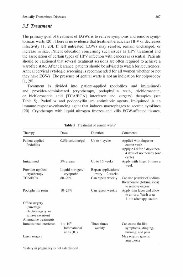
Sexually Transmitted Diseases 207
3.5 Treatment
The primary goal of treatment of EGWs is to relieve symptoms and remove symp-tomatic warts [20]. There is no evidence that treatment eradicates HPV or decreasesinfectivity [1, 20]. If left untreated, EGWs may resolve, remain unchanged, orincrease in size. Patient education concerning such issues as HPV treatment andthe association of certain types of HPV infection with cancers is essential. Patientsshould be cautioned that several treatment sessions are often required to achieve awart-free state. After clearance, patients should be advised to watch for recurrences.Annual cervical cytologic screening is recommended for all women whether or notthey have EGWs. The presence of genital warts is not an indication for colposcopy[1, 20].
Treatment is divided into patient-applied (podofilox and imiquimod)and provider-administered (cryotherapy, podophyllin resin, trichloroacetic,or bichloroacetic acid [TCA/BCA] interferon and surgery) therapies (seeTable 5). Podofilox and podophyllin are antimitotic agents. Imiquimod is animmune response-enhancing agent that induces macrophages to secrete cytokines[20]. Cryotherapy with liquid nitrogen freezes and kills EGW-affected tissues,
Table 5 Treatment of genital wartsa
Therapy Dose Duration Comments
Patient-appliedPodofilox
0.5% solution/gel Up to 4 cycles Applied with finger orcotton swab
Apply b.i.d for 3 days then4 days of no therapy (onecycle)
Imiquimod 5% cream Up to 16 weeks Apply with finger 3 times aweek
Provider-appliedcryotherapy
Liquid nitrogen/cryoprobe
Repeat applicationsevery 1–2 weeks
TCA/BCA 80–90% Can repeat weekly Can use powder of sodiumBicarbonate (baking soda)
to remove excessPodophyllin resin 10–25% Can repeat weekly Apply thin layer and allow
to air dry; Wash area1–4 h after application
Office surgery(curettage,electrosurgery, orscissor excision)
Alternative treatmentsIntralesional interferon 1 × 10⁶
Internationalunits (IU)
Three timesweekly
Can cause flu-likesymptoms, stinging,burning, and pain
Laser surgery May require generalanesthesia
aSafety in pregnancy is not established.

208 A.C. Thornton et al.
which results in sloughing and wart destruction [20]. TCAs/BCAs are causticagents that destroy warts by chemical coagulation of proteins. Due to the lowviscosity of these agents, care must be taken to prevent the solution from runningonto unaffected areas (treated areas should be allowed to dry before the patient sitsor stands) [1]. Surgical removal includes curettage, electrocautery/electrotherapy,and ablative therapy (laser). Intralesional injections or interferon administeredtopically have not been shown to be effective as a primary or adjunct therapy forEGWs [20]. Cidofovir and 5-fluorouracil (5-FU/ipinephrine/bovine collagen gel)cream are under development.
There are no guidelines regarding which treatment to use first for EGWs. Choiceof modalities should be guided by patient preference number and location of warts,and the patient’s ability to follow directions, and clinical expertise [1]. Expertssuggest that treatment should be changed or the patient referred to a specialistwhen three treatment sessions have resulted in no improvement, if there is incom-plete clearance after six treatment sessions, or when continued treatment extendsbeyond the manufacturer’s recommendations [1]. Clinicians must monitor patientprogress and avoid overtreatment. There is no single treatment modality that isvastly superior to other therapies [20]. All wart treatments may cause mild localirritation, ulceration, or erosion. Ablative modalities can result in hypopigmentation,hyperpigmentation, or scarring. Pregnancy and immunodeficiency can be associatedwith larger or more numerous EGWs [22]. There are reports of immunocompro-mised patients having EGWs with high-risk types of HPV [22]. Although currenttreatments are imperfect, most patients can eventually be wart free. There is noreported resistance of genital HPV currently.
Two new prophylactic HPV vaccines are currently on the market and demonstratehigh-level protection (90% or better) from infection, persistent infection and high-grade lesions [21]. One of the vaccines, Gardasil (Merck), protects against HPVtypes 6, 11, 16, and 18 and the other, Cervarix (GlaxoSmithKline), protects againsttypes 16 and 18 [21].
4 Diseases Characterized by Urethritis and Cervicitis
Urethritis in men and cervicitis in women are among the most common diagnoses insexually transmitted disease clinics. The majority of cases of urethritis and cervicitisare caused by infection with Neisseria gonorrhoeae and Chlamydia trachomatis.Other agents that cause urethritis are Trichomonas vaginalis, Herpes simplex, andMycoplasma genitalium. Ureaplasma urealyticum biovar 2 has also recently beenlinked to urethritis.
4.1 Urethritis
4.1.1 Clinical Description
Urethritis refers to inflammation of the urethra manifested by dysuria, pyuria, ordischarge. Discharge may range from scant and mucoid to grossly purulent. It may

Sexually Transmitted Diseases 209
occur in males or females, but clinically appears more frequently in men. Althoughdysuria may suggest a urinary tract infection in females, in males, urethritis isusually due to a sexually transmitted disease (STD).
Per the CDC 2006 STD Treatment Guidelines [1], the criteria for diagnosis ofurethritis are the following:
a. Mucopurulent or purulent discharge.b. A Gram stain of urethral secretions demonstrating greater than or equal to 5
WBCs per oil immersion field. The diagnosis of gonococcal urethritis dependson the demonstration of intracellular Gram-negative diplococci.
c. A positive leukocyte esterase test on a first-void urine or microscopic exami-nation of first-void urine demonstrating greater than or equal to 10 WBCs perhigh-power field.
Because gonococcal urethritis is so often symptomatic, individuals usuallypresent for diagnosis. In contrast, chlamydial infection is often asymptomatic, evenin men. With the advent of highly sensitive and specific non-invasive tests based onnucleic acid amplification (NAATs) (see below) that can be performed using urinein men and a self-administered vaginal swab in women, screening asymptomaticindividuals for Chlamydia or gonorrhea is feasible in a variety of non-clinical sites.
4.1.2 Epidemiology
Both gonorrhea and Chlamydia genital infections are extremely common. In 2006,over 350,000 cases of gonorrhea and 1 million cases of Chlamydia were reportedto the CDC [23]. However, many cases of both diseases are not reported because ofasymptomatic disease and providers’ failures to report. The most striking element ofchlamydial and gonorrheal epidemiology is their association with young adult age.Both infections are most prevalent in those 15–24 years of age. This association hasbeen linked to anatomic changes, immune responses, and sexual networks. Unlikesyphilis, which tends to occur in geographically identified high prevalence areas inthe United States, Chlamydia and gonorrhea are widespread. Although both occurin advantaged and disadvantaged populations, the latter often bear a dispropor-tionate burden of disease. Similarly, racial differences are prominent. Nationwide,African-Americans, American Indians/Alaskan natives, and Hispanic populationsare disproportionately affected by both diseases. There is no biologic reason knownfor this disparity, and differences are more likely to be due to factors such as accessto care and social capital.
4.1.3 Etiology
Neisseria gonorrhoeae
N. gonorrhoeae is a fastidious Gram-negative diplococcus. The organism causesdisease by attaching to columnar epithelium via pili, invading mucosa using Opa

210 A.C. Thornton et al.
(opacity-related proteins), and porins. Porin proteins, Por A and Por B, havebeen used to serotype gonococci. Opa proteins exhibit a wide range of antigenicvariability, which may affect their ability to invade cells.
Chlamydia trachomatis
C. trachomatis is an obligate intracellular pathogen, which makes diagnostic cul-ture techniques more difficult than those for gonorrhea and makes nucleic acidamplified tests a particularly important tool in the diagnosis of infection. Of thethree species recognized in the genus Chlamydia (i.e., trachomatis, pneumoniae,and psittaci) only the trachomatis species is associated with genital infection. Thespecies is comprised of 19 different serovars that differ in sequence in at least oneof their variable regions of the major outer membrane MOMP encoded by the geneomp1. Serovars A, B, Ba, and C are typically associated with trachoma, one of theworld’s most common causes of reversible blindness. Serovars D, E, F, G, and H aretypically associated with urethritis/cervicitis, although L2a, one of the lymphogran-uloma venereum serovars (L1, L2a, L2b, and L3), has recently been associatedwith urethritis. Heat-shock proteins have been studied in relation to the scarringassociated with chlamydial infection.
4.1.4 Diagnosis
Nucleic acid amplified tests (NAATs) such as polymerase chain reaction (PCR),ligase chain reaction (LCR), or temperature-mediated amplification (TMA) are themost sensitive diagnostic tests for both gonorrhea and Chlamydia and are highlyspecific. Urine in men and self-administered vaginal swabs in women have highsensitivity and specificity for the detection of Chlamydia and gonorrhea. Ratesare similar to urethral and cervical swabs. For gonorrhea, culture has the advan-tage of being inexpensive and it may be used for antibiotic susceptibility testing.Sensitivity of N. gonorrhoeae culture, depending on the laboratory and collectionconditions, ranges from 65 to 100%. A Gram stain of a urethral discharge typi-cally shows the Gram-negative diplococci associated with neutrophils in ≥95% ofsymptomatic males. Gram-negative diplococci seen extracellularly are consideredequivocal. Approximately 50–70% of culture-positive asymptomatic men will havea positive Gram stain. Although Gram stains of endocervical discharge can detectN. gonorrhoeae, they are rarely used to diagnose gonorrhea in females because oftheir low sensitivity.
NAATs offer strong advantages over culture in diagnosing chlamydial infection.First, culture requires highly trained personnel and meticulous technique. Second,even in the best of hands, culture only has sensitivity of 70–80%. Gram stains of theexudate from a patient with C. trachomatis infection are non-specific and show onlynongonococcal urethritis (NGU), i.e., the presence of neutrophils and no intracellu-lar diplococci. In most studies, C. trachomatis has been associated with 30–50% ofall cases of NGU. It is also found in approximately 20–30% of gonococcal urethri-tis. No clinical features reliably distinguish chlamydial and nonchlamydial NGU. In

Sexually Transmitted Diseases 211
the absence of a positive culture for gonorrhea or Chlamydia, NGU is not consid-ered to be caused by either of the two organisms. Evidence to support this beliefincludes the observation that neither gonorrhea nor Chlamydia is isolated fromthe sexual partners of patients with culture-negative NGU. In addition, individu-als with chlamydial NGU respond better to tetracyclines than nonchlamydial NGU.Although treatment with doxycycline suppresses symptoms of NGU in 90% of men,35–50% of those with negative cultures for Chlamydia have persistent pyuria or ure-thral inflammation 6 weeks after treatment. Persistence occurs in less than 20% ofthose with chlamydial NGU.
4.1.5 Treatment
The CDC’s current treatment recommendations for urethritis are shown inTable 6. Although penicillin was the treatment of choice for N. gonorrhoeae forthree decades the rapid spread of penicillinase-producing N. gonorrhoeae, or PPNG,precipitated a need for alternative treatments. The CDC treatment guidelines forgonococcal (GC) infections have been developed with regard to availability of medi-cations and the resistance patterns in the United States. Because concomitant genitalinfection with C. trachomatis is common, patients with a positive Gram stain or cul-ture for gonorrhea should be given additional treatment for C. trachomatis in theabsence of results of testing that specifically exclude it.
Chlamydial genital infection responds well to doxycycline. Single-doseazithromycin has been shown to eradicate C. trachomatis and alleviate symp-toms. Some practitioners prefer this regimen’s use when compliance is in doubt.Erythromycin is also effective, but desirability is limited by its gastrointestinaltoxicity. Ampicillin is recommended for pregnant woman who cannot tolerate themacrolides. For all types of urethritis discussed, patients with HIV infection shouldbe treated the same as those without HIV infection.
4.1.6 Special Considerations
Partner notification: Treatment of partners should be considered as important astreating the index case of urethritis. All sexual contacts of patients with gonorrheaor chlamydial genital infection in the 60 days prior to the index case’s diagnosisshould be treated and, if possible, screened for STDs.
Recurrent or persistent urethritis: If non-compliance or re-exposure is unlikely,individuals who remain symptomatic after treatment for NGU should be tested forTrichomonas by culture and treated with metronidazole 2 g orally in a single doseplus Azithromycin 1 g [1]. Trichomonas testing may also be done using sensitiveand specific NAATs.
Disseminated gonorrhea: Disseminated gonorrhea infection (DGI) occurs rarely,in 0.2–1.9% of mucosal cases [24, 25]. The exact mechanism for disseminationis unknown, although it appears to be associated with certain strains and resis-tance to complement-mediated killing by normal human serum. Patients usuallypresent with an asymptomatic mucosal infection, relatively mild systemic illness,

212 A.C. Thornton et al.
Table 6 Treatment of urethritis and cervicitis [1, 49]
Treatment of urethritis orcervicitisa due to For Neisseria gonorrhoeae For Chlamydia trachomatis
Neisseria gonorrhoeae:
Because of a high associationof N. gonorrhoeae infectionwith concomitant chlamydialinfection, patients withgonorrhea should be treatedfor Chlamydia.
Rectal gonorrhea is treated inthe same manner as urethritiswith comparable successrates
Ceftriaxone 125 mg IM in asingle dose
or
Cefixime 400 mg orally in asingle dose or 400 mg bysuspension (200 mg/5 ml)
plus
Treatment for Chlamydia ifchlamydial infection has notbeen ruled out (see columnto the right)
Alternative regimens:
Spectinomycinb 2 g in a singleintramuscular (IM) dose
or
Single-dose cephalosporinregimens
Azithromycin 1 g p.o. in asingle dose
or
Doxycycline 100 mg p.o. b.i.d.for 7 days
Alternative regimens:
Erythromycin base 500 mg p.o.qid for 7 days orErythromycin ethylsuccinate800 mg p.o. qd for 7 days, orOfloxacin 300 mg b.i.d. for7 days
If only erythromycin can beused and a patient cannottolerate the high-doseregimens, the following canbe used:
Erythromycin base 250 mg p.o.qid for 14 days orErythromycin ethylsuccinate400 mg p.o. qid for 14 days
For treatment of pharyngealgonorrhea
Pharyngeal gonorrhea is moredifficult to treat than urethral,cervical, or rectal gonorrhea.Oral cephalosporins are notrecommended for thispurpose
Ceftriaxone 125 mg IM
plus
Treatment for Chlamydia (seecolumn to the right)
As listed above for treatment ofC. trachomatis
Chlamydia trachomatis If Gram stain or culture doesnot show GC, the patientneed only be treated for C.trachomatis (see column tothe right)
As listed above for treatment ofC. trachomatis
Nongonococcal urethritis No treatment required As listed above for treatment ofC. trachomatis
aThese regimens are recommended for all adult and adolescent patients, regardless of travelhistory or sexual behavior.bSpectinomycin is currently not available in the United States.
asymmetric polyarthralgia, tenosynovitis, and a characteristic skin rash, called thearthritis–dermatitis syndrome. Other patients present with suppurative arthritis andare less likely to have a rash or tenosynovitis. Recommended treatment is ceftri-axone 1 g q 24 h. Cefotaxime and ceftizoxime are alternative treatments. Therapy

Sexually Transmitted Diseases 213
should be continued for 10–14 days [1]. Spectinomycin is recommended as an alter-native but is not available currently in the United States. Quinolones should not beused unless specific testing demonstrates susceptibility.
4.1.7 Gonorrhea and Chlamydia Resistance
Gonorrhea resistance: Isolated reports of penicillinase producing N. gonorrhoeae(PPNG) in the mid-1970s gave way to widespread PPNG by the 1980s, resulting inthe abandonment of penicillin analogs as the drugs of choice for GC. Use of alterna-tive agents including sulfa drugs, tetracyclines, and, most recently, quinolones hasbeen followed by the development of resistance.
In 1986, the CDC established a surveillance program, the Gonococcal IsolateSurveillance Project (GISP), to monitor trends in the antimicrobial susceptibilityof gonococcal isolates in the United States [26]. The GISP includes 26 publiclyfunded STD clinics and 5 regional laboratories. At each clinic, urethral isolates areobtained from the first 25–30 men diagnosed with gonorrhea each month. Theseisolates are shipped to one of the regional laboratories, where the susceptibilitiesof the organisms to a panel of antibiotics are determined. Worldwide surveillanceis performed through the World Health Organization (WHO) via a network oflaboratories, entitled the gonococcal antimicrobial-susceptibility program (GASP).
The best known form of beta-lactam resistance for N. gonorrhoeae is plasmidmediated. Reports in the late 1980s led to the abandonment of ampicillin as thedrug of choice for the treatment of gonorrhea. Surveillance throughout the Americasand Caribbean has shown a decrease in the incidence of PPNG from about 15% in1990 to less than 10% in 1995 [27]. Resistance to beta-lactams may also occurthrough chromosomal mutations. These usually produce low-level penicillin resis-tance and work through mechanisms other than beta-lactamase production, such asan alteration in the penicillin-binding proteins. Despite decreases in the incidence ofPPNG, chromosomal beta-lactam resistance has increased, as has plasmid-mediatedhigh-level resistance to tetracycline [27].
Cases of gonorrhea caused by N. gonorrhoeae resistant to fluoroquinolones(QRNG) have increased dramatically in many parts of the world and are becom-ing widespread in parts of Asia. Quinolone resistance results from point mutationsin genes contained in the Quinolone Resistance Determining Region (QRDR). Inparticular, mutations in the DNA gyrase gene, gyrA, may confer resistance toquinolones, whereas another locus, parC, which specifies a topoisomerase, con-tributes to quinolone resistance [28]. QRNG strains have increased in the UnitedStates and worldwide. Because of this, the CDC has recommended that quinolonesnot be used in the treatment of gonorrhea unless supported by local epidemiology.In the United States during 1996, less than 0.05% of 4,639 clinical isolates testedfrom surveillance laboratories had minimum inhibitory concentrations (MICs) >1.0 μg/ml to ciprofloxacin.
C. trachomatis resistance: Tetracycline-resistant C. trachomatis was firstdescribed in 1990 [29]. Isolates with this type of resistance formed 100-fold fewerinclusions in the presence of tetracycline than sensitive strains, suggesting that
214 A.C. Thornton et al.
approximately 1% of the isolates were resistant. Culture of resistant organisms intetracycline-containing medium yielded populations that were uniformly tetracy-cline resistant. Isolates that survived continued serial passage became tetracyclinesensitive again in antibiotic-free medium. It appeared that not all organisms genet-ically capable of expressing the tetracycline resistance phenotype did so. Thisheterogeneous expression of tetracycline resistance is referred to as heterotypicresistance. In one study, the tetracycline-resistant isolates were also resistant todoxycycline, erythromycin, sulfamethoxazole, and clindamycin but sensitive torifampin, ciprofloxacin, and ofloxacin; in a second study, sensitivity to erythromycinand azithromycin was maintained. The significance of resistance in clinical diseaseremains to be defined.
4.2 Management of Women with Cervicitis
4.2.1 Clinical Description
Cervicitis refers to inflammation of the uterine cervix. The syndrome of mucopu-rulent cervicitis (MPC) is defined by mucopurulent or purulent discharge and maypresent to the patient as a vaginal discharge. Diagnosis of MPC is made when aswab, inserted into the cervical os, after swabbing vaginal secretions from the vault,reveals a yellow or green discharge visible on removal. Some but not all investigatorsconsider cervicitis present if the endocervix is friable, that is, it bleeds when a cottonor dacron swab is inserted into the endocervical canal. Women with cervicitis dueto gonorrhea or C. trachomatis infection may be asymptomatic, experience vaginaldischarge, have lower abdominal discomfort, or present with an acute pelvic inflam-matory disease (PID) characterized by moderate to severe lower abdominal pain andcervical motion tenderness. The most severe consequence of cervicitis is ascensionof infection to the upper genital tract, resulting in infertility from obstruction of thefallopian tubes. Obstructed fallopian tubes resulting from salpingitis associated withPID may result in ectopic pregnancy.
4.2.2 Epidemiology
The epidemiology of gonorrhea and C. trachomatis is similar in men and womenexcept that women tend to be slightly younger than men. C. trachomatis infection iscommon in adolescent girls, often asymptomatic, and commonly recurs. It is recom-mended that all sexually active female adolescents be screened for C. trachomatis.High rates of chlamydial infection are found also in women aged 20–24, particularlythose with a new sexual partner.
4.2.3 Etiology
Both gonorrhea and C. trachomatis may cause cervicitis or MPC. Because asubstantial number of cases remain symptomatic after treatment for those twoorganisms, there may be other etiologies as well.

Sexually Transmitted Diseases 215
4.2.4 Treatment
The treatment of cervicitis is similar to that of urethritis (see Table 6) [1].
4.2.5 Other Considerations
Sexual partners of women with gonorrhea or Chlamydia infection whose last sexualcontact was within 60 days before symptom onset should be treated and, if possible,screened to increase case finding [1].
5 Sexually Transmitted Pharyngitis
While H. simplex infection and C. trachomatis can infrequently cause pharyngitis,sexually transmitted pharyngitis is almost synonymous with gonococcal pharyngi-tis. Gonococcal pharyngitis is relatively difficult to treat effectively as compared togenital tract infection. Although newer antibiotic regimens are more effective thanprevious ones, the cure rate for pharyngitis remains less than that for uncompli-cated GC genital infection. In a review of clinical trials performed after 1980, morethan 95% of genital and rectal gonococcal infections were cured compared to only83.7% of pharyngeal infections in women and 79.2% of pharyngeal infections inmen [30]. For this reason, the recommendations for therapy in pharyngeal gonor-rhea do not include oral cefixime, which achieves lower drug levels than ceftriaxonegiven intramuscularly. The recommended regimen is ceftriaxone. Spectinomycin isan unreliable treatment for gonorrhea pharyngitis [1]. Although not recommendedspecifically by the CDC, in some areas azithromycin 2 g as a one time dosemay be an alternative. With the exception of cefixime, oral cephalosporins suchas cefpodoxime have unacceptable failure rates. Quinolones should not be usedunless susceptibilities are done or the regional epidemiology suggests susceptibility[1, 31]. Treatment of C. trachomatis infection should be initiated with treatment ofpharyngeal gonorrhea because of the frequency of co-infection.
6 Sexually Transmitted Proctitis, Proctocolitis, and Enteritis
6.1 Clinical Description
Infection of the gastrointestinal tract may occur from anal intercourse (proctitis) orsexual activity that includes fecal–oral contact (enteritis). Proctocolitis may occurwith either route. Proctitis is characterized by anorectal pain, tenesmus, and rec-tal discharge and may be caused by N. gonorrhoeae, C. trachomatis includinglymphogranuloma venereum (LGV) serovars, H. simplex, or T. pallidum [1].
6.2 Diagnosis
Patients with acute proctitis who practice receptive anal intercourse should beexamined with anoscopy to establish a specific diagnosis.
216 A.C. Thornton et al.
6.3 Treatment
Although proctitis due to N. gonorrhoeae responds well to regimens recommendedfor uncomplicated N. gonorrhoeae infection, the recommended treatment if pusis present on examination of the rectum is ceftriaxone 125 mg IM plus doxycy-cline 100 twice daily for 7 days. Rectal chlamydial infection with the non-LGVserovars responds to the regimens recommended for urethritis. LGV may causea hemorrhagic proctitis associated with regional lymphadenitis. LGV is treatedwith doxycycline 100 mg orally twice daily for 21 days or alternatively witherythromycin base 500 mg orally four times daily for 21 days [1].
7 Epididymitis
Both N. gonorrhoeae and C. trachomatis may ascend the male genital tract to causeepididymitis. Symptoms typically include unilateral pain that radiates to the testicleand tenderness with palpation. The differential diagnosis of epididymitis includestesticular torsion, which is a surgical emergency, more common in adolescents,and often presents with pain that suddenly gets worse. The recommended treat-ment regimen for epididymitis, like that for proctitis, is ceftriaxone 125 mg IM plusdoxycycline 100 mg orally twice daily for 10 days. The recommended alternativeregimen is ofloxacin 300 mg orally twice daily for 10 days or levofloxacin 500 mgonce daily for 10 days [1].
8 Diseases Characterized by Vaginal Discharge
Abnormal vaginal discharge is one of the leading reasons for visits to primarycare physicians in the United States. Symptoms of vaginitis include the following:abnormal discharge, itching, odor, or vaginal pain. Discharge may be mild andcolorless, heavy, frothy or curdled, and white, yellow, or grey. This spectrum of pre-sentations adds to the difficulty of diagnosis based solely on observation. Therefore,clinical diagnosis is based on a variety of components including observed signs,simple clinical tests, and potentially laboratory-based testing. Differential diagnosismay include physiologic discharge, chemical or irritant vaginitis, atrophic vagini-tis, or vaginitis due to infectious agents. The three most common infectious causesof vaginitis are bacterial vaginosis, Candida albicans (the causative agent of yeastinfections), and T. vaginalis (the causative agent of trichomoniasis) [32, 33].
Patients should undergo a speculum exam with careful examination of the cervixfor discharge and to collect diagnostic swab samples. In the absence of cervical dis-charge, the vaginal mucosa should be examined and sampled as well. Tests that canreadily be performed in the physician’s office include measurement of pH and wetpreparation microscopy using both normal saline and potassium hydroxide (KOH)solutions. Samples also may be referred to a centralized laboratory for C. trachoma-tis and N. gonorrhoeae testing, Gram’s stain, culture for yeast or T. vaginalis, orDNA-based testing for T. vaginalis.

Sexually Transmitted Diseases 217
8.1 Bacterial Vaginosis
8.1.1 Clinical Description
Bacterial vaginosis (BV) is a syndrome with unclear etiology that results when thebalance of the normal flora in the vaginal micro-environment is disrupted. Ratherthan a true infectious disease, BV is more likely the result of changes to the florain response to a disruptive event. The largest change is generally a reduction ofLactobacillus spp., in the vagina, which results in a pH change. These two eventsthen create a more permissive environment for bacteria such as Gardnerella vagi-nalis, Mycoplasma hominis, and Prevotella spp., and anaerobes such as Bacteroidesspp. Recent studies using DNA-based techniques suggest that megasphaera phy-lotype and an as-yet unidentified bacterium are frequently present in women withBV [34].
8.1.2 Etiology
The lack of a distinct etiology hinders our understanding of the transmission mecha-nisms for this disease. Transmission is associated with sexual activity in some casesbut can occur in women with no history of sexual behaviors. Sexual activity is a riskfactor for this disease (higher rates of disease are found in sexually active women),as is having multiple sex partners or acquisition of a new partner. However, usingan intrauterine deviceand douching, which are not related to sexual behaviors, arealso risk factors, thus supporting the theory that BV results from activities that aredisruptive to the vaginal environment. From this perspective, sexual activity may bea risk factor as a result of the disruption of both the pH and the vaginal mucosa thatoccur as part of sexual activity rather than the transmission of a specific organism.In all likelihood, a combination of events (infectious, chemical, and/or mechan-ical) is responsible for BV, explaining the wide variation in presentation of thedisease.
8.1.3 Diagnosis
BV is characterized by fishy-smelling, homogenous, white discharge that adheresto the vaginal mucosa. Vulvovaginal itching and burning and pain during coitus arecommonly associated with BV. Clinical diagnosis is based on a set of observationscalled Amsel’s criteria [35]. The presence of at least three of the following criteria isindicative of BV: (1) homogenous, adherent discharge with little evidence of inflam-mation; (2) vaginal pH > 4.5; (3) amine (fishy) odor after addition of 10% KOHsolution to wet prep vaginal fluid; and (4) presence of bacteria-coated epithelial cells(clue cells) seen on wet prep microscopy. Improved specificity of diagnosis can beachieved with use of Gram’s stain of a vaginal smear. Criteria for a diagnosis of BVusing Nugent’s scoring [36] include 0–4 points for the lack of lactobacilli morpho-types (higher score when fewer are present); 0–4 points for gardnerella/bacteroidesmorphotypes (higher score when more are present); and 0–2 points for the presence

218 A.C. Thornton et al.
of curved, Gram-variable rods (higher score when more are present). A total scoreof >7 is diagnostic for BV.
8.1.4 Treatment
Consequences of untreated BV can be quite serious. In pregnant women, BV hasbeen associated with adverse outcomes such as preterm labor, preterm delivery,premature rupture of membranes, and chorioamnionitis. Pelvic inflammatory dis-ease and endometritis are also associated with BV. Finally, BV has been stronglyimplicated as increasing risk of acquisition of HIV [37, 38].
Table 7 summarizes the various therapies recommended by the CDC for BV.Oral and intravaginal forms of metronidazole and clindamycin are the cornerstonesto therapy for BV. There are minimal differences between the two drugs in regard toefficacy, so choice may be guided by differences in cost, mode of administration, andadverse effects [39]. Antimicrobial testing of anerobic Gram-negative rods recov-ered from women treated for BV may reveal an increase in clindamycin-resistantorganisms. After treatment of BV, recurrence may occur in up to 30% of womenwithin 3 months [39]. A newer BV treatment, tinidazole, is a nitroimidazole-likemetronidazole. In a recent trial, tinidazole 1 g daily for 5 days and 2 g daily for2 days were significantly superior to placebo [40].
8.2 Trichomoniasis
T. vaginalis in women has been found in different genitourinary sites: vagina, ure-thra, Bartholin’s and Skene’s glands, and endocervix. Presentation in women canvary from asymptomatic to severe vaginitis with vaginal discharge, pruritus, dysuria,and abdominal pain. In men, the organism has been isolated from the urethra, urine,semen, external genitalia, epididymitis, and prostate. Men with T. vaginalis infectioncan be asymptomatic or complain of urethral discharge or dysuria. Unfortunately,men are not routinely screened or treated for this infection and as a consequencethere is a lack of information about the disease in men.
8.2.1 Etiology and Epidemiology
Trichomoniasis is caused by the parasitic protozoan T. vaginalis. This motile organ-ism is associated with the squamous epithelial cells of the vagina. T. vaginalisis the most prevalent non-viral STI both globally and within the United States.The estimated number of cases occurring annually is 170 million worldwide and5 million in the United States alone [41]. The infection is more often identifiedin women but is transmitted sexually. Men appear to carry the organism but donot frequently develop overt disease. Interestingly, the prevalence of T. vaginalisincreases with age, peaking in women 40–45 years old. In settings with high riskfor other sexually transmitted infections, the prevalence of T. vaginalis in adult
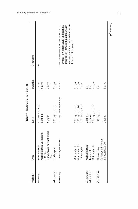
Sexually Transmitted Diseases 219
Tabl
e7
Tre
atm
ento
fva
gini
tis[1
]
Vag
initi
sD
rug
Dos
eD
urat
ion
Com
men
ts
Bac
teri
alM
etro
nida
zole
500
mg
p.o.
b.i.d
.7
days
51M
etro
nida
zole
vagi
nalg
el0.
75%
5g
b.i.d
.5
days
Clin
dam
ycin
vagi
nalc
ream
2%5
gqh
s7
days
Alte
rnat
ive
Lin
dam
ycin
300
mg
p.o.
b.i.d
.7
days
Preg
nanc
yC
linda
myc
inov
ules
100
mg
intr
avag
inal
qhs
3da
ysD
ueto
conc
erns
ofin
crea
sed
adve
rse
even
ts(l
owbi
rthw
eigh
tand
neon
atal
infe
ctio
ns),
intr
avag
inal
clin
dam
ycin
crea
msh
ould
only
beus
eddu
ring
the
first
half
ofpr
egna
ncy
Met
roni
dazo
le50
0m
gp.
o.b.
i.d.
7da
ysM
etro
nida
zole
250
mg
p.o.
t.i.d
.7
days
Clin
dam
ycin
300
mg
p.o.
b.i.d
.7
days
T.va
gina
lis
Alte
rnat
ive
Met
roni
dazo
le2
gp.
o.1×
Tin
idaz
ole
2g
p.o.
1×M
etro
nida
zole
500
mg
p.o.
b.i.d
.7
days
Can
didi
asis
Fluc
onaz
ole
150
mg
p.o.
1×In
trav
agin
alcr
eam
sB
utoc
onaz
ole
2%5
gqh
s3
days
(Con
tinu
ed)

220 A.C. Thornton et al.
Tabl
e7
(con
tinue
d)
Vag
initi
sD
rug
Dos
eD
urat
ion
Com
men
ts
But
ocon
azol
e2%
sust
aine
dre
leas
e5
gqh
s1×
Clo
trim
azol
e1%
5g
qhs
7–14
days
Mic
onaz
ole
2%5
gqh
s7
days
Terc
onaz
ole
0.8%
5g
qd3
days
Terc
onaz
ole
0.4%
5g
qd7
days
Vagi
nals
uppo
sito
ries
Mic
onaz
ole
100
mg
qd7
days
Mic
onaz
ole
200
mg
qd3
days
Mic
onaz
ole
1,20
0m
gqd
1da
yTe
rcon
azol
e80
mg
qd3
days
Nys
tatin
100,
000
uqh
s7
days
Rec
urre
ntV
VC
Clo
trim
azol
e10
0m
gqd
7da
ysC
lotr
imaz
ol10
0m
g2
tabl
ets
qd3
days
Clo
trim
azol
500
mg
1×O
intm
ent
Tio
cona
zole
6.5%
5g
1×
Indu
ctio
nC
hoos
eon
eof
the
abov
efo
rin
duct
ion
for
7–14
days
then
star
tmai
nten
ance
14da
ysC
ontin
ueun
tilpa
tient
isas
ympt
omat
ican
dcu
lture
sar
ene
gativ
e

Sexually Transmitted Diseases 221
women is often equal to the prevalence of C. trachomatis and N. gonorrhoeaecombined.
8.2.2 Diagnosis
A common misconception is that the majority of women with T. vaginalis infectionare symptomatic. This is an artifact of the definition of trichomoniasis that includesthe description of fulminate frothy discharge and often punctuate micro-abrasions ofthe ectocervix (“strawberry cervix”). In studies using DNA-based diagnostic meth-ods, over 40% of women with T. vaginalis had no clinical symptoms of infection[42]. Another factor that leads to the misconception of exclusively symptomaticdisease is that the majority of cases are diagnosed based on wet prep microscopy.Unfortunately, in many cases, the organism load is insufficient for capture in thesample used for wet prep. This occurs in part because as samples await microscopicexamination, in as few as 10 min at room temperature, the organisms lose motility,a requirement for a diagnosis of trichomoniasis. Therefore, the cases diagnosed bythis method have higher organism burden, which frequently leads to a higher prob-ability of symptoms. Wet prep microscopy has been shown to be less sensitive thanculture, which itself is less sensitive than DNA-based testing. In many settings, wetprep microscopy is only 50–70% sensitive, thus many cases of T. vaginalis infec-tion are undiagnosed. There are commercially available rapid diagnostic tests (TheAffirm VP III, OSOM Trichomonas Rapid Test, and Xenostrip) which appear to beas good as or slightly better than wet prep microscopy.
For many years, infection with T. vaginalis has been considered to be merely anuisance. However, data are now available that suggest that T. vaginalis is associatedwith PID and adverse outcomes of pregnancy. In addition, longitudinal data demon-strate that infection with T. vaginalis increases the risk of HIV acquisition withinthe next 3 months by 2.7-fold even when other behavioral risk factors are taken intoaccount [43]. Given the high prevalence of this disease, particularly in populationsat risk for HIV infection, it is clear that the use of more sensitive diagnostic tests iswarranted.
8.2.3 Treatment
The effective treatment of trichomoniais in the United States has relied solely onthe use of metronidazole until the recent FDA approval of tinidazole (see Table 7).Currently, cost more than efficacy favors metronidazole for the treatment ofT. vaginalis infections. Clinical resistance to metronidazole has been reported since1962 [44]. Antibiotic susceptibility testing for T. vaginalis is not standardized.Metronidazole doses as high as 1 g three times daily coupled with intravaginalmetronidazole have been successfully used for refractory cases. However, sideeffects (nausea, vomiting, etc.) of metronidazole may be prohibitive [44]. In onestudy, patients with refractory trichomoniasis were treated successfully with highdoses of tinidazole (2–3 g orally plus 1–1.5 g intravaginally for 14 days) [45].There is cross-reactivity between metronidazole and tinidazole, and, as there are

222 A.C. Thornton et al.
no effective alternatives, desensitization followed by treatment is recommended formetronidazole-allergic patients.
8.3 Vulvovaginal Candidiasis
8.3.1 Clinical Description
Vulvovaginal candidiasis (VVC) typically presents with acute pruritus and vaginaldischarge. VVC is frequently accompanied by vulvar pruritus as well as dysuria,dysparenuria, and vaginal burning. Symptoms can be exacerbated in the weekpreceding the onset of menstrual flow.
8.3.2 Etiology and Epidemiology
VVC is predominately caused by C. albicans or Candida glabrata. These fungiare normal flora of the gut and urogenital tract, associated with mucosal sur-faces. Similar to BV, yeast “infection” is often a result of a disturbance of theenvironmental balance that results in yeast proliferation.
It is estimated that 75% of women will have at least one episode of VVC in theirlifetimes. Risk factors for developing VVC include pregnancy, diabetes mellitus,recent use of antibiotics, and corticosteroid use. VVC is not a sexually transmittedinfection. The incidence of VVC increases with age, peaking in the fourth and fifthdecade of life. Sporadic VVC usually occurs without an obvious precipitating factor,with the exception of uncontrolled diabetes. There may be a relationship betweenVVC and HIV [37], but further research into this area is needed.
8.3.3 Diagnosis
Speculum examination and collection of samples is required for clinical diagnosis.Diagnosis is based on the presence of a thick, white adherent discharge that, unlikethe case with BV, is not homogeneous, but rather has a clumpy, or “cottage cheese-like,” appearance. VVC is associated with vulvar itching, vaginal burning, and painon urination and/or coitus. Wet preparations for microscopy, as with all vaginitis,should be prepared using both normal saline and 10% KOH solutions. Microscopywill reveal yeast and pseudohyphae. The pH is typically <4.5, and higher valuesmay be indicative of mixed infections. Culture for yeast is available from refer-ence laboratories and is recommended when signs indicate infection but no yeast orpseudohyphae are observed microscopically. Additionally, the diagnosis of recur-rent VVC should be confirmed by culture. VVC due to azole-resistant strains ofC. albicans is rare [45]. Most clinically resistant VVC appears to be with non-albicans species, particularly with C. glabrata.
In addition to uncomplicated VVC, more severe cases can occur. These casesrequire prolonged antifungal therapy. Factors related to risk of complicated can-didiasis include uncontrolled diabetes, immunosuppression, history of recurrentcandidiasis, and recent antibiotic use.

Sexually Transmitted Diseases 223
8.3.4 Treatment
Patient preference should influence the choice of treatment for VVC. A varietyof effective topical azole agents are available and there is no strong evidence thatone formulation has superior cure rates over the other (see Table 7). Oral systemicazole agents achieve comparable therapeutic cure rates; however, only fluconazoleis recommended by the CDC at this time [1]. Fewer than 5% of women experiencesevere or recurrent VVC, defined as ≥4 episodes per year. In contrast to compli-cated infection, these cases are infrequently associated with uncontrolled diabetes orimmunosuppression. In recurrent cases, a more rigorous laboratory work-up and his-tory is required prior to initiation of therapy. These patients should receive inductiontherapy (14-day course of an agent in Table 7) to achieve negative vaginal culturesand then a maintenance regimen should be instituted for 6 months [1].
Little information regarding optimal treatment of C. glabrata vaginal infection isavailable. C. glabrata isolates have intrinsic reduced susceptibility to azoles. Boricacid 600 mg gelatin capsule administered vaginally once daily for 14 days has beenshown to be moderately effective [1, 46].
9 Pelvic Inflammatory Disease
9.1 Clinical Description
Pelvic inflammatory disease (PID) remains a diagnostic and treatment challenge. Itis typically defined as an infection of the female genital tract above the cervix andmay include salpingitis, endometritis, tubuloovarian abscess (TOA), and/or frankperitonitis. The majority of women have complete recovery; however, long-termsequelae of PID can be severe and include ectopic pregnancy, infertility, and chronicpelvic pain. In the United States, the population most commonly affected by PIDappears to be the young, nonwhite, unmarried urban dweller as well as those witha history of PID, multiple sexual partners, previous or current STDs, and cigarettesmoking.
9.2 Diagnosis
The diagnosis of PID is imprecise and should be considered in any woman withpelvic pain. Definitive diagnosis can be made by culture of involved areas, butthis frequently involves invasive procedures such as culdocentesis, endometrialbiopsy, and/or laparoscopy. The differential diagnosis is extensive and shouldinclude ectopic pregnancy, ovarian torsion, flare of endometriosis, ruptured ovariancyst, appendicitis cholecystitis, colitis, gastroenteritis, pyelonephritis, nephrolithia-sis, and bowel perforation. The CDC recommends initiating antibiotic therapy forPID in patients with adnexal, lower abdominal, or cervical motion tenderness [1].

224 A.C. Thornton et al.
The presence of fever, an elevated erythrocyte sedimentation rate (ESR) and/or C-reactive protein (CRP), and cervical or vaginal discharge with proven chlamydialor gonorrheal infection support the diagnosis of PID [1]. The findings of hydros-alpinx, pyosalpinx with thickened tubular walls with or without free fluid in thepelvis, or tuboovarian complex are considered to definitively establish a diagnosis ofPID [1].
C. trachomatis and N. gonorrhoeae are well-documented causes of PID. A ran-domized, controlled trial found that identifying and treating women with chlamydialcervical infections reduced the incidence of PID [47]. PID is frequently a polymi-crobial infection with bacteria such as M. hominis, H. influenzae, G. vaginalis,Staphylococci, Group B Streptococci, E. coli, and anaerobes. Thus, the consensusis that PID necessitates broad-spectrum antibiotic therapy. Anaerobes are par-ticularly frequent in women with TOA and with PID in the presence of HIVinfection or bacterial vaginosis [47]. Microbial species associated with BV areoften recovered from upper genital tract or cul-de-sac specimens of women withacute PID.
9.3 Treatment
Given the serious consequences of PID, prevention and early treatment should be apriority. The CDC recommends hospitalization for patients who are pregnant, non-compliant, failing outpatient regimens, or not tolerating oral antibiotics; who havesevere symptoms (nausea, vomiting, or high fever) or have a TOA; and in whom asurgical emergency cannot be excluded [1]. Table 8 lists the CDC’s treatment rec-ommendations for PID. All patients, whether treated on an inpatient or outpatientbasis, should have follow-up within 3 days of initiation of therapy. If no improve-ment is noted despite 72 h of appropriate treatment, then additional testing, otherdiagnoses, or surgical referral should be considered [1]. Some recommend rescreen-ing for C. trachomatis and N. gonorrhoeae 4–6 weeks after completion therapy.Evaluation of male sexual partners of patients with PID is an essential component oftreatment.
10 Sexually Transmitted Ectoparasitic Infections
Ectoparasitic infestations are common worldwide and can be endemic. In the UnitedStates, both scabies and Pediculosis pubis (crab lice) are sexually transmitted [1].Lindane, a drug used for both scabies and crab lice, should be used cautiouslybecause there are reports of central nervous system (CNS) toxicity and death.The CDC recommends that lindane not be used immediately after bathing, as thisincreases the absorption, and also not be prescribed for persons with extensivedermatitis or for pregnant women or children <2 years of age [1].

Sexually Transmitted Diseases 225
Table 8 Treatment of PID [1, 49]
Type of treatment Duration
Parenteral
Regimen A2 g cefotetan intravenously (i.v.) q 12 h
or
2 g cefoxitin i.v. q 6 h
plus
100 mg doxycycline p.o. or i.v. q 12 h
Continue for at least 48 h after theoccurrence of significant clinicalimprovement; Doxycycline should thenbe given orally for a total of 14 days
Regimen B900 mg clindamycin i.v. q 8 h
plus
Gentamicin 2 mg/kg IV/IM loading dosethen 1.5 mg/kg q 8 (can substitute withsingle-dose gentamicin)
Continue for at least 48 h after clinicalimprovement. Doxycycline 100 mg (asabove) or Clindamycin, 450 mg orally4 × a day should be given until day 14of treatment
Alternative3 g ampicillin/sulbactam i.v. q 6 h
plus
Doxycycline (as above)
Same principles as above
Oral
Regimen ACeftriaxone 250 mg IM in a single dose
plus
Doxycycline 100 mg p.o. b.i.d. with orwithout metronidazole 500 mg p.o.b.i.d.
Continue both for 14 days
or
Other parenteral third-generationcephalosporin (ceftizoxime orcefotaxime)
plus
Doxycycline 100 mg p.o. b.i.d. with orwithout metronidazole as above
Continue for 14 days
Alternative Oral Regimens
If parenteral cephalosporin therapy is not feasible, then use of fluoroquinolones (levofloxacin500 mg qd for 14 days) with or without metronidazole as above may be considered if thecommunity prevalence and individual risk of gonorrhea is low. Tests for gonorrhea must beperformed. If the test is positive, parenteral cephalosporin is recommended or treatmentdirected toward antimicrobial susceptibilities of the isolate if cultures were performed [49]

226 A.C. Thornton et al.
10.1 Pediculosis Pubis
Crab lice may infest the pubic and perianal areas and can extend to the beard, axilla,and eyelashes (phthiriasis palpebrarum). The CDC recommends 1% permethrincream or pyrethrins with piperonyl butoxide applied to affected areas and washedoff after 10 min [1]. Treatment should be applied to all hairy areas of the body. Dueto reported increases in resistance to pediculicides, malathion may be used whentreatment failures are suspected. Ivermectin 250 μg/kg repeated in 2 weeks is alsorecommended as an alternative regimen [1]. Lindane is no longer recommended asfirst-line therapy because of increased toxicity (seizure and aplastic anemia) [1].Cure rates of 60% have been reported for single treatment with 1% permethrin [48].Reportedly, a second treatment 1 week later will increase the cure rate to 72%, anda 93% cure rate is achieved with a single treatment of 5% permethrin [48].
10.2 Scabies
The CDC recommends 5% permethrin cream (wash off 8–14 h later) or ivermectin200 μg/kg orally, repeated in 2 weeks for the treatment of scabies. An alternativeregimen is 1 oz of 1% lindane (wash off in 8 h) [1]. Patients, especially those whoare HIV positive, can develop crusted scabies (Norwegian scabies). Recommendedtreatment is with 200 μg/kg of ivermectin as a single dose in immunocompetentpatients and two doses in immunocompromised patients [48]. Some expertsrecommend ivermectin on days 1, 15, and 29 [1].
Pyrethroid (permethrin) resistance appears to be emerging in the form of target-site resistance known as a knockdown resistance gene (kdr). The kdr appears to beunaffected regardless of the concentration of permethrin [48]. Lindane resistance inscabies has been reported in the United States, and lindane resistance is reportedas “commonplace” in Peru [48]. If a patient is not cured after a second treatmentwith a product, then treatment should be changed to a drug with a different activeingredient in case the organism is resistant.
11 Other Considerations
Hepatitis A and hepatitis B can be transmitted sexually. Hepatitis and other viralillnesses such as HIV are discussed elsewhere in this book. As part of preventionand risk reduction, at-risk individuals should be counseled to receive hepatitis Bvirus (HBV) vaccination [4].
Key Points
• STD management must include screening and treatment of sexual partners toprevent transmission and severe complications.

Sexually Transmitted Diseases 227
• The CDC guidelines are readily available and provide the current standard fortherapy of STDs.
• Earlier case detection is crucial for disease control; thus, rapid diagnostic testingshould be used when available.
• Prevention of STDs must be emphasized as concurrent STDs facilitate thetransmission of HIV.
• Fluoroquinolones are no longer recommended in gonococcal infections due toincreasing resistance.
• The emergence of resistance has impacted the recommended management ofsexually transmitted ectoparasitic infections.
References
1. Workowski KA, Berman SM (2006). Sexually transmitted diseases treatment guidelines,2006. MMWR Recomm Rep 55:1–94
2. Cohen MS (1998). Sexually transmitted diseases enhance HIV transmission: No longer ahypothesis. Lancet 351(Suppl 3):5–7
3. Eng T, Butler W (1997). The Neglected Health and Economic Impact of STD’s. The HiddenEpidemic: Confronting Sexually Transmitted Disease. Washington, DC: National AcademyPress
4. CDC (1998). HIV prevention through early detection and treatment of other sexually trans-mitted disease-US recommendations of the advisory committee for HIV and STD prevention.MMWR 47:1–24
5. Spinola SM (2008). Chancroid and Haemophilus ducreyi. In: Holmes K, Sparling P, StammW, et al. (eds.) Sexually Transmitted Diseases, 4th ed. New York: McGraw Hill, 689–699
6. Tyndall M, Malisa M, Plummer FA et al. (1993). Ceftriaxone no longer predictably cureschancroid in Kenya. J Infect Dis 167:469–471
7. Knapp JS, Back AF, Babst AF et al. (1993). In vitro susceptibilities of isolates of Haemophilusducreyi from Thailand and the United States to currently recommended and newer agents fortreatment of chancroid. Antimicrob Agents Chemother 37:1552–1555
8. Wald A (1999). New therapies and prevention strategies for genital herpes. Clin Infect Dis28:S4–S13
9. Lawrence C, Wald A (2008). Genital Herpes. In: Holmes K, Sparling P, Stamm W, et al. (eds.)Sexually Transmitted Diseases, 4th ed. New York: McGraw Hill.
10. Fife KH, Crumpacker CS, Mertz GJ et al. (1994). Recurrence and resistance patterns of herpessimplex virus following cessation of >6 years of chronic suppression with acyclovir. J InfectDis 177:542–550
11. Kessler HA, Hurwitz S, Farthing C et al. (1996). Pilot study of topical trifluridine for thetreatment of acyclovir-resistant mucocutaneous herpes simplex disease in patients with AIDS(ACTG 172). AIDS Clinical Trials Group. J Acquir Immune Defic Syndr Hum Retrovirol12:147–152
12. Sparling PF, Swartz MN, Musher DM et al. (2008). Clinical Manifestations of Syphilis. In:Holmes K, Sparling P, Stamm W, et al. (eds) Sexually Transmitted Diseases, 4th ed. NewYork: McGraw Hill.
13. CDC (2006). Primary and secondary syphilis – United States, 2003–2004. MMWR 55:269–273
14. Tramont EC (1995). Syphilis in adults: From Christopher Columbus to Sir Alexander Flemingto AIDS. Clin Infect Dis 21:1361–1369
15. CDC (2008). Syphilis testing algorithms using treponemal tests for initial screening – Fourlaboratories, New York City, 2005–2006. MMWR 57:872–875

228 A.C. Thornton et al.
16. Sanchez PJ, Wendel GD (1997). Syphilis in pregnancy. Clin Perinatol 24:71–9017. Riedner G, Rusizoka M, Todd J et al. (2005). Single-dose azithromycin versus penicillin G
benzathine for the treatment of early syphilis. N Engl J Med 353:1236–124418. Holmes KK (2005). Azithromycin versus penicillin G benzathine for early syphilis. N Engl J
Med 353:1291–129319. Rolfs RT, Joesoef MR, Hendershot EF et al. (1997). A randomized trial of enhanced therapy
for early syphilis in patients with and without human immunodeficiency virus infection. TheSyphilis and HIV Study Group. N Engl J Med 337:307–314
20. Wiley DJ, Douglas J, Beutner K et al. (2002). External genital warts: Diagnosis, treatment,and prevention. Clin Infect Dis 35:S210–S224
21. Winer RL, Koutsky LA (2008). Genital Human Papillomavirus Infection. In: Holmes KK,Sparling PF, Stamm W, et al. (eds.) Sexually Transmitted Diseases, 4th ed. New York:McGraw Hill, 489–508
22. Beutner KR, Wiley DJ, Douglas JM et al. (1999). Genital warts and their treatment. Clin InfectDis 28 (Suppl 1):S37–S56
23. Prevention CDC (2007). Sexually Transmitted Disease Surveillance, 2006. In: ServicesUSDoHaH, (ed.) Atlanta, GA, 2007
24. Barr J, Danielson D (1971). Septic gonococcal arthritis. BMJ 1:482–48525. Sharp JT (1974). Editorial: Gonococcal infections and arthritis. Arthritis Rheum 17:
511–51226. Gorwitz RJ, Nakashima AK, Moran JS et al. (1993). Sentinel surveillance for antimicro-
bial resistance in Neisseria gonorrhoeae – United States, 1988–1991. The gonococcal isolatesurveillance project study group. MMWR CDC Surveill Summ 42:29–39
27. Ison CA, Dillon JA, Tapsall JW (1998). The epidemiology of global antibiotic resistanceamong Neisseria gonorrhoeae and Haemophilus ducreyi. Lancet 351(Suppl 3):8–11
28. Giles JA, Falconio J, Yuenger JD et al. (2004). Quinolone resistance-determining region muta-tions and por type of Neisseria gonorrhoeae isolates: Resistance surveillance and typing bymolecular methodologies. J Infect Dis 189:2085–2093
29. Jones RB, Van der Pol B, Martin DH et al. (1990). Partial characterization of Chlamydiatrachomatis isolates resistant to multiple antibiotics. J Infect Dis 162:1309–1315
30. Moran JS (1995). Treating uncomplicated Neisseria gonorrhoeae infections: Is the anatomicsite of infection important? Sex Transm Dis 22:39–47
31. CDC (1998). Fluoroquinolone-resistant Neisseria gonorrhoeae – San Diego, California, 1997.MMWR Morb Mortal Wkly Rep 47:405–408
32. Anderson MR, Klink K, Cohrssen A (2004). Evaluation of vaginal complaints. J Am MedAssoc 291:1368–1379
33. Carr PL, Rothberg MB, Friedman RH et al. (2005). “Shotgun” versus sequential testing. Cost-effectiveness of diagnostic strategies for vaginitis. J Gen Intern Med 20:793–799
34. Fredricks DN, Fiedler TL, Thomas KK et al. (2007). Targeted PCR for detection of vaginalbacteria associated with bacterial vaginosis. J Clin Micro 45:3270–3276
35. Amsel R, Totten PA, Spiegel CA et al. (1983). Nonspecific vaginitis. Diagnostic criteria andmicrobial and epidemiologic associations. Am J Med 74:14–22
36. Nugent RP, Krohn MA, Hillier SL (1991). Reliability of diagnosing bacterial vaginosisis improved by a standardized method of gram stain interpretation. J Clin Micro 29:297–301
37. Moodley P, Connolly C, Sturm AW (2002). Interrelationships among human immunodefi-ciency virus type 1 infection, bacterial vaginosis, trichomoniasis, and the presence of yeasts.J Infect Dis 185:69–73
38. Martin HL, Richardson BA, Nyange PM et al. (1999). Vaginal lactobacilli, microbial flora, andrisk of human immunodeficiency virus type 1 and sexually transmitted disease acquisition. JInfect Dis 180:1863–1868
39. Nyirjesy P (2008). Vulvovaginal candidiasis and bacterial vaginosis. Infect Dis Clin NorthAm 22:637–652

Sexually Transmitted Diseases 229
40. Livengood CH, III, Ferris DG, Wiesenfeld HC et al. (2007). Effectiveness of two tinidazoleregimens in treatment of bacterial vaginosis: A randomized controlled trial. Obstet Gynecol110:302–309
41. Cates W, Jr. (1999). Estimates of the incidence and prevalence of sexually transmitted diseasesin the United States. American Social Health Association Panel. Sex Transm Dis 26:S2–S7
42. Van der Pol B, Kraft B, Williams JA (2006). Use of an adaptation of a commercially availablePCR assay aimed at diagnosis of Chlamydia and gonorrhea to detect Trichomonas vaginalisin urogenital specimens. J Clin Micro 44:366–373
43. Van Der Pol B, Kwok C, Pierre-Louis B et al. (2008). Trichomonas vaginalis infection andhuman immunodeficiency virus acquisition in African women. J Infect Dis 197:548–554
44. Wendel KA, Workowski KA (2007). Trichomoniasis: Challenges to appropriate management.Clin Infect Dis 44 (Suppl 3):S123–S129
45. Sobel JD, Nyirjesy P, Brown W (2001). Tinidazole therapy for metronidazole-resistant vaginaltrichomoniasis. Clin Infect Dis 33:1341–1346
46. Sobel J (2008). Vulvovaginal Candidiasis. In: Holmes KK, Sparling PF, Stamm W, et al. (eds.)Sexually Transmitted Diseases, 4th ed. New York: McGraw Hill
47. Scholes D, Stergachis A, Heidrich FE et al. (1996). Prevention of pelvic inflammatory diseaseby screening for cervical chlamydial infection. N Engl J Med 334:1362–1366
48. Meinking T (1999). Infestations. Curr Probl Dermatol 11:73–12049. CDC (2007). Update to CDC’s Sexually Transmitted Diseases Treatment Guidelines,
2006: Fluoroquinolones No Longer Recommended for Treatment of Gonococcal Infections.MMWR 56:332–336

Gastrointestinal Tract Infections
Robert E. Post and Barry L. Hainer
1 Introduction
Gastrointestinal (GI) tract infections are caused by a wide variety of fungi, viruses,and bacteria, and all areas of the GI tract are affected. Infections range from mild,where disease is self-limited and supportive care is the treatment, to severe, wherehospitalization and intravenous fluids and antibiotics are required for survival. Inrecent years, the increasing antibiotic resistance of various bacteria has becomean important aspect of GI infection treatment and has resulted in augmentation oftherapy.
2 Oral and Esophageal Infections
2.1 Fungal Infections
2.1.1 Overview
Humans are constantly exposed to fungi in their daily activities, especially whileeating. Among human yeast isolates, Candida albicans is the most prevalent andthe most common cause of oropharyngeal and esophageal fungal infections. Whenisolated from the oral cavity, Candida can be found in 31–60% of healthy people[1]. Candidiasis is a common infection, especially in those who are immunocom-promised [2]. Over the past several decades, the prevalence of these infectionshas increased; this is most likely due to the increased number of patients withHIV/AIDS, cancer patients, and transplant patients [3]. In some patients, candidi-asis of the oral mucosa may be the first sign of infection with HIV. In thosewith more advanced HIV, esophageal candidiasis is as common as oropharyngealcandidiasis [4]. Factors that contribute to a predisposition to develop candidiasis
R.E. Post (B)Department of Family Medicine, Medical University of South Carolina, Charleston, SC, USAe-mail: [email protected]
231A.G. Mainous III, C. Pomeroy (eds.), Management of Antimicrobials in InfectiousDiseases, Infectious Disease, DOI 10.1007/978-1-60327-239-1_12,C© Springer Science+Business Media, LLC 2001, 2010

232 R.E. Post and B.L. Hainer
include age (infants and the elderly), HIV infection, diabetes mellitus, use of H2blockers, mucositis (chemotherapy induced), radiation, dentures, trauma, surgery, ormalignancy. Humans are repeatedly exposed to Candida in food and other sources,including the hospital environment, and personnel [3].
2.1.2 Clinical Presentation
Thrush or oral candidiasis can present in a variety of ways; these include incidentalfindings in asymptomatic patients or complaints of a burning tongue, dysphagia, anda painful mouth. Clinically, thrush presents as diffuse erythema with white plaqueson the surfaces of the oropharyngeal mucosa. These plaques can be wiped away toreveal a raw, erythematous, and sometimes bleeding base [3].
Candidal esophagitis most commonly presents with dysphagia, odynophagia,and retrosternal pain. Sometimes, AIDS patients present with fever or epigas-tric pain or they may be asymptomatic. Proof of diagnosis can only be made bybiopsy of esophageal plaques. Biopsies are taken during endoscopy, and the typicalappearance is yellow-white plaques on an erythematous background [3].
2.1.3 Treatment
Nystatin is generally an effective treatment for oral candidiasis, but compliance andtolerance can be a problem in immunocompromised patients where this fungistaticantifungal is less effective and requires lengthy therapy. For HIV-positive patients,the azole class of antifungals has replaced nystatin as the treatment of choice.Clotrimazole, 10 mg troches administered five times daily, has been shown to beeffective for mild to moderate cases. Ketoconazole, 200 mg daily for 10–14 days,has been shown to treat greater than 80% of cases. Ketoconazole is highly effec-tive even in patients with malignancy or HIV/AIDS. Treatment with ketoconazolehas been limited by concerns for hepatotoxicity and reliability of gastric absorp-tion. Fluconazole, 50–100 mg daily, has also been shown to have a cure rate ofapproximately 80%. Fifty milligrams daily of fluconazole for 10 days and 200 mgdaily for 5 days have been shown to cure AIDS-related thrush. AIDS patients are athigh risk for symptomatic recurrences. Fluconazole, 100 mg daily, has also beenshown to be effective for prophylaxis of thrush in AIDS patients. Itraconazole,100–200 mg daily, has been shown to be as effective as fluconazole for acutetreatment [3].
Oral or intravenous fluconazole is the drug of choice for treatment of candi-dal esophagitis. In several studies, oral itraconazole has shown similar clinicalresponse rates to fluconazole, with both treatments being well tolerated. Intravenousamphotericin B can be used for azole-refractory cases. Low-dose amphotericin B,0.3–0.5 mg/kg daily for 10 days, is usually effective for moderate disease [3].
With increasing resistance of fluconazole, drugs from the echinocandin class,specifically micafungin and anidulafungin, have been increasingly studied for treat-ment of candidiasis. In one study, micafungin was shown to have similar efficacyto fluconazole with HIV-positive patients [2]. In another recent study, anidulafunginwas found to have statistically similar efficacy to fluconazole; however, the relapse

Gastrointestinal Tract Infections 233
rate at a 2-week follow-up was significantly higher for the anidulafungin group [4].Some studies have also shown that use of echinocandins has bred cross-resistanceto other drugs in this class. Fluconazole remains the first-line treatment for candidaesophagitis [2].
3 Gastric Infections
3.1 Helicobacter pylori
3.1.1 Overview
Helicobacter pylori is one of the main causes of chronic peptic ulcer disease. Itis also the main cause of chronic gastritis and the development of gastric can-cer, which is responsible for 1 million deaths worldwide each year. There havebeen conflicting study results as to whether this bacteria is also linked with GERDand hyperemesis gravidarum [5, 6]. This bacteria is found in 48% of patientswith peptic ulcer disease; in developing countries, it is found in 80% of middle-aged adults [5, 7]. In the United States, African-Americans, Hispanics, and olderadults have a higher prevalence than the general population. Increased prevalenceof H. pylori infection is usually correlated with lower income and lower educationlevels [5].
3.1.2 Clinical Presentation
Clinically, patients with peptic ulcer disease generally have the following symp-toms: gnawing or burning epigastric pain that occurs 2–5 h after meals or on anempty stomach and nighttime pain that is relieved by food intake, antacids, or anti-secretory agents (such as H2 blockers or proton pump inhibitors (PPI’s)). Otherpossible symptoms include indigestion, vomiting, anorexia, intolerance of fattyfoods, and heartburn. There is more likely to be a positive family history of H. pyloriinfection in these patients. Physical examination is an unreliable tool in making thediagnosis. The ELISA serum antigen test is a useful tool to test for initial infectionwith a sensitivity of 85% and a specificity of 79%. However, it cannot be used totest for eradication of the bacteria. The urea breath test has a higher sensitivity andspecificity (95–100% and 91–98%, respectively) and can be used to confirm erad-ication; however, a patient needs to have stopped PPI therapy 2 weeks prior to thetest for results to be reliable. The most direct diagnostic test is an endoscopic biopsy,which has a sensitivity greater than 95% and a specificity of 100%. This success indiagnosis has to be weighed with the invasive nature and expense associated withupper gastroesophageal endoscopy [7].
3.1.3 Etiology
Patients are usually exposed to and infected by H. pylori as children and retainthe infection throughout life unless treated. The mode of transmission in unknown

234 R.E. Post and B.L. Hainer
at this time, but current theories suggest oral–oral, fecal–oral, maternal–infant,conjugal, or iatrogenic routes. In countries where H. pylori prevalence is greaterthan 50%, there are some data suggesting that having a partner infected with H.pylori increases risk of infection by six times. The overall prevalence of pep-tic ulcer disease has been declining in developed countries for many decades, inpart due to improved sanitation, but also due to increased antibiotic treatment ofH. pylori [5].
H. pylori infection creates an environment in the gastric mucosa that favors ulcerformation and these include the following: increased resting and meal-stimulatedgastrin levels, decreased gastric mucus production, and decreased duodenal mucosalbicarbonate secretion. The key to reducing ulcer reoccurrence is to eliminate theH. pylori [7]. Patients with H. pylori infection and duodenal ulcers have a gas-tric acid secreting rate three times higher than those who are not infected. Somepatients infected with H. pylori have lower than normal rates of gastric acid secre-tion, and it is in these patients that gastric ulcers are more common [5]. Treatmenthas been shown to reduce duodenal ulcer reoccurrence from 67 to 6% and gastriculcer reoccurrence from 59 to 4% [7].
3.1.4 Treatment
Treatment of H. pylori is vital in successful treatment of peptic ulcer disease. It iswell known that eradication of the bacteria hastens the healing of ulcers and preventsrelapses and complications [5]. Treatments should be administered for 7–14 daysand have a success rate of 80–90% [7, 8]. Recommended treatment regimens can befound in Table 1.
The triple therapy regimen (option number 1 in Table 1) is the most commontreatment used. However, a fall in the H. pylori eradication rate has been observedover the last few years due to increased clarithromycin resistance, and thus newtreatment regimens are being proposed. One such European-based regimen, known
Table 1 Treatment regimens for Helicobacter pylori infection
1. Proton pump inhibitor (omeprazole 20 mg twice daily or lansoprazole 30 mg twice daily)and amoxicillin 1 g twice daily or Flagyl 500 mg twice dailyand clarithromycin 500 mg twice daily
2. Ranitidine bismuth citrate (not available in the United States) 400 mg twice dailyand clarithromycin 500 mg twice daily or metronidazole 500 mg twice dailyand tetracycline 500 mg twice daily or amoxicillin 1 g twice daily
3. Pantoprazole 40 mg twice dailyand levofloxacin 500 mg dailyand amoxicillin 1 g twice daily
4. Bismuth subsalicylate 525 mg four times dailyand metronidazole 250 mg four times dailyand tetracycline 500 mg four times dailyand an H2 blocker for 28 days or a proton pump inhibitor for 14 days
Information obtained from [7]

Gastrointestinal Tract Infections 235
as the sequential therapy regimen, is becoming more widely studied. This regimenincludes a PPI twice daily and amoxicillin 1 g twice daily for 5 days followedby a twice-daily regimen of a PPI, clarithromycin 500 mg, and tinidazole forfive more days. Early results are promising, with a treatment success rate of over90% [9].
Another area of recent study is the use of probiotics in the treatment of H. pylori[10]. Probiotics are bacteria that aid the host in improving immunity and the nutri-tional and microbial balance in the gastrointestinal tract. Most probiotics sold todayare a group known as the lactic acid bacteria. These include the lactobacillus, strep-tococcus, and bifidobacteria species, which are all normal and beneficial residentsof the human gastrointestinal tract [11]. Animal studies have shown that probioticsreduce the gastric inflammation associated with H. pylori infection. Lactobacillihas been shown, in vitro, to reduce the growth of H. pylori and inhibit adhesionto the gastric mucosa [11]. The majority of human studies have shown improve-ment in eradication when probiotics were added to the conventional treatmentregimens. However, no studies have demonstrated definitive H. pylori eradicationwith probiotics on their own [10, 11].
4 Intestinal Infections
4.1 Viral Infections
4.1.1 Rotavirus
During childhood, rotaviruses are the most common cause of severe gastroenteri-tis, and, by the age of 5 years, most children will be infected by them. In childrenyounger than 5 years, they cause 114 million cases of diarrhea each year worldwide.Six hundred thousand of these children die each year, and 98% of these deaths occurin developing countries. Infection with rotavirus can help confer future immunity.Forty percent of children with one infection are protected against further infections,75% are protected against further diarrhea, and 88% are protected against futuresevere disease. The goal of the oral rotavirus vaccines is to try to duplicate thisimmunity. RotaTeq is the vaccine currently available in the United States. It is com-posed of human–bovine pentavalent strains to induce immunity to the most commonantigenic types, P[8]G1–G4, which account for approximately 90% of strains world-wide. This vaccine is given in three doses, the first at 6–12 weeks of age, and thesecond and third doses at 4–10 week intervals to complete the three total doses by32 weeks of age. It is indicated for preterm infants, since they are at the greatest riskfor rotavirus infection [12]. The Rotavirus Efficacy and Safety Trial (REST) showedthis vaccine to decrease gastroenteritis hospital admission by 59%. Overall, it pre-vents severe rotavirus gastroenteritis by 90–95%. Unlike older rotavirus vaccines,RotaTeq was not found to be associated with increased risk of intussusception [13].An additional rotavirus vaccine, Rotarix, is pending review by the Food and DrugAdministration (FDA).

236 R.E. Post and B.L. Hainer
4.1.2 Norovirus
Human noroviruses are the number one cause of non-bacterial gastroenteritis out-breaks in the world. In the United States alone, there are over 23 million infectionsper year. Norovirus infections should not be taken lightly; they can cause hospital-ization in children and deaths in nursing home outbreaks. Since 2002, outbreaksof noroviruses on cruise ships have been well publicized in the media. Kaplanet al. have developed criteria for the identification of noroviruses as the cause ofnon-bacterial gastroenteritis outbreaks. These include a stool culture that is neg-ative for bacteria, vomiting in over half of cases identified, a mean (or median)incubation period of 24–48 h, and a mean (or median) illness duration of 12–60 h.These criteria carry a sensitivity of 68% and a specificity of 99%. Transmission ofnorovirus can occur by person to person, fecal–oral, contaminated food and water,and aerosolization of vomitus [14]. There is no specific therapy for norovirus infec-tion. Treatment is supportive by maintaining hydration and correcting electrolytedisturbances through oral and intravenous hydration [15].
5 Acute Infectious Diarrhea
5.1 Overview
Diarrhea is a common problem. Worldwide, over 2 million deaths each year can beattributed to diarrhea. In the United States, between 211 and 375 million episodesof acute diarrhea occur each year, with almost 1 million hospitalizations and 6,000deaths each year [16]. Acute diarrhea is defined as bowel movements at least threetimes per day or at least 200 g of stool per day for less than 14 days [16, 17].Three diarrhea-free days intervening in a stretch of a diarrhea constitutes a new acuteepisode [18]. Most cases of diarrhea are viral and self-limited, about 50% last lessthan 24 h [16]. About 10% of diarrhea cases can be attributed to bacterial pathogensin developed countries [19]. Most episodes of diarrhea in the United States occur inthe winter months and are caused by noroviruses and rotaviruses, which have beenexplained in detail earlier in this chapter [16].
5.2 Clinical Presentation
Diagnosis can mainly be made through history. The elapsed time when symptomsstarted, after the ingestion of possibly contaminated food, can be a clue to diagnosisas well as what particular foods a patient ate. A past medical history, includingpossible compromised immunity, antibiotic history, travel, and sexual orientation areall important pieces of information for narrowing a differential diagnosis. The maingoal of the physical examination is to assess the degree of dehydration. Laboratorystudies are generally unreliable. Stool cultures are widely used, but the blanket useof them has made them expensive, with the cost per positive result ranging from$900 to $1,000 [18].

Gastrointestinal Tract Infections 237
5.3 Treatment
The initial therapy for all cases of diarrhea is rehydration. Oral rehydration treat-ment (ORT), which includes fluids with glucose and sodium, is preferred. Thisis because sodium absorption increases when coupled with glucose, and waterabsorption increases with sodium [16, 17, 19]. Adults in developed countries areencouraged to drink fluids and eat soup or salted crackers. Another common treat-ment is the combination of the “BRAT” diet, consisting of bananas, rice, applesauce,and toast, with the avoidance of dairy. Loperamide, an agent that inhibits peristalsisin the gut, has been shown to reduce the duration of diarrhea by up to 24 h in adults[16]. However, it should be avoided in patients with bloody diarrhea or febrile dis-ease where it can possibly prolong the duration of disease [16, 18]. It should also beavoided in children [18]. Bismuth subsalicylate has been shown to reduce the dura-tion of norovirus-associated diarrhea in children by 20–27 h [16]. It has also beenshown to increase sodium and water reabsorption in the gut, bind enterotoxins, andhas a direct antibacterial effect [17].
6 Traveler’s Diarrhea
6.1 Overview
Acute diarrhea is the most common illness among travelers, occurring in up to 55%of people who travel from developed countries to developing countries, including46% of Americans. Most cases develop within the first 14 days of travel and canlast up to 4 days without treatment [20].
6.2 Clinical Presentation
Traveler’s diarrhea (TD) is defined as three or more uncomfortable, unformed stoolsin 24 h with at least one of the following symptoms: fever, nausea, vomiting, abdom-inal cramps, tenesmus, or bloody stools [20, 21]. TD is more common in childrenthan in adults, and they are at higher risk for dehydration. Parents should seek imme-diate treatment for their child if he or she has signs of at least moderate dehydration,bloody diarrhea, fever higher than 102◦F, or intractable vomiting. Young childrencan normally have three or more loose stools daily, so diarrhea in this age group isdefined as double the frequency of unformed stool [20].
6.3 Etiology
Regions where the risk of acquiring TD are highest include Africa, South Asia, LatinAmerica, and the Middle East. Other risk factors include an immunocompromisedstate and patients taking H2 blockers or proton pump inhibitors. The main mode of

238 R.E. Post and B.L. Hainer
transmission is fecal–oral; contaminated food and water are the main reservoirs ofinfectious organisms. Unsafe foods include raw vegetables, unpeeled fruits, rawor poorly cooked meats and seafood, unpasteurized dairy products, cold sauces,salsas, foods that are cooked and then reheated, and tap water. Escherichia coli(especially in Mexico, Latin America, the Caribbean, and Africa), Campylobacter(especially in Thailand and Nepal), Shigella, and non-typhoid Salmonella are allcommon pathogens (to be discussed in more detail later in this chapter). Viruses arenot common causes, but noroviruses have been responsible for outbreaks on cruiseships [20]. In 20–50% of cases, no offending pathogen is identified [21].
6.4 Treatment
The best treatment is prevention. The most common travel advice is to boil, cook, orpeel the foods and beverages they are about to consume [20]. Even with this advice,the incidence of TD has not decreased in the last 50 years. In fact, most studies donot support a link between personal hygiene and preventing TD. This is likely due tothe fact that most travelers eat their meals at restaurants, which have been found tobe an independent risk factor for developing TD [21]. Antibiotic prophylaxis is notroutinely recommended, despite fluoroquinolones being up to 90% effective [20,21]. This practice is due to concern for increased bacterial resistance and travel-ers developing a false sense of security [20]. Bismuth subsalicylate is up to 62%effective in preventing TD [21]. However, it should be avoided in patients takingdoxycycline for malaria prophylaxis because it interferes with its absorption [20].Pregnant women and those taking anticoagulants should also avoid subsalicylates.Probiotics such as Lactobacillus have shown 15–63% effectiveness for prophylaxis[20, 21].
Empiric antibiotic treatment should be started within the first 24 h of symp-toms. Fluoroquinolones, particularly ciprofloxacin (500 mg twice daily orally for1–3 days), have been the drug of choice for empiric TD treatment [20]. However,Campylobacter resistance to fluoroquinolones has been on the rise, particularly inThailand, where it increased from 0% in 1987 to 96% in 1997 [21]. In places wherefluoroquinolone-resistant Campylobacter is prevalent, azithromycin, 500 mg dailyfor 1–3 days or 1,000 mg in a single dose for adults or 10 mg/kg daily for 3 days forchildren, is recommended [20, 21]. Azithromycin is the empiric drug of choice forchildren and pregnant women. Rifaximin, 200 mg three times daily orally for 3 days,is an effective choice for people traveling to countries where noninvasive E. coli iscommon, such as Mexico [20]. Rifaximin does not effectively treat Campylobacteror Shigella, however [21]. Loperamide is considered safe to use in individuals olderthan 2 years of age when combined with antibiotic therapy and can usually helpreduce symptoms to 1 day. As opposed to bismuth subsalicylate, loperamide isalso safe for use in pregnant women. Oral rehydration therapy is also important,because dehydration is the main complication. Children can continue their usualfeeding regimens including breast-feeding on demand. In those who do not respondto antibiotics, parasitic causes should be evaluated [20].

Gastrointestinal Tract Infections 239
7 Hemorrhagic E. coli Colitis
7.1 Overview
In North America, E. coli O157:H7 is the most common Shiga-toxin-producingE. coli strain and the most common cause of hemolytic uremic syndrome (HUS)and hemorrhagic colitis [22, 23]. A popular misconception is that ground beef isa common vehicle of transmission and that outbreaks are common. Actually, mostcases are sporadic, and ground beef transmission is not common, though it can occurdue to internalization of the organism during processing [23, 24].
7.2 Clinical Presentation
Clinically, E. coli O157:H7 infection can range from asymptomatic to diarrhea (70–90% bloody) with complications of HUS or thrombotic thrombocytopenic purpura(TTP). After an incubation period of 1–7 days (3 days on average), abdominalcramps are initially followed by 1–3 days of watery, non-bloody diarrhea. Bloodydiarrhea begins on the second to third day of illness. Fever is not a common finding,occurring in less than 30% of cases, and vomiting occurs in about 30–60% of cases[22, 24]. Abdominal pain is usually worse than in gastroenteritis caused by otherbacterial pathogens, and patients may have abdominal tenderness on examinationand painful defecation [23]. Overall mortality from these infections is 3–7% [24].The complication of HUS appears 2–14 days after the onset of diarrhea and occursin 2–15% of cases [22, 24]. Complications are more common in children and theelderly, with the most consistent risk factor for development of HUS being age lessthan 5 years or greater than 65 years. The mortality rate from HUS has been reportedto be 3–17%, but has been seen as high as 87% in the elderly [24].
7.3 Etiology
The reservoir of infection is in the gut of cattle [24]. Modes of transmission includelarge herbivore (bovine) contact, contact with infected water that may have runoff from grazing land, ingesting contaminated water, ingesting unpasteurized milk,ingesting unwashed salad or other vegetables (e.g., lettuce, radish, sprouts), day carecenters, and person-to-person contact [23, 24]. Fecal shedding of E. coli O157:H7may last for up to 2 months after acute infection, but a patient is most conta-gious during the acute diarrheal phase [22, 23]. The Shiga toxin is the main causeof the clinical features of this disease. The Shiga toxin mediates both the localand systemic disease [23, 24]. Local intestinal effects cause the development ofbloody diarrhea with hemorrhage and edema of the lamina propria. HUS resultsfrom microvascular disease when the Shiga toxin enters the vasculature and bindsto receptors on endothelial cells that are abundant in the kidneys, brain, and red

240 R.E. Post and B.L. Hainer
blood cells. A pro-inflammatory reaction occurs which triggers endothelial damage,TTP, hemolysis, and occlusion of renal and cerebral microvasculature. This reactionleads to renal failure and other complications, such as seizures and cerebrovascularaccidents. Laboratory markers of HUS development include decreasing hemoglobinlevels, fragmented RBCs on pathology, increasing LDH, decreasing platelet count,and increasing creatinine and blood urea nitrogen levels [24]. The treatment of HUSis beyond the scope of this text.
7.4 Treatment
Antibiotics are not recommended in the treatment of E. coli O157:H7, as drugslike TMP–SMX and fluoroquinolones can induce production of the Shiga toxin andthus increase the risk of developing HUS [16, 23, 24]. There is also no evidenceto show decreased duration of illness or increased fecal excretion of the pathogenwith antibiotic use [23]. Antimotility drugs, narcotic pain medicine, and NSAIDsare also not recommended [23]. Supportive care is the only recommended care atthis time [16]. Intravenous rehydration has been recommended for its renal protec-tive properties [23]. The best treatment for E. coli O157:H7 infection is prevention.This includes proper food and kitchen hygiene and reduction of contact with farmanimals [24].
8 Non-hemorrhagic E. coli
8.1 Overview
Enterotoxigenic E. coli (ETEC) is the most common cause of bacterial gastroenteri-tis in the world, accounting for 20% of cases. It is the most common of the six typesof diarrhea causing E. coli and is a frequent cause of diarrhea in children less than 2years. About 25% of cases occur in adults.
8.2 Clinical Presentation
Overall, the clinical picture mimics that of Vibrio cholerae. Symptoms include vom-iting and watery diarrhea with varying levels of dehydration. Fever is not common.The diarrhea usually persists for 3–4 days and is self-limited. Diagnosis is made bydetection of the toxins in the stool.
8.3 Etiology
It is commonly found in countries such as Mexico, Bangladesh, Egypt, andThailand. Infection is usually due to the ingestion of contaminated food and waterand illness is caused by toxins formed by the pathogen.

Gastrointestinal Tract Infections 241
8.4 Treatment
Treatment is supportive, with rehydration being the most important factor. Withproper hydration, mortality is less than 1%. Adding zinc to the therapy has beenfound to reduce the duration of illness in children [25]. Please refer to Section 5 forantimicrobial options.
9 Campylobacter
9.1 Overview
Campylobacter species are the most common cause of bacterial gastroenteritis in thedeveloped world [26, 27]. About 400 million cases occur worldwide, and only 2.5million of these happen in the United States [27]. Risk factors for disease includemales less than 5 years or 20–29 years, recent foreign travel, handling and ingestionof raw or undercooked poultry, unpasteurized dairy, contaminated water, ingestionof poultry at a restaurant, and contact with farm animals [26, 28].
9.2 Clinical Presentation
Symptoms occur 1–10 days after exposure and include diarrhea (possibly bloody),abdominal pain, fever, and occasionally vomiting. Some patients may be asymp-tomatic. Symptoms generally resolve in 2–5 days. Serious complications includereactive arthritis, which occurs in 1% of patients, and Guillain–Barre syndrome,which occurs in 0.1% of cases [26, 29]. Five to ten percent of patients need hos-pital admission, and mortality is 0.05%. Diagnosis is only through isolation ofthe pathogen in stool specimens because the symptoms are similar to many otherbacterial gastrointestinal diseases [26].
9.3 Etiology
Campylobacter jejuni is the most common cause of disease, accounting for about90% of cases, and is isolated from poultry, cattle, sheep, and goats [26, 27]. Diseaseoccurs from the ingestion of contaminated poultry, red meat, water, or unpasteurizedmilk [27].
9.4 Treatment
Treatment is supportive, since most cases are short in duration and self-limiting.Antibiotics can be considered if the patient has severe or prolonged illness, and thedrugs of choice are erythromycin and azithromycin [26, 27, 29]. Ciprofloxacin and

242 R.E. Post and B.L. Hainer
tetracycline are other alternatives, but ciprofloxacin resistance emerged after fluo-roquinolones were added to feed for farm animals [26, 27]. Macrolide resistancehas been reported in the range of 0.6–12% depending on the country. This antibi-otic resistance is important because macrolide-resistant Campylobacter species havebeen associated with more severe illness [27, 29]. Prevention includes infection con-trol at the farm level down to proper washing and cooking of meat and poultry andproper hand washing at the individual level [26].
10 Non-typhoidal Salmonella
10.1 Overview
In the United States, one of the most common bacterial causes of gastroenteritisis non-typhoidal Salmonella enterica. Salmonellosis accounts for about 30% ofdeaths, due to food-borne illnesses in the United States [30].
10.2 Clinical Presentation
There are a wide range of clinical presentations for non-typhoidal salmonella infec-tions, but the most common presentations include acute onset of fever, waterydiarrhea, and abdominal cramping [31, 32]. Illness usually occurs about 6–72 hafter infection. Fecal shedding occurs for up to 1 month in adults and 7 weeks inchildren less than 5 years of age [31]. Approximately 5% of individuals with gas-trointestinal illness caused by non-typhoidal Salmonella will develop bacteremia.This is important because a possible complication of this is the development ofinfectious endarteritis, especially of the abdominal aorta, which has been found tocarry up to 38% mortality [31]. Focal infections can also occur, as there have beenreports from Asia and Africa of meningitis, subdural empyema, and brain abscessas a complication of non-typhoidal Salmonella infection [32].
10.3 Etiology
More than 95% of cases of Salmonella infection are food-borne; outbreaks aregenerally caused by contaminated eggs, poultry, dairy, and beef [28, 30, 31].Contaminated pork, fruit, and vegetables can also cause disease. It is also importantto remember that reptiles (particularly lizards, snakes, and turtles) and amphibians(particularly frogs and newts) have Salmonella in their gastrointestinal tracts in upto 90% of individual animals [28, 30]. The amount of households in the UnitedStates with these animals as pets has significantly increased, and about 6% of non-typhoidal Salmonella infections each year can be attributed to contact with them[30]. The highest incidence of non-typhoidal Salmonella infections is in childrenless than 1 year of age. The risk factors for this population include riding in a shop-ping cart next to meat or poultry, exposure to reptiles, ingesting concentrated baby

Gastrointestinal Tract Infections 243
formula up to 5 days before infection, day care, and travel outside the United States[28, 30]. Breast-feeding has found to be a protective factor in this population [28].
10.4 Treatment
For over the past 10 years due to their growth-promoting effects, antibiotics havebeen added to animal feed. However, this practice has bred resistance to these drugssuch as fluoroquinolones in Salmonella species [28, 32]. Fluoroquinolones havebeen shown to possibly decrease the duration of symptoms by 1–3 days. However,there has been no benefit shown in reducing the time to stool clearance of thepathogen [31]. The mainstay of treatment is supportive care. Antibiotic treatment isnot recommended for otherwise healthy individuals but is recommended for thosewho are severely ill [31, 33]. Children less than 1 year of age should be treated, espe-cially those less than 3 months [31]. First-line drugs for Salmonella infection includefluoroquinolones, TMP–SMX, ampicillin, and third-generation cephalosporins, thelatter being the preferred antibiotic for children. In the 1990s, increasing resistanceto ampicillin and TMP–SMX was found. During this period of time the emergenceand spread of Salmonella typhimurium definitive phage type 104 (DT104) began,which is resistant to these two drugs as well as streptomycin, tetracycline, and chlo-ramphenicol [31, 33]. In the early 2000s, this subtype was found to account for8–9% of Salmonella isolates in the United States [31]. Multidrug-resistant strainsare more virulent than sensitive strains, having a reported 10-fold increase in mor-tality rate [32]. As with many of the infections mentioned in this chapter, preventionis the key to controlling these diseases. Many Salmonella infections can be pre-vented by hand washing; proper cleaning of cooking utensils and cooking surfaces;and thorough cooking of eggs, meat, and poultry [30].
11 Typhoid Fever
11.1 Overview
Typhoid fever is common throughout the world. The CDC estimates that there are21.6 million typhoid cases each year, with about 200,000 patients dying annually.The majority of these cases occur outside the United States [34].
11.2 Clinical Presentation
Common clinical features of the disease include high-grade fevers, diarrhea, vom-iting, abdominal pain, myalgias, and hepatomegaly. Other features could includetoxicity, splenomegaly, headache, intestinal perforation, jaundice, and ileus. Thegold standard for diagnosing typhoid fever is a positive blood culture, which occursin only 40–60% of cases. Much of the diagnosis of typhoid fever is made clinically,which is difficult because it shares features with many other illnesses [34].

244 R.E. Post and B.L. Hainer
11.3 Etiology
Typhoid fever is caused by Salmonella typhi. A milder illness is caused by S. paraty-phi. Disease caused by S. typhi is about 10 times more common than that causedby S. paratyphi. Mode of transmission is commonly through ingestion of contam-inated water or food. Person-to-person transmission through poor hygiene and acontaminated water supply are also common [34].
11.4 Treatment
First-line treatment was previously with chloramphenicol or amoxicillin. In the1990s, multidrug-resistant typhoid emerged and led to widespread use of flu-oroquinolones for treatment. In recent years, fluoroquinolone resistance hasbeen reported in Asia. The World Health Organization still recommends fluoro-quinolones as the first-line treatment. In cases of quinolone resistance, ceftriaxone orazithromycin can be used. Two typhoid vaccines are available in the United States,with 60–80% efficacy, and should be taken at least 2 weeks before travel. One is anoral vaccine, which is contraindicated in pregnant women, children under 6 years,and immunocompromised patients. A booster may be required every 5 years. Anintramuscular vaccine is available for travelers older than 2 years, and a boostermay be required every 2 years [34]. Combined hepatitis A/typhoid fever vaccinesare not licensed in the United States, but are available in Europe, and have beenshown to be safe and effective [35].
12 Shigella
12.1 Overview
There are 140 million cases and over 600,000 deaths each year from Shigella speciesinfection, known as shigellosis or acute bacillary dysentery. It is a disease commonlyseen in children in developing countries and can be attributed to overcrowding andunsanitary living conditions [36].
12.2 Clinical Presentation
The symptoms of shigellosis are passage of loose stools mixed with blood andmucus, fever, crampy abdominal pain, and tenesmus. Cases range from asymp-tomatic and mild (typically produced by Shigella sonnei) to severe (typicallyproduced by S. flexneri or S. dysenteriae type I). Some children can experiencefevers up to 104◦F, convulsions, rectal prolapse, and even malnutrition. S. dysente-riae type I infection may cause arthralgias, hemolytic uremic syndrome, intestinal

Gastrointestinal Tract Infections 245
perforation, hemorrhage, and toxic megacolon. Diagnosis is made by the clinicalpresence of blood and mucus in the stool along with many fecal leukocytes seen onmicroscopic stool exam and confirmation by stool culture [36].
12.3 Etiology
The mode of transmission is fecal–oral, and it takes as few as 10–100 organismsto cause infection. It is commonly transmitted by person-to-person contact or bycontaminated water and food, such as raw vegetables. An infected individual is con-tagious as long as they excrete the organism in the stool, which can last for up to 4weeks [36].
12.4 Treatment
Antibiotic treatment in shigellosis has been shown to reduce the duration of diar-rhea by an average of 2.4 days [16]. Effective therapies in the past have includedtrimethoprim–sulfamethoxazole, tetracycline, ampicillin, and nalidixic acid, butincreased bacterial resistance now limits their efficacy [16, 36]. Drugs that are com-monly used now include the fluoroquinolones (particularly ciprofloxacin, 500 mgorally twice daily for 3–5 days), but even these have shown increasing resistancein countries such as India and Bangladesh over recent years [36]. Ciprofloxacin isthe only fluoroquinolone that has been shown to be safe in children [16]. The onlyantibiotics shown to be effective in multidrug-resistant shigellosis are ceftriaxoneand azithromycin. Treatment should also include oral rehydration and small, fre-quent meals. The most important prevention strategy is frequent hand washing withplenty of soap and water [36]. There is no commercial vaccine available at this timefor shigellosis [17, 36].
13 Vibrio cholerae
13.1 Overview
Cholera is caused by V. cholerae and is generally indistinguishable from other typesof acute bacterial diarrhea. Cholera only affects the intestine and does not producesystemic symptoms. Cholera is endemic in Africa, Asia, Central America, and SouthAmerica [37].
13.2 Clinical Presentation
Symptoms of vomiting and voluminous “rice-water” diarrhea develop abruptly.Adults with severe cholera may lose 1/2 to 1 l of fluid per hour, leading to severe

246 R.E. Post and B.L. Hainer
dehydration [38]. About 90% of cases are mild to moderate in severity [37]. Insevere cases, cholera can cause 50% mortality if left untreated, mostly withinthe first day of illness. This can be reduced to 1% with adequate supportive care[37, 38].
13.3 Etiology
The disease is caused by a toxin produced from the bacteria that binds to the intesti-nal mucosal cells and causes diarrhea and dehydration [37]. The incubation periodis 18–120 h [38]. The mode of transmission is fecal–oral, and epidemics can occurafter disasters where water and food supplies become contaminated [37]. A doseof 100,000 to 100 million bacteria is required for infection in healthy individualsbecause gastric acid is protective against infection, acting as a natural barrier [37,38]. In those who have decreased gastric acid levels, such as patients with H. pylori,a lower infectious dose can cause infection [37]. Other risk factors include poverty,lack of development, high population density, low education, and lack of previousexposure [39].
13.4 Treatment
The mainstay of treatment of cholera is replacement of fluids, orally for mild dis-ease with no signs of dehydration, intravenously for severe disease. Doxycycline300 mg daily for 1–3 days is the preferred antibiotic. Patients will recover in about4–5 days with supportive treatment alone, and about 2–3 days with antibiotic treat-ment. Patients with mild disease should not receive antibiotics and prophylaxis isnot recommended [38].
In the last 20 years, there have been reports of resistance to tetracycline,ampicillin, kanamycin, streptomycin, sulfa drugs, trimethoprim, and gentamicin.Fluoroquinolones have been effective, but quinolone resistance has been reportedin India [38]. Prevention of cholera is achieved by having satisfactory sanitation andclean drinking water [37]. Oral cholera vaccines have been shown to be effectivewithout serious side effects [38].
14 Yersinia
Infection with Yersinia enterocolitica is not common, with the CDC estimating onlyabout 17,000 cases each year in the United States [40]. A well-known mode oftransmission is through the ingestion of raw or undercooked pork [40, 41]. Yersiniais unique in its ability to survive and reproduce at temperatures close to 0◦C[42]. Infection usually affects young children, where symptoms are non-specificand include fever, diarrhea (sometimes bloody), and abdominal pain. In adults,symptoms include abdominal pain that mimics appendicitis. Symptoms develop 1week after exposure and last for 3–28 days in children and 1–2 weeks in adults.

Gastrointestinal Tract Infections 247
Complications include monoarticular arthritis, erythematic nodosum, and reactivepolyarthritis, the latter in patients who are HLA-B27 positive [40, 43]. Diagnosis isdetermined by stool or blood culture. Most cases are self-limited. Antibiotic treat-ment does decrease seriousness and length of illness. Y. enterocolitica in vitro issusceptible to aminoglycosides, tetracyclines, TMP–SMX, and ciprofloxacin. It isresistant to penicillin, ampicillin, and first-generation cephalosporins. Preventionincludes hand washing and avoiding raw meat, unpasteurized milk, and unchlori-nated water [40].
15 Bacillus
Bacillus cereus causes two types of illness: diarrhea and emesis. The diarrheal type,which is caused by heat-labile toxins, is found in meats, soups, vegetables, sauces,and dairy. The emetic type, which is caused by a heat-stable toxin, is found in rice,noodles, pasta, and pastry. Illness occurs because the spores of the pathogen arehardy and can survive cooking or pasteurization. They then germinate and multi-ply in food that is not properly refrigerated. The incidence of the disease is notfully known, due to frequent misdiagnosis and since the symptoms are commonlyconfused with other gastrointestinal illnesses. Diarrheal illness starts 8–16 h afteringestion whereas the emetic illness starts 30 min–6 h after ingestion. Both types ofdisease are mild and usually resolve within 24 h [44].
16 Listeria
Infection with Listeria monocytogenes is rare, with an incidence of only about 0.7case per 100,000 individuals [45]. Most reported cases are of bacteremia and menin-gitis caused by the pathogen and are beyond the scope of this chapter [46]. Infectioncan also present as a febrile gastroenteritis, which is rare and self-limiting. Thisinfection is characterized by fever and non-bloody diarrhea within 24–48 h of inges-tion of contaminated food and resolves spontaneously. Outbreaks have occurredwith Mexican-style cheeses, as well as chocolate milk, various deli meats, rice salad,corn salad, and shrimp [45, 46]. Listeria can survive in near-freezing temperatures,similar to bacillus species [46]. To avoid infection, individuals should avoid rawmilk and soft cheeses, especially in pregnant women and the elderly [45].
17 Clostridium difficile
17.1 Overview
Clostridium difficile is the most common cause of infectious diarrhea in hospitalizedpatients and one of the top four most common infections in nursing home resi-dents. It is the organism responsible for 3 million cases of diarrhea each year and

248 R.E. Post and B.L. Hainer
about one-fifth of cases of antibiotic-associated diarrhea [47, 48]. Mortality from C.difficile-associated diarrhea (CDAD) is 17% overall, with a higher percentage in theelderly.
17.2 Clinical Presentation
Common clinical signs of CDAD include mild to moderate watery diarrhea, lowerabdominal pain and cramping, fever, malaise, nausea, anorexia, and leukocytosis[47–49]. In the elderly, they may present with altered mental status, weakness, andweight loss [48]. Colitis can develop in 1–3% of patients. Clinical signs include dif-fuse abdominal pain, distention, and high fever. Those who are the most severelyill may develop toxic megacolon, which has an overall mortality of 25–40% or pos-sibly bowel perforation [47, 49]. Recurrent diarrhea occurs in 10–40% of patientstreated [49]. The gold standard for diagnosis is testing for C. difficile toxins in a stoolsample, particularly toxin B, which has a specificity of 99–100% if found [48].
17.3 Etiology
The mode of transmission is by the fecal–oral route. A patient is colonized by expo-sure to C. difficile spores, and if there is a disturbance of the normal colonic flora,usually by recent antibiotic use, then the patient is at increased risk to develop thedisease [48].
The most frequent antibiotics associated with this are clindamycin and thecephalosporins [49]. C. difficile binds to colonic mucosa and produces two toxins,known as cytotoxin A and B. These toxins work in tandem, with toxin B thought tobe 1,000 times more powerful than toxin A. They work to break down the tight junc-tions between mucosal cells, thus causing cellular leakage and thus watery diarrhea.If the infection is left untreated, then apoptosis of cells occurs, causing ulcerationof the gut mucosa and subsequent development of pseudomembranes, leading topseudomembranous colitis [48].
There have been increasing reports of a hypervirulent strain of C. difficile fromNorth America and Europe over the past 8 years [49, 50]. This strain is knownas the pulse field type 1, PCR ribotype 027 (NAP1/027) strain. This strain pro-duces 16 times the amount of toxin A and 23 times the amount the toxin B thanwild-type C. difficile [49]. Type 027 strains have been found to have high-level flu-oroquinolone resistance and thus exposure to these antibiotics is a major risk factorfor developing CDAD caused by these strains [49]. High relapse and mortality rateshave been reported with this strain, especially in the elderly [51]. Type 027 strainsisolated from Canada were found to be susceptible to clindamycin, metronidazole,vancomycin, meropenem, and rifampin. However, they were resistant to bacitracin,clarithromycin, cephalosporins, and fluoroquinolones [49].

Gastrointestinal Tract Infections 249
17.4 Treatment
The first step in treatment of this infection is to discontinue the antibiotic contribut-ing to the infection [47, 48]. In patients with mild to moderate infection, first-linetreatment is oral metronidazole, 250–500 mg every 8 h or oral vancomycin, 125 mgevery 6 h, for 10–14 days. Clinical improvement occurs in 85–95% of patientswithin 5 days [47, 48]. Vancomycin is safe in women who are pregnant or breast-feeding. Both antibiotics have showed response rates of 85% in the past, but inrecent years metronidazole response rates have dropped to 62–74% [47]. Part of thiscan be attributed to the hypervirulent C. difficile strain (see above) that has emergedover the past few years.
Relapse occurs in about 25% of cases, and these cases are usually treatedwith a repeat course of vancomycin or metronidazole. For severe and refractorycases, intracolonic vancomycin, enemas of “donor stool,” surgery (total colectomy),and intravenous immunoglobulin (IVIG) are all options [47, 50]. However, theeffectiveness of IVIG has not been established in randomized control trials [51].
Prevention of the spread of C. difficile is crucial. The simplest methods includefrequent hand washing with soap and water, cleaning contaminated surfaces withbleach, and contact isolation precautions with infected patients [48]. Probiotics,such as Lactobacillus, have shown modest benefits in some trials, but no probiotichas reliably been effective as prophylaxis and thus remains controversial [49–51].Vaccines that contain inactivated toxins A and B have been developed, but trials totest them have yet to occur [49, 50].
18 Parasites
18.1 Giardia
18.1.1 Overview
Giardia intestinalis aka Giardia lamblia is the most common gastrointestinal pro-tozoal infection in humans [52]. It is estimated that over 280 million people areinfected annually, but that most of these patients are in developing nations with aprevalence of only about 2–7% in developed countries.
18.1.2 Clinical Presentation
Many patients with Giardia infection are asymptomatic, but those that are affectedcan have a range of symptoms, which include nausea and vomiting, abdominalcramping, and diarrhea. The most severe cases can occur in children with a failure-to-thrive syndrome that results from chronic diarrhea and malabsorption. Symptomsusually occur about 7–14 days after infection and can persist from a few days toseveral weeks [52, 53]. No gold standard exists for diagnosis, but the first methodusually chosen is stool examination to attempt to observe the cysts or trophozoites

250 R.E. Post and B.L. Hainer
of the pathogen. If direct stool examination is negative, then other diagnostic optionsinclude examination of duodenal aspirate and stool ELISA [52].
18.1.3 Etiology
Modes of transmission include contaminated water and food and person-to-persontransmission by a fecal–oral route during sexual activity [53]. Patients found to beat the highest risk include travelers to foreign countries (especially Mexico, SouthAmerica, Asia, Africa, and the former Soviet Union), children in day care, andhomosexual men [52].
18.1.4 Treatment
The first-line treatment regimen is metronidazole, 250 mg three times daily for 5–10days [52, 53]. If this is not effective, a higher dose regimen of 500–800 mg, threetimes daily for 5 days, can be used [52]. Resistance to metronidazole is reported in20% of cases, with recurrence of Giardia infection in up to 90% of cases. An effec-tive alternative to metronidazole is tinidazole, 100 mg three times daily for 7 days.The recommended regimen for pregnant patients is paromomycin, 500 mg threetimes daily for 5–10 days. If cases are refractory to treatment with metronidazole ortinidazole, then other possible drugs that could be used are furazolidone, quinacrine,and albendazole [53].
19 Entamoeba histolytica
Entamoeba histolytica is estimated to have infected over 50 million individualsworldwide, causing 40,000–110,000 deaths each year. Most cases occur in develop-ing countries. Many patients are symptomatic, but those that do develop symptomsgenerally have bloody diarrhea with abdominal pain. Fever is rare. Infection occursfrom ingestion of cysts in contaminated water. The cysts develop into the activeamoeba, which destroys epithelial cells in the bowel. The drug of choice for treat-ment is metronidazole 750 mg three times daily for 7 days. Cyst carriers shouldbe treated for at least 10 days. To date, there are no reports of high levels ofmetronidazole resistance [53].
20 Cryptosporidium
20.1 Overview
Infection with Cryptosporidium species affects about 300,000 patients in the UnitedStates annually. In developing countries, it is found in an average of 12.7% of cases

Gastrointestinal Tract Infections 251
of diarrhea. Peak incidence is in warm months, with a peak in the United Statesfrom July through September [54].
20.2 Clinical Presentation
Cryptosporidium is associated with HIV infection and is found in approximately16% of patients who have AIDS and diarrhea. Symptoms occur about 1 weekafter infection and commonly present as watery diarrhea with malabsorption. AIDSpatients with a CD4 count less than 50 can present with more severe disease whichis similar to cholera with voluminous watery stools. Diagnosis is by microscopicexamination of stool [54].
20.3 Etiology
Infection with this pathogen is associated with ingestion of contaminated drinkingwater. Other infections have been documented with contaminated recreational water,such as swimming pools. Food-borne infections are less common, but associatedwith apple cider, unpasteurized milk, chicken salad, and raw produce. Person-to-person transmission is common, because cysts shed in the stool are virulent, and hasbeen associated with outbreaks in day care centers and hospitals [54].
20.4 Treatment
Treatment includes fluid and electrolyte replacement, with oral rehydration beingthe preferred method. Antimotility drugs have been shown to decrease symptoms inmild cases. In AIDS patients, antiretroviral therapy has shown to improve diarrhea.Antibiotics to consider include nitazoxanide, 500 mg twice daily for 3 days, andparomomycin, 500 mg three times daily for 5–10 days [54].
21 Diverticulitis
21.1 Overview
Diverticular disease of the colon is rare in developing nations, but a common prob-lem in the Western hemisphere. Diverticular disease includes diverticulosis anddiverticulitis. Diverticula are outpouchings that occur at weak points of the colonicwall where small blood vessels enter the circular muscle layer [55]. Diverticulosis isassociated with a diet high in fat, high in refined carbohydrates, and low in fiber [55,56]. Other risk factors include physical inactivity, constipation, obesity, smoking,

252 R.E. Post and B.L. Hainer
and non-steroidal anti-inflammatory (NSAID) use [56]. The incidence of divertic-ulosis increases with age. Approximately one-third of the population greater than45 years and 50–70% of the population greater than 80 years have diverticulosis.Sixty-five percent of patients have disease at the level of the sigmoid colon.
21.2 Clinical Presentation
Fever, malaise, and left lower quadrant or suprapubic abdominal pain are commonpresenting symptoms. Patients may also present with change in bowel habits, nau-sea, vomiting, and urinary symptoms [55]. Abdominal or perirectal fullness mayalso be present [56]. A tender mass in the left lower quadrant is the most commonfinding on physical exam [55]. A complete blood count usually demonstrates an ele-vated white blood cell count with a left shift. A computed tomography (CT) scan isthe best radiologic study to aid in the diagnosis of diverticulitis. It has a sensitivityof 85–97% and a specificity of almost 100% [55, 56]. The presence of divertic-ula, inflammation of the pericolic fat or other tissues, bowel-wall thickness greaterthan 4 mm, or a peridiverticular abscess suggests the diagnosis of diverticulitis [56].Colonoscopy is contraindicated in patients who have acute diverticulitis because ofthe increased risk for perforation, but can be performed 6 weeks after resolution ofthe acute disease process [55, 56].
21.3 Etiology
The pathogenesis of diverticulitis is not fully known. It is thought that it is a compli-cation of diverticulosis that occurs when these outpouchings become infected andpossibly perforated [55, 56]. Diverticulitis occurs in 15–20% of patients who havediverticulosis [55]. Anaerobes are the most commonly isolated organisms, alongwith E. coli and streptococci [56].
21.4 Treatment
Patients who have mild diverticulitis (i.e., those patients who can tolerate oral intakeand who have only mild symptoms with no peritoneal signs) can be treated as anoutpatient with 7–10 days of broad-spectrum oral antibiotics that include coverageagainst anaerobic bacteria [55, 56]. Treatment regimens can be found in Table 2.A clear liquid diet is recommended until symptoms resolve. Then the diet can beadvanced slowly as tolerated by the patient. Symptoms should improve within thefirst 2–3 days.
Hospitalization is indicated if the patient is unable to tolerate oral intake or hasincreasing pain or fever. In this case, the patient should not be allowed anythingby mouth (NPO) and intravenous fluids and antibiotics should be started [55, 56].

Gastrointestinal Tract Infections 253
Table 2 Treatment regimens for diverticulitis
Oral1. Metronidazole, 500 mg every 6–8 h
and ciprofloxacin, 500–750 mg every 12 h2. Metronidazole, 500 mg every 6–8 h
and trimethoprim–sulfamethoxazole (TMP/SMX), one double strength tablet every 12 h3. Amoxicillin–clavulanic acid, 875 mg every 12 h
Intravenous1. Metronidazole, 500 mg every 6–8 h
and ciprofloxacin, 400 mg every 12 h2. Metronidazole, 500 mg every 6–8 h
and ceftriaxone, 1–2 g every 24 h3. Ampicillin–sulbactam, 3 g every 6 h
Information from [56]
Intravenous regimens can also be found in Table 2. Improvement should occur in2–3 days. If it does not, surgical intervention or percutaneous drainage of a possibleabscess may be necessary. Computerized tomographic (CT) imaging may be helpfulin these steps. If the patient does respond to IV antibiotics, then the patient can beswitched to one of the oral regimens to complete a 7–10 day course, and diet canbe slowly advanced as the patient tolerates. Fifty to eighty-five percent of patientsrespond well to antibiotics during their first episode of diverticulitis, and only lessthan 10% require surgical intervention [55, 56].
22 Human Papilloma Virus and Anal Cancer
Squamous cell carcinoma of the anus is a rare tumor that accounts for 1.5% of allcases of gastrointestinal tract cancer in the United States. Its incidence has risenover the last 25 years to a current incidence of 1.5 per 100,000. It occurs morefrequently in men who engage in anoreceptive intercourse and in these patientswith HIV infection [57, 58]. Other risk factors for anal cancer include low CD4count, cigarette smoking, immunosuppressant therapy following transplants, historyof persistent high-risk human papillomavirus (HPV) infection, and infection withmultiple HPV genotypes [57]. HPV has been detected in up to 90% of anal cancerswith HPV subtype 16 being the most common, appearing in about 70% of cases[57–59].
In the past few years, one of the major developments that promises to have animpact on reducing the incidence of anal cancer is the prophylactic HPV vaccine[58, 59]. The vaccine currently available in the United States and many countriesin Europe protects against HPV subtypes 6, 11, 16, and 18. In phase III trials, itwas shown to have 100% efficacy at preventing disease from these subtypes. Thevaccine is administered in three total doses: one initial dose, then another doseeach at 2 and 6 months after the initial dose. It has been shown that this vaccineis purely for prophylaxis, as it will not be effective in those who already have HPV

254 R.E. Post and B.L. Hainer
infection. Currently, the vaccine has only been approved for females aged 9–26years. It has not been approved for use in males; however, studies are promising.In one study, antibody levels were found to be higher in 9- to 15-year-old boysthan in girls of the same age group [59]. Therefore, there is potential that thisvaccine could reduce the incidence of anal cancer in not only females but malesas well.
23 Inflammatory Bowel Disease
23.1 Overview
Inflammatory bowel disease (IBD), which includes Crohn’s disease (CD) and ulcer-ative colitis (UC), is a chronic inflammatory disorder of the gastrointestinal tract.IBD has been shown to occur in genetically susceptible patients, particularly thosewith a defect in the NOD2/CARD15 gene. A defect in this gene causes an aber-rant immune response to enteric flora and dietary factors [60, 61]. Most IBD lesionsoccur in the distal parts of the GI tract, where bacteria are the most numerous [60].
23.2 Etiology
There is evidence that the enteric flora themselves play an important role in thedevelopment of IBD. Patients with IBD have higher concentrations of enteric bac-teria and an altered balance of these bacteria, which is known as dysbiosis [60,61]. This altered composition has been found to contain increased concentrations ofaggressive bacteria, such as Bacteroides species and E. coli, and decreased concen-trations of protective bacteria, such as Bifidobacterium and Lactobacillus species.Some studies have shown that dysbiosis may be the trigger for the chronic inflamma-tion associated with IBD [61, 62]. Recent research in mice suggests that a possibledeficiency in a transcription factor that regulates tumor necrosis factor productionin mucosal cells may contribute to increased inflammation when these cells have anexaggerated response to gastrointestinal flora [63].
23.3 Treatment
Antibiotics have widely been used for the septic complications of IBD, whichinclude abscesses, peritonitis, fistulae, fissures, and toxic megacolon. However, theuse of antibiotics as primary treatment or as adjunctive treatment (with aminos-alicylates and steroids) has not been established. Despite this lack of evidence,they are still frequently used. Metronidazole and ciprofloxacin are the most widelystudied antibiotics with mixed results [61]. Rifaximin has shown promise in earlystudies, with safety and efficacy in CD, UC, and pouchitis, showing both positive

Gastrointestinal Tract Infections 255
clinical and endoscopic results [60, 61]. There is a concern for development of C.difficile-associated diarrhea with frequent antibiotic use as well as future antibioticresistance.
Overall, antibiotics have not been found to be superior to other treatmentsnor do they increase efficacy of these other treatments in CD. Metronidazole andciprofloxacin in particular have been found to be as effective as aminosalicylates andsteroids [61, 64]. However, they have been shown to be more effective than placebo[60, 64]. There has also been evidence found of increased efficacy in colonic diseaseversus ileal disease. If the patient does have acute left colonic disease, metronida-zole (10–20 mg/kg/day), ciprofloxacin (500 mg twice daily), a combination of thesetwo antibiotics, or rifaximin (400 mg twice daily) can be treatment options [61].
Antibiotics studied in UC have shown no benefit except in the case of pouch-itis [60, 61]. Twenty-five to thirty-two percent of patients with UC will requiresurgical treatment. The preferred procedure in this case is a total colectomy withileal pouch–anal anastomosis, and the most common complication of this surgery isan inflammation of the pouch reservoir, known as pouchitis. Within 5 years of thesurgery, the risk of developing an acute episode of pouchitis is 50% and there is a5% chance of developing chronic pouchitis. For treatment of pouchitis, a regimensimilar to that for acute left colonic CD is recommended. Clinical improvement isgenerally seen in 1–2 days. However, in chronic pouchitis, antibiotics have beenshown to have a lower efficacy [61].
As mentioned above, there is evidence showing that there is immunologicalintolerance to enteric bacteria in patients with IBD, leading to the proposal thatmanipulation of the enteric flora with probiotics may be therapeutic in IBD. Mostevidence supports the use of probiotics in pouchitis [61, 62]. Most trials have used aprobiotic mixture known as VSL#3, which contains a very high bacterial concentra-tion (each packet containing 450 billion viable bacteria) of eight different bacterialspecie: four strains of lactobacilli, three strains of bifidobacteria, and one strain ofStreptococcus salivarius subsp. thermophilus [62]. Several studies have found thatgiving VSL#3 after antibiotics for pouchitis has beneficial results. There is alsosome promising results using VSL#3 in UC and CD [61, 62]. Overall, more studyis needed on this subject.
24 Hepatitis
24.1 Hepatitis A
24.1.1 Overview
Hepatitis A infections have significantly decreased since the introduction of twoeffective vaccines in the United States in the mid-1990s. From 1990 to 2004,the number of yearly cases dropped from 32,000 to 5,609 [65, 66]. In manyindustrialized countries, such as the United States, the immunization rate is lowerthan in developing nations due to improved hygiene and sanitation [67]. The

256 R.E. Post and B.L. Hainer
Hepatitis A virus (HAV) exists in freshwater, seawater, wastewater, and soil. Itis able to resist detergents, acids, and freezing, but does not resist chlorine orformalin. Initial diagnostic tests for infection include liver function tests andhepatitis serology. Antihepatitis A IgM testing can be used as a confirmatorytest, but should not be used as a screening test due to a high false-positiverate [65].
24.1.2 Clinical Presentation
In children, only 30% of those infected with HAV develop symptoms, which areusually flu like. Jaundice does not generally occur in infected children younger than6 years. Children can shed the virus in their stool for up to 6 months, even thosewho are asymptomatic.
The vast majority of infected adults (greater than 80%) are affected for up to 8weeks. In adults, about 70% of those infected develop symptoms, including jaun-dice. There are two phases to the illness: the pre-icteric and icteric phases. Thepre-icteric phase is characterized by fever, malaise, anorexia, nausea, vomiting,abdominal pain, headache, tender hepatomegaly, splenomegaly, bradycardia, andposterior cervical lymphadenopathy that lasts for 5–7 days. The icteric phase is cat-egorized by conjugated bilirubinuria followed by depigmented stools and jaundicethat lasts for 4–30 days [65].
24.1.3 Etiology
The mode of transmission is the oral–fecal route. Often, this occurs by intake ofcontaminated food or water or person-to-person contact. In the United States, mostfood-borne infections of HAV are associated with ingestion of green onions andstrawberries; outside the United States, shellfish is a major cause [65].
24.1.4 Treatment
Treatment is strictly supportive as there is no antiviral therapy available. Thisincludes rest, balanced nutrition, and avoidance of alcohol, acetaminophen, andother hepatotoxins. Approximately 30% of symptomatic patients require hospital-ization [65]. Prevention can be achieved with the hepatitis A vaccine, which canprovide protection for 12 years in immunocompetent adults and for 6 years in chil-dren [67]. Lifelong immunity can only be achieved through infection [66]. HepatitisA serum immune globulin can give short-term immunity for 3–5 months and canprevent infection in 80–85% of patients prior to exposure and in 69–89% of patientswithin 2 weeks of exposure to HAV [65].
Gastrointestinal Tract Infections 257
24.2 Hepatitis B
24.2.1 Overview
Hepatitis B virus (HBV) commonly causes liver disease, with well over 1 milliondeaths each year. The incidence of the disease, however, has decreased in the UnitedStates over the past 20 years. From the late 1980s to 2001, the number of acutecases of HBV infection decreased from 300,000 to 79,000 per year. This is likelydue to the development of the HBV vaccine, which will be discussed later in moredetail [68].
24.2.2 Clinical Presentation
Acute HBV infection is characterized by nausea, anorexia, fatigue, low-grade fever,and epigastric or right upper quadrant pain. In 70% of adults and 90% of childrenless than 5 years of age, acute infection is subclinical. In 1–3 months, the symptomsof acute disease generally resolve. Alanine transaminase (ALT), aspartate transam-inase (AST), and bilirubin levels can all be elevated. In approximately 1% of acuteinfections, hepatic failure can occur from large-scale hepatocellular necrosis [68].The following are the markers used in HBV infection: hepatitis B surface antigen(HBsAg), which is used to diagnose acute infection and carriers, hepatitis B coreantibody (anti-HBc), which shows HBV infection at some point during the patient’slife, hepatitis B core antibody IgM (IgM anti-HBc), which shows recent infection,hepatitis B e-antigen (HBeAg), which shows the patient is infectious when elevated,and hepatitis B surface antibody (anti-HBs), which shows recovery and/or immunity(natural or vaccine acquired) [66].
The criteria for chronic HBV infection are being HBsAg positive for more than6 months, serum HBV DNA greater than 100,000 copies/mL, persistent elevationof ALT and AST, and a liver biopsy showing chronic hepatitis. Each year, 12% ofpatients with chronic HBV infection progress to cirrhosis, and a minority of thesepatients progress to hepatocellular carcinoma. In patients with chronic hepatitis Binfection, their lifetime risk of death from cirrhosis or hepatocellular carcinomais between 12 and 25%. Hepatocellular carcinoma can develop 25–30 years afterdeveloping chronic infection, and screening patients is controversial.
24.2.3 Etiology
HBV’s mode of transmission is through blood and other body fluids such as salivaand semen. In the United States, Canada, and Western Europe about 80% of infec-tions are transmitted sexually or by intravenous drug use. However, in China,Southeast Asia, and sub-Saharan Africa, 5–20% of infections are acquired duringchildhood or by vertical transmission from mother to child [68].
258 R.E. Post and B.L. Hainer
24.2.4 Treatment
The treatment for acute HBV infection is supportive care. For chronic HBV infec-tion, the goal of treatment is to prevent end-stage liver disease by suppressing viralreplication [68]. The first-line treatment is interferon-alpha, which causes serocon-version in 46% of patients within the first 12 months. However, it has many sideeffects and is not well tolerated. Lamivudine, a nucleoside reverse transcriptaseinhibitor, is another medicine that can be used as first-line therapy or in patients whofailed interferon. The main concern with this treatment is viral resistance, whichdevelops at 15–20% per year of treatment; after stopping treatment, relapse is rela-tively common. A third treatment is adefovir dipivoxil, another reverse transcriptaseinhibitor, which can be used after failure of lamivudine treatment. It has been shownto breed no resistance, but there is the risk of nephrotoxicity [66, 68].
The HBV vaccine has become a standard in medical care. It has been used forover 20 years and has been found to be safe and effective [67]. The vaccine is rec-ommended for all children and also for high-risk groups, such as medical workers,intravenous drug users, patients with a history of sexually transmitted diseases ormultiple sex partners, men who have sex with men, and hemodialysis patients. Thevaccine is a series of three injections given over 6 months, with 95% effectivenessin children and 90% in adults. Due to its high effectiveness, the CDC recommendsstarting a vaccination series on all patients even if it might not be completed. Thevaccine can be given safely during pregnancy. If a woman tests positive for HBsAgduring pregnancy, then it is recommended for the infant to receive the HBV vaccineand HBV immune globulin within 12 h of birth, which reduces the risk of verticaltransmission from 10 to 3% [68].
24.3 Hepatitis C
24.3.1 Overview
Hepatitis C virus (HCV) is the most common blood-borne infection in the UnitedStates, with a prevalence of almost 3 million people [69]. HCV accounts for approx-imately 40% of chronic liver disease in the United States. HCV is the number onecause for liver transplantation in the United States [66, 69].
24.3.2 Clinical Presentation
Most patients with HCV show no symptoms. Only 20–30% of patients have jaun-dice during the acute phase. A hallmark of the disease is a fluctuating pattern ofelevated alanine transaminase (ALT) [66]. Fifty to seventy percent of patients withacute HCV infection develop chronic hepatitis [72]. Seventeen percent of patientswith chronic infection will develop cirrhosis, and chronic infection carries a mortal-ity rate of 4% [66]. Patients with cirrhosis have a 1–2% annual risk of developinghepatocellular carcinoma [69].

Gastrointestinal Tract Infections 259
There are two different diagnostic tests for establishing the presence of a hepati-tis C infection. The first is an enzyme immunoassay screen that detects anti-HCVantibodies with a sensitivity of 98%. If this first test is positive, then a second test, aserologic anti-HCV assay or a nucleic acid test (e.g., polymerase chain reaction), isperformed to confirm the diagnosis [66]. Liver biopsy is recommended to determinethe need for antiviral therapy based on the degree of inflammation and amount offibrosis [69].
24.3.3 Etiology
HCV’s mode of transmission is infected blood and blood products. Intravenous druguse accounts for 60% of acute infections [66]. Other risk factors include householdcontact, dialysis for multiple years, accidental needle sticks, blood transfusions priorto 1992, intranasal cocaine use, sex with multiple partners, and body piercing [66,69]. Household contacts and sexual partners of HCV-positive patients should betested as they have been shown to have a higher prevalence than the general popula-tion [70]. The groups most at risk are people aged 25–40 years, African-Americans,and Mexican-Americans [66]. Co-infection with HIV is common; approximately40% of Americans infected with HIV and up to 90% of intravenous drug users whohave HIV are also infected with HCV. This is likely due to the two viruses’ similarmodes of transmission [69]. Chronic HCV infection has also been shown to increasethe risk of other viral infections. Therefore patients with this disease should beimmunized against HAV and HBV, as well as possibly varicella and influenza [71].
24.3.4 Treatment
Standard HCV therapy includes a combination of interferon-alpha (IFN) and rib-avirin [66, 73]. Treatment lasts for 24–48 weeks depending on the genotype ofhepatitis C present in the patient. Patients are considered cured when they have asustained virologic response (SVR), defined as undetectable HCV RNA 6 monthsafter therapy is completed [74]. The sustained response rates if IFN alone are only10–20% [73]. Patients who are likely to have an SVR are females under 40 yearswithout cirrhosis who do not ingest alcohol and have infection with genotypes 2or 3 [66]. Multiple studies have also shown lower treatment response in African-American patients compared to Caucasian patients [72]. Patients over 60 years,uncontrolled substance abusers, and patients with a comorbid condition that is wors-ened by anemia are those in whom treatment with interferon is contraindicated [66].HCV treatment is also contraindicated in pregnancy [66, 69, 74]. Ribavirin has noeffect against HCV on its own and therefore is used only as adjunctive therapy withIFN. Pegylated IFN, a polyethylene glycol molecule attached to the IFN, has beenshown to have SVR’s of 54–60% when used in combination with ribavirin [73, 74].This combination of drugs has become the standard of care for treatment of HCVinfection [74]. The contraindications to ribavirin are severe anemia, ischemic heartdisease, and renal failure because the drug is known to cause anemia [73]. Insomnia,

260 R.E. Post and B.L. Hainer
depression, and neutropenia are other well-known side effects of HCV treatment[69, 74].
24.4 Hepatitis D
Hepatitis D virus (HDV) is a virus that can only be replicated by the hepatitis Bvirus. The vast majority of patients with HDV infection (90%) are asymptomatic[66]. The mode of transmission is through blood and blood products, and a patientcan only be infected with the virus if they already have HBV infection or are infectedwith both HBV and HDV simultaneously, which is seen mostly among IV drugabusers in the United States. Patients with preexisting HBV who develop an HDVsuperinfection have an 80% chance of developing chronic liver disease. There is novaccine for HDV prevention. The current standard of care for treatment of HDV isinterferon, which has limited efficacy. Recent studies support the use of pegylatedinterferon for treatment of HDV, but this is still experimental. Ribavirin has beenshown to provide no additional benefit in these studies [72].
24.5 Hepatitis E
The hepatitis E virus (HEV) is similar to HAV in terms of size and structure.Clinical symptoms are similar to those of HAV infection, which include jaundice,fatigue, abdominal pain, anorexia, nausea, and vomiting [75]. HEV infection hasalso been shown to cause acute pancreatitis, particularly in young, male patients[76]. The gold standard to diagnose the infection is through polymerase chain reac-tion studies. Mortality of HEV infection is 1–4% in non-pregnant patients, but canincrease to upward of 20% in pregnant females. HEV infection in utero can causeloss of the pregnancy [75]. The mode of transmission is through fecally contam-inated water; transmission through contaminated food is rare. Infection is rare inthe United States, but the virus has infected more than 2 billion people worldwideand is a cause of 20–40% of acute liver failure in endemic areas [75, 76]. HEVis usually associated with travel to areas such as North Africa and Southern Asia[66]. Treatment is supportive. Most important is prevention, which includes diligenthand washing and hygiene, avoiding consumption of water in known endemic areas,and ensuring that meats, especially pork, are well cooked. There is no vaccine forprevention that is commercially available at this time [66, 75].
25 Spontaneous Bacterial Peritonitis
25.1 Overview
One of the major complications of cirrhosis is spontaneous bacterial peritoni-tis (SBP). SBP carries a lifetime incidence of 10–30% and is the most common

Gastrointestinal Tract Infections 261
infection in patients with cirrhosis [77]. SBP is defined as an infection of asciticfluid with an ascitic fluid polymorphonuclear leukocyte (PMNL) count of greaterthan or equal to 250 cells/mm3 in the absence of a known source of infection [77,78]. Therefore, paracentesis should be performed in all patients who present withascites to rule out SBP. Despite the fact that approximately 60% of cultures of asciticfluid in SBP are negative, these should still be obtained. Cirrhotic patients with SBPhave a reported mortality rate of 50–70% after 1 year and 70–75% after 2 years.This is due to the advanced stage of cirrhosis that is usually present in patients whodevelop SBP. The majority of the mortality is due to septic shock, multiorgan fail-ure, and variceal bleeds. Therefore, patients who survive an episode of SBP shouldbe evaluated for liver transplantation [77, 79].
25.2 Clinical Presentation
The majority of patients with SBP are asymptomatic, but some will present withfever, nausea, diffuse abdominal pain, renal insufficiency, or ileus [77, 79]. Aboutone half of patients with SBP will develop encephalopathy [77].
25.3 Etiology
SBP is mainly caused by gram-negative bacteria, particularly E. coli and Klebsiellaspecies. E. coli is isolated in approximately 70% of cases and Klebsiella species in10% of cases [79]. However, gram-positive bacteria are becoming more common asthe cause of SBP [77].
25.4 Treatment
Patients with a PMNL count greater than or equal to 250 cells/mm3 or those withPMNL counts less than 250 cells/mm3 but with signs and symptoms of infectionshould be administered empiric antibiotic therapy to treat SBP [78]. Timely treat-ment is important because untreated SBP carries an acute mortality of 50% [79]. Thegold standard antibiotic class is third-generation cephalosporins, mainly cefotaxime.Two grams intravenously every 8–12 h for 5–10 days is recommended [77–79], andthis has been shown to successfully treat 75–90% of cases of SBP [79]. Albumin,1.5 g/kg intravenously within 6 h of detection and 1 g/kg on day 3, is added tothis regimen to reduce the incidence of renal impairment and improve short-termsurvival [77–79]. Oral ofloxacin, 400 mg twice daily, is an effective alternative inpatients without severe hepatic encephalopathy, vomiting, shock, and a creatininelevel greater than 3 mg/dL [78].
The recurrence of SBP is 40–70% within 1 year [77, 79]. Therefore, after anepisode of SBP, patients should receive long-term prophylaxis with norfloxacin,
262 R.E. Post and B.L. Hainer
400 mg orally daily, or trimethoprim–sulfamethoxazole (TMP–SMX), one singlestrength tablet daily. Those with gastrointestinal hemorrhage should receive nor-floxacin, 400 mg twice daily, or TMP–SMX, one single strength tablet twice daily,for a total of 7 days as short-term prophylaxis [78].
Fluoroquinolone resistance has been increasing among gram-negative bacteria.This is contributed to long-term prophylaxis with norfloxacin. Resistance to TMP–SMX has also become a growing concern. This can possibly be counteracted byrotating antibiotics or by restricting the use of prophylaxis to those with the highestrisk of developing a recurrence of SBP [79].
Key Points
• C. albicans is the most common cause of oral and esophageal fungal infections. Itmore commonly presents in immunocompromised individuals and can be treatedwith a variety of antifungal agents.
• H. pylori is a common, but treatable, cause of peptic ulcer disease.• Acute diarrhea has a myriad of causes that can be differentiated by obtaining a
detailed history, including travel history and recent medication use. Prevention isthe key to avoiding acute diarrhea.
References
1. Vazquez JA (2000). Therapeutic options for the management of oropharyngeal and esophagealcandidiasis in HIV/AIDS patients. HIV Clin Trials 1:47–59
2. Pace F, Pallotta S, Antinori S (2007). Nongastroesophageal reflux disease-related infectious,inflammatory, and injurious disorders of the esophagus. Curr Opin Gastroenterol 23:446–451
3. Vazquez JA, Sobel JD (2002). Mucosal candidiasis. Infect Dis Clin N Am 16:793–8204. Krause DS, Simjee AE, Rensburg C et al. (2004). A randomized, double-blind trial of anidu-
lafungin versus fluconazole for the treatment of esophageal candidiasis. Clin Infect Dis39:770–775
5. Makola D, Peura DA, Crowe SE (2007). Helicobacter pylori infection and related gastroin-testinal diseases. J Clin Gastroenterol 41(6):548–558
6. Goldberg D, Szilagyi A, Graves L (2007). Hyperemesis gravidarum and Helicobacter pyloriinfection. Obstet Gynecol 110:695–703
7. Ramakrishnan K, Salinas R (2007). Peptic ulcer disease. Am Fam Physician 76:1005–10128. Zagari RM, Bianchi-Porro G, Fiocca R et al. (2007). Comparison of 1 and 2 weeks of
omeprazole, amoxicillin, and clarithromycin treatment for Helicobacter pylori eradication:The HYPER study. Gut 56:475–479
9. Zullo A, De Francesco V, Hassan C et al. (2007). The sequential therapy regimen forHelicobacter pylori eradication: A pooled-data analysis. Gut 56:1353–1357
10. Laesbros-Pantoflickova D, Corthesy-Theulaz I, Blum AL (2007). Helicobacter pylori andprobiotics. J Nutr 137(3 Suppl 2):812S–818S
11. Penner R, Fedorak RN, Madsen KL (2005). Probiotics and nutraceuticals: Non-medicinaltreatments of gastrointestinal diseases. Curr Opin Pharmacol 5:596–603
12. Grimwood K, Buttery JP (2007). Clinical update: Rotavirus gastroenteritis and its prevention.Lancet 370:302–304

Gastrointestinal Tract Infections 263
13. Vesikari T, Matson DO, Dennehy P et al. (2006). Safety and efficacy of a pentavalent human-bovine (WC3) reassortant rotavirus vaccine. N Engl J Med 354:23–33
14. Estes MK, Prasad BV, Atmar RL (2006). Noroviruses everywhere: Has something changed?Curr Opin Infect Dis 19:467–474
15. CDC (2006). Norovirus: Technical fact sheet. Atlanta, GA: US Department of Health andHuman Services, CDC; 2006. (Accessed on Jul 15, 2008 http://www.cdc.gov/ncidod/dvrd/revb/gastro/noro-factsheet.pdf)
16. Thielman NM, Guerrant RL (2004). Acute infectious diarrhea. N Engl J Med 350:38–4717. Gadewar S, Fasano A (2005). Current concepts in the evaluation, diagnosis and management
of acute infectious diarrhea. Curr Opin Pharm 5:559–56518. Ilnyckyj A (2001). Clinical evaluation and management of acute infectious diarrhea in adults.
Gastroenterol Clin N Am 30:599–60919. Marcos LA, DuPont HL (2007). Advances in defining etiology and new therapeutic
approaches in acute diarrhea. J Infect 55:385–39320. Yates J (2005). Traveler’s diarrhea. Am Fam Physician 71:2095–210021. Shlim DR (2005). Update in traveler’s diarrhea. Infect Dis Clin N Am 19:137–14922. Panoz GZ, Betsi GI, Falagas ME (2006). Systematic review: Are antibiotics detrimental or
beneficial for the treatment of patients with Escherichia coli O157:H7 infection? AlimentPharmacol Ther 24:731–742
23. Tarr PI, Gordon CA, Chandler WL (2005). Shiga-toxin-producing Escherichia coli andhemolytic uraemic syndrome. Lancet 365:1073–1086
24. Todd WTA, Dundas S (2001). The management of VTEC O157 infection. Int J FoodMicrobiol 66:103–110
25. Qadri F, Svennerholm A, Faruque ASG, Sack RB (2005). Enterotoxigenic Escherichiacoli in developing countries: Epidemiology, microbiology, clinical features, treatment, andprevention. Clin Microbiol Rev 18:465–483
26. Galanis E (2007). Campylobacter and bacterial gastroenteritis. CMAJ 177:570–57127. Gibreel A, Taylor DE (2006). Macrolide resistance in Campylobacter jejuni and
Campylobacter coli. J Antimicrob Chemother 58:243–25528. DuPont HL (2007). The growing threat of foodborne bacterial enteropathogens of animal
origin. Clin Infect Dis 45:1352–136129. Moore JE, Barton MD, Blair IS et al. (2006). The epidemiology of antibiotic resistance in
Campylobacter. Microbes Infect 8:1955–196630. Linam WM, Gerber MA (2007). Changing epidemiology and prevention of Salmonella
infections. Pediatr Infect Dis J 26:747–74831. Hohmann EL (2001). Nontyphoidal salmonellosis. Clin Infect Dis 32:263–26932. Graham SM (2002). Salmonellosis in children in developing and developed countries and
populations. Curr Opin Infect Dis 15:507–51233. Molbak K (2005). Human health consequences of antimicrobial drug–resistant Salmonella
and other foodborne pathogens. Clin Infect Dis 41:1613–162034. Bhutta ZA (2006). Current concepts in the diagnosis and treatment of typhoid fever. BMJ
333:78–8235. Luxemburger C, Dutta AK (2005). Overlapping epidemiologies of Hepatitis A and typhoid
fever: The needs of the traveler. J Travel Med 12:S12–S2136. Sur D, Ramamurthy T, Deen J, Bhattacharya SK (2004). Shigellosis: Challenges & manage-
ment issues. Indian J Med Res 120:454–46237. Zuckerman JN, Rombo L, Fisch A (2007). The true burden and risk of cholera: Implications
for prevention and control. Lancet Infect Dis 7:521–53038. Sack DA, Sack RB, Nair GB et al. (2004). Cholera. Lancet 363:223–23339. Griffith DC, Kelly-Hope LA, Miller MA (2006). Review of reported cholera outbreaks
worldwide, 1995–2005. Am J Trop Med Hyg 75:973–97740. DeBerardis B, Torresini G, Brucchi M et al. (2004). Yersinia enterocolitica intestinal infection
with ileum perforation: Report of a clinical observation. Acta Biomed 75:77–81

264 R.E. Post and B.L. Hainer
41. Fredriksson-Ahomaa M, Korkeala H (2003). Low occurrence of pathogenic Yersinia enteroco-litica in clinical, food, and environmental samples: A methodological problem. Clin MicrobiolRev 6:220–229
42. Skovgaard N (2007). New trends in emerging pathogens. Int J Food Microbiol 120:217–22443. Bottone EJ (1997). Yersinia enterocolitica: The charisma continues. Clin Microbiol Rev
10:257–27644. Ehling-Schulz M, Fricker M, Scherer S (2004). Bacillus cereus, the causative agent of an
emetic type of food-borne illness. Mol Nutr Food Res 48:479–48745. Schlech WF (2003). Foodborne Listeriosis. Clin Infect Dis 31:770–77546. Swaminathan B, Gerner-Smidt P (2007). The epidemiology of human listeriosis. Microbes
Infect 9:1236–124347. Razavi B, Apisarnthanarak A, Mundy LM (2007). Clostridium difficile: Emergence of
hypervirulence and fluoroquinolone resistance. Infection 35:300–30748. Crogan NL, Evans BC (2007). Clostridium difficile: An emerging epidemic in nursing homes.
Geriatr Nurs 28:161–16449. Kuijper EJ, van Dissel JT, Wilcox MH (2007). Clostridium difficile: Changing epidemiology
and new treatment options. Curr Opin Infect Dis 20:376–38350. Miller MA (2007). Clinical management of Clostridium difficile-associated disease. Clin
Infect Dis 45:S122–S12851. Balagopal A, Sears CL (2007). Clostridium difficile: New therapeutic options. Curr Opin
Pharm 7:455–45852. Vesy CJ, Peterson WL (1999). Review article: The management of giardiasis. Aliment
Pharmacol Ther 13:843–85053. Ali V, Nozaki T (2007). Current therapeutics, their problems, and sulfur-containing-amino-
acid metabolism as a novel target against infections by “amitochondriate” protozoan parasites.Clin Microbiol Rev 20:164–187
54. Huang DB, White AC (2006). An updated review on Cryptosporidium and Giardia.Gastroenterol Clin N Am 35:291–314
55. Dominguez EP, Sweeney JF, Choi YU (2006). Diagnosis and management of diverticulitisand appendicitis. Gastroenterol Clin N Am 35:367–391
56. Jacobs DO (2007). Diverticulitis. N Engl J Med 357:2057–206657. Uronis HE, Bendell JC (2007). Anal cancer: An overview. Oncologist 12:524–53458. Palefsky J (2006). Human papillomavirus-related tumors in HIV. Curr Opin Oncol 18:
463–46859. Stanley M (2007). Prophylactic HPV vaccines: Prospects for eliminating ano-genital cancer.
Br J Cancer 96:1320–132360. Andoh A, Fujiyama Y (2006). Therapeutic approaches targeting intestinal microflora in
inflammatory bowel disease. World J Gastroenterol 12:4452–446061. Perencevich M, Burakoff R (2006). Use of antibiotics in the treatment of inflammatory bowel
disease. Inflamm Bowel Dis 12:651–66462. Gionchetti P, Rizzello F, Lammers KM et al. (2006). Antibiotics and probiotics in treatment
of inflammatory bowel disease. World J Gastroenterol 12:3306–331363. Hecht GA (2008). Inflammatory bowel disease – live transmission. N Engl J Med 358:
528–53064. Kozuch PL, Hanauer SB (2008). Treatment of inflammatory bowel disease: A review of
medical therapy. World J Gastroenterol 14:354–37765. Brundage SC, Fitzpatrick AN (2006). Hepatitis A. Am Fam Physician 73:2162–216866. Wilson TR (2005). The ABCs of hepatitis. Nurse Pract 30:12–2167. Zuckerman JN (2006). Vaccination against hepatitis A and B: Developments, deployment,
and delusions. Curr Opin Infect Dis 19:456–45968. Lin KW, Kirchner JT (2004). Hepatitis B. Am Fam Physician 69:75–82, 8669. Stribling R, Sussman N, Vierling JM (2006). Treatment of hepatitis C infection. Gastroenterol
Clin N Am 35:463–486

Gastrointestinal Tract Infections 265
70. Brusaferro S, Barbone F, Andrian P et al. (1999). A study on the role of the family and otherrisk factors in HCV transmission. Eur J Epidemiol 15:125–132
71. Isaguliants MG (2007). Functionality of the immune system in patients with chronic hepatitisC: Trial by superinfections and vaccinations. Expert Rev Vaccines 6:527–537
72. Tan J, Lok ASF (2006). Update on viral hepatitis: 2006. Curr Opin Gastroenterol 23:263–26773. Rosenberg PM (2001). Hepatitis C: A hepatologist’s approach to an infectious disease. Clin
Inf Dis 33:1728–173274. Bacon BR, McHutchinson JG (2007). Into the light: Strategies for battling hepatitis C. Am J
Manag Care 13:S319–S32675. Emerson SU, Purcell RH (2007). Hepatitis E. Pediatr Infect Dis J 26:1147–114876. Jaroszewicz J, Flisiak R, Kalinowska A et al. (2005). Acute hepatitis E complicated by acute
pancreatitis: A case report and literature review. Pancreas 30:382–38477. Kuiper JJ, van Buuren HR, de Man RA (2007). Ascites in cirrhosis: A review of management
and complications. Neth J Med 65:283–28878. Heidelbaugh JJ, Sherbondy M (2006). Cirrhosis and chronic liver failure: Part II.
Complications and treatment. Am Fam Physician 74:767–77779. Koulaouzidis A, Bhat S, Karagiannidis A et al. (2007). Spontaneous bacterial peritonitis.
Postgrad Med J 83:379–383

Endocarditis
Eric Matheson
1 Introduction
Infective endocarditis (IE) is an important cause of morbidity and mortality, andthe incidence of this disease among the elderly, recipients of prosthetic valves, andintravenous drug abusers has been increasing. The emergence of microbial resis-tance has complicated treatment of this disease. Primary prevention, early diseaserecognition, and prompt treatment are vital in reducing the incidence of IE as wellas its complications.
2 Clinical Description
Infective endocarditis is described as acute or subacute and is further delineated asleft-sided, right-sided, native valve, or prosthetic valve. These distinctions help clin-icians determine the most likely pathogen and choose appropriate empiric therapy.Right-sided IE occurs in only 5–10% of patients with IE. Intravenous drug abusers(IVDAs) comprise the majority of these patients, and the prevalence of right-sided IE in these patients is 30 times that of the general population. Interestingly,5–10% of patients with right-sided IE are not IVDAs [2, 3]. Among non-IVDApatients, hospitalization appears to increase the risk of right-sided IE. Risk factors inthis setting include intravenous and intracardiac catheterization, abdominal surgery,hyperalimentation, and indwelling pacemakers [4–6].
Left-sided IE occurs more frequently than right-sided IE. Patients typi-cally have underlying cardiac abnormalities (acquired or congenital), prostheticvalves, poor dentition, and/or HIV infection. Dental, respiratory, genitourinary, orgastrointestinal procedures may predispose high-risk patients (those with underlying
E. Matheson (B)Department of Family Medicine, Medical University of South Carolina, Charleston, SC, USAe-mail: [email protected]
267A.G. Mainous III, C. Pomeroy (eds.), Management of Antimicrobials in InfectiousDiseases, Infectious Disease, DOI 10.1007/978-1-60327-239-1_13,C© Springer Science+Business Media, LLC 2001, 2010

268 E. Matheson
valvular abnormalities). Infection with highly virulent organisms such as staphylo-cocci and Pseudomonas spp. can cause IE in the absence of underlying risk factorsor cardiac abnormalities [2, 3, 7, 8].
The clinical presentations of IE are variable and depend upon the pathogen,the duration of the infection, the heart valves affected, and the mode of diseaseacquisition. Clinical manifestations include bacteremia and systemic embolization.Vasculitis is a more prominent feature of subacute bacterial endocarditis (SBE)because immune complexes do not have time to form in acute IE [1, 9, 10]. Fever isthe most frequent finding in patients with IE, and this diagnosis should be enter-tained in patients with fever of unknown origin. It is noteworthy, however, thatroughly 19% of patients with culture-negative IE are afebrile [11]. Associated non-specific symptoms of IE include weight loss, chills, night sweats, malaise, fatigue,nausea, cough, and arthralgias. Elderly patients may have few symptoms, and theirability to mount a febrile response may be blunted [1, 2, 4, 12, 13]. Other clin-ical manifestations include neurological symptoms (stroke or focal deficits dueto embolic phenomenon), mental status changes, subarachnoid hemorrhage (sec-ondary to mycotic aneurysm rupture), splenomegaly (abscess or infarct), and flanktenderness (renal infarcts) [1, 2, 12].
A variety of cardiac complications may evolve in IE, including myocardi-tis, perivalvular abscess formation, mycotic aneurysm formation, and conductiondefects. In addition, myocardial infarction may occur as a result of coronary arteryembolism [2, 14]. The presence of new or changing insufficiency murmurs may alsodevelop; however, their absence does not exclude the diagnosis of IE. Right-sidedmurmurs are rare and their presence should further heighten the suspicion of IE [2,4, 7, 12, 15]. Another important cardiac complication is congestive heart failure, fre-quently out of proportion to or in the absence of valvular abnormalities. This may besecondary to microbial antigenic mimicry, resulting in the formation of antibodiesdirected against myocardial proteins [16].
The vasculitis observed in SBE is a consequence of immune complex formationand deposition. Immune complex formation and deposition occurs predominately inthe kidney, spleen, and skin [16]. The clinical presentation may be dominated by iso-lated immunological response phenomena without other signs and symptoms [17].Renal involvement can result in glomerulonephritis with subsequent hematuria, pro-teinuria, and urinary red cell casts [1, 4, 12, 16, 18]. Cutaneous lesions, includingpetechiae and splinter hemorrhages, develop in as many as half of patients; how-ever, these findings are neither sensitive nor specific for SBE. Osler’s nodes – small,tender nodules found on the pads of the fingertips – are uncommon but may appearlater in the course of the disease [1, 2, 7, 12, 18]. Janeway lesions are non-tendermacules that form on the fingers, palms, and soles. They are another uncommoncutaneous manifestation of IE that may result from systemic septic embolization orhypersensitivity angiitis [2].
Systemic embolization complicates the clinical picture in 22–50% of patientswith IE [9]. Patients with right-sided valvular vegetations may develop compli-cations, including pulmonary embolism, pneumonia, pulmonary hypertension, andlung abscess formation [1, 2, 4]. In contrast, left-sided vegetations embolize to the

Endocarditis 269
major arterial beds in the central nervous system (CNS), heart, spleen, bowel, andextremities. This typically results in ischemia, infarction, hemorrhage, or abscessformation in the involved organ [1, 2, 5, 7, 9, 12, 19]. Embolic events appear to bemore common in left-sided IE and in IE caused by Staphylococcus aureus, Candidaspp., and the HAECK organisms [7, 9, 20].
Laboratory abnormalities frequently observed in patients with IE include anelevation in erythrocyte sedimentation rate, positive rheumatoid factor, cryoglob-ulinemia, leukocytosis, anemia, and elevations in blood urea nitrogen and creatinine[1, 4, 7, 16, 17]. It is important to keep in mind that these abnormalities are notsensitive or specific for diagnosing IE, as they may also be present in a variety ofother diseases.
The Duke criteria for diagnosis of IE classify patients suspected of having IE intothree categories based on the presence of specific pathological and clinical criteria[9, 19] (Table 1). Definitive IE requires the culture or histological demonstrationof microorganisms in a vegetation or an intracardiac abscess or histological evi-dence of endocarditis in a vegetation or an intracardiac abscess. In addition, thepatient must manifest two major criteria, one major and three minor criteria, orfive minor criteria (Table 1). When criteria for definitive IE are lacking but thediagnosis cannot be rejected, the patient is considered to have possible IE. The
Table 1 Duke’s clinical criteria for diagnosis of infective endocarditis
Major criteriaPositive blood culture (No. 1 or 2)1. Typical microorganisms consistent with IE from two separate blood cultures:
a. S. viridans, S. bovis, or HACEK group, orb. Community-acquired S. aureus or enterococci in the absence of primary focus
2. Microorganisms consistent with IE from persistently positive blood cultures:a. At least two positive cultures of blood samples drawn at least 12 h apartb. All of three or a majority of at least four separate blood cultures with the first and last sample
drawn at least 1 h apartEvidence of endocardial involvement (No. 1 or 2)1. Positive echocardiogram for IE defined as
a. Oscillating intracardiac mass on valve or supporting structures, in the path of regurgitant jets,or on implanted material in the absence of an alternative anatomic explanation, or
b. Abscesses, orc. New partial dehiscence of prosthetic valve
2. New valvular regurgitation (worsening or changing of preexisting murmur not sufficient)
Minor Criteria1. Predisposition: IVDA or predisposing heart condition2. Fever: temperature of at least 38◦C3. Vascular phenomena: major arterial emboli, septic pulmonary infarcts, mycotic aneurysm,
intracranial hemorrhage, conjunctival hemorrhages, and Janeway lesions4. Immunological phenomena: glomerulonephritis, Osler’s nodes, Roth spots, and rheumatoid
factor5. Microbiological evidence: positive blood culture but does not meet a major criterion as noted
above or serological evidence of active infection with organism consistent with IE6. Echocardiographic findings: consistent with IE but do not meet a major criterion as noted above
Data from [19].
270 E. Matheson
diagnosis is rejected when there is a resolution of clinical manifestations within4 days of antibiotics, when there is no pathological evidence of disease after at least4 days of antibiotics, or when there is a firm alternative diagnosis to explain thesymptoms [19].
Bacteremia is a major diagnostic criterion of IE; however 1–5% of patients willhave negative blood cultures. Culture-negative IE occurs predominately in patientsinfected with members of the HAECK group and in those with IE due to unusualorganisms such as Chlamydia or fungi, as these microorganisms may often takeweeks to grow in culture [1, 7, 9, 11, 21]. Culture-negative IE may also result fromthe initiation of antimicrobial therapy prior to culturing the blood. In fact, adminis-tration of antibiotics prior to obtaining blood cultures may reduce bacterial recoveryrate by 35–40% [1, 9, 22]. Patients with culture-negative IE appear to have highermortality rates, especially those with prosthetic valve endocarditis (PVE). This ispresumably due to the delay in diagnosis and treatment [7, 23].
Echocardiographic findings are also essential to the diagnosis of IE by the Dukecriteria [19]. Echocardiography has been particularly instrumental in the diagno-sis of IE in patients with right-sided IE and culture-negative IE [7]. Transthoracicechocardiography (TTE) has played an important diagnostic role in the initial man-agement of patients suspected of having IE. It is rapid, noninvasive, and highlyspecific for detecting valvular vegetations [24]. A major disadvantage of TTE isits low degree of sensitivity, and negative testing necessitates further imaging inpatients strongly suspected of having IE. Furthermore, the ability of TTE to detectPVE and perivalvular abscess formation is limited. The usefulness of TTE is alsolimited in diagnosing IE in patients who have chronic obstructive pulmonary diseaseand in patients who are obese [2, 9, 24]. Despite these shortcomings, a good qualityTTE is still recommended as the initial procedure of choice for patients in whomthe suspicion of IE is low [9].
Transesophageal echocardiography (TEE) has greater sensitivity than does TTE,while maintaining a high degree of specificity, especially in patients with PVE [1,2, 7, 9, 20, 24]. The most recent American Heart Association (AHA) guidelinesrecommend TEE as the procedure of choice in patients suspected of having PVE,for patients in whom the risk of IE is intermediate or high, in patients who aredifficult to image, and in patients with high risk of complications [25].
Despite its high degree of sensitivity and specificity, TEE may be falsely negativein cases where vegetations are smaller than the limits of resolution, embolization ofvegetations has occurred, or views are inadequate to detect small abscesses [10, 24].Patients strongly suspected of having IE should undergo repeat TEE 7–10 days afteran initial negative test [9]. In patients who have undergone both TTE and TEE, thenegative predictive value is 95% [10].
2.1 Etiology
The most commonly encountered pathogens in bacterial IE are streptococci, staphy-lococci, and enterococci. The HAECK organisms (Haemophilus spp., Actinobacterspp., Cardiobacterium spp., Eikenella spp.) and gram-negative bacteria (Klebsiella

Endocarditis 271
spp., Pseudomonas spp., and Escherichia spp.) are less frequently offenders. Otherrare but important pathogens include Rickettsia spp., Bartonella spp., Legionellaspp., Brucella spp., Neisseria spp., Mycobacterium spp., fungi, and Norcardia spp.These unusual organisms as well as members of the HAECK group are responsiblefor the majority of culture-negative endocarditis [1, 7–9, 26].
Staphylococcus aureus is the predominant pathogen in right-sided IE, both inIVDAs and other patients. Candida spp. have also been implicated as etiologi-cal agents in both groups of patients. gram-negative IE occurs more frequently inIVDAs [2, 4, 7, 9]. In contrast, left-sided valvular structures are most often tar-geted by streptococci, especially S. viridans. Staphylococcus aureus and enterococciare also frequently isolated pathogens. Enterococci are most likely among elderlypatients [1, 2, 7, 8, 12, 19].
PVE has become increasingly important owing to the increasing number ofpatients undergoing valve replacements. More than 60,000 patients receive a pros-thetic heart valve each year, and 3–6% ultimately develop IE [26]. Early PVE,occurring within the first 60 days after valve replacement, is most often caused bystaphylococci, predominately S. epidermidis. Mortality rates in early PVE rangefrom 30 to 80%. Streptococci are the predominant infecting microorganisms in latePVE, with mortality rates ranging from 20 to 40% [1, 2, 9, 27, 28]. The mortalityrate of PVE is higher than that of native valve IE [2, 7, 28].
Two uncommon but important pathogens causing IE deserve special mention.Streptococcus bovis is predominately isolated in elderly patients with IE. Becauseof its tendency to form multiple, multivalvular vegetations, S. bovis often results inheart failure, necessitating extensive surgical repair. This results in higher mortal-ity rates. Importantly, the presence of S. bovis mandates a complete examination ofthe upper and lower gastrointestinal (GI) tract due to its association with GI malig-nancy [29–31]. Staphylococcus lugdunensis is a highly virulent coagulase-negativeorganism causing approximately 1% of cases of acute IE [32]. Metastatic seeding,perivalvular seeding with abscess formation, and rapid valve destruction frequentlyoccur despite appropriate antibiotic therapy [9, 33].
With each passing year, antibiotic-resistant organisms are responsible for a largerand larger number of IE cases. The pathogens that have been particularly prob-lematic are S. aureus, S. pneumoniae, Enterococcus spp., and Candida spp. Ratesof penicillin-resistant pneumococci have decreased somewhat in recent years butremain high at approximately 25% in most regions of the United States [34].Although pneumococci are not a predominant cause of IE, mortality associated withinfection is high [35].
The staphylococci developed early resistance to penicillin, necessitating the useof penicillinase-resistant penicillins, that is, methicillin. The capacity of the staphy-lococci to elude eradication with even these drugs, however, has been increasingat an alarming rate, with resistance rates above 30% [36]. Methicillin-resistantS. aureus (MRSA) IE is most commonly seen in IVDAs and in hospitalized patients,although cases of community-acquired MRSA IE have become increasingly com-mon, accounting for approximately 21% of cases [36–38].
The enterococci have long been an important cause of IE, and their suscepti-bility to the penicillins decreased many years ago. The increasing resistance of

272 E. Matheson
these organisms to aminoglycosides has compromised the ability to treat entero-coccal infections; however, perhaps more concerning has been the emergence ofvancomycin-resistant enterococci (VRE). The number of VRE infections, includingIE, has been steadily increasing, and the lack of efficacious therapeutic alternativeshas significantly hindered efforts to eradicate infection [8, 39].
2.2 Antimicrobial Therapy
Antibiotics for the treatment of infective endocarditis should be bactericidal anddirected against the microorganism cultured from the blood. IVDAs and otherpatients with community-acquired infection may present with acute infective endo-carditis. In this setting, three blood cultures should be obtained as soon as possible(within 1 h of presentation), and empiric antibiotic therapy should be initiatedimmediately thereafter [1, 2]. Combined therapy with ampicillin–sulbactam isrecommended in patients with acute IE because S. aureus is the most commonoffending pathogen [2, 40]. Patients already on antibiotics, patients in whom infec-tion with MRSA is suspected, patients with PVE (high likelihood of S. epidermidis),and patients with beta-lactam allergy should be treated with a combination regimenof vancomycin plus gentamicin, plus ciprofloxacin [40].
Table 2 Current recommendation for the treatment of SBE
Microorganism Primary treatment Alternative treatment
StreptococciPCN sensitiveNative valve
PCN G (12–18 MU/d) × 4 weekORPCN G (12–18 MU/d)Plus gentamicin (3 mg/kg/d) × 2
weeks
Ceftriaxone (2 g/d) × 4 weekORCeftriaxone (2 g/d) plus
gentamicin (3 mg/kg/d) × 2weeks
ORVancomycin (30 mg/kg/d) × 4
weeksPCN-resistant native
valve andprosthetic valve
PCN G (24 MU/d) × 4 week plusgentamicin (3 mg/kg/d) × 2 weeks
Ceftriaxone (2 g/d) × 4 weeksplus gentamicin (3 mg/kg/d)× 2 weeks
ORVancomycin (30 mg/kg/d) × 4
weeksStaphylococciMethicillin sensitive
Nafcillin (12 g/d) × 6 week plusgentamicin (3 mg/kg/d) × 3–5 days
Cefazolin (6 g/d) × 6 weeksplus gentamicin (3 mg/kg/d)× 3–5 days
Prosthetic valve Nafcillin (12 g/d) × 6 weeks plusrifampin (900 mg/d) × 6 weeks plusgentamicin (3 mg/kg/d) × 2 weeks
Methicillin resistantNative valveProsthetic valve
Vancomycin (30 mg/kg/d) × 4–6weeks
Vancomycin (30 mg/kg/d) × 6 weeksplus rifampin (900 mg/d) × 6weeks plus gentamicin (3 mg/kg/d)× 2 weeks

Endocarditis 273
Table 2 (continued)
Microorganism Primary treatment Alternative treatment
Enterococci (nativeor prostheticvalve)
PCN sensitive
PCN G (18–30 MU/d) × 4–6 weeksplus gentamicin (3 mg/kg/d) × 4–6weeks
Ampicillin (12 g/d) plusgentamicin (3 mg/kg/d) ×4–6 weeks
ORVancomycin (30 mg/kg/d) × 6
weeks plus gentamicin(3 mg/kg/d) × 6 weeks
Vancomycinresistant
Linezolid (1200 mg/d)
HACEK group(native orprosthetic valve)
Ceftriaxone (1 g/d) × 4 weeks Ampicillin–sulbactam(12 g/d)plus gentamicin (3 mg/kg/d)× 4–6 weeks
ORVancomycin (30 mg/kg/d) × 6
weeks plus gentamicin(3 mg/kg/d) × 6 weeks
Reprinted with Permission Circulation. 2007;116:1736–1754 © 2007 American Heart AssociationInc.
The treatment of subacute bacterial endocarditis differs from that of acute infec-tive endocarditis in that antibiotic therapy can be delayed until after blood cultureshave been obtained. Three sets of blood cultures (each set being one aerobic bottleplus one anaerobic bottle) should be obtained, and each sample should contain atleast 10 mL of serum. Collection should be from different venipuncture sites andshould be performed at least 1 h apart. With appropriate technique, blood cultureswill reveal the offending pathogen in 95% of patients [1, 2, 9, 19]. Once the microor-ganism is identified, directed antibiotic therapy can be initiated. Table 2 summarizesthe current regimens recommended for the treatment of native and prosthetic valveSBE.
3 Antimicrobial Therapy of Resistant Organisms
With the rapid proliferation of antibiotic-resistant pathogens in recent years, thetreatment of endocarditis has become increasingly difficult. Treatment is particu-larly difficult in the case of antibiotic-resistant S. aureus, which now accounts for atleast 10% of all cases of endocarditis [41, 42]. While the standard of care for treat-ment is vancomycin, the failure rate remains high at nearly 20% [31]. One potentialalternative to vancomycin is linezolid, which has been shown to be effective in rab-bit models [43]. However, human trials with linezolid are lacking, so treatment withlinezolid should be used only with great caution. More recently, daptomycin hasbeen approved by the Food and Drug Administration for treatment of right-sidedendocarditis based on a study which demonstrated similar if not better efficacythan vancomycin [44]. Finally, tigecycline has demonstrated good efficacy againstMRSA in rat models of IE, but human trials are lacking, so this medication should be

274 E. Matheson
used only if other choices fail [45]. Several medications currently being investigatedwhich show promise include dalbavancin, telavancin, oritavancin, and ceftobiprole[42].
Enterococci have become increasingly resistant to current therapy, and aminogly-coside adjuvant therapy is becoming less efficacious. Most troubling is the rapidlyincreasing resistance to vancomycin. The seriousness of the situation is compoundedby the fact that no reliable, alternative treatment is currently available for patientswith VRE IE. The use of third-generation cephalosporins as well as previous van-comycin use predisposes patients to develop enterococcal infections, especiallythose patients with VRE [8, 39].
Patients with beta-lactamase-producing enterococcal IE should be treated with anaminoglycoside plus ampicillin–sulbactam or vancomycin. High-level aminoglyco-side resistance, however, will preclude the bactericidal synergy of these agents, andsurgical intervention is often necessary [46].
Enterococcal infections resistant to vancomycin pose a serious therapeuticdilemma. Occasionally, patients may be infected with VRE that have low levelsof resistance to ampicillin. These infections may be treated with very high-doseampicillin–sulbactam in combination with an aminoglycoside [39]. In the presenceof VRE highly resistant to ampicillin, no reliable bactericidal regimen is currentlyavailable. However, case reports show some success with linezolid [47]. In addi-tion, some institutions are using other antibiotics including quinupristin/dalfopristin,daptomycin, and tigecycline but data on the efficacy of such treatment islacking [48].
In an attempt to prevent the ongoing emergence of VRE, guidelines outliningthe appropriate use of vancomycin have been developed by the Hospital InfectionControl Practices Advisory Committee (HICPAC). Their recommendations deemvancomycin use appropriate in the following situations: (1) treatment of seriousinfection caused by beta-lactam-resistant gram-positive bacteria, (2) treatment ofserious infections with gram-positive bacteria in patients with serious allergiesto beta-lactams, and (3) treatment of antibiotic-associated colitis when it is lifethreatening or fails to respond to metronidazole. Importantly, vancomycin use isappropriate for the treatment of IE due to penicillin-resistant streptococci andMRSA and for IE in patients with serious penicillin allergies [39]. Health-careproviders have an obligation to both educate themselves regarding the appropriateuse of antibiotics and comply with current guidelines outlining the appropriate useof antimicrobial agents.
3.1 Surgical Therapy
Surgery is an important adjunct to antibiotics in treating patients with IE. In fact,surgery is needed in nearly half of all cases of IE [49]. A primary indication forsurgery is the presence of progressively worsening or refractory congestive heartfailure (CHF) [50]. Valve replacement or repair is recommended early in the course

Endocarditis 275
of CHF, as rates of morbidity and mortality are lower at this time [51]. In fact,delaying surgery in CHF patients until severe ventricular dysfunction developsnearly doubles the rate of mortality [1, 9, 52, 53].
Combined medical and surgical treatment is clearly superior to medical treatmentalone in patients with cardiac complication, prosthetic valve endocarditis, and IEwith resistant microorganisms [50]. The role of valve replacement in patients withPVE who lack serious cardiac or CNS complications is not clearly defined, and somepatients have been successfully treated with only antibiotics [28, 52, 54]. Factorswhich should be considered prior to valve replacement surgery include the patient’sage and comorbidities, the type of prosthetic valve involved, and the expertise of thesurgical team [54].
The size of valvular vegetations and subsequent embolic phenomena are alsoimportant factors in determining the need for and timing of surgery. Typically,patients with small vegetations or asymptomatic aortic valve vegetations can bemanaged with medical therapy alone. If, however, the vegetations continue toincrease in size despite appropriate medical treatment, then surgery should beconsidered [9, 47, 52].
Embolic phenomena frequently occur when large, mobile valvular vegetationsare present, and surgical repair should be considered. If embolic phenomena recurdespite adequate antimicrobial therapy, surgery is generally recommended regard-less of vegetation size [40, 55]. Surgery may also be employed in an attempt toprevent or reduce the number of embolic events. In this setting, surgery should beperformed early in the course of IE when the likelihood of systemic embolization ishigher and in patients with other predictors of post-IE complications [9, 49, 54].
Other indications for which adjunctive surgery should be considered are the fol-lowing: (1) progressive glomerulonephritis despite appropriate antimicrobials; (2)infection with highly virulent organisms; (3) perivalvular extension with or withoutabscess formation in patients with native valve IE; and (4) cardiovascular instability.In addition, surgery may be the only effective options in patient with IE due to fun-gal or other highly resistant organisms for which medical therapy alone is limited[1, 9, 28, 37, 40, 52, 53].
3.2 Antibiotic Prophylaxis
Given the increasing incidence of IE among certain segments of the population,the medical community has focused intensely on primary prevention as a meansfor reducing morbidity and mortality. Historically, the American Heart Associationhas recommended antibiotic prophylaxis prior to a wide variety of surgical anddental procedures for patients with structural heart disease to prevent IE [56].In 2007, the American Heart Association Guidelines for antibiotic prophylaxischanged, significantly reducing the number of patients who need antibiotic prophy-laxis [57]. These changes resulted from a series of case–control studies, suggestingthat antibiotic prophylaxis does little to reduce the risk of IE and is associated withnoteworthy iatrogenic complications [47]. Table 3 outlines the current conditions for

276 E. Matheson
Table 3 Cardiac conditions associated with the highest risk of adverse outcome from endocarditisfor which prophylaxis with dental procedures is reasonable
Prosthetic cardiac valve or prosthetic material used for cardiac valve repairPrevious IECongenital heart disease (CHD)a
Unrepaired cyanotic CHD, including palliative shunts and conduitsCompletely repaired congenital heart defect with prosthetic material or device, whether placed by
surgery or by catheter intervention, during the first 6 months after the procedureb
Repaired CHD with residual defects at the site or adjacent to the site of a prosthetic patch or aprosthetic device (which inhibit endothelialization)
Cardiac transplantation recipients who develop cardiac valvulopathy
Reprinted with Permission Circulation. 2007;116:1736–1754 © 2007 American Heart AssociationInc.aExcept for the conditions listed above, antibiotic prophylaxis is no longer recommended for anyother form of CHD.bProphylaxis is reasonable because endothelialization of prosthetic material occurs within 6months after the procedure.
which antibiotic prophylaxis is recommended, and Table 4 outlines the appropriateantibiotic dosing.
Antibiotic prophylaxis is reasonable for procedures on respiratory tract orinfected skin, skin structures, or musculoskeletal tissue only for patients with under-lying cardiac conditions associated with the highest risk of adverse outcome fromIE (Table 3). Antibiotic prophylaxis solely to prevent IE is not recommended forgenitourinary or gastrointestinal tract procedures.
Table 4 Regimens for a dental procedure
Situation AgentRegimen: Single dose 30–60 min beforeprocedure
Adults Children
Oral Amoxicillin 2 g 50 mg/kgUnable to take oral
medicationAmpicillin ORCefazolin or ceftriaxone
2 g IM or IV 50 mg/kg IM or IV
1 g IM or IV 50 mg/kg IM or IVAllergic to penicillins or
ampicillin – oralCephalexina,b ORClindamycin ORAzithromycin or
clarithromycin
2 g600 mg500 mg
50 mg/kg20 mg/kg15 mg/kg
Allergic to penicillins orampicillin and unableto take oralmedication
Cefazolin orceftriaxoneb OR
Clindamycin
1 g IM or IV600 mg IM or IV
50 mg/kg IM or IV20 mg/kg IM or IV
Reprinted with Permission Circulation. 2007;116:1736–1754 © 2007 American Heart AssociationInc.IM, intramuscular; IV, intravenous.aOr other first- or second-generation oral cephalosporin in equivalent adult or pediatric dosage.bCephalosporins should not be used in an individual with a history of anaphylaxis, angioedema, orurticaria with penicillin or ampicillin.

Endocarditis 277
4 Fungal Endocarditis
Fungal endocarditis (FE) is rare and predominately occurs in hospitalized patients,in immunocompromised patients, in patients with prosthetic valves, or IVDAs[58–61]. Candida spp. and Aspergillus spp. cause the majority of FE; however, othernon-albicans Candida, Cryptococcus spp., Histoplasma spp., and Coccidiomycesspp. may be etiologic [59, 61]. Despite available antifungal therapy, mortalityremains greater than 50% [62]. Factors predisposing patients to the development ofFE include prosthetic cardiac devices, widespread and prolonged use of antibiotics,invasive diagnostic procedures, previous bacterial endocarditis (BE), chemotherapy,and open heart surgery [58–62].
There are no characteristic clinical features of FE that distinguish it from BE.Fever, constitutional symptoms, splenomegaly, and neurological complications maybe observed in both FE and BE. Massive and fatal embolization to more deep-seated organs (i.e., brain, mesentery, and heart) occurs with greater frequency inFE than in BE, most likely as a consequence of the larger vegetation size. Bloodcultures can be negative in up to 50% of patients, but echocardiography will oftenreveal large vegetations which are usually left-sided and may be non-valvular.Diagnosis is often made by biopsy or surgical removal of a systemic embolus[58–60, 62].
The current recommendation for the treatment of FE is surgical replace-ment of the involved valves with adjunctive antifungal therapy [25, 58, and 60].Amphotericin B (AmB) is the antifungal agent of first choice, and many advocatethe concomitant use of flucytosine and rifampin as synergist agents. Due to the highfailure rate of current therapy, some practitioners use fluconazole, itraconazole, andvoriconazole; however, these medications appear no more effective than traditionaltherapy. The optimal dose of antifungal therapy and the duration of therapy are notclearly defined. A dose of 2.5–5 mg/kg/d of the liposomal preparation of ampho-tericin B (AmB) for at least 6 weeks has been advocated; however, no randomized,controlled trials have been conducted to substantiate this. The liposomal form ofAmB can be used in higher doses and appears to have less toxicity than does thenon-liposomal preparation [58, 60].
Despite the inherent role of surgery in treating FE, additional foci of infectionoften persist, resulting in reinfection of newly implanted prosthetic valves. FE mayreemerge several years after initial diagnosis and treatment, necessitating meticu-lous follow-up. Although not substantiated by randomized controlled trials, someadvocate the use of life-long antifungal prophylaxis to prevent recurrence of FE[59–61].
The problem of antibiotic resistance is not unique to bacteria. Resistant fun-gal infections are also emerging, particularly among the Candida spp. Previousexposure to antifungal agents, that is, AmB and the azoles, appears to predis-pose patients to develop resistant FE, particularly fluconazole monotherapy andprophylaxis [59, 63]. Although C. albicans azole resistance occurs most often inlate-stage AIDS patients with oropharyngeal candidiasis, the azole resistance ofother Candida species (C. glabrata and C. krusei) is increasing as a result ofwidespread fluconazole use [63].

278 E. Matheson
Fungal resistance to AmB is rare. It is interesting to note, however, that somepatients with fluconazole-susceptible C. albicans infections developed resistancenot only to fluconazole and other azoles but also to AmB after azole therapy. Ithas been postulated that exposure to fluconazole results in the depletion of thefungal membrane ergosterol content, resulting in resistance to AmB [63]. Severalnew antifungal agents are being examined for potential use in the treatment of FE.One group consists of the β-1,3-glucan synthase inhibitors caspofungin, micafungin,anidulafungin, which are used in the treatment of Candida spp. Another group con-sists of the second-generation triazoles which include voriconazole, posaconazole,and ravuconazole, which are effective against a broad range of fungal infections[63–65]. Unfortunately, in spite of the recent advances in antifungal agents, theoptimum treatment for fungal endocarditis remains unclear.
Key Points
• IE caused by antibiotic-resistant bacteria is increasing, and therapy should alwaysbe guided by susceptibility testing of isolates.
• MRSA PVE should be treated with vancomycin + gentamicin + rifampin.• Optimal treatment for VRE IE has not been defined; infectious disease consulta-
tion is recommended.• Surgery has an important role in many cases of IE, especially those due to highly
resistant bacteria of fungi.
References
1. Stamboulina D, Carbone E (1997). Recognition, management and prophylaxis of endocardi-tis. Drugs 54:730–744
2. Pawsat DE, Lee JY (1998). Inflammatory disorders of the heart. Emerg Med Clin North Am16:673–679
3. Sandre RM, Shafran SD (1996). Infective endocarditis: Review of 135 cases over 9 years.Clin Infect Dis 22:276–286
4. Nandakumar R, Raju G (1997). Isolated tricuspid valve endocarditis in nonaddicted patients:A diagnostic challenge. Am J Med Sci 314:207–212
5. Gagliardi JP, Nettles RE, McCarty DE et al. (1998). Native valve infective endocarditis inelderly and younger adult patients: Comparison of clinical features and outcomes with use ofthe Duke criteria and the Duke endocarditis database. Clin Infect Dis 26:1165–1168
6. Cacoub P, Leprince P, Nataf P et al. (1998). Pacemaker infective endocarditis. Am J Cardiol82:480–484
7. Kjerulf A, Tvede M, Aldershvile J et al. (1998). Bacterial endocarditis at a tertiary hospital-how do we improve diagnosis and delay of treatment? Cardiology 89:79–86
8. Hoesley CJ, Cobbs CG (1999). Endocarditis at the millennium. J Infect Dis 179:s360–s3659. Bayer AS, Bolger AF, Taubert KA et al. (1998). Diagnosis and management of infective
endocarditis and its complications. Circulation 98:2936–294810. Bayer AS (1993). Infective endocarditis. Clin Ifect Dis 17:313–32011. Hoen B, Selton-Suty C, Lacassin F et al. (1993). Infective endocarditis in patients with neg-
ative blood cultures: Analysis of 88 cases from a one-year nationwide survey in France. ClinInfect Dis 17:313–320

Endocarditis 279
12. Lamas CC, Eykyn SJ (1998). Hospital acquired native valve endocarditis: Analysis of 22 casespresenting over 11 years. Hear 79:442–447
13. Selton-Suty C, Hoen B, Grentzinger A et al. (1997). Clinical and bacteriological characteris-tics of infective endocarditis in the elderly. Heart 77:260–263
14. Pasic M, Hetzer R (1998). Myocardial complications of acute mitral valve endocarditis.Circulation 98:489
15. Siddiq S, Missri J, Silverman DI (1996). Endocarditis in an urban hospital in the 1990s. ArchIntern Med 156:2425–2458
16. Brown M, Griffin GE (1998). Immune responses in endocarditis. Heart 79:1–217. Agarwal A, Clements J, Sedmak D et al. (1997). Subacute bacterial endocarditis masquerad-
ing as type III essential mixed cryoglobulinemia. J Am Soc Nephrol 8:1971–197618. Lamas CC, Eykyn SJ (1997). Suggested modifications to the Duke criteria for the clinical
diagnosis of native valve prosthetic valve endocarditis: Analysis of 118 pathologically provencases. Clin Infect Dis 25:713–719
19. Durak DT, Lukes AS, Bright DK (1994). New criteria for diagnosis of infective endocarditis:Utilization of specific echocardiographic findings: Duke Endocarditis Service. Am J Med96:200–209
20. Lancellotti P, Galiuto L, Albert A et al. (1996). Relative value of clinical and transesophagealechocardiographic variables for risk stratification in patients with infective endocarditis.Cardiol Clin 14:351–362
21. Saccente M, Cobbs CG (1996). Clinical approach to infective endocarditis. Cardiol Clin14:351–362
22. Litler WA (1998). Antimicrobial therapy for bacterial endocarditis on native valves. J InfectDis 36:137–139
23. Nissen H, Neilsen PF, Frederiksen M et al. (1992). Native valve infective endocarditis in thegeneral population: A 10-year survey of the clinical picture during the 1980’s. Eur Heart J13:872–877
24. Shively BK, Gurule FT, Roldan CA et al. (1991). Diagnostic value of transesophagealcompared with transthoracic echocardiography in infective endocarditis. J Am Coll Cardiol18:391–397
25. Baddour LM, Wilson WR, Bayer AS et al. (2005). Infective endocarditis: Diagnosis, antimi-crobial therapy, and management of complications: A statement for healthcare professionalsfrom the committee on rheumatic fever, endocarditis, and kawasaki disease, council oncardiovascular disease in the young, and the councils on clinical cardiology, stroke, andcardiovascular surgery and anesthesia, American Heart Association – Executive summary:Endorsed by the Infectious Diseases Society of America. Circulation 111:3167–3184
26. Tacconelli E, Tumbarello M, de Gaetano K et al. (1998). Native valve endocarditis due to anocardia-like organism. Clin Infect Dis 27:902
27. Vongpatanasin W, Hillis LD, et al. (1996). Prosthetic Heart Valves. N Engl J Med 335:407–416
28. John MDV, Hibberd PL, Karchmer AW et al. (1998). Staphylococcus aureus prosthetic valveendocarditis: Optimal management and risk factors for death. Clin Infect Dis 26:1302–1309
29. Kupferwasser J, Darius H, Muller AM et al. (1998). Clinical and morphological characteristicsin Streptoccus bovis endocarditis: A comparison with other causative microorganisms in 177cases. Heart 80:276–280
30. Klein RS, Catalano MT, Edberg SC et al. (1979). Streptococcus bovis septicemia andcarcinoma of the colon. Ann Intern Med 91:560
31. Chang FY, Peacock JE, Jr., Musher DM et al. (2003). Staphylococcus aureus bacteremia:Recurrence and the impact of antibiotic treatment in a prospective multicenter study. Medicine82:333–339
32. Anguera I, Del Rio A, Miro JM, Matinez-Lacasa X (2005). Staphylococcus lugdunensis infec-tive endocarditis: Description of 10 cases and analysis of native valve, prosthetic valve, andpacemaker lead endocarditis clinical profiles. Heart 91:e10

280 E. Matheson
33. Lessing MP, Crook DW, Bowler IC et al. (1996). Native valve endocarditis caused byStaphylococcus lugdunensis endocarditis. Q J Med 89:855–858
34. Active Bacterial Core Surveillance (ABCs) Report Emerging Infections Program NetworkStreptococcus pneumoniae, 2006-provisional.
35. Aronin SI, Mukherjee SK, West JC et al. (1998). Review of pneumococcal endocarditis inadults in the penicillin era. Clin Infect Dis 26:165–171
36. Chang FY, MacDonald BB, Peacock JE, Jr. (2003). A prospective multicenter study ofStaphylococcus aureus Bacteremia: Incidence of endocarditis, Risk factors for mortality, andclinical impact of methicillin resistance. Medicine 82(5):322–332
37. Kim YS, Liu Q, Chow LL et al. (1998). Comparative efficacy of trovafloxacin in experimentalenocarditis caused by ciprogloxacin-sensitive, methicillin-resistant Staphylococcus Aureus.Antimicro Agents Chemother 42:3325–3327
38. Mallory MA, Nettles RE, Alspaugh A et al. (1998). Community-acquired prostheticvalve endocarditis due to methicillin-resistant Staphylococcus aureus. Antimicrob AgentsChemother 42:3325–3327
39. Murray BE (1997). Vancomycin-resistant enterococci. Am J Med 101:284–29340. Bonow RO, Carabello BA, Chatterjee K et al. (2006). ACC/AHA 2006 guidelines for the
management of patients with valvular heart disease. A report of the American College ofCardiology American Heart Association Task Force on Practice Guidelines (Writing commit-tee to revise the 1988 guidelines for the management of patients with valvular heart disease).Circulation 114:e156–e166
41. Fowler VG, Jr, Miro JM, Hoen B et al. (2002). Staphylococcus aureus endocarditis: Aconsequence of medical progress. JAMA 288:75
42. Drees M, Boucher H (2006). New agents for Staphylococcus aureus endocarditis. Curr OpinInfect Dis 19:544–550
43. Dailey CF, Leto-Fang CL, Buchana LV et al. (2001). Efficacy of linezolid in treatment ofexperimental endocarditis caused by methicillin-resistant Staphylococcus aureus. AntimicrobAgents Chemother 45:2304–2308
44. Fowler VG, Jr, Boucher HW, Corey GR et al. (2006). Daptomycin versus standard therapy forStaphylococcus aureus bacteremia and endocarditis. N Engl J Med 355:653–665
45. Murphy TM, Deitz JM, Petersen PJ et al. (2000). Therapeutic efficacy of GAR-936, a novelglycylcycline, in a rat model of experimental endocarditis. Antimicrob Agents Chemother44:3022–3027
46. Landman D, Quale JM (1997). Management of infections due to resistant enterococci: Areview of therapeutic options. J Antimicrob Chemother 40:161–170
47. Pendergast BD (2006). The changing face of infective endocarditis. Heart 92:879–88548. Stevens MP, Edmond MB (2005). Endocarditis due to vancomycin-resistant enterococci: Case
report and review of the literature. Clin Infect Dis 43:113449. Yamaguchi H, Kiyoyuki E (2007). Surgical treatment of active infective mitral valve
endocarditis. Ann Thorac Cardiovasc Surg 13:150–15550. Beyon RP, Bahl VK, Prendergast BD (2006). Infective endocarditis. Bri Med J 333:334–33651. Zirakzadeh A, Patel R (2006). Vancomycin resistant enterococci: Colonization, infection,
detection and treatment. Mayo Clin Proc 81(4):529–53652. Moon MR, Stinson EB, Miller DC (1997). Surgical treatment of endocarditis. Prog Cardivasc
Dis 40:239–26453. Bauernschmitt R, Jakob HG, Vahl CF et al. (1998). Operation for infective endocarditis:
Results after implantation of mechanical valves. Ann Thorac Surg 26:13100–1311154. Fowler VG, Sexton DJ (1997). Editorial response: The role of valve replacement in the
treatment of prosthetic valve endocarditis. Clin Infect Dis 25:1448–145855. Habib G (2006). Management of infective endocarditis. Heart 92:124–13056. Dajani AS, Taubert KA, Wilson W (1997). Prevention of bacterial endocarditis:
Recommendations by the American Heart Association. Clin Infec Dis 25:1448–1558

Endocarditis 281
57. Wilson W, Taubert KA, Gewitz M (2007). Prevention of infective endocarditis: Guidelinesfrom the American Heart Association: A guideline from the American Heart AssociationRheumatic Fever, Endocarditis, and Kawasaki Disease Committee, Council on CardiovascularDisease in the Young, and the Council on Clinical Cardiology, Council on CardiovascularSurgery and Anesthesia, and the Quality of Care and Outcomes Research InterdisciplinaryWorking Group. Circulation 116(15):1736–1754. 2007 Oct 9 Epub 2007 Apr 19. Erratum in:Circulation. 116(15):e376–377
58. Hogevik H, Alestig K (1996). Fungal endocarditis – A report on seven cases and a briefreview. Infection 1:17–19
59. Ellis M (1997). Fungal endocarditis. J Infect 35:99–10360. Gilbert HM, Peters ED, Lang SJ et al. (1996). Successful treatment of fungal prosthetic valve
endocarditis: Case report and review. Clinical Infect Dis 22:348–35461. Melgar GR, Nasser RM, Gordon SM et al. (1997). Fungal prosthetic valve endocarditis in 16
patients. Medicin 76:94–10362. Pierrotti L, Baddor L (2002). Fungal endocarditis, 1995–2000. Chest 122:302–31063. Joly V, Carbon C (1998). Editorial response: Unfortunate in-vitro selection of resistant
Candida albicans with severe clinical consequences. Clin Infect Dis 27:692–69464. Ellis ME, Al-abdely H, Sandridge A et al. (2001). Fungal endocarditis: Evidence in the world
of literature, 1965–1995. Clin Infect Dis 32:50–6265. Petrikkos G, Skiada A (2007). Recent advances in antifungal chemotherapy. Int J Antimicrob
Agents 30:108–117

Infections of the Central Nervous System
Nicoline Schiess and Avindra Nath
1 Introduction
Infections of the central nervous system (CNS) cause a major health and economictoll on society, due to the significant morbidity and mortality associated with theseinfections. Proper recognition and early treatment can have a notable and positiveimpact on the outcome of most of these infections.
2 Acute Meningitis
The presentation of acute meningitis is dramatic, with clinical manifestations com-monly developing over only a few hours to days. Typical presenting symptoms arehigh fever, headache, photophobia, stiff neck, and altered mental state. Headacheand vomiting, due to raised intracranial pressure, may be an early manifestation,particularly in young children. Infants, immune-suppressed individuals, and theelderly may not develop neck stiffness, thus lacking this cardinal sign of meningealirritation.
2.1 Etiology
The leading causes of acute meningitis are viruses and bacteria. The most fearedof all is meningococcal meningitis. The presence of purpura or petechial rash onthe trunk, lower extremities, and mucous membranes, including conjunctiva, but theabsence in the nail beds, is highly suggestive of meningococcemia. Patients withfulminant meningococcal septicemia may develop the Waterhouse–Friderichsen
N. Schiess (B)Division of Neuroimmunology and Neuroinfectious Disease, Johns Hopkins Department ofNeurology, Baltimore, MD, USAe-mail: [email protected]
283A.G. Mainous III, C. Pomeroy (eds.), Management of Antimicrobials in InfectiousDiseases, Infectious Disease, DOI 10.1007/978-1-60327-239-1_14,C© Springer Science+Business Media, LLC 2001, 2010

284 N. Schiess and A. Nath
syndrome, characterized by cardiovascular collapse associated with hypoadrenalismand disseminated intravascular coagulation.
The leading cause of acute bacterial meningitis in children is Haemophilusinfluenzae; however, its incidence has dramatically decreased following the intro-duction of an effective vaccine. H. influenzae meningitis in children is frequentlyassociated with epiglottis or otitis media. The most common cause of bacterialmeningitis in adults is Streptococcus pneumoniae. Medical conditions commonlyassociated with pneumococcal meningitis include pneumonia, otitis media, headtrauma or cerebrospinal fluid (CSF) leaks, and alcoholism. Pneumococci are alsothe most common cause of meningitis in children with sickle cell anemia and otherasplenic states. Asplenic patients are predisposed to pneumococcal disease becausethey are unable to mount an antibody response.
Listeria monocytogenes is an important cause of neonatal meningitis and is trans-mitted via the maternal genital tract during delivery. Diabetics, renal transplantpatients, alcoholics, the elderly, and sometimes other individuals may also developListeria meningitis after ingestion of contaminated (usually unpasteurized) food.Listeria causes a rhombencephalitis in about 10% of listeriosis cases [1], affectingpredominantly the posterior fossa structures.
Patients with neutropenia are at particular risk for meningitis due toPseudomonas aeruginosa and other enterobacteria. Because these patients areunable to mount an effective inflammatory response, they may have minimalmeningeal symptoms despite serious infection.
The most common causes of viral meningitis are enteroviruses followed byarboviruses. However, establishing the exact viral etiology is often of little clinicalimportance, because no specific treatment is currently available for these viruses,with the exception of HIV infection. HIV may present with an aseptic meningitisduring the acute state of illness.
It is important for the clinician to differentiate infectious meningitis from thenon-infectious causes of acute meningitis, including systemic lupus erythemato-sus, rare reactions to non-steroidal anti-inflammatory drugs, and Behcet’s syndrome.Mollaret’s meningitis is of uncertain etiology, but herpes simplex virus-2 has beenimplicated in some cases [2].
2.2 Laboratory Diagnosis
In a patient with suspected meningitis, it is urgent to establish the diagnosis bya CSF evaluation, because treatment delay is the most critical factor in determin-ing morbidity and mortality from bacterial meningitis. Nevertheless, it has becomecommon practice to obtain a computed tomographic (CT) or magnetic resonanceimaging (MRI) scan prior to performing a lumbar puncture, even in the absenceof focal neurological signs. Such a practice may be justified in situations whereneuroimaging can be obtained immediately, where the patient does not appear seri-ously ill, or if there is uncertainty about the neurological findings. However, in theabsence of focal neurological signs and/or papilledema, a lumbar puncture can and

Infections of the Central Nervous System 285
should be performed without first obtaining a CT or an MRI scan. Following thelumbar puncture, antimicrobial therapy should be started promptly, pending results.In cases where antibiotics were given prior to the lumbar puncture, the puncturemay be done after starting antibiotics, as antibiotics take about 24 h to sterilizethe CSF.
In general, viral meningitis causes a lymphocytosis in the CSF with a nor-mal protein and glucose, while bacterial meningitis causes an elevation of thepolymorphonuclear cells and protein with a decrease in glucose in the CSF.
2.3 Treatment of Bacterial Meningitis
2.3.1 Empirical Therapy of Community-Acquired Bacterial Meningitis
Adults
The most common organism causing bacterial meningitis in adults is S. pneumoniae.However, an increasing number of strains are resistant to penicillin, and some strainsare resistant to third-generation cephalosporins. Cephalosporin-resistant strains aremore frequent in children than in adults [3]. For relatively penicillin-resistant strains(minimal inhibitory concentration 0.1–1.0 μg/ml), a third-generation cephalosporin(e.g., cefotaxime or ceftriaxone) is the drug of choice. For highly penicillin-resistant strains or cephalosporin-resistant strains (minimal inhibitory concentration>2.0 μg/ml), vancomycin with or without rifampicin is the antibiotic combinationof choice. Recommended drug therapies for meningitis, due to other organisms,are summarized in Table 1. Patients with neutropenia and meningitis should beempirically treated with a third-generation cephalosporin (e.g., ceftazidime toprovide Pseudomonas coverage) and vancomycin, pending culture and sensitivityresults.
Infants and Children
Antibiotic coverage for bacterial meningitis in children should be effective againstH. influenzae, S. pneumoniae, and Neisseria meningitidis. Thus, a third-generationcephalosporin (ceftriaxone or cefotaxime) and vancomycin are recommended as ini-tial therapy. Cefuroxime, also a third-generation cephalosporin, should be avoidedin children because of reports of delayed sterilization of the CSF and an associationwith hearing loss in children when this drug was used.
Neonates
Antibiotic coverage chosen to treat bacterial meningitis in neonates should be effec-tive against group B streptococci, Escherichia coli, and L. monocytogenes. Thus,therapy should include ampicillin and also cefotaxime or an aminoglycoside.

286 N. Schiess and A. Nath
Table 1 Antimicrobial therapy of CNS bacterial infections based on pathogen
Organism Antibiotic Comments
Streptococcus pneumoniaeSensitive to penicillin Penicillin GRelatively resistant to
penicillinCeftriaxone
orCefotaxime
Highly resistant topenicillin
Highly resistant tocephalosporins
Vancomycin plusCefotaxime or ceftriaxoneVancomycin plus rifampicin
Neisseria meningitidis Penicillin GorAmpicillin
Ceftriaxone or cefotaximefor penicillin-resistantstrains
Rifampin, minocycline, orciprofloxacin for closecontacts of index case
Gram-negative bacilli(except P. aeruginosa)
Ceftriaxone or cefotaxime
P. aeruginosa Ceftazidime May add aminoglycoside inthe first week oftreatment
L. monocytogenes Ampicillin Cephalosporins are inactiveStaphylococcus NafcillinMethicillin-resistant
staphylococcusVancomycin May add rifampicin
Streptococcus agalactiae Ampicillin or penicillinH. influenzae Cefotaxime or ceftriaxone Increasingly strains are
resistant to ampicillin orchloramphenicol or both
Enterobacteriaceae Cefotaxime or ceftriaxone
Other Categories of Patients
The neutropenic patient should be treated with antibiotics effective againstPseudomonas, Staphylococcus, and Listeria. Asplenic patients should be coveredfor S. pneumonia and H. influenzae. Older adults (>50 years of age) should betreated with a third generation cephalosporin (ceftriaxone or cefotaxime) in com-bination with ampicillin to provide coverage for S. pneumoniae, E. coli, Listeria,and H. influenzae.
2.3.2 Adjunctive Steroid Therapy
The role of steroids in meningitis has been controversial over the last two decades.Currently, the recommendations of the American Academy of Pediatrics are thatin the setting of H. influenzae b meningitis, the use of steroids may be bene-ficial if given before or within 1 h of antimicrobial therapy. However, in other
Infections of the Central Nervous System 287
cases, including pneumococcal or meningococcal meningitis, the decision shouldbe individualized and the risk/benefit ratio weighed [4].
3 Subacute or Chronic Meningitis
Subacute or chronic meningitis is characterized by a gradual onset. These syn-dromes may persist for weeks, months, or years. The clinical signs can includeheadache, fever, stiff neck, and altered consciousness. Lower cranial nerve palsiesmay accompany basilar meningitis.
3.1 Etiology
Lyme disease is due to infection with Borrelia burgdorferi, and nervous systeminvolvement usually presents as a chronic meningitis. CNS involvement shouldbe suspected if a combination of facial nerve palsy (often bilateral) and asep-tic meningitis is present. Later in the course, the disease may manifest as anencephalomyelitis, although several patterns of peripheral neuropathies, radiculopa-thy, and myositis have also been described. The characteristic skin lesion, erythemachronicum migrans, is often accompanied by a secondary annular skin lesion. Othersymptoms may include myalgias, arthralgias, dysesthesias, or abdominal pain.
Fungal meningitis occurs primarily in immunosuppressed individuals.Cryptococcus neoformans is the most common cause of meningitis in patients withHIV infection and in transplant patients. Other fungal infections that may occurin individuals with defects in cell-mediated immunity include coccidioidomycosisand histoplasmosis. Patients with granulocytopenia are at risk of developinginfections due to Candida, Aspergillus fumigatus, and Zygomycetes organisms(mucormycosis).
Tuberculous meningitis may occur in non-immunosuppressed hosts as well asin HIV-infected patients. Patients with HIV are more likely to have symptomssuggestive of extrameningeal tuberculosis.
Treponema pallidum is a spirochete that causes syphilis. It is now recognized thatCNS disease can occur through all stages of infection. Several well-characterizedCNS syndromes have been described with T. pallidum and can be broadly classi-fied into those with meningeal involvement (asymptomatic, subacute meningitis,meningovascular syphilis), which occur early and are common, and those withparenchymal involvement (general paresis, tabes dorsalis, and gumma), which occurseveral years after the primary infection and are rare. In contrast to the pre-antibioticera, neurosyphilis today is identified in young patients most often with HIVco-infection, and early symptomatic syndromes and asymptomatic neurosyphilis areidentified more frequently than late neurosyphilis syndromes.
Important non-infectious causes of subacute or chronic meningitis include thefollowing: sarcoidosis, systemic lupus erythematosus, vasculitides, carcinomatousmeningitis, and Vogt–Koyanagi–Harada syndrome.

288 N. Schiess and A. Nath
One disease entity to also keep in mind when an HIV-infected patient presentswith a subacute meningoencephalitis is the immune reconstitution inflammatorysyndrome (IRIS). IRIS clinically presents as an unexpected, symptomatic dete-rioration in HIV-infected patients weeks or months after starting highly activeantiretroviral therapy (HAART) when the patient is showing an improvement inviral load and CD4 cells counts. It occurs secondary to the recovery of the immunesystem from HAART with subsequent immune assault on the central nervoussystem.
3.2 Laboratory Diagnosis
Fungal meningitis is most common among immunosuppressed patients and is alsooften associated with thrombocytopenia. With this in mind, platelet counts should beperformed prior to performing a lumbar puncture. Neuroimaging (CT or MRI scan),preferably with contrast, should also be performed in these patients because fungalmeningitis is often associated with intracranial mass lesions. Fungi are typicallydifficult to isolate and culture from the CSF because the infection occurs primar-ily in the basilar cisterns, and fungi are seldom present in the lumbar thecal sacwhere the LP is performed. If repeated cultures are negative and it remains essen-tial to establish a diagnosis, then a cisternal puncture may be considered. Biopsyof the basilar meninges may also be performed, but it carries a high morbidity rate.Biopsies of the cortical meninges are typically negative and thus not very practi-cal. For Cryptococcus species, antigen detection by latex agglutination is the mostsensitive and specific test available. However, in HIV-infected patients, cryptococcimay be detected by India ink in nearly 80% of patients [5].
In tuberculous meningitis, similar to other chronic meningitides, a lymphocyticpleocytosis is commonly seen in the CSF. However, early in the course of the illness(2–3 days), polymorphonuclear cells may be present, which later get replaced bylymphocytes. Following initiation of antituberculous therapy, polymorphonuclearcells may reappear in the CSF. Cultures require 3–6 weeks and are positive in about75% of patients.
Two types of serological tests are available for diagnosing syphilis, trepone-mal, and non-treponemal tests. A large number of conditions have been describedin which false-positive syphilis serologies may occur. Most experts recommenda newly reactive positive CSF VDRL as a criterion for diagnosing asymptomaticneurosyphilis.
For diagnosis of Lyme disease, positive or equivocal results on an ELISA, an IFA,or an immunodot assay require supplemental testing with a Western blot assay. Anegative result on the Western blot or the ELISA indicates that there is no serologicevidence of infection by B. burgdorferi.
While polymerase chain reaction (PCR) has revolutionized clinical diagnosesfor several central nervous system infections, particularly viral and some bacterialinfections, it has not turned out to be particularly useful in the diagnosis of fungalinfections or for spirochetes despite its extreme sensitivity [6, 7].

Infections of the Central Nervous System 289
3.3 Treatment
For all types of fungal meningitis, intravenous amphotericin B is the treatment ofchoice. The major side effect of amphotericin B is renal impairment; hence, renalfunctions should be monitored frequently, typically every other day for the firstmonth and then weekly for the duration of therapy. Adding flucytosine allows thedose of amphotericin B to be reduced and thus decreases the chance of develop-ing nephrotoxicity. However, at serum flucytosine concentrations of >100 μg/ml,bone marrow suppression may occur. For this reason, drug levels should be moni-tored regularly. The risk of bone marrow suppression should be carefully weighedin patients with AIDS, who may be on other drugs with similar side effects. Inpatients with AIDS and fungal meningitis, maintenance therapy with fluconazole isnecessary for life [8].
3.3.1 Coccidial Meningitis
Treatment of coccidial meningitis requires the use of intravenous and intrathecalamphotericin B or oral azole antifungal drugs such as fluconazole. AmphotericinB may be administered via an Ommaya reservoir. Patients may have to be treatedindefinitely.
3.3.2 Cryptococcal Meningitis
In one clinical trial, non-AIDS patients treated with amphotericin B plus flucona-zole required 36% fewer days of hospitalization than those receiving amphotericinB alone [9]. In non-AIDS patients, a negative or low cryptococcal antigen titersuggests that the infection is adequately treated. However, in AIDS patients, CSFcryptococcal antigen may remain positive despite adequate treatment due to therelease of antigen from dead cells or slow clearing of the antigen from the CSF.Hence, clinical status is the best indicator for therapy response and repeated CSFevaluations are not needed. Serum cryptococcal antigen titers are useful in patientson maintenance therapy. A rising titer indicates a relapse and requires confirma-tion with cultures. Poor prognostic factors, in AIDS patients, include high titer ofcryptococcal antigen in CSF, low serum albumin level, and low CD4 cell count.Together with subtherapeutic doses of amphotericin B, these prognostic factors arestrongly associated with failure to achieve negative CSF cultures after 2 weeks oftreatment [10].
3.3.3 Tuberculous Meningitis
Initiation of empiric chemotherapy should not await the results of CSF cultures.Isoniazid and pyrazinamide have excellent penetration of the blood–CSF and ofthe blood–brain barriers even under non-inflamed conditions and, hence, formthe backbone of all anti-tuberculosis therapy. In non-AIDS patients, treatment of

290 N. Schiess and A. Nath
tuberculous meningitis should be initiated with isoniazid, rifampicin, and pyraz-inamide. Pyridoxine is given to prevent isoniazid-induced peripheral neuropathy.When antimicrobial resistance is suspected, ethambutol may also be added. Once aclinical response is noted, and sensitivity confirmed, pyrazinamide and ethambutolmay be discontinued (usually after 2 months of treatment). Isoniazid and rifampicinshould be continued for 9–12 months. Patients suspected of having possible resistanttuberculosis should be referred to a specialist for management.
In patients with HIV infection, it is recommended that treatment of tuberculousmeningitis be initiated with four drugs: isoniazid, rifampicin, either ethambutol orpyrazinamide as the third drug, and either streptomycin, rifabutin, or clofazimine asthe fourth drug [11]. The dosage for isoniazid is two to three times that of non-AIDSpatients (i.e., 10–15 mg/kg/day). For this reason, AIDS patients should be care-fully monitored for the development of peripheral neuropathy. Ethambutol causes adose-related optic neuropathy, while rifampicin, pyrazinamide, and isoniazid can behepatotoxic.
For HIV-negative children with tuberculous meningitis, the American Academyof Pediatrics recommends the use of isoniazid, rifampicin, pyrazinamide, and strep-tomycin for 2 months followed by isoniazid and rifampicin for another 10 months[12]. They recommend that liver function be monitored for the first several months.For children with HIV and tuberculous meningitis, the ideal drug regimen is stilluncertain, though the Academy also recommends that the use of corticosteroidsbe considered in this patient population; steroids decrease cerebral edema and theinflammatory reaction, which, in and of itself, can cause damage [13]. In thesechildren, consultation with a specialist is indicated.
3.3.4 Neurosyphilis
Penicillin is the drug of choice for neurosyphilis treatment. However, the total dose,the most appropriate formulation, and the duration of therapy remain a subject ofdebate. The CDC recommends intravenous crystalline penicillin G, 12–24 millionunits daily in divided dosages at 4 h intervals for 10–14 days [14]. Lower dosagesdo not provide adequate CSF levels of the drug. An alternative regimen is the use ofprocaine penicillin, 2.4 million units given intramuscularly daily, plus probenecid,500 mg orally four times daily for 10–14 days [14].
The interactions of HIV and syphilis are still not completely understood.However, patients with syphilis and HIV infection are at increased risk for treatmentfailure. Higher dosages of penicillin given for 10–14 days offer no clear advantageover benzathine penicillin [15]. Careful observation coupled with a low thresholdfor repeat CSF evaluation remains the recommended management strategy for thesepatients [16].
In patients allergic to penicillin, suggested alternatives include tetracyclines,chloramphenicol, and ceftriaxone; however, use of these alternatives in the HIVpopulation has not been extensively studied and should be used with caution.

Infections of the Central Nervous System 291
3.3.5 Lyme Disease
Intravenous ceftriaxone (2 g IV daily) or cefotaxime (2 g IV every 8 h) is the treat-ment of choice for nervous system involvement with Lyme disease, due to theirstrong CSF penetration and long half-life [17, 18]. However, controlled trials haveshown that intravenous penicillin [17] and doxycycline may be just as effective [19,20]. For all of the above regimens, the American Academy of Neurology’s recom-mended duration of therapy is 14 days, though the duration in studies has variedfrom 10 to 28 days [21] (see Table 2).
Table 2 AAN practice guidelines for the treatment of CNS Lyme disease [29]
1. Parenteral ceftriaxone, cefotaxime, and penicillin are safe and effective treatment options2. Oral doxycycline is a reasonable alternative for CNS Lyme disease not involving the brain
parenchyma3. Prolonged courses of the above antibiotics do not improve outcome and can be associated
with adverse events. They are therefore not recommended
4 Acute Encephalitis
When encephalitis develops, evidence of diffuse or, less commonly, focal braindysfunction accompanies or overshadows signs of meningeal irritation. Patientscharacteristically exhibit altered attention and consciousness, ranging from con-fusion to lethargy or coma. Motor function may be abnormal, with weakness,altered tone, or incoordination, reflecting dysfunction of the cortex, basal gan-glia, or cerebellum. Severe cases may cause difficult-to-control generalized or focalseizures. Some patients exhibit myoclonus or tremor. Acute viral encephalitis almostalways is accompanied by a fever; however, hypothalamic involvement may lead tohyperthermia or hypothermia, autonomic dysfunction with vasomotor instability, ordiabetes insipidus. Spinal cord infection is usually inconspicuous but can result inflaccid weakness, with acute loss of reflexes in the most severe cases.
4.1 Etiology
Herpes simplex virus type 1 (HSV) encephalitis is the most common identified causeof severe, sporadic viral encephalitis in the United States. Although immunologi-cal mechanisms are important in HSV latency and its peripheral reactivation, HSVencephalitis usually occurs in immunocompetent hosts and does not appear to berelated to immunosuppression. The infection commonly involves the frontal andtemporal lobes, so seizures with fever are a common initial manifestation.
Other viruses that cause encephalitis include cytomegalovirus virus (CMV),varicella zoster virus (VZV), and West Nile virus (WNV). CMV encephalitis canresult in significant neurological disability in the setting of immunosuppression,particularly AIDS. Besides the well-characterized CMV retinitis, this organism

292 N. Schiess and A. Nath
characteristically causes a ventriculitis in AIDS patients. Cranial neuropathies, nys-tagmus, and progressive ventricular enlargement may accompany CMV ventri-culitis. Nearly all patients with CMV encephalitis have systemic CMV infection.
Varicella zoster virus (VZV) encephalitis in HIV-infected patients may presentas stroke-like syndromes and a retinitis, due to a vasculopathy caused by the virus.
In 1999, West Nile virus (WNV) was introduced into New York City and hassubsequently spread to all continental US states except Maine. WNV, a type of fla-vivirus, is now the most widely distributed arbovirus on the globe; its spread ismost likely due to migratory birds and mosquitoes [22]. Other arboviruses such asSaint Louis, Western equine, and Eastern equine viruses also cause encephalitis inthe United States but vary in their geographical distribution.
Mumps and lymphocytic choriomeningitis virus are among the less commoncauses of viral encephalitis. Rabies results in a devastating and nearly invariablyfatal encephalitis. Viral transmission characteristically results from the bite of arabid animal, although in cases involving bats, the patient is often unaware of abite. The interval between the bite and the onset of disease in most cases is 1–2months. This delay affords an opportunity for prophylactic post-exposure immu-nization after the bite. Once the virus enters peripheral and central nervous systempathways, immune defenses are unable to suppress further replication and spreadof infection. Hydrophobia with reflexive intense contraction of the diaphragm andaccessory respiratory and other muscles is induced upon attempts to drink or evenby the sight of water. As the disease progresses from the paralytic form to theencephalitic form, patients succumb to respiratory failure or cardiovascular collapse.Intensive supportive care may extend survival in rare cases.
4.2 Laboratory Diagnosis
When evaluating a patient suspected of encephalitis, neuroimaging with head CTscanning or cranial MRI is generally the initial diagnostic measure. The MRI is sig-nificantly more sensitive and often reveals highly characteristic abnormalities. Heavysedation or even general anesthesia may sometimes be required to perform the MRI,due to agitation and inability to cooperate with the study. The MRI is also sensitive insuggesting other similar presenting pathologies such as brain abscess, vasculitis, ordemyelination. In the case of HSV encephalitis, MRI may show virtually pathog-nomonic increased signal abnormalities on T2-weighted sequences in the medialtemporal, insular cortical regions, and inferior frontal cingulate gyri in patients.
In patients with CMV encephalitis, MRI may show ependymal or meningealenhancement and areas of focal infarction or necrosis may be visualized. Progressiveventricular enlargement may suggest CMV ventriculitis. A prominent pleocytosiswith polymorphonuclear leukocytes may occur in patients with CMV ventriculitis;this is a unique finding because other viral CNS infections cause a lymphocytosis inthe CSF.
Rabies is usually suspected on the basis of a history of animal bite or otherexposure, although in as many as one-third of cases, no such history is obtained.

Infections of the Central Nervous System 293
Although reports on MRI findings in patients with rabies are limited, the MRI canshow T2 hyperintense lesions in the brainstem, thalami, hippocampus, and subcor-tical white matter. Gadolinium post-contrast enhancement is not present until thepatient is comatose.
4.3 Treatment
Effective antiviral therapy is available against HSV, CMV, and varicella; the lattertwo generally cause encephalitis in immune-compromised patients, while HSV isnot linked to immunosuppression [23]. In immunocompromised patients, long-termtherapy may be necessary; however, the length of treatment should be deter-mined on a case-by-case basis. Treatment of acute viral encephalitis other than theherpes viruses or HIV is directed at symptom relief, supportive care, and prevent-ing and managing complications. Strict isolation is not essential, although whenenteroviral infection is suspected, precautions in handling stool and careful handwashing should be instituted. Persons with measles, chickenpox, rubella, or mumpsvirus infections should observe the usual precautions of isolation from susceptibleindividuals.
HSV encephalitis should be considered a medical emergency, due to its char-acteristically aggressive course, and antiviral therapy with intravenous acyclovirshould be administered at the time the diagnosis is considered. Drug excretion isthrough the kidney, and thus acyclovir should be administered cautiously in patientswith impaired renal function. If the diagnosis subsequently cannot be confirmed,then acyclovir can be safely discontinued. Cases of resistance to acyclovir have beenreported, typically in immunocompromised patients. In HSV encephalitis patientswhose virus is resistant to acyclovir, foscarnet or cidofovir can be used.
CMV- and VZV-related encephalitis should be treated with ganciclovir or fos-carnet; however, the evidence of the drugs’ efficacy in these conditions is chieflylimited to case reports and small series. The emergence of CMV strains resistant toboth agents has been observed, and CMV encephalitis has developed in the face ofmaintenance ganciclovir therapy for CMV retinitis.
In the case of possible exposure to a rabid animal, the decision to vaccinatedepends on the type of possible exposure: an open wound or a disrupted mucousmembrane exposed to saliva may warrant post-exposure prophylaxis whereas con-tact of saliva with intact skin may not. Prompt local wound care should includethorough washing with soap and water, then application of iodine or 70% ethanol.In the absence of previous vaccination, both passive (rabies immune globulin ofhuman origin) and active (diploid cell vaccines) immunizations are administered.Rabies immunoglobulin should be injected in and around the wound and shouldnot be administered on the same limb where the vaccine is given. Current tis-sue culture-derived vaccines have a low incidence of adverse reactions in contrastto earlier vaccines. Supportive treatment of rabies has been the standard of care;however, the one reported survivor in the literature was treated with induction ofcoma (Willoughby, 2005 #17).

294 N. Schiess and A. Nath
4.3.1 Symptomatic Management of Encephalitis
Headache and fever can usually be managed with judicious doses of acetaminophen.Severe hyperthermia (>40◦C) may require vigorous therapy, but mild temperatureelevations may serve as a natural defense mechanism and are best left untreated.Even in cases with severe manifestations, some patients may achieve remarkablerecovery if provided vigorous supportive therapy. Meticulous care in an inten-sive care unit setting with respiratory and nutritional support is justified. Althoughseizures sometimes complicate encephalitis, prophylactic anticonvulsants are notroutinely recommended. If status epilepticus ensues, then appropriate therapyshould be instituted to prevent secondary brain injury and hypoxia.
Generally, steroids should probably be avoided in the treatment of encephalitisbecause they inhibit host immune responses. There are no controlled studies on theuse of steroids in patients with viral encephalitis. However, in the presence of sig-nificant cerebral edema with impending brain herniation, high-dose corticosteroidtherapy (4–6 mg dexamethasone every 4–6 h) should be considered. While there is atheoretical concern about the slowing of viral clearance in the face of corticosteroidtherapy, treatment of the accompanying vasogenic edema is imperative.
4.4 Prognosis
The prognosis of encephalitis depends on its cause. Arbovirus encephalitides havevariable mortality rates. Eastern equine encephalitis has the highest mortality rate ofall arboviruses, while California virus has the lowest. The mortality rates for mostviral encephalitides are greater in children under 4 years of age and in the elderly.Non-fatal encephalitis caused by Eastern equine, Western equine, West Nile, andSt. Louis viruses leaves a relatively high rate of neurologic sequelae. Encephalitisassociated with mumps or lymphocytic choriomeningitis virus is rarely associatedwith death, and sequelae are infrequent. Hydrocephalus has been reported as a latesequela of mumps meningitis and encephalitis in children. In patients with herpesencephalitis, the age of the patient and the level of consciousness at the time of initi-ation of therapy determine the outcome. Relapse has been observed in rare patientsdespite seemingly adequate antiviral treatment. This relapse usually occurs within afew weeks of the resolution of the acute illness and often results in severe sequelae.
5 Chronic Encephalitis
5.1 Etiology
Most chronic encephalitis syndromes have characteristic neurological manifesta-tions that help distinguish them from each other. Chronic encephalitis, due to

Infections of the Central Nervous System 295
HIV infection, commonly manifests as a dementing illness. These symptoms areusually recognized in the later stages of the illness when the CD4 counts are <100cells/mm3. The symptoms of HIV dementia affect three main functional categories:cognitive, motor, and behavioral. The primary cognitive symptom is forgetfulness,associated with impaired concentration and difficulty in reading. Lower extremityweakness and impaired balance are among the early motor signs. The most com-monly observed behavioral symptoms are apathy and social withdrawal, which areoften mistakenly diagnosed as depression. Occasionally, organic psychosis suchas acute mania may be a primary manifestation of HIV dementia. Survival hasimproved since the introduction of HAART. Subtypes of HIV dementia have beendefined in relation to their temporal course.
In patients with HIV infection, a new neurological syndrome called CNS-immune reconstitution inflammatory syndrome (CNS-IRIS) has emerged. Thesepatients may develop a subacute progressive encephalitis that eventually results incoma and death, despite adequate treatment with antiretroviral drugs. Risk factorsfor development of CNS-IRIS include initiation of antiretroviral therapy in patientswith low CD4 cells counts (<50 cells/mm3), rapid drop in plasma viral load, andpersistent HIV in CSF despite control in the peripheral blood. IRIS may also occurin patients with CNS opportunistic infections following initiation of antiretroviraltherapy; these patients develop neurological deterioration due to enhanced inflam-mation as the cell-mediated immune response is reconstituted. Occasionally, theCNS opportunistic infection may manifest itself only after the initiation of antiretro-viral therapy. In these patients, it is thought that either the pathogen is brought intothe brain by the cellular components of the cell-mediated immune response or thelymphocytes release cytokines that activate latent reservoirs of the pathogen in thebrain.
Progressive multifocal leukoencephalopathy (PML) is a demyelinating diseasethat occurs in immunosuppressed patients and is caused by JC virus, a papo-vavirus widely distributed among humans. PML is most commonly seen in theAIDS population. PML may also occur in patients on chemotherapeutic drugsfor treatment of cancer, organ transplant, or autoimmune diseases. Recently, PMLcases were reported in patients treated with natalizumab, which blocks T cell traf-ficking into the brain, and in patients treated with rituximab, an antibody to Bcells.
Subacute sclerosing panencephalitis (SSPE) is caused by measles virus. Patientsusually have a history of measles rash at <2 years of age, and it is speculated thatsuch early host exposure allows the emergence of persistent defective virus repli-cation. Due to an effective vaccination strategy against measles virus, the incidenceof SSPE has markedly decreased in recent years. SSPE usually begins with cog-nitive and behavioral changes and progresses to include motor dysfunction withprominent myoclonus, choreoathetosis, dystonia, and rigidity. It usually causes aprogressive deterioration over 1–3 years, eventually resulting in rigid quadriparesisand a vegetative state.
296 N. Schiess and A. Nath
5.2 Laboratory Diagnosis
In a patient who presents with a clinical picture of chronic encephalitis, it is impor-tant to exclude non-infectious causes of dementia and other neurodegenerativediseases (see Table 3). There are no laboratory or neuroimaging study results thatare specific for HIV dementia; the diagnosis is established by excluding other causesof dementia and encephalitis. CSF examinations in HIV-infected patients frequentlyshow an elevated total protein, mild pleocytosis, increased total immunoglobulinfraction, and oligoclonal bands. Neuroimaging studies are generally nonspecific.
Table 3 Treatable,non-infectious causes to beconsidered in chronicencephalitis
AnxietyDepressionAlcoholRecreational drugsMedication side effects and drug interactionsMetabolic encephalopathyHypothyroidismVitamin B12 deficiency
In patients with PML, MRI is the preferred neuroimaging modality. It showsa hyperintense lesion on T2-weighted images in the affected regions. Contrastenhancement is seen in approximately 5–10% of pathologically confirmed cases.The CSF protein may be elevated and myelin basic protein may be detected. PCR forJC virus has sensitivity of 43–92% and specificity of 92–100%. In the United States,PCR for JC virus is now routinely used to establish the diagnosis of PML [24].
In SSPE, the electroencephalogram (EEG) shows periodic complexes with syn-chronous bursts of two or three slow waves per second, recurring at 5–8 s intervalsin the myoclonic stage. CT or MRI scan of the brain shows generalized atrophy. TheCSF protein, glucose, and cells are usually normal but the CSF is characterized bya high immunoglobulin concentration, oligoclonal bands, and abundant intrathecalsynthesis of antibody to measles virus antigens. Serum measles antibody titers arealso high. These findings are usually characteristic for diagnosis, but brain biopsymay be needed in atypical cases.
5.3 Treatment
Treatment for HIV has evolved into a specialty of its own and is a rapidly changingand evolving field. Patients with HIV encephalitis and dementia are best referred toa specialist for management. The relative CSF penetration of antiretroviral drugs isprovided in Table 4 [25].
Currently there is no effective therapy for PML. Cytosine arabinoside has beenshown to inhibit JC viral replication in vitro; however, a randomized, double-blind trial that intrathecally and intravenously administered the drug in patientswith AIDS-associated PML demonstrated no benefit [26]. Since the introduction

Infections of the Central Nervous System 297
Table 4 Characteristics of antiretroviral drugs with high penetration into the CSF and mosteffective control of CSF viral load
Generic name Drug class AbbreviationCommon side effects(comments)
Zidovudine Nucleoside reversetranscriptase (RT)inhibitor
AZT, ZDV Bone marrow suppression,GI upset, headache,myopathy
Abacavir Nucleoside RTinhibitor
ABC GI upset, hypersensitivityreaction
Nevirapine Non-nucleoside RTinhibitor
NVP Rash
Indinavir/ritonavir Protease inhibitor IDV/RTV Kidney stones,hyperbilirubinemia (takeon an empty stomach)
Lopinavir/ritonavir Protease inhibitor LPV/RTV Hepatitis, pancreatitis,increased triglycerides
of HAART, survival in AIDS-associated PML has appeared to improve [24]. Thus,immune reconstitution seems to be the best treatment for PML if it can be achieved.This can be particularly challenging in non-AIDS PML, especially in patients withcancer or organ transplants. There is no established treatment for SSPE, althoughcase reports suggest that interferon with inosiplex may stabilize symptoms in somepatients [27].
5.4 Prognosis
Prior to HAART, HIV dementia was typically rapidly progressive, with a meansurvival of about 6 months, less than half the average survival of non-dementedAIDS patients [28]. Occasionally, patients may remain mildly demented and cog-nitively stable until death. Similarly, CNS-IRIS also seems to have high morbidityand mortality rates if not recognized early.
PML usually progresses to death within a mean of 6 months. In approximately9% of patients with AIDS-associated PML, survival may exceed 12 months, oftenwith partial or nearly complete clinical and radiographic recovery.
SSPE has a dismal prognosis. Once patients become symptomatic, coma anddeath generally follow within a few years.
6 Space-Occupying Lesion Syndrome
Cerebral abscesses result in focal neurological deficits with specific manifestationsdependent on the site of the lesion. Symptoms may include visual field deficits,focal seizures, aphasias, hemiparesis or hemisensory deficits, cranial nerve palsies,

298 N. Schiess and A. Nath
or cerebellar dysfunction. Non-focal symptoms, such as a confused state or a per-sonality disorder, may be an initial manifestation, but as the disease progresses,focal symptoms eventually appear. If multiple cerebral abscesses are present, thenmultifocal symptoms may result.
6.1 Etiology
Although a number of bacterial, fungal, and parasitic infections may cause cerebralabscesses, this section primarily discusses the management of cerebral toxoplasmo-sis, which since the emergence of HIV infection has become the most frequent causeof cerebral abscess. The causative agent of toxoplasmosis is a coccidian parasiteToxoplasma gondii. Cats serve as natural reservoirs of Toxoplasma, although virtu-ally any animal that ingests material contaminated with oocytes can get infected.Clinical manifestations typically evolve over several weeks and include focal signsreferable to the site of the abscess.
6.2 Diagnosis
Cerebral toxoplasmosis is the most likely diagnosis in an immunosuppressed patientwho presents with focal neurological signs and multiple cerebral ring-enhancinglesions on neuroimaging. A lack of response to such therapy should alert the clini-cian about the possibility of other conditions, such as CNS lymphoma or progressivemultifocal leukoencephalopathy. In patients with solitary lesions, the possibility ofcerebral lymphoma is more likely. In patients who are seronegative for toxoplas-mosis or are on prophylactic therapy for toxoplasmosis, other diagnoses should beconsidered.
PCR for Toxoplasma on CSF has a 100% specificity for documented or presumedencephalitis with a sensitivity of about 50%. Neuroimaging techniques demonstratethe abscesses usually as multiple ring-enhancing lesions with mass effect and sur-rounding edema. An MRI is more sensitive than a CT scan in demonstrating theselesions.
6.3 Treatment
The choice of drugs for treating cerebral toxoplasmosis is limited. Pyrimethamineand sulfonamide should be used in combination. Clindamycin is an alternate choice.While spiramycin is effective against Toxoplasma, it has poor CNS penetration;however, it achieves high concentrations in the placenta and is useful for treatmentof toxoplasmosis during pregnancy. Because long-term maintenance therapy is com-mon, particularly in patients with AIDS, a wider choice of antibiotics is urgentlynecessary, due to potential problems with drug resistance and side effects.

Infections of the Central Nervous System 299
7 Conclusion
Despite major medical advances in the past century, infections of the central nervoussystem remain a major burden on society. A high index of suspicion, early recogni-tion, and prompt treatment by the clinician remain the best tools to save lives anddecrease resultant morbidity from these infections. However, multiple drug-resistantmicrobes are emerging with the broad-spectrum use of medications, and thus appro-priate clinically focused diagnosis and treatment should be the ultimate goal andstandard of care.
Key Points
• In patients with bacterial meningitis, delay in treatment is the most critical factorin determining morbidity and mortality; hence, establishing an etiological diag-nosis by CSF evaluation in patients suspected of bacterial meningitis is of utmosturgency.
• HSV encephalitis should be treated with intravenous acyclovir; early administra-tion is associated with better prognosis.
• In immunosuppressed patients, CMV infection may cause a ventriculitis withpolymorphonuclear leukocytes in the CSF.
• An immunosuppressed HIV-infected patient with focal neurological signs andmultiple cerebral ring-enhancing lesions on neuroimaging should be empiricallytreated for cerebral toxoplasmosis and followed by repeated neuroimaging.
• Steroids may be used in select situations, such as postviral encephalitis, orin conjunction with antimicrobial therapy in bacterial meningitis, tuberculousmeningitis, or immune reconstitution syndrome with HIV infection.
References
1. Antal EA, Dietrichs E, Loberg EM et al. (2005). Brain stem encephalitis in listeriosis. ScandJ Infect Dis 37:190–194
2. Berger J (1991). Benign aseptic (Mollaret’s) meningitis after genital herpes. Lancet 337:1360–1361
3. Fiore AE, Moroney JF, Farley MM et al. (2000). Clinical outcomes of meningitis caused byStreptococcus pneumoniae in the era of antibiotic resistance. Clin Infect Dis 30:71–77
4. Pediatrics AAo (2006). Haemophilus influenzae Infections. In: Pediatrics AAo, (ed). RedBook: 2006 Report of the Committee on Infectious Diseases, 27th ed. IL:Elk Grove
5. Bogaerts J, Rouvroy D, Taelman H et al. (1999). AIDS-associated cryptococcal meningitis inRwanda (1983–1992): Epidemiologic and diagnostic features. J Infect 39:32–37
6. Folgueira L, Delgado R, Palenque E et al. (1994). Noriega AR. Polymerase chain reactionfor rapid diagnosis of tuberculous meningitis in AIDS patients [see comments]. Neurology44:1336–1338
7. Macher A, Goosby E, Beller M (1995). PCR and the misdiagnosis of active tuberculosis[letter; comment] [published erratum appears in N Engl J Med 1995 Apr 13; 332(15):1043].N Engl J Med 332:128–129

300 N. Schiess and A. Nath
8. Saag MS, Cloud GA, Graybill JR et al. (1999). A comparison of itraconazole versus flucona-zole as maintenance therapy for AIDS-associated cryptococcal meningitis. National Instituteof Allergy and Infectious Diseases Mycoses Study Group [see comments]. Clin Infect Dis28:291–296
9. Lu CH, Chang WN, Chang HW (1999). The prognostic factors of cryptococcal meningitis inHIV-negative patients. J Hosp Infect 42:313–320
10. Robinson PA, Bauer M, Leal MA et al. (1999). Early mycological treatment failure in AIDS-associated cryptococcal meningitis. Clin Infect Dis 28:82–92
11. Treatment of tuberculosis (2003). MMWR Recomm Rep 52:1–7712. American Academy of Pediatrics Committee on Infectious Diseases (1992). Chemotherapy
for tuberculosis in infants and children. Pediatrics 89:161–16513. Kaojarern S, Supmonchai K, Phuapradit P et al. (1991). Effect of steroids on cerebrospinal
fluid penetration of antituberculous drugs in tuberculous meningitis. Clin Pharmacol Ther49:6–12
14. Center for Disease Control (1989). Sexually transmitted diseases treatment guidelines.MMWR 38(suppl 8):9
15. Emskotter T, Jenzevski H, Pulz M et al. (1988). Neurosyphilis in HIV infection – persistenceafter high-dose penicillin therapy. J Neuroimmunol 20:153–155
16. Marra CM, Longstreth WT, Jr., Maxwell CL et al. (1996). Resolution of serum and cere-brospinal fluid abnormalities after treatment of neurosyphilis. Influence of concomitant humanimmunodeficiency virus infection. Sex Transm Dis 23:184–189
17. Pfister HW, Preac-Mursic V, Wilske B et al. (1989). Cefotaxime vs penicillin G for acuteneurologic manifestations in Lyme borreliosis. A prospective randomized study. Arch Neurol46:1190–1194
18. Logigian EL, Kaplan RF, Steere AC (1999). Successful treatment of Lyme encephalopathywith intravenous ceftriaxone. J Infect Dis 180:377–383
19. Kohlhepp W, Oschmann P, Mertens HG (1989). Treatment of Lyme borreliosis. Randomizedcomparison of doxycycline and penicillin G. J Neurol 236:464–469
20. Dotevall L, Hagberg L (1999). Successful oral doxycycline treatment of Lyme disease-associated facial palsy and meningitis. Clin Infect Dis 28:569–574
21. Loewen PS, Marra CA, Marra F (1999). Systematic review of the treatment of early Lymedisease. Drugs 57:157–173
22. Kramer LD, Styer LM, Ebel GD (2008). A global perspective on the epidemiology of WestNile virus. Annu Rev Entomol 53:61–81
23. Keating MR (1999). Antiviral agents for non-human immunodeficiency virus infections.Mayo Clin Proc 74:1266–1283
24. Berger JR, Major EO (1999). Progressive multifocal leukoencephalopathy. Semin Neurol19:193–200
25. Letendre S, Marquie-Beck J, Capparelli E et al. (2008). Validation of the CNS penetration-effectiveness rank for quantifying antiretroviral penetration into the central nervous system.Arch Neurol 65:65–70
26. Levy RM, Major E, Ali MJ et al. (2001). Convection-enhanced intraparenchymal delivery(CEID) of cytosine arabinoside (AraC) for the treatment of HIV-related progressive multifocalleukoencephalopathy (PML). J Neurovirol 7:382–385
27. Anlar B, Yalaz K, Kose G et al. (1998). Beta-interferon plus inosiplex in the treatment ofsubacute sclerosing panencephalitis. J Child Neurol 13:557–559
28. Bouwman FH, Skolasky RL, Hes D et al. (1998). Variable progression of HIV-associateddementia. Neurology 50:1814–1820
29. Halperin JJ, Shapiro ED, Logigian E et al. (2007). Practice parameter: Treatment of ner-vous system Lyme disease (an evidence-based review): Report of the Quality StandardsSubcommittee of the American Academy of Neurology. Neurology 69:91–102

Uncomplicated Skin and Soft Tissue Infections
Ivar L. Frithsen and Cassandra D. Salgado
1 Introduction
Uncomplicated skin and soft tissue infections (SSTI) include the follow-ing: impetigo, folliculitis, carbuncles, furuncles, simple abscesses, and celluli-tis/erysipelas [1]. This chapter outlines the common bacterial etiologies of theseinfections as well as reviews current guidelines for their treatment as related tooutpatient management. Information regarding the management of bites from ani-mals and from humans will also be presented. Community-associated methicillin-resistant Staphylococcus aureus (CA-MRSA) will be discussed as an importantconsideration in the treatment of skin and soft tissue infections. Table 1 outlinesthe common pathogens causing the various skin infections described in this chapter.Table 2 provides detailed adult and pediatric dosing information for the antibioticsmentioned in this chaptter.
2 CA-MRSA
In several US cities, CA-MRSA is now the most common organism isolatedfrom emergency department patients with skin and soft tissue infections [2].Approximately 85% of CA-MRSA infections involve the skin or soft tissue, withabscess and folliculitis being the most common presentations [3]. Specific popu-lations have been considered to be at higher risk for developing CA-MRSA skininfections and include the following: athletes, children, military personnel, intra-venous drug abusers, prison inmates, and certain ethnic populations [4]. However,many patients with CA-MRSA have no previously identified risk factors and theindex of suspicion for the presence of this organism should be based on knowledgeof local prevalence rates. Outbreaks of skin infections of any type among patients insettings that involve close personal contact such as families or sports teams shouldraise suspicion of possible CA-MRSA involvement [5].
I.L. Frithsen (B)Department of Family Medicine, Medical University of South Carolina, Charleston, SC, USAe-mail: [email protected]
301A.G. Mainous III, C. Pomeroy (eds.), Management of Antimicrobials in InfectiousDiseases, Infectious Disease, DOI 10.1007/978-1-60327-239-1_15,C© Springer Science+Business Media, LLC 2001, 2010

302 I.L. Frithsen and C.D. Salgado
Table 1 Bacterial etiology of skin infections
Diagnosis Bacterium
ImpetigoNonbullous Staphylococcus aureus and Streptococcus pyogenes
Community-associated methicillin-resistant Staphylococcusaureus
Bullous Staphylococcus aureusCommunity-associated methicillin-resistant Staphylococcus
aureusFolliculitis/furuncle/carbuncle Staphylococcus aureus
Community-associated methicillin-resistant Staphylococcusaureus
Hot tub folliculitis Pseudomonas aeruginosaAbscess Polymicrobial
Staphylococcus aureusCommunity-associated methicillin-resistant Staphylococcus
aureusCellulitis/erysipelas Streptococcus pyogenes
Group B Streptococci Staphylococcus aureusCommunity-associated methicillin-resistant Staphylococcus
aureusBites Polymicrobial with aerobes/anaerobes
Pasteurella, staphylococcal, and streptococcal speciesCapnocytophaga canimorsus Eikenella corrodens
Table 2 Treatment of skin infectionsa
Community-associated methicillin-resistant Staphylococcus aureusFirst choice: Trimethoprim–sulfamethoxazole (adults) 160/800 mg BID for 10 daysTrimethoprim-sulfamethoxazole (children) 40/200 per 5 ml, 5 ml per 10 kg/dose, (maximum
20 ml) BID for 10 daysAlternatives: Clindamycinb (adults) 300–450 mg TID for 10 daysClindamycinb (children) 10–20 mg/kg/day divided into 3 doses for 10 daysDoxycycline (for use in adults only) 100 mg BID for 10 daysLinezolid (adults) 600 mg BID for 10 daysLinezolid (children) 10 mg/kg/dose BID for 10 days
ImpetigoFirst choice for adults and children: Topical mupirocin 2% TID for 7 days or retapamulin 1% BID
for 5 days.Alternatives: Amoxicillin–clavulanate (adults) 875/125 mg BID for 7 daysAmoxicillin–clavulanate (children) 25 mg/kg/day divided into 2 doses for 7 daysCephalexin (adults) 250 mg QID or 500 mg BID for 7 daysCephalexin (children) 25 mg/kg/day divided into 4 doses for 7 daysPenicillin allergic: Clindamycin (adults) 300–450 mg TID for 7 daysClindamycin (children) 10–20 mg/kg/day divided into 3 doses for 7 days
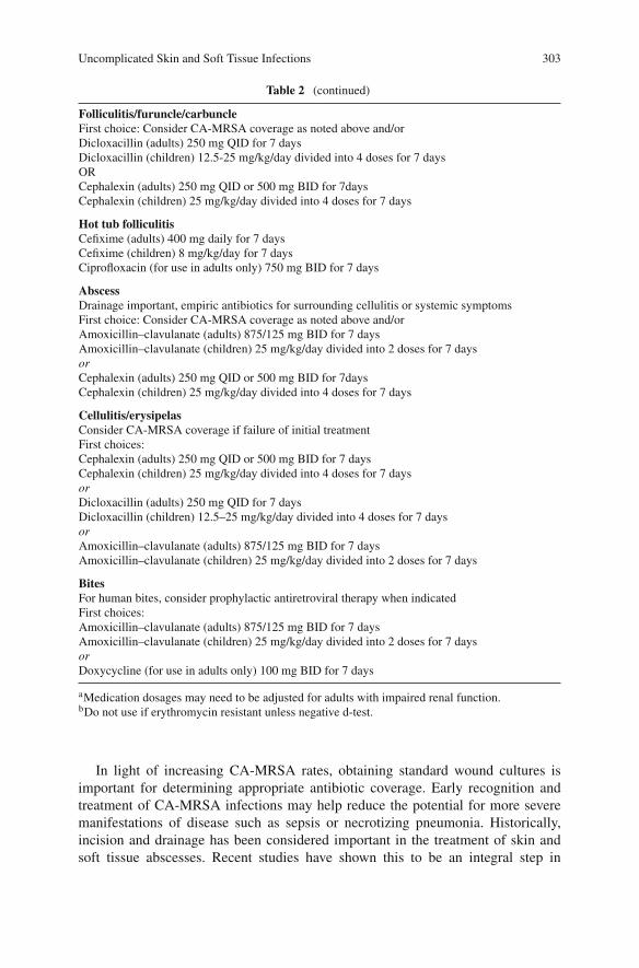
Uncomplicated Skin and Soft Tissue Infections 303
Table 2 (continued)
Folliculitis/furuncle/carbuncleFirst choice: Consider CA-MRSA coverage as noted above and/orDicloxacillin (adults) 250 mg QID for 7 daysDicloxacillin (children) 12.5-25 mg/kg/day divided into 4 doses for 7 daysORCephalexin (adults) 250 mg QID or 500 mg BID for 7daysCephalexin (children) 25 mg/kg/day divided into 4 doses for 7 days
Hot tub folliculitisCefixime (adults) 400 mg daily for 7 daysCefixime (children) 8 mg/kg/day for 7 daysCiprofloxacin (for use in adults only) 750 mg BID for 7 days
AbscessDrainage important, empiric antibiotics for surrounding cellulitis or systemic symptomsFirst choice: Consider CA-MRSA coverage as noted above and/orAmoxicillin–clavulanate (adults) 875/125 mg BID for 7 daysAmoxicillin–clavulanate (children) 25 mg/kg/day divided into 2 doses for 7 daysorCephalexin (adults) 250 mg QID or 500 mg BID for 7daysCephalexin (children) 25 mg/kg/day divided into 4 doses for 7 days
Cellulitis/erysipelasConsider CA-MRSA coverage if failure of initial treatmentFirst choices:Cephalexin (adults) 250 mg QID or 500 mg BID for 7 daysCephalexin (children) 25 mg/kg/day divided into 4 doses for 7 daysorDicloxacillin (adults) 250 mg QID for 7 daysDicloxacillin (children) 12.5–25 mg/kg/day divided into 4 doses for 7 daysorAmoxicillin–clavulanate (adults) 875/125 mg BID for 7 daysAmoxicillin–clavulanate (children) 25 mg/kg/day divided into 2 doses for 7 days
BitesFor human bites, consider prophylactic antiretroviral therapy when indicatedFirst choices:Amoxicillin–clavulanate (adults) 875/125 mg BID for 7 daysAmoxicillin–clavulanate (children) 25 mg/kg/day divided into 2 doses for 7 daysorDoxycycline (for use in adults only) 100 mg BID for 7 days
aMedication dosages may need to be adjusted for adults with impaired renal function.bDo not use if erythromycin resistant unless negative d-test.
In light of increasing CA-MRSA rates, obtaining standard wound cultures isimportant for determining appropriate antibiotic coverage. Early recognition andtreatment of CA-MRSA infections may help reduce the potential for more severemanifestations of disease such as sepsis or necrotizing pneumonia. Historically,incision and drainage has been considered important in the treatment of skin andsoft tissue abscesses. Recent studies have shown this to be an integral step in

304 I.L. Frithsen and C.D. Salgado
management of these infections with drainage considered more important thanappropriate antibiotic selection [6]. When choosing an antibiotic for treatmentof a known or suspected CA-MRSA infection, it is necessary to understand themechanism of resistance. Unlike penicillin resistance, which is conferred via pro-duction of beta-lactamase, methicillin resistance is conferred through expressionof an altered penicillin binding protein [7]. This mechanism of resistance makesthe use of combination beta-lactam/beta-lactamase inhibitor antibiotics ineffective.Trimethoprim–sulfamethoxazole (TMP–SMX) and tetracyclines are effective first-line agents in the treatment of CA-MRSA skin infections [3]. Clindamycin may alsobe considered, but since inducible resistance to Staphylococcus aureus can occur, itshould be used only when the susceptibility profile is favorable.
3 Impetigo
3.1 Clinical Presentation
Impetigo is a highly contagious skin infection that most commonly affects children,although any age group can be affected [8]. Clinically, impetigo may be dividedinto two forms: bullous and nonbullous, with nonbullous impetigo accounting forthe majority of cases. Body sites most often affected are around the mouth and noseor on the hands and forearms. Nonbullous impetigo begins as a single erythematousmacule or papule that rapidly progresses to a vesicle. These vesicles then burst toform characteristic honey colored crusts that can become pruritic [8]; a commonaffect of scratching is spread to the surrounding areas causes clusters of lesions toform. Bullous impetigo commonly presents as a larger blister containing yellow,serous fluid with clear margins and no surrounding erythema. Yellow crusting andoozing occurs once the bullae rupture [8, 9].
3.2 Epidemiology
Impetigo is the most common pediatric skin infection; it most commonly affectschildren aged 2–5 [8]. A study in the United Kingdom found that the annual inci-dence of impetigo was 2.8% for children under 4 years and 1.6% for those aged 5–15years [10]. Nonbullous impetigo accounts for over 70% of cases. Spread can occurrapidly among close contacts and schoolmates either via direct contact or throughcontaminated objects such as toys. The highest incidence has been noted to occur inwarmer climates or in the summer months and in populations with crowded livingconditions and poor hygiene [8].
3.3 Microbiology
Both forms of impetigo are mainly caused by Staphylococcus aureus withStreptococcus pyogenes contributing to nonbullous forms [8]. If subtypes of

Uncomplicated Skin and Soft Tissue Infections 305
Staphylococcus aureus are those that produce toxins associated with staphylococcalscalded skin syndrome (such as exfolatin), then larger bullae may be noted on clin-ical exam [11]. CA-MRSA has also been described with impetigo, but the actualincidence of this is unknown.
3.4 Treatment
Impetigo is generally a self-limiting condition that rarely leads to systemic illness orscarring; however, untreated cases can take up to 2 weeks to heal, which can causesignificant discomfort for those afflicted and increase the risk of spread to contacts.Since the diagnosis of impetigo is made on the basis of characteristic clinical appear-ance, wound cultures are not commonly obtained. Therefore, empirical treatmentis aimed at the commonly encountered organisms noted above. Of note, post-streptococcal glomerulonephritis occurs in fewer than 1% of cases and treatmentof lesions has not been shown to be effective in reducing this complication [10].
A systematic review of the treatment options for impetigo showed that 7 daysof topical mupirocin was more effective than placebo [10]. The authors also notedthat topical antibiotics may be more effective than some oral antibiotics and thatthere have been no high-quality studies comparing oral antibiotics to placebo [10].Despite the limited evidence supporting their use, oral antibiotics are commonlyrecommended and prescribed for treatment of impetigo. These include amoxicillinplus clavulanic acid, cephalosporins, and macrolides, although increasing rates oferythromycin resistance have been reported. Evidence is also lacking for use of oralantibiotics in cases of severe or extensive impetigo, but, practically speaking, it maybe more feasible for some patients to be treated systemically [12]. Clinicians shouldconsider adding coverage for CA-MRSA in the event of a known outbreak or uponinitial treatment failure.
4 Folliculitis/Furuncle/Carbuncle
4.1 Clinical Presentation
Folliculitis is an inflammatory reaction to a bacterial infection localized to thehair follicle and affecting the superficial skin layers [13]. Patients may initiallydevelop erythematous, pruritic lesions that can become purulent and can subse-quently rupture and drain. Typical lesion locations include the head, back, buttocks,and extremities. Systemic manifestations of folliculitis are rare [14]. Hot tub fol-liculitis is a distinct entity that results from exposure to contaminated water inhot tubs, whirlpools, or swimming pools. Patients can present with a variety oflesions located on body parts that have been under water, although areas coveredby bathing suits are affected more frequently [14]. A similar condition involvingnodular lesions of the palms and soles has been reported in children and is referredto as, “hot hand–foot syndrome” [15].
306 I.L. Frithsen and C.D. Salgado
Furuncles are single lesions similar to folliculitis with the exception that theyextend into deeper layers of the skin, including the subcutaneous tissue. Lesions arefirm, tender, erythematous and can spontaneously drain purulent material. They arecommonly found on the face, neck, axilla, and buttocks, as these tend to be moistand hairy areas. Systemic manifestations of furuncles are also rare. Carbuncle is theterm used to describe an aggregate of multiple furuncles. Carbuncles are commonlylocated on the back of the neck, the back, or the thighs. Carbuncles can be painfuland commonly involve systemic symptoms such as fever and malaise. Furunclesand carbuncles are sometimes referred to as boils [14].
4.2 Epidemiology
Poor hygiene is a risk factor for developing folliculitis. Hot tub folliculitis gener-ally presents within 48 h of contact with contaminated water and outbreaks havebeen reported [16]. Furuncles and carbuncles commonly arise from folliculitis. Riskfactors for the development of furuncles or carbuncles include obesity, diabetes,chronic kidney disease, impaired neutrophil function, intravenous or subcutaneousdrug abuse, and corticosteroid use [14]. These conditions can occur in patients ofany age.
4.3 Microbiology
Staphylococcus aureus, including CA-MRSA, is associated with most cases offolliculitis [3]. Pseudomonas aeruginosa is the pathogen responsible for the vastmajority of cases of hot tub folliculitis. Staphylococcus aureus is also the mostcommon causative agent for furuncles and carbuncles [14].
4.4 Treatment
Folliculitis will likely resolve spontaneously in immunocompetent patients withoutscarring. Warm compresses and topical antibiotics have been used to promote heal-ing and provide symptomatic relief. Proper hygiene should be advocated to avoidrecurrent episodes [14]. Small furuncles can be managed in the same manner asfolliculitis; however, larger furuncles and carbuncles require incision and drainage.Systemic antibiotic therapy is indicated if the patient is febrile or extensive cellulitisis present surrounding the lesion [5]. Cultures obtained upon incision and drainagecan help direct antibiotic therapy when indicated. Empiric antibiotic choices shouldinclude agents effective against staphylococci such as semi-synthetic penicillins orfirst generation cephalosporins. Trimethoprim–sulfamethoxazole is a good choiceif CA-MRSA is suspected. Fluoroquinolones and third- and fourth-generationcephalosporins are effective in treating Pseudomonas infections and should be usedto treat hot tub folliculitis.

Uncomplicated Skin and Soft Tissue Infections 307
5 Abscess
5.1 Clinical Presentation
An abscess is a collection of exudate located within the dermal or deeper layersof the skin. Patients present with single or multiple painful, tender, erythematousnodules that can occur anywhere on the body. These nodules are fluctuant andcommonly have an overlying pustule [5].
5.2 Epidemiology
Abscesses can affect individuals of any age. Patients may present with abscesses invarious stages of progression. When patients present early, there may be an area ofinduration with erythema, but no fluctuance is noted. Patients presenting later mayhave spontaneous rupture of the abscess with development of surrounding celluli-tis. Systemic symptoms are rare but are more often noted when there is an area ofcellulitis surrounding the abscess [17].
5.3 Microbiology
Abscesses are usually polymicrobial with normal skin flora and other organismsfrequently isolated. Guidelines published in 2005 noted that Staphylococcus aureuswas isolated as a solitary organism from about 25% of drainable abscess [5].However, a more recent study found that CA-MRSA was isolated from more than80% of abscesses, thus having profound implications for treatment [2].
5.4 Treatment
Incision and drainage is the mainstay of treatment for abscess. In addition to open-ing the skin layer, probing is performed to break up loculations. Packing the woundafter drainage is commonly performed in order to prevent the incision from closing,which could result in the abscess reforming. However, a pilot study that examinedthe utility of packing after incision and drainage of an abscess found no reduc-tion in morbidity among patients that did not receive packing. Additionally, thestudy reported that patients who did get packing experienced more pain and usedmore pain medication than those whose wounds were not packed [18]. Treatmentwith antibiotics following incision and drainage remains a topic of debate. Forpatients with simple abscess and no surrounding cellulitis who have no systemicsymptoms, antibiotics are not necessary [17]. If cellulitis or systemic symptomsare present or if the patient is immunocompromised, then antibiotic therapy shouldbe directed toward Staphylococcus aureus [17]. Cephalexin can be used, although

308 I.L. Frithsen and C.D. Salgado
a strong suspicion for CA-MRSA should be maintained and empiric treatmentwith TMP–SMX can be included. Regardless of the decision to use antibiotics,obtaining exudate cultures and close follow-up are important for treatment ofabscesses.
6 Cellulitis/Erysipelas
6.1 Clinical Presentation
Cellulitis and erysipelas are terms used to describe similar infections of the skin.Cellulitis is an infection of the skin that involves the deeper dermal layers, includingsubcutaneous fat. Erysipelas is an infection that affects the upper dermis and super-ficial lymphatics. Patients who have both of these conditions present with rapidlyspreading areas of painful, erythematous warm and edematous skin. Localized lym-phadenopathy and dimpling of the skin known as peau d’orange can be present.Vesicles or bullae filled with clear fluid are also commonly noted. Systemic symp-toms include fever, tachycardia, hypotension, and leukocytosis; these can be presentbefore skin manifestations are noted. Erysipelas may be distinguished from celluli-tis by the presence of a clearly demarcated border of erythema and raised lesions[5].
6.2 Epidemiology
Among 320,000 members of a Utah health plan, Cellulitis accounted for 2.2% ofoutpatient office visits in 1999 [11]. Extremities are the most common site forcellulitis/erysipelas, although these infections can occur anywhere on the body. Itgenerally occurs where there has been a break in the skin such as an abrasion,although the portal of entry for bacteria is often not identified [17]. Cellulitis anderysipelas can occur in patients of any age, although erysipelas is more common ininfants, young children, and older adults. Erysipelas has also been noted to occurmore often on the lower extremities [5].
6.3 Microbiology
Cellulitis is still most commonly caused by beta-hemolytic (group A/B) strep.Species however, Staphylococcus aureus, including CA-MRSA, is becoming morecommon. Cellulitis can be caused by a wide variety of organisms depending onthe circumstances involving development of the infection. Examples include vib-rio infections following exposure to saltwater and Haemophilus influenzae, causingperiorbital cellulitis in children [17]. Erysipelas is commonly caused by S. pyogeneswith rare involvement of group B Strep or Staphylococcus aureus.

Uncomplicated Skin and Soft Tissue Infections 309
6.4 Treatment
An important first step in the treatment of cellulitis or erysipelas is obtaining athorough exposure history, as this could provide information that would directthe clinician to consider infection with a less common organism such as vibriomentioned above. Empiric antibiotic therapy generally consists of a first genera-tion cephalosporin such as cephalexin, a penicillinase-resistant penicillin such asdicloxacillin or a beta-lactam/beta-lactamase inhibitor combination such as amox-icillin/clavulanate [3]. One consideration in treating cellulitis is whether or not toobtain blood cultures. A recent review that included five separate studies noted thatroutine blood cultures for uncomplicated cellulitis were not helpful [19]. However,in the presence of systemic symptoms it is important to consider that a more seri-ous infection may be present and could include hematogenous spread. In thesecircumstances, further diagnostic studies, including blood cultures, may be bene-ficial. Close follow-up is important and in the event of treatment failure, coverageto include less common etiologic organisms and CA-MRSA should be considered.
7 Bites
7.1 Clinical Presentation
Each year millions of animal bites occur in the United States with more than 90% ofthese due to the bites of cats and of dogs [20]. Human bites have been reported as thethird most common mammalian bite and present complex management issues [21].Up to one-half of children will be bitten by a dog during childhood, although themajority of bites are minor and do not require medical attention [22]. Complicationsfrom infected bite wounds can result in significant morbidity, including sepsis,meningitis, and endocarditis [23]. Older patients, asplenic patients, those with liverdisease, and alcoholics may be more likely to develop serious complications [24].Patients who seek medical attention less than 8 h after being bitten are less likelyto have an established infection as compared to those who present later [20]. Dogbites occur about six times more often than cat bites, but cat bites are more likely tobecome infected [20, 23].
7.2 Epidemiology
Bites from animals are common, as there are over 160 million dogs and cats inthe United States [25]. Dog bites account for up to 80% of animal bites, and thisis reflected in the fact that the majority of published data on animal bites pertainsto dogs [20]. It was estimated that 4.5 million dog bites occurred in the UnitedStates in 1994, which is the most recent year for which published data are avail-able on the overall number of bites [26]. Also in 1994, there were an estimated
310 I.L. Frithsen and C.D. Salgado
333,687 emergency department (ED) visits for dog bites [27], resulting in an esti-mated 5,991 hospitalizations [28]. A 2001 report estimated that there were 368,245patients treated in emergency departments with a reported rate similar to previousstudies, indicating that rates of dog bites have remained stable. The highest dogbite rates among ED patients occurred in children aged 5–9 and incidence ratesdeclined steadily with increasing age. Dog bites are more likely on the head or facefor children and are more common on the arm or hand for adults [29].
Recent data on rates of human bites are not readily available. Human bites canbe divided into actual bites or those resulting from a closed fist injury. Bites havebeen described to occur under a variety of circumstances, for example, during vio-lent encounters such as assaults, among children, and accidentally during sports orsexual activity. Bites also have been reported to occur while attempting to maintainan airway during medical emergencies such as an epileptic seizure. An importantconsideration in the evaluation of a human bite is the possibility of blood-bornepathogen exposure such as HIV or hepatitis [30].
7.3 Microbiology
Organisms infecting animal or human bite wounds are those from the mouth ofthe biting animal, normal human skin microbes, and other secondary infectiousorganisms. Animal and human bite wounds commonly involve both anaerobic andaerobic species. Common pathogens include Pasteurella species, staphylococci, andstreptococci. Capnocytophaga canimorsus has been reported to cause sepsis andis particularly dangerous for asplenic patients and those with underlying hepaticdysfunction including alcoholics [32]. Human bites have been noted to carry thehighest risk of infection with up to 50% infection rates reported from one study[31]. Human bites are typically polymicrobial, but Eikenella corrodens is one gram-negative bacillus often recovered along with other organisms. It is not unusual formore than 10 organisms to be cultured from human bite wounds, and one study of18 human bites in children revealed a total of 97 different bacterial isolates with arange of 1–8 per bite [32].
7.4 Treatment
As with any open wound, the initial treatment should include irrigation, cleans-ing, and debridement of damaged tissue. Thorough exploration of the woundshould be performed to determine if tendon, muscle, or bone damage has occurred.Radiographs should be performed if bone damage is suspected or to detect thepresence of a foreign body such as a tooth [32]. Primary closure of bite woundsremains controversial. Empiric antibiotic therapy for animal or human bites is amox-icillin/clavulanate or doxycycline. Due to the large number of potential pathogensisolated from animal and human bite wounds, cultures can be helpful in determin-ing appropriate antibiotic coverage. With animal bites it is important to establish the

Uncomplicated Skin and Soft Tissue Infections 311
rabies status, which will be determined by local health department officials whenthe bite is reported; in most states this reporting is mandatory. Tetanus immuniza-tion status should be assessed and a booster given when indicated. Transmission ofhuman immunodeficiency virus, syphilis, and hepatitis B have been reported fol-lowing human bite wounds, so it is important to determine the health status of thebiter [33]. Prophylactic treatment with antiretroviral agents may be considered forhuman bites based on a risk analysis [30].
Key Points
• Community-associated methicillin-resistant Staphylococcus aureus (CA-MRSA)is a common cause of skin/soft tissue infections and should be consideredregardless of risk factors.
• Empiric antibiotic selection depends on the type of infection and exposurehistory.
• Wound cultures obtained when initiating empiric therapy will allow detectionof resistant organisms and guide subsequent choice of antibiotic in the event oftreatment failure.
• Incision and drainage is often essential to allow timely wound healing.
References
1. Rosen T (2005). Update on treating uncomplicated skin and skin structure infections. J DrugsDermatol 4(6):s9–s14
2. Moran G, Krishnadasan A, Gorwitz R, et al.(2006). Methicillin-resistant S. aureus infectionsamong patients in the emergency room. NEJM 355(7):666–674
3. Elston D (2007). Community-acquired methicillin-resistant Staphylococcus aureus. J AmAcad Dermatol 56:1–16
4. Cohen P (2007). Community-acquired methicillin-resistant Staphylococcus aureus skin infec-tions: A review of epidemiology, clinical features, management and prevention. Intl JDermatol 46:1–11
5. Stevens D, Bisno A, Chambers H, et al.(2005). Practice guidelines for the diagnosis andmanagement of skin and soft-tissue infections. Clin Infect Dis 41:1373–1406
6. Fridkin S, Hageman J, M M, et al.(2005). Methicillin-resistant Staphylococcus aureus diseasein three communities. N Engl J Med 352:1436–1444
7. Eady E, Cove J (2003). Staphylococcal resistance revisited: Community-acquired methicillinresistant Staphylococcus aureus – an emerging problem for the management of skin and softtissue infections. Curr Opin Infect Dis 16(2):103–124
8. Cole C, Gazewood J (2007). Diagnosis and treatment of impetigo. Am Family Phys75(6):859–864
9. Jacobs M, Jones R, Giordano P (2007). Oral beta-lactams applied to uncomplicated infectionsof skin and skin stuctures. Diag Microbiol Infect Dis 57:55S–65S
10. George A, Rubin G (2003). A systematic review and meta-analysis of treatments for impetigo.Br J Gen Pract 53:480–487
11. Stulberg D, Penrod M, Blatny R (2002). Common bacterial skin infections. Am Family Phys66:119–124

312 I.L. Frithsen and C.D. Salgado
12. Koning S, Verhagen A, van Suijlekon-Smit L, et al.(2003). Interventions for impetigo.Cochrane Database of Systematic Reviews (2):CD003261
13. Ruocco E, Donnarumma G, Baroni A, et al.(2007). Bacterial and viral skin diseases. DermatolClin 25(4):663–676
14. Lopez F, Lartchenko S (2006). Skin and soft tissue infections. Infectious Disease ClinicsNorth Am 20(4):759–772
15. Yu Y, Cheng A, Wang L, et al.(2007). Hot tub folliculitis or hot hand-foot syndrome causedby Pseudomonas aeruginosa. J Am Acad Dermatol 57:596–600
16. CDC (2000). Pseudomonas dermatitis/folliculitis associated with pools and hot tubs –Colorado and maine, 1999–2000. MMWR 49(48):1087–1091
17. Rogers R, Perkins J (2006). Skin and soft tissue infections. Prim Care Clin Office Practice33:697–710
18. O‘Malley G, Williams E, Dominici P, et al. (2007). Packing simple cutaneous abscess afterincision and drainage is painful and probably unnecessary. Ann Emerg Med 50(3):s58
19. Mills A, Chen E (2005). Are blood cultures necessary in adults with cellulitis? Ann EmergMed 45(5):548–549
20. Steele M, Ma J, Nakase J, et al.(2007). Epidemiology of animal exposures presenting toemergency departments. Acad Emerg Med 14(5):398–403
21. Goldstein E (1992). Bite wounds and infection. Clin Infect Dis 14(3):633–63822. Beck A, Jones B (1985). Unreported dog bites in children. Public health rep 100:315–32123. Talan D, Citron D, Abrahamian F, et al.(1999). Bacteriologic analysis of infected dog and cat
bites. NEJM 340(2):85–9224. Wareham D, Michael J, Warwick S, et al.(2007). The dangers of dog bites. J Clin Path 60:
328–32925. Industry statistics and trends. (Accessed at http://www.appma.org/press_industrytrends.asp).26. Sacks J, Kresnow M, Houston B (1996). Dog bites: How big a problem? Injury Prevention
2:52–5427. Weiss H, Friedman D, Coben J (1998). Incidence of dog bite injuries treated in emergency
departments. J Am Med Assoc 279(1):51–5328. Quinlin K, Sacks J (1999). Hospitalizations for dog bite injuries. J Am Med Assoc
281(3):232–23329. CDC (2001). Non-fatal dog bite related injuries treated in hospital emergency departments
United States, 2001. MMWR 52(26):605–61030. Pretty I, Anderson G, Sweet D (1999). Human bites and the risk of human immunodeficiency
virus transmission. Am Jf Forensic Med Pathol 20(3):232–23931. Mann R, Hoffeld T, Rarmer C (1977). Human bites of the hand: Twenty years of experience.
J Hand Surg 2(2):97–10432. Brook I (2003). Microbiology and management of human and animal bite wound infections.
Prim Care Clin Office Practice 30:25–3933. Brook I (2005). Management of human and animal bite wounds: An overview. Adv Skin
Wound Care 18:197–203

Septic Arthritis and Osteomyelitis
Camelia E. Marculescu
1 Introduction
Septic arthritis and osteomyelitis are two of the most common diseases that affectbone and joints. Septic arthritis is a true rheumatologic emergency, mandatingimmediate joint drainage and antibiotics. Adult osteomyelitis most commonly arisesfrom open fractures, diabetic foot infections, or surgical treatment of closed injuries.This chapter provides an overview of the etiology, diagnosis, and antimicrobialmanagement of septic arthritis and adult osteomyelitis.
2 Septic Arthritis
2.1 Overview
The incidenceof septic arthritis in the United States is increasing; the average patientis older, the antimicrobial resistance is rising, and the diagnosis and presentationremains problematic. The incidence of gonococcal arthritis is decreasing, whereastwo virulent pathogens methicillin-resistant Staphylococcus aureus and group Bstreptococcus have become more important. Septic arthritis is more often a conse-quence of occult bacteremia. Breaks in the skin allow staphylococci or streptococcithe initial access into the bloodstream. Gram-negative septic arthritis probablyarises from bacteremia originating from the urinary or the gastrointestinal tract.Therapeutic joint injections with corticosteroids and rarely arthroscopy can resultin iatrogenic septic arthritis [27].
C.E. Marculescu (B)Division of Infectious Diseases, Department of Internal Medicine, Medical University of SouthCarolina, Charleston, SC, USAe-mail: [email protected]
313A.G. Mainous III, C. Pomeroy (eds.), Management of Antimicrobials in InfectiousDiseases, Infectious Disease, DOI 10.1007/978-1-60327-239-1_16,C© Springer Science+Business Media, LLC 2001, 2010

314 C.E. Marculescu
2.2 Etiology
Most cases of septic arthritis are caused by gram-positive organisms, whereasenteric gram-negative rods account for 43% of the community-acquired bacteremiasbut cause only 10% of septic arthritis [12, 18].
a. Staphylococcus aureus is the most common cause of septic arthritis, whichaccounts for 44% of cases (Table 1). MRSA may be increasing in importancein septic arthritis. In the last 5 years at Caritas St. Elizabeth’s Medical Centerand Tufts-New England Medical Center, 25% of septic arthritis was caused byMRSA [27]. In this series, cases were associated with chronic illness, olderage, and health-care exposure. Community-acquired MRSA septic arthritis hasoccurred in some regions of the United States [8].
Table 1 Organisms isolated in 2407 cases of septic arthritis
Organism No. of isolates (%)
S. aureus 1,066 (44.3)S. pyogenes 183 (7.6)S. pneumoniae 156 (6.5)Haemophilus influenzae 104 (4.3)Mycobacterium tuberculosis 101 (4.2)Escherichia coli 91 (3.8)Coagulase-negative staphylococci 84 (3.5)N, gonorrhoeae 77 (3.2)S. agalactiae 69 (2.9)Pseudomonas aeruginosa 36 (1.5)N. meningitidis 28 (1.2)Salmonella sp. 25 (1)Other gram-negative rods 110 (4.6)Other β-hemolytic streptococci 104 (4.3)Polymicrobial 33 (1.4)Fungi 4 (0.2)Miscellaneous 136 (5.7)
Adapted with permission from Ross et al. [28].
b. Hemolytic streptococci (Streptococcus agalactiae) are pathogens encounteredespecially in the elderly patients, those with diabetes, cirrhosis, and neurologicdisease [7], and cause about 10% of septic arthritis [21]. Functional outcomesare typically poor in septic arthritis, due to group B streptococci and otherβ-hemolytic streptococci [27].
c. Pneumococcus causes up to 6% of septic arthritis cases. Pneumococcal septicarthritis is notable for a high frequency of bacteremia and polyarticular disease.Only 50% have an underlying focus of pneumococcal disease such as pneumo-nia. Mortality in adults is high (19%), although in 95% of survivors, functionaloutcomes are good [27]. Drug resistance may be an increasing problem [28].

Septic Arthritis and Osteomyelitis 315
d. Gonococcal septic arthritis is a distinct clinical syndrome with a good prognosis.Seventy-five percent of cases occur in women; menses and pregnancy increasethe risk of disseminated gonococcal infection [23]. Seventy-two percent of casesare polyarticular and knee involvement is most common. The characteristic hem-orrhagic pustules of disseminated gonococcal infection are found in 42% ofpatients and tenosynovitis in 21%. Urinary signs or symptoms are present inonly 32% [27]. Gonococci are recovered from joint fluid in less than 50% ofcases. This is probably largely due to the difficulty in recovering these fastidiousorganisms from cultures but may also indicate that some cases of gonococcalarthritis are immune mediated [23].
e. Enteric gram-negative rods cause about 10% of adult septic arthritis especiallyin populations at risk such as intravenous drug users and elderly patients withcomorbid conditions [3, 10]. Recent studies suggest that with prompt diagnosisand therapy, outcomes in older patients may be relatively good [27]. Surprisingly,a urinary source of gram-negative septic arthritis is found only in 50% of elderlypatients [20].
f. Meningococcal septic arthritis. The incidence of arthritis in invasive meningo-coccal disease is as high as 14%. The joint fluid may be serous or purulent [27].Typical presentation is a mono- or oligoarthritis involving large joints, especiallythe knee. It has been suggested that the arthritis has an immunologic basis, sincesynovial fluid immune complexes have been detected in several of these patients[9, 30]. The outcome of meningococcal septic arthritis is usually excellent [30].
g. Coagulase-negative staphylococci. Typically coagulase-negative staphylococciare considered contaminants when isolated from the joint space in native septicarthritis, but they can be true pathogens after procedures such as arthroscopyand anterior cruciate ligament reconstruction (ACL). It tends to present in anindolent fashion.
Postarthroscopic septic arthritis can be cured with 2 weeks of parenteral antimi-crobial therapy, whereas septic arthritis that develops following ACL reconstructionrequires arthroscopic debridement and 6 weeks of antimicrobial therapy [1, 14].
2.2.1 Clinical Presentation and Diagnosis
The diagnosis of septic arthritis can be easily established in patients present-ing with typical symptoms such as fever, warm, swollen, and exquisitely painfuljoint. However, high-grade fever is present only in 58% and leukocytosis only in50–60% of patients [11, 35]. Joint pain can be blunted in immunosuppressedpatients. Synovial fluid WBC count is variable. A synovial fluid WBC value of≥50,000 cells/mm3 is a commonly used threshold for empiric antimicrobial ther-apy. At least one-third of the patients with native septic arthritis have synovial fluidWBC count < 50,000 cells/mm3. The yield of synovial fluid culture is increased byinoculation of fluid into blood culture bottles [13]. In cases of undiagnosed arthritis,gram stain and culture of the synovial fluid should be routinely obtained; however,gram staining is insensitive for the diagnosis of septic arthritis [27]. All patients with

316 C.E. Marculescu
suspected septic arthritis should have blood cultures obtained, since bacteremia canbe present in at least one-third of the patients with septic arthritis [27]. In gonococcalseptic arthritis, less than 50% of synovial fluid cultures are positive, and diagnosisis often based on a compatible clinical syndrome and the isolation of Neisseriagonorrhoeae from cultures of the cervix, the urethra, the rectum, or the oropharynx.
2.3 Therapy and Outcome
Septic arthritis is a rapid and destructive disease of the joint space; therefore,broad-spectrum antimicrobial therapy should be initiated before culture data areavailable. Because synovial fluid tests lack precision for diagnosing septic arthritis,the threshold for starting antibiotics should be low.
– In cases with a negative gram stain of the synovial fluid and if the patient is atlow risk for methicillin-resistant S. aureus (MRSA) or gonorrhea, cefazolin is areasonable initial empirical choice for treatment of native septic arthritis.
– In elderly or immunocompromised patients felt to be at risk for gram-negative septic arthritis, empiric antimicrobial therapy should include abroad-spectrum single agent active against streptococci, methicillin-susceptibleS. aureus (MSSA), and gram-negative bacilli such as cefepime.
– Patients with MRSA risk factors such as hemodialysis, diabetes, recent hos-pitalization, or nursing home admission should be empirically covered withvancomycin. Community-acquired MRSA infections have been reported invarious regions of the Unites States, and it is reasonable to initiate empiricantimicrobial therapy directed against MRSA in patients with septic arthritis inthese regions [19]. Limited, but favorable data support the use of linezolid anddaptomycin as alternative agents for MRSA septic arthritis.
– Critically ill patients with septic arthritis should receive broad-spectrum antibi-otics (e.g., combination therapy with vancomycin and cefepime).
– Sexually active patients that present with clinical syndrome suggestive of gono-coccal infection should receive ceftriaxone. Septic arthritis that occurs as a resultof human or animal bites should receive agents active against oral flora such asampicillin–sulbactam.
– Intravenous drug users with septic arthritis may be empirically covered withvancomycin and an antipseudomonal agent.
Recommendations for empiric antimicrobial therapy are summarized in Table 2.Once the causative organism has been identified, the antimicrobial therapy shouldbe narrowed.
Duration of therapy: In general, it is recommended to complete at least3 weeks of antimicrobial therapy, which may include a period of step-down oraltherapy. Gonococcal septic arthritis can be treated with 2 weeks of ceftriaxone.

Septic Arthritis and Osteomyelitis 317
Table 2 Empiric antibiotic therapy of suspected septic arthritis
Gram stain of synovial fluid Antibiotic therapy
Gram-positive cocciNo risk factors for MRSA (see text) Cefazolin, 2 g IV q 8 hMRSA risk factors or β-lactam allergy Vancomycin, 1 g IV q 12 hGram-negative cocci (presumptive
Neisseria sp.)Ceftriaxone, 1 g IV q 24 h
Gram-negative rods Cefepime, 2 g IV q 8 h orpiperacillin–tazobactam, 4.5 g IV q 6 h
No organisms on gram stainPreviously healthy, low MRSA risk Cefazolin, 2 g IV q 8 hMRSA risks present Vancomycin, 1 g IV q 12 h plus cefepime, 2 g IV
q 8 h or piperacillin–tazobactam, 4.5 gIV q 6 h
Abbreviation: MRSA, methicillin-resistant S. aureus.Adapted with permission. Ross, Septic arthritis [27].
Courses of 4–6 weeks are recommended for patients with septic arthritis that involvesternoclavicular and sacroiliac joints [27, 29].
Joint drainage decompresses the joint, improves blood flow, and removes bac-teria, toxins, and proteases. Arthrocentesis should be repeated daily until effusionsresolve and cultures are negative. Surgical drainage is recommended for native hipseptic arthritis, for failure to respond after 5–7 days of arthrocentesis and antibi-otics, for pus in the joint space, and for soft tissue extension of infection [27].The shoulder joint should be drained surgically or under radiologic guidance [26,32]. No good data show a superiority of surgical drainage versus arthrocentesis.Aggressive rehabilitation is needed to prevent joint contractures and muscle atrophy,and mobilization should be done as soon as pain allows [27].
3 Adult Osteomyelitis
3.1 Overview
Osteomyelitis is a disease very heterogeneous in its pathophysiology, in its clini-cal presentation, and in its management. It is felt to be one of the most difficultinfectious diseases to treat. The hallmarks of osteomyelitis are the formation ofthe sequestra and progressive bone destruction. Osteomyelitis can result fromhematogenous seeding, from contiguous spread of infection, or from direct inoc-ulation of the microorganisms into the bone. There are several classifications ofosteomyelitis: the two major classifications are those by Waldwogel [34] andCierny–Mader [6]. The Waldvogel classification takes into account the duration ofdisease (acute or chronic), the mechanism of infection (hematogenous or contigu-ous), and the presence of vascular insufficiency. The Cierny–Mader classification

318 C.E. Marculescu
Table 3 Cierny–Mader staging system
Anatomic type Physiologic class
Stage 1: Medullary osteomyelitis A Host: Normal hostStage 2: Superficial osteomyelitis B Host: Systemic compromise (Bs)Stage 3: Localized osteomyelitis Local compromise (Bl)Stage 4: Diffuse osteomyelitis Systemic and local compromise (Bls)
C Host: Treatment worse than the disease
Systemic or local factors that affect immune surveillance, metabolism, and local vascularity
Systemic (Bs) Local (Bl)Malnutrition Chronic lymphedemaRenal, hepatic failure Venous stasisDiabetes mellitus Major vessel compromiseChronic hypoxia ArteritisImmune disease Extensive scarringMalignancy Radiation fibrosisExtremes of age Small vessel diseaseImmunosuppression or neuropathy Complete loss of sensationImmune deficiency Tobacco abuse
Adapted after Calhoun and Manring [5], with permission.
is determined by the status of disease process, and it is based on the portion ofthe bone affected, the physiologic status of the host, and other risk factors (seeTable 3).
3.2 Etiology
The specific microorganism isolated from patients with bacterial osteomyelitis isoften associated with the age of the host or a common clinical scenario (seeTable 4). Hematogenous osteomyelitis is almost always a monomicrobial infection,with S. aureus being the most prevalent causative organism [31]. Coagulase-negative staphylococci, aerobic gram-negative bacteria, and Peptostreptococcusspp. are also frequently isolated in long bone osteomyelitis [31]. Posttraumaticosteomyelitis resulting from open fractures is often polymicrobial. Staphylococciand gram-negative aerobic and anaerobic bacilli are most frequently isolated. Othermicroorganisms such as enterococci, fungi, and atypical mycobacteria have beenimplicated in the etiology of osteomyelitis as well. Staphylococcus aureus andcoagulase-negative staphylococci are the most common microorganisms encoun-tered in vertebral osteomyelitis. Vertebral osteomyelitis caused by gram-negativeaerobic bacilli and Candida spp. are more commonly seen in intravenous drug users,immunosuppressed patients, and in the postoperative setting [31]. Contiguous-focusosteomyelitis associated with diabetes mellitus or vascular insufficiency is oftenpolymicrobial (S. aureus, β-hemolytic streptococci, enterococci, anaerobes).

Septic Arthritis and Osteomyelitis 319
Table 4 Microorganisms isolated from patients with bacterial osteomyelitis
Microorganism Most common clinical association
S. aureus (susceptible or resistant tomethicillin)
Most frequent microorganism in any type ofosteomyelitis
Coagulase-negative staphylococci orPropionibacterium
Foreign body-associated infection
Enterobacteriaceae or P. aeruginosa Common in nosocomial infectionsStreptococci or anaerobic bacteria Associated with bites, fist injury caused by
contact with another person’s mouth, diabeticfoot lesions, and decubitus ulcers
Salmonella or S. pneumoniae Sickle cell diseaseBartonella henselae Human immunodeficiency virus infectionPasteurella multocida or Eikenella
corrodensHuman or animal bites
Aspergillus, Mycobacterium aviumcomplex, or Candida albicans
Immunocompromised patients
M. tuberculosis Populations in which tuberculosis is prevalentBrucella, Coxiella burnetii (chronic Q
fever), or other fungi found in specificgeographic areas
Populations in which these pathogens areendemic
Adapted with permission from Lew and Waldwogel [16].
3.3 Diagnosis
The typical presentation of acute osteomyelitis is with acute pain and febrileepisode. Presence of chronic drainage, sinus tract or pain, relapses in the sameaffected area, and exposed bone are symptoms that suggest chronic osteomyelitis.In general, persistence of clinical symptoms for more than 10 days is sugges-tive of chronic osteomyelitis. However, the hallmark of chronic osteomyelitis isthe presence of necrosis on bone histology rather than duration of symptoms.Chronic osteomyelitis is a long-standing infection that lasts over months or years. Itimplies persistence of microorganisms, low-grade inflammation, sequestrum, andfistulous tract formation. Laboratory data are primarily useful as a benchmarkagainst which treatment response is measured. Leukocyte count is not a reliableindicator of infection, since its sensitivity is only 26% [4]. Sedimentation ratehas a sensitivity between 50 and 90%; it could be normal early in the course ofdisease, and it decreases slowly over months in response to therapy. C-reactive pro-tein (CRP) has a sensitivity of 71% and is more reliable than sedimentation ratefor follow-up. CRP increases within hours from infection and returns to normalwithin 1 week after effective therapy [4], with the caveat that in rheumatologicor inflammatory conditions, these markers are less reliable and more difficult tointerpret.
Imaging studies are helpful when diagnosis of osteomyelitis is equivocal or whenthere is a need for the determination of the extent of soft tissue/bone abnormali-ties in preparation for surgical intervention [18]. In hematogenous osteomyelitis,

320 C.E. Marculescu
radiographic changes usually reflect the destructive process but lag at least 2 weeksbehind the evolution of infection. The earliest changes are swelling of the soft tissue,periosteal thickening, and/or elevation, and focal osteopenia [18]. A recent meta-analysis of 23 studies in chronic osteomyelitis showed that leukocyte scintigraphyhas 84% accuracy in the peripheral skeleton and PET-FDG is superior for the axialskeleton [33]. Indications and limitations of selected imaging studies are presentedin Table 5.
The diagnosis and the determination of the cause of osteomyelitis rest on the iso-lation of the pathogen(s) from the bone lesion or from the blood or the joint culture.Superficial cultures obtained from the drainage site are usually contaminated withskin flora. The use of antibiotics prior to surgical culture ascertainment reduces thesensitivity of the culture; therefore, whenever possible, cultures of the bone shouldbe obtained prior to initiation of antimicrobial therapy. Cultures of the bone biopsyyield a microbiologic diagnosis in 94% of cases [36]. In general, antimicrobial ther-apy should be stopped for 10–14 days prior to any diagnostic procedure, unless softtissue infection is present, unless microbiology is already known, unless patient isseptic, or unless the risk of secondary bacteremia and hematogenous infection ishigh. Sinus tract cultures are not reliable in predicting the microorganism isolatedfrom the infected bone [17]. However, sinus tract cultures that grow S. aureus showa positive correlation with bone cultures [18].
The gold standard in diagnosing chronic osteomyelitis is bone biopsy obtainedbefore or at the time of surgical debridement. Cultures of the bone yield a microbi-ologic diagnosis in 94% of cases [17]. At least three intraoperative tissue samplesfor aerobes and anaerobes should be obtained routinely to facilitate the distinctionbetween an intraoperative contaminant and a true pathogen [2, 4].
3.4 Treatment
Appropriate therapy of osteomyelitis includes the following: adequate drainage,thorough debridement, obliteration of dead space, wound protection, and specificantimicrobial coverage. If the patient is a compromised host, then efforts should bemade to correct or improve the host defects (e.g., good nutrition, diabetes control,smoking cessation programs).
a. Surgical management of osteomyelitis can be very challenging. The goal ofdebridement is to leave healthy, viable tissue until punctuate bleeding is noted(“paprika” sign) [18]. Adequate debridement may leave a large bone defect termed“dead space.” The adequate management of dead space may include local tissueflaps or free flaps, cancellous bone grafting, antibiotics-impregnated acrylic beads,or antibiotic-impregnated biodegradable beads [18]. Stabilization of the bone isneeded when movement at the site of infection is present and can be achieved byusing screws, rods, and/or an external fixator. Adequate soft tissue coverage of thebone is necessary to arrest osteomyelitis and can be achieved with the use of splitthickness grafts for small defects or free muscle flaps and free vascularized muscleflaps for larger defects [18, 24].

Septic Arthritis and Osteomyelitis 321
Table 5 Indications and limitations of selected imaging studies in chronic osteomyelitis
Imagingmodality Advantages/indications Limitations
Tri-phase bonescan
– High sensitivity (∼70%); candifferentiate cellulitis fromosteomyelitis, specificity 25%;negative bone scan excludesosteomyelitis
– Trauma, hardware, surgery,neuropathy, diabetes mellitus cancause a false-positive bone scan
– Specificity 38–79%– Can be positive for 1–2 years after
treatmentGallium scan – More specific than bone scan (67%)
– Returns to normal after successfultherapy
– Can be combined with Tc scan
– False positive from fractures, tumor– Image takes longer
WBC scan (In orTc)
– Increased specificity compared tobone scan in complicatingconditions
– Good accuracy in peripheral skeleton
– Loss of sensitivity for axial skeleton– Does not delineate anatomy in the
extremities, especially whencirculatory compromise is present
CT – Exceptional bony detail; best fordetection of sequestrum, small fociof gas in the medullary canal
– Important in presurgical evaluationof the extent
– Metallic implants-artifacts
MRI – Highly sensitive (greater than CT)for detecting osteomyelitis as earlyas 3–5 days
– Differentiates between soft tissueand bone infection
– Determines the extent of infection– Helps planning the optimal surgical
management– Defines critical adjacent structures– Differentiates osteomyelitis from
neoplasm or neuropathic joint indiabetic foot, avascular necrosisfrom osteomyelitis, infections inpatients with rheumatoid arthritis
– Not useful in whole-bodyexaminations
– Metal implants-artifacts
PET-FDG – High accuracy, especially in theaxial skeleton
– Allows differentiation betweenosteomyelitis and soft tissueinfections
– Early bone healing can be confusedwith osteomyelitis
– Avoid in the first 3–6 months inpostsurgical/posttraumatic bonehealing
– Limited usefulness in prosthetic jointinfections or tumor
Data from Pineda et al. [25].
b. Antimicrobial management: Following culture ascertainment, a parenteralantimicrobial regimen is begun to cover the clinical suspected pathogens. Once theorganism is identified, a specific antibiotic class(es) can be selected by appropriatesensitivity method (see Table 6). The optimal duration of therapy in osteomyelitis is

322 C.E. Marculescu
unknown. Many experts advocate a total duration of 4–6 weeks of parenteral therapyfrom the last major debridement surgery. The duration of therapy is primarily basedon experimental studies that showed that 4 weeks of therapy was more effective insterilizing the bone than was 2 weeks [22]. Furthermore, it takes up to 6 weeks forthe debrided bone to be covered with vascularized soft tissue. Outpatient intravenousantimicrobial therapy is typically given via a long-term intravenous catheter such
Table 6 Suggested antimicrobial therapy for selected microorganisms in adults with chronicosteomyelitis
Microorganism First choicea Alternativea
Staphylococcus spp.,oxacillinsusceptible
Nafcillin sodium, 1.5–2 g IV q 4 h Vancomycin, 15 mg/kg IV q 12 hor orCefazolin, 1–2 g IV q 8 h Levofloxacin, 500–750 mg PO or
IV q 24 h + rifampin,300–450 mg PO q 12 hb
Staphylococcus spp.,oxacillin resistant
Vancomycin, 15 mg/kg IV q 12 h Linezolid, 600 mg PO or IV q 12 horLevofloxacin, 500–750 mg PO or
IV q 24 h+ rifampin,300–450 mg PO q 12 hb
Enterococcus spp.,penicillinsusceptiblec
Aqueous crystalline penicillin G,24–30 million units IV q 24 hcontinuously or in six divideddoses
Vancomycin, 15 mg/kg IV q 12 h
orAmpicillin sodium, 12 g IV q 24 h
continuously or in six divideddoses
Enterococcus spp.,penicillinresistantc
Vancomycin, 15 mg/kg IV q 12 h Linezolid, 600 mg PO or IV q 12 h
P. aeruginosa d Cefepime, 1–2 g IV q 12 h Ciprofloxacin, 750 mg POor or 400 mg IV q 12 hMeropenem, 1 g IV q 8 hor orImipenem, 500 mg IV q 6–8 h Ceftazidime, 2 g IV q 8 h
Enterobacter spp. Meropenem, 1 g IV q 8 h Cefepime, 1–2 g IV q 12 hor orImipenem, 500 mg IV q 6–8 h Ciprofloxacin, 750 mg PO or
400 mg IV q 12 hβ-Hemolytic
streptococciAqueous crystalline penicillin G,
20–24 million units IV q 24 h bycontinuous infusion or in sixdivided doses
Vancomycin, 15 mg/kg IV q 12 h
orCeftriaxone, 1–2 g IV q 24 h Clindamycin, 600–900 mg IV q
8 hAqueous crystalline penicillin G,20–24 million units IV q 24 h bycontinuous infusion or in sixdivided doses

Septic Arthritis and Osteomyelitis 323
Table 6 (continued)
orCeftriaxone 1–2 g IV q 24 hor
Propionibacteriumacnes andCorynebacterium
spp.
Vancomycin, 15 mg/kg IV q 12 h
aDose adjustment necessary for renal impairment.bLevofloxacin–rifampin combination therapy for patients managed by debridement with retention.cAddition of aminoglycoside for bactericidal synergy is optional. Considerations in choice of anagent are similar to those noted for treatment of enterococcal endocarditis.dAddition of an aminoglycoside is optional.Adapted with permission from Sia et al. Prosthetic joint infections, Infect Clin North Am.2005:19; 885–914.
as peripherally inserted central catheter (PICC line) [31]. Oral therapy using drugswith excellent bioavailability such as fluoroquinolones, clindamycin, trimethoprim–sulfamethoxazole, metronidazole, fluconazole, and linezolid may be considered inselect patients [15].
Key Points
• The incidence of septic arthritis in the United States is increasing, S. aureus beingthe main pathogen causing septic arthritis.
• Septic arthritis is a true rheumatologic emergency, mandating immediate jointdrainage and antibiotics.
• Osteomyelitis can result in severe morbidity. Bone biopsy is the gold standard ofdiagnosis.
• Microbiologic diagnosis is crucial for appropriate antimicrobial therapy. Bonecultures should be obtained in the absence of antimicrobial therapy wheneverpossible.
• Surgical management is crucial in the management of chronic osteomyelitis. Itis typically followed by directed, long-term intravenous antimicrobial therapy,which is often administered on an outpatient basis.
References
1. Armstrong RW, Bolding F, Joseph R (1992). Septic arthritis following arthroscopy: Clinicalsyndromes and analysis of risk factors. Arthroscopy 8:213–223
2. Atkins BL, Athanasou N, Deeks JJ et al. (1998). Prospective evaluation of criteria formicrobiological diagnosis of prosthetic-joint infection at revision arthroplasty. The OSIRIScollaborative study group. J Clin Microbiol 36(10):2932–2939

324 C.E. Marculescu
3. Bayer AS, Chow AW, Louie JS et al. (1977). Gram-negative bacillary septic arthritis: Clinical,radiographic, therapeutic, and prognostic features. Semin Arthritis Rheum 7:123–132
4. Berbari EF, Osmon DR Osteomyelitis. (Accessed at PIER- ACP http://acponline.org/physicians/diseases/d592/d592.html)
5. Calhoun JH, Manring MM (2005). Adult osteomyelitis. Infect Dis Clin North Am 19(4):765–786
6. Cierny, G., Mader JT, Penninck JJ (2003). A clinical staging system for adult osteomyelitis.Clin Orthop 414:7–24
7. Farley MM (2001). Group B streptococcal dissease in nonpregnant adults. Clin Infect Dis33:556–561
8. Fridkin SK, Hageman JC, Morrsion M et al. (2005). Methicillin-resistant Staphylococcusaureus disease in three communities. New Engl J Med 352:1436–1444
9. Goedvolk CA, von Rosentiel IA, Bos AP (2003). Immune complex associated complicationsin the subacute phase of meningococcal disease: Incidence and literature review. Arch DisChild 88:927–930
10. Goldenberg DL, Brandt KD, Catheart ES (1974). Acute arthritis due to gram-negative bacilli:A clinical characterization. Medicine (Baltimore) 53:197–208
11. Goldenberg DL, Cohen AS (1976). Acute infectious arthritis. Am J Med 60:369–37712. Haug JB, Harthug S, Kalager T (1994). Bloodstream infection in a Norwegian university
hospital, 1974–1979 and 1988–1989: Changing epidemiology, clinical features and outcome.Clin Infect Dis 19:246–256
13. Hughes JG, Vetter EA, Patel R (2001). Culture with BACTEC Peds Plus/F bottle com-pared with conventional methods for detection of bacteria in synovial fluid. J Clin Microbiol39:4468–4471
14. Indelli PF, Dillingham M, Fanton G (2002). Septic arthritis in posoperative anterior cruciatereconstruction. Clin Orthop 398:182–188
15. Lazzarini L, Mader JT, Calhoun JH (2004). Osteomyelitis in long bones. J Bone Joint SurgAm 86-A(10):2305–2318
16. Lew DP, Waldvogel FA (1997). Osteomyelitis. New Engl J Med 336(14):999–100717. Mackowiak PA, Jones SR, Smith JW (1978). Diagnostic value of sinus-tract cultures in
chronic osteomyelitis. JAMA 239(26):2772–277518. Mader JT, Lazzarini L, Calhoun JH (2003). Adult long bone osteomyelitis. In: Calhoun J,
Mader JT (ed). Musculoskeletal infectiond. New York:Marcel Dekker19. Naimi TS, LeDell KH, Como-Sabetti K (2003). Comparison of community-acquired
and health-care associated methicillin-resistant Staphylococcus aureus infection. JAMA290:2976–2984
20. Newman ED, Davis DE, Harrington TM (1988). Septic arthritis due to gram-negative bacilli:Older patients with good outcome. J Rheumatol 15:659–662
21. Nolla JM, Gomez-Vaquero C, Corbella X (2003). Group B streptococcus pyogenic arthritis innonpregnant adults. Medicine 82:119–127
22. Norden CW, Shinners E, Niederriter K (1986). Clindamycin treatment of experimentalchronic osteomyelitis due to Staphylococcus aureus. J Infect Dis 153(5):956–959
23. O’Brien JP, Goldenberg DL, Rice PA (1983). Disseminated gonococcal infection: A prospec-tive analysis of 49 patients and a review of pathophysiology and immune mechanisms.Medicine 62:395–406
24. Parsons B, Strauss E (1976). Surgical management of chronic osteomyelitis. Am J Surg188(1A Suppl):57–66
25. Pineda C, Vargas A, Rodriguez AV (2006). Imaging of osteomyelitis: Current concepts. InfectDis Clin North Am 20(4):789–825
26. Pioro MH, Mandell BF (1997). Septic arthritis. Rheum Dis Clin North Am 23:239–25827. Ross JJ (2005). Septic arthritis. Infect Dis Clin North Am 19(4):799–81728. Ross JJ, Saltzman CL, Carling P et al. (2003). Pneumococcal septic arthritis: Review of 190
cases. Clin Infect Dis 36(3):319–327

Septic Arthritis and Osteomyelitis 325
29. Ross JJ, Shamsuddin H (2004). Sternoclavicular septic arthritis: Review of 180 cases.Medicine 83:139–148
30. Schaad UB (1980). Arthritis in disease due to Neisseria meningitidis. Rev Infect Dis 2:880–888
31. Sia IG, Berbari EF (2006). Osteomyelitis. Best Pract Res Clin Rheumatol 20(6):1065–108132. Smith JW, Piercy EA (1995). Infectious arthritis. Clin Infect Dis 20(2):225–23033. Termaat MF, Raijmakers PGHM, Scholten HJ et al. (2005). The accuracy of diagnostic imag-
ing for the assessment of chronic osteomyelitis: A systematic review and meta-analysis.J Bone Joint Surg- Am 87(11):2464–2471
34. Waldvogel FA, Medoff G, Swartz MN (1970). Osteomyelitis. A review of clinical features,therapeutic considerations, and unusual aspects. New Engl J Med 282(4):198–206
35. Weston VC, Jones AC, Bradbury N (1999). Clinical features and outcome of septic arthritisin a single UK Health District 1982–1991. Ann Rheum Dis 58:214–219
36. Zuluaga AF, Galvis W, Saldarriaga JG et al. (2006). Etiologic diagnosis of chronic osteomyeli-tis: A prospective study. Arch Int Med 166(1):95–100
Human Immunodeficiency Virus (HIV):Acquired Immune Deficiency Syndrome (AIDS)
Frank Romanelli
1 Introduction
Within the United States, the first clinical cases of HIV infection were reportedas early as 1981 [1]. These initial cases involved previously healthy gay, whitemales who presented to clinicians with various opportunistic infections includ-ing Pneumocystis jiroveci pneumonia (PCP) [1]. With little epidemiologic dataavailable, homosexuality appeared to be the most significant risk factor for this“new” disease and the term gay-related immune deficiency (GRID) became pop-ular. After more data revealed that sexual preference was not a true risk factor andthat a virus was the causative organism, GRID would later be re-termed HIV.
Following the report of these initial cases, HIV infection rates across the UnitedStates increased. Lack of knowledge regarding the exact mechanisms by which thedisease was transmitted contributed to behaviors and policies that failed to stem thisexpansion in disease incidence and prevalence. In the mid-1980s, the virus was iso-lated and a screening test became available. Because no therapies were yet defined,acquisition of HIV infection at this time predictably resulted in the development ofAIDS; life expectancies following an AIDS diagnosis typically ranged from 2 to 4years.
As researchers garnered more information regarding the pathophysiology asso-ciated with HIV infection, various therapies became available. The era of antiretro-viral therapy was heralded in 1987 by the introduction of the nucleoside reversetranscriptase inhibitor zidovudine [2]. Soon after its introduction it became clearthat the use of zidovudine alone would not be sufficient to control or eradicateHIV infection. The Centers for Disease Control (CDC) reported that prior to theadvent of combination therapy involving protease inhibitors, AIDS was the numberone cause of death among American males aged 25–44 [3]. Subsequently, severaladditional antiretroviral drugs were developed and multi-drug therapy became the
F. Romanelli (B)Clinical Specialist in HIV/AIDS, College of Pharmacy, University of Kentucky, Lexington,KY, USAe-mail: [email protected]
327A.G. Mainous III, C. Pomeroy (eds.), Management of Antimicrobials in InfectiousDiseases, Infectious Disease, DOI 10.1007/978-1-60327-239-1_17,C© Springer Science+Business Media, LLC 2001, 2010
328 F. Romanelli
standard of care. Combination therapy has significantly impacted the life expectan-cies of patients living with HIV but is not a cure for the disease. Despite the lack ofcurative approaches, the efficacy of current drug therapies has changed HIV man-agement to require an emphasis on chronic care. While highly beneficial, the useof combination antiretroviral therapy is still associated with significant limitations,including adverse side effects, compromises in quality of life, and a high economicburden.
2 Clinical Course
The clinical course of HIV varies from individual to individual. Infection initiallyresults in an acute phase that is characterized by high levels of viral replication anddissemination [4]. Typically the acute phase manifests as constitutional symptomsincluding rash, fever, weight loss, fatigue, night sweats, and adenopathy; patientsfrequently attribute these non-specific symptoms to other causes. These symptomsmay occur within days to weeks of initial infection and typically last approximately14 days [4]. Because HIV antibodies may take weeks to achieve detectable levels,routine antibody testing to establish a diagnosis during this early time may produce“false negatives,” a phenomenon called the “window period” [4].
Following the acute phase, an asymptomatic period usually follows and is char-acterized by a reduction in viral burden [4]. The reduction in viral load is thoughtto be due to immune responses that are partially effective in limiting viral replica-tion. Following this initial drop in viral load, a set point of viral activity is usuallyachieved. Individuals experiencing higher viral load set points have a faster progres-sion to AIDS. Eventually viral replication begins to exceed the immune responsewith resultant declines in CD4+ cell counts and increases in viral load. The declinein functional CD4+ cells is associated with increased susceptibility to numerousopportunistic infections which may or may not be life-threatening [4].
3 Epidemiology
HIV infection rates are usually reported as sub-divided into either an “HIV” or an“AIDS” category [4]. Persons are termed as being “HIV positive (HIV+)” after twoconsecutive HIV enzyme-linked immunosorbent assay (ELISA) tests are positive,and these findings have been confirmed via a Western blot. An HIV+ patient is saidto have AIDS if the CD4+ cell count is or ever has been less than 200 cells/mm[3] or if the patient develops one of the many opportunistic infections termedas “AIDS-defining illnesses” [5]. These AIDS-defining illnesses include, amongothers, tuberculosis, toxoplasmosis, Pneumocystis jiroveci pneumonia (PCP), andKaposi’s sarcoma (Table 1) [6].
Worldwide, approximately 40 million people are infected with HIV; of those,approximately 18 million are female [7]. Globally, in 2006 alone, an estimated 2.6million individuals died from AIDS [7]. The majority of infections worldwide are

Human Immunodeficiency Virus (HIV) 329
Table 1 CommonAIDS-defining illnesses � Pneumocystis jiroveci pneumonia
� Wasting syndrome� Kaposi’s sarcoma� Esophageal or pulmonary candidiasis� Mycobacterium avium–intracellulare complex� Cytomegalovirus retinitis and other CMV diseases� HIV encephalopathy� Extra-pulmonary cryptococcosis� Toxoplasmosis� Lymphoma� Cryptosporidiosis� Tuberculosis� Histoplasmosis� Progressive multifocal leukoencephalopathy� Coccidioidomycosis� Salmonella septicemia
transmitted by heterosexual contact (75%), and the disease continues to be a majorpublic health issue and crisis in most underdeveloped and unindustrialized nations[7]. The visible effects of HIV and AIDS are probably most evident in the continentof Africa, where, in many countries, HIV disease alone has decreased average lifespans by decades. Limited access to medications and prevention education continuesto contribute to the HIV dilemma in resource-poor areas. In regard to pediatric infec-tions, vertical transmission is responsible for more than 90–95% of all childhoodcases of HIV [1, 3, 7].
In the United States, approximately 1,000,000 individuals are believed to beliving with either HIV and/or AIDS as reported through 2005 [8].
Approximately 31% of these individuals are women. Unlike global estimates,AIDS rates in the United States have steadily declined. Patients with HIV infec-tion are living longer. In 1996a 17% decrease in deaths from AIDS was reported.Unfortunately, HIV infection rates remain unchanged. In some subsets of popula-tions, such as younger gay men and heterosexual women, a trend toward increasedinfection rates has been noted. In the United States, a disproportionate burdenof HIV infection occurs among minorities (especially African-Americans andHispanics). Increases are also observed among people living in poor, rural areas. Themajority of American HIV-infected individuals continue to reside in urban areas,with Washington, DC, having the highest infection rate per person. Men who havesex with men (MSM) compromise the largest exposure category in the United States(35%), followed by those with high-risk heterosexual contact (20%) and injectiondrug users (14%) [8].
4 Transmission
Compared to most viral particles, HIV is difficult to transmit. The virus requiressignificant exposure in order for transmission to occur. HIV has not been shown tobe transmitted by casual contact and it appears to be unstable when exposed to the

330 F. Romanelli
environment [9]. Transmission almost always involves exposure to blood, semen, orvaginal fluids from HIV-seropositive individuals.
Exchange of bodily fluids most often occurs through sexual contact [9]. Analintercourse, likely because of its traumatic nature, carries the highest risk of dis-ease transmission. In terms of risk, vaginal intercourse and receptive oral sex withejaculation follow anal intercourse. A great deal of speculation has surrounded thetrue risk associated with oral sex. Epidemiologic trials in this area are limited bythe extent of truthfulness regarding sexual practices that are reported by study par-ticipants. When used appropriately, barrier methods including latex condoms anddental dams have been shown to reduce the rate of disease transmission. In patientswith latex allergies, the use of more costly polyurethane-based condoms and/or den-tal dams should be advised [10]. It should be noted that while effective at reducingrisk, neither dental dams nor latex condoms will entirely eliminate risk. Abstinenceis the only practice which will entirely preclude the risk of HIV transmission fromsexual contact.
Intrauterine transmission of HIV remains the leading cause of pediatric infec-tions in the United States [9, 11]. The use of zidovudine (AZT) has been shown toeffectively reduce the potential risk of perinatal transmission by approximately 68%[12, 13]. For HIV-infected pregnant women, zidovudine should be administered atdoses of 100 mg PO five times per day beginning at 14–34 weeks of gestation, fol-lowed by an intravenous zidovudine 2 mg/kg load and, during delivery, a 1 mg/kg/hinfusion. The neonate should then be administered at 2 mg/kg of zidovudine POq6h for the first six weeks of life [11]. Some researchers have explored the util-ity of simpler, shorter antiretroviral regimens involving the non-nucleoside reversetranscriptase inhibitor nevirapine.
Due to their propensity to induce resistance in infected women, these short-course antiretroviral therapies to prevent maternal–child transmissions have raisedconcerns. Pregnancy is not considered a reason to defer appropriate antiretrovi-ral therapy. In caring for HIV-positive women who become pregnant and wish tocontinue antiretroviral therapy, it may be advisable to include AZT as part of anythree-drug regimen because of its proven efficacy in protecting the fetus. Even thesole use of AZT during pregnancy, labor, or by the newborn infant may reducetransmission; treatment should be offered at whatever stage the women enters thehealth-care system. Current CDC recommendations emphasize the importance ofHIV testing as part of the routine care of pregnant females and an “opt-out” strategyis recommended.
Injection drug users (IDUs) are at risk for HIV transmission from the exchangeof potentially contaminated needles and syringes. While rehabilitation treatmentremains paramount, IDUs should be intensively counseled regarding the importanceof using clean needles and associated paraphernalia (e.g., “works”) whenever pos-sible. If sterile needles/syringes are not available, disinfection of needles/syringeswith at least two washings of a minimum of 30 s in duration using undiluted house-hold bleach (i.e., Clorox R©) may be advised [13]. Some states and other jurisdictionshave enacted pharmacy syringe sales and/or syringe exchange programs as publichealth responses to the epidemic.

Human Immunodeficiency Virus (HIV) 331
Standard OSHA precautions have lowered the incidence of exposures withinoccupational settings. Standard precautions dictate that all bodily fluids should beconsidered infectious regardless of the source patient; thus appropriate precautionsand barriers should always be employed (e.g., face masks, gloves, gowns, and eyeguards). Needles should never be re-capped or broken by health-care profession-als. Used needles and other sharps such as scalpels should be disposed of in properneedle receptacles as per OSHA standards [17]. The risk of contracting HIV froma needlestick is approximately 0.32% [14, 16]. The risk of seroconversion is likelycorrelated with the source patient’s viral load.
The use of post-exposure prophylaxis (PEP) has been demonstrated to reducethe risk of occupational transmission by upwards of 79% [14]. When an expo-sure has occurred, the extent of contact with potentially infectious materials (i.e.,blood, mucous) should guide the selection of the PEP regimen to be used. Incases involving less severe exposures (i.e., mucous membrane), a dual nucleosidereverse transcriptase inhibitor-based regimen may be appropriate (i.e., zidovudineplus lamivudine, or lamivudine plus stavudine, or tenofovir plus emtricitabine)[15]. In more serious exposures (i.e., needlesticks), a three-drug regimen, whichincludes two nucleoside reverse transcriptase inhibitors and a protease inhibitor ora non-nucleoside reverse transcriptase inhibitor, should be used (i.e., any two basicregimen selections plus lopinavir/ritonavir or efavirenz). Specific drug selectionsmay vary depending upon individual factors including the patient’s past medicalhistory and the antiretroviral history of the source patient (if available). Drug ther-apy should be initiated as soon as possible (ideally within 2 h of exposure) and becontinued for a minimum of 30 days. Exposed individuals should be offered HIVELISA testing at baseline, 6 weeks, 12 weeks, and 6 months post-exposure. Theuse of PEP following non-occupational exposures (i.e., sexual) is controversial andshould be considered within 72 h of exposure and on a case-by-case basis [18].
5 Diagnosis
As previously mentioned, the HIV ELISA test is the accepted screening assay forHIV. This test detects the presence of HIV antibodies within the serum. While highlysensitive, the ELISA is also relatively non-specific; thus, it is important to test fur-ther to rule out a false-positive test result. In cases where a patient’s HIV ELISA isreactive, the test should be repeated and then confirmed with a Western blot assay.In an attempt to improve screening processes, some rapid and alternative testingsystems have been developed. OraQuick Advance R© is an ELISA system approvedfor use with oral fluids or fingerstick blood specimens with a 20 min turnaroundtime. Additionally, OraQuick Advance R© is CLIA waived by the FDA, thus allow-ing various health-care professionals to perform the test outside of the auspices of aclinical laboratory [19].
In the earlier years of the epidemic, HIV testing was advised for high-risk groups(e.g., MSM, IDU) and limited by stringent consenting rules. Recently, the CDCamended its earlier recommendations regarding the screening and diagnosis of HIV

332 F. Romanelli
and now advocates that all persons aged 13–64 years of age be screened as a partof routine health examinations [20]. Additionally, HIV screening is now considereda test of dissent rather than consent. The impetus for wider screening practices isbased on data which indicate that as many as one-third of all HIV-infected patientsin the United States are unaware of their status. By expanding screening protocols,the CDC hopes to capture more of these unknown diagnoses as well as de-stigmatizethe entire HIV testing procedure. Regardless of the initial screening test employed,patients should be informed of their HIV status only after confirmation with aWestern blot assay. Since antibody production following infection with HIV maynot occur for 3–6 months, a negative ELISA test does not negate the possibility ofinfection [20]. Patients testing negative should be advised to consider repeat testing6 months later.
6 Pathogenesis
HIV exists in two distinct forms, HIV-1 and HIV-2. HIV-1 is known to be morevirulent than is HIV-2 [21]. HIV-1 compromises approximately 99% of all casesin the United States. In contrast, HIV-2 is more prevalent in Africa. Unlike otherviral entities, HIV has a remarkable capacity both to replicate and to mutate. Thevirus produces up to 1010 new particles per day, while committing 104 errors perbase pair incorporated into its genome [21]. This high rate of production coupledwith an error-prone replication cycle translates into a high propensity for the rapidemergence of drug resistance.
Once HIV enters the bloodstream it seeks out and infects CD4+ cells [4, 21].CD4+ cells, named for their CD4+ receptors, play critical roles in the recruitment,maintenance, and control of immune system functions (Fig. 1). HIV infects CD4+
cells through mechanisms involving interactions with the CD4+ receptor as wellas with one of two co-receptors (CCR5 or CXCR4) [22, 23]. Viral strains, whichare preferentially trophic for the CXCR4 co-receptor, have been associated withgreater virulence and patients infected with these strains have an increased likeli-hood of progression to AIDS. HIV carries out its replication cycle within infectedCD4+ cells. As a retrovirus, HIV’s baseline genetic material is RNA. Using theenzyme reverse transcriptase, HIV will transcribe its viral RNA into viral DNA. Thisviral DNA is then integrated into host cell human DNA via an enzyme known asintegrase. Once viral DNA is incorporated into the host CD4+ cell, it will be inadver-tently translated into viral proteins. Viral proteins are large polypeptides which mustbe cleaved for activation by “scissor-like” enzymes known as proteases. Once thesepolypeptides have been cleaved and activated, the virus leaves the host CD4+ cellto infect another cell. The processes involved in the departure of the newly formedviral particles result in the death of the host cell. Thus viral load increases, whileCD4+ cell counts decrease. As the CD4+ cell counts decrease, the infected personbecomes increasingly susceptible to various opportunistic infections (OIs) includingtuberculosis, Kaposi’s sarcoma, toxoplasmosis, candidiasis, and various other dis-eases (Table 1) [6] as well as increased rates of some cancers. The infected patient

Human Immunodeficiency Virus (HIV) 333
Fig. 1 HIV replication cycle
usually dies not as a result of HIV disease itself but rather from secondary infectionsor malignancies that occur as a result of ongoing immune destruction. As effectivetherapies have become more available in the United States, it is now recognizedthat many patients with HIV infection will die from other non-HIV-related causes.This highlights the role of primary care in the treatment of HIV-infected patients.

334 F. Romanelli
These individuals will require attention to standard health-related issues includingdiet and exercise, cancer screening, counseling regarding healthy lifestyles, man-agement of chronic diseases, and attention to prevention and wellness issues thatmaximize overall health.
The incidences of many OIs seen in patients infected with HIV have been corre-lated with specific CD4+ cell count breakpoints. Therefore, prophylactic regimensagainst various OIs are initiated at specific cell counts in the hopes of circumvent-ing disease acquisition [6]. For example, when the CD4+ cell count of an individualpatient falls below 200 cells/mm3, a prophylactic regimen of Bactrim DS R© 1 POQD should be prescribed to prevent PCP. When the CD4+ cell count falls below 50cells/mm3, a regimen of azithromycin 1,200 mg weekly should be prescribed forprophylaxis against Mycobacterium avium complex (MAC). The addition of theseprophylactic regimens often complicates medication regimens for patients infectedwith HIV and contributes to the overall incidence of drug-related adverse effects.In cases where CD4+ cell counts rise above prophylactic breakpoints for a specifiedperiod of time and in the presence of a sufficiently controlled viral load, it may beappropriate to discontinue these drugs [6].
7 Pharmacotherapeutics
Primary care providers and extenders play distinct roles in the screening and refer-ral process involved in HIV infection. As the life expectancies of patients infectedwith HIV continue to increase, primary care providers will likely encounter greaternumbers of HIV-infected patients within their practices. These patients will facemany of the same health-related issues that non-HIV-infected patients encounter.Additionally, these patients may suffer from medication or disease-related adverseeffects such as hyperlipidemia, peripheral neuropathies, depression, and others.Many of these conditions can be managed symptomatically and in consultation withan HIV specialist. In terms of the therapeutic management of HIV disease itself, thecomplicated nature of the disease and drug therapies is best managed by providersand care teams with specialized expertise in this area. This is underscored by theserious consequences and implications on future pharmacotherapeutic options thatcan occur if drug therapy is mismanaged.
There are currently five classes of antiretroviral drugs available for treatment ofpatients with HIV infection (Table 2). Each of these classes of medications actsto inhibit a specific process in the HIV replication cycle. The nucleoside reversetranscriptase inhibitors (NRTIs) were the first class of antiretrovirals to be intro-duced. The NRTIs act to inhibit HIV replication by binding to reverse transcriptaseand halting viral DNA synthesis. The nucleotide reverse transcriptase inhibitorsare structurally distinct when compared to the nucleoside reverse transcriptaseinhibitors and require a lesser extent of intracellular activation, potentially increas-ing this class of drug’s potency and making them less amenable to resistance. Thenon-nucleoside reverse transcriptase inhibitors (NNRTIs) act in a similar fashion to
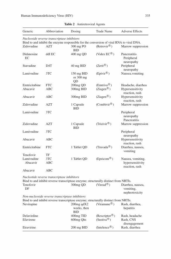
Human Immunodeficiency Virus (HIV) 335
Table 2 Antiretroviral Agents
Generic Abbreviation Dosing Trade Name Adverse Effects
Nucleoside reverse transcriptase inhibitorsBind to and inhibit the enzyme responsible for the conversion of viral RNA to viral DNA.Zidovudine AZT 300 mg PO
BID(Retrovir R©) Marrow suppression
DidanosineEC
ddI EC 400 mg QD (Videx EC R©) PancreatitisPeripheralneuropathy
Stavudine D4T 40 mg BID (Zerit R©) Peripheralneuropathy
Lamivudine 3TC 150 mg BIDor 300 mgQD
(Epivir R©) Nausea,vomiting
Emtricitabine FTC 200mg QD (Emtriva R©) Headache, diarrheaAbacavir ABC 300mg BID (Ziagen R©) Hypersensitivity
reaction, rashAbacavir ABC 300mg BID (Ziagen R©) Hypersensitivity
reaction, rashZidovudine AZT 1 Capsule
BID(Combivir R©) Marrow suppression
Lamivudine 3TC PeripheralneuropathyPancreatitis
Zidovudine AZT 1 CapsuleBID
(Trizivir R©) Marrow suppression
Lamivudine 3TC Peripheralneuropathy
Abacavir ABC Hypersensitivityreaction, rash
Emtricitabine FTC 1 Tablet QD (Truvada R©) Diarrhea, nausea,vomiting
Tenofovir TFLamivudine
Abacavir3TCABC
1 Tablet QD (Epzicom R©) Nausea, vomiting,hypersensitivityreaction, rash
Abacavir ABC
Nucleotide reverse transcriptase inhibitorsBind to and inhibit reverse transcriptase enzyme; structurally distinct from NRTIs.Tenofovir
DF300mg QD (Viread R©) Diarrhea, nausea,
vomiting,nephrotoxicity
Non-nucleoside reverse transcriptase inhibitorsBind to and inhibit reverse transcriptase enzyme; structurally distinct from NRTIs.Nevirapine 200mg qdX2
weeks, thenBID
(Viramune R©) Rash, diarrhea,hepatitis
Delavirdine 400mg TID (Rescriptor R©) Rash, headacheEfavirenz 600mg Qhs (Sustiva R©) Rash, CNS
disengagementEtravirine 200 mg BID (Intelence R©) Rash, diarrhea

336 F. Romanelli
Table 2 (continued)
Generic Abbreviation Dosing Trade Name Adverse Effects
Protease inhibitorsBind to and inhibit protease enzyme. Protease enzyme normally cleaves and activates HIV
pro-proteins.Saquinavir
(Hard gel)500mg BID
(boosted)(Invirase R©) Nausea, vomiting,
diarrheaRitonavir 600mg BID (Norvir R©) Multiple drug
interactions, GIdistress, perioraltingling
Indinavir 800mg Q8h (Crixivan R©) Nephrolithiasis,increased bilirubin
Nelfinavir 1250mg BID (Viracept R©) Diarrhea, nauseaFosamprenavir 700mg BID
(variable)(Lexiva R©) Nausea, vomiting,
diarrheaLopinavir/Ritonavir 2 Capsules
BID(Kaletra R©) GI upset
Atazanavir 400mg QD (Reyataz R©) Increased bilirubin,decreased lipids
Tipranavir 500mg BID(boosted)
(Aptivus R©) GI upset
Darunavir 600 mg BID(boosted)
(Prezista R©) GI upset
Receptor antagonists inhibitorsPrevent receptor medicated fusion between HIV and CD4+ cell receptors.Enfuvirtide 90mg SQ BID (Fuzeon R©) Injection site
reactionsMaraviroc
(CCR5)150 mg PO
BID (varied)(Selzentry R©) Cardiovascular,
increased risk ofmalignancy?
Integrase inhibitorsBind and inhibit the integrase enzyme. Integrase is responsible for the integration of viral DNA
into host cell DNA.Raltegravir 400 mg PO
BID(Isentress R©) Diarrhea, N/V
Multi- class combinationsEmtricitabine
TenofovirEfavirenz
FTCTFSustiva R©
1 Tablet Qhs (Atripla R©) Diarrhea, nausea,vomiting, CNSdisengagement
inhibit viral DNA synthesis but do not become incorporated into the growing viralDNA chain. Protease inhibitors (PIs) are powerful antiretrovirals that inhibit HIVreplication by binding to viral proteases and preventing the proteolytic activation ofHIV polyproteins. Enfuvirtide is the sole commercially available fusion inhibitor,and it acts to inhibit HIV by preventing receptor-mediated binding of the virus toCD4+ cells. While efficacious, the drug is limited by its high cost and subcutaneousroute of administration. Recently, two new classes of antiretroviral drugs have beenintroduced (e.g., co-receptor antagonists and integrase inhibitors). Currently only

Human Immunodeficiency Virus (HIV) 337
one drug is available in each of these two new classes. Maraviroc (Selzentry R©) isa CCR5 receptor antagonist intended for use in patients who are treatment expe-rienced [22, 23]. Maximum efficacy of the agent would be expected in cases ofinfection with a CCR5-specific strain of HIV. Therefore, a tropic assay to deter-mine the co-receptor specificity of any particular strain is required prior to use ofthe drug [24]. Research and development toward the integrase class has been ongo-ing for several years and was previously hindered by the significant adverse effectsoften encountered by experimental agents in this class. Raltegravir (Isentress R©)represents the first and only integrase inhibitor. Similar to maraviroc, raltegraviris intended for use in treatment-experienced patients. The drug has few interactionsand a very favorable side effect profile [25].
The initiation of antiretroviral therapy in newly infected patients is undertakenon a case-by-case basis with tailoring to the patient’s medical condition, desire fortherapy, ability to adhere to complex medical regimens, and co-morbid conditions.Generally, therapy should be initiated when the CD4+ cell count falls below 350cells/mm3 [5]. Previously viral load values had been a consideration for the initi-ation of therapy; however, CD4+ cell counts are now considered the most criticalparameter.
Furthermore, ongoing studies are underway to determine if initiation of therapyat higher CD4+ counts may be associated with better outcomes. Clinical guidelinesfor the management of HIV also stress quality-of-life issues surrounding the initia-tion of antiretroviral therapy [5]. Antiretroviral therapy may be associated with sig-nificant adverse effects that contribute to overall morbidity. Decisions about when itis appropriate to initiate antiretroviral therapy should always involve patient-specificfactors. Current consensus regarding the initiation of therapy focuses on preservingquality of life by balancing starting medications at a point where immune function ismaximally preserved with consideration of side effects, lifestyle impact, and costs.
The efficacy of antiretroviral drug regimens is monitored using viral load assays.Viral load is a method of quantifying viral burden by measuring the number of cir-culating viral RNA strands in the bloodstream. Standard viral load testing has alower threshold of 50 copies/ml, below which test results will be reported as “unde-tectable.” Standard viral load testing can be performed by one of two methods:polymerase chain reaction (PCR) or branch chain DNA (bDNA). Because thesetwo methods can produce differing results, the same testing method should be usedconsistently in any given patient. An undetectable viral load is a reflection of goodviral control; however, it does not indicate that a patient is “cured” or no longercontagious.
Early in the epidemic, antiretroviral monotherapy was considered the standard ofcare in the management of HIV. However, because of the rapid development of resis-tance, monotherapy has been shown to result in inadequate and even detrimentaleffects. Current standard of care advocates the use of potent multi-drug combina-tions [5]. Most commonly these combination regimens are composed of two NRTIsand one PI or two NRTIs plus the NNRTI efavirenz. Once combination antiretro-viral therapy is initiated, the goal of treatment should be an undetectable viral load(<50 copies/ml) [5].

338 F. Romanelli
Research has shown that the development of antiretroviral resistance is signif-icantly less likely to occur when patients achieve and maintain undetectable viralloads [5, 26]. In cases where a previously undetectable viral load has increasedor where viral load is unresponsive to therapy, patients should be started on a newmulti-drug regimen, ideally composed of antiretrovirals to which the patient is treat-ment naïve. When patients have exhausted all standard regimens, clinicians shouldconsider “salvage therapy” with fusion inhibitors or various alternative drug combi-nations. Addition of a single new additional drug to a failing regimen is inadequateand often yields the same results as monotherapy. In cases of drug toxicity or adverseeffects, it is appropriate to substitute for the offending agent with another drug ofthe same class [5]. Drug doses should not be adjusted in response to adverse effectsexcept in cases of renal insufficiency.
The success of current and future combination antiretroviral therapy is highlydependent upon patient adherence [27, 28]. Adherence is a challenge for many HIV-infected patients, due to various factors including pill burdens, drug costs, adverseeffects, and lifestyle impacts. When initiating antiretroviral therapy and selectingspecific drug regimens, clinicians should consider potential changes to the patient’scurrent lifestyle as well as the quality of life [5]. In counseling HIV-infected patients,clinicians should be especially certain to emphasize the importance of strict adher-ence to prescribed regimens. Clinicians should also be prepared to offer practicaladvice and suggestions to enhance the understanding and adherence of HIV-infectedpatients.
Drug interactions are a concern when managing this patient population.Interactions which might result in sub-therapeutic antiretroviral drug concentra-tions should always be considered as they may result in the premature developmentof resistance. Management of drug interactions in HIV-infected individuals is aparticular challenge because of the large numbers of medications used for themanagement of the virus itself as well as for the prophylaxis and treatment of oppor-tunistic infections. Appropriate references should always be consulted to identifyand evaluate potential drug interactions in this population [5, 29].
Many patients placed on potent multi-drug antiretroviral regimens achieveand maintain undetectable viral loads. While sustained undetectable viral loadsare achieved in large numbers of patients, it appears as though some level ofvirus remains dormant in lymph nodes, CNS tissue, and other stealth cellularcompartments [30]. In cases where antiretroviral medications have been withdrawn,these previously dormant virions begin once again to actively replicate. Therefore,until further trials are completed, it is not feasible to discontinue antiretroviralmedications even after long-term sustained undetectable viral loads.
Recognizing that most antiretroviral regimens require stringent adherence andmay be associated with severe adverse effects, some researchers have experimentedwith the concept of structured treatment interruptions (STIs) of antiretroviral drugtherapy. STIs involve planned and limited interruptions in antiretroviral drug therapyin patients with undetectable viral loads. Conceptually, STIs are based on a premisethat drug discontinuation in a patient with controlled viremia may result in an “auto-inoculation” against HIV. Unfortunately, most trials examining the results of STIs

Human Immunodeficiency Virus (HIV) 339
have not demonstrated any discernable efficacy in this regard and are associatedwith the risk of the induction of resistance and future treatment failures. Therefore,STIs are currently not a recommended therapeutic intervention.
The search for new and effective antiretroviral medications continues. Mostof the research in this area has focused on developing new antiretrovirals whichtarget receptors/co-receptors or unique viral enzymes such as integrase. Effortshave also been made to simplify existing dosing regimens in order to enhanceadherence [31, 32]. Examples of combination antiretroviral product intended toease dosing are Truvada R©, which contains 300 mg of tenofovir and 200 mg ofemtricitabine per tablet, and Epzicom R©, which contains 300 mg of lamivudineand 600 mg of abacavir per tablet. Perhaps the most significant recent additionto the antiretroviral armamentarium is the combination product Atripla R©, whichcontains 300 mg of emtricitabine, 300 mg of tenofovir, and 600 mg of efavirenzper tablet. Atripla R© represents the first single-dose, triple-drug tablet intended forthe management of HIV.
8 Resistance Testing
A controversial issue in the management of HIV-infected individuals has been therole of antiretroviral resistance testing (Table 3) [27]. Antiretroviral resistance test-ing can be accomplished either genotypically or phenotypically. Genotyping relatesresistance to either the presence or the absence of specific mutations at the geneticsites encoding for various HIV proteins including reverse transcriptase and pro-tease. Phenotyping more closely resembles traditional antibiotic sensitivity testing
Table 3 Resistance testing: comparison of genotypic and phenotypic resistance assays
Relative advantages Relative limitations
Genotypic Ease of availabilityShorter time to results (days)Less technically demandingMutations will likely precede
phenotypic resistanceLess costly when compared to
phenotyping
Indirect measure of susceptibilityMay not correlate directly with
phenotypeExpert interpretation requiredInsensitive for the detection of minor
speciesReliance upon known mutations in
mapped areas of the HIV genomeLack of laboratory standardization
Phenotypic Direct measure of susceptibilityMore familiar reporting results (IC50
or IC90)
Limited availabilityLonger time to results (weeks)Technically demandingInsensitive for detecting minor speciesClinically significant breakpoints
undefinedLack of laboratory standardizationCostly

340 F. Romanelli
as it involves live viral cultures and varying drug concentrations. Many inherentlimitations to resistance testing exist (i.e., cost, lack of interpretation guidelines,lack of laboratory standardization.). Despite these limitations, most clinicians agreethat antiretroviral resistance testing should play some role in the management ofHIV-infected patients. Clinical guidelines support the use of resistance testing incertain situations including use before initiating therapy in treatment-naïve patientsand to manage cases of antiretroviral treatment failure [5]. There is no consensus onwhich assay (genotypic or phenotypic) should be employed by clinicians. The useof either test should be undertaken with an understanding of the limitations of eachtesting modality and the high cost of this technology. Phenotypic assays, which aremore costly, might be better reserved for more complicated patients with extensivehistories of antiretroviral exposure.
9 Conclusion
Since its first reported US cases in the early 1980s, clinicians and researchers alikehave learned a great deal about HIV. AIDS has forced sensitive issues such assexuality, discrimination, and death into the auspices of many clinical practices.As researchers continue the search for an effective cure, clinicians are helpingseropositive patients lead healthier, more productive lives. The advent of potentantiretrovirals has dramatically changed the prognosis of this disease. Chronic man-agement and longer life expectancies are now a reality; however, these are notwithout costs. Antiretroviral therapy often compromises quality of life and placestremendous financial burdens on patients. Therapy is always limited by the potentialdevelopment of efficacy-compromising resistance mutations. Additionally, medica-tions must be taken indefinitely as they do not represent a “cure.” These advanceshave primarily been realized in industrialized nations, highlighting inequities aroundthe globe. Even in the United States, the disproportionate impact of HIV/AIDS onminority populations reflects disturbing social disparities.
Complacency would likely result in a rapid return to the desperate situations oftenencountered in the late 1980s and the early 1990s. Research must continue in thearea of drug and vaccine development. Effective and safe medications that inhibitnovel enzyme targets will be needed if the advances seen over the last 20 years areto continue. Equally important, clinicians must continue to treat HIV-seropositivepatients with empathy and caring, always considering the widespread implicationsof HIV disease on the patient’s quality of life.
Key Points
• HIV is most commonly transmitted via sexual fluids, exposure to blood or bloodyfluids, and via injection drug use.
• HIV infects and destroys CD4+ cells, leaving patients susceptible to a number ofserious opportunistic infections (OI). Prophylactic antibiotics are often employedto protect susceptible individuals from various OIs.

Human Immunodeficiency Virus (HIV) 341
• Access to antiretroviral drug therapy in industrialized nations has resulted infewer HIV-infected patients progressing to AIDS. Therapy typically involves theuse of three concurrent antiretroviral agents.
• Antiretroviral therapy for HIV infection does not represent a cure, requires life-long treatment, and has several limitations including high cost, multiple adverseeffects, and a low ceiling to the development of resistance.
• As life expectancies of HIV-infected patients continue to increase, the provi-sion of primary care to optimize overall health for these patients will becomeincreasingly critical.
• Antiretroviral resistance is a common cause of treatment failure. Resistance canbe delayed and/or prevented with stringent adherence to drug regimens. Thepresence of resistance can be detected through the use of either genotypic orphenotypic resistance testing.
References
1. CDC (1981). Pneumocystis pneumonia – Los Angeles. MMWR 30:250–2522. Broder S, Fauci AS (1988). Progress in drug therapies for HIV infection. Public Health Rep
103:224–2293. CDC (2003). HIV/AIDS surveillance report, Vol. 154. Kahn JO, Walker B (1998). Acute human immunodeficiency virus type 1 infection. N Engl J
Med 339:33–395. Panel on Antiretroviral Guidelines for Adult and Adolescents. Guidelines for the use
of antiretroviral agents in HIV-infected adults and adolescents (2007). Department ofHealth and Human Services. December 1, 2007; 1–101. (Accessed on Dec 1, 2007http://aidsinfo.nih.gov/contentfiles/AdultandAdolescentGL.pdf)
6. Treating Opportunistic Infection Among HIV-Infected Adults and Adolescents (2004).Recommendations from the CDC, NIH, and HIV Medicine Association. (Accessed on Feb16, 2007 http://www.aidsinfo.nih.gov/ContentFiles/TreatmentofOI_AA.pdf)
7. Anonymous (2006). CDCs global AIDS outlook gives preview of WHO report. Prevalencedeclines in some hot spots. AIDS Alert 21:3–4
8. Anonymous (2006). The global HIV/AIDS pandemic. MMWR 55(31):841–8449. Katz MH, Gerberding JL (1998). The care of persons with recent sexual exposure to HIV.
Ann Intern Med 128:306–31110. Gallo MF, Grimes DA, Lopez LM et al. (2006). Non-latex versus latex male condoms for
contraception. Cochrane Database Syst Rev 25:CD00355011. Sperling RS, Stratton PA, O’Sullivan MJ et al. (1992). A survey of the use of zidovudine
in pregnant women with human immunodeficiency virus infection. New Engl J Med 326:857–861
12. Connor EM, Sperling RS, Gelber R et al. (1994). Reduction of maternal-infant transmissionof human immunodeficiency virus type 1 with zidovudine treatment. New Engl J Med 331:1173–1180
13. Shapshak P, McCoy CB, Shah SM et al. (1994). Preliminary laboratory studies of inactivationof HIV-1 in needles and syringes containing infected blood using undiluted household bleach.J Acqui Immune Defic Syndr 7:754–759
14. Gerberding JL (1996). Prophylaxis for occupational exposure to HIV. Ann Intern Med125:497–501
15. Anonymous (1995). Case control study of HIV seroconversion in health-care workers afterpercutaneous exposures to HIV-infected blood-France, United Kingdom, United States,January 1988-August 1994. MMWR 44:929–933

342 F. Romanelli
16. Gerberding JL (1995). Management of occupational exposures to blood-borne viruses. NewEngl J Med 332:444–451
17. Anonymous (2005). Updated US Public Health Service Guidelines for the Managementof Occupational Exposures to HIV and Recommendations for Postexposure Prophylaxis.MMWR 55:RR–9
18. Anonymous (2005). Antiretroviral postexposure prophylaxis after sexual, injection-drug use,or other nonoccupational exposure to HIV in the United States. MMWR 54:RR–2
19. Delaney KP, Bransom BM, Uniyal A et al. (2006). Performance of an oral rapid HIV-1/2 test:Experience from four CDC studies. AIDS 120:1655–1660
20. Branson BM, Handsfield HH, Lampe MA et al. (2006). Revised recommendations forHIV testing of adults, adolescents, and pregnant women in health-care settings. MMWR55(RR14):1–17
21. Havlir DV, Richman DD (1996). Viral dynamics of HIV: Implications for drug developmentand therapeutic strategies. Ann Intern Med 124:987–994
22. Repik A, Richards KH, Clapham PR (2007). The promise of CCR% antagonists as newtherapies for HIV-1. Curr Opin Investig Drugs 8:130–139
23. Biswas P, Tambussi G, Lazzarin A (2007). Access denied? The status of co-receptor inhibitionto counter HIV entry. Expert Opin Pharmacother 8:923–933
24. Meanwell NA, Kadow JF (2007). Maraviroc, a chemokine CCR5 receptor antagonist for thetreatment of HIV infection and AIDS. Curr Opin Investig Drugs 8:669–681
25. Markowitz M, Nguyen BY, Gotuzzo E et al. (2007). Rapid and durable antiretroviral effectof the HIV-1 Integrase inhibitor raltegravir as part of combination therapy in treatment-naivepatients with HIV-1 infection: Results of a 48-week controlled study. J Acquir Immune DeficSynd 46:125–133
26. Bangsberg DR, Kroetz DL, Deeks SG (2007). Adherence-resistance relationship to combina-tion HIV antiretroviral therapy. Curr HIV/AIDS Rep 4:65–72
27. Hirsch MS, Conway B, D’Aquila RT et al. (1998). Antiretroviral drug resistance testing inadults with HIV. J Am Med Assoc 279:1984–1991
28. Friedland GH (1997). Adherence: The achilles’ heal of highly active antiretroviral therapy.Improving the management of HIV disease. Int AIDS Soc USA 5:13–15
29. Tseng AL, Foisy MM (1997). Management of drug interactions in patients with HIV. AnnPharmacother 31:1040–1058
30. Schrager LK, D’Souza P (1998). Cellular and anatomical reservoirs of HIV-1 in patientsreceiving potent antiretroviral combination therapy. J Am Med Assoc 280:67–71
31. Machtinger EL, Bangsberg DR (2007). Seven steps to better adherence: A practicalapproachto promoting adherence to antiretroviral therapy. AIDS Read 17:43–51
32. Killingley B, Pozniak A (2007). The first once-daily single-tablet regimen for the treatmentof HIV-infected patients. Drugs Today 43:427–442

Part IIISpecial Considerations

Strategies for Optimal Antimicrobial Use
Arch G. Mainous III and Paul Little
1 Introduction
Optimalantimicrobialuse is essential in the face of escalating antibiotic resistance.The problem of antibiotic resistance affects all sectors of the health-care system –the patient, the health-care team, the payer, and the public health system. Previousantibiotic use has consistently been identified as a risk factor for individual coloniza-tion with resistant pneumococcus [1]. Community-wide consumption of antibioticsis strongly associated with infection or colonization with resistant organisms [2].Antibiotic resistance has been shown to be proportional to the volume of antimicro-bial consumption, and reductions in resistance require a proportional reduction inconsumption [3, 4]. In fact, in a recent double-blind placebo-controlled trial com-paring placebo with the macrolides azithromycin and clarithromycin, it showedthe direct effect of antibiotic exposure on resistance in the oral streptococcal florain healthy volunteers [5]. Both macrolides significantly increased the carriage ofmacrolide-resistant streptococci compared with the placebo in these healthy adults.
The overall goal of reducing the prescribing of antibiotics should be an effortto minimize antibiotic resistance while appropriately delivering quality health care.There are some data to indicate that decreasing antibiotic use does lead to decreasedresistance. In Finland, a nationwide reduction in the outpatient use of macrolideantibiotics resulted in a reduction of resistant group A streptococcus from 16.5 to8.6% within 4 years. Practitioners were regulated to substitute macrolides for otherantibiotics, and although macrolide use decreased, overall antibiotic use did notchange [6]. Icelandic researchers have reduced the proportion of resistant pneumo-coccal infections through an intervention program delivered to patients and to thehealth-care team [7]. Effective strategies must be identified, and all groups mustpromote and participate in efforts to reduce antibiotic resistance. Otherwise, thecurrent concern of a “post-antimicrobial era,” in which antimicrobial agents will no
A.G. Mainous III (B)Department of Family Medicine, Medical University of South Carolina, Charleston, SC, USAe-mail: [email protected]
345A.G. Mainous III, C. Pomeroy (eds.), Management of Antimicrobials in InfectiousDiseases, Infectious Disease, DOI 10.1007/978-1-60327-239-1_18,C© Springer Science+Business Media, LLC 2001, 2010

346 A.G. Mainous III and P. Little
longer be effective, may become a reality. We will present a discussion of a varietyof strategies that have been used to promote judicious antibiotic use.
1.1 Controlling Antibiotic Prescribing
In order to identify strategies to reduce antibiotic overuse and resistance, contribut-ing factors must be understood (see Table 1). Overuse of antibiotics may relate tomisinformation on the part of the practitioner and patient. For example, when pre-sented scenarios that were consistent with an upper respiratory tract infection withdiscolored nasal discharge, the physicians, the pharmacists, and the patients werelikely to prescribe, to recommend, and to desire antibiotics [8, 9]. Diagnostic andprognostic uncertainty may contribute to physicians prescribing antibiotics primar-ily for viral infections in order to cover the “chance” of bacterial infection or ofcomplications. Clinical guidelines have been developed to improve diagnostic cer-tainty with the hopes of therefore improving the management of common infections[10, 11]. Other strategies have focused on using biomarkers like C-reactive proteinor procalcitonin to provide an indication as to whether an acute respiratory infec-tion is likely to be viral or bacterial. In one trial, procalcitonin-guided therapy usedas an adjunct to guidelines reduced antibiotic use without compromising patientoutcomes [12].
As resolution of symptoms may serendipitously correlate with the course ofantibiotics, patients’ past use of antibiotics, for self-limiting illnesses, may alsoinfluence prescribing. Barriers to changing provider behavior include satisfyingpatient expectations without decreasing patient-care productivity. The patient edu-cation required to dispel the myth of antibiotic necessity is time consuming, andphysicians are often fearful that the lack of prescription may negatively affect
Table 1 Factors contributing to overuse of antibiotics
Contributing factor Explanation
Experience Provider: habit of antibiotic prescribing initiated in the pre-antibioticresistance era
Patient: perceived efficacy of prior antibiotic therapy for viralinfections
Lack of education Provider: suboptimal approach to diagnosis and treatment of commonviral and bacterial infections
Patient: belief that all infections require antibiotic therapyExpectations Provider: belief that patients expect an antibiotics prescription, and
that lack of prescription will damage the relationshipPatient: expectation that antibiotics are effective for common viral
infectionsEconomics Provider: time required to explain the lack of need for antibiotic
therapy, importance of patient satisfactionPatient: lost productivity during illness, need to return to work, need to
return child to day care

Strategies for Optimal Antimicrobial Use 347
the doctor–patient relationship [13]. However, one study that examined the rela-tionship between the amount of time spent by a physician in an encounter fora presumed viral respiratory infection when antibiotics are or are not prescribedshowed no significant difference [14]. Patients often request antibiotics for paramed-ical reasons such as upcoming examinations or prophylactically for travel, and timeconstraints often make it easier for practitioners to prescribe than to explain why notto prescribe.
When considering pathogens causing nosocomial infections, improving inpa-tient antimicrobial use and infection control practices are necessary. Hospitals andtheir intensive care units are common sites for the breeding of resistant bacteria,which require more expensive antibiotics, lead to prolonged hospitalization, cre-ate an increase in cost of care, and an increase in morbidity and mortality [15].Several guidelines and clinical strategies have been published for optimizing antibi-otic use and curtailing antibiotic resistance in hospitals [16, 17]. The Centersfor Disease Control and Prevention have produced a wide variety of tools forpreventing antibiotic resistance in health-care settings which provide guidancefor hospitalized adults, for children, for long-term care, for surgical patients,and for dialysis patients (http://www.cdc.gov/drugresistance/healthcare/default.htmaccessed on 2009). Although these guidelines must be tailored to each hospital sys-tem to target specific problems of antimicrobial resistance, local circumstances, andresources, there is some evidence that compliance with the guidelines will lead tolower levels of resistance [18].
1.2 Quality Improvement Initiatives
Over the past decade, numerous strategies have been implemented to curb antibioticoveruse. A recent systematic review of 55 separate trials indicated that broad-basedinterventions could have a community level impact on antibiotic use of 17–117prescriptions per 1,000 person years [19]. Quality improvement efforts have beeneffective at reducing antibiotic use in ambulatory settings, but much room forimprovement remains. The conclusion was that targeting all acute respiratory infec-tions rather than single conditions in single age groups would have the greatestimpact.
Despite the considerable amount of money spent on clinical research, relativelylittle attention has been given to ensuring that the findings of research are imple-mented in routine clinical practice. In order to implement changes in medical care,it is important to focus on interventions promoting change and to target interventionsto the appropriate audience. Traditional teaching methods (i.e., didactic lectures andcontinuing medical education) and other forms of passive dissemination of informa-tion (i.e., recommendations for clinical care, clinical practice guidelines, audiovisualmaterials, and electronic publications) have not been shown effective in changingphysician behavior [19, 20]. Provision of drug-cost information in the computer-ized patient record has not been shown to affect overall mean prescription cost orprescribing patterns [21].

348 A.G. Mainous III and P. Little
On the other hand, systematic reviews have documented that educational out-reach visits, computerized and manual reminders, and interactive educationalmeetings (participation of health-care providers in workshops that include dis-cussion or practice) are effective in promoting behavioral changes among healthprofessionals [19]. Educational outreach visits, also known as academic detailing,use a combination of techniques to improve physicians’ clinical decision mak-ing [22, 23]. Multifaceted interventions, which include a combination of auditand feedback, reminders, local consensus processes, and/or marketing are con-sistently effective interventions [19]. Point-of-care delivery of clinical guidelinesand evidence-based recommendations has been shown to impact clinical practice[24]. Peer education (physician to physician) to improve quality of care and toreduce the cost of antibiotic prescribing was effective in office practices [25]. Amultidisciplinary continuous improvement approach has been shown to increaseclinical prevention efforts and to improve the delivery of diabetes care [26, 27].Unfortunately, these more effective methods are not routinely used in medical edu-cation [28]. In a recent meta-analysis of continuing medical education trials, passivemethods for disseminating information were shown to have a small impact onphysician performance [29]. The most effective interventions are interactive, usemultiple methods, and are designed for a small group of physicians from a singlediscipline.
Physicians and other health-care providers claim awareness of the problem ofantibiotic resistance, which has been created by the overuse of antibiotics. However,given the fact that inappropriate antibiotic use continues, this simple awareness isnot sufficient to affect prescribing behavior. Several strategies have been applied toimprove antibiotic prescribing in the outpatient and inpatient settings; these strate-gies include focusing on the system, on the provider, and on the patient (see Table 2).These strategies are often implemented in closed health-care systems and consist oftightly controlled interventions. However, when these interventions are applied toa less restrictive system and to a wider array of prescribers maintaining efficacyremains a challenge.
1.3 Antibiotic Control Policies
Clinical guidelines have been produced to control the use of antibiotics in hospitalsystems [16]. Antibiotic control programs are implemented in an effort to opti-mize antibiotic use while minimizing antibiotic costs [30]. The success of theseprograms depends on cooperation of multidisciplinary teams, including hospitaladministrators, clinicians, infectious diseases specialists, infection control teams,microbiologists, and hospital pharmacists [15]. All team members must promotebasic hospital infection control practices such as hand washing. Pharmacists andinfection control teams are involved in monitoring drug use, in the surveillance andin the reporting of antimicrobial resistance patterns, and in the detection of patientscolonized with potentially resistant and communicable bacteria. Antibiotic control

Strategies for Optimal Antimicrobial Use 349
Table 2 Strategies to promote optimal use of antimicrobial agents
Administrativeinterventions
Denial of claims for inappropriate useFinancial incentives and penalties
Antibiotic control programs Antibiotic order formsAutomatic stop dates (limiting and optimizing duration of use)Restriction for specific indicationsRestriction of specific classesImproved diagnostic techniques
Point-of-care decisionsupport
Computer-assisted management programs
Provider education Academic detailingPeer educationLocal opinion leadersInteractive educational meetings
Patient intervention Public/media campaignsDelayed prescription
Clinical guidelines Locally developed with input from participating physiciansDeveloped by an outside party (governmental agency, medical
society, etc.)Audit and feedback Generated and delivered by members of the health-care team
Generated and delivered by an outside party (e.g., governmentalagency, health-care organization)
Multifaceted intervention Quality improvement initiative involving educational interventions(to provider and patient), audit and feedback, etc.
programs include antibiotic order forms developed by a team of infectious disease“experts,” which reflect preferred dosing intervals. Programs may limit the durationof antibiotic therapy (automatic stop dates) or institute restrictions on antibiotic usefor specific indications. Many antibiotic restriction programs require clinical justifi-cation for the specific antibiotic order prior to dispensing by the pharmacy. A recentstudy examined the impact on antibiotic use in three different units of a hospitalwhen guided by an infectious disease specialist [31]. Antibiotic consumption wasrecorded yearly from 1998 to 2005 in the three units. On Unit A, after approval bythe head of the unit with the infectious disease specialist involved as a consultantupon request, restricted antibiotics were prescribed. On Unit B, restricted antibioticshad to be approved by an infectious disease specialist; all other antibiotics were pre-scribed by the physicians on the ward. On Unit C, all the antibiotics were prescribedby an infectious disease specialist. In Unit C, a significant decreasing trend in antibi-otic consumption, in defined daily doses per patient day and per admission, and costof antibiotics per patient was observed; in Unit B, a decreasing trend in antibioticconsumption per patient was also seen.
Antibiotic control policies are usually institutional interventions, creating bar-riers to inappropriate practices and limiting prescriber autonomy. Administrativeinterventions may also come from governmental agencies, which enforce specificpractices by laws, by regulations, or by recommendations. These policies oftenrequire added personnel and must be maintained indefinitely to continue to achievedesired results. Success of antibiotic control policies depends on the definition

350 A.G. Mainous III and P. Little
of success. Antibiotic order forms are effective in controlling antibiotic use andreducing antibiotic costs. Antibiotic restriction policies are effective in alteringspecific resistance patterns. When held to a more important definition of success,such impact on overall resistance patterns and overall patient outcomes, antibi-otic control policies have not been appropriately evaluated. Finally, the “hasslefactor” of administrative interventions may create dissatisfaction among practicingphysicians.
1.4 Computer-Assisted Decision Support
Integration of data from the microbiology laboratory, from the pharmacy, from themedical record, and from the financial databases can assist physicians to makedecisions in a timely fashion. Antimicrobial susceptibility data, pharmacokineticinformation about the individual patient, specific patient factors, and financial dataof antimicrobial choices can be presented to the physician at the point of care andin an effort to improve antimicrobial prescribing, cost, and patient outcomes [32].Electronic clinical decision support might help in both diagnosis and treatmentdecisions. Although, few studies have directly addressed the question of elec-tronic decision support to improve antibiotic prescribing beyond a few small pilotstudies, those studies suggest that clinical decision support may hold a particularpromise [33, 34]. Recent data from a trial of a stand-alone personal digital assistant(PDA)-based clinical decision support system for the diagnosis and managementof acute respiratory tract infections in the outpatient setting and overall adherencewith an electronic decision support system for recommendations for the five mostcommon diagnoses (pharyngitis, otitis media, sinusitis, bronchitis, and upper res-piratory tract infection) were 82% [35]. When antibiotics were prescribed (in 53%of cases), adherence with the decision support system-recommended antibiotic washigh (76%).
Antibiotic costs have been significantly reduced using a computer-assisted man-agement program for antibiotics in a small intensive care unit [36]. The computerprogram recommends antibiotic regimens and courses of therapy for individualpatients and provides immediate feedback to the provider at the point of care. Duringa 1-year intervention period, in 545 patients managed in the intensive care unit, therewas a documented improvement in quality of patient care and in medication costswhen compared to retrospective data. Decreases were noted in medications adminis-tered to patients with known allergies, excess drug dosages, antibiotic-susceptibilitymismatches, mean number of days of excessive drug dosages, and adverse eventscaused by antibiotics. In addition, cost of antibiotics was reduced threefold as weretotal hospital costs and hospital length of stay. The advantages of the computerizeddecision support tool were demonstrated in this study and allowed for more effi-cient data retrieval. Physicians therefore had more time available to them to spendon other medical decisions. However, in systems where the use of computer systemsis less prevalent, this intervention might be costly and less effective.

Strategies for Optimal Antimicrobial Use 351
1.5 Provider Education
Many systematic reviews have shown that passive dissemination of information, orpassive education, is generally ineffective in changing physician practice [19, 28].A more important measure, of course, would be to evaluate the impact of physicianeducation on patient health outcomes, but these data are scarce. When educationis tailored to change specific behaviors, and tailored to specific providers or situa-tions, this type of intervention is more effective. Interactive educational meetings,where providers participate in workshops that include discussion or demonstrationof skills, have been consistently effective [19]. In addition, educational outreachvisits or academic detailing has been effective, particularly when the visit is con-ducted by a peer or local opinion leader. To ensure success, academic detailingactivities must include several techniques, including assessment of baseline knowl-edge of each provider, identification of local opinion leaders, and use of positivereinforcement of improvements in clinical practice [23].
In a now classic study, investigators evaluated the effect of three educationalmethods on antibiotic prescribing in office practices in Tennessee [25]. The threeeducational methods included a mailed brochure, a 15-min visit by a pharmacist(drug educator), and a 15-min visit by a physician counselor. Topics of educationalactivities were three antibiotics contraindicated for office practice (chlorampheni-col, clindamycin, and tetracycline for children < 8 years old) as a measure of thequality of care and the use of oral cephalosporins as a measure of the cost of care.Based on their use of the mentioned antibiotics, 372 physicians were selected forthe interventions. The mailed brochure had no measurable effect on prescribing,and the pharmacist visit had only a modest effect. The physician visit correspondedwith a subsequent 44% reduction in patients receiving contraindicated drugs, and a21% reduction in patients receiving and in prescriptions for oral cephalosporins.Therefore, the intervention waseffective in improving the quality of care and inreducing the cost of care, particularly when the message was delivered by a peer.Further studies of academic detailing activities have demonstrated a benefit of physi-cian visits and small group education over simple mailing of educational materials[22, 37, 38].
The advantages of academic detailing programs and education outreach visitsinclude the ability to tailor the discussion to the learner’s level of understandingand to his or her scope of practice. However, programs are very dependent on thepeer educator’s abilities as well as the physician’s active participation in the discus-sion. Since most improvement initiatives involve some sort of educational program,the positive aspects of academic detailing programs should be highlighted whenpossible. More practically speaking, however, the effectiveness of an educationalintervention is greatly limited by the size of the population in which the change isdesired. When implementing a change in a small group of physicians, the cost andtime involved in academic detailing may be worth the investment. However, it isimpractical to think that an academic detailing program could be implemented in alarge provider group such as a health maintenance organization or a state health-careplan. The cost to send a drug educator (either peer physician or pharmacist) to meet

352 A.G. Mainous III and P. Little
with each provider in the system would be exorbitant, and even if effective, wouldonly affect the prescribing of the target clinical question such as antibiotic pre-scribing for common infections. Considering the number of target clinical questionsthat could be subject for improvement, this method becomes even more impracticaloutside the research environment but could potentially be addressed by interactivecomputer- or web-based systems.
1.6 Clinical Guidelines
Variation and uncertainty regarding the appropriate treatment of common medicalconditions, including infectious diseases, may be reduced by adhering to evidence-based clinical guidelines. Clinical practice guidelines are defined as “consensusstatements that are systematically developed to assist practitioner and patient deci-sions about appropriate health care for specific clinical circumstances” [39]. Practiceguidelines are often used in educational interventions and have gained increas-ing popularity as a means of influencing physicians’ practice patterns. Despitetheir popularity, clinical practice guidelines have failed to, in most circumstances,dramatically change practice patterns.
The treatment of infectious diseases, like other common conditions, has beeninundated with the development of clinical practice guidelines. In July of 2009, theNational Guideline Clearinghouse, a public resource of the Agency for HealthcareResearch and Quality listed 242 guidelines under the heading of bacterial infectionsand mycoses (http://www.guideline.gov/ accessed on 2009). Further, there were 81guidelines just for respiratory tract infections. This mass quantity of guidelinesmakes it very hard for providers to be compliant, and they may become somewhatcynical of outside entities providing guidelines on which they are unfamiliar as away to evaluate their practice patterns.
The impact of clinical practice guidelines has been disappointing and may beaccounted for by the belief from physicians that they are not written for practic-ing physicians but rather focus on the current state of scientific knowledge [40].Physicians also may disagree or distrust guidelines written by governmental agen-cies or “experts” from other institutions and are more apt to adopt clinical guidelineswhen they or their peers are involved in development. Finally, physicians fear theimplications of clinical practice guidelines on nonclinical factors such as liabilityand financial incentives from payers. The cost of developing and of implementingclinical practice guidelines should not be dismissed. Delivery of clinical practiceguidelines is probably best accomplished during academic detailing exercises withpeer educators rather than by passive dissemination (i.e., mass mailings).
In six urban and rural hospitals, a practice guideline for the management ofcommunity-acquired pneumonia (in low-risk patients) was evaluated, with theobjective to switch from intravenous to oral therapy within 3 days and to dischargepatients within 4 days of hospitalization [41]. Guidelines were developed by theresearch team and physicians and nurses were selected from each hospital as “cham-pions” to endorse the use of the guideline. Despite multidisciplinary involvement in

Strategies for Optimal Antimicrobial Use 353
dissemination of the practice guideline through educational sessions, the practiceguideline had no effect on length of stay or on the patient outcomes.
In a Midwestern staff-model health maintenance organization, clinical guide-lines for the treatment of uncomplicated cystitis were developed by local physiciansand each clinical practice site determined their own process for guideline imple-mentation [42]. The goal of the cystitis clinical practice guideline was to reducetreatment duration, to reduce the use of urine cultures, and to increase the numberof nurse-coordinated cases, all without compromising patient outcomes. Use of theguideline was associated with desirable changes in antibiotic use, in nurse coordina-tion of care, and in cost of care with comparable clinical outcomes. Therefore, thisguideline designed with local input and tailored implementation was successful inachieving a desirable change in care of patients with uncomplicated cystitis.
Sometimes, practice guidelines can have the unintended effect of modifying prac-tice in a way that makes it difficult for providers to deliver care. One study examinedemergency physician (EP) understanding of the Centers for Medicare and MedicaidServices (CMS) core measures for community-acquired pneumonia (CAP) guide-lines and sought to assess the impact on their antibiotic prescribing patterns [43].Nearly all of the physicians correctly understood the time-based guidelines forantibiotic administration, reported institutional commitment to meet these core mea-sures, and stated that they had a department-based CAP protocol. Importantly, morethan half of the respondents (55%) reported prescribing antibiotics to patients theydid not believe had pneumonia in an effort to comply with the CMS guidelines. Only40% of these physicians indicated a belief that the guidelines improve patient care.
With the rising concern of appropriate antibiotic use, there will inevitably be anincreased number of clinical practice guidelines for infectious diseases. As practi-tioners, it will be important to evaluate their impact on the process of care, on thepatient-care outcomes, and, more importantly, on the antibiotic resistance patterns.Advantages of using clinical practice guidelines in educational efforts include theability to target those who need the education and the mostly the streamlining ofcare. However, the cost and labor intensity of implementation is not minor. In addi-tion, the ability of these techniques to succeed outside the research environment isyet to be seen.
1.7 Audit and Feedback
Audit and feedback, or any summary of clinical performance, has had variablesuccess on promoting changes in physician behavior [19]. Audit of physicianperformance requires a strict review of their practice activities for a given clinicalcondition or specific delivery of service. Feedback implies a report on their spe-cific practices or patient outcomes and a comparison of this feedback to an externalsource. The external source may be a published clinical practice guideline, a nationalaverage, or the “best practice” among their specific physician group practice [40]. Inorder for an audit and feedback loop to be effective, physicians must first recognizethat their practice needs to be improved. Obviously, providers requesting feedback

354 A.G. Mainous III and P. Little
and recognizing a need for change are more likely to be successful in implementinga change. Next, the person receiving the feedback must be able to act on the feed-back. In addition, feedback should be provided at the point of care, or prospectively,during the providers participation in the care system. Retrospective feedback onwhat “should have been done” is less effective than feedback that can affect changein future patient-care activities.
To demonstrate the effect of unsolicited, retrospective feedback on prescribing,the Australian government conducted a randomized controlled trial [20]. Feedbackon 2 years of prescribing was provided to more than two thousand practitioners, intwo sets of graphical displays over 6 months. Prescribing patterns were comparedto peer prescribing and were related to five main drug groups. The interventiongroup also received educational newsletters related to the drug groups. The controlgroup received no information on their prescribing. There were no demonstrablechanges in prescribing in the intervention group when compared to the controlgroup – feedback did not affect the variability in prescribing. Therefore, this unso-licited government sponsored feedback data had no impact on individual physicianpractices.
From the other extreme, a group of physicians performed an internal audit andfeedback examining their use of antibiotics for the treatment of otitis media [44].Their examination was based on data presented in the Cochrane review for antibi-otic use in otitis media, and they found significant antibiotic prescribing for thiscommon condition [45]. In their practice, they implemented a new system of carefor otitis media including a patient handout, an analgesic treatment, and a deferredprescription for amoxicillin. A similar practice acted as the control group in whichstandard treatment regimens were continued. During the 12-month interventionperiod, antibiotic prescriptions fell 32% in the intervention practice, but only 12% inthe control practice. This successful intervention reflects the physicians’ readinessfor change and their practice culture and the internal development of a new systemof care – which may be difficult to extrapolate to other practice settings.
When efforts to change are borne from within those requiring the change ratherthan imposed from an outside organization, the likelihood of success is greatlyincreased. Physicians may perceive an outside audit with feedback as a threat totheir clinical competence, self-esteem, or autonomy [40]. Allowing physicians todetermine the external standard for comparison of clinical audits and providingprospective feedback at the point of care are important factors for successful auditand feedback interventions.
1.8 Delayed Prescriptions
Delayed antibiotic prescribing is one promising method of promoting judiciousantibiotic use in primary care [46, 47]. Delayed prescribing refers to the strategywhere a clinician provides an antibiotic prescription to be used by the patient butonly if symptoms are not starting to settle in the time frame expected for the nat-ural history or if symptoms are getting significantly worse during that time frame.

Strategies for Optimal Antimicrobial Use 355
Systematic reviews 1:2 suggest delayed prescribing can reduce antibiotic prescrip-tions (with relative risk ranging from 0.25 to 0.54), which has been confirmedoutside trial settings in a longitudinal observational study from primary care [11,46, 48]. In a review of the six studies that supplied data on the proportion of patientswho filled the antibiotic prescription, the post-intervention median rate of antibioticuse in the study control groups was 75% compared with 37.5% in the interventiongroups [19]. However, the question arises why not just avoid prescribing antibi-otics altogether when the patient presents with an apparently uncomplicated illness?There are two important reasons: First, we have relatively poor information aboutwho is at risk of subsequently developing rare but important complications of infec-tions – delayed prescribing is one way of providing extremely rapid access toantibiotics if symptoms are unexpectedly getting significantly worse (i.e., a rapidsafety net). Second, the prescribing of antibiotics allows a ready compromise wherethere are very high patient expectations for antibiotics.
However, there are potential disadvantages of delayed prescribing. There is thepossibility of mixed messages about the role of antibiotics – but if clear guidance isgiven about when to use antibiotics this should not be a great problem, since beliefsabout antibiotics, when delayed prescribing is used, are very similar to beliefs whenno offer of antibiotics is made [47, 49]. There are some data that suggest a decreasein patient satisfaction with delayed prescriptions [46]. If patients expect antibioticsand are denied them by their physician, then there are data to suggest that theycan obtain them without a prescription [50, 51]. Thus, evidence-based guidance topatients is particularly important. A second issue is the possibility of an increasedrisk of complications. The data on clinical trials on delayed prescribing suggestcomplications are very rare. Patients should also be given clear advice about thenatural history and the importance of returning if there are signs of developingcomplications.
1.9 Multifaceted Interventions
Multifaceted interventions are described as those that combine audit and feedback,point-of-care reminders, local input in clinical guideline development, and the sup-port of local opinion leaders [19]. Multifaceted interventions may also be referredto as continuous quality improvement (CQI) initiatives and are often attractive tophysicians for several reasons: First, the focus lies on improving the delivery andquality of health care, rather than on individual physician behaviors or the bottomline of cost [40]. Second, there is no mandate of change in individual physician prac-tice but rather a focus on the efficiency of delivery of care. Many health systems areimplementing CQI activities in specific areas of patient care and clinical service, butfew randomized clinical trials exist to document the benefit of this approach.
A large study that examined a multifaceted approach in the outpatient settingfocused on improving the treatment of uncomplicated acute bronchitis in adults[52]. Four office practices were selected for the study: one practice was provided

356 A.G. Mainous III and P. Little
with a full intervention, one practice received a limited intervention, and two prac-tices served as the control sites. In the full intervention site, household educationalmaterials were mailed to all patients (i.e., magnets, pamphlets, or a letter fromthe medical director of the practice) regarding appropriate management of com-mon infections. Office-based educational material specific for acute bronchitis wasdelivered to the office for the examination rooms. Clinicians were detailed on thepatient education activities included in the intervention, and they were providedwith antibiotic prescribing rates for acute bronchitis at their site during the previouswinter. They participated in an interactive educational session on evidence-basedmanagement of acute bronchitis, and how to say “no” to patient demands for antibi-otics. These sessions were led by the medical director and the opinion leader ofeach practice and were attended by all disciplines. In the limited intervention site,office-based educational materials were distributed to the nursing manager at thepractice, and they were displayed in the patient examination rooms. The controlsites provided usual care.
The study was conducted over a 3-month period, with baseline data from thesame months of the prior year. Although antibiotic prescribing rates were similaramong the four practices in the baseline period, prescribing fell significantly at theintervention site (from 74 to 48%), but, during the study period, it did not change inthe control or limited intervention site. Prescriptions for nonantibiotic medications(i.e., bronchodilators) did not differ among sites nor did return office visits for bron-chitis or pneumonia. Therefore, in this focused intervention, antibiotic utilization foracute bronchitis improved in one office practice using a multifaceted intervention.
Ideally, using a multifaceted intervention that is tailored to each practice groupwould be the ideal way to improve the system of care. Realistically, however, thecontinued success of such programs in everyday clinical practice is less likely, par-ticularly when the focus of interventions expands to include other acute and chronicillnesses. In addition, the generalizability of this controlled intervention to a widerprescribing community requires further study. Furthermore, there have been fewstudies addressing the overall impact on patients – either symptomatic outcomes orthe development of complications.
2 Conclusions
Although specific antibiotic selection and restriction policies in the hospital settingare important in altering microbial susceptibility patterns, an overall reduction inantibiotic use in a wider population and in the outpatient setting is more likely tosignificantly impact antibiotic resistance. Education of providers, the developmentand implementation of clinical practice guidelines, audit and feedback activities,delayed prescribing strategies, and multifaceted interventions have all demonstratedan effect in altering antibiotic prescribing in a research setting. However, the abilityto translate these research activities into clinical practice and on a wider basis affectantibiotic use has not been consistently accomplished. Addressing antibiotic use andresistance is one of the most urgent priorities in confronting emerging infectious

Strategies for Optimal Antimicrobial Use 357
disease threats [53]. All providers must examine their own practices to identify howthey can reduce unnecessary antimicrobial use. Professional societies, health-careorganizations, and the Centers for Disease Control and Prevention must also beinvolved. With partnerships and cooperation of members of the health-care teams,the effectiveness of currently available antibiotics may be sustained and the threatof antibiotic resistance minimized.
Key Points
• Physicians and other health-care providers claim awareness of the problem ofantibiotic resistance, created by the overuse of antibiotics. However, given thefact that inappropriate antibiotic use continues, this simple awareness is notsufficient to affect prescribing behavior.
• Antibiotic control policies, delayed prescription, decision support models, aca-demic detailing, clinical guidelines, audit and feedback, and multifaceted inter-ventions have documented efficacy in altering antibiotic prescribing in controlledhealth-care settings.
References
1. Dowell SF, Schwartz B (1997). Resistant pneumococci: Protecting patients through judicioususe of antibiotics. Am Fam Physician 55:1647–1654
2. Schneider-Lindner V, Delaney JA, Dial S, et al. (2007). Antimicrobial drugs and community-acquired methicillin-resistant Staphylococcus aureus, United Kingdom. Emerging Infect Dis13:994–1000
3. Austin DJ, Kristinsson KG, Anderson RM (1999). The relationship between the volume ofantimicrobial consumption in human communities and the frequency of resistance. Proc NatlAcad Sci 96:1152–1156
4. van de Sande-Bruinsma N, Grundmann H, Verloo D, et al. (2008). European AntimicrobialResistance Surveillance System Group. European Surveillance of Antimicrobial ConsumptionProject Group. Antimicrobial drug use and resistance in Europe. Emerg Infect Dis 14:1722–1730
5. Malhotra-Kumar S, Lammens C, Coenen S, et al.(2007). Effect of azithromycin and clar-ithromycin therapy on pharyngeal carriage of macrolide-resistant streptococci in healthyvolunteers: A randomized, double-blind, placebo-controlled study. Lancet 369:482–490
6. Seppala H, Klaukka T, Vuopio-Varkila J, et al. (1997). The effect of changes in the consump-tion of macrolide antibiotics on erythromycin resistance in group A streptococci in Finland.N Engl J Med 337(7):441–446
7. Stephenson J (1996). Icelandic researchers are showing the way to bring down rates ofantibiotic-resistant bacteria. J Am Med Assoc 275(3):175–176
8. Mainous AG III, Hueston WJ, Eberlein C (1997). Colour of respiratory discharge andantibiotic use. Lancet 350(9084):1077
9. Mainous AG III, Zoroob RJ, Oler MJ, et al. (1997). Patient knowledge of upper respiratoryinfections: Implications for antibiotic expectations and unnecessary utilization. J Fam Pract45(1):75–83
10. Dowell SF, Schwartz B, Phillips WR (1998). Appropriate use of antibiotics for URIs in chil-dren: Part I. Otitis media and acute sinusitis. The Pediatric URI Consensus Team. Am FamilyPhys 58(5):1113–1118

358 A.G. Mainous III and P. Little
11. Tan T, Little P, Stokes T (2008). Antibiotic prescribing for self limiting respiratory tractinfections in primary care: Summary of NICE guidance. BMJ 337:232–233
12. Briel M, Schuetz P, Mueller B, et al. (2008). Procalcitonin-guided antibiotic use vs a stan-dard approach for acute respiratory tract infections in primary care. Arch Intern Med 168:2000–2007
13. Butler CC, Rollnick S, Pill R, et al. (1998). Understanding the culture of prescribing:Qualitative study of general practitioners’ and patients’ perceptions of antibiotics for sorethroats. BMJ 317(7159):637–642
14. Coco A, Mainous AG, (2005). Relation of time spent in an encounter with the use of antibi-otics in pediatric office visits for viral respiratory infections. Arch Pediatr Adolesc Med159:1145–1149
15. Struelens MJ (1998). The epidemiology of antimicrobial resistance in hospital acquiredinfections: Problems and possible solutions. BMJ 317:652–654
16. Yokoe DS, Mermel LA, Anderson DJ, et al. (2008). A compendium of strategies to pre-vent healthcare associated infections in acute care hospitals. Infect Control Hosp Epidemiol29:S12–S21
17. Brouqui P, Puro V, Fusco FM, et al. (2009). Infection control in the management of highlypathogenic infectious diseases: Consensus of the European Network of Infectious Disease.Lancet Infect Dis 9:301–311
18. Larson EL, Quiros D, Giblin T, et al. (2007). Relationship of antimicrobial control policies andhospital and infection control characteristics to antimicrobial resistance rates. Am J CriticalCare 16:110–120
19. Ranji SR, Steinman MA, Shojania KG, et al. (2008). Interventions to reduce unneces-sary antibiotic prescribing: A systematic review and quantitative analysis. Med Care 46:847–862
20. O’Connell DL, Henry D, Tomlins R, (1999). Randomised controlled trial of effect of feedbackon general practitioners’ prescribing in Australia. BMJ 318:507–511
21. Ornstein SM, MacFarlane LL, Jenkins RG, et al. (1998). Medication cost information in acomputer-based patient record system: Impact on prescribing in a family medicine clinicalpractice. Arch Fam Med 8:118–121
22. Avorn J, Soumerai SB, (1983). Improving drug-therapy decisions through educationaloutreach. N Engl J Med 308:1457–1463
23. Soumerai SB, Avorn J, (1990). Principles of educational outreach (“Academic Detailing”) toimprove clinical decision making. J Am Med Assoc 263:549–556
24. Headrick LA, Speroff T, Pelecanos HI, (1992). Efforts to improve compliance with theNational Cholesterol Education Program guidelines. Results of a randomized controlled trial.Arch Intern Med 152(12):2490–2496
25. Schaffner W, Ray WA, Federspiel CF, et al. (1983). Improving antibiotic prescribing inoffice practice. A controlled trial of three educational methods. J Am Med Assoc 250(13):1728–1732
26. Geiger WJ, Neuberger MJ, Bell GC, (1998). Implementing the US preventive servicesguidelines in a family practice residency. Fam Med 25:447–451
27. Fox CH, Mahoney MC, (1998). Improving diabetes preventive care in a family practiceresidency program: A case study in continuous quality improvement. Fam Med 30(6):441–445
28. Davis DA, Thomson MA, Oxman AD, et al. (1995). Changing physician performance. Asystematic review of the effect of continuing medical education strategies. J Am Med Assoc274:700–705
29. Mansouri M, Lockyer J, (2007). A meta-analysis of continuing medical education effective-ness. J Continuing Educ Health Profess 27:6–15
30. Lohr KN, Brook RH, Kaufman MA, (1979). Quality of care in the New Mexico Medicaidprogram (1971–1975): The effect of the New Mexico Experimental Medical Care ReviewOrganization on the use of antibiotics for common infectious diseases. Med Care 18:1–129

Strategies for Optimal Antimicrobial Use 359
31. Beovic B, Kreft S, Seme K, et al. (2009). The impact of total control of antibiotic prescrib-ing by infectious disease specialist on antibiotic consumption and cost. J Chemotherapy 21:46–51
32. Mason WH (1998). Strategies to promote appropriate antimicrobial use. Pediatr Infect Dis J17(8):747–748
33. Linder J, Schnipper JL, Volk LA, et al. (2007). Clinical decision support to improve antibi-otic prescribing for acute respiratory infections: Results of a pilot study. AMIA. . . AnnualSymposium Proceedings/AMIA Symposium 468–472
34. Linder JA, Jung E, Housman D, et al. (2007). The Acute Respiratory Infection QualityDashboard: A performance measurement tool in an electronic health record. AMIA. . . AnnualSymposium Proceedings/AMIA Symposium 1035
35. Rubin MA, Bateman K, Donnelly S, et al. (2006). Use of a personal digital assistant for man-aging antibiotic prescribing for outpatient respiratory tract infections in rural communities. JAm Med Informatics Assoc 13:627–634
36. Evans RS, Pestotnik SL, Classen DC, et al. (1998). A computer assisted management programfor antibiotics and other antiinfective agents. N Engl J Med 338:232–238
37. DeSantis G, Harvey KJ, Howard D, et al. (1994). Improving the quality of antibiotic pre-scription patterns in general practice. The role of educational intervention. Med J Aust160:502–505
38. Ekedahl A, Andersson SI, Hovelius B, et al. (1995). Drug prescription attitudes and behaviorof general practitioners. Effects of a problem-oriented educational programme. Eur J ClinPharmacol 47:381–387
39. Woolf SH, (1990). Practice guidelines: A new reality in medicine: I. Recent developments.Arch Intern Med 150:1811–1818
40. Greco PJ, Eisenberg JM, (1993). Changing physicians’ practices. N Engl J Med 329:1271–1274
41. Rhew DC, Riedinger MS, Sandhu M, (1998). A prospective, multicenter study of a pneumoniapractice guideline. Chest 114(1):115–119
42. O’Connor PJ, Solberg LI, Christianson J, et al. (1996). Mechanism of action and impact of acystitis clinical practice guideline on outcomes and costs of care in an HMO. Jt Comm J QualImprov 22(10):673–682
43. Nicks BA, Manthey DE, Fitch MT (2009). The Centers for Medicare and Medicaid Services(CMS) community-acquired pneumonia core measures lead to unnecessary antibiotic admin-istration by emergency physicians. Acad Emergency Med 16:184–187
44. Cates C, (1999). An evidence based approach to reducing antibiotic use in children with acuteotitis media: Controlled before and after study. BMJ 318:715–716
45. Glasziou PP, Hayem M, Del Mar CB (1997). Antibiotic versus placebo for acute otitis mediain children. In: Cochrane Collaboration. Cochrane Library. Issue I. Oxford: Update Software
46. Spurling G, Del Mar C, Dooley L, et al. (2007). Delayed antibiotics for symptoms andcomplications of respiratory infections. Cochrane Database Syst Rev CD00417
47. Little P, Rumsby K, Kelly J, et al. (2005). Information leaflet and antibiotic prescribing strate-gies for acute lower respiratory tract infection: A randomized controlled trial. J Am MedAssoc 293:3029–3035
48. Sharland M, Kendall H, Yeates D et al. (2005). Antibiotic prescribing in general practiceand hospital admissions for peritonsillar abscess, mastoiditis, and rheumatic fever in children:Time trend analysis. BMJ 331:328–329
49. Little P, Gould C, WilliamsonI, et al. (1997). Reattendance and complications in a randomisedtrial of prescribing strategies for sore throat: The medicalising effect of prescribing antibiotics.BMJ 315:350–352
50. Mainous AG III, Everett CJ, Post RE, et al. Availability of antibiotics for purchase without aprescription on the Internet. Ann Fam Med 2009; 7(5):431–435
51. Mainous AG III, Cheng AY, Garr RC, et al. (2005). Non prescribed antimicrobial drugs inLatino community, South Carolina. Emerg Infect Dis 11:883–888

360 A.G. Mainous III and P. Little
52. Gonzales R, Steiner JF, Lum A, et al. (1999). Decreasing antibiotic use in ambulatory practice:Impact of a multidimensional intervention on the treatment of uncomplicated acute bronchitisin adults. J Am Med Assoc 281:1512–1519
53. Spellberg B, Guidos R, Gilbert D, et al. (2008). The epidemic of antibiotic-resistant infections:A call to action for the medical community from the Infectious Disease Society of America.Clin Infect Dis 46:155–164

Infections in the Immunocompromised Host
Cristina Baker and Vicki A. Morrison
1 Introduction
Infection is one of the primary causes of morbidity and mortality in the immuno-compromised host. Among those who are at greatest risk for infection due to defectsin immunity are patients with cancer or HIV infection, neutropenic individuals,recipients of solid organ (SOT) and hematopoietic stem cell transplants (HSCT),and those whose underlying conditions require treatment with TNF-α inhibitors.Successful therapy for these conditions must be directed not only at managing theunderlying disease but also at preventing and treating complications. In the fol-lowing chapter, the predisposing factors and potential infectious complications ofthese disorders will be presented, as will the changing epidemiology of these infec-tions. Finally, a general approach to prophylaxis and treatment of specific infections,including those caused by resistant organisms, will be provided.
2 The Cancer Patient
2.1 Fever in the Cancer Patient
Over two-thirds of patients with malignancy will have a fever at sometime duringtheir clinical course. Fever in the cancer patient may occasionally be caused by thetumor itself. Malignancies most likely to cause fever include lymphoma, renal can-cer, and hepatic cancer. About 10% of patients with metastatic disease, particularlythose with liver metastases, may also have fever. More commonly, however, feverin the cancer patient is attributable to infection.
C. Baker (B)Staff Physician, Infectious Diseases, Hennepin County Medical Center, University of Minnesota,Minneapolis, MN, USAe-mail: [email protected]
361A.G. Mainous III, C. Pomeroy (eds.), Management of Antimicrobials in InfectiousDiseases, Infectious Disease, DOI 10.1007/978-1-60327-239-1_19,C© Springer Science+Business Media, LLC 2001, 2010

362 C. Baker and V.A. Morrison
2.2 Immunodeficiencies and Associated Malignancies
Risk factors for infection in cancer patients are related both to humoral and cellularimmune defects that are inherent to the primary disease process and to immunosup-pression caused by therapy for the malignancy. Several hematologic malignancieshave a characteristic spectrum of infection as a result of specific immune defects.
2.2.1 Chronic Lymphocytic Leukemia (CLL)
Patients with chronic lymphocytic leukemia (CLL) have a variety of immunedefects, the most critical of which is hypogammaglobulinemia [1]. This deficit ismore common and profound with advanced-stage disease and is not reversible bytherapy, even with a concomitant hematologic response. The most common sites ofinfection are not only mucosal-lined surfaces, especially the respiratory tract, butalso the urinary tract and skin/soft tissue.
Bacterial pathogens most frequently isolated in these patients includeStaphylococcus aureus, Streptococcus pneumoniae, Haemophilus influenzae, andvarious enteric gram-negative organisms such as Escherichia coli and Klebsiellapneumoniae. Fungal (including Candida and Aspergillus) and viral infections (espe-cially herpesviruses) are much less common, most often occurring in patients withadvanced stage disease and/or chemotherapy-induced neutropenia. However, theintroduction of purine analog therapy has altered the spectrum of infection becausethese drugs cause defects in cell-mediated immunity [2]. Pathogens seen in the set-ting of purine analog therapy include mycobacterial species, Listeria, Nocardia,Aspergillus, herpesviruses, and Pneumocystis.
2.2.2 Multiple Myeloma
Impaired humoral immunity is the hallmark of multiple myeloma (MM).Hypogammaglobulinemia is prominent in these patients, resulting from bothdecreased synthesis and increased catabolism of immunoglobulins. As in CLL, theimmunoglobulin levels decline with disease progression. However, in contrast toCLL, an improvement in immunoglobulin levels may accompany a hematologicresponse to therapy. Humoral responses to immunization are also deficient in thesepatients. The mechanisms of these humoral immune defects are primarily related toimpairment in the number and function of the residual polyclonal B cells.
Strep. pneumoniae and other encapsulated organisms such as H. influenzae andNeisseria meningitidis are commonly implicated in infection patients with multiplemyeloma and fungal and viral infections are less frequent. Infections with entericgram-negative organisms, Candida, and Aspergillus are common in the setting ofneutropenia, most often occurring in heavily pretreated multiple myeloma patientsand in those with extensive marrow involvement. As in CLL, there is a predilectionfor these infections to occur at mucosal surfaces.

Infections in the Immunocompromised Host 363
2.2.3 Hairy Cell Leukemia
Cell-mediated immune defects, specifically T-lymphocyte dysfunction, are promi-nent in hairy cell leukemia. Quantitative and qualitative defects in granulocytesand monocytes are also present. Typically, infections are caused by commongram-positive and gram-negative bacteria and occur at mucosal sites and withinskin or soft tissue. However, disseminated infections with not only opportunis-tic organisms – especially atypical mycobacteria – but also Candida, Aspergillus,Pneumocystis, and cytomegalovirus (CMV) are also seen, related to the monocy-topenia that characterizes this disorder.
2.2.4 Hodgkin’s Lymphoma
Patients with Hodgkin’s lymphoma have underlying defects in cell-mediated immu-nity, predisposing them to infections caused by isolates such as Listeria, Candida,herpesvirues, and Pneumocystis. Strep. pneumoniae bacteremia may occur in thesetting of prior, extensive combined-modality therapy or relapse. Lastly, in the smallnumber of patients who now undergo staging laparotomy with splenectomy, infec-tions caused by encapsulated organisms such as Strep. pneumoniae, H. influenzae,and N. meningitidis may occur, more commonly in children than in adults.
2.2.5 Myelodysplastic Syndrome and Large Granular Lymphocytic Leukemia
Neutropenia is common in myelodysplastic syndrome (MDS) and large granularlymphocytic leukemia (LGLL), the latter being a T-lymphocyte disorder. In patientswith MDS, bacterial pneumonia and skin abscesses are common infections [3].Recurrent bacterial infections, especially involving the skin, sinuses, and perirec-tal area, complicate the course of patients with LGLL. Organisms typically foundin the setting of LGLL include Staph. aureus and Pseudomonas aeruginosa, withopportunists being less frequent.
2.3 Infection Based on Location of Malignancy
In addition to specific immune defects related to the primary disease process, thelocation of the malignancy may be crucial in predisposing to infectious complica-tions. Obstruction of the respiratory tract, gastrointestinal (GI) tract, genito-urinary(GU) tract, and biliary tract by a tumor mass can result in infection. Central nervoussystem (CNS) dysfunction resulting from a primary or metastatic CNS tumor or car-cinomatous / lymphomatous meningitis may predispose to infections. The loss of agag reflex can lead to aspiration and pneumonia. Likewise, a neurogenic bladder canresult in urinary tract infections. Infections in these settings are most often caused bythe usual colonizing organisms at that site. However, nosocomial infections, some-times with multidrug-resistant organisms, are of great significance in hospitalized

364 C. Baker and V.A. Morrison
patients and those patients receiving broad-spectrum antibiotic therapy with subse-quent suppression of normal colonizing flora. In these settings, empiric treatmentshould be broadened to cover for potentially resistant organisms until culture resultsare available.
2.4 Infection Related to Treatment
Therapy administered for a malignancy can lead to additional immunosuppression.Cytotoxic chemotherapy may result in disruption of normal barriers to infection(skin, mucosa) or immune defects, including neutropenia, which has its own uniquespectrum of infections (see next section). Specific chemotherapeutic agents, such asthe purine analogs, result in T-lymphocyte defects that may persist for over a yearafter discontinuation of therapy. Corticosteroids used in the therapy of malignanciescan cause qualitative defects in phagocyte and in monocytes/macrophages function.Radiation therapy can lead to damage of the normally intact, protective mucosalsurfaces of the respiratory, GI, and GU tracts and to defects in cellular immunity.Alemtuzumab, used to treat CLL and lymphoma, results in profound defects in cel-lular immunity with significant reductions in B, T, and NK cells developing shortlyafter initiation of therapy and persisting for at least 9 months after discontinuationof treatment.
A variety of iatrogenic infections may arise in these patient populations. Theplacement of central lines for venous access or monitoring purposes, indwellingurinary catheters, and stents in obstructed regions such as the biliary tree may pre-dispose to infection. Infections involving central venous access devices (CVAD)include local exit site infection, catheter tunnel infection, or catheter-related blood-stream infections. These infections are most commonly caused by staphylococcalspecies, with Staphylococcus epidermidis being the most frequent isolate in cases ofcatheter-related bacteremia. Integumentary defects from venipuncture, intravenousline placement, or bone marrow biopsy allow the introduction of colonizing skinflora, including nosocomial pathogens, past normally intact barriers, resulting inlocal or systemic infection.
2.5 Management of Infections in Cancer Patients
2.5.1 Antimicrobial Prophylaxis
Both preventive and therapeutic approaches to infectious complications have beenexamined in the cancer population. According to practice guidelines outlined bythe National Comprehensive Cancer Network (NCCN), no bacterial or fungal pro-phylaxis is recommended for patients receiving standard chemotherapy for solidtumors and whose episode of neutropenia is expected to last for less than 7 days.Viral prophylaxis in this population is only recommended in patients with priorHSV episodes. In patients with hematologic malignancies such as CLL and multiple

Infections in the Immunocompromised Host 365
myeloma that result in humoral immune dysfunction, bacterial prophylaxis is rec-ommended. Updated guidelines for prophylaxis for the cancer patient can be foundon the National Comprehensive Cancer Network website (www.nccn.org) [4].
2.5.2 Myeloid Growth Factors and Intravenous Immunoglobulin
Myeloid growth factors are advocated for use in cancer patients receiving myelosup-pressive chemotherapy, which is likely to result in at least a 20% incidence of febrileneutropenia. The American Society of Clinical Oncology has devised guidelines forthe optimal use of these agents for primary and secondary prevention of neutrope-nia [5]. Because of hypogammaglobulinemia present in some disorders, the use ofimmunoglobulin replacement has been examined, but studies have not supported itsrole in the therapy of established infections.
2.5.3 Antimicrobial Therapy
The general approach to antimicrobial therapy for infections in cancer patientsshould be based on an understanding of the underlying immune defects andcorresponding predisposition for infections with specific organisms, as well asrecognition of the local colonizing flora. Knowledge of local resistance patternsof nosocomial pathogens is important to consider, and clinicians should be aware ofthe hospital antibiograms for the institutions in which they practice.
2.5.4 Management of Catheter-Related Infections
Catheter-related infections are unfortunately common in patients with cancer.Whereas infection along the catheter tunnel generally requires catheter removalfor cure, exit-site infections can usually resolve with appropriate antimicrobialtherapy. The majority of catheter-related bacteremias can be successfully treatedwithout catheter removal. However, in the setting of persistent bacteremia, it maybe necessary to remove the catheter to eradicate the infection, particularly if infec-tion is due to isolates such as Corynebacterium jeikeium, P. aeruginosa, Bacillus,atypical mycobacteria, or Candida species. Guidelines regarding treatment ofcatheter-related infections have been established by the IDSA (www.idsociety.org)[6].
3 The Neutropenic Patient
3.1 Sources of Neutropenia
Myeloablative therapy is increasingly utilized for patients with various types of can-cer. As a result, a larger proportion of cancer patients will experience at least oneepisode of therapy-related neutropenia, which may last from days to several weeks.Infection risk is related not only to the absolute neutrophil count (ANC) but also to

366 C. Baker and V.A. Morrison
the rate of decline in the ANC and the duration of neutropenia. The risk of infec-tion rises with an ANC of less than 1,000/mm3, but increases exponentially as theANC declines to <500/mm3. The definition of fever in this population has been arbi-trarily defined as a single oral temperature of ≥38.3◦C (101◦F). At least one-halfof the febrile neutropenic patients are found to have either an established infec-tion or an occult infection. Over 20% of those with profound neutropenia (ANC <100/mm3) will be bacteremic. Signs and symptoms of infection may be masked inthese patients, making infection diagnosis more difficult. A major factor in predict-ing a successful outcome from infection in the neutropenic host is recovery of theANC.
3.2 Etiologic Agents of Infection
In addition to the skin, infections in neutropenic patients most commonly occurin the oral cavity, upper/lower respiratory tract, gastrointestinal tract, and perirec-tal area. The most common etiologic agents of infection are the organisms thatnormally colonize these sites. Use of broad-spectrum antimicrobial agents in thesepatients has an impact on the colonizing flora, with resultant eradication of the nor-mal flora at mucosal sites, particularly anaerobes, and the potential for colonizationand overgrowth by nosocomial and/or drug-resistant isolates such as methicillin-resistant Staph. aureus and vancomycin-resistant enterococci. The majority ofbacterial infections in neutropenic patients are caused by enteric gram-negative (E.coli, K. pneumoniae, P. aeruginosa) and common gram-positive isolates (coagulase-negative staphylococci, Staph. aureus, α-hemolytic streptococci). Fungal infectionsare most frequently caused by Candida, followed by Aspergillus. Fluconazole pro-phylaxis may result in the emergence of isolates such as Candida krusei andCandida glabrata. Reactivation of HSV and other herpesviruses in HSCT patientsis also seen.
3.3 Treatment
In the past three decades, much commentary has been published with regard tothe antimicrobial approach in the neutropenic patient. In 1997, the first in a seriesof practice guidelines was commissioned by IDSA. The updated IDSA guidelinesremain the best reference of the present recommendations for care in these patients(www.idsociety.org) [7]. Considerations for the use of oral antimicrobial prophy-laxis include anticipated profound neutropenia (ANC < 100/mm3), mucositis, severeperiodontal disease, postobstructive infections, or other immune system compro-mise. Adjunctive therapy in the febrile neutropenic patient is also addressed in theseguidelines. The use of empirical antiviral treatment, granulocyte transfusions, ormyeloid growth factors is not routinely recommended.

Infections in the Immunocompromised Host 367
4 Infections in the Hematopoietic Stem Cell TransplantRecipient
4.1 Immunodeficiencies Associated with HSCT
In the HSCT recipient, profound defects in cell-mediated and humoral immunitycontribute to the increased risk for infection. Early in the post-transplant period,prolonged neutropenia occurs secondary to myelosuppression from the preparativeregimen. Defects in both B- and T-lymphocyte function are pronounced in the allo-geneic transplant recipient. Graft-versus-host disease (GVHD), as well as therapyrendered for this complication, results in increased immunosuppression, includ-ing subnormal immunoglobulin production and a hyposplenic state (with chronicGVHD). Lastly, the use of T-cell depleted marrow products results in pronouncedand prolonged deficiencies in CD3+, CD4+, and CD8+ T lymphocytes. The use ofperipheral blood stem cell products and the ancillary use of myeloid growth factorshave been found to result in a shorter time to engraftment, thus a briefer period ofneutropenia and less risk for infectious complications.
4.2 Etiologic Agents of Infection
There is a characteristic spectrum of post-transplant infectious complications inHSCT patients, related to the interval following transplantation [8]. The firstmonth, post-transplant, is characterized by marrow aplasia with marked neutrope-nia. Integumentary defects, such as those from mucositis and CVADs, representportals of entry for colonizing organisms. Total body irradiation, when utilized,results in additional cellular immune deficiency. Bacterial infections caused byvarious common gram-positive isolates (Staph. aureus, coagulase-negative staphy-lococci, viridans streptococci) and less commonly gram-negative enteric organismspredominate in this period. In patients who are seropositive for HSV, a high rateof reactivation may occur. BK virus reactivation can lead to bladder infection andhemorrhagic cystitis.
The spectrum of infection changes in the interval from 1 to 3 months, post-transplant, as engraftment with resolution of neutropenia has generally occurredby this time [9]. Acute GVHD may have its onset in allogeneic recipients duringthis period. Major pathogens causing infection during this time include CMV, aden-ovirus, Aspergillus, other non-Candida fungi, VZV, and human herpes virus-6 [10].
Following day 100, post-transplant, chronic GVHD may complicate the courseof allogeneic transplant recipients. The incidence of pneumococcal bacteremiarises dramatically due to the functional asplenic state induced by chronicGVHD. Bacterial sinopulmonary infections, especially with encapsulated organ-isms, become more common. The incidence of VZV infections, mostly dermatomal,also increases. Infections with CMV and Aspergillus continue to occur through thisphase [11].

368 C. Baker and V.A. Morrison
4.3 Prophylaxis and Treatment
Prophylactic antimicrobial use in the HSCT population has been extensively studied[12]. Currently, the NCCN recommends that a fluoroquinolone, an anti-mold agent,and an antiviral agent be given as prophylaxis to all allogenic HSCT recipients.Because HSCT recipients who are CMV seropositive are at risk for CMV reactiva-tion, the use of either CMV-negative or filtered blood products has been advocatedin seronegative patients to prevent acquisition. In addition, prophylactic gancicloviror preemptive ganciclovir therapy for patients identified as being at high risk forCMV disease by CMV antigen detection or polymerase chain reaction testing isrecommended [13]. In the setting of autologous HSCT, anti-mold prophylaxis isonly recommended in patients with mucositis. Following transplant, patients whohave received alemtuzumab or have GVHD are generally given Pneumocystis pro-phylaxis. The NCCN has developed a complete set of guidelines for prophylaxis inHSCT patients (www.nccn.org).
Routine use of antimicrobial prophylaxis has led to the emergence of distinc-tive pathogens. Ubiquitous fluoroquinolone use has resulted in increased rates ofinfection with α-hemolytic streptococci and fluoroquinolone-resistant strains ofE. coli and P. aeruginosa. Similarly, selective pressure due to fluconazole prophy-laxis has resulted in an emergence of Candida species, such as C. krusei and C.glabrata, which are inherently resistant to fluconazole. Ganciclovir-resistant CMVinfection also has the potential to become an increasing problem in HSCT recipients.
The approach to therapy of established infection is similar to that used forother highly immunocompromised hosts. Due to high rates of infection with drug-resistant organisms, selection of empiric antimicrobial therapy with activity againstextended-spectrum beta-lactamase (ESBL)-producing organisms such as K. pneu-moniae, P. aeruginosa, Enterobacter cloacae, E. coli, MRSA, and VRE should beconsidered in the appropriate clinic scenarios. Recovery of neutropenia is an impor-tant factor, affecting outcome especially with Candida and Aspergillus infection.Guidelines for the treatment of these infections were published by IDSA in 2003 and2000, respectively [14,15]. The Aspergillus guidelines have been updated in 2008,to incorporate recommendations about newer drugs such as the echinocandins andnew azoles (voriconazole, posaconazole).
5 Infections in the Solid Organ Transplant (SOT) Recipient
5.1 Immunodeficiencies in SOT Recipients
Due to defects in both cellular and humoral immunity, the course of SOT recipi-ents is often complicated by infection. However, improvements in graft and patientsurvival and a decline in infection-related mortality have been seen related to betterselection of candidates, progress in the regulation of chronic immunosuppressive

Infections in the Immunocompromised Host 369
therapy, better selection of candidates, improved antimicrobial prophylaxis, andadvances in surgical techniques. Cell-mediated immune defects predominate in theSOT recipient, further exacerbated by immunosuppressive therapy needed follow-ing SOT. Corticosteroids, though now used in lower dosages than in the past, resultin defects in both cell-mediated and humoral immunity. Other immunosuppres-sive agents utilized, such as azathioprine, cyclosporine, tacrolimus, methotrexate,mycophenolate mofetil, sirolimus, and monoclonal anti-T-cell globulins (OKT3,antithymocyte globulin), also result in significant and prolonged immune defects.Additional factors that contribute to the type, severity, and mortality of infection inthe SOT recipient include underlying medical conditions (e.g., diabetes, hepatitis),the specific organ transplanted, and the duration of the surgical transplant procedure.The incidence of infection and subsequent mortality are lowest in renal transplantrecipients and highest in the heart–lung recipients [16–18]. Liver transplant surgeryhas its own attendant complications, related to the length and technical difficulty ofthe procedure and anastomotic connections to nonsterile sites such as the biliary treeand intestine [19].
5.2 Etiologic Agents of Infection
The majority of infections following SOT occur within 4 months after solidorgan transplant. However, a temporal sequence of infections in the post-transplantperiod has been recognized. Most infections in the first month, post-transplant, areeither pre-existent preoperative infections (hepatitis), routine postoperative infec-tions (pneumonia, wound or line-related infections), or HSV reactivation. Variousinfections are seen 2–6 months post-transplant. Bacterial infections may involvesites such as the bladder or sinuses. In addition, opportunistic organisms such asmycobacteria, Nocardia, Listeria, Aspergillus, Cryptococcus, CMV, Toxoplasma,and Pneumocystis may be implicated. In the period of greater than 6 months,post-transplant, three clinical groups with characteristic infectious complicationsare seen. Approximately 60–75% of patients require minimal immunosuppressionand have good graft function at this time. Common infections such as respiratorytract infections, diverticulitis, and cholecystitis may occur in these patients but maypresent in an atypical manner or have more serious sequelae due to the chronicimmunosuppression. From 10 to 15% of patients will have chronic recrudescentviral infections related to long-standing immunosuppression, and these infectionsmay lead to end-organ damage. Etiologies include BK and JC viruses (urethralstricture, hemorrhagic cystitis), hepatitis B or C (subacute or chronic hepatitis),Epstein–Barr virus (post-transplant lymphoproliferative disorders), CMV (espe-cially retinitis), adenovirus, and VZV. The remaining 10–20% of patients whohave poor allograft function and require large amounts of immunosuppressive ther-apy due to acute/chronic allograft rejection are at greatest risk of life-threateningopportunistic infections, with immunomodulatory viruses, such as CMV, beingamong the most common pathogens.
370 C. Baker and V.A. Morrison
The etiologic agents of infection are somewhat dictated by the site of infec-tion in the SOT recipient. Skin infections are commonly due to HSV and VZV,with Papillomavirus and various dermatophytes being less frequent. The skin mayalso represent a target organ for disseminated infection due to a variety of bacte-ria (including atypical mycobacteria), fungi, and viruses. The incidence of woundinfections varies with the type of transplant and is particularly common in livertransplant recipients.
In the era before routine prophylaxis, the incidence of urinary tract infection fol-lowing renal transplantation ranged from 35 to 80%. However, with the institution oftrimethoprim–sulfamethoxazole (TMP–SMX) prophylaxis, the incidence has nowdropped to less than 10%. Common urine isolates include enteric gram-negativeorganisms, Staphylococcus saprophyticus, Enterococcus, and Candida. Less fre-quently isolated are Mycoplasma, Mycobacterium species, Papovavirus (BK, JC),and CMV.
Frequent sources of septicemia in transplant recipients are pathogens originat-ing from the lung, GI or GU tracts, biliary tract, urinary tract, skin/soft tissue, andintravascular catheters. Polymicrobial bacteremia is not uncommon, especially inthe setting of liver, pancreas, or small intestine transplants. Common isolates areenteric gram-negative organisms, Enterococcus, Staph. aureus, Candida species,and CMV.
A variety of pulmonary infections may arise, post-transplant. Community-acquired pneumonia may be caused by traditional organisms such as Strep.pneumoniae or other isolates such as respiratory syncytial virus. Although pul-monary tuberculosis may occur in these patients, many mycobacterial infectionsare disseminated. Etiologic agents such as Histoplasma, Blastomyces, and coccid-ioidomycosis need to be considered in endemic areas. Opportunistic infections withCMV, Legionella, Pneumocystis, and Aspergillus are also common.
Intra-abdominal infections are more problematic in patients who undergo trans-plant procedures involving the abdomen. Candida species may cause lesionsthroughout the gastrointestinal tract, as may CMV or HSV. Hepatitis may be viralin etiology (CMV, hepatitis C, VZV, HSV, adenovirus Type 5) or be a presenta-tion of disseminated fungal infection (Candida, Aspergillus, Histoplasma). Liverabscesses and cholangitis may occur in the setting of biliary tract obstruction afterliver transplant.
The greatest risk for CNS infection occurs in the first 4 months followingSOT, although these infections may also occur later in patients whose course iscomplicated by chronic rejection and intensified immunosuppressive therapy. Thepresentation of these processes is often subtle, with focal neurologic signs infre-quently present. Pyogenic bacteria are uncommonly isolated, with opportunistssuch as Listeria, Nocardia, Mycobacterium species, Aspergillus, Cryptococcus,Toxoplasma, and Polyomavirus more frequently found.
Lastly, infections may be transmitted from the donor to the recipient by theallograft. Agents transmitted in this manner include CMV, HSV, hepatitis B andC, human immunodeficiency virus (HIV), and Toxoplasma gondii. Nosocomial

Infections in the Immunocompromised Host 371
outbreaks of infection also may occur, related to either the nosocomial flora at thetransplant center or the water supply (as Legionella outbreaks).
5.3 Prophylaxis and Treatment
Prophylactic antimicrobial regimens are generally utilized in SOT patients. Themost commonly used agent is TMP–SMX, which is typically given for a mini-mum period of 6 months following SOT. Not only does this provide prophylaxisfor various urinary tract pathogens, it is also protective against organisms suchas Toxoplasma, Pneumocystis, Nocardia, and Listeria. Unfortunately, the long-term use of TMP–SMX in this setting is a risk factor for the emergence ofTMP–SMX-resistant E. coli. Ciprofloxacin may also be utilized for urinary tractprophylaxis, although its spectrum of coverage is more limited than that of TMP–SMX. Similarly, prophylaxis with ciprofloxacin in this setting has led to an increasein the rates of fluoroquinolone-resistant E. coli.
Although the use of isoniazid prophylaxis is controversial, it may be consid-ered for patients with additional risk factors for active tuberculous disease, recentskin test conversion, or recipients of organs from skin test-positive donors. Nystatinsuspension or clotrimazole troches are used to reduce the risk of candidal infec-tions, especially when patients are receiving broad-spectrum antimicrobials orheightened immunosuppressive therapy for episodes of rejection. Prophylactic acy-clovir administered for a month, post-transplant, in HSV-seropositive recipients hasmarkedly reduced the incidence of HSV infections. In settings in which immuno-suppressive therapy is intense, ganciclovir prophylaxis may be utilized to preventCMV infection; alternatively, monitoring and pre-emptive ganciclovir may be used.Lastly, pre-transplant vaccination may be considered for pneumococcal infection,influenza, hepatitis B, and VZV, although data on the latter are limited. Typically,these vaccines are postponed during the first 3 months after SOT because the recipi-ent is likely to have a poor immunologic response. No live virus vaccines should beadministered following SOT due to immunosuppression.
With these prophylactic measures, awareness must be present as to the poten-tial for development of drug-resistant isolates, which may be problematic in theseimmunocompromised patients. Drug-resistant isolates of importance in the SOTrecipient are similar to those found in the setting of HSCT. When instituting broad-spectrum antimicrobial therapy in the setting of acute illness, coverage of bacterialpathogens – including extended-spectrum beta-lactamase (ESBL)-producing gramnegatives – multidrug-resistant P. aeruginosa, VRE, and MRSA should be consid-ered. Likewise, antifungal treatment with an echinocandin should be instituted iffungal infection is suspected in a patient on prophylactic fluconazole. Finally, if asystemic viral illness is suspected in a patient on prophylactic acyclovir or ganci-clovir, the possibility of acyclovir-resistant HSV and/or ganciclovir-resistant CMVinfection should be taken into account when selecting treatment.
372 C. Baker and V.A. Morrison
6 Infection in the Patient with HIV/AIDS
6.1 Immunodeficiencies in Patients with HIV/AIDS
A wide range of immune system defects are associated with HIV infection.Aberrations in all branches of the immune system have been described, includingdysfunction of T, B, and NK cells; neutrophils; and antigen-presenting cells. Diseaseprogression and immunosuppression are primarily related to viral replication andsubsequent damage to CD4+ T cells. Before the development of antiretroviraltherapy, the vast majority of AIDS-related deaths were due to opportunistic infec-tions. Much of the early improvement in survival was due to the institution ofPneumocystis prophylaxis and management of opportunistic infections. Over thelast two decades, the development of increasingly effective antiretroviral therapyhas led to a dramatic decrease in infectious complications.
6.2 Etiologic Agents of Infection
The etiologic agents of infection in the setting of HIV infection relate to the level ofimmunodeficiency. The CD4+ T-cell count is routinely used as a surrogate estimateof susceptibility to infection. At a CD4+ T-cell count of less than 200 cells/mm3,a patient is considered at increased risk for recurrent pneumonia (pneumococcus,Pneumocystis), oral candidiasis, and progressive multifocal leukoencephalopathycaused by JC virus. CD4+ T-cell counts less than 100 cells/mm3 lead to increasedsusceptibility to pathogens such as toxoplasmosis, Cryptococcus, and CMV. Whenthe CD4+ T-cell count drops below 50 cells/mm3, the risk of infection withMycobacterium avium complex is of concern. Other opportunistic infections thatcan occur at low CD4+ T-cell counts include tuberculosis, endemic fungal infec-tions (histoplasmosis, coccidiomycosis), cryptosporidiosis, worsening of chronicviral hepatitis, HSV, VZV, and human papillomavirus leading to warts on the anus,cervix, esophagus, penis, urethra, vagina, and vulva. Finally, patients with HIV arealso at risk for complications such as Kaposi’s sarcoma, non-Hodgkin’s lymphoma,and HIV-related encephalopathy.
6.3 Treatment
Opportunistic infections in patients with HIV infection may be challenging to man-age because eradication is usually dependent on appropriate antimicrobial treatmentand the use of antiretroviral therapy to control the virus and increase the CD4+ T-cellcount. As many HIV-positive patients are on multiple drugs, particular considera-tion must be given to interactions between antimicrobials and antiretroviral therapyleading to reduced drug efficacy or toxicity. Prophylaxis approaches are based on thepatient’s CD4+ T-cell count. Standard of care includes the initiation of TMP–SMX

Infections in the Immunocompromised Host 373
when CD4+ T cells fall below 200 cells/mm3 (dapsone or aerosolized pentamidine ifsulfa allergic) and azithromycin at CD4+ T cells less than 50 cells/mm3. All patientswith HIV infection should be screened for latent TB infection with a tuberculin skintest (TST). QuantiFERON testing should be used with caution in this populationdue to concerns about sensitivity of the test in immunocompromised persons [20].As with a negative TST, a negative QuantiFERON test alone may not be sufficientto rule out TB in an immunocompromised host. Treatment for latent tuberculosisshould be initiated for all HIV-positive patients with a tuberculin skin test reac-tion (TST) ≥ 5 mm or a positive QuantiFERON test. Current recommendations forinfection prophylaxis and treatment are summarized by the United States PublicHealth Service and the Infectious Disease Society of America and are available atwww.aidsinfo.nih.gov [21].
7 Infections in Recipients of TNF-α Inhibitors
7.1 Immunodeficiencies in Recipients of TNF-α Inhibitors
TNF-α inhibitors (adalimumab, etanercept, infliximab) are a relatively new class ofdrugs used to treat autoimmune diseases such as rheumatoid arthritis and Crohn’sdisease. By blocking TNF-α, these drugs lead to a reduction of inflammation in suchpatients. However, inhibiting this important cytokine leads to a disruption in the reg-ulation of the immune system. Because TNF-α plays a central role in the formationof granulomas, treatment with these agents leads to an increased susceptibility togranulomatous infections.
7.2 Etiologic Agents of Infection
The most common pathogen reported with TNF-α inhibitor therapy has beenMycobacterium tuberculosis. Patients receiving TNF-α inhibitors are susceptible tonew tubercular infection, as well as reactivation of old pulmonary and/or extrapul-monary disease. Infections caused by Histoplasma, Aspergillus, Cryptococcus, andListeria have also been reported. In addition, severe bacterial infection with agentssuch as Strep. pneumoniae may occur in these patients.
7.3 Treatment
Before initiating therapy with TNF-α inhibitors, patients should be screened forlatent TB infection. A tuberculin skin test (TST) or QuantiFERON test should beperformed and isoniazid given for 9 months if the screening test is positive or ifthe patient has a history of a positive skin test or known exposure to an active TBcase. In the event that a patient is thought to have significant immunosuppression,

374 C. Baker and V.A. Morrison
the QuantiFERON test should not be used in place of the TST. There are no spe-cific recommendations for prevention of fungal infection in these patients. Risk ofinfection, due to Listeria, may be attenuated by avoiding unpasteurized dairy prod-ucts. Successful management in these patients requires a high index of suspicion forinfection and regular clinical assessment.
8 Conclusion
Primary care physicians and physician extenders are able to manage most infectionsin the immunocompromised host. However on occasion, additional consultation isadvisable. In the event that an infection is potentially serious or when problemsoccur with treatment, it may be necessary to consult an infectious diseases spe-cialist. Specific instances in which additional consultation can be useful includetreatment of multidrug-resistant organisms, management of infection control issues,and complicated, severe infections.
Key Points
• Knowledge of local resistance patterns of nosocomial pathogens is important toconsider when treating immunosuppressed individuals.
• Catheter-related bloodstream infections caused by organisms such as Staph.aureus, Pseudomonas, C. jeikeium, atypical mycobacteria, or fungi require theremoval of the catheter.
• Antimicrobial prophylaxis is warranted in almost all solid organ and hematopoi-etic stem cell transplant recipients for at least 6 months following transplantation.
• Antimicrobial prophylaxis to prevent opportunistic disease in those infected withHIV is based on the patient’s CD4+ T-cell count.
• Patients being treated with TNF-α inhibitors are at high risk for tuberculosis andfungal infections.
• The benefits of broad-spectrum prophylactic medications in the immunocom-promised patient must be weighed carefully against the propensity to select forresistant organisms.
References
1. Morrison VA (1998). The infectious complications of chronic lymphocytic leukemia. SeminOncol 25:98–106.
2. Morrison VA, Rai KR, Peterson B, et al. (2001). The impact of therapy with chlorambucil, flu-darabine, or fludarabine plus chlorambucil on infections in patients with chronic lymphocyticleukemia: An intergroup study (CALGB 9011). J Clin Oncol 19:3611–3621.
3. Pomeroy C, Oken MM, Rydell RE, Filice GA (1991). Infection in myelodysplastic syn-dromes. Am J Med 90:338–344.

Infections in the Immunocompromised Host 375
4. National Comprehensive Cancer Network (2007). Practice Guidelines in Oncology(Prevention and Treatment of Cancer-Related Infections), Volume 2.
5. Smith TJ, Khatcheressian J, Lyman GH, et al. (2006). 2006 Update of recommendations forthe use of white blood cell growth factors: An evidence-based clinical practice guideline.J Clin Oncol 24(19):3187–3205.
6. Mermel LA. Farr BM, Sheretz, RJ, et al. (2001). Guidelines for the management ofintravascular catheter-related infection. Clin Infect Dis 32:1249–1272.
7. Hughes WT, Armstrong, D, Bodey GP, et al. (2002). 2002 Guidelines for the use ofantimicrobial agents in neutropenic patients with cancer. Clin Infect Dis 34:730–751.
8. Meyers JD (1985). Infection in Recipients of Bone Marrow Transplants. In: Remington JS,Swartz MN, (eds) Current Clinical Topics in Infectious Diseases. New York: McGraw-Hill,262–292.
9. Mossad SB, Longworth DL, Goormastic M, Serkey JM, Keys TF, Bolwell BJ (1996). Earlyinfectious complications in autologous bone marrow transplantation; a review of 219 patients.Bone Marrow Transplant 18:265–271.
10. Morrison VA, Haake RF, Weisdorf DJ (1993). The spectrum of non-Candida fungal infectionsfollowing bone marrow transplantation. Medicine 72:78–89.
11. Anaissie E, Bodey GP, Kantarjian H, et al. (1989). New spectrum of fungal infections inpatients with cancer. Rev Infect Dis 11:369–378.
12. Serody JS, Shea TC (1997). Prevention of infections in bone marrow transplant recipients.Infect Dis Clin North Am 11:459–477.
13. Stocchi R, Ward KN, Fanin R, Baccarani M, Apperley, JF (1999). Management ofhuman cytomegalovirus infection and disease after allogeneic bone marrow transplantation.Haematologica 84:71–79.
14. Pappas PG. Rex JH, Sobel JD, et al. (2004). Guidelines for treatment of candidiasis. ClinInfect Dis 38:161–189.
15. Stevens DA. Kan VL, Judson MA, et al. (2000). Practice guidelines for diseases caused byAspergillus. Infectious Diseases Society of America. Clin Infect Dis 30(4):696–709.
16. Sia IG, Paya CV (1998). Infectious complications following renal transplantation. Surg ClinNorth Am 78:95–112.
17. Chaparro C, Kesten S (1997). Infections in lung transplant recipients. Clin Chest Med 18:339–351.
18. Thaler SJ, Rubin RH (1996). Opportunistic infections in the cardiac transplant patient. CurrOpin Cardiol 11:191–203.
19. Singh N (1998). Infectious diseases in the liver transplant recipient. Seminars in GastrointestDis 9:136–146.
20. Mazurek GH, Jereb J, Lobue P, et al. (2005). Guidelines for using the QuantiFERON R©-TB Gold test for detecting Mycobacterium tuberculosis Infection, United States. MMWRRecomm Rep 54:49–55.
21. Benson CA, Kaplan JE, Masur H, et al. (2004). Treating opportunistic infections amongHIV-infected adults and adolescents: Recommendations from CDC, the National Institutes ofHealth, and the HIV Medicine Association/Infectious Diseases Society of America. MMWRRecomm Rep 53(RR-15):1–112.

Future Trends in Antimicrobial Use
Arch G. Mainous III
1 Overview
The rapidly expanding challenge of antibiotic resistance impacts patients across theglobe. Antibiotic-resistant organisms are becoming a massive clinical challenge inthe twenty-first century. As outlined in the preceding chapters, all medical prac-titioners must be aware of the implications of drug resistance when prescribingtherapy, as it is becoming very difficult to eradicate infections caused by theseantibiotic-resistant pathogens. Pharmaceutical companies do not have vast newdrugs in the pipeline. In many ways, we have come to the point as frightening as thepreantibiotic era: For patients with multidrug-resistant bacteria, there is no magicbullet.
It is becoming ever clearer that strategies to successfully deal with the rise inantibiotic-resistant pathogens must view this threat as a global problem. The risein antibiotic resistance is directly related to many human activities. Consequently,what does the future hold for therapy for infectious diseases and antibiotic resistanceif behaviors remain unchanged? The purpose of this chapter is to discuss severalfactors that contribute to the rise in resistant organisms and some potential newapproaches to addressing this critical medical problem.
2 Overuse and Misuse of Antibiotics
Patient expectations for receiving antibiotics play an important role in the overuse ofantibiotics. For example, discolored nasal discharge is a normal self-limited phase ofa viral upper respiratory tract infection (URI). Randomized placebo-controlled trialshave shown no significant effect of antibiotics on purulent rhinitis or discolorednasal discharge [1]. In the treatment of acute bronchitis, the color of sputum is not
A.G. Mainous III (B)Department of Family Medicine, Medical University of South Carolina, Charleston, SC, USAe-mail: [email protected]
377A.G. Mainous III, C. Pomeroy (eds.), Management of Antimicrobials in InfectiousDiseases, Infectious Disease, DOI 10.1007/978-1-60327-239-1_20,C© Springer Science+Business Media, LLC 2001, 2010

378 A.G. Mainous III
related to the effectiveness of antibiotics [2]. However, when patients were presentedwith a scenario of a clinical syndrome that was consistent with, although not labeledas, a “common cold” that was 5 days in duration and accompanied by sore throat,cough, and runny nose with discolored (yellow, green, brown) nasal discharge, 79%of patients thought that antibiotics would be effective [3].
A lack of knowledge of appropriate use of antibiotics is not limited to patients;it also includes health care. The trend toward practicing defensive medicine (i.e.,prescribing antibiotics “just in case”) has accelerated the development of antibioticresistance. In a survey of primary care physicians in four geographic areas of theUnited States, respondents indicated that antibiotics would be prescribed by 59% ofthe physicians in the URI scenario with discolored nasal discharge [4]. In scenariosof acute bronchitis, antibiotics were prescribed in 93% of cases with discoloredsputum and 44% of those with clear sputum.
To decrease overuse and misuse of antibiotics, the practitioner prescribing ofantibiotics needs to be impacted. A variety of interventions have been introduced toreduce unnecessary antibiotic prescribing [5]. These interventions include targetingspecific populations or health systems as well as guidelines disseminated more gen-erally. Some interventions have been effective but the impact could be improved.It would not be fair to assume that there has not been any headway in controllingoveruse of antibiotics. Although initiatives in the general medical community mayhave been effective, they also have had some unintended negative effects relatedto development of resistant organisms. In a recent study examining antibiotic pre-scribing in outpatient settings in the United States between 1995 and 2002, antibioticprescribing for respiratory infections, in which antibiotics would be rarely indicated,did exhibit a decrease; unfortunately, the proportion of broad-spectrum antibioticsincreased significantly [6].
Physician education is still needed. Thus providing physicians and other health-care professionals with educational tools, clinical pathways, and feedback aboutprescribing habits may be a good way to help physicians change practices, therebyconfronting the public health problem of antibiotic resistance. One novel and inno-vative strategy that is being investigated in several trials is the utility of electronicclinical decision support. By placing clinical decision support into an electronichealth record or a personal digital assistant, the provider can be prompted withpoint of care questions and answers regarding diagnosis, treatment, and guidelinesfor care [7]. These decision support programs can give a provider an immediate,and potentially prompted, ability to see current guidelines as well as first- andsecond-line treatment options. Decision support for targeted prescribing of antibi-otics should help decrease the inappropriate use of broad-spectrum antibiotics.Moreover, the Agency for Healthcare Research and Quality in 2009 began fundingseveral trials to improve antibiotic prescribing for respiratory infections, therebysuggesting that evidence of new strategies will be available in the not-too-distantfuture.
A second strategy for reducing antibiotic usage is to encourage the develop-ment and use of vaccines. There are currently approved antibacterial vaccines
Future Trends in Antimicrobial Use 379
for pertussis, tetanus, diphtheria, meningococcus, pneumococcus, Haemophilusinfluenzae, cholera, typhoid, and anthrax. Successful development of vaccinesis a time-intensive process [8]. Further, adherence to vaccine recommendationsrequires significant work but can be particularly effective. However, vaccinesfor streptococcal, staphylococcal, and other bacterial infections are being investi-gated. Following the demonstrated effectiveness of the H. influenzae type b (Hib)vaccine and Streptococcus pneumoniae vaccines, a group A streptococcus vac-cine is being developed [9–11]. A group A streptococcus vaccine could havea significant impact on the use of antibiotics because even though guidelinesand recommendations suggest testing for group A streptococcus before treat-ing a sore throat, many physicians still treat empirically. Considering that thereare more than 11 million physician office visits in the United States each yearfor pharyngitis, an effective vaccine should decrease the providers’ suspicionof group A streptococcus and lead to decreased antibiotic prescribing for sorethroats.
Similarly, we face an increasing threat from methicillin-resistant Staphylococcusaureus (MRSA). Recent data show that more than 50% of S. aureus isolatesrecovered in US hospitals are MRSA [12]. MRSA is continuing to appear incommunity samples. Consequently, a S. aureus vaccine would appear to havesignificant utility as another strategy to combat MRSA. Attempts to develop aS. aureus vaccine have been pursued for years. While some formulations haveshown promise in ameliorating clinical disease, few, if any, of the S. aureusvaccines developed have adequately prevented new infection. Recent ongoingresearch is investigating a vaccine that appears to have significant promise foraddressing MRSA, particularly community-associated MRSA. A large percent-age of the community-acquired MRSA strains in the United States producePanton–Valentine leukocidin (PVL), which is associated with severe infections.The new vaccine strategy targets the Panton–Valentine leukocidin, which appearsto be an effective strategy [13]. As exciting as this research is on the new vac-cine strategy, application to the general population is unfortunately somethingfor the future, until the effectiveness and production can reach recommendedlevels.
Finally, an exciting new area of medicine is the potential modulation ofthe patient’s own immune system which may provide alternative or adjunctiveapproaches to the use of antibiotics for the treatment of infectious diseases.Immunomodulation represents a novel approach therapy for infectious diseasethat depends on bolstering host immunity rather than direct antimicrobial activity.Immunomodulators can be divided into two categories: those that are specific topathogens and those that are not specific to pathogens. Although few immunomod-ulators have been evaluated at this time, this strategy may become a significant onein this era of antibiotic-resistant pathogens [14]. As our understanding of the roleof cytokines and other immunomodulators expands, these agents may play a largerrole in the treatment of infections. Optimization of nutrition and stress reductionmay allow reduction in the incidence of infectious disease and avoid the need forantibiotics [15].

380 A.G. Mainous III
3 Migration and Lack of Regulation of Antibiotic Use
Initiatives to control antibiotic resistance must include a global perspective. Localor even country-specific initiatives are important but not sufficient; efforts must beundertaken within the context of a larger global population. Travel and immigrationhave highlighted the fact that countries are not closed systems, and microorganismsdo not recognize national borders. All nations, especially developed nations with theresources and expertise, must deal with policies and organisms that originate aroundthe globe.
For example, in 2006 the estimated proportion of multidrug-resistant tubercu-losis was 2.95 and 15.3% for new and previously treated tuberculosis cases [16].This number can be misleading because the drug-resistant cases are not randomlydistributed around the globe. In some Western European countries, there are essen-tially no multidrug-resistant cases of tuberculosis; however, in the former SovietUnion almost half of the tuberculosis cases were resistant to one drug and 20% hadmultidrug resistance. A recent study showed that clustering of multidrug-resistanttuberculosis cases in Europe was associated with origination in Eastern Europeancountries [17, 18]. Perhaps not coincidentally, in a study of self-medication withantibiotics in 19 European countries, rates of self-medication were particularly highin Eastern Europe. Although over the counter sale of antibiotics is illegal through-out Europe, the acquisition of antibiotics from pharmacies without a prescription iscommon in Eastern Europe.
A risk factor for self-diagnosis and self-medication with antibiotics is theavailability of drugs, and opportunity encourages use. Most initiatives regardinginappropriate direct human consumption of therapeutic antibiotics focus nearlyexclusively on controlling prescribing by health-care providers. Corresponding esti-mates of the reservoir of antibiotics in the community are then based on evaluationsof prescribing behavior.
In many countries, either antibiotics are legally available without a prescriptionor existing regulations are not uniformly enforced. Studies indicate that in coun-tries with little regulation, a substantial amount of antibiotic misuse occurs [19].Data from many countries suggest that self-medication is common and frequentlyinappropriate [20, 21]. Antibiotics are often purchased without a proper indication,in insufficient quantities, and sometimes even when they are contraindicated. InBangladesh, for instance, about half of the purchases of antibiotics were in quan-tities which fulfilled the requirements for a single day’s dose [22]. In a relativelyaffluent district of Manila, the Philippines, 90% of antibiotic purchases were for 10or fewer tablets or capsules [21].
The process of migration and travel not only allows resistant organisms to movefrom an area with high use of self-medication with antibiotics but also allows forcultural norms regarding self-medication with antibiotics to travel from one area toanother. Industrialized countries are not immune to the problem of self-prescriptionof inappropriate antibiotics. In a town on the US side of the US–Mexican border,75% of the respondents had purchased prescription medications in Mexico with-out a prescription [23]. Even though the United States has strict regulations on the

Future Trends in Antimicrobial Use 381
dispensing of antibiotics without a prescription, recent evidence has shown thatacquisition of antibiotics without a prescription is a common phenomenon in theLatino community in the United States [24, 25]. Approximately 20% of Latino res-idents in South Carolina have purchased antibiotics without a prescription in theUnited States.
Health beliefs and practices are integrated into one’s ethnic and cultural orienta-tion. Antibiotics are commonly available without a prescription in Latin Americancountries. Some belief systems that have roots in Latin American cultures mayencourage the overuse of antibiotics and particularly the use of non-prescribedantibiotics. Latinos past experiences play a significant role in their practice of self-diagnosis and self-medication. In a study using focus groups, a US resident fromMexico said “If I already know my symptoms and what medicine heals me, I’m notgoing to go to the doctor so they can give me a prescription there. If I go, they’llcharge me just for the visit, and then I have to go buy the prescription and that’smore money, another cost. If I know what medication I’m going to take and willwork for me, I’ll go buy it and it’s cheaper for me” [25]. Such entrenched beliefs arehard to change and will require considerable effort to control this potentially largepool of unregulated antibiotics.
A more concerning trend is that this phenomenon of acquisition of antibioticswithout a prescription in a country with strict regulation may move from inconspic-uous activities in ethnic neighborhoods to the larger general population. A recentstudy documented the phenomenon of selling antibiotics without a prescription onthe Internet [26]. Some prescription medication is sold without the purchaser pre-senting a valid prescription which is a violation of the Federal Food, Drug, andCosmetic Act. Other vendors attempt to circumvent this law by providing onlinediagnoses and prescriptions based on health histories without a physical exam andwithout an ongoing relationship between patient and physician, practices that arenot considered appropriate standard of care.
In the study of Internet vendors, 138 unique vendors selling antibiotics without aprior prescription were evaluated. Penicillins were available on 94.2% of the sites,macrolides on 96.4%, fluoroquinolones on 61.6%, and cephalosporins on 56.5%.A total of 98.6% ship to the United States. The mean delivery time is 8 days with46.1% expecting delivery in greater than 7 days. Among those selling macrolides(n = 133), 93.3% would sell azithromycin in quantities consistent with greater thana single course of medication. Vendors who sell antibiotics without a prescriptionare more likely to sell quantities greater than a single course and more likely to takemore than 7 days for the antibiotics to reach the customer than those that requirea medical interview. The above strategies of unregulated vendor sells of antibioticsencourage self-medication and low quality of care. Currently, it is unclear how manycourses of antibiotics are sold on the Internet or how many people are using thesesites but if left uncontrolled it could be a significant portal for unregulated antibioticsin the United States.
There may be no simple solutions to improve the use of antibiotics in coun-tries with antibiotics available over the counter. Even instituting regulations onaccess to medications may not be enough. Substantial patient educational initiatives

382 A.G. Mainous III
coordinated across countries may be necessary. A key to controlling resistancerequires cooperation and shared belief systems from patients, physicians, and othersin the health-care system regarding appropriate treatment.
4 Use of Antibiotics in Household Cleaning Products
Concern has been growing over the use of antibacterial household and personalhygiene products as another factor contributing to antibiotic resistance [27]. Thererecently has been a proliferation of household products containing antibacterialagents. In particular, many contain triclosan, which has mechanisms for killing bac-teria similar to many systemic antibiotics and which has other mechanisms thatcan confer resistance to antibiotics used to treat human disease [28]. One concernis that the use of these products contributes to an environment with low, sub-effective concentrations of antibiotics in the environment thereby contributing tothe development of antibiotic-resistant organisms.
Currently, the results regarding the impact of antibacterial cleaning products onantibiotic resistance have been mixed. One study found bacterial contaminationwith pan-resistant Acinetobacter and Klebsiella, multidrug-resistant Pseudomonas,and MRSA on the surfaces of dispensers of hand soap with 2% chlorhexidine[29]. Gram-negative isolates could multiply in the presence of 1% chlorhexidine.In contrast, in a study in which households were randomly assigned to use eitherantibacterial or nonantibacterial cleaning and hygiene products for 1 year, the studydid not find an increased risk with using antibacterial products [30]. Antibacterialproduct use did not lead to a significant increase in antibiotic resistance after 1 yearnor did it have an effect on bacterial susceptibility to triclosan.
There is good evidence to suggest that high standards of hygiene in the homecan have a significant impact in reducing the number of infections arising in thehome. With an interest of consumers in reducing their risk of infections, therehas been substantial growth in soaps and cleaning products with antibacterialingredients. Because the relationship between antibacterial household products andantibiotic resistance is still unclear, some have called for the removal of antibac-terial ingredients in these products where the mechanism of antibiotic resistancehas been characterized [31]. Further examination of these cleaning products is war-ranted along with additional education of consumers of the potential role of certainingredients on development of antibiotic resistance.
5 Expanding Use of Antibiotics in Nonmedical Settings:Animal Feed
As mentioned above in regard to antibiotics obtained without a prescription, ratherthan seeing the only reservoir of antibiotics that may lead to resistance as thosecoming from human prescriptions, other sources contribute to one large reser-voir. A great deal of interest has been generated in the link between the use

Future Trends in Antimicrobial Use 383
of antibiotics in food animal feeds and the extent to which the practice con-tributes to the development of antibiotic resistance. In agriculture, antibiotics areused as feed additives to promote growth and feed efficiency. Antibiotics fromalmost every clinical class are permitted for use in some country as feed addi-tives. This agricultural strategy is found in the production of poultry, swine, cattle,and aquaculture. Prophylactic antibiotics are used “nontherapeutically” in healthyanimals.
Evidence has continued to accumulate suggesting a relationship between the useof antibiotics in animal feed as a growth promoter and the development of resis-tant pathogens, particularly vancomycin-resistant enterococci (VRE) [32]. Otherstudies have found significantly higher prevalence of multidrug-resistant E. coliin animals supplied with antibiotics in feed as compared with those from organicfarms [33]. Antibiotics added to animal feed not only reduce the normal intestinalflora which compete with the host for nutrients but also reduce harmful gut bacteriawhich may decrease performance and growth by causing subclinical disease. Theclass of antimicrobial drugs used and the animal species involved may determinethe relative importance of each mechanism [34]. Although the quantity of antibi-otics used in feed varies, the concentration is often referred to as “nontherapeutic.”The resulting concentration in the gastrointestinal tract of the animal is sufficientto inhibit the susceptible bacteria and change the composition of the bacterial gutflora.
The resistant bacteria is exposed to humans through a variety of channels. First,it can be present in the meat that one consumes [35], making the actual con-sumer food product a possible route of exposure. Other routes include air, water,and soil near the food animal production facilities. The major source of resis-tant pathogens is via waste disposal, which can contaminate ground water andsurface water as well as create contaminated dust. An additional source that hasbeen recently identified is in food crops grown in soils irrigated with contaminatedwater that may be relatively distant from the initial food animal production facility.Resistant pathogens have been found on food crops irrigated with this contaminatedwater [36].
In terms of human disease risk, the use of antibiotics in agriculture creates a largereservoir of nonhospitalized individuals carrying resistant organisms. Consequently,when these individuals enter the hospital or health-care settings, they are often col-onized and may be a major source of resistant infections thereby increasing therisk of antibiotic resistance in the health-care setting. A large number of peopleexposed to a low risk in the community setting may cause more cases of resistantinfections.
It has been suggested that antimicrobial agents should not be used for growthpromotion in animals if they are used in human therapeutics or are known to selectfor cross-resistance to antimicrobial drugs used in human medicine [32]. The HazardAnalysis and Critical Control Point (HACCP) in the United States regulates the farmand the food consumers eat, but regulations are not very effective within or near thefarm, which is where much of the waste contamination occurs. The HACCP placesno regulation on food animal production to contain the risks of antibiotic resistance.Adherence to the World Health Organization recommendations [37] will ensure a

384 A.G. Mainous III
systematic approach toward replacing antimicrobial growth promoters with safernon-antimicrobial drug alternatives. The European Community countries enteredthis process in December 1998 when four growth promoters (tylosin, spiramycin,bacitracin, and virginiamycin) were banned because of their structural relatednessto therapeutic antimicrobial drugs used for humans [38].
The contribution of agriculture to the reservoir of antibiotic-resistant organismsis significant as well as the downstream consequences for public health, and soattention to this issue in the future is critical.
6 Conclusion
We hope that this book helps practitioners to choose optimal antimicrobial therapyfor treatment of infectious diseases in their patients. It is clear that the emer-gence of antimicrobial resistance poses many challenges to both clinicians andtheir patients. We have responsibilities both to our individual patients and to thepublic. Practitioners must avoid the overuse and misuse of antibiotics and educatepatients about the potential dangers of unnecessary antibiotics. A renewed emphasison infection control, on infection prevention, and on vaccination is needed to fur-ther reduce the use of antibiotics. Public policy to support worldwide public healthefforts to monitor and to respond to antibiotic resistance is critical, and a global per-spective is necessary. New indications for antibiotic use in humans as well as useof these drugs in animals must be embarked upon with a heightened awareness ofthe risks of antibiotic resistance. Antibiotic resistance will change the practice ofmedicine – we must all be prepared!
Key Points
• Rising antibiotic resistance is a global problem emanating from human activities.Successful strategies to curb this trend will require cooperation and coordinatedstrategies.
• Future strategies to deal with infectious diseases and antibiotic resistance mayinclude a greater emphasis on electronic clinical decision support, vaccines, andimmunomodulation.
• Antibiotic resistance is a global problem that is exacerbated by migrationand travel. Coordination of policies regarding regulation of antibiotics acrosscountries is necessary to effectively deal with the problem.
• The emergence of antimicrobial resistance poses challenges to society, patients,and clinicians. In industrialized countries, it is incumbent upon practitioners toact as first-line implementers of strategies to decrease antimicrobial resistance byavoiding the overuse and misuse of antibiotics and educating patients about thepotential dangers of unnecessary antibiotics.

Future Trends in Antimicrobial Use 385
References
1. Todd JK, Todd N, Damato J et al. (1984). Bacteriology and treatment of purulent nasopharyn-gitis: A double blind placebo-controlled evaluation. Pediatr Infect Dis 3:226–232
2. Stott NCH, West RR (1976). Randomised controlled trial of antibiotics in patients with coughand purulent sputum. BMJ 6035:556–559
3. Mainous AG, III, Zoorob RJ, Oler MJ et al. (1997). Patient knowledge of colds: Implicationsfor antibiotic expectations and unnecessary utilization. J Family Pract 45:75–83
4. Mainous AG, III, Hueston WJ, Eberlein C (1997). Colour of respiratory discharges andantibiotic use. Lancet 350:1077
5. Ranji SR, Steinman MA, Shojania KG et al. (2008). Interventions to reduce unnecessaryantibiotic prescribing: A systematic review and quantitative analysis. Med Care 46:847–862
6. Roumie CL, Halasa NB, Grijalva CG et al. (2005). Trends in antibiotic prescribing for adultsin the United States – 1995 to 2002. J Gen Intern Med 20:697–702
7. Rubin MA, Bateman K, Donnelly S et al. (2006). Use of a personal digital assistant for man-aging antibiotic prescribing for outpatient respiratory tract infections in rural communities.J Am Med Inform Assoc 13:627–634
8. National Vaccine Advisory Committee (1999). Lessons learned from a review of thedevelopment of selected vaccines. Pediatrics 104:942–950
9. MMWR (1998). Progress toward eliminating Haemophilus influenzae type b diseaseamong infants and children – United States, 1987–1997. MMWR Morb Mortal Wkly Rep47(46):993–998
10. De Wals P, Erickson L, Poirier B et al. (2009). How to compare the efficacy of conjugatevaccines to prevent acute otitis media?. Vaccine 27:2877–2883
11. Georgousakis MM, McMillan DJ, Batzloff MR et al. (2009). Moving forward: A mucosalvaccine against group A streptococcus. Expert Rev Vaccines 8:747–760
12. Arias CA, Murray BE (2009). Antibiotic-resistant bugs in the twenty-first century – A clinicalsuper-challenge. N Engl J Med 360:439–443
13. Brown EL, Dumitrescu O, Thomas D et al. (2009). The Panton-Valentine leukocidin vaccineprotects mice against lung and skin infections caused by Staphylococcus aureus USA300.Clin Microbiol Infect 15:156–164
14. Poole K (2003). Overcoming multidrug resistance in gram-negative bacteria. Current Opinionin Investigational. Drugs 4:128–139
15. Glaser R, Rabin B, Chesney M et al. (1999). Stress induced immunomodulation: Implicationsfor infectious diseases? J Am Med Assoc 281:2268–2270
16. Grant A, Gothard P, Thwaites G (2008). Managing drug resistant tuberculosis. BMJ337:a1110
17. Devaux I, Kremer K, Heersma H et al. (2009). Clusters of multidrug-resistant mycobacteriumtuberculosis cases, Europe. Emerg Infect Dis 15:1052–1060
18. Grigoryan L, Haaijer-Ruskamp FM, Burgerhof JGM et al. (2006). Self-medication withantimicrobial drugs in Europe. Emerg Infect Dis 12:452–459
19. Drug Utilization Group, Latin America (1997). Multi-center study on self-medication andself-prescription in six Latin American countries. Clin Pharmacol Ther 61:488–493
20. Hui L, LI XS, Zeng XJ, Dai YH et al. (1997). Patterns and determinants of use of antibi-otics for acute respiratory tract infection in children in China. Pediatr Infectious Dis J 16:560–564
21. Lansang MA, Lucas-Aquino R, Tupasi TE et al. (1990). Purchase of antibiotics without aprescription in Manila, The Philippines. Inappropriate choices and doses. J Clin Epidemiol43:61–67
22. Hossain MM, Glass RI, Khan MR (1982). Antibiotic use in a rural community in Bangladesh.Int J Epidemiol 11:402–405
23. Casner PR, Guerra LG (1992). Purchasing prescription medication in Mexico without aprescription: The experience at the border. West J Med 156:512–516

386 A.G. Mainous III
24. Mainous AGIII, Cheng AY, Garr RC et al. (2005). Nonprescribed antimicrobial drugs inLatino Community, South Carolina. Emerg Infect Dis 11:883–888
25. Mainous AGIII, Diaz VA, Carnemolla M (2008). Factors affecting Latino adults’ use ofantibiotics for self-medication. J Am Board Fam Med 21:128–134
26. Mainous AGIII, Everett CJ, Post RE et al. (2009). Availability of antibiotics for purchasewithout a prescription on the Internet. Ann Fam Med 7(5):431–435
27. Gilbert P, McBain AJ (2003). Potential impact of increased use of biocides in consumerproducts on prevalence of antibiotic resistance. Clin Microbiol Rev 16:189–208
28 Aiello AE, Larson E (2003). Antibacterial cleaning and hygiene products as an emerging riskfactor for antibiotic resistance in the community. Lancet Infect Dis 3:501–506
29. Brooks SE, Walczak MA, Hameed R et al. (2002). Chlorhexidine resistance in antibiotic-resistant bacteria isolated from the surfaces of dispensers of soap containing chlorhexidine.Infect Control Hosp Epidemiol 23:692–695
30. Aiello AE, Marshall B, Levy SB (2005). Antibacterial cleaning products and drug resistance.Emerg Infect Dis 11:1565–1570
31. Tan L, Nielsen NH, Young DC et al. (2002). Use of antimicrobial agents in consumer products.Arch Dermatol 138:1082–1086
32. Wegener HC, Aarestrup FM, Jensen LB et al. (1999). Use of antimicrobial growth promotersin food animals and Enterococcus faecium resistance to therapeutic antimicrobial drugs inEurope. Emerg Infect Dis 5:329–335
33. Sato K, Bartlett PC, Saeed MA (2005). Antimicrobial susceptibility of Escherichia coli iso-lates from dairy farms using organic vs conventional production methods. J Am Vet MedAssoc 226:589–594
34. McDonald CL, Kuehnert MJ, Tenover FC et al. (1997). Vancomycin-resistant enterococcioutside the health care setting: Prevalence, sources, and public health. Emerg Infect Dis3:311–317
35. Silbergeld EK, Graham J, Price LB (2008). Industrial food production, antimicrobial resis-tance, and human health. Annu Rev Public Health 29:151–169
36. Islam M, Doyle MP, Phatak SC et al. (2004). Persistence of enterohemorrhagic Escherichiacoli O157:H7 in soil and on leaf lettuce and parsley grown in fields treated with contaminatedmanure composts or irrigation water. J Food Prot 67:1365–1370
37. World Health Organization (1997). The medical impact of the use of antimicrobials in foodanimals. Report from a WHO meeting; Berlin, Germany 1997 Oct 13–17. Geneva:WorldHealth Organization
38. Commission regulation of amending council directive 70/524/EEC concerning additivesin feedingstuffs as regards withdrawal of the authorisation of certain antibiotics (1998).Document No.:VI/7767/98. European Commission, Brussels, Belgium

Subject Index
A
The letters ‘f’ and ‘t’ following the locators refer to figures and tables respectively.
AAP, see American Academy of Pediatrics(AAP)
ABCs, see Active Bacterial Core surveillance(ABCs)
ABPA, see Allergic BronchopulmonaryAspergillosis (ABPA)
Abscessclinical presentation, 307epidemiology, 307microbiology, 307treatment
incision and drainage, 307–308Absolute neutrophil count (ANC), 365–366ABU, see Asymptomatic bacteriuria (ABU)ACHSP, see Advisory Committee for HIV and
STD prevention (ACHSP)Acinetobacter baumannii, 56–57
beta-lactams, resistance, 56glycylcyclines (tigecycline), 56
ACIP, see Advisory Committee onImmunization (ACIP)
ACL, see Anterior cruciate ligament (ACL)reconstruction
Active Bacterial Core surveillance (ABCs),2, 12
Acute upper respiratory illnesses (ARIs), 14See also Upper Respiratory Infections
(URIs)Adamantanes, 85–86
amantadine/rimantadinedosing recommendations, 85tside effects, 85–86
Advisory Committee for HIV and STDprevention (ACHSP), 196
Advisory Committee on Immunization(ACIP), 98
Agency for Healthcare Research andQuality, 380
AHA, see American Heart Association (AHA)guidelines
Alanine transaminase (ALT), 258Allergic bronchopulmonary aspergillosis
(ABPA), 133–134Allergic rhinitis, 154, 156–157ALT, see Alanine transaminase (ALT)American Academy of Pediatrics (AAP), 102,
152, 155, 157, 286, 290American Heart Association (AHA)
guidelines, 270American Society of Clinical Oncology, 365Aminoglycosides, 53
infections, treatment, 53drug exclusion, 53gentamicin/tobramycin/amikacin, 53
Amphotericin B, 127, 140–146, 232, 277, 289candidal infections, dosages for, 141t
Amsel’s criteria, 218ANC, see Absolute neutrophil count (ANC)Anterior cruciate ligament (ACL) reconstruc-
tion, 317Antibacterial vaccines, 380–381Antibiotic prophylaxis, endocarditis, 275–276
AHA guidelines, 275cardiac conditions, risk associated, 276tiatrogenic complications, 276regimens for dental procedure, 276t
Antibioticsawareness/uses, 15fin children/adolescent, 14controlled trials, 18t–20teducational interventions, 15Get Smart: Know When Antibiotics
Work, 16migration/lack of regulation, uses, 382–384
internet vendors, 383Latin American cultures, 383multidrug-resistant tuberculosis, 382
A.G. Mainous III, C. Pomeroy (eds.), Management of Antimicrobials in InfectiousDiseases, Infectious Disease, DOI 10.1007/978-1-60327-239-1,C© Springer Science+Business Media, LLC 2001, 2010
387
388 Subject Index
Antibiotics (cont.)prescription medication on internet, 383self-diagnosis/self-medication, risk
factors, 382travel and immigration, 382
in nonmedical settings, 384–386antibiotics in agriculture, 385multidrug-resistant E. coli, 385“nontherapeutic” concentration, 385prophylactic antibiotics, 385resistant bacteria, exposures, 385VRE, 385
objectives, 16overuse/misuse, 379–381
clinical decision support, 380vaccination, 380–381
prescriptions, importance, 14URIs in children, 14use in household cleaning products, 384
antibacterial ingredients, 384bacterial contamination, 384triclosan, 384
Antifungal therapy, “golden age,” 128, 138Antimicrobial agents, 1–23
cell physiology and genetics, 7–10cellular physiologic mechanisms, 7chromosomal mutations, 8–9genetic basis for resistance, 7–8inducible mechanisms, 9–10
measuring antimicrobial resistance, 10–12laboratory testing, 10–11surveillance, 11–12See also Antimicrobial resistance
promoting factors/control measures, 12–23community-acquired pathogens, 12–16hospital-acquired pathogens, 16–23
resistant pathogens, epidemiology of, 2–7antimicrobial resistance level, 2–6morbidity/mortality, implications for,
6–7Antimicrobial resistance, 10–12
laboratory testing, 10–11clinical resistance, 11micro organisms, isolation of, 10phenotype/genotype-based assays, 10in vitro chloroquine resistance, 11
surveillance system, 11–12ABCs/EIP network, 12active or passive, 11definition, 11fluoroquinolone resistance, 12internal surveillance systems, 11macrolide resistance, 12
population-based surveillance, 12VISA/VRSA infections, 12
Antimicrobial use, future trends, 379–386antibiotics
migration/lack of regulation, 382–384overuse/misuse, 379–381use in household cleaning products, 384use in nonmedical settings, 384–386
See also AntibioticsAntiretroviral drugs, classes, 337
maraviroc (selzentry R©), 337NRTIs/NNRTIs, 337raltegravir (isentress R©), 337
Antiretroviral therapy, 72, 251, 288, 295, 330,333, 338–339, 372–373
Antivirals, 102, 117–120, 124–125, 293ARIs, see Acute upper respiratory illnesses
(ARIs)Arthritis–dermatitis syndrome, 213Aspartate aminotransferase (AST), 68–70, 257Aspergillosis, 132–134
diagnosis, 133ABPA, 133–134galactomannan, 133imaging studies, 134
pathophysiology, 132–133taxonomic classification, 132treatment of, 143–145
amphotericin B, 143combination antifungal therapy,
144–145echinocandins/posaconazole, 144voriconazole/itraconazole, 143
Aspergillus, 127, 140AST, see Aspartate aminotransferase (AST)Asymptomatic bacteriuria (ABU), 192–193
definition/etiology/therapy, 192–193antimicrobial therapy, 192long-term indwelling catheters, 192secondary urosepsis, 192urinary catheterization, 192urologic procedures, 193
Autoimmune diseases, 203, 295, 373“Autoinoculation,” 339Avian influenza, 111–114, 116, 112tAzoles, 138–140
fluconazole/voriconazole, 139itraconazole/posaconazole, 140
BBacillus cereus, 247
diarrhea and emesis, cause, 247Bacteria/fungi, differences, 128–129

Subject Index 389
Bacterial gastroenteritis, 240Campylobacter species, 241ETEC, 240non-typhoidal Salmonella enterica, 242
Bacterial prophylaxis, 365Bananas/rice/applesauce/toast (BRAT)
diet, 236Basic virology of influenza, 109–112
avian influenza viruses, 112characteristics/pathogenicity, 112tHPAI/OIE, 112
genetic changespoint mutation, 110RNA segment reassortment, 110
host immune responses, 112viral structure, 109
characteristics of influenzaviruses, 110t
HA/NA glycoprotein, 109influenza A/B/C, 109RNA segments, 109subtypes, influenza A viruses, 109
virulence features, 111human virus subtypes H1/H2/H3, 111mouse model, H1N1, 111NS1 protein, 111
B. catarrhalis, 156BDNA, see Branch chain DNA (bDNA)Behcet’s syndrome, 197, 284Beta-lactam antibiotics, 47–52
agents, 51ampC gene, 51antimicrobials, 48aztreonam (monobactams), 48BJM classification system, 48carbapenems, 48classification, 49tESBLs
detection (disk diffusion/E-test R©), 50non resistant combinations, 50pinpoint mutations in genes, 48production, 49–50, 50fresistance to penicillins/
cephalosporins/aztreonam,50
extended-spectrum cephalosporins, 47ceftazidime and cefepime, 48first/third/fourth-generation
agents, 48gram-negative spectrum, 47
amino group, 47carboxypenicillin ticarcillin, 47of penicillin G/piperacillin, 47
imipenem/meropenem/doripenem, 48MBLs, see Metallo-beta-lactamases
(MBLs)non-beta-lactam agents, 52oxyiminocephalosporins, 49penicillin agent subtypes/activity, 47t
Bitesclinical presentation
dog/cat bites, 309infected bite wounds, complica-
tions, 309epidemiology, 309–310microbiology
common pathogens, 310treatment
antibiotic therapy for human/animalbites, 310
of human bites, antiretroviral agents,311
BJM, see Bush, Jacoby, and Medeiros (BJM)classification system
Blastomyces dermatitidis, 136–137diagnosis, 137epidemiology, 137taxonomic classification, 136
Blastomycosistreatment of, 146
Boils, see Furuncles/carbunclesBrain
abscess, 242, 292blood–brain barriers, 36, 289
Branch chain DNA (bDNA), 338BRAT, see Bananas/rice/applesauce/toast
(BRAT) dietBritish Thoracic Society, 174Bronchitis, acute
clinical descriptionasthma/allergic bronchospastic
disorders, 162cough, early indication of
illness, 162epidemiology, 162etiology, 163evaluation
bacterial infections, patients at high/lowrisk, 163
treatmentantibiotic treatment, 163bronchodilators, use of, 164
Bullous impetigo, 304Bush, Jacoby, and Medeiros (BJM)
classification system, 48–49, 49t,51, 53–55, 57

390 Subject Index
CCAB/F, see Catheter-associated bacteriuria/
funguria (CAB/F)Campylobacter, 241–242
clinical presentation, 241etiology, 241treatment, 241–242
CA-MRSA, see Community-associatedmethicillin resistant Staphylococcusaureus (CA-MRSA)
CA-MRSA infectionpeople at higher risk, 301TMP-SMX/tetracyclines, treating
agents, 304Cancer patients, 361–365
fever in, 361immuno deficiencies/malignancies,
362–363CLL, 362hairy cell leukemia, 363Hodgkin’s lymphoma, 363MDS/LGLL, 363multiple myeloma, 362
infection related to treatment, 364biliary tree/CVAD, 364cytotoxic chemotherapy, 364radiation therapy, 364
location of malignancy, infections,363–364
management of infections, 364–365antimicrobial prophylaxis/therapy,
364–365catheter-related infections, 365myeloid growth factors/intravenous
immunoglobulin, 365Candida, 127, 140Candidial infections, 129–132
diagnosis, 132epidemiology, 129–130
bloodstream infections/differences in,130t–131t
changing epidemiology, 130–131invasive candidiasis, risk,
131–132species commonly causing, 130t
taxonomic classification, 129Candidiasis, 231
candidal esophagitis, 232factors, predisposition, 231oral candidiasis, 232thrush or oral candidiasis, 231
CAP, see Community-acquired pneumonia(CAP)
Carbuncles/furunclesclinical presentation
erythematous, early indication, 305fever and malaise, symptoms, 306hot hand–foot syndrome, 305hot tub folliculitis, 305
epidemiologyrisk factors, 306
microbiology, 306treatment, 306
Catheter-associated bacteriuria/funguria(CAB/F), 191
Catheter-associated UTI (CAUTI),191–192
definition/presentation/epidemiology, 191bacterial (and fungal) colonization, 191CAB/F, 191
prevention, 191antimicrobial-coated catheters, 191indwelling catheters, 191
treatment, 192CAUTI, see Catheter-associated UTI
(CAUTI)CD, see Crohn’s disease (CD)CDAD, see C. difficile-associated diarrhea
(CDAD)CDC, see Centers for Disease Control
(CDC)CD4+ cells, 329, 331, 335, 337C. difficile-associated diarrhea (CDAD),
248, 255Cell physiology/genetics, antimicrobial
resistance, 7–10cellular physiologic mechanisms, 7
antibiotic permeability, 7bacterial porins, 7β-lactam-based antibiotics, 7chloroquine/tetracycline resistance, 7
chromosomal mutations, 8–9co-resistances of microbial agents, 9gene cassettes, 9M. tuberculosis, 8plasmids/segments of genes,
implications, 9short term resistant patterns, 8
genetic basis for resistance, 7–8genetic alterations, types of, 7public health and clinical implica-
tions, 8tinducible mechanisms, 9–10
Cellulitis/erysipelasclinical presentation, 308epidemiology, 308

Subject Index 391
microbiologybeta-hemolytic (group A/B) strep,
cause, 308periorbital cellulitis, cause, 308
treatment, 309Centers for Disease Control (CDC), 2–6,
12, 16, 19, 70–71, 75, 86, 116,120–122, 195, 202, 205, 211,213–215, 218, 223–227, 243, 246,258, 290, 330, 333–335, 349, 359
Centers for Medicare and Medicaid Services(CMS), 169, 175, 177, 355
Central nervous system (CNS) infections,283–299
acute encephalitis, 291–294etiology, 291–292laboratory diagnosis, 292–293prognosis, 294treatment, 293–294
acute meningitis, 283–287etiology, 283–284laboratory diagnosis, 284–285treatment of bacterial meningitis,
285–287chronic encephalitis, 294–297
etiology, 294–295laboratory diagnosis, 296prognosis, 297treatment, 296–297
space-occupying lesion syndrome,297–298
diagnosis, 298etiology, 298treatment, 298
subacute or chronic meningitis, 287–291etiology, 287–288laboratory diagnosis, 288treatment, 289–291
Central venous access devices (CVAD),364, 367
Cerebral spinal fluid (CSF), 34–35, 90, 135,138, 205, 284–285, 288–292,295–298
Cervarix (GlaxoSmithKline) vaccine, 208Cervicitis, management of women with,
214–215clinical description, 214
MPC/PID, 214epidemiology, 214–215etiology, 215treatment, 215
Chemoprophylaxis, 83–84oseltamivir/zanamivir, 83
prophylactic antiviral therapy, assessment,83–84
CHF, see Congestive heart failure (CHF)Chlamydia pneumoniae, 152Chronic lymphocytic leukemia (CLL), 362
bacterial pathogens, 362enteric gram-negative organisms, 362fungal/viral infections, 362hypogammaglobulinemia, 362purine analog therapy, pathogens, 362
Chronic peptic ulcer disease, 232chronic gastritis, 232endoscopic biopsy, 233gastroesophageal endoscopy, 233Helicobacter pylori, 232symptoms, 233urea breath test, 233
Cierny–Mader classification, 319–320Clinical and Laboratory Standards Institute
(CLSI), 5t, 10, 49–50Clinical features of influenza, 115–117
avian subtypes, influenza A in humans,116–117
fatality rate, 117incubation period, 116multi-organ system failure, 116
seasonal influenza in adults, 115–116bacterial pathogens/pneumonia, 115incubation period, 115inhibition/transmission/replication, 114pneumonia, primary or secondary, 115postinfluenza asthenia, 115risk factors, 115symptoms/physical findings, 115
seasonal influenza in children, 116childhood complications, 116respiratory symptoms, 116
Clinical Laboratory Standards Institute (CLSI),10, 49–50
Clinical pulmonary infection score (CPIS),179–180, 179t
CLL, see Chronic lymphocytic leukemia(CLL)
Clorox R©, undiluted household bleach, 333Clostridium difficile, 247–249
clinical presentation, 248etiology, 248treatment, 249
“donor stool,” surgery (total colectomy),249
oral metronidazole, 249probiotics/vancomycin, 249

392 Subject Index
CLSI, see Clinical and Laboratory StandardsInstitute (CLSI)
CMS, see Centers for Medicare and MedicaidServices (CMS)
CMV, see Cytomegalovirus virus (CMV)CNS, see Central nervous system (CNS)
infectionsCNS immune reconstitution inflammatory
syndrome (CNS-IRIS), 295, 297CNS-IRIS, see CNS immune reconstitu-
tion inflammatory syndrome(CNS-IRIS)
Coccidial meningitis, 289Coccidioides immitis/posadasii, 134–135
diagnosis, 135epidemiology, 135infection treatment, 145
amphotericin B deoxycholate, 145fluconazole therapy, 145posaconazole, 145
taxonomic classification, 134–135Colonoscopy, 252Common cold
clinical description, 151epidemiology
direct/indirect costs of URIs, 152mechanisms of transmission, 152
etiologyviruses, major pathogens, 152
treatmentantimicrobial treatment trials, results,
153over-the-counter decongestants, 153
Community-acquired bacterial meningitis,285–286
adults, 285asplenic patients, 286infants and children, 285neonates, 285neutropenic patient, 286
Community-acquired pneumonia (CAP), 45,169–176, 355
diagnosis/tests, 171–173, 173tcase-fatality rate, 171cavitary lung findings, 172chest radiographs, 172clinical signs and symptoms, 172Legionella sp./psittacosis pneumonias,
172physical examination, 172pulse oximetry screening, 172respiratory symptoms, 172UAT, 173
epidemiology, 169–170mortality rates, US, 170penicillin, development of, 170
etiology, 170–171, 171fbacteria, typical/atypical, 170severity of illness, 171, 171t
management/treatment, 173–176antimicrobial agent, selection, 175clinical severity, 173–174CURB-65 scoring system, 174, 175tempiric treatment, recommendations,
175–176, 176tfirst antibiotic dose, 175IDSA/ATS guidelines, 175major components, 173PSI, scoring system, 174t
Community-acquired viral infections, 79–103herpes simplex viruses, 86–94influenza virus, 79–86respiratory syncytial virus, 99–103varicella zoster virus, 94–98
Community-associated methicillin resis-tant Staphylococcus aureus(CA-MRSA), 303
Computer tomography (CT) scans, 134,153–154, 186–187, 252–253, 285,288, 292, 296, 298, 323t
Congestive heart failure (CHF), 68t, 162,274–275
Continuous quality improvement (CQI), 357Coronaviruses, 152, 163Coxsackieviruses, see HerpanginaCQI, see Continuous quality improvement
(CQI)Craniofacial congenital abnormalities, see
West Nile virus (WNV)C-reactive protein (CRP), 224, 321, 348Crohn’s disease (CD), 254–255CRP, see C-reactive protein (CRP)Cryptococcal meningitis, 289Cryptococcus
infection, treatment of, 146neoformans, 137–138
diagnosis, 138epidemiology, 137–138taxonomic classification, 137
Cryptosporidium, 250–251clinical presentation, 251etiology, 251treatment, 251
CSF, see Cerebral spinal fluid (CSF)CT, see Computer tomography (CT) scansCURB-65 scoring system, 174t

Subject Index 393
CVAD, see Central venous access devices(CVAD)
Cystitis, 184–186definition/presentation/epidemiology, 184
postmenopausal, 184risk factors, 184symptoms, 184
diagnosis, 184–185patient-initiated therapy, 184positive dipstick or microscopy test,
184post-therapy testing, 184
therapy, 185–1863-day therapy, 185single-dose fosfomycin, 186single-dose therapy, 185TMP–SMX (first-line agent), 185uncomplicated cystitis, 185t
treatment ofMidwestern staff-model health
maintenance organization, role, 355Cytomegalovirus virus (CMV), 291–293, 331t,
363, 367–372
DDefinitive phage type 104 (DT104), 243DFA, see Direct immunofluorescence test
(DFA)DGI, see Disseminated gonorrhea infection
(DGI)Diarrhea, 236–237
clinical presentation, 236treatment, 236–237
Direct immunofluorescence test (DFA),197, 203
Directly observed therapy (DOT), 63–64, 66t,70, 73
Directly observed therapy single session(DOT-SS), 196
Directly observed treatment-short course(DOTS), 70
Disseminated gonorrhea infection(DGI), 213
Diverticulitis, 251–253clinical presentation, 252etiology, 252treatment, 252–253, 253t
DOT, see Directly observed therapy (DOT)DOTS, see Directly observed treatment-short
course (DOTS)DOT-SS, see Directly observed therapy single
session (DOT-SS)“Double sickening” phenomenon, 153
DRSP, see Drug-resistant S. pneumoniae(DRSP)
Drug-resistant S. pneumoniae (DRSP), 2,12–13
Drug-resistant TB disease, treatment, 72–74anti-tuberculous drugs, 72drug resistance in patients, assessment, 72gastrointestinal side effects, 72long-term antibiotic therapy, 74potential regimens for management
of, 73tre-treatment regimen, principles, 73–74second-line agents, 72
cycloserine, 72PaserTM granules, 72pyridoxine, 72
surgery indications, 74Drug-susceptible TB disease, treatment,
63–72DOT/combination antimicrobial
preparations, 70–71advantages, 70DOTS, 70drug toxicity, 71epidemic control, 70fixed-dose combinations, 70
general, 64–70AST level, 68–70clinical signs, 64culture-positive pulmonary TB, drug
regimens, 65t–66tdrug–drug interactions, 70first-line agents, 64gastrointestinal symptoms, 64INH/RIF/PZA/EMB, drugs, 64initial treatment, dosage
recommendations, 67tlocal health department, 64pyridoxine/INH, 68treatment algorithm for
tuberculosis, 69ftuberculosis, drugs, 68t
patients co-infected with HIV, 71–72concurrent HIV infection, 71CYP450 enzymes, 71drug resistance, 71immunosuppression, 71paradoxical reaction or IRIS, 71–72rifamycins and drugs, interactions, 71TB–HIV co-infection, 71
DT104, see Definitive phage type 104(DT104)
Dysbiosis, 254

394 Subject Index
EEchinocandins, 128, 138, 141–145, 142t, 232,
368, 371E. coli colitis, hemorrhagic, 238–240
clinical presentation, 239etiology, 239treatment, 240
antimotility drugs, 240intravenous rehydration, 240TMP–SMX/fluoroquinolones, 240
See also Hemolytic uremic syndrome(HUS)
E. coli, non-hemorrhagic, 240–241clinical presentation, 240etiology, 240treatment, 240–241
EEG, see Electroencephalogram (EEG)EGWs, see External genital warts (EGWs)Eikenella corrodens, 310EIP, see Emerging Infections Programs (EIP)
networkElectroencephalogram (EEG), 296ELISA, see Enzyme-linked immunosorbent
assay (ELISA) testsEMB, see Ethambutol (EMB)Emergency physician (EP), 355Emerging Infections Programs (EIP) network,
12Encephalitis
acute, 291–294etiology, 291–292laboratory diagnosis, 292–293prognosis, 294treatment, 293–294
chronic, 294–297antiretroviral drugs, characteristics,
297tetiology, 294–295laboratory diagnosis, 296prognosis, 297treatable/non-infectious causes, 296ttreatment, 296–297
CMV, 293Eastern equine, 294HSV, 293non-fatal, 294VZV, 293
Endocarditis, 267–278antimicrobial therapy of resistant
organisms, 273–276antibiotic prophylaxis,
275–276surgical therapy, 274–275
clinical description, 267–270antimicrobial therapy, 272–273etiology, 270–272
fungal endocarditis, 277–278See also Infective endocarditis (IE)
Entamoeba histolytica, 250Enterococcus species, 40–42
broad-spectrum antibiotics, 40drug’s killing effect, 40E. faecalis and E. faecium, 40facultative anaerobic organisms, 40group D streptococcus, 40resistance to antibiotics, 40risk factors for VRE, 40–41susceptible to β-lactams, 40treatment, 41
daptomycin, 41gentamicin or streptomycin, 42linezolid, 41quinupristin-dalfopristin, 41tigecycline, 41
vanA gene cluster mechanism, 40vancomycin resistant, 40VRE/VSE infections, comparison, 41See also Gram-positive bacteria,
antimicrobial resistanceEnterotoxigenic E. coli (ETEC), 240Enzyme-linked immunosorbent assay (ELISA)
tests, 75, 128, 233, 250, 289,331, 334
Enzyme reverse transcriptase, 335viral RNA into viral DNA, transcrip-
tion, 335integrase/protease, 335
EP, see Emergency physician (EP)Epidemiology, influenza, 113–115
avian subtypes, 113–114H7N7/H7N3 subtype, 113HPAI H5N1, 113human infection and disease, 114thuman-to-human transmission, 114subtype H9N2, 114
pandemic/novel strains, 115seasonal influenza, 113
factors affecting, 113influenza A/B, viruses/outbreaks, 113
Epididymitis, 216, 221Epstein–Barr virus, see Candidial infectionsErythrocyte sedimentation rate (ESR), 224, 269ESBLs, see Extended-spectrum β-lactamases
(ESBLs)Escherichia coli, 53–54
aminopenicillins, 54

Subject Index 395
beta-lactam agents, 54BJM functional group II, 53efflux pumps or target-site modification, 54ESBL-producing bacterium, 54narrow-spectrum enzyme (TEM-1), 53pinpoint mutations, 54SMZ/TMP, 53in UTIs, 53
ESR, see Erythrocyte sedimentation rate (ESR)ETEC, see Enterotoxigenic E. coli (ETEC)Ethambutol (EMB), 63, 66t–68t, 73t, 290Eustachian tube, 156–157Exfolatin, see Staphylococcal scalded skin
syndromeExtended-spectrum β-lactamases (ESBLs), 3,
9, 48–51, 54–56Extensively resistant tuberculosis (XDR-TB),
61–63External genital warts (EGWs), 206–207
FFDA, see Food and Drug Administration
(FDA)FE, see Fungal endocarditis (FE)Federal Food, Drug, and Cosmetic Act, 383Fetal varicella syndrome, 95, 98Fluconazole, 127, 130–131, 139, 141–142,
144–146, 219t, 223, 232, 277–278,289, 325, 369, 371, 374
Fluorescent treponemal antibody absorption(FTA-ABS), 203
Fluoroquinolones, 52gram-negative activity, 52
ciprofloxacin and levofloxacin, 52gemifloxacin and moxifloxacin, 52
multidrug efflux pumps, 52mutational resistance, 52
Folliculitisclinical presentation
erythematous, early indication, 306hot hand–foot syndrome, 306hot tub folliculitis, 306
epidemiology, 306microbiology, 306treatment
systemic antibiotic therapy, 307warm compresses/topical antibiotics,
use of, 306Food and Drug Administration (FDA), 14,
57, 84, 91–92, 102, 222, 235,273, 334
FTA-ABS, see Fluorescent treponemalantibody absorption (FTA-ABS)
Fungal endocarditis (FE), 277–278antifungal agents, 278antifungal therapy, 277C. albicans azole resistance, 277clinical features, 277developmental factors, 277echocardiography/blood cultures, 277resistant fungal infections, 277treatment, 277
Fungal meningitis, 287–288Fungi, importance of, 128Furuncles/carbuncles
clinical presentationerythematous, early indication, 306fever and malaise, symptoms, 306hot hand–foot syndrome, 306hot tub folliculitis, 306
epidemiology, 306microbiology, 306treatment, 306–307
GGardasil (Merck) vaccine, 208GASP, see Gonococcal antimicrobial-
susceptibility program (GASP)Gastric infections (Helicobacter pylori),
232–235clinical presentation, 233etiology, 233treatment, 234, 234t
lactic acid bacteria, 234sequential therapy regimen, 234triple therapy regimen, 234
See also Chronic peptic ulcer diseaseGastrointestinal (GI) tract, 366
infections, 231–262acute infectious diarrhea, 236–237Bacillus, 247campylobacter, 241–242Clostridium difficile, 247–249cryptosporidium, 250–251diverticulitis, 251–253Entamoeba histolytica, 250gastric infections (Helicobacter pylori),
232–235hemorrhagic E. coli colitis, 238–240hepatitis, 255–260HPV/anal cancer, 253–254inflammatory bowel disease, 254–255intestinal infections, 235–236Listeria, 247non-hemorrhagic E. coli, 240–241non-typhoidal Salmonella, 242–243
396 Subject Index
Gastrointestinal (cont.)oral/esophageal (fungal) infections,
231–232parasites, Giardia, 249–250SBP, 260–262Shigella, 244–245traveler’s diarrhea, 237–238typhoid fever, 243–244Vibrio cholerae, 245–246Yersinia, 246–247
Gay-related immune deficiency (GRID), 330GBS, see Guillain-Barre Syndrome (GBS)GC, see Gonococcal (GC) infectionsGenital ulcer disease (GUD), 197–205
chancroid, 197–198clinical description, 197diagnosis, 198epidemiology, 197–198etiology, 198treatment, 198, 198t
genital lesionsulcerative/nonulcerative lesions, 197
HSV, 199–202See also HSV in sexually transmitted
diseasessyphilis, 202–205
See also SyphilisGenito-urinary (GU) tract, 366, 372Get Smart program, 16GI, see Gastrointestinal (GI) tractGISP, see Gonococcal Isolate Surveillance
Project (GISP)“Global Comparative Aspergillosis Study,” 143Global pandemics, 79Gonococcal antimicrobial-susceptibility
program (GASP), 213Gonococcal arthritis, 315–317Gonococcal (GC) infections, 211Gonococcal Isolate Surveillance Project
(GISP), 213Graft-versus-host disease (GVHD), 131,
369–370Gram-negative bacteria, 45–58
aminoglycosides, 53beta-lactam antibiotics, 47–52characteristic features, 46development pipeline, lack of
agents, 58examples of resistance mechanisms, 46tfluoroquinolones, 52gram-negative organisms, 53–57
Acinetobacter baumannii, 56–57Escherichia coli, 53–54
Klebsiella pneumoniae, 54–55Pseudomonas aeruginosa, 55–56
infections, primary care providers, 57–58Gram-negative infections, primary care
providers, 57–58Gram-positive bacteria, antimicrobial
resistance, 33–42Enterococcus species, 40–42MRSA, 37–39
community-associated, 38–39healthcare-associated, 37–38See also Methicillin-resistant S. aureus
(MRSA)Staphylococcus aureus, 36–37Streptococcus pneumoniae, 33–36VISA/VRSA, 39–40
definitions, 39VRE, 40
GRID, see Gay-related immune deficiency(GRID)
Group A beta-hemolytic streptococcaltonsillopharyngitis
treatment of tonsillopharyngitis, use in,160–161
“short-course” therapy withazithromycin, study, 161
Group A streptococcal carriers, 161Group A streptococcus vaccine, 381GU, see Genito-urinary (GU) tractGUD, see Genital ulcer disease (GUD)Guillain-Barre Syndrome (GBS), 82–83, 116,
122, 241GVHD, see Graft-versus-host disease (GVHD)
HHAART, see Highly active antiretroviral
therapy (HAART)HACCP, see Hazard Analysis and Critical
Control Point (HACCP)Haemophilus influenzae, 152Hairy cell leukemia, 365
bacterial microorganisms, 365T-lymphocyte dysfunction, 365
HAP, see Hospital-Acquired Pneumonia(HAP)
HAP and HCAP, 176–180diagnosis, 178–179epidemiology, 176–177
“community” infection, 177definition, HAP, 177HAP vs. CAP, advantage, 176morbidity and mortality rates, 177VAP, 177

Subject Index 397
etiology/prevention, 177–178, 178tcolonization of oropharynx, 177colonizing bacteria, 177ICU, 177mechanical ventilation, 177older ventilation systems, 177
treatment, 179–180antimicrobial therapy, 179broad-spectrum antimicrobials, 179CPIS, 179–180, 180tde-escalation of therapy, 179empiric therapy, 179multidrug-resistant bacteria, 179VAP specific antibiograms, 179
Hazard Analysis and Critical Control Point(HACCP), 385
HCAP, see Healthcare-Associated Pneumonia(HCAP)
Healthcare-associated pneumonia (HCAP),169–170, 176–180
Healthcare Infection Control PracticesAdvisory Committee, 22
Hematopoietic stem cell transplants (HSCT),369–371
Hemolytic uremic syndrome (HUS), 238bloody diarrhea/abdominal pain, 239complications/mortality rate, 239incubation period, 239Shiga-toxin-producing E. coli strain, 239
Hepatitis, 255–260hepatitis A, 255–256
antihepatitis A IgM testing, 256clinical presentation, 256etiology, 256hepatitis A virus (HAV), 256pre-icteric and icteric phases, 256treatment, 256
hepatitis B, 256–258clinical presentation, 257etiology, 257HBV infection, markers, 257hepatitis B virus (HBV), 257treatment, 258
hepatitis C, 258–260clinical presentation, 258–259etiology, 259hepatitis C virus (HCV), 258treatment, 259–260
hepatitis D, 260virus (HDV), 260
hepatitis E, 260virus (HEV), 260
Hepatocellular carcinoma, 257–258
“Herd immunity,” 82Herpangina, 88Herpes simplex viruses (HSV), community-
acquired viral infections, 86–94clinical syndromes, 87–91
CNS infections, 90HIV/HSV, 89–90HSV in pregnancy, 90–91keratoconjunctivitis, 89mucocutaneous infection, 87–88neonatal disease, 89
pregnancy, 92–93antiviral medications, 92primary infection, treatment, 93recurrent infection, treatment, 93
resistance in immunocompromisedpatients, 93–94
acyclovir-resistant HSV infection, 93antiviral-susceptibility testing, 93cidofovir, 93–94first-line therapy (foscarnet), 93valacyclovir/famciclovir, cross
resistance, 93treatment, 91–92
famciclovir, 91of genital HSV infections, 92t
virology, 86–87double-stranded DNA viruses, 86HSV II infection, serologic evidence,
87, 87tHSV I infections, factors influencing,
86–87primary and recurrent infections, 86risk of transmission, 87
Herpes zoster, 95CNS syndromes caused by VZV, 95immunocompromised host, 95ophthalmic branch complications, 95postherpetic neuralgia, 95unilateral vesicular rash, 95zoster oticus, 95
Hib, see H. influenzae type b (Hib) vaccineHICPAC, see Hospital Infection Control
Practices Advisory Committee(HICPAC)
Highly active antiretroviral therapy (HAART),288, 295, 297
Highly pathogenic avian influenza (HPAI),112–114, 112t, 117
High-resolution computed tomography scan(HRCT), 134
H. influenzae B vaccine, 156H. influenzae type b (Hib) vaccine, 381

398 Subject Index
Histoplasma capsulatum, 136diagnosis, 136epidemiology, 136taxonomic classification, 136
Histoplasmosistreatment of, 146
HIV, see Human immunodeficiency virus(HIV)
HIV/AIDS, 329–341clinical course, 330–331
CD4+ cells, 331constitutional symptoms, 330viral replication, 331“window period,” 330
diagnosis, 334HIV ELISA test, 334HIV screening, 334OraQuick Advance R©, 334Western blot assay, 334
epidemiology, 331–332“AIDS-defining illnesses,” 331, 331tAIDS rates, US, 332ELISA tests, 331“HIV” or an “AIDS” category, 331“HIV positive (HIV+)”, 331Western blot, 331
pathogenesis, 334–336Bactrim DS R©, prophylactic regimen,
335CD4+ cells, HIV infection, 335CXCR4 co-receptor, 335enzyme reverse transcriptase, 335HIV-1/HIV-2, comparison, 334–335HIV replication cycle, 336fopportunistic infections (OIs), 335
pharmacotherapeutics, 337–340antiretroviral drugs, classes, 337antiretroviral monotherapy, 338“autoinoculation,” 339combination antiretroviral product,
examples, 339drug interactions, management, 339medication or disease-related adverse
effects, 337multi-drug regimen, 338“salvage therapy,” 338viral enzymes, 339viral load testing, 338
resistance testing, 340–341antiretroviral resistance testing, 340genotypic and phenotypic resistance
assays, comparison, 340ttransmission, 332–334
drug selection, 334Injection drug users (IDUs), 333intrauterine transmission, HIV, 332maternal–child transmissions, 333“opt-out” strategy, 333PEP regimen, 333perinatal transmission, 332reverse transcriptase inhibitor-based
regimen, 333sexual contact, 332three-drug regimen, 333
HIV/AIDS, infection, 372etiologic agents, 372immuno deficiencies, 372treatment, 372
QuantiFERON test/TST, 374“HIV positive (HIV+)”, 205, 227, 232, 331,
373HIV–TB co-infection, 61H1N1 influenza virus, 86Hodgkin’s lymphoma, 363
combined-modality therapy or relapse, 363encapsulated organisms, 363laparotomy/splenectomy, 363
Hospital-acquired pneumonia (HAP), 176–180Hospital Infection Control Practices Advisory
Committee (HICPAC), 274Hot hand–foot syndrome, 305Hot tub folliculitis
fluoroquinolones/cephalosporins, treatmentof, 306
HPAI, see Highly pathogenic avian influenza(HPAI)
HPV/anal cancer, 253–254phase III trials, 253risk factors, 253squamous cell carcinoma, 253
HRCT, see High-resolution computedtomography scan (HRCT)
HSCT, see Hematopoietic stem cell transplants(HSCT)
HSCT recipients, infections inetiologic agents, 367
bacterial sinopulmonary infections, 367post-transplant infections, 370
immuno deficiencies, 368prophylaxis/treatment, 368
anti-mold prophylaxis, 368Aspergillus guidelines, 368drug resistant organisms, 368Pneumocystis prophylaxis, 368prophylactic/preemptive ganciclovir
therapy, 368

Subject Index 399
HSV in sexually transmitted diseases, 199–202clinical description, 199
antigenic types (HSV-1/HSV-2), 199genital HSV, 199infection/symptoms, 199primary genital herpes, 199serologic testing, 199type-specific IgG testing, 199
treatment, 200–202acyclovir resistance, 201antiviral therapy, 200, 202cidofovir/foscarnet/trifluridine, 201episodic/suppressive therapy, 200of genital herpes in persons infected
with HIV, 200tof HSV infection, 200tpharmacotherapy, 200transmission/prevention, 201
Human immunodeficiency virus (HIV), 6,36, 62, 64, 66, 68, 71–76, 89–98,122–123, 131, 138, 195–198,200–206, 211, 218, 221–222, 224,227, 232, 251, 253, 259, 284, 287,290–293, 295–298, 310–311, 321t,329–342, 361, 373–374
and HSV, 89–90HIV-1 seropositive individuals, 89infections, 90
Human papilloma virus (HPV), 206–208clinical description, 206
biopsy/acetowhite test, 206EGWs, 206squamous intraepithelial
lesions, 206diagnosis, 206
instrumentation (colposcopy/anoscopy/urethroscopy), 206
speculum examination, 206epidemiology, 206etiology, 206–207treatment, 207–208
cryotherapy, 207of genital wartsa, 208tpatient-applied/provider-administered
therapies, 207podofilox/podophyllin, antimiotic
agents, 207prophylactic HPV vaccines,
207surgeries/injections, 207
HUS, see Hemolytic uremic syndrome(HUS)
Hypogammaglobulinemia, 362, 365
IIBD, see Inflammatory bowel disease (IBD)ICU, see Intensive care unit (ICU)IDSA, see Infectious Diseases Society of
America (IDSA)IDUs, see Injection drug users (IDUs)IFI, see Invasive fungal infections (IFI)Immune reconstitution inflammatory syndrome
(IRIS), 71–72, 288, 295, 297Immunocompromised host, infections in,
361–374cancer patient, 364–365HIV/AIDS, 374HSCT recipient, 367–368neutropenic patient, 365–366SOT recipient, 368–371TNF-α inhibitors, 373–374
Immunomodulation therapy, 381Impetigo
clinical presentationbullous/nonbullous impetigo, 304
epidemiology, 304microbiology, 304treatment
oral antibiotics recommended, 305topical antibiotics, use of, 305topical mupirocin, use of, 305
Infectious Diseases Society of America(IDSA), 23, 58, 175,365–366, 368
Infective endocarditis (IE), 267antibiotic prophylaxis, see Antibiotic
prophylaxis, endocarditisantimicrobial therapy, 272–274
acute infective endocarditis, 272combined therapy, 272empiric antibiotic therapy, 272treatment of SBE, 272t–273t
clinical description, 267, 270AHA guidelines, 270bacteremia, 270cardiac complications, 268clinical presentations, 268diagnosis, Duke’s clinical criteria, 269techocardiographic findings, 270laboratory abnormalities, 269left-sided/right-sided IE, 267neurological symptoms, 268non specific symptoms, 268Osler’s nodes, 268right-sided valvular complications, 268risk factors, 267TEE/TTE, 270

400 Subject Index
Infective endocarditis (cont.)etiology, 270–272
bacterial/uncommon pathogens, 270gram-negative bacteria, 270–271HAECK organisms, 270MRSA, 271predominant/isolated pathogens, 271PVE and mortality rates, 271VRE infections, 272
FE, see Fungal endocarditis (FE)resistant organisms, antimicrobial therapy,
273–274daptomycin, 273enterococci/enterococcal infections,
274tigecycline, 273–274vancomycin use, 273–274
surgical therapy, 274–275cardiovascular instability, 275CHF, 274perivalvular extension, 275progressive glomerulonephritis, 275valve replacement surgery, 275
INF-γ, see Interferon-γ (INF-γ)Inflammatory bowel disease (IBD), 254–255
etiology, 254NOD2/CARD15 gene defect, 254treatment, 254–255
CD/UC, 255colonic disease vs. ileal disease, 255metronidazole/ciprofloxacin, 254pouchitis, 255rifaximin, 254
Influenza, 109–125basic virology, 109–112
avian influenza viruses, 112genetic changes, 110–111host immune responses, 112viral structure, 109virulence features, 111See also Basic virology of influenza
clinical features, 115–117avian subtypes of influenza A in
humans, 116–117seasonal influenza in adults/children,
116See also Clinical features of influenza
diagnosis, 117bronchoalveolar lavage, 117immunofluorescence or enzyme
immunoassays, 117polymerase chain reaction-based
assays, 117
sporadic cases of influenza, 117epidemiology, 112–115
avian subtypes, 113–114pandemic/novel strains, 114seasonal influenza, 112
prevention, 121–125antiviral prophylaxis, 124–125vaccination in adults, 121–122vaccination in children, 123–124vaccine basics, 121
treatment with antivirals, 117–119adamantanes, 118antiviral resistance, 120neuroaminidase inhibitors, 119–120
Influenza A or B virus, 152Influenza vaccine, 81–83
antibody titers/antigenic variation, 83dosing recommendations, 83teffectiveness of, 82t“herd immunity,” 82influenza A strains/B strain, 81recommendations, 82side effects, 82–83
Influenza virus, 79–86antiviral resistance, 86clinical syndromes, 80–81
cervical adenopathy, 80characteristics/symptoms of infection,
80myositis and rhabdomyolysis, 81primary influenza pneumonia, 80risk factors, 80secondary bacterial pneumonia, 80
diagnosis, 81epidemiology, 80prevention, 81–84
chemoprophylaxis, 83–84vaccine, see Influenza vaccine
treatment, 84–86adamantanes, 85–86neuraminidase inhibitors, 84
virology, 79–80antigenic shift/drift, 79infection or vaccination, 79influenza A virus, 79orthomyxovirus family, 79
Injection drug users (IDUs), 87, 89,332–333
Intensive care unit (ICU), 5t, 22, 55, 131, 171,174, 177, 294, 349, 352
Interferon-γ (INF-γ), 74–75International Union Against Tuberculosis and
Lung Disease, 6

Subject Index 401
Intestinal infections (viral infections), 235–236norovirus, 235–236rotavirus, 235
Intravenous drug abusers (IVDAs), 267,271–272, 277, 301
Intravenous immunoglobulin (IVIG), 98, 249,365
Invasive candidiasis, treatment of, 141–143amphotericin B/candidiasis, 142echinocandins/candidiasis, 142–143fluconazole, 142
Invasive fungal infections (IFI), 127–146aspergillosis, 132–134
diagnosis, 133pathophysiology, 132–133taxonomic classification, 132
bacteria/fungi, differences between,128–129
blastomyces dermatitidis, 136–137diagnosis, 137epidemiology, 137taxonomic classification, 136
candidial infections, 129–132diagnosis, 132epidemiology, 129–130taxonomic classification, 129
Coccidioides immitis/posadasii, 134–135diagnosis, 135epidemiology, 135taxonomic classification, 134–135
cryptococcus neoformans, 137–138diagnosis, 138epidemiology, 137–138taxonomic classification, 137
fungi, importance of, 128histoplasma capsulatum, 136
diagnosis, 136epidemiology, 136taxonomic classification, 136
treatment strategies/options, 138–146aspergillosis, 143–145azoles, 138–140blastomycosis, 146coccidioidal infection, 145cryptococcus infection, 146echinocandins, 141histoplasmosis, 146invasive candidiasis, 141–143polyene antifungals, 140–141
IRIS, see Immune reconstitution inflammatorysyndrome (IRIS)
IVDAs, see Intravenous drug abusers (IVDAs)IVIG, see Intravenous immunoglobulin (IVIG)
JJarisch–Herxheimer reaction, 204–205
KKaposi’s sarcoma, 331, 335, 372Keratoconjunctivitis, 89
corneal opacification, 89recurrent viral shedding, 89superficial or deep structures, infections of,
89Klebsiella pneumoniae, 54–55
beta-lactamase inhibitor, 55community-acquired pneumonia, 54efflux pumps/NNIS system, 55extended-spectrum cephalosporins, 55harbor resistance mechanisms, 54hospital-acquired/ventilator-associated
pneumonia, 54multidrug resistance, 55mutation of DNA gyrase, 55oxyiminocephalosporins, 55SHV-1 (BJM class IIb), 54topoisomerase IV mutation, 55
Klebsiella pneumoniae carbapenemase(KPCs), 49t, 51–52
KPCs, see Klebsiella pneumoniae carbapene-mase (KPCs)
LLAIV, see Live attenuated virus (LAIV)Large granular lymphocytic leukemia (LGLL),
366Latent tuberculosis infection (LTBI), 61, 74–75LCR, see Ligase chain reaction (LCR)LGLL, see Large granular lymphocytic
leukemia (LGLL)LGV, see Lymphogranuloma venereum (LGV)Ligase chain reaction (LCR), 210Listeria, 247, 284, 363, 373–374Live attenuated virus (LAIV), 121–122, 124LTBI, see Latent tuberculosis infection (LTBI)Lyme disease, 287, 291
Borrelia burgdorferi, 287disease manifestation/characteristics, 287
Lymphogranuloma venereum (LGV), 197,205, 210, 216
MMAC, see Mycobacterium avium complex
(MAC)MBLs, see Metallo-beta-lactamases (MBLs)MDR, see Multidrug-resistant (MDR) bacteriaMDR-TB, see Multidrug-resistant tuberculosis
(MDR-TB)

402 Subject Index
MDS, see Myelodysplastic syndrome (MDS)Meningitis, 289–290
acute, 283–287etiology, 283–284laboratory diagnosis, 288treatment of bacterial meningitis,
285–287bacterial, 285–287
Haemophilus influenzae (children), 284Streptococcus pneumoniae (adults), 284
coccidial, treatment of, 289cryptococcal, treatment of, 289–290fungal, 287–288
Cryptococcus neoformans, 287granulocytopenia, risk factors, 287
lyme diseaseAAN practice guidelines, 291ttreatment of, 291
meningococcal, 283neonatal, 284
Listeria monocytogenes, 284neurosyphilis
treatment of, 290–291subacute or chronic, 287–291
etiology, 287–288laboratory diagnosis, 288
tuberculoustreatment of, 289–291
viralenteroviruses/arboviruses, 284
Meningococcal meningitis, 283Metallo-beta-lactamases (MBLs), 51
“carbapenemases,” 51ESBL-producing K. pneumoniae, 52KPCs, 51–52Stenotrophomonas maltophilia, 51
Methicillin-resistant S. aureus (MRSA), 22,271, 318
community-associated, 38–39CA-MRSA infections, 39daptomycin, 39linezolid, 39MRSA infections, 38SCCmec IV/V, 38SSTIs, 38treatment, 39tygecycline, 39vancomycin therapy, 39
healthcare-associated, 37–38device-associated infections, 37hospital-acquired infections, 37risk factors, 37
S. aureus bacteremia/MRSA,comparison, 38
nosocomial pneumonia, 39Methicillin-susceptible S. aureus (MSSA),
38–39, 318MHA-TP, see Microhemagglutination assay
for T. pallidum antibody (MHA-TP)Microhemagglutination assay for T. pallidum
antibody (MHA-TP), 203MICs, see Minimum inhibitory concentrations
(MICs)Minimum inhibitory concentrations (MICs),
10, 41, 50, 214MM, see Multiple myeloma (MM)Mollaret’s meningitis, 284MPC, see Mucopurulent cervicitis (MPC)MRSA, see Methicillin-resistant S. aureus
(MRSA)MSSA, see Methicillin-susceptible S. aureus
(MSSA)Mucocutaneous infection, 87–88
chemotherapy or radiation therapy, 88genital herpes (HSV), 88HSV exposure, 87primary HSV II genital infection,
symptoms, 88recurrent infections, 88
neurologic/systemic complications, 88prodrome, 88
symptoms in childrens/adults, 88Mucopurulent cervicitis (MPC), 214Multidrug-resistant (MDR) bacteria, 3t, 46–47,
52, 55, 57, 61–63, 71, 73–76Multidrug-resistant tuberculosis (MDR-TB), 2,
6, 8, 61–63, 74–76, 382Multiple myeloma (MM), 362
encapsulated organisms, 363humoral immune defects, 362impaired humoral immunity, 362
Mycobacterium avium complex (MAC), 321t,335, 372
Mycoplasma pneumoniae, 152Myelodysplastic syndrome (MDS), 363Myeloma, 362, 365Myelosuppressive chemotherapy, 365
NNAATs, see Nucleic acid amplified tests
(NAATs)National Ambulatory Medical Care Survey, 16National Comprehensive Cancer Network
(NCCN), 365, 368National Guideline Clearinghouse, 354

Subject Index 403
National Jewish Medical and Research Center,63
National Nosocomial Infections SurveillanceSystem (NNIS), 54–55, 130
NCCN, see National Comprehensive CancerNetwork (NCCN)
Neonatal HSV infection, 89inoculation of newborns, 89neonatal HSV/disseminated infections, 89primary maternal infection, 89vertical transmissions, 89
Neuraminidase inhibitors, 84, 117oseltamivir, 84
treatment/effects, 84zanamivir, 84
treatment/effects, 84Neutropenia, 363, 366, 368
etiologic agents, 369–371bacterial infections, 369fluconazole prophylaxis, 368fungal infections, 370
sources ofANC, infection risk, 365myeloablative therapy, 365single oral temperature, 365
treatment, 371N. gonorrhoeae resistant to fluoroquinolones
(QRNG), 213–214NGU, see Nongonococcal urethritis (NGU)NNIS, see National Nosocomial Infections
Surveillance System (NNIS)NNRTIs, see Non-nucleoside reverse
transcriptase inhibitors (NNRTIs)Nonbullous impetigo, 304“Non-culture” method, 132–133Nongonococcal urethritis (NGU), 211–212Non-group A streptococcal pharyngitis, 161Non-nucleoside reverse transcriptase inhibitors
(NNRTIs), 337Non-steroidal anti-inflammatory (NSAID) use,
252, 284NRTIs, see Nucleoside reverse transcriptase
inhibitors (NRTIs)NSAID, see Non-steroidal anti-inflammatory
(NSAID) useNucleic acid amplified tests (NAATs),
209–210, 212Nucleoside reverse transcriptase inhibitors
(NRTIs), 5t, 333, 337–338
OOIE, see Organization for Animal Health
(OIE)OIs, see Opportunistic infections (OIs)
Opportunistic infections (OIs), 295, 329, 335,339, 372
Optimal antimicrobial use, strategies forantibiotic control policies
administrative interventions, 351–352antibiotic control programs/team
experts, aim, 350antibiotic use in different units of
hospital, impact, 351basic hospital infection control
practices, promotion, 350pharmacists/infection control teams,
role of, 350antibiotic resistance/volume of
consumption, proportionality,347
audit and feedbackCochrane review for antibiotic use in
otitis media, 356clinical guidelines
CAP guidelines, study, 355community-acquired pneumonia,
management of, 354National Guideline Clearinghouse, 354practice guidelines, definition, 354selection of ‘champions’ (physi-
cians/nurses), 354for treatment of uncomplicated cystitis,
355use in educational efforts, advantages,
355computer-assisted decision support
advantages, 352antibiotic costs, impact on, 352PDA clinical decision support system,
role, 352controlling antibiotic prescribing, goal,
347–349diagnostic certainty improvement,
clinical guidelines, 348overuse of antibiotics, contributing
factors, 348tprocalcitonin-guided therapy, 348viral/bacterial infection assessment,
strategies, 348delayed prescriptions
avoiding antibiotics, rea-sons/consequences, 357
disadvantages, 357multifaceted interventions
CQI initiatives, 357treatment of uncomplicated acute
bronchitis in adults, study, 357–358

404 Subject Index
Optimal antimicrobial (cont.)‘post-antimicrobial era,’ concern, 347–348provider education
educational methods on antibioticprescribing, effects, 353
educational outreach visits/academicdetailing, techniques/advantages,353
interactive educational meetings,effectiveness, 353
quality improvement initiativesantibiotic use in outpatient/inpatient
settings, strategies, 350educational outreach visits, 350multifaceted interventions, 350peer education, 350point-of-care delivery, impact, 350single age groups, impact on, 349strategies to promote optimal
antimicrobial use, 351ttraditional teaching methods,
effectiveness, 349Oral/esophageal (fungal) infections, 231–232
clinical presentation, 231–232treatment, 232
ketoconazole/fluconazole/itraconazole,232
nystatin/clotrimazole, 232See also Candidiasis
Oral rehydration treatment (ORT), 236Organization for Animal Health (OIE), 112ORT, see Oral rehydration treatment (ORT)Osteomyelitis in adults, 319–325
Cierny–Mader staging system, 320tdiagnosis, 321–322
chronic osteomyelitis, 321CRP, sensitivity, 321imaging studies, 321–322microbiologic diagnosis, 322selected imaging studies, indica-
tions/limitations, 323tetiology, 320–321
bacterial osteomyelitis, microorganismswith, 321t
contiguous-focus osteomyelitis, 320hematogenous osteomyelitis, 320posttraumatic osteomyelitis, 320vertebral osteomyelitis, 320
treatment, 322–325antimicrobial management, 323–325,
324t–325tmajor debridement surgery, 324oral therapy, 325
“paprika” sign, 322PICC line, 325surgical management, 322therapy of osteomyelitis, 322
Otitis media, acutediagnosis, essential components for
acute onset of illness, 155presence of middle ear effusion, 155signs/symptoms of middle ear
inflammation, 155epidemiology
H. influenzae B vaccine, use of, 156predominant in males/younger children,
156etiology
Eustachian tube dysfunction, impact,156
otitis media, causes, 157suppurative otitis media, cause, 156
treatmentantibiotic therapy, recommendations,
157intramuscular dose therapy of
ceftriaxone, 157WASP approach, 157
treatment failures, management ofbroad/narrow-spectrum antibiotics, use
of, 158
PP-aminosalicylic acid (PAS), 63, 68t, 72–73,
73tPanton Valentine leukocidin (PVL) gene, 381“Paradoxical reaction,” 71–72Parasites, Giardia, 249–250
clinical presentation, 249–250etiology, 250treatment, 250
PAS, see P-aminosalicylic acid (PAS)Pathogens
community-acquired, 12–16antibiotic, see Antibioticsantimicrobial exposure, 12–13combination tools, 13day care attendance, 13new drug development, 14“prescription pads,” 16–17, 17fS. pneumoniae, 13types/implications for control measures,
13tvaccines, 13
gram-negative, 45–46hospital-acquired, 16–23

Subject Index 405
antimicrobial stewardship, 23control of resistance, measures, 22extended-spectrum β-lactam antibiotics,
22face-to-face educational interventions,
23K. pneumoniae, 22nosocomial pathogens, 16preventing transmission, guidelines, 22ventilator-associated pneumonia, 23VRE/MRSA, 22
PBPs, see Penicillin binding proteins (PBPs)PCP, see Pneumocystis jiroveci pneumonia
(PCP)PCR, see Polymerase chain reaction (PCR)PCR-based testing, 81PDA, see Personal digital assistant (PDA)Pelvic inflammatory disease (PID), 214,
223–226clinical description, 223–224diagnosis, 224treatment, 224–226, 225t
Penicillinase-producing N. gonorrhoeae(PPNG), 211, 213
Penicillin binding proteins (PBPs), 7, 34, 213PEP, see Post-exposure prophylaxis (PEP)Peptide nucleic acid fluorescent in situ
hybridization (PNA-FISH), 128Periorbital cellulitis, 308Peripherally inserted central catheter (PICC
line), 325Peritonitis, spontaneous bacterial, 260–262Personal digital assistant (PDA), 352Pharyngitis, sexually transmitted, 215
gonococcal/gonorrhea pharyngitis, 215PICC line, see Peripherally inserted central
catheter (PICC line)PID, see Pelvic inflammatory disease (PID)Pinpoint mutations in genes, 48, 54PIs, see Protease inhibitors (PIs)“Plaster mold,” 129PML, see Progressive multifocal leukoen-
cephalopathy (PML)PMNL, see Polymorphonuclear leukocyte
(PMNL) countPNA-FISH, see Peptide nucleic acid
fluorescent in situ hybridization(PNA-FISH)
Pneumocystis jiroveci pneumonia (PCP), 329,331t, 335
Pneumonia, 169–180CAP, 170–176
diagnosis/tests, 172–173
epidemiology, 169etiology, 170–171management/treatment, 173–176See also Community-acquired
pneumonia (CAP)HAP/HCAP, 176–180
diagnosis, 178epidemiology, 176–177etiology/prevention, 177–178treatment, 179–180See also HAP and HCAP
Pneumonia severity index (PSI), scoringsystem, 174t
Polyene antifungals, 140–141amphotericin B, 141
Polymerase chain reaction (PCR), 81, 96, 101,128, 152, 197, 199, 203, 210, 248,259–260, 288, 296, 298, 338, 368
Polymorphonuclear leukocyte (PMNL) count,261
Posaconazole, 127–128, 139–140, 144–145,278, 368
Post-exposure prophylaxis (PEP), 97–98, 293,333–334
IVIG, 98VariZIG, 97–98VZIG, 97
PPI’s, see Proton pump inhibitors (PPI’s)PPNG, see Penicillinase-producing N.
gonorrhoeae (PPNG)Pregnancy, HSV in, 90–91
maternal–fetal transmission, 90rate of, 91f
risk factors, 90vertical transmission/pregnancy-related
morbidity, 90Prevention of influenza, 121–125
antiviral prophylaxis, 124–125adamantanes, 124oseltamivir and zanamivir, 124pandemic influenza, 125
vaccination in adults, 121–123antivirals for treatment/prophylaxis,
118mortality reduction, 122risk groups, age, 122targeted groups for influenza
vaccination, 123tTIV-LAIV comparative trial, 122vaccine efficacy, 121
vaccination in children, 123–124drug and dosage administration, 124febrile illness, 122

406 Subject Index
Prevention of influenza (cont.)laboratory-confirmed influenza, 123LAIV vs. TIV efficacy rates, advantage,
123side effects, 122
vaccine basics, 121antigenic drift/shift, 121human strains, influenza A/B, 121inactivated compound (TIV), 121live attenuated virus (LAIV), 121vaccine manufacture, 121WHO/CDC, 121
Procalcitonin-guided therapy, 348Proctitis/proctocolitis/enteritis, sexually
transmitted, 216clinical description, 216diagnosis, 216treatment, 216
Progressive multifocal leukoencephalopathy(PML), 295–298, 331t
Prosthetic valve endocarditis (PVE), 270–272,275
Protease inhibitors (PIs), 330, 337Proton pump inhibitors (PPI’s), 233, 237Pseudomonas aeruginosa, 55–56, 306
antimicrobial efflux pumps, 56BJM class I, 55–56class II (ESBLs)/class III (carbapene-
mases), 56hospital-acquired infections, 55imipenem/carbapenem resistance, 55monotherapy, 56mutational resistance, 55OprD, downregulation, 56tigecycline, 55
PSI, see Pneumonia severity index (PSI),scoring system
Pulse oximetry screening, 100, 172PVE, see Prosthetic valve endocarditis (PVE)PVL, see Panton Valentine leukocidin (PVL)
genePyelonephritis, 186–189
definition/presentation/epidemiology, 186diagnosis, 186–187
CT or ultrasonography, 186–187pre-therapy urine culture, 186pyelonephritis, 186pyuria or bacteriuria, 186
therapy, 187–189antibiotic therapy, 189empiric therapy, 187fluoroquinolones, 189intravenous therapy, 187
levofloxacin, 189non-fluoroquinolone based therapy, 189resistant uropathogens, 189uncomplicated pyelonephritis, 188t
QQRDR, see Quinolone Resistance Determining
Region (QRDR)QRNG, see N. gonorrhoeae resistant to
fluoroquinolones (QRNG)Quinolone Resistance Determining Region
(QRDR), 213
RRamsey Hunt Syndrome, 95Rapid plasma reagin (RPR), 203Recurrent UTI, 190–191
definition/epidemiology, 190prevention, 190–191
antimicrobial therapy, 190topical estrogen therapy, 190
Reiter’s syndrome, 197Resistant pathogens, epidemiology of, 2–7
level of antimicrobial resistance, 2–6antimicrobial therapy, 2chloroquine-resistant malaria, 2Mycobacterium tuberculosis strains, 6parasitic resistance, 2public health burden, 2–5, 3t–5t
morbidity/mortality, implications for, 6–7drug resistance, 7methicillin-resistant/susceptible,
mortality rates, 6multidrug-resistant HIV, 6
public health threats, 2amantadine/rimantadine resistant, 2methicillin-resistant (Staphylococcus
aureus), 2multidrug-resistant tuberculosis, 2oseltamivir-resistant (influenza virus), 2vancomycin-resistant (Enterococcus), 2
Respiratory syncytial virus, 99–103childhood infection, 101
bronchiolitis or pneumonia, 101signs and symptoms, 101
diagnosis, 101disease pathogenesis, 99–100infection in adults and elders, 101
symptoms, 101tracheobronchitis, 101
infection in infants, 100apnea, 100clinical/laboratory findings, 100complications/recurrences, 100

Subject Index 407
neonatal infection, 100prevention, 102–103
infection control guidelines, 102–103,103t
palivizumab, 102treatment, 101–102
bronchodilators/anti-inflammatoryagents, 101–102
ribavirin therapy, 102virology and epidemiology, 99
infectious sources/transmission, 99lower respiratory-tract infections, 99re-infection rates, 99risk factors, 99single-stranded RNA virus, 99
REST, see Rotavirus Efficacy and Safety Trial(REST)
Reyes syndrome, 82, 95“Rice-water” diarrhea, 245RifamateTM/RifaterTM, 70RotaTeq vaccine, 235Rotavirus Efficacy and Safety Trial (REST),
235RPR, see Rapid plasma reagin (RPR)
SSalmonella, non-typhoidal, 242–243
clinical presentation, 242etiology, 242treatment, 243
definitive phage type 104 (DT104), 243first-line drugs, 243fluoroquinolones, 243
Salmonellosis, 242Salvage therapy, 144–145, 338S. aureus vaccine, 381SBE, see Subacute bacterial endocarditis
(SBE)SBP, see Spontaneous bacterial peritonitis
(SBP)SCCmec, see Staphylococcal cassette
chromosome mec (SCCmec)SCT, see Stem cell transplant (SCT)SENTRY Antimicrobial Surveillance Program,
130Septic arthritis, 315–319
etiology, 316–317clinical presentation/diagnosis,
317–318Coagulase-negative staphylococci, 317Enteric gram-negative rods, 317Gonococcal septic arthritis, 317Hemolytic streptococci, 316Meningococcal septic arthritis, 317
organisms isolated in 2407 cases, 316tPneumococcus, 316Staphylococcus aureus, 316
therapy/outcome, 318–319duration of therapy, 318–319empiric antibiotic therapy, 319tjoint drainage, 319
Sexually transmitted diseases (STDs), 195–227ectoparasitic infections, 226–227epididymitis, 216genital ulcer disease, 197–205HPV, 206–208pelvic inflammatory disease, 223–226pharyngitis, 215proctitis/proctocolitis/enteritis, 216urethritis/cervicitis diseases, 208–215
treatment of urethritis and cervicitis,212t
vaginal discharge diseases, 216–223See also Individual
Sexually transmitted ectoparasitic infections,226–227
pediculosis pubis, 226ivermectin/permethrin, 226
scabies, 227lindane resistance, 227Norwegian scabies, 227pyrethroid (permethrin) resistance, 227
SHEA, see Society for HealthcareEpidemiology of America (SHEA)
Shigellaclinical presentation, 244–245etiology, 245treatment, 245
ceftriaxone/azithromycin, 245effective therapies, 245fluoroquinolones/ciprofloxacin, 245
Shigellosis or acute bacillary dysentery, 244Sinus inflammation, 153–154Sinusitis, acute
clinical description“double sickening” phenomenon,
symptoms, 153non-sinus-related diagnoses, CT results,
153sinus inflammation, 153
epidemiologychildren/adults, increased risk factors,
154etiology
bacterial sinusitis, 154fungal sinusitis, 154viral infection, major cause, 154

408 Subject Index
Sinusitis, acute (cont.)treatment
antibiotics, placebo-controlledtrials/results, 154–155
short-course therapy/narrow-spectrumagents, recommended, 155
Skin and soft tissue infections (SSTI), 301–311SMZ/TMP, see Sulfamethoxa-
zole/trimethoprim (SMZ/TMP)Society for Healthcare Epidemiology of
America (SHEA), 23Solid organ transplant (SOT) recipient,
infections, 368–371etiologic agents, 372
allograft, 370chronic recrudescent viral infections,
369CNS infection, 370HSV reactivation, 369intra-abdominal infections, 370opportunistic organisms, 369postoperative infections, 369pre-existent preoperative infections,
369skin/wound infections, 370urinary tract infection, 370
immunodeficiencies, 372cell-mediated/humoral immunity
defects, 369immunosuppressive agents, utilized,
369liver transplant surgery, 369
prophylaxis/treatment, 371antifungal treatment, 371broad-spectrum antimicrobial therapy,
371ganciclovir prophylaxis, 371isoniazid prophylaxis, 371pre-transplant vaccination, 371TMP–SMX agent, 372urinary tract prophylaxis, 371
SOT, see Solid organ transplant (SOT)recipient, infections
Space-occupying lesion syndrome, 297–298diagnosis, 298etiology, 298treatment, 298
Spontaneous bacterial peritonitis (SBP),260–262
clinical presentation, 261etiology, 261treatment, 261–262
SSPE, see Subacute sclerosing panencephalitis(SSPE)
SSTI, see Skin and soft tissue infections (SSTI)SSTI, uncomplicated
abscessclinical presentation, 309epidemiology, 307, 309microbiology, 310treatment, 307–308
bacterial etiology of skin infections, 302tbites
clinical presentation, 304epidemiology, 304microbiology, 304treatment, 310
CA-MRSA infectionpeople at higher risk, 301TMP-SMX/tetracyclines, treating
agents, 304cellulitis/erysipelas
clinical presentation, 308epidemiology, 308microbiology, 308treatment, 309
folliculitis/furuncle/carbuncleclinical presentation, 305–306epidemiology, 306microbiology, 306treatment, 306
impetigoclinical presentation, 304epidemiology, 304microbiology, 304treatment, 305
treatment of skin infections, 302t–313t“Standard of care” therapy, 2, 129, 144, 203,
259, 260, 273, 293, 330, 338, 372,383
Staphylococcal cassette chromosome mec(SCCmec), 36–38
Staphylococcal scalded skin syndrome, 305Staphylococcus aureus, 36–37, 156, 306
colonizing organism, 36methicillin resistants, 36micrococcaceae, 36PBP2a and MRSA, 37positive coagulase test, 36SCCmec (types I–V), 37synthetic penicillins, 36virulence factors, 36See also Gram-positive bacteria,
antimicrobial resistance

Subject Index 409
STDs, see Sexually transmitted diseases(STDs)
St. Elizabeth’s Medical Center, 316Stem cell transplant (SCT), 361Stevens–Johnson syndrome, 88STIs, see Structured treatment interruptions
(STIs)Strep. pneumonia, 157Streptococcus pneumoniae, 33–36, 152
antibiotic-resistant strain, 34colonizing organism, 34in nasopharynx, 34phosphorylcholine, 34pneumolysin, 34prevention
Pneumovax R© and Pnu-Immune R©, 367-valent pneumococcal conjugate
vaccine (Prevnar R©), 36serotype identification system, 33susceptible to penicillin, 35treatment
cephalosporins, 36failure or penicillin allergy, 35for infections and resistant, 35meningitis, 35pneumonia and bacteremia, 35vancomycin, 36
treatment for infections, 35type-specific protective immunity, 33See also Gram-positive bacteria,
antimicrobial resistanceStreptococcus pneumoniae vaccines, 381Structured treatment interruptions (STIs), 339Subacute bacterial endocarditis (SBE), 268,
272–273Subacute sclerosing panencephalitis (SSPE),
295motor dysfunction, 295vaccination, 295
Sulfamethoxazole/trimethoprim (SMZ/TMP),53–54
“Susceptible dose dependent,” 142Syphilis, 202–205
clinical description, 202gummas, 202latent syphilis, 202secondary syphilis, 202symptoms, 202tertiary (late) syphilis, 202treponemes, 202
epidemiology, 202–203HIV, transmission/infection, 203men sex with men (MSM), 202
etiology and diagnosis, 203dark-field microscopy or DFA test, 203nontreponemal tests, 203PCR-based tests, 203serologic tests, 203
treatment, 203–205, 204tazithromycin/ceftriaxone, 204Jarisch–Herxheimer reaction, 204neurosyphilis, 205penicillin-allergic patients, 205Penicillin G, 203primary and secondary syphilis, 205second-line therapies, 203
TTD, see Traveler’s diarrhea (TD)TEE, see Transesophageal echocardiography
(TEE)Temperature-mediated amplification (TMA),
210Tenosynovitis, 213, 317Thrombotic thrombocytopenic purpura (TTP),
239TTP–hemolytic uremic syndrome, 91
TMA, see Temperature-mediated amplification(TMA)
TMP-SMX, see Trimetho-prim/sulfamethoxazole(TMP-SMX)
TNF-α, see Tumor necrosis factor-α (TNF-α)inhibitors
TOA, see Tubuloovarian abscess (TOA)Tonsillitis/pharyngitis
clinical description, 158–159epidemiology, 159etiology
chlamydia and mycoplasma, role of,160
clinical prediction rule, 160Group A beta-hemolytic streptococcus,
159rapid strep testing, cost-effective
approach, 160respiratory viruses, common cause, 159
treatmentgroup A beta-hemolytic streptococcal
tonsillopharyngitis, 160–161group A streptococcal carriers, 161non-group A streptococcal pharyngitis,
161Toxic shock syndrome, 36Transesophageal echocardiography (TEE), 270Transthoracic echocardiography (TTE), 270
advantages/disadvantages, 270

410 Subject Index
Traveler’s diarrhea (TD), 237–238clinical presentation, 237etiology, 237treatment, 238
azithromycin/rifaximin/loperamide,238
bismuth subsalicylate, 238empiric antibiotic treatment, 238fluoroquinolones, 238oral rehydration therapy, 238
Treatment of influenza, 117–120antiviral resistance, 120
drug administration, 120NA/M2 mutations, 120resistance/cross resistance, 120single point mutation, 120
antivirals: adamantanes, 118amantadine and rimantadine, 118anticholinergic side effects, 118M2 protein (influenza A), 118serum concentration, 118teratogenic effects, 119
antivirals: neuroaminidase inhibitorsoseltamivir, 118–119zanamivir, 118–119
Trimethoprim/sulfamethoxazole (TMP-SMX),2, 155, 158, 184–189, 243, 247,262, 304, 308, 372
Trisomy 21, 156TST, see Tuberculin skin test (TST)TTE, see Transthoracic echocardiography
(TTE)TTP, see Thrombotic thrombocytopenic
purpura (TTP)Tuberculin skin test (TST), 74–75, 373Tuberculosis, 61–76
drug resistance, 63antibiotics resistance (RIF plus INH),
63MDR-TB, risk factors, 63mutations, spontaneous, 63tuberculosis management practices, 63
epidemiology, 61–63cure rate, drug susceptible, 62drug resistance/prevalances, 62MDR-TB (INHor RIF), 62mortality rate, 61Mycobacterium tuberculosis, 61XDR-TB, 62
preventive regimens for TB disease, 74–76anti-TNF-α antibodies, 74candidates for preventive therapy, 75tELISA test, 75
HIV-infected individuals, 74immunodeficiency, 74immunosuppression, severe, 76INF-γ, whole blood assay, 74–75INH/RIF/PZA, LTBI treatment, 75LTBI, treatment, 74MDR-TB, risk factors, 76QuantiFERON R© -TB Gold test, 75re-infection, 74tuberculin skin test, 74–75
treatment of drug-resistant TB disease,72–74
See also Drug-resistant TB disease,treatment
treatment of drug-susceptible TB disease,63–72
See also Drug-susceptible TB disease,treatment
Tubuloovarian abscess (TOA), 224Tufts-New England Medical Center, 316Tumor necrosis factor-α (TNF-α) inhibitors,
361infections, 373–374
etiologic agents, 373immunodeficiencies, 373treatment, 373
Typhoid fever, 243–244clinical presentation, 243etiology, 244treatment, 244
combined hepatitis A/typhoid fevervaccines, 244
first-line treatment, 244oral vaccine/intramuscular vaccine, 244
UUAT, see Urinary antigen testing (UAT)UC, see Ulcerative colitis (UC)Ulcerative colitis (UC), 254–255Upper Respiratory Infections (URIs), 151–164Urethritis diseases, 209–214
Chlamydia resistance, 214clinical description, 209
CDC 2006 STD treatment guidelines,209
diagnosis, 210–211gonorrhea or Chlamydia, 211gram-negative diplococci, 210
epidemiology, 209etiology, 210
Chlamydia trachomatis, 210Neisseria gonorrhoeae, 210
gonorrhea resistance, 213–214

Subject Index 411
chromosomal mutations, 213drug agents, 213GASP/GISP, 213QRNG/QRDR, 213
special considerations, 211–213disseminated gonorrhea, 213partner notification, 211recurrent or persistent urethritis, 212
treatmenterythromycin/ampicillin, 211GC infections, 211single-dose azithromycin, 211
Urinary antigen testing (UAT), 173Urinary tract infections (UTI), 45, 183–193
asymptomatic bacteriuria, 192–193definition/etiology/therapy, 192–193
catheter-associated UTI, 191–192definition/presentation/epidemiology,
191prevention, 191treatment, 192
complicated UTI, 189–190cystitis, 184–186
definition/presentation/epidemiology,184
diagnosis, 184–185therapy, 185–186
pyelonephritis, 186–189definition/presentation/epidemiology,
186diagnosis, 186–187therapy, 187–189
recurrent UTI, 190–191definition/epidemiology, 190prevention, 190–191
URIs, see Upper Respiratory Infections (URIs)URIs and acute bronchitis
acute bronchitisclinical description, 162epidemiology, 162etiology, 163evaluation, 163treatment, 163–164
acute otitis mediaclinical description, 155epidemiology, 155–156etiology, 156–157treatment, 157–158
acute sinusitisclinical description, 153epidemiology, 154etiology, 154treatment, 154–155
common coldclinical description, 151epidemiology, 152etiology, 152treatment, 153
tonsillitis/pharyngitisclinical description, 158–159epidemiology, 159etiology, 159–160treatment, 160–161
uncomplicated infectionsmechanisms of transmission, 152
UTI, see Urinary tract infections (UTI)
VVaccination, 79, 81–84, 98, 103, 121–123, 196,
258, 293, 295, 371, 386Vaginal discharge, diseases characterized by,
216–223bacterial vaginosis (BV), 217–218
clinical description, 217diagnosis, 218etiology, 217–218treatment, 218
treatment of vaginitis, 219t–220tTrichomoniasis, 218–222
diagnosis, 221etiology and epidemiology, 221treatment, 222
vulvovaginal candidiasis (VVC), 222–223clinical description, 222diagnosis, 223etiology and epidemiology, 222treatment, 223
Valacyclovir therapy, 91Vancomycin-intermediate S. aureus (VISA)
infections, 12, 39–40Vancomycin-resistant enterococci (VRE)
infections, 22, 40–42, 272, 274,366, 371, 383, 385
Vancomycin-resistant S. aureus (VRSA)infections, 12, 39–40
Vancomycin-susceptible Enterococcus (VSE),41
VAP, see Ventilator-associated pneumonia(VAP)
Varicella, 94–95incubation period, 95infection complications, 95perinatal infection, 95symptoms, 94–95
Varicella Zoster Immune Globulin (VZIG), 95,97–98

412 Subject Index
Varicella zoster virus (VZV), 94–98, 292antiviral therapy, 96–97
acyclovir, 96chronic corticosteroids, 96famciclovir, 96neuralgia, 96placebo-controlled trial, 96valacyclovir, 96
clinical syndromes, 94–95herpes zoster, 95varicella, 94–95
diagnosis, 96epidemiology, 94
herpes zoster, 94preventative measures, 94secondary varicella, 94serologic immunity, 94VZV transmission, 94
prevention, 97–98post-exposure prophylaxis, 97–98vaccination, 98
VDRL, see Venereal Disease ResearchLaboratory (VDRL)
Venereal Disease Research Laboratory(VDRL), 203, 288
Ventilator-associated pneumonia (VAP), 23,37, 39, 54, 177–180
Vibrio cholerae, 245–246clinical presentation, 245–246etiology, 246treatment, 246
doxycycline/quinolone resistance, 246Vibrio infections, 308Viral load testing
PCR or bDNA, 338Viral meningitis, 284Viral prophylaxis, 364
VISA, see Vancomycin-intermediate S. aureus(VISA) infections
Vogt–Koyanagi–Harada syndrome, 287Voriconazole, 127, 139, 143, 145, 277, 368VRE, see Vancomycin-resistant enterococci
(VRE) infectionsVRSA, see Vancomycin-resistant S. aureus
(VRSA) infectionsVSE, see Vancomycin-susceptible Enterococ-
cus (VSE)VZIG, see Varicella Zoster Immune Globulin
(VZIG)VZV, see Varicella zoster virus (VZV)
WWait and see Protocol (WASP), 157WASP, see Wait and see protocol (WASP)Waterhouse–Friderichsen syndrome, 283Western blot assay, 288, 334West Nile virus (WNV), 292Wet prep microscopy, 218, 221WHO, see World Health Organization (WHO)WNV, see West Nile virus (WNV)World Health Organization (WHO), 6, 121,
213, 244, 385Wound cultures, 311
XXDR-TB, see Extensively resistant tuberculosis
(XDR-TB)
YYersinia, 246–247
ZZygomycosis, 127–128, 141


![Menschenhaut [Human skin]](https://static.fdokumen.com/doc/165x107/6326d24f24adacd7250b1364/menschenhaut-human-skin.jpg)