Topical antibiotics for skin infections Chronic plaque psoriasis ...
72
OXYCODONE PRESCRIBING | ACCESS TO HPV VACCINE | AMIODARONE | OESTRADIOL Issue 78 April 2017 www.bpac.org.nz A focus on skin: Topical antibiotics for skin infections Chronic plaque psoriasis Childhood eczema Isotretinoin for acne Fluorouracil and imiquimod bpac nz better medicine
-
Upload
khangminh22 -
Category
Documents
-
view
3 -
download
0
Transcript of Topical antibiotics for skin infections Chronic plaque psoriasis ...

OXYCODONE PRESCRIBING | ACCESS TO HPV VACCINE | AMIODARONE | OESTRADIOL
Issue 78 April 2017www.bpac.org.nz
A focus on skin:Topical antibiotics for skin infectionsChronic plaque psoriasisChildhood eczemaIsotretinoin for acneFluorouracil and imiquimod bpacnz
better medicine
EDITOR-IN-CHIEFProfessor Murray Tilyard
EDITORRebecca Harris
CONTENT DEVELOPMENTDr Chris BookerMark CaswellDr Adrian LaurenceDr Sharyn WillisDave Woods
REPORTS AND ANALYSISJustine BroadleyDr Alesha Smith
DESIGNMichael Crawford
WEBBen King
MANAGEMENT AND ADMINISTRATIONKaye BaldwinLee Cameron
ACKNOWLEDGEMENTWe would like to acknowledge the following people for their guidance and expertise in developing this edition:
Dr Anna Fenton, ChristchurchDr Peter Moodie, WellingtonDr Amanda Oakley, HamiltonDr Diana Purvis, AucklandAssociate Professor Marius Rademaker, HamiltonAssociate Professor Mark Thomas, AucklandLeanne Te Karu, TaupoDr Nikki Turner, AucklandDr Neil Whittaker, Nelson
The information in this publication is specifically designed to address
conditions and requirements in New Zealand and no other country. BPAC NZ
Limited assumes no responsibility for action or inaction by any other party
based on the information found in this publication and readers are urged to
seek appropriate professional advice before taking any steps in reliance on
this information.
Printed in New Zealand on paper sourced from well-managed sustainable
forests using mineral oil free, soy-based vegetable inks
Issue 78 April 2017
Best Practice Journal (BPJ)ISSN 1177-5645 (Print)ISSN 2253-1947 (Online)
BPJ is published and owned by bpacnz LtdLevel 8, 10 George Street, Dunedin, New Zealand.
Bpacnz Ltd is an independent organisation that promotes health care interventions which meet patients’ needs and are evidence based, cost effective and suitable for the New Zealand context.
We develop and distribute evidence based resources which describe, facilitate and help overcome the barriers to best practice.
Bpacnz Ltd is currently funded through a contract with PHARMAC.
Bpacnz Ltd has six shareholders: Procare Health, South Link Health, General Practice NZ, the University of Otago, Pegasus and The Royal New Zealand College of General Practitioners
CONTACT US: Mail: P.O. Box 6032, Dunedin Email: [email protected] Phone: 03 477 5418 Free-fax: 0800 27 22 69
www.bpac.org.nz
SOUTH LINK HEALTH
Te Whare Tohu Rata o Aotearoa
12
16
3
Best Practice Journal – Issue 78 1www.bpac.org.nz
CONTENTS
Issue 78 April 2017
3 Topical antibiotics for skin infections: should they be prescribed at all?
Clinical indications for the use of topical antibiotics are continuing to narrow, driven by increasing resistance rates in New Zealand.
8 Topical antibiotics for skin infections: when are they appropriate?
In the community, many patients have skin and soft tissue infections that are relatively minor, and do not usually require antibiotics. A prescription for a topical antiseptic (rather than a topical antibiotic) is a pragmatic next step.
12 Oxycodone prescribing: New Zealand solutions to a global problem
Inappropriate prescribing of opioids for non-cancer pain is an international problem. In this article we examine an initiative that was launched by the Capital and Coast District Health Board (CCDHB) to reduce prescribing of oxycodone.
16 Chronic plaque psoriasis: an overview of treatment in primary care
Most patients with psoriasis have chronic plaque psoriasis, the majority of whom can be managed in primary care.
19 Choosing a topical treatment for patients with chronic plaque psoriasis
Topical medicines for treating chronic plaque psoriasis include emollients, topical corticosteroids and topical calcipotriol.
24 Monitoring patients with moderate to severe psoriasis
Treatments in secondary care include phototherapy, methotrexate, ciclosporin, acitretin or TNF inhibitors. There needs to be a clear understanding between the dermatologist and general practitioner regarding the responsibility for monitoring patients.
30 Access to HPV vaccine widened The human papillomavirus virus (HPV) vaccine is now subsidised for
males and females aged from 9-26 years.

34
50
62
2 Best Practice Journal – Issue 78 www.bpac.org.nz
CONTENTS
Issue 78 April 2017
34 Childhood eczema: improving adherence to treatment basics
Emollients form the basis of treatment for all patients with eczema, and must be used in adequate quantities. Regular use reduces the risk of flares and the need for topical corticosteroids.
40 Topical corticosteroids for childhood eczema: clearing up the confusion
Topical corticosteroids can be used at the lowest effective potency needed to control symptoms; however, avoid the term “use sparingly”.
46 Amiodarone brand-change and a reminder on patient monitoring
50 Prescribing isotretinoin for patients with acne in primary care
Isotretinoin is recommended for patients with moderate acne resulting in scarring or distress, or for acne that persists following other treatments. Low-dose isotretinoin, is effective for most patients.
56 Oestradiol patches now fully subsidised: what is their place in the treatment of menopausal symptoms?
Transdermal oestradiol patches are now fully subsidised, without the need for Special Authority approval, and may be considered as a treatment for menopausal symptoms that affect quality of life.
62 How to use fluorouracil and imiquimod for non-melanoma skin cancer in a general practice setting
Fluorouracil and imiquimod creams are topical treatments, suitable for some patients with non-melanoma skin cancers including actinic keratoses, superficial BCC and SCC in situ.
68 Research update : Targeted testing for abdominal aortic aneurysm
This study provides clarity as to which patients in general practice benefit most from opportunistic investigation for AAA.

What prescribers need to know: In New Zealand, there has been an increasing rate of
antibacterial resistance in Staphylococcus aureus, a frequent causative organism in skin infections such as impetigo and infected eczema
Research shows that this increasing resistance has occurred in association with high rates of topical antibiotic use
Infectious disease experts believe that there are now few clinical situations in which topical antibiotics are appropriate, and that in the near future they may no longer be recommended at all
Clinical indications for the use of topical antibiotics are continuing to narrow, driven by increasing resistance rates in New Zealand. Questions as to whether these medicines should be prescribed at all are now being asked. It is a rapidly changing landscape and for the third time in as many years we are highlighting the issue of antimicrobial resistance and updating recommendations on the appropriate use of topical antibiotics.
For further information, see: “Topical antibiotics for skin infections: when are they appropriate?”, Page 8.
Resistance to topical antibiotics: why it mattersConcerns about antimicrobial resistance (AMR) are increasing in New Zealand and worldwide.1, 2 AMR is now considered to be one of the most significant issues in healthcare.1 Education for both prescribers and patients to promote the responsible use of antibiotics is an important strategy to reduce AMR.
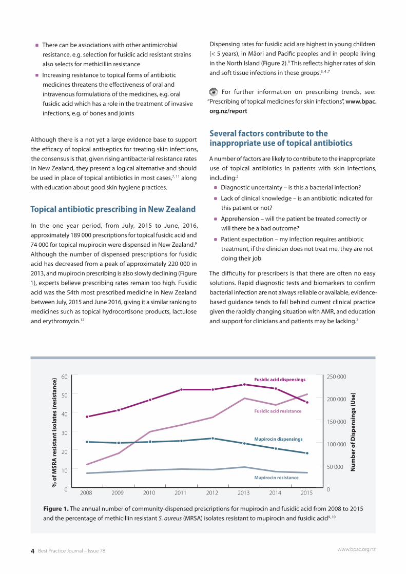
Excessive use of topical antibiotics is known to be a key driver of AMR, and is directly responsible for increasing antibacterial resistance in S. aureus.3, 4 There have been calls to restrict the use of topical antibiotics for some time, both in New Zealand and internationally.5 ,6 It is clear that this need is becoming more urgent.3, 7 New Zealand research has shown significant associations between increases in the use of topical mupirocin and fusidic acid and rapidly rising resistance rates in S. aureus (Figure 1).3, 4 Similar findings have been reported from the United Kingdom and Australia.5, 8 It is noteworthy that topical fusidic acid is not licensed for use in the United States and rates of fusidic acid resistance are negligible.5
There are three major reasons why resistance to topical antibiotics is important:5, 7
Increasing resistance leads to ineffective treatment with these medicines
Topical antibiotics for skin infections: should they be prescribed at all?
DERMATOlOgy ANTIbIOTIC RESISTANCE & STE wARDSHIP
Best Practice Journal – Issue 78 3www.bpac.org.nz
0
10
20
30
40
50
60
0
50 000
100 000
150 000
200 000
250 000
2008 2009 2010 2011 2012 2013 2014 2015
Year
% o
f MSR
A re
sist
ant i
sola
tes
(res
ista
nce)
Num
ber o
f Dis
pens
ings
(Use
)
Fusidic acid dispensings
Mupirocin dispensings
Mupirocin resistance
Fusidic acid resistance
4 Best Practice Journal – Issue 78 www.bpac.org.nz
There can be associations with other antimicrobial resistance, e.g. selection for fusidic acid resistant strains also selects for methicillin resistance
Increasing resistance to topical forms of antibiotic medicines threatens the effectiveness of oral and intravenous formulations of the medicines, e.g. oral fusidic acid which has a role in the treatment of invasive infections, e.g. of bones and joints
Although there is a not yet a large evidence base to support the efficacy of topical antiseptics for treating skin infections, the consensus is that, given rising antibacterial resistance rates in New Zealand, they present a logical alternative and should be used in place of topical antibiotics in most cases,7, 11 along with education about good skin hygiene practices.
Topical antibiotic prescribing in New Zealand
In the one year period, from July, 2015 to June, 2016, approximately 189 000 prescriptions for topical fusidic acid and 74 000 for topical mupirocin were dispensed in New Zealand.9 Although the number of dispensed prescriptions for fusidic acid has decreased from a peak of approximately 220 000 in 2013, and mupirocin prescribing is also slowly declining (Figure 1), experts believe prescribing rates remain too high. Fusidic acid was the 54th most prescribed medicine in New Zealand between July, 2015 and June 2016, giving it a similar ranking to medicines such as topical hydrocortisone products, lactulose and erythromycin.12
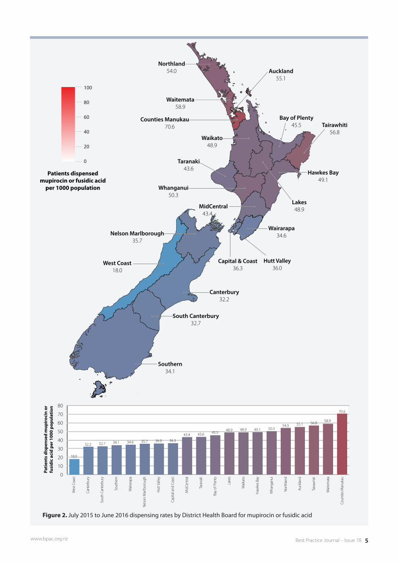
Dispensing rates for fusidic acid are highest in young children (< 5 years), in Māori and Pacific peoples and in people living in the North Island (Figure 2).9 This reflects higher rates of skin and soft tissue infections in these groups.3, 4 ,7
For further information on prescribing trends, see: “Prescribing of topical medicines for skin infections”, www.bpac.org.nz/report
Several factors contribute to the inappropriate use of topical antibiotics
A number of factors are likely to contribute to the inappropriate use of topical antibiotics in patients with skin infections, including:2
Diagnostic uncertainty – is this a bacterial infection?
Lack of clinical knowledge – is an antibiotic indicated for this patient or not?
Apprehension – will the patient be treated correctly or will there be a bad outcome?
Patient expectation – my infection requires antibiotic treatment, if the clinician does not treat me, they are not doing their job
The difficulty for prescribers is that there are often no easy solutions. Rapid diagnostic tests and biomarkers to confirm bacterial infection are not always reliable or available, evidence-based guidance tends to fall behind current clinical practice given the rapidly changing situation with AMR, and education and support for clinicians and patients may be lacking.2
Figure 1. The annual number of community-dispensed prescriptions for mupirocin and fusidic acid from 2008 to 2015 and the percentage of methicillin resistant S. aureus (MRSA) isolates resistant to mupirocin and fusidic acid9, 10
0
10
20
30
40
50
60
70
80
Wes
t Coa
st
Cant
erbu
ry
Sout
h Ca
nter
bury
Sout
hern
Wai
rara
pa
Nelso
n M
arlb
orou
gh
Hutt
Valle
y
Capi
tal a
nd C
oast
Mid
Cent
ral
Tara
naki
Bay
of P
lent
y
Lake
s
Wai
kato
Haw
kes B
ay
Wha
ngan
ui
North
land
Auck
land
Taira
whi
ti
Wai
tem
ata
Coun
ties M
anuk
au
Pati
ents
dis
pens
ed m
upir
ocin
or
fusi
dic
acid
per
100
0 po
pula
tion
18.0
32.2 32.7 34.1 34.6 35.7 36.0 36.343.4 43.6 45.5
48.9 48.9 49.1 50.354.0 55.1 56.8 58.9
70.6
Auckland55.1
Bay of Plenty45.5
Canterbury32.2
Hutt Valley36.0
Capital & Coast36.3
Wairarapa34.6
South Canterbury32.7
Southern34.1
West Coast18.0
Nelson Marlborough35.7
MidCentral43.4
Whanganui50.3
Taranaki43.6
Waikato48.9
Counties Manukau70.6
Waitemata58.9
Northland54.0
Lakes48.9
Hawkes Bay49.1
Tairawhiti56.8
Patients dispensed mupirocin or fusidic acid
per 1000 population
100
80
60
40
20
0
Best Practice Journal – Issue 78 5www.bpac.org.nz
Figure 2. July 2015 to June 2016 dispensing rates by District Health Board for mupirocin or fusidic acid
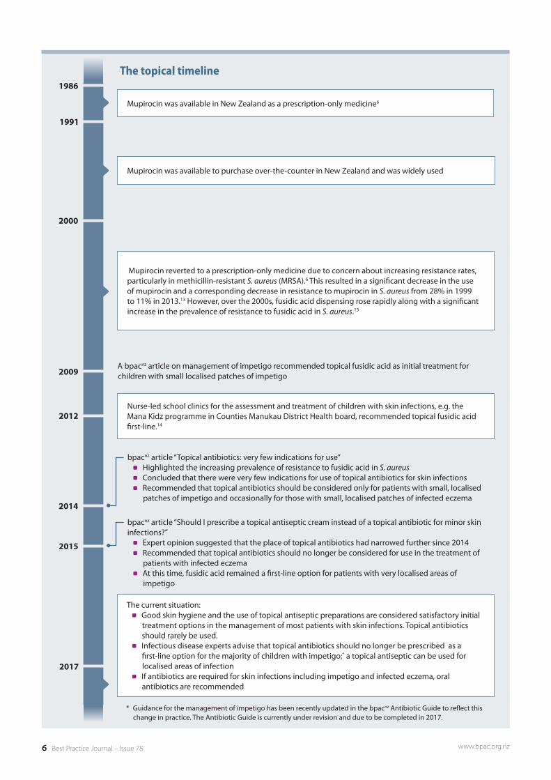
Mupirocin reverted to a prescription-only medicine due to concern about increasing resistance rates, particularly in methicillin-resistant S. aureus (MRSA).6 This resulted in a significant decrease in the use of mupirocin and a corresponding decrease in resistance to mupirocin in S. aureus from 28% in 1999 to 11% in 2013.13 However, over the 2000s, fusidic acid dispensing rose rapidly along with a significant increase in the prevalence of resistance to fusidic acid in S. aureus.13
Mupirocin was available to purchase over-the-counter in New Zealand and was widely used
A bpacnz article on management of impetigo recommended topical fusidic acid as initial treatment for children with small localised patches of impetigo
Nurse-led school clinics for the assessment and treatment of children with skin infections, e.g. the Mana Kidz programme in Counties Manukau District Health board, recommended topical fusidic acid first-line.14
bpacnz article “Topical antibiotics: very few indications for use” Highlighted the increasing prevalence of resistance to fusidic acid in S. aureus Concluded that there were very few indications for use of topical antibiotics for skin infections Recommended that topical antibiotics should be considered only for patients with small, localised
patches of impetigo and occasionally for those with small, localised patches of infected eczema
The current situation: Good skin hygiene and the use of topical antiseptic preparations are considered satisfactory initial
treatment options in the management of most patients with skin infections. Topical antibiotics should rarely be used.
Infectious disease experts advise that topical antibiotics should no longer be prescribed as a first-line option for the majority of children with impetigo;* a topical antiseptic can be used for localised areas of infection
If antibiotics are required for skin infections including impetigo and infected eczema, oral antibiotics are recommended
bpacnz article “Should I prescribe a topical antiseptic cream instead of a topical antibiotic for minor skin infections?”
Expert opinion suggested that the place of topical antibiotics had narrowed further since 2014 Recommended that topical antibiotics should no longer be considered for use in the treatment of
patients with infected eczema At this time, fusidic acid remained a first-line option for patients with very localised areas of
impetigo
* Guidance for the management of impetigo has been recently updated in the bpacnz Antibiotic Guide to reflect this change in practice. The Antibiotic Guide is currently under revision and due to be completed in 2017.
The topical timeline
Mupirocin was available in New Zealand as a prescription-only medicine6
1986
1991
2000
2009
2012
2014
2015
2017
6 Best Practice Journal – Issue 78 www.bpac.org.nz
Best Practice Journal – Issue 78 7www.bpac.org.nz
Additional information is available from the Goodfellow Unit:
www.goodfellowunit.org/gems/stop-using-topical-antibiotics
www.goodfellowunit.org/podcast/topical-antibiotics-emma-best
Acknowledgement: Thank you to Associate Professor Mark Thomas, Infectious Diseases Specialist, University of Auckland
and Auckland City Hospital for expert review of this article.
References:1. Pullon H, Gommans J, Thomas M, et al. Antimicrobial resistance in New
Zealand: the evidence and a call for action. NZMJ 2016;129:105–12.
2. Lipsky B, Dryden M, Gottrup F, et al. Antimicrobial stewardship in wound care:
a position paper from the British Society for Antimicrobial Chemotherapy and
European Wound Management Association. 2016;71:3026–35.
3. Williamson D, Ritchie S, Best E, et al. A bug in the ointment: topical
antimicrobial usage and resistance in New Zealand. NZMJ 2015;128:103–9.
4. Heffernan H, Bakker S, Woodhouse R, et al. Demographics, antimicrobial
susceptibility and molecular epidemiology of Staphylococcus aureus in New
Zealand, 2014. 2015.
5. Howden B, Grayson M. Dumb and dumber – the potential waste of a useful
antistaphylococcal agent: emerging fusidic acid resistance in Staphylococcus
aureus. Clin Infect Dis 2006;42:394–400.
6. Upton A, Lang S, Heffernan H. Mupirocin and Staphylococcus aureus: a
recent paradigm of emerging antibiotic reistance. J Antimicrob Chemother
2003;51:613–7.
7. Vogel A, Lennon D, Best E, et al. Where to from here? The treatment of impetigo
in children as resistance to fusidic acid emerges. NZMJ 2016;129:77–83.
8. Chaplin S. Topical antibacterial and antiviral agents: prescribing and resistance.
Prescriber 2016;27:29–36.
9. Ministry of Health. Pharmaceutical Claims Collection. 2016.
10. Environmental Science and Research. Antimicrobial resistance data from
hospital and community laboratories, 2008-15. Available from: https://surv.esr.
cri.nz/antimicrobial/general_antimicrobial_susceptibility.php (Accessed Feb,
2017).
11. Leitch C, Leitch A, Tidman M. Quantitative evaluation of dermatological
antiseptics. Clin Exp Dermatol 2015;40:912–5.
12. bpacnz. 2016 Annual Report. 2016. Available from: www.bpac.org.nz (Accessed
Feb, 2017).
13. Williamson D, Monecke S, Heffernan H, et al. High usage of topical fusidic acid
and rapid clonal expansion of fusidic acid-resistant Staphyoccus aureus: A
cautionary tale. Clin Infect Dis 2014;59:1451–4.
14. Tsai J-YC, Anderson P, Broome L, et al. Antimicrobial stewardship using
pharmacy data for the nurse-led school-based clinics in Counties Manukau
District Health Board for management of group A streptococcal pharyngitis
and skin infection. NZMJ 2016;129:29–38.
15. Llor C, Bjerrum L. Antimicrobial resistance:risk associated with antibiotic
overuse and initiatives to reduce the problem. Ther Adv Drug Saf
2014;5:229–41.
A strategy that may help to address some of these factors is to have a practice policy in place that offers guidance on when to use, and when not to use topical antibiotics, and how to discuss this with patients. Knowing that the practice offers a consistent approach may give prescribers more confidence in their decision-making.
How can prescribers manage patient expectations for an antibiotic?
Patients with skin infections usually expect treatment with antibiotics, even if their infection is minor. Patient education is an important aspect in reducing the use of all antibiotics, both topical and oral.
Successful interventions to help clinicians prescribe antibiotics rationally include:15
Clear communication that reinforces appropriate use of antibiotics and the increasing problem of resistance; topical antiseptics are recommended first-line for minor skin infections and if an antibiotic is required, oral treatment will be necessary
Setting realistic expectations regarding the natural history of the skin infection and the likely time for resolution; many minor skin infections are self-limiting, “a nuisance rather than a problem” to the patient, and when this is the case, best practice is to not prescribe at all.8
Individualised prescribing; consider factors such as the patient’s age, severity of the infection, co-morbidities, family and household circumstances
A delayed prescription for an oral antibiotic may be of value in some situations provided the patient knows when and why to get the prescription dispensed
Guidance is likely to change again Many issues remain unresolved regarding the appropriate use of topical antibiotics, such as clear evidence of effectiveness of other treatments including topical antiseptics. Experts are calling for informed debate and further research, and it is hoped that this will help determine the best course of action. In New Zealand, a randomised controlled trial is currently underway, comparing the effectiveness of hygiene measures, topical antiseptics and topical fusidic acid in the management of children with impetigo.7
At this stage, we recommend following the pragmatic advice: use topical antiseptics, along with good skin hygiene, to treat minor skin infections, and oral antibiotics for more severe or extensive infection. The clinical evidence is not yet conclusive, but the problem of rapidly rising resistance cannot be ignored. Watch this space.

8 Best Practice Journal – Issue 78 www.bpac.org.nz
Topical antibiotics for skin infections: when are they appropriate?
DERMATOlOgy ANTIbIOTIC RESISTANCE & STE wARDSHIP INFEC TIONS
What prescribers need to know: In primary care, many skin infections are relatively
minor and do not need to be treated with antibiotics. Management should focus on good skin hygiene measures and a trial of a topical antiseptic
Do not prescribe topical antibiotics for patients with infected eczema, for wound management, for other skin infections, or first-line for impetigo. If antibiotic treatment is required, prescribe an oral medicine
Topical antibiotics may be appropriate as a second-line option for patients with areas of localised impetigo, if first-line management with hygiene measures and topical antiseptics has not resolved the lesions or for Staphylococcus aureus nasal decolonisation
If a topical antibiotic is prescribed, patients should be instructed to use it for no longer than seven days. The practice of saving an unfinished tube as a “first-aid” measure for household members should be strongly discouraged
For further information on the changing role of topical antibiotics in New Zealand, see: “Topical antibiotics for skin infections – should they be prescribed at all?”, Page 3.
Few clinical situations require topical antibiotics
In the community, many patients have skin and soft tissue infections that are relatively minor, e.g. scrapes and scratches or mild folliculitis. These types of infections do not usually require antibiotic treatment as they will generally improve with good skin hygiene measures, e.g. cleaning and covering the lesion.1 A prescription for a topical antiseptic (rather than a topical antibiotic) is a pragmatic next step if hygiene interventions are not sufficient, although guidance on the use of antiseptics varies and there is a relative lack of evidence for their effectiveness.2–4
If a patient has an infection that requires antibiotic treatment, e.g. they have extensive infection, systemic symptoms or co-morbidities that place them at higher risk of infection or poor healing, in most cases prescribe an oral not a topical antibiotic.

Best Practice Journal – Issue 78 9www.bpac.org.nz
Due to increasing resistance, infectious diseases experts recommend that topical antibiotics should have a very limited role in clinical practice.2, 5 Currently the two clinical situations where their use may still be appropriate are:
As a second-line option for patients with areas of localised impetigo (e.g. less than three lesions) if first-line management with hygiene measures and topical antiseptics has not resolved the lesions within an appropriate timeframe, e.g. five to seven days. If a topical antibiotic is prescribed, fusidic acid should be used; mupirocin is reserved for treating MRSA infection. In many cases of impetigo, treatment with an oral antibiotic is more appropriate
Some patients with recurrent skin infections due to S. aureus may require nasal decolonisation with either fusidic acid or mupirocin once susceptibility is known. If the isolate is resistant to both topical antibiotics or there is active infection, oral antibiotics may be required (see below)
See sidebar “Topical antibiotics and antiseptics available in New Zealand” for information on available medicines
If topical antibiotics are prescribed, instruct the patient to use the medicine for up to seven days only and to discard the tube after this time. The practice of saving an unfinished tube as a “go-to” first-aid measure for household members should be strongly discouraged.5
Limit the use of fusidic acid in the management of impetigo
Impetigo is regarded as a self-limiting condition although treatment is often initiated to hasten recovery and to reduce the spread of infection.7 There is, however, a lack of quality evidence-based research on the optimal management of impetigo.2 Recent expert opinion is that first-line management in the majority of children with mild to moderate impetigo is good skin hygiene and topical antiseptic preparations.
For further information on current views on the role of topical antibiotics, see: www.goodfellowunit.org/podcast/topical-antibiotics-emma-best
Skin hygiene measures in children with impetigo should start with the “clean, cut (nails) and cover” message, which also applies to patients with other skin infections or injuries.2 Advise parents or caregivers to use a clean cloth soaked in warm water to gently remove crusts from lesions. Infectious diseases experts then recommend the application of a topical antiseptic such as hydrogen peroxide or povidone-iodine. These antiseptic preparations can also assist in softening the
Topical antibiotics and antiseptics available in New ZealandTwo topical antibiotics and two topical antiseptics for use on the skin are currently subsidised in New Zealand.*
The topical antibiotics are:6
Fusidic acid (sodium fusidate) cream or ointment 2% – 15 g tube, fully subsidised
Mupirocin ointment 2% – 15 g tube, partially subsidised
The topical antiseptics are:6
Hydrogen peroxide cream 1% – 15 g tube, fully subsidised (also available over-the-counter in a 10 g or 25 g tube)
Povidone-iodine ointment 10% – 25 g tube, fully subsidised (also available over-the-counter in a range of tube sizes)
* Other topical antibiotics are available, e.g. for acne and for infections of the eye and nose, including fusidic acid as an eye gel (Fucithalmic). Compound preparations, e.g. fusidic acid + betamethasone valerate (Fucicort, partly subsidised) and hydrocortisone + natamycin + neomycin (Pimafucort, fully subsidised) are also available. The use of these medicines is not covered in this resource, however, similar restraints with prescribing should also apply to these medicines.
For a snapshot of national and individual prescribing data for these topical medicines, see: “Prescribing of topical medicines for skin infections”. www.bpac.org.nz/report
10 Best Practice Journal – Issue 78 www.bpac.org.nz
There is some evidence to support this practice and/or the use of an activated oxygen bleach product.13
Antibiotics are indicated if skin infections are recurrent despite other measuresIf the patient continues to have recurrent skin infections despite optimal care and hygiene measures, personal decolonisation with antibiotics may be required and also considered for family members.9 A nasal swab to determine whether the patient has S. aureus nasal colonisation should be requested, if this has not already been done. Consider discussing an appropriate decolonisation regimen with an infectious diseases expert as advice is likely to vary due to local resistance patterns. There is a lack of consensus on the most effective decolonisation method and increasing antibiotic resistance continues to drive research into alternative options both in New Zealand and internationally. For example, the antiseptic povidone-iodine used intranasally has been suggested as an alternative to a topical antibiotic, but consistent evidence for its effectiveness is lacking.12, 14
Topical antibiotic treatment – if topical antibiotics are recommended, the appropriate topical antibacterial (either mupirocin or fusidic acid as guided by the sensitivity results) should be applied to the anterior nares, twice daily, for five to seven days. They should not be administered if the patient still has an active skin infection as the skin infection can be a source from which nasal carriage is re-established. Good personal hygiene measures and environmental decolonisation measures should be ongoing.
Oral antibiotic treatment – although international guidelines do not recommend the routine use of oral antibiotics for decolonisation there may be a role for this strategy when first-line measures have been unsuccessful or when there is active infection.15 Prescribing a combination of oral antibiotics (usually two) has been found to be effective for decolonisation, however, they may need to be used concurrently with topical antibiotics to achieve eradication of S. aureus from the nose.12, 15 The choice of oral antibiotics should usually be made after a discussion with an infectious diseases physician or clinical microbiologist and in addition should be guided by the sensitivity results from nasal swabs. A typical oral regimen for an adult with recurrent skin infections would include both rifampicin* (e.g. 300 mg, twice daily) and flucloxacillin (e.g. 500 mg, three or four times daily).** 12, 15 Both oral antibiotics are taken for one week and then repeated for one week each month for three to six months.
* Rifampicin requires specialist approval for prescription. This can be obtained from an infectious disease specialist or a clinical microbiologist at a community laboratory and the prescription endorsed accordingly.
** Alternative antibiotics to flucloxacillin include co-trimoxazole or doxycycline (used in combination with rifampicin)
crusted areas. Parents and caregivers should be advised to keep the affected areas covered with dressings to reduce the spread of infection to others. The child should be excluded from school or pre-school until the lesions have dried up or for 24 hours after oral antibiotic treatment has been initiated.8 If required, assess and treat other household members who may be infected.
Oral antibiotics are recommended if: Lesions are extensive or there is widespread infection
Systemic symptoms are present
Good hygiene and topical antiseptic treatment has failed
The first choice for an oral antibiotic should be flucloxacillin. An appropriate dose for a child is:
Flucloxacillin: 12.5 mg/kg/dose, four times daily, for five days (maximum 500 mg/dose)
Alternative oral antibiotics if there is allergy or intolerance to flucloxacillin include erythromycin, co-trimoxazole (first choice if MRSA is present) and cefalexin.
Topical fusidic acid should only be considered as a second-line option for areas of very localised impetigo (e.g. less than three lesions) if the first-line measures have been unsuccessful.
Decolonisation for patients with recurrent skin infections
Patients with recurrent skin infections and their family members may require decolonisation to reduce S. aureus carriage. The initial focus should be on good personal hygiene and environmental decolonisation.9 If this approach has been unsuccessful and the patient continues to have recurrent skin infections, antibiotics may be required.
Advise intensification of personal hygiene measures Patients should be advised to intensify personal hygiene practices and not to share items such as razors, towels or linen. The regular use of antibacterial soaps or washes and weekly dilute bleach baths is often advocated, although the evidence base for this is variable.8–11 One approach is to prescribe triclosan 1% as a liquid soap to reduce the bacterial load on the skin. This can be used daily for five to seven days then reduced to once or twice weekly. Triclosan 1% is fully subsidised in a 500 mL bottle, provided the patient has recurrent S. aureus infection and the prescription is endorsed accordingly.
Environmental decolonisation is recommendedEnvironmental measures should include cleaning of regularly touched surfaces and frequent washing of clothes, towels and linen.9 The use of heat, e.g. hot water, hot dryer cycle or ironing, when laundering towels and linen is often recommended.
Best Practice Journal – Issue 78 11www.bpac.org.nz
Many patients with mild bacterial skin infections do not require antibiotics
Folliculitis is often self-limitingFolliculitis is a collective term for a group of skin conditions which can be due to bacterial infection but can be also caused by fungi and viruses. A sterile folliculitis can be the result of occlusion, e.g. from the use of emollients (particularly paraffin-based ointments), or adhesive dressings.16 In addition, environmental factors, e.g. hot, humid weather, shaving and other forms of hair removal, medicines such as topical or oral corticosteroids and immunosuppression may all contribute to folliculitis.16, 17
Superficial folliculitis is a mild, self-limiting condition and patients usually do not require topical or oral antibiotic treatment. Management should focus on effective skin hygiene, avoiding or treating any underlying cause and topical antiseptics.17 If the skin lesions are spreading, persistent or recurrent, oral antibiotics, such as flucloxacillin may need to be considered.
Furuncles (boils) and carbuncles are treated with incision and drainage
Larger lesions such as furuncles and carbuncles that extend into the subcutaneous tissue and are fluctuant should be managed with incision and drainage alone. Patients do not usually need antibiotic treatment unless there is associated cellulitis or the patient becomes systemically unwell.18 An oral antibiotic, e.g. flucloxacillin, would be appropriate in these situations.
Take a pragmatic approach to the management of skin infections
Although management for skin infections in primary care cannot be directed by a conclusive evidence base, the consensus from infectious diseases experts is that, given the rise in antibacterial resistance rates in New Zealand, topical antiseptics and education about good skin hygiene practices presents a pragmatic approach when managing patients with skin infections. Inappropriate use of topical antibiotics has been clearly shown to be associated with rapidly rising resistance. Clinicians need to be mindful of this and alter their management accordingly.
Acknowledgement: Thank you to Associate Professor Mark Thomas, Infectious Diseases Specialist, University of Auckland
and Auckland City Hospital for expert review of this article.
References1. Tsai J-YC, Anderson P, Broome L, et al. Antimicrobial stewardship
using pharmacy data for the nurse-led school-based clinics in
Counties Manukau District Health Board for management of group A
streptococcal pharyngitis and skin infection. NZMJ 2016;129:29–38.
2. Vogel A, Lennon D, Best E, et al. Where to from here? The treatment
of impetigo in children as resistance to fusidic acid emerges. NZMJ
2016;129:77–83.
3. Cooke J. When antibiotics can be avoided in skin inflammation and
bacterial colonization: a review of topical treatments. Curr Opin Infect
Dis 2014;27:125–9.
4. Leitch C, Leitch A, Tidman M. Quantitative evaluation of dermatological
antiseptics. Clin Exp Dermatol 2015;40:912–5.
5. Williamson D, Ritchie S, Best E, et al. A bug in the ointment:
topical antimicrobial usage and resistance in New Zealand. NZMJ
2015;128:103–9.
6. New Zealand Formulary (NZF). NZF v56. 2017. Available from: www.nzf.
org.nz (Accessed Feb, 2017).
7. Yeoh D, Bowen A, Carapetis J. Impetigo and scabies - disease burden and
modern treatment strategies. J Infection 2016;72:S61-7.
8. Impetigo (school sores). Ministry of Health. 2017. Available from: http://
www.health.govt.nz/your-health/conditions-and-treatments/diseases-
and-illnesses/impetigo-school-sores (Accessed Feb, 2017).
9. Creech C, Al-Zubeidi D, Fritz S. Prevention of recurrent staphylococcal
skin infections. Infect Dis Clin N Am 2015;29:429–64.
10. Fritz S, Camins B, Eisenstein K, et al. Effectiveness of measures to
eradicate Staphylococcus aureus carriage in patients with comunity-
associated skin and soft tissue infections: a randomized trial. Infect
Control Hosp Epidemiol 2011;32:872–80.
11. Kaplan S, Forbes A, Hammerman W, et al. Randomised trial of
‘bleach baths’ plus routine hygienic measures vs routine hygienic
measures alone for prevention of recurrent infections. Clin Infect Dis
2014;58:679–82.
12. Septimus E, Schweizer M. Decolonization in prevention of health
care-associated infections. Clin Microbiol Rev 2016;29:201–22.
13. Bockmuhl D. Laundry hygiene - how to get more than clean. J Appl
Microbiol 2017;epub ahead of print. doi:doi: 10.1111/jam.13402
14. Anderson M, David M, Scholz M, et al. Efficacy of skin and nasal
providone-iodine preparation against mupirocin-resistant methicillin-
resistant Staphylococcus aureus and S. aureus within the anterior nares.
Antimicrob Agents Chemother 2015;59:2765–73.
15. Liu C, Bayer A, Cosgrove S, et al. Clinical practice guidelines by the
Infectious Disease Society of America for the treatment of methicillin
resistant Staphylococcus aureus infections in adults and children. Clin
Infect Dis;52:e18-55.
16. Oakley A. Folliculitis. DermNet NZ. Available from: http://www.
dermnetnz.org/topics/folliculitis/ (Accessed Feb, 2017).
17. Cunliffe T. Folliculitis and boils (furuncles/carbuncles). Primary Care
Dermatology Society. http://www.pcds.org.uk/clinical-guidance/
folliculitis-an-overview
18. Ibler K, Kromann C. Recurrent furunculosis - challenges and managment:
a review. Clin Cosmet Investig Dermatol;7:59–64.
12 Best Practice Journal – Issue 78 www.bpac.org.nz
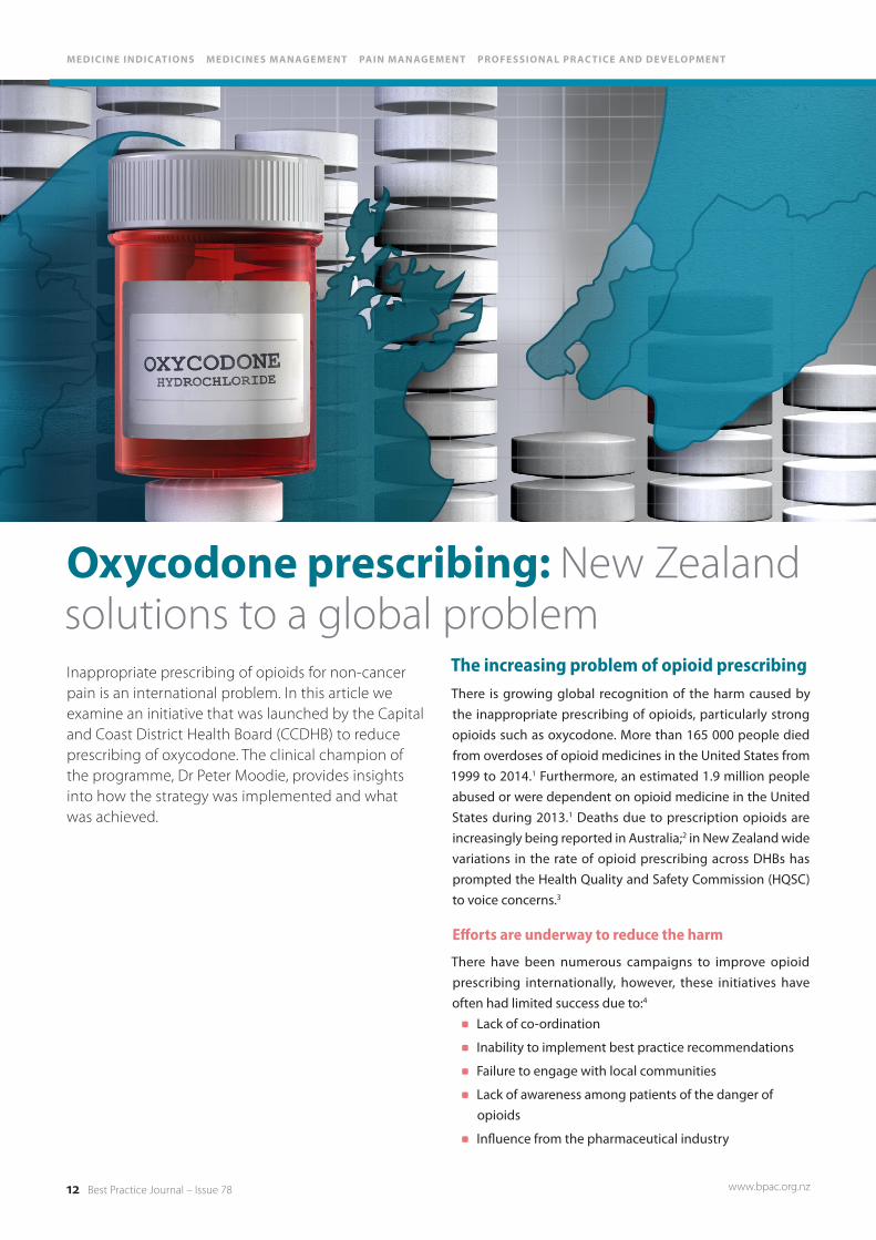
Inappropriate prescribing of opioids for non-cancer pain is an international problem. In this article we examine an initiative that was launched by the Capital and Coast District Health Board (CCDHB) to reduce prescribing of oxycodone. The clinical champion of the programme, Dr Peter Moodie, provides insights into how the strategy was implemented and what was achieved.
Oxycodone prescribing: New Zealand solutions to a global problem
The increasing problem of opioid prescribingThere is growing global recognition of the harm caused by the inappropriate prescribing of opioids, particularly strong opioids such as oxycodone. More than 165 000 people died from overdoses of opioid medicines in the United States from 1999 to 2014.1 Furthermore, an estimated 1.9 million people abused or were dependent on opioid medicine in the United States during 2013.1 Deaths due to prescription opioids are increasingly being reported in Australia;2 in New Zealand wide variations in the rate of opioid prescribing across DHBs has prompted the Health Quality and Safety Commission (HQSC)to voice concerns.3
Efforts are underway to reduce the harm
There have been numerous campaigns to improve opioid prescribing internationally, however, these initiatives have often had limited success due to:4
Lack of co-ordination
Inability to implement best practice recommendations
Failure to engage with local communities
Lack of awareness among patients of the danger of opioids
Influence from the pharmaceutical industry
MEDICINE INDIC ATIONS MEDICINES MANAgEMENT PAIN MANAgEMENT PROFESSIONAl PR AC TICE AND DE VElOPMENT
Best Practice Journal – Issue 78 13www.bpac.org.nz
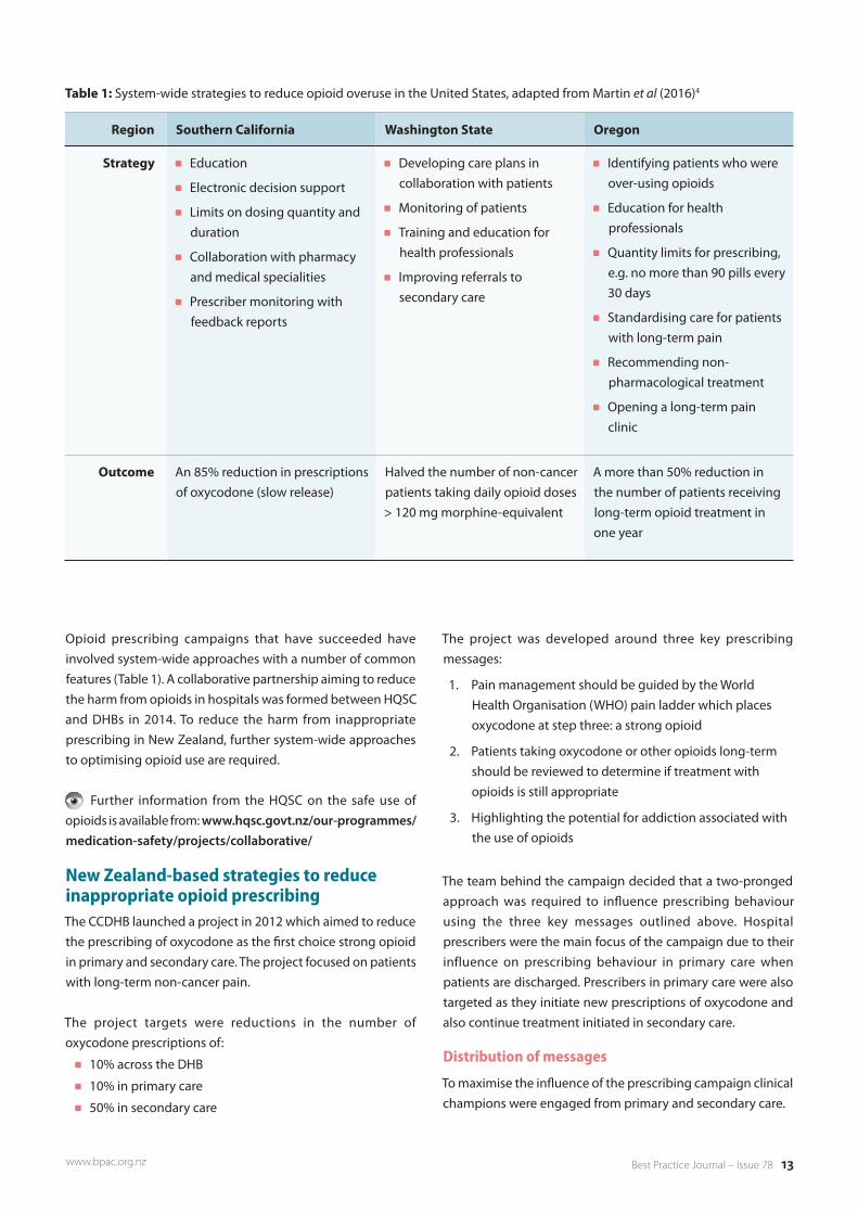
Opioid prescribing campaigns that have succeeded have involved system-wide approaches with a number of common features (Table 1). A collaborative partnership aiming to reduce the harm from opioids in hospitals was formed between HQSC and DHBs in 2014. To reduce the harm from inappropriate prescribing in New Zealand, further system-wide approaches to optimising opioid use are required.
Further information from the HQSC on the safe use of opioids is available from: www.hqsc.govt.nz/our-programmes/medication-safety/projects/collaborative/
New Zealand-based strategies to reduce inappropriate opioid prescribingThe CCDHB launched a project in 2012 which aimed to reduce the prescribing of oxycodone as the first choice strong opioid in primary and secondary care. The project focused on patients with long-term non-cancer pain.
The project targets were reductions in the number of oxycodone prescriptions of:
10% across the DHB
10% in primary care
50% in secondary care
The project was developed around three key prescribing messages:
1. Pain management should be guided by the World Health Organisation (WHO) pain ladder which places oxycodone at step three: a strong opioid
2. Patients taking oxycodone or other opioids long-term should be reviewed to determine if treatment with opioids is still appropriate
3. Highlighting the potential for addiction associated with the use of opioids
The team behind the campaign decided that a two-pronged approach was required to influence prescribing behaviour using the three key messages outlined above. Hospital prescribers were the main focus of the campaign due to their influence on prescribing behaviour in primary care when patients are discharged. Prescribers in primary care were also targeted as they initiate new prescriptions of oxycodone and also continue treatment initiated in secondary care.
Distribution of messages
To maximise the influence of the prescribing campaign clinical champions were engaged from primary and secondary care.
Table 1: System-wide strategies to reduce opioid overuse in the United States, adapted from Martin et al (2016)4
Region Southern California washington State Oregon
Strategy Education
Electronic decision support
Limits on dosing quantity and duration
Collaboration with pharmacy and medical specialities
Prescriber monitoring with feedback reports
Developing care plans in collaboration with patients
Monitoring of patients
Training and education for health professionals
Improving referrals to secondary care
Identifying patients who were over-using opioids
Education for health professionals
Quantity limits for prescribing, e.g. no more than 90 pills every 30 days
Standardising care for patients with long-term pain
Recommending non-pharmacological treatment
Opening a long-term pain clinic
Outcome An 85% reduction in prescriptions of oxycodone (slow release)
Halved the number of non-cancer patients taking daily opioid doses > 120 mg morphine-equivalent
A more than 50% reduction in the number of patients receiving long-term opioid treatment in one year
14 Best Practice Journal – Issue 78 www.bpac.org.nz
The approach in primary care
The “top 20” oxycodone primary care prescribers were identified in each PHO within the CCDHB via the Pharmaceutical Collection data warehouse. Support for practices with relatively high rates of oxycodone prescribing was provided by pharmacist facilitators which included:
An oxycodone practice audit accredited by the Royal New Zealand College of General Practitioners
Campaign posters
Practice education forums
Peer review groups
A multidisciplinary pain management education session was held that was attended by 96 clinicians including general practitioners, nurses and pharmacists.
The approach in secondary care
The hospital utilisation of oxycodone for 2011/12 was analysed. Education sessions were delivered by a specialty pain team and staff from the hospital pharmacy to nurses, house surgeons and registrars in the three wards with the highest oxycodone use.
A series of campaign posters was developed which were changed on a weekly basis. A booklet summarising opioid prescribing messages from the bpacnz pain management guidelines with a reminder to contact the pain team for advice and a one-page information sheet was distributed across all wards. The oxycodone prescribing campaign was featured on the hospital intranet site.
The effect of the campaign on oxycodone prescribing
The oxycodone campaign resulted in a 24% reduction in the number of oxycodone scripts written across the DHB and a 20% reduction in the number of oxycodone items dispensed.5 The targets of a 10% reduction in the number of oxycodone prescriptions in primary care and 50% reduction in the number of oxycodone hospital prescriptions were achieved.5 Before the oxycodone prescribing campaign, the CCDHB were reportedly
the third lowest DHB for oxycodone usage; following the campaign they were ranked the lowest DHB for oxycodone use.5 The amount of harm reduction the campaign achieved is hard to quantify, however, the financial savings in reduced medicine use amounted to at least $50 000.5
Prescribing changes in primary careThere was a 22% decrease in the annual prescribing of oxycodone across 18 general practices in the CCDHB following the campaign.5
Prescribing changes in secondary care There were substantial decreases in the rate of oxycodone prescribing in the hospital following the campaign with the goal of a 50% reduction being met by most wards (Table 2).5
Personalised reports for oxycodone prescribing in primary are available from: www.bpac.org.nz/Report/2016/February/oxycodone.aspx
Further information on opioid prescribing across individual DHBs is available from: www.hqsc.govt.nz/our-programmes/health-quality-evaluation/projects/atlas-of-healthcare-variation/opioids/
Best practice points for the use of opioids for non-cancer pain:
Maximise appropriate non-opioid treatments first
Morphine is the first-line strong opioid for non-cancer pain unless the patient is intolerant
Use shared decision-making and ensure the patient is educated about the risks and benefits of opioid treatment
Table 2: Percentage reduction of in-hospital use of oxycodone from June 2011 – July 2012, compared with March 2012 – February 20135
ward Percentage reduction
general surgery/vascular 68%
Orthopaedics 58%
Cardiothoracic/cardiology 54%
general medicine, oncology, renal 35%
Avoid prescribing more than three days’ supply unless circumstances clearly warrant additional opioid treatment
Prescribe opioids with caution in elderly patients: take into account renal function and consider prescribing lower doses
Make sure the patient is aware that opioids can affect their work duties and driving
Best Practice Journal – Issue 78 15www.bpac.org.nz
Analysis of the prescribing campaign
Dr Peter Moodie led the CCDHB opioid prescribing campaign. He works as a general practitioner at the Karori Medical Centre and was Medical Director of PHARMAC until 2013. Dr Moodie provides insight into how the prescribing campaign was undertaken and what was learnt from it.
1. what were the challenges faced during the prescribing campaign?The greatest challenges for the project were data; accurate data and relevant data. In New Zealand we are blessed with an amazing data repository called the “Pharmhouse” [Pharmaceutical Collection data warehouse]. Every script dispensed in community pharmacies goes into that database and virtually everything on the prescription is searchable, albeit with the patient’s name encrypted.
The downsides are that secondary care data is not included unless their prescription is dispensed outside the hospital and you have to know what you are doing when interrogating the data.
Once you have the data, putting it into a meaningful format is again critical. It is possible to work out who initiated a prescription when there is chain of scripts for the same person as although the NHIs are encrypted, it is always with the same encryption. This means that if a script was initiated when a patient was discharged it can be followed to see who then continued the prescription.
2. How was the programme received by prescribers in primary care, in particular the use of individualised prescriber reports?How was all of this received? Well if you are like me, you know what you prescribe and don’t need anyone else to tell you; like I knew that I never used oxycodone…well I thought I didn’t. When confronted with the data I had lots of excuses: “The other doctor was away and I had to write the script”, or “They came out of hospital on it and I just had to repeat it” or “I knew I shouldn’t have but I can’t quite remember the reason why I did it”. And remember I was the clinical champion for the project!
In other words, we can all get defensive but for groups that do not get audited often it can be even more challenging.
For example when pointing out that anaesthetists were often big prescribers, they often blamed it on the orthopaedic surgeon who thought it was a good drug. Why? We all rationalise.
3. was it possible to identify which specific aspects of the campaign were effective?The seminars were well attended and the prescribing data was useful but we found that it had to be presented in a manner which encouraged feedback. Simply handing it out didn’t do much.
4. Do you think a similar approach could be successful in other DHbs?The programme should go nation-wide, however, it is important that pressure groups are not allowed to dilute the messages.
5. what were the learning points that prescribers could take from the campaign? What were the greatest learnings? Firstly, every time we reach for a controlled drug pad and start to write oxycodone or fentanyl, we should ask ourselves why we aren’t writing a script for morphine. There are lots of reasons and one of them is possibly, “it’s not actually morphine – it’s just strong codeine” – yeah right. The other and more insidious is “the pain clinic uses it and I don’t want to be old fashioned”. Finally feedback from peers is critical. Secondary care needs honest feedback from their peers and likewise primary care.
Acknowledgement: Thank you to Dr Peter Moodie, Karori
Medical Centre, Wellington for his assistance with this article.
References1. Dowell D, Haegerich T, Chou R. CDC guideline for prescribing opioids for
chronic pain - United States, 2016. MMWR Recomm Rep 2016;65:1–49. doi:
10.15585/mmwr.rr6501e1
2. Jammal W, Gown G. Opioid prescribing pitfalls: medicolegal and regulatory
issues. Aust Prescr 2015;38:198–203. doi:10.18773/austprescr.2015.069
3. Sheppard L. Pain in the news: Press release on opioid prescribing from the NZ
Health Quality and Safety Commission. 2015. Available from: www.nzps.org.
nz/blog/pain-in-the-news-press-release-on-opioid-prescribing-from-the-nz-
health-quality-safety-commission (Accessed Jul, 2016)
4. Martin L, Laderman M, Hyatt J, et al. Addressing the opioid crisis in the United
States. 2016. Available from: www.ihi.org/Pages/default.aspx (Accessed Jul,
2016)
5. Balram A. Oxycodone: project evaluation. 2013. Available from: On request

16 Best Practice Journal – Issue 78 www.bpac.org.nz
Chronic plaque psoriasis: an overview of treatment in primary care
DERMATOlOgy
KEy PR AC TICE POINTS:
Most patients with psoriasis have chronic plaque psoriasis, the majority of whom can be managed in primary care
Emollients can reduce pruritus, plaque scale and restore skin pliability
Additional first-line topical medicines include intermittent courses of topical corticosteroids, topical calcipotriol, or both in combination
Patients with psoriasis require life-long treatment and are at increased risk of cardiovascular disease, depression, inflammatory bowel disease and diabetes
Guidance on selecting topical treatments and tailoring treatment to the affected body area is available in a second article: “Choosing a topical treatment for patients with chronic plaque psoriasis”. Guidance on monitoring patients with moderate to severe psoriasis is available in the third article:
“Monitoring patients with moderate to severe psoriasis”.
Psoriasis is an immune-mediated chronic inflammatory skin disease which causes red, scaly plaques. Approximately one-third of patients develop symptoms before age 20 years and prevalence increases with age; most patients develop symptoms before the age of 35 years.1, 2 There are no reliable estimates of prevalence in New Zealand, but in the United States and United Kingdom approximately 3% of adults are affected and less than 1% of children aged 12 years and under.1–3 Evidence suggests Māori and Pacific peoples have similar rates of psoriasis as New Zealand Europeans.4
Approximately 15% of patients with psoriasis have psoriatic arthritis, i.e. joint involvement or inflammation of tendons, ligaments or joint capsule insertions (enthesitis).1 Patients with significant inflammatory joint disease should be referred to a rheumatologist as systemic medicines, such as methotrexate or other disease modifying agents, are often used early to reduce the risk of permanent joint destruction and simultaneously may improve skin symptoms.3
Patients with psoriasis have an increased risk of other conditions, including fatty liver, cardiovascular disease, diabetes, inflammatory bowel disease and depression, and should be regularly assessed for symptoms and signs.3 Psoriasis is also
Best Practice Journal – Issue 78 17www.bpac.org.nz
associated with a number of ophthalmic conditions, usually uveitis; expert opinion is that ocular involvement may occur in up to 10% of people with psoriasis.5
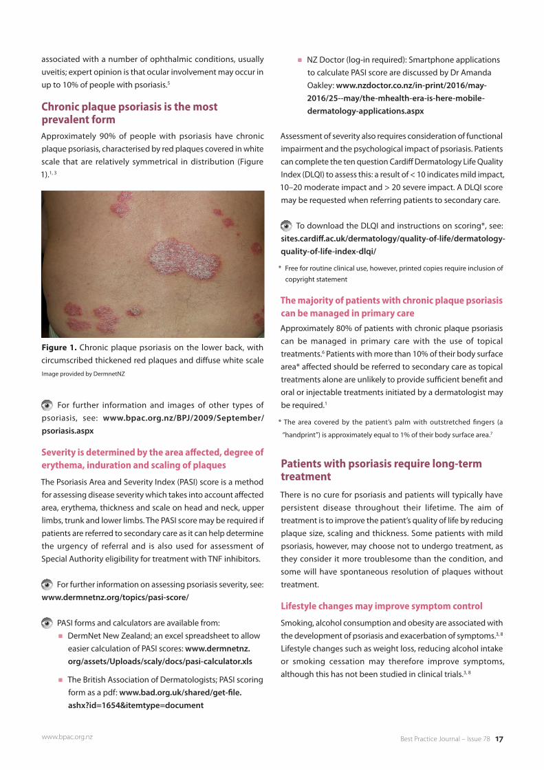
Chronic plaque psoriasis is the most prevalent form Approximately 90% of people with psoriasis have chronic plaque psoriasis, characterised by red plaques covered in white scale that are relatively symmetrical in distribution (Figure 1).1, 3
Figure 1. Chronic plaque psoriasis on the lower back, with circumscribed thickened red plaques and diffuse white scale Image provided by DermnetNZ
For further information and images of other types of psoriasis, see: www.bpac.org.nz/BPJ/2009/September/psoriasis.aspx
Severity is determined by the area affected, degree of erythema, induration and scaling of plaques
The Psoriasis Area and Severity Index (PASI) score is a method for assessing disease severity which takes into account affected area, erythema, thickness and scale on head and neck, upper limbs, trunk and lower limbs. The PASI score may be required if patients are referred to secondary care as it can help determine the urgency of referral and is also used for assessment of Special Authority eligibility for treatment with TNF inhibitors.
For further information on assessing psoriasis severity, see: www.dermnetnz.org/topics/pasi-score/
PASI forms and calculators are available from: DermNet New Zealand; an excel spreadsheet to allow
easier calculation of PASI scores: www.dermnetnz.org/assets/Uploads/scaly/docs/pasi-calculator.xls
The British Association of Dermatologists; PASI scoring form as a pdf: www.bad.org.uk/shared/get-file.ashx?id=1654&itemtype=document
NZ Doctor (log-in required): Smartphone applications to calculate PASI score are discussed by Dr Amanda Oakley: www.nzdoctor.co.nz/in-print/2016/may-2016/25--may/the-mhealth-era-is-here-mobile-dermatology-applications.aspx
Assessment of severity also requires consideration of functional impairment and the psychological impact of psoriasis. Patients can complete the ten question Cardiff Dermatology Life Quality Index (DLQI) to assess this: a result of < 10 indicates mild impact, 10–20 moderate impact and > 20 severe impact. A DLQI score may be requested when referring patients to secondary care.
To download the DLQI and instructions on scoring*, see: sites.cardiff.ac.uk/dermatology/quality-of-life/dermatology-quality-of-life-index-dlqi/
* Free for routine clinical use, however, printed copies require inclusion of copyright statement
The majority of patients with chronic plaque psoriasis can be managed in primary care
Approximately 80% of patients with chronic plaque psoriasis can be managed in primary care with the use of topical treatments.6 Patients with more than 10% of their body surface area* affected should be referred to secondary care as topical treatments alone are unlikely to provide sufficient benefit and oral or injectable treatments initiated by a dermatologist may be required.1
* The area covered by the patient’s palm with outstretched fingers (a
“handprint”) is approximately equal to 1% of their body surface area.7
Patients with psoriasis require long-term treatment
There is no cure for psoriasis and patients will typically have persistent disease throughout their lifetime. The aim of treatment is to improve the patient’s quality of life by reducing plaque size, scaling and thickness. Some patients with mild psoriasis, however, may choose not to undergo treatment, as they consider it more troublesome than the condition, and some will have spontaneous resolution of plaques without treatment.
Lifestyle changes may improve symptom control
Smoking, alcohol consumption and obesity are associated with the development of psoriasis and exacerbation of symptoms.3, 8 Lifestyle changes such as weight loss, reducing alcohol intake or smoking cessation may therefore improve symptoms, although this has not been studied in clinical trials.3, 8
18 Best Practice Journal – Issue 78 www.bpac.org.nz
Emollients should be recommended to all patients with chronic plaque psoriasisEmollients can be applied frequently and liberally, and used on symptomatic and asymptomatic skin, as they help restore skin pliability and reduce plaque scale and pruritus.* 9 A variety of emollients are available fully subsidised and the most appropriate emollient is one a patient prefers and uses. If patients find soaps irritating, an emollient soap substitute, e.g. emulsifying ointment, can also be prescribed. In clinical trials of topical corticosteroids in patients with mild to severe chronic plaque psoriasis, a wide range of patients (15–47%) show improvement with the use of emollients only.6
* Clinicians may need to add instructions to “apply liberally” in prescribing software.
Topical corticosteroids alone or in combination with calcipotriol are the first-line addition to emollientsTopical corticosteroids, topical calcipotriol and these medicines in combination provide additional benefit over and above the effect of emollients for patients with chronic plaque psoriasis.1 These topical medicines should be applied in sufficient quantities to cover symptomatic plaques. Second-line topical treatments for mild chronic plaque psoriasis include products containing coal or synthetic tar at concentrations of 0.5–12%, and keratolytics such as topical salicylic acid, used at concentrations of 2–5%.
For further information on prescribing topical treatments, see: “Choosing a topical treatment for patients with chronic plaque psoriasis”, Page 19.
It is essential to give the patient realistic expectations regarding topical treatments: advise patients to expect partial resolution rather than complete clearance. In clinical trials of topical calcipotriol, corticosteroids or combination treatment, on average PASI scores improve by 40–70%, so patients will often have some remaining symptoms.10, 11 Psoriasis affecting the face, flexures, genitalia, scalp, palms and soles and nails is typically more difficult to treat.1
Follow-up in primary careA follow-up appointment is recommended four to six weeks after treatment is initiated for adults, or two weeks after for children.1, 3
Emphasise appropriate durations for the use of topical corticosteroids and that patients should leave at least four weeks between courses of topical corticosteroids on the same area of skin; severe adverse effects are more likely when patients continue treatment beyond recommended timeframes or without appropriate intervals between courses.
For appropriate durations of treatment with topical corticosteroids, see: Figure 1 in “Choosing a topical treatment for patients with chronic plaque psoriasis”, Page 20.
Topical calcipotriol can be used on an ongoing basis, however, patients may prefer not to use any treatment during periods of remission in order to have a break from daily applications. Continued emollient use can help to improve skin pliability and should be encouraged.9
When assessing the patient, also consider: The development of joint involvement (psoriatic arthritis)
The patients’ quality of life; stress may exacerbate psoriasis, and the severity of symptoms can influence a patient’s mental health 3
The patient’s increased risk of other conditions such as cardiovascular disease, diabetes, fatty liver, inflammatory bowel disease and depression
Relapses of psoriasis are expectedRelapse should not be regarded as treatment failure, but relapse frequency and the effect on quality of life should be taken into account when considering referral to secondary care. A meta-analysis reported that 88% of patients relapsed within six months of a course of topical treatment, with no consistent evidence that any treatment had lower rates of relapse than another.12
When to refer
Discussion with a dermatologist or rheumatologist is appropriate at any point during treatment if:1
Patients develop joint involvement
Symptoms spread to 10% or more of the body, or patients have a PASI score ≥ 10
Psoriasis is having a major effect on the patient’s wellbeing, e.g. a DLQI score of ≥ 10
Patients develop ocular complications
Assessment of DLQI and PASI score may be necessary for referral. Referral to a psychologist may be appropriate for patients with psoriasis that has worsened significantly due to stress. Annual influenza vaccination is recommended for patients taking oral or injectable medicines for the treatment of chronic plaque psoriasis.13
Acknowledgement: Thank you to Dr Amanda Oakley,
Honorary Associate Professor and Dermatologist, Waikato
District Health Board for expert review of this article.
References are available from the bpacnz website. See: www.bpac.org.nz/2017/psoriasis-1.aspx

Choosing a topical treatment for patients with chronic plaque psoriasis
Best Practice Journal – Issue 78 19www.bpac.org.nz
KEy PR AC TICE POINTS:
Finding a treatment that works for patients may require trial and error
First-line topical medicines include emollients, potent or very potent topical corticosteroids, topical calcipotriol, or a combination of these medicines
Keratolytics such as topical salicylic acid or products containing coal tar may reduce scaling and be beneficial for patients who have responded poorly to other topical medicines
Treatment needs to be both a science and an art The appropriate treatment for patients with chronic plaque psoriasis will depend on the location and characteristics of the plaques, as well as the patient’s response and tolerance, so can require trial and error. Patient preference is an important factor to consider when selecting topical medicines as treatments that are used regularly are more likely to be successful.
Emollients are recommended as the basis of treatment for all patients with psoriasis (Table 1). There is little evidence, however, to guide the choice of emollient or optimal frequency of application.1, 2 In practice, patients can be prescribed the
product they prefer. Prescribing an emollient dispensed in a pump bottle may reduce the risk of bacterial contamination of the emollient.
Potent topical corticosteroids, topical calcipotriol or both medicines in combination significantly improve the symptoms of patients with chronic plaque psoriasis. A recommended order for trialling these medicines is shown in Figure 1.
Selecting an appropriate topical formulation
Emollients, topical corticosteroids, topical calcipotriol and the combination of topical corticosteroid + calcipotriol are available in a variety of formulations.* Creams, gels and lotions are useful for spreading over larger plaques.2 Scalp preparations are typically liquid solutions to enable the product to spread between hair follicles. Ointments are generally more effective for patients with trunk or limb psoriasis and thick scale, however, patients may find them less cosmetically appealing on exposed skin and less convenient as they may stick to clothing on covered skin. Patients may prefer applying an ointment overnight rather than during the day.3
* N.B. Topical calcipotriol is subsidised as an ointment; from 1 April, 2017 the scalp solution and cream formulations were delisted due to discontinuation of supply.
DERMATOlOgy
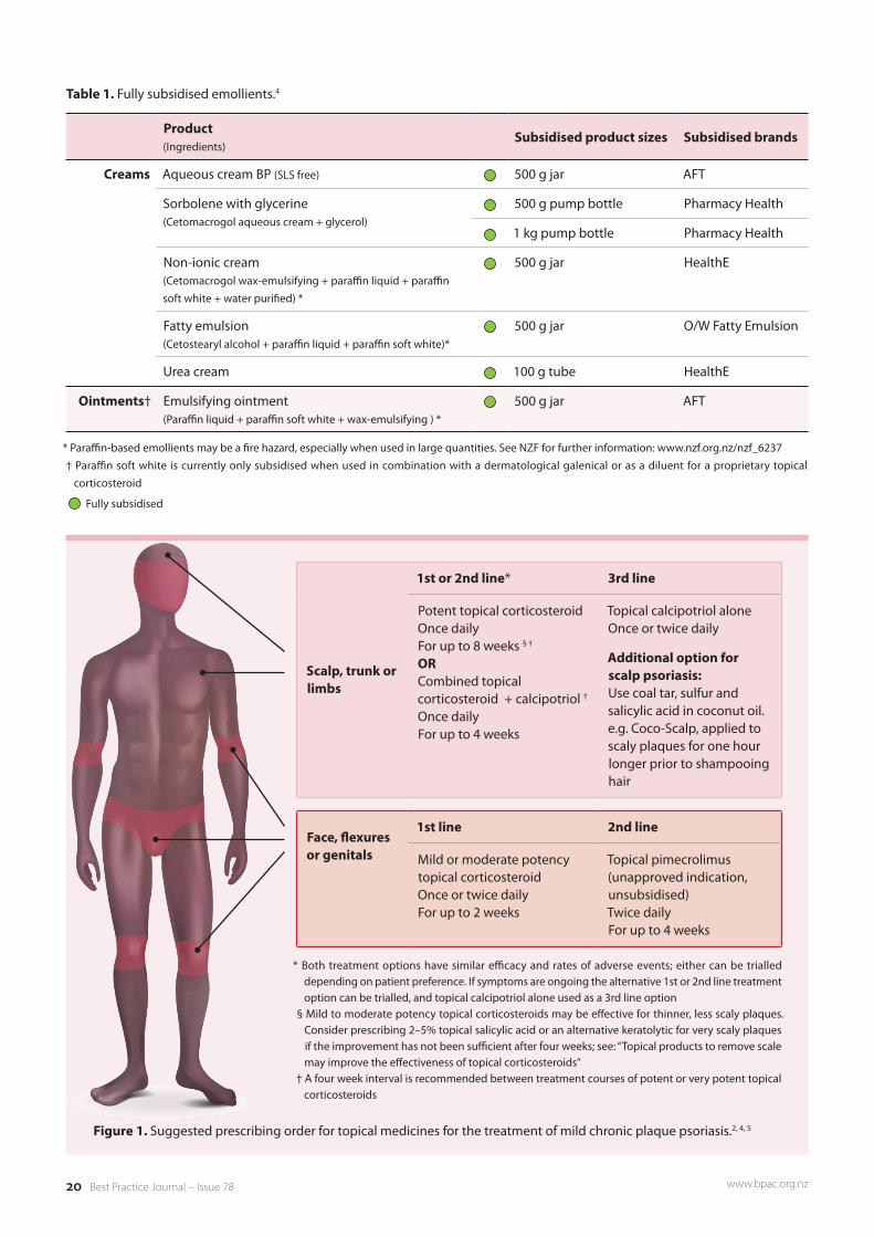
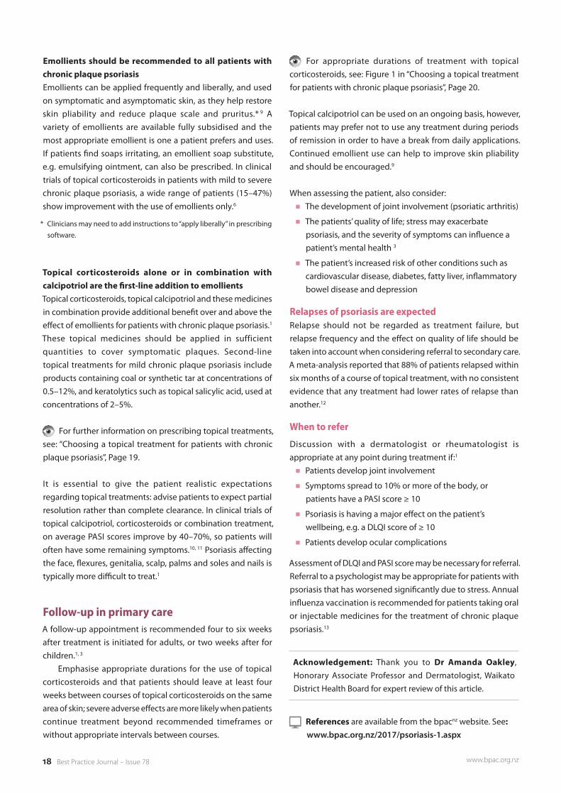
Figure 1. Suggested prescribing order for topical medicines for the treatment of mild chronic plaque psoriasis.2, 4, 5
20 Best Practice Journal – Issue 78 www.bpac.org.nz
Scalp, trunk or limbs
1st or 2nd line* 3rd line
Potent topical corticosteroidOnce dailyFor up to 8 weeks § †
ORCombined topical corticosteroid + calcipotriol †
Once dailyFor up to 4 weeks
Topical calcipotriol aloneOnce or twice daily
Additional option for scalp psoriasis:Use coal tar, sulfur and salicylic acid in coconut oil. e.g. Coco-Scalp, applied to scaly plaques for one hour longer prior to shampooing hair
Face, flexures or genitals
1st line 2nd line
Mild or moderate potency topical corticosteroidOnce or twice dailyFor up to 2 weeks
Topical pimecrolimus(unapproved indication, unsubsidised)Twice dailyFor up to 4 weeks
* Both treatment options have similar efficacy and rates of adverse events; either can be trialled depending on patient preference. If symptoms are ongoing the alternative 1st or 2nd line treatment option can be trialled, and topical calcipotriol alone used as a 3rd line option
§ Mild to moderate potency topical corticosteroids may be effective for thinner, less scaly plaques. Consider prescribing 2–5% topical salicylic acid or an alternative keratolytic for very scaly plaques if the improvement has not been sufficient after four weeks; see: “Topical products to remove scale may improve the effectiveness of topical corticosteroids”
† A four week interval is recommended between treatment courses of potent or very potent topical corticosteroids
Table 1. Fully subsidised emollients.4
Product(Ingredients)
Subsidised product sizes Subsidised brands
Creams Aqueous cream BP (SLS free) 500 g jar AFT
Sorbolene with glycerine(Cetomacrogol aqueous cream + glycerol)
500 g pump bottle Pharmacy Health
1 kg pump bottle Pharmacy Health
Non-ionic cream(Cetomacrogol wax-emulsifying + paraffin liquid + paraffin soft white + water purified) *
500 g jar HealthE
Fatty emulsion(Cetostearyl alcohol + paraffin liquid + paraffin soft white)*
500 g jar O/W Fatty Emulsion
Urea cream 100 g tube HealthE
Ointments† Emulsifying ointment (Paraffin liquid + paraffin soft white + wax-emulsifying ) *
500 g jar AFT
* Paraffin-based emollients may be a fire hazard, especially when used in large quantities. See NZF for further information: www.nzf.org.nz/nzf_6237† Paraffin soft white is currently only subsidised when used in combination with a dermatological galenical or as a diluent for a proprietary topical
corticosteroid
Fully subsidised

Best Practice Journal – Issue 78 21www.bpac.org.nz
Topical corticosteroids alone or in combination with calcipotriol are the first-line addition to emollientsTopical corticosteroids alone can be used as a first-line treatment for chronic plaque psoriasis affecting any part of the body (Figure 1). A range of topical corticosteroids are available partly or fully subsidised in New Zealand (Table 2).
Safe prescribing of topical corticosteroids: maximising benefit and minimising risk
Topical corticosteroids should be used intermittently, with short courses of two to eight weeks, depending on location of use and potency (Figure 1). Prolonged use of potent to very potent topical corticosteroids is associated with an increased risk of skin atrophy, striae and adrenal suppression.1, 2 In addition, ongoing use of topical corticosteroids can paradoxically result in poor control of psoriasis.2 Applying topical corticosteroids to widespread areas, e.g. 10% or more of the body, is not recommended due to the increased potential for systemic absorption; patients with psoriasis this widespread should be referred to a dermatologist as treatment with oral medicines is likely to be necessary.*
The use of emollients, bath oils and products containing salicylic acid may improve the response to topical corticosteroids (see below).2
Topical corticosteroids combined with antibacterial and antifungal medicines should not be routinely used as they provide no additional benefit for the majority of patients with psoriasis.
* The area covered by the patient’s palm with outstretched fingers (a “handprint”) is approximately equal to 1% of their body surface area.8
Combination topical corticosteroid + calcipotriol is also an appropriate first-line treatmentCalcipotriol is a topical vitamin D analogue indicated for the treatment of psoriasis. Combination treatment with both topical corticosteroids and topical calcipotriol is an appropriate first-line option for patients with psoriasis on the scalp, trunk or limbs (Figure 1).2 Combination treatment can be prescribed either as a pre-mixed formulation containing betamethasone dipropionate, available as a gel or ointment (Table 2), or topical calcipotriol (available fully subsidised as an ointment) and a topical corticosteroid can be prescribed separately for concurrent use; there is not clear evidence whether the pre-mixed combination formulation or use of each product separately gives better results.6
The combination product requires one application per day as opposed to two applications when these medicines are prescribed separately. However, prescribing separately enables a different potency of topical corticosteroid to be selected if required, e.g. if a potent topical corticosteroid is stepped down
to a mild or moderate potency topical corticosteroid as plaques improve.
The use of calcipotriol is associated with local adverse effects (see below), however, combining treatment with a topical corticosteroid results in less adverse effects than the use of calcipotriol alone.5
Calcipotriol alone is effective but associated with high rates of local adverse effects
Calcipotriol alone can be considered as a treatment for psoriasis on the scalp, trunk or limbs, applied once or twice daily to affected areas (Figure 1).4 However, local adverse effects such as burning, pruritus, peeling, dryness or erythema may be experienced by up to 35% of patients using calcipotriol.3 These typically reduce with ongoing use so patients can be encouraged to persist with treatment if tolerable. Use on the face is not recommended, and calcipotriol is more likely to irritate the flexures and groin than topical corticosteroids.11, 12 Patients should wash their hands after applying calcipotriol to prevent inadvertent application to other areas, such as the face.
Systemic effects from vitamin D analogues, such as hypercalcaemia and altered parathyroid hormone levels, are rare unless patients have renal disease or impaired calcium metabolism, or are applying more than 100 g per week, i.e. one tube of calcipotriol ointment or approximately three tubes of calcipotriol + betamethasone dipropionate gel or ointment per week.3, 4 There are no studies on the safety of calcipotriol during pregnancy, however, expert opinion is that use on localised areas during pregnancy or breastfeeding is unlikely to result in harm from systemic absorption.12–14
Emollients containing urea or salicylic acid may reduce the effectiveness of topical calcipotriol and should be applied at different times.9 If patients are undergoing phototherapy, calcipotriol should be applied after treatment sessions as phototherapy inactivates calcipotriol.10
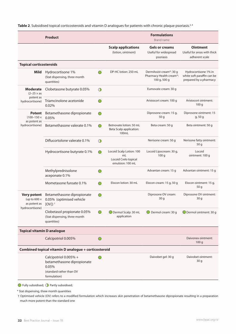
Topical products to remove scale may improve the effectiveness of topical corticosteroidsFor patients with thick scale, the use of a keratolytic, such as topical salicylic acid or urea, a coal tar preparation (Table 3), or oils such as olive oil or coconut oil, may soften plaques prior to application of topical corticosteroids.1, 2 Coco-Scalp ointment (containing coal tar) and topical salicylic acid or urea can be prescribed fully subsidised (see: “Prescribing topical salicylic acid”.
Coal tar products left on the skin may cause staining of clothes or skin.5 Patients may find coal tar products used during bathing, such as bath oils or shampoos, more convenient.
22 Best Practice Journal – Issue 78 www.bpac.org.nz
Table 2. Subsidised topical corticosteroids and vitamin D analogues for patients with chronic plaque psoriasis.2, 4
ProductFormulations
Brand name
Scalp applications (lotion, ointment)
gels or creamsUseful for widespread
psoriasis
OintmentUseful for areas with thick
adherent scale
Topical corticosteroids
Mild Hydrocortisone 1%(Stat dispensing, three month quantities)
DP-HC lotion: 250 mL DermAssist cream*: 30 gPharmacy Health cream*:
100 g, 500 g
Hydrocortisone 1% in white soft paraffin can be prepared by a pharmacy
Moderate(2–25 × as potent as
hydrocortisone)
Clobetasone butyrate 0.05% Eumovate cream: 30 g
Triamcinolone acetonide 0.02%
Aristocort cream: 100 g Aristocort ointment: 100 g
Potent (100–150 × as potent as
hydrocortisone)
Betamethasone dipropionate 0.05%
Diprosone cream: 15 g, 50 g
Diprosone ointment: 15 g, 50 g
Betamethasone valerate 0.1% Betnovate lotion: 50 mLBeta Scalp application:
100mL
Beta cream: 50 g Beta ointment: 50 g
Diflucortolone valerate 0.1% Nerisone cream: 50 g Nerisone fatty ointment: 50 g
Hydrocortisone butyrate 0.1% Locoid Scalp Lotion: 100 mL
Locoid Crelo topical emulsion: 100 mL
Locoid Lipocream: 30 g, 100 g
Locoidointment: 100 g
Methylprednisolone aceponate 0.1%
Advantan cream: 15 g Advantan ointment: 15 g
Mometasone furoate 0.1% Elocon lotion: 30 mL Elocon cream: 15 g, 50 g Elocon ointment: 15 g, 50 g
Very potent(up to 600 × as potent as
hydrocortisone)
Betamethasone dipropionate 0.05% (optimised vehicle [OV]) †
Diprosone OV cream: 30 g
Diprosone OV ointment: 30 g
Clobetasol propionate 0.05%(Stat dispensing, three month quantities)
Dermol Scalp: 30 mL application
Dermol cream: 30 g Dermol ointment: 30 g
Topical vitamin D analogue
Calcipotriol 0.005% Daivonex ointment:100 g
Combined topical vitamin D analogue + corticosteroid
Calcipotriol 0.005% + betamethasone dipropionate 0.05%(standard rather than OV formulation)
Daivobet gel: 30 g Daivobet ointment:30 g
Fully subsidised; Partly subsidised;
* Stat dispensing, three month quantities† Optimised vehicle (OV) refers to a modified formulation which increases skin penetration of betamethasone dipropionate resulting in a preparation
much more potent than the standard one
Best Practice Journal – Issue 78 23www.bpac.org.nz
Table 3. Coal tar products for patients with psoriasis. All products shown are available over-the-counter.
Subsidy Proportions of coal tar and other ingredients Product sizes
Coco-Scalp ointment * Coal tar 12% + salicylic acid 2% + sulphur 4% 40 g
EgoPsoryl TA gel** Coal tar solution + sulfur-precipitated + phenol 30, 75 g
Scytera foam 2% coal tar 12, 100 g
Polytar bath oil Tar 7.5% + cade oil 7.5% + coal tar 2.5% + arachis oil extract of coal tar 7.5%
350 mL
Ionil-T shampoo Coal tar 4.25% + salicylic acid 2% 200 mL
Neutrogena T/gel shampoo Coal tar 0.5% 200 mL
Polytar plus shampoo Coal tar 4% 150 mL
Sebitar shampoo Coal tar solution 1% + tar 1% + salicylic acid 2% 15, 250, 500 mL
Fully subsidised; Unsubsidised
* Coco-Scalp can be left on the scalp for an hour or longer, e.g. overnight, and then washed off 4
** Use with caution on face and flexures. Do not use under occlusion.
Prescribing topical salicylic acidSubsidised topical salicylic acid may be prepared in the pharmacy, with prescribers specifying the concentration (recommended at 2–5%) and base. Salicylic acid is also present in coal tar combination products (Table 3) and in many unsubsidised over-the-counter skincare products.Subsidised topical salicylic acid can be prescribed in two ways:1. Added to a proprietary topical corticosteroid
formulation (Table 2). These prescriptions must be endorsed by a dermatologist to be eligible for subsidy.
2. Added to an emollient; patients can apply the salicylic acid in an emollient base to soften plaques before applying a topical corticosteroid.2 There is no evidence regarding how long the interval should be between applications. Salicylic acid in an emollient base can be prescribed fully subsidised by a general practitioner, e.g. salicylic acid powder 5% in white soft paraffin, 100 g.
Topical salicylic acid should: Not be prescribed to:15
– Women who are pregnant, due to the potential for systemic absorption
Be used with caution in:
– Patients using topical calcipotriol: salicylic acid may reduce the effectiveness of topical calcipotriol and these medicines should be applied at different times of the day.4, 9
– Children aged under five years: topical salicylic acid use is recommended only in small patches and in concentrations of 0.5% or less.16
– Patients with widespread psoriasis or significant hepatic or renal impairment: the potential for toxicity, i.e. salicylism, is increased.3, 4
Patients should be aware that a sudden onset of symptoms such as difficulty breathing, swelling in the face or feeling faint may indicate acute hypersensitivity, although this is uncommon.17
For further information on prescribing topical products prepared in the pharmacy, see “Section C: Extemporaneously compounded products and galenicals”, in the Pharmaceutical Schedule: www.pharmac.govt.nz/healthpros/Schedule
Acknowledgement: Thank you to Dr Amanda Oakley,
Honorary Associate Professor and Dermatologist, Waikato
District Health Board for expert review of this article
References are available from the bpacnz website. See: www.bpac.org.nz/2017/psoriasis-2.aspx

24 Best Practice Journal – Issue 78 www.bpac.org.nz
Monitoring patients with moderate to severe psoriasis
DERMATOlOgy
Patients with moderate to severe psoriasis are usually managed in secondary care with the following treatments:
Phototherapy: suitable for most patients if topical treatments are not sufficient
Methotrexate: the preferred oral treatment for patients with chronic plaque psoriasis
Ciclosporin: typically used after trialling methotrexate
Acitretin: used if methotrexate and ciclosporin are inappropriate or unsuccessful
TNF inhibitors: initiated if other treatments are unsuccessful
Clinicians in primary care may provide repeat prescriptions for these treatments. There needs to be a clear understanding between the dermatologist and general practitioner regarding the responsibility for monitoring patients and requesting blood tests; adverse effects of these medicines can be serious and potentially fatal.
Phototherapy is suitable for most patients if topical treatments are not sufficientPhototherapy is used for patients with moderate to severe
chronic plaque psoriasis who have not responded sufficiently to topical treatments.1 It is most effective for thin plaques on the trunk and limbs.
Narrow band UV-B (NB-UVB) is the preferred form of phototherapy, which involves multiple exposures over six to twelve weeks, that are initially a minute or less and extended over subsequent sessions.2, 3 Other forms of phototherapy, such as psoralen with UV-A exposure (PUVA), are no longer preferred treatments for chronic plaque psoriasis but may be used in select patients.
Tanning beds or sun exposure cannot be used as substitutes for phototherapy. They are less effective in the treatment of psoriasis and have a much wider range of wavelengths than NB-UVB phototherapy with a higher risk of skin damage.4, 5
Patients should not apply topical products prior to phototherapy sessions
Patients are advised not to apply topical products such as salicylic acid or sunscreen prior to phototherapy sessions, as they can interact with UV-B treatment. Calcipotriol, if used, should be applied after rather than before treatment, as it is inactivated by UV light.5
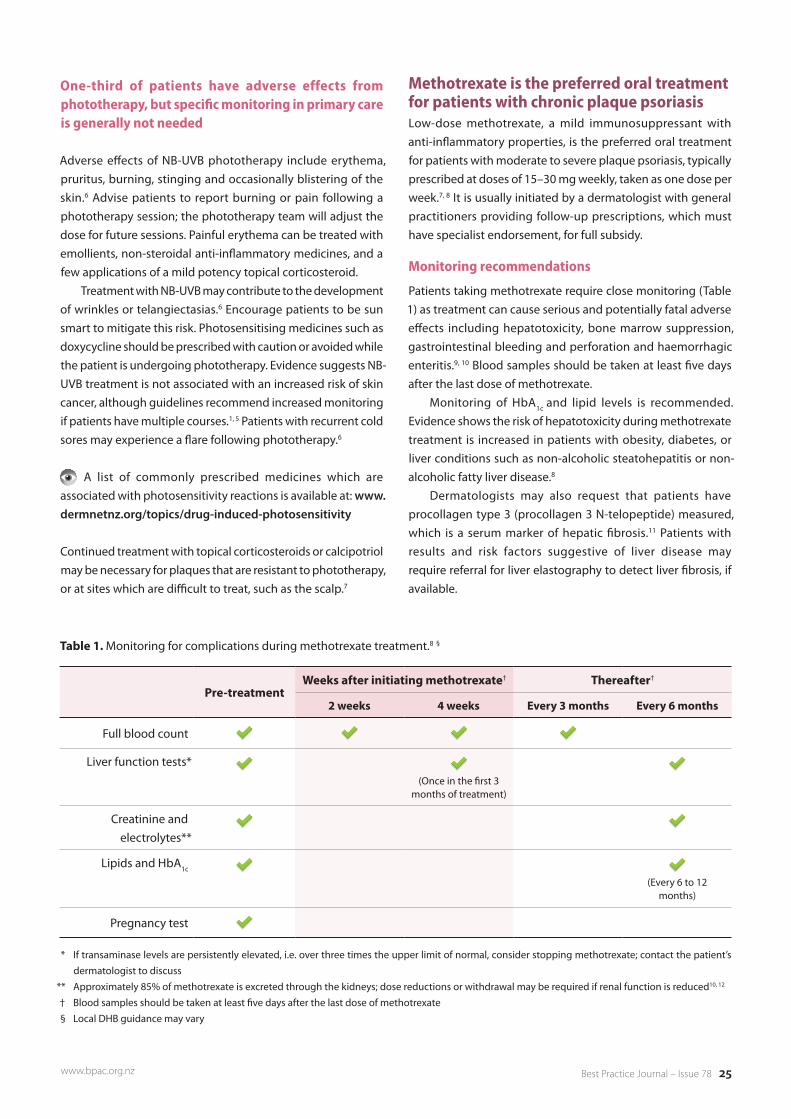
Best Practice Journal – Issue 78 25www.bpac.org.nz
One-third of patients have adverse effects from phototherapy, but specific monitoring in primary care is generally not needed
Adverse effects of NB-UVB phototherapy include erythema, pruritus, burning, stinging and occasionally blistering of the skin.6 Advise patients to report burning or pain following a phototherapy session; the phototherapy team will adjust the dose for future sessions. Painful erythema can be treated with emollients, non-steroidal anti-inflammatory medicines, and a few applications of a mild potency topical corticosteroid.
Treatment with NB-UVB may contribute to the development of wrinkles or telangiectasias.6 Encourage patients to be sun smart to mitigate this risk. Photosensitising medicines such as doxycycline should be prescribed with caution or avoided while the patient is undergoing phototherapy. Evidence suggests NB-UVB treatment is not associated with an increased risk of skin cancer, although guidelines recommend increased monitoring if patients have multiple courses.1, 5 Patients with recurrent cold sores may experience a flare following phototherapy.6
A list of commonly prescribed medicines which are associated with photosensitivity reactions is available at: www.dermnetnz.org/topics/drug-induced-photosensitivity
Continued treatment with topical corticosteroids or calcipotriol may be necessary for plaques that are resistant to phototherapy, or at sites which are difficult to treat, such as the scalp.7
Methotrexate is the preferred oral treatment for patients with chronic plaque psoriasisLow-dose methotrexate, a mild immunosuppressant with anti-inflammatory properties, is the preferred oral treatment for patients with moderate to severe plaque psoriasis, typically prescribed at doses of 15–30 mg weekly, taken as one dose per week.7, 8 It is usually initiated by a dermatologist with general practitioners providing follow-up prescriptions, which must have specialist endorsement, for full subsidy.
Monitoring recommendations
Patients taking methotrexate require close monitoring (Table 1) as treatment can cause serious and potentially fatal adverse effects including hepatotoxicity, bone marrow suppression, gastrointestinal bleeding and perforation and haemorrhagic enteritis.9, 10 Blood samples should be taken at least five days after the last dose of methotrexate.
Monitoring of HbA1c and lipid levels is recommended. Evidence shows the risk of hepatotoxicity during methotrexate treatment is increased in patients with obesity, diabetes, or liver conditions such as non-alcoholic steatohepatitis or non-alcoholic fatty liver disease.8
Dermatologists may also request that patients have procollagen type 3 (procollagen 3 N-telopeptide) measured, which is a serum marker of hepatic fibrosis.11 Patients with results and risk factors suggestive of liver disease may require referral for liver elastography to detect liver fibrosis, if available.
Table 1. Monitoring for complications during methotrexate treatment.8 §
Pre-treatmentweeks after initiating methotrexate† Thereafter†
2 weeks 4 weeks Every 3 months Every 6 months
Full blood count
Liver function tests*(Once in the first 3
months of treatment)
Creatinine and electrolytes**
Lipids and HbA1c
(Every 6 to 12 months)
Pregnancy test
* If transaminase levels are persistently elevated, i.e. over three times the upper limit of normal, consider stopping methotrexate; contact the patient’s dermatologist to discuss
** Approximately 85% of methotrexate is excreted through the kidneys; dose reductions or withdrawal may be required if renal function is reduced10, 12
† Blood samples should be taken at least five days after the last dose of methotrexate§ Local DHB guidance may vary
26 Best Practice Journal – Issue 78 www.bpac.org.nz
Pulmonary toxicity is less common in patients with psoriasis taking low-dose methotrexate than in patients with rheumatoid arthritis and specific monitoring of lung function is not required; however, inform patients of potential symptoms, such as dry cough, which could indicate pulmonary toxicity.8, 12 Bone marrow suppression is rare during low-dose methotrexate treatment; when it does occur, this is often associated with prescribing or dispensing errors, reduced renal function or medicines interactions.11
Other adverse effects of methotrexate include fatigue, nausea and headaches; less commonly, mouth ulcers; and rarely, hair loss.13
Safe prescribing of repeat methotrexate prescriptions
Cases of serious toxicity, including deaths, have occurred when methotrexate has been taken daily instead of weekly, often due to prescribing or dispensing errors.
Key points for safe prescribing include:8, 9, 14
Prescribe methotrexate as a specific weekly dose, rather than “as directed”
Prescribe 5 mg folic acid, once per week, to be taken at least two days after methotrexate. Methotrexate and folic acid tablets are both yellow.
Write days of administration out in full on prescriptions
Prescribe one strength of tablet at a time, where possible. If both 10 mg and 2.5 mg tablets are prescribed, point out to patients the differences in tablets, especially if doses change.
Warn patients to report symptoms that could indicate:
– Bone marrow suppression, e.g. fever, sore throat, mouth ulcers
– Hepatotoxicity, e.g. jaundice, abdominal pain
– Pulmonary toxicity, e.g. dry cough, dyspnoea
Methotrexate interacts with many medicines which can result in increased toxicity and serious adverse effects. For example, serious bone marrow suppression can occur due to interaction with folate antagonists, particularly trimethoprim or co-trimoxazole (trimethoprim + sulfamethoxazole).8 Check for drug interactions using the NZF interactions checker: www.nzf.org.nz/nzf_1
Immunisation with live vaccines during treatment should be avoided
Methotrexate can cause birth deformities and is contraindicated during pregnancy and lactation.15,16 Whether use in males poses a risk is controversial, however, guidelines and manufacturers advise that both males and females should use appropriate contraception during, and for at least three months following, treatment.10, 11
Advise patients to limit alcohol intake to no more than one to two standard drinks once or twice a week10
Ensure that patients receive clear instructions in written form. A patient information sheet is available from: www.saferx.co.nz/methotrexate-patient-guide.pdf
Discuss with the patient’s dermatologist if their symptoms fail to improve: methotrexate can be titrated according to clinical response, at recommended increments of 2.5–5 mg per week.10 It may take up to four weeks for a response after increasing doses.17
For further information on adverse effects associated with methotrexate use, see: www.bpac.org.nz/BPJ/2014/October/safer-prescribing.aspx
Ciclosporin is typically used after trialling methotrexateFor most patients with chronic plaque psoriasis ciclosporin is a second-line oral treatment, after methotrexate.7 Ciclosporin can be prescribed by any medical practitioner but is likely to be initiated in secondary care by a dermatologist. For the treatment of chronic plaque psoriasis it is typically taken in short courses of two to four months in doses of 2.5–5 mg per kg per day.7, 10
Monitoring for nephrotoxicity or hypertension
Ciclosporin can cause nephrotoxicity and hypertension and frequent monitoring is recommended, particularly during the first months of use (Table 2). Older patients or those with obesity or diabetes are at an increased risk of nephrotoxicity.18
Common adverse effects
Adverse effects from ciclosporin use include headache, respiratory symptoms such as cough and rhinitis, musculoskeletal pain or paraesthesia. Hypertrichosis (an increased growth rate of existing hair) occurs in approximately 5% of patients.17 There is an increased risk of non-melanoma skin cancer in patients taking ciclosporin who have also had psoralen with ultraviolet light (PUVA) treatment.17,18
Safe prescribing of repeat ciclosporin prescriptions
Key points for safe prescribing include:10, 18
Check interactions when initiating other medicines: Ciclosporin is metabolised by cytochrome P450 3A4 and interactions can cause changes in ciclosporin concentrations or the concentration of other medicines. The NZF interactions checker is available at: www.nzf.org.nz/nzf_1. Patients should avoid consuming grapefruit or grapefruit juice as this may increase ciclosporin levels.17
Patients should not take ciclosporin if they are also undergoing phototherapy17
Best Practice Journal – Issue 78 27www.bpac.org.nz
Immunisation with live vaccines during treatment should be avoided. The efficacy of other vaccinations may be reduced.
Discuss contraception and pregnancy plans with female patients: ciclosporin is not contraindicated during pregnancy but has been associated with adverse pregnancy outcomes. Discuss the use of ciclosporin during pregnancy with the patient’s dermatologist. Use during breastfeeding is not advised as infants require monitoring of blood levels of ciclosporin and regular review for signs of toxicity.15
Ask patients to report symptoms of infections, e.g. fever, chills, sore throat, mouth ulcers, as these may take longer to clear or increase in severity while taking ciclosporin. Infections may also indicate leucopenia.
Ask patients to report symptoms of liver toxicity such as jaundice and abdominal pain
Acitretin may be used if methotrexate and ciclosporin are inappropriate or unsuccessful
Acitretin is an oral retinoid which is typically used in patients with chronic plaque psoriasis if other treatments are not tolerated or have been unsuccessful; it is less effective than methotrexate or ciclosporin and has mucocutaneous adverse effects.20 Acitretin is highly teratogenic and should generally be avoided in women of reproductive age.
Acitretin is fully subsidised with Special Authority approval. Female patients must have been informed of the teratogenic potential of acitretin and agree to use appropriate contraception while taking acitretin as well as for at least two years afterwards.16, 20 Two forms of contraception are recommended, e.g. condoms and an intrauterine device or hormonal contraceptive. In addition, since progesterone-only pills must be taken within a three hour window these are not recommended as one of the forms of contraception.21 Acitretin is teratogenic regardless of the treatment duration or dosage used.20
Monitor patients for elevated triglycerides and lipid levels or altered liver function testsFrequent monitoring of lipid profile is recommended (Table 3) as up to one-third of patients taking acitretin develop hypertriglyceridaemia or hypercholesterolaemia and patients with psoriasis are already at increased risk of cardiovascular disease.20, 22
Elevations in triglycerides while taking acitretin should be managed by lifestyle changes. Discuss with a dermatologist if a patient meets one of the following criteria:22
Five year cardiovascular risk is > 20%
Triglyceride levels are ≥ 5.7 mmol/L AND patients have at least one risk factor, e.g.:
– Elevated blood pressure (> 170/100 mmHg)
– Angina
– Ischaemic heart disease
– Peripheral arterial disease
– Diabetic nephropathy
– A total cholesterol:high density lipoprotein ratio > 8
– A genetic lipid disorder
Depending on a patient’s circumstances, stopping acitretin may be appropriate; changes in lipid profile usually improve four to eight weeks after acitretin is stopped.20
Acitretin causes dryness of mucous membranes
Patients commonly experience dry lips and mouth, and may experience stomatitis, taste disturbances or symptoms such as nose bleeds, rhinitis and dryness of the eyes.20 A topical emollient for dry lips and eye drops can help, especially in patients who use contact lenses.20 Additional emollients may be required for use on areas of dry skin, or topical corticosteroids if patients develop asteatotic dermatitis.
Acitretin can also cause thinning of the skin and nails and alopecia, which can increase sun sensitivity. Thinning of the skin affects the whole body but is particularly noticeable on the palms and soles of the feet, and can be accompanied by redness and scaling. Patients may have difficulty grasping objects or placing pressure on the feet while walking.20 Up to 75% of patients experience some degree of hair loss, with alopecia occurring in 10% of patients.20
Safe prescribing of repeat acitretin prescriptions Ensure females of childbearing potential use two
forms of contraception and reinforce the need to avoid pregnancy for at least two years post-treatment23
Advise female patients of childbearing potential not to drink alcohol.20, 21 This can increase the metabolism of acitretin into etretinate, which has a half-life of 168 days and contributes to teratogenicity after stopping treatment.17
Patients taking acitretin should not donate blood during treatment and for two years following treatment21
Tetracyclines should not be used with acitretin as both increase the risk of intracranial hypertension10
TNF inhibitors can be initiated if other treatments are unsuccessfulThe TNF inhibitors adalimumab and etanercept are subsidised with Special Authority approval for the treatment of moderate to severe plaque psoriasis.10 Applications for Special Authority approval must be made by a dermatologist and patients must have trialled at least three other treatments of phototherapy, methotrexate, ciclosporin or acitretin, as well as have a psoriasis area and severity index (PASI) score > 15 on treatment.*10
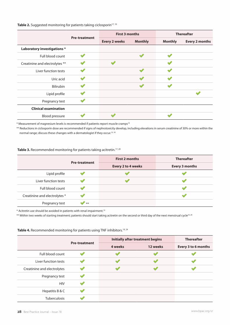
28 Best Practice Journal – Issue 78 www.bpac.org.nz
Table 2. Suggested monitoring for patients taking ciclosporin17, 19
Pre-treatmentFirst 3 months Thereafter
Every 2 weeks Monthly Monthly Every 2 months
laboratory investigations *
Full blood count
Creatinine and electrolytes **
Liver function tests
Uric acid
Bilirubin
Lipid profile
Pregnancy test
Clinical examination
Blood pressure
* Measurement of magnesium levels is recommended if patients report muscle cramps19
** Reductions in ciclosporin dose are recommended if signs of nephrotoxicity develop, including elevations in serum creatinine of 30% or more within the normal range; discuss these changes with a dermatologist if they occur.10, 18
Table 3. Recommended monitoring for patients taking acitretin.17, 20
Pre-treatmentFirst 2 months Thereafter
Every 2 to 4 weeks Every 3 months
Lipid profile
Liver function tests
Full blood count
Creatinine and electrolytes *
Pregnancy test **
* Acitretin use should be avoided in patients with renal impairment.10
** Within two weeks of starting treatment; patients should start taking acitretin on the second or third day of the next menstrual cycle10, 20
Table 4. Recommended monitoring for patients using TNF inhibitors.19, 24
Pre-treatmentInitially after treatment begins Thereafter
4 weeks 12 weeks Every 3 to 6 months
Full blood count
Liver function tests
Creatinine and electrolytes
Pregnancy test
HIV
Hepatitis B & C
Tuberculosis
Best Practice Journal – Issue 78 29www.bpac.org.nz
Renewal applications can be made by general practitioners on the recommendation of a dermatologist.
* For further information on the PASI score, see: “Chronic plaque psoriasis: an overview of treatment in primary care”
Monitoring recommendations and safe prescribing of repeat prescriptions
Patients should be tested to exclude tuberculosis, HIV, hepatitis B or C, or active infection prior to initiating TNF inhibitor treatment (Table 4).19 Monitoring of full blood count, liver and renal function is required as TNF inhibitors can increase the risk of infections, and have infrequently been associated with thrombocytopenia or leucopoenia, renal impairment, and autoimmune-like syndromes.10, 19
The use of TNF inhibitors is not recommended in patients who have developed cancer within the past five years or in patients with moderate to severe heart failure.10, 24
Injection site reactions and flu-like symptoms are commonTNF inhibitors are administered by subcutaneous injection and pain and irritation at the injection site are reported by 15–37% of patients, usually lasting for three to five days.24, 25 Following a dose patients may develop flu-like symptoms, such as chills, headache, musculoskeletal pain and nausea. Patients typically do not find injection site reactions or flu-like symptoms severe enough to withdraw from treatment and these adverse reactions may improve with subsequent doses.24, 25
Safe prescribing of TNF inhibitors: Women who are pregnant or breastfeeding should not
use TNF inhibitors19
Immunisation with live vaccines during treatment should be avoided24
TNF inhibitors should be withdrawn if patients develop a serious infection, or in patients who have new onset or worsening of heart failure25
Advise patients to report worsening fever, sore throat, bruising or bleeding as these may be symptoms of blood disorders such as aplastic anaemia or pancytopaenia10, 24
Acknowledgement: Thank you to Dr Amanda Oakley,
Honorary Associate Professor and Dermatologist, Waikato
District Health Board for expert review of this article.
References:1. Scottish Intercollegiate Guidelines Network (SIGN). Guideline 121: Diagnosis
and management of psoriasis and psoriatic arthritis in adults. 2010. Available from: www.sign.ac.uk/guidelines/fulltext/121/index.html (Accessed Jan, 2017).
2. Chen X, Yang M, Cheng Y, et al. Narrow-band ultraviolet B phototherapy versus broad-band ultraviolet B or psoralen-ultraviolet A photochemotherapy for psoriasis. Cochrane Database Syst Rev 2013;10:CD009481. doi:10.1002/14651858.CD009481.pub2
3. Paul C, Gallini A, Archier E, et al. Evidence-based recommendations on topical
treatment and phototherapy of psoriasis: systematic review and expert opinion of a panel of dermatologists. J Eur Acad Dermatol Venereol 2012;26 Suppl 3:1–10. doi:10.1111/j.1468-3083.2012.04518.x
4. National Psoriasis Foundation. Light therapy. 2015. Available from: www.psoriasis.org/sites/default/files/light_therapy-12-7-2015.pdf (Accessed Jan, 2017).
5. Menter A, Korman NJ, Elmets CA, et al. Guidelines of care for the management of psoriasis and psoriatic arthritis: Section 5. Guidelines of care for the treatment of psoriasis with phototherapy and photochemotherapy. J Am Acad Dermatol 2010;62:114–35. doi:10.1016/j.jaad.2009.08.026
6. Mehta D, Lim HW. Ultraviolet B phototherapy for psoriasis: review of practical guidelines. Am J Clin Dermatol 2016;17:125–33. doi:10.1007/s40257-016-0176-6
7. National Institutes for Health and Care Excellence (NICE). Psoriasis: assessment and management. 2012. Available from: www.nice.org.uk/guidance/cg153 (Accessed Jan, 2017).
8. Rademaker M, Gupta M, Andrews M, et al. The Australasian Psoriasis Collaboration view on methotrexate for psoriasis in the Australasian setting. Australas J Dermatol 2016; [Epub ahead of print]. doi:10.1111/ajd.12521
9. Waitemata District Health Board. Methotrexate. Safe prescribing - once a week! 2014. Available from: www.saferx.co.nz/full/methotrexate.pdf (Accessed Jan, 2017).
10. New Zealand Formulary (NZF). NZF v54. 2016. Available from: www.nzf.org.nz (Accessed Dec, 2016)
11. Warren RB, Weatherhead SC, Smith CH, et al. British Association of Dermatologists’ guidelines for the safe and effective prescribing of methotrexate for skin disease 2016. Br J Dermatol 2016;175:23–44. doi:10.1111/bjd.14816
12. Kalb RE, Strober B, Weinstein G, et al. Methotrexate and psoriasis: 2009 National Psoriasis Foundation Consensus Conference. J Am Acad Dermatol 2009;60:824–37. doi:10.1016/j.jaad.2008.11.906
13. Robinson S, Gibson S, George E, et al. Tolerability and adherence problems in patients on a stable dose of methotrexate: results of a multicentre survey. Musculoskeletal Care 2016;14:152–5. doi:10.1002/msc.1129
14. Rex Medical Ltd. Trexate New Zealand Datasheet. 2014. Available from: www.medsafe.govt.nz/profs/datasheet/t/trexatetab.pdf (Accessed Jan, 2017).
15. Butler DC, Heller MM, Murase JE. Safety of dermatologic medications in pregnancy and lactation: Part II. Lactation. J Am Acad Dermatol 2014;70:417.e1-10. doi:10.1016/j.jaad.2013.09.009
16. Murase JE, Heller MM, Butler DC. Safety of dermatologic medications in pregnancy and lactation: Part I. Pregnancy. J Am Acad Dermatol 2014;70:401.e1-14. doi:10.1016/j.jaad.2013.09.010
17. Menter A, Korman NJ, Elmets CA, et al. Guidelines of care for the management of psoriasis and psoriatic arthritis: section 4. Guidelines of care for the management and treatment of psoriasis with traditional systemic agents. J Am Acad Dermatol 2009;61:451–85. doi:10.1016/j.jaad.2009.03.027
18. Rosmarin DM, Lebwohl M, Elewski BE, et al. Cyclosporine and psoriasis: 2008 National Psoriasis Foundation Consensus Conference. J Am Acad Dermatol 2010;62:838–53. doi:10.1016/j.jaad.2009.05.017
19. Nast A, Gisondi P, Ormerod AD, et al. European S3-Guidelines on the systemic treatment of psoriasis vulgaris. Update 2015. EDF in cooperation with EADV and IPC. Zurich: European Dermatology Forum 2015. Available from: www.euroderm.org/edf/index.php/edf-guidelines/category/5-guidelines-miscellaneous (Accessed Jan, 2017).
20. Ormerod AD, Campalani E, Goodfield MJD, et al. British Association of Dermatologists guidelines on the efficacy and use of acitretin in dermatology. Br J Dermatol 2010;162:952–63. doi:10.1111/j.1365-2133.2010.09755.x
21. Douglas Pharmaceuticals Limited. Novatretin datasheet. 2011. Available from: www.medsafe.govt.nz/profs/datasheet/n/novatretincap.pdf (Accessed Feb, 2017).
22. Chan BCY, Reid N, Armour K, et al. Hypertriglyceridaemia with acitretin use: a proposal for its management in the context of overall cardiovascular risk. Br J Dermatol 2014;171:665–7. doi:10.1111/bjd.13027
23. Ministry of Health. Application for subsidy by Special Authority. Acitretin. Form SA1476. 2017. Available from: www.pharmac.govt.nz/2017/02/01/SA1476.pdf (Accessed Feb, 2017).
24. Smith CH, Anstey AV, Barker JNWN, et al. British Association of Dermatologists’ guidelines for biologic interventions for psoriasis 2009. Br J Dermatol 2009;161:987–1019. doi:10.1111/j.1365-2133.2009.09505.x
25. Menter A, Gottlieb A, Feldman SR, et al. Guidelines of care for the management of psoriasis and psoriatic arthritis: Section 1. Overview of psoriasis and guidelines of care for the treatment of psoriasis with biologics. J Am Acad Dermatol 2008;58:826–50. doi:10.1016/j.jaad.2008.02.039

30 Best Practice Journal – Issue 78 www.bpac.org.nz
Access to HPV vaccine widened
IMMuNISATION MEDICINE SubSIDy PublIC HEAlTH SExuAl HEAlTH
The human papillomavirus virus (HPV) vaccine reduces the incidence of HPV-related diseases, including genital warts and cervical cancer, as well as anogenital and oropharyngeal cancers. It is most effective if it is given before sexual activity begins. The quadrivalent HPV vaccine (Gardasil) has been part of the National Immunisation Schedule since 2008 and is currently subsidised for females aged under 20 years and additional groups at increased risk, e.g. transplant patients.1
PHARMAC recently announced changes to subsidised access to the HPV vaccine in New Zealand.2 From 1 January, 2017:
A nonavalent vaccine (Gardasil 9) which protects against five additional HPV types will be subsidised
Males will now be eligible for subsidised access
The age range for subsidised access will be extended to 9–26 years
The dosing schedule will change for children aged 9–14 years
HPV vaccination: how successful has the program been?
Coverage has remained low: The HPV vaccination program in New Zealand began in 2008 and has been subsidised for females up to age 20 years. Since 2010, approximately 50% of females in the eligible age range have received HPV vaccination.3 The Ministry of Health aims to increase coverage with a target of 75% of girls aged 12 years being vaccinated by the end of 2017.3
More time is required to evaluate the effect on rates of cervical cancer: Girls who were vaccinated in the first year of the school-based program will now be aged approximately 20 years. Females who were vaccinated outside of the school-based program at an older age will now be aged in their early to mid-20s, the youngest age at which cervical cancer is usually diagnosed.4 The effect of New Zealand’s HPV vaccination programme is therefore likely to become apparent over the next decade. Rates of cervical cancer averaged 6.6 per 100,000 women in the five years prior to introduction of the HPV vaccine (2003–2007), with rates in recent years of 6.3 per 100,000 in 2012–2013, and 5.4 per 100,000 in 2014.4, 5 In Australia, which has one of the highest rates of HPV vaccine uptake in the

Best Practice Journal – Issue 78 31www.bpac.org.nz
world, the rate of high grade cervical abnormalities has fallen in women aged under 20 years, with a 46% reduction in 2011 compared to the years prior to introduction of the HPV vaccine. A smaller reduction of 12% was also observed in women aged 20–24 years.6
The incidence of genital warts has decreased: The majority of cases of genital warts are caused by HPV types 6 and 11, which are included in the HPV vaccine.7 The incidence of genital warts in New Zealand has reduced since the HPV vaccination program began, with the greatest reduction in incidence seen in females in their teens and those aged 20–24 years.8,
9 Dispensing rates of medicines indicated for the treatment of genital warts have also decreased.10 From 2009 to 2012, genital warts cases decreased by 32% at sexual health clinics and decreased by 52% at family planning clinics.8 In Australia, where a vaccine coverage of 70% has been achieved, a 93% reduction in the incidence of genital warts in eligible age groups has been recorded.11
The new HPV vaccine will protect against five additional HPV types
The existing HPV vaccine provides immunity against four HPV types: 6, 11, 16 and 18. The new nonavalent HPV vaccine provides immunity against nine HPV types: 6, 11, 16, 18, 31, 33, 45, 52 and 58. Clinical trials of the nonavalent vaccine show that it generates an equivalent antibody response compared to the quadrivalent vaccine against HPV types 6, 11, 16, and 18.12
The new nonavalent vaccine may offer broader protection against cervical cancer and other HPV-related cancers. HPV types 16 and 18 are estimated to cause approximately 70% of cancers of the cervix, vagina and anus, and 30–40% of cancers of the vulva, penis and oropharynx.13 The HPV types 31, 33, 45, 52 and 58, covered by the nonavalent vaccine are thought to cause up to 20% of cervical cancers and to contribute up to approximately 20% of other HPV-related cancers.14, 15
In a large clinical trial comparing the effectiveness of the quadrivalent and nonavalent vaccines in women aged 16 to 26 years, there were no overall differences in the rate of high-grade cervical, vulvar or vaginal pre-malignant lesions. However, among women who did not have HPV infection and had normal pap smear results at the start of the trial, the nonavalent vaccine reduced the incidence of high-grade cervical, vulvar or vaginal pre-malignant lesions attributable to HPV types 31, 33, 45, 52, 58 by more than 96% (95% confidence interval 81–100%); 30 women out of approximately 6000 women who received the quadrivalent HPV vaccine developed high-grade cervical, vulvar or vaginal pre-malignant lesions due to these HPV types compared to one out of approximately 6000 who received the nonavalent HPV vaccine.12, 16
Eligibility criteria for the subsidised HPV vaccine will be widened
Females and males aged 9 to 26 years will be eligible for subsidised HPV vaccination from January, 2017:
A maximum of two doses will be funded for males and females aged 14 years and under.
A maximum of three doses will be funded for patients aged 15–26 years, transplant patients (including people receiving stem cell treatments) and those aged 26 years and under who have received chemotherapy.
Males will now be eligible for HPV vaccination
Since the HPV vaccine programme began more evidence has accumulated regarding potential benefits to males. Clinical trials have shown HPV vaccination in males can reduce the incidence of genital warts and anogenital cancer.17, 18 It is also expected to reduce the incidence of oropharyngeal cancer; the incidence of this cancer in males in New Zealand has doubled from 1995 to 2010.19
Modelling suggests that heterosexual males will benefit from vaccination as current uptake rates among eligible females are approximately 50%, and an uptake of 80% in females is required for maximal herd immunity for heterosexual males.20 The current vaccination strategy also does not benefit men who have sex with men, as they do not benefit from herd immunity conferred by vaccinated female partners and are at higher risk for anogenital and oropharyngeal warts and cancers than heterosexual men.21, 22
In Australia, both boys and girls have received school-based HPV vaccination since 2013.23
People aged 9 to 26 years now eligible for free HPV vaccination
The HPV vaccine will not confer protection against previously acquired infection, however, it does protect against future infections of HPV types not yet acquired. First HPV infections typically occur within a few years of becoming sexually active, and data in New Zealand show that approximately 8% of adolescents are sexually active by age 13 years.8
The new nonavalent vaccine is indicated and subsidised from age 9 years: the same age as people included in clinical trials of the vaccine.12, 24 Vaccination will typically begin at age 11 years as part of the school-based HPV vaccination programme, which is consistent with international programmes which offer vaccination from ages 11–13 years.18, 23 This is expected to move from Year 8 to Year 7 in the future in order to align with the tetanus, diphtheria and acellular pertussis (Tdap) immunisations.2, 3, 20
People in the eligible age range who are too old to receive school-based vaccination can be vaccinated in primary care.
32 Best Practice Journal – Issue 78 www.bpac.org.nz
Although the immune response to the nonavalent vaccine is highest in younger individuals aged 9 to 15 years, in clinical trials of nonavalent vaccine over 99% of males and females aged 16 to 26 years showed antibody responses to all nine HPV types.24
The dosing schedule will change for children aged 9–14 years
The dosing schedule for HPV vaccines will change from January, 2015:*2, 16
People aged 9–14 years: two doses at least six months apart**
People aged 15–26 years: three doses, at zero, two and six months
* Transplant patients, those receiving stem cell treatments and people who have received chemotherapy require three doses at zero, two and six months, regardless of age, unless an alternative schedule is advised28
** If females aged under 14 years have already had two doses of the quadrivalent HPV vaccine , a third dose will be required if the two doses were administered less than six months apart28
The dosing schedule has been reduced for younger people because research has shown a two-dose course is as effective as the three-dose course in this age group.25–27 Reducing the number of vaccines in the schedule will decrease the cost of the vaccination programme and may increase patient compliance with the schedule.
Offer eligible patients HPV vaccination from 1 January 2017
Primary care clinicians are encouraged to discuss HPV vaccination with eligible patients, particularly with the following groups:
Non-immunised school-aged males in Year 9 and above, as they will miss the school-based programme
Non-immunised females and males aged under 27 years; those aged 26 years will have limited opportunity to receive free vaccination before turning 27 years*
Non-immunised people aged 14 years, who might be eligible for a two-dose vaccine regimen before turning 15 years when they would require the three-dose regimen
Non-immunised people aged between 9 and 26 years who declined their school vaccination programme
* Provided the first dose is given prior to turning 27 years, the subsidised course can still be completed
Updating recall processes ensures young males who do not complete their HPV vaccine dosing schedule in school are followed-up.28
For further information on the HPV vaccine programme, see: www.immune.org.nz/education-and-training/hpv-vaccination-module
Transitioning between subsidised vaccines
A transition period will occur while stocks of the quadrivalent HPV vaccine are used up. Once this occurs in early 2017, practices can then administer the new nonavalent vaccine. Females who have started the quadrivalent vaccine course will be able to complete this in early 2017. If patients seek HPV vaccination in early 2017, it is recommended that the quadrivalent vaccine is administered if it is still available.28 If stocks of the quadrivalent vaccine are used up prior to patients finishing their dosing schedule, they will be able to switch to the new nonavalent HPV vaccine.28 The school-based vaccination programme will administer the nonavalent HPV vaccine from 2017, and the quadrivalent vaccine will be delisted from 1 October, 2017.2, 28
Acknowledgement: Thank you to Dr Nikki Turner, Director
of the Immunisation Advisory Centre (IMAC) and Associate
Professor in the Division of General Practice and Primary
Health Care, University of Auckland for expert review of this
article.
References are available from the bpacnz website. See: www.bpac.org.nz/2016/hpv.aspx
Best Practice Journal – Issue 78 33www.bpac.org.nz
Questions which clinicians may be asked during discussion with parents or patients include:
what are the benefits of the new vaccine? The new vaccine and wider subsidised access is likely to result in a greater reduction in the incidence of cervical cancer and other HPV-related diseases in both males and females.
If HPV is a sexually transmitted infection, why does my child need this now? HPV vaccination is most effective when it is given before sexual activity begins. Vaccination is recommended between the ages of 11 to 13 years as the vast majority of New Zealand adolescents of this age will not have been exposed to HPV.8 Vaccinating at this age should not be interpreted as an expectation that the child is about to engage in sexual activity or that permission is given for sexual activity to begin. Research shows that HPV vaccination does not influence the age of onset of sexual activity, condom usage, number of sexual partners, or rates of STI diagnosis, pregnancy or termination of pregnancy.29
Does the vaccine contain the HPV virus? The vaccine contains virus-like particles which are made with recombinant DNA, as is the case with previous HPV vaccines.8, 30 These are recognised by the immune system and allow the body to generate antibodies, but are not capable of causing infection.30
will it hurt? The most frequent adverse effects in females are local injection site reactions such as pain (90%), swelling (40%) and erythema (34%).24 Rates of injection site reactions are lower in males, and tend to increase with subsequent doses.24 Injection site reactions may be more painful and with greater swelling after the nonavalent vaccine than the current quadrivalent vaccine.12 Approximately 55% of patients can be expected to experience some form of systemic symptom, such as
HPV vaccination information for parents and patients
headache, pyrexia, nausea, dizziness or fatigue following administration; these rates are similar to the current quadrivalent vaccine.12
I heard that the vaccine isn’t safe. HPV vaccination has been subject to intense scrutiny by members of the public, news media and some academics raising concerns over adverse effects.31–35 These have tended to focus on the development of rare conditions in a small number of people, such as complex regional pain syndrome (CRPS), postural orthostatic tachycardia syndrome (POTS), autoimmune diseases and primary ovarian failure, which have featured in some case reports.35–39 A highly publicised Coroner’s court case in New Zealand involved an investigation into the death of a young woman a few months after receiving the final dose of the quadrivalent HPV vaccine.
In all of these cases, assessments by regulatory agencies, the Coroner’s court verdict or monitoring of adverse event rates have not supported the conclusion that HPV vaccination was the cause of these events.36–39
The new nonavalent vaccine has been approved by Medsafe as well as authorities overseas, including in the United States and Europe, and is subject to ongoing monitoring. If patients develop unexpected signs or symptoms following vaccine administration, clinicians can submit a Medicines Adverse Reaction Report (https://nzphvc.otago.ac.nz/report/).
Clinicians should encourage parents to obtain information regarding the HPV vaccine from reliable sources, such as the Ministry of Health and Immunisation Advisory Centre:
www.immune.org.nz/diseases/human-papillomavirus
www.health.govt.nz/your-health/conditions-and-treatments/diseases-and-illnesses/cervical-cancer/hpv-immunisation/
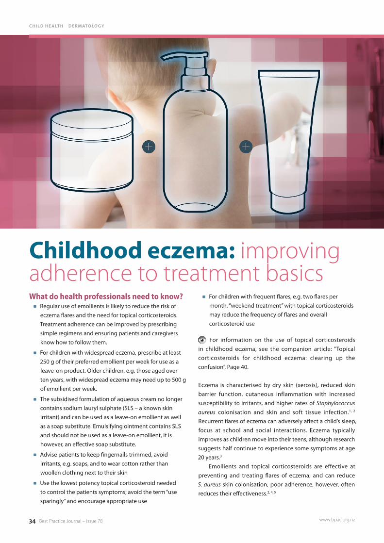
34 Best Practice Journal – Issue 78 www.bpac.org.nz
Childhood eczema: improving adherence to treatment basics
CHIlD HEAlTH DERMATOlOgy
What do health professionals need to know? Regular use of emollients is likely to reduce the risk of
eczema flares and the need for topical corticosteroids. Treatment adherence can be improved by prescribing simple regimens and ensuring patients and caregivers know how to follow them.
For children with widespread eczema, prescribe at least 250 g of their preferred emollient per week for use as a leave-on product. Older children, e.g. those aged over ten years, with widespread eczema may need up to 500 g of emollient per week.
The subsidised formulation of aqueous cream no longer contains sodium lauryl sulphate (SLS – a known skin irritant) and can be used as a leave-on emollient as well as a soap substitute. Emulsifying ointment contains SLS and should not be used as a leave-on emollient, it is however, an effective soap substitute.
Advise patients to keep fingernails trimmed, avoid irritants, e.g. soaps, and to wear cotton rather than woollen clothing next to their skin
Use the lowest potency topical corticosteroid needed to control the patients symptoms; avoid the term “use sparingly” and encourage appropriate use
For children with frequent flares, e.g. two flares per month, “weekend treatment” with topical corticosteroids may reduce the frequency of flares and overall corticosteroid use
For information on the use of topical corticosteroids in childhood eczema, see the companion article: “Topical corticosteroids for childhood eczema: clearing up the confusion”, Page 40.
Eczema is characterised by dry skin (xerosis), reduced skin barrier function, cutaneous inflammation with increased susceptibility to irritants, and higher rates of Staphylococcus aureus colonisation and skin and soft tissue infection.1, 2 Recurrent flares of eczema can adversely affect a child’s sleep, focus at school and social interactions. Eczema typically improves as children move into their teens, although research suggests half continue to experience some symptoms at age 20 years.3
Emollients and topical corticosteroids are effective at preventing and treating flares of eczema, and can reduce S. aureus skin colonisation, poor adherence, however, often reduces their effectiveness.2, 4, 5
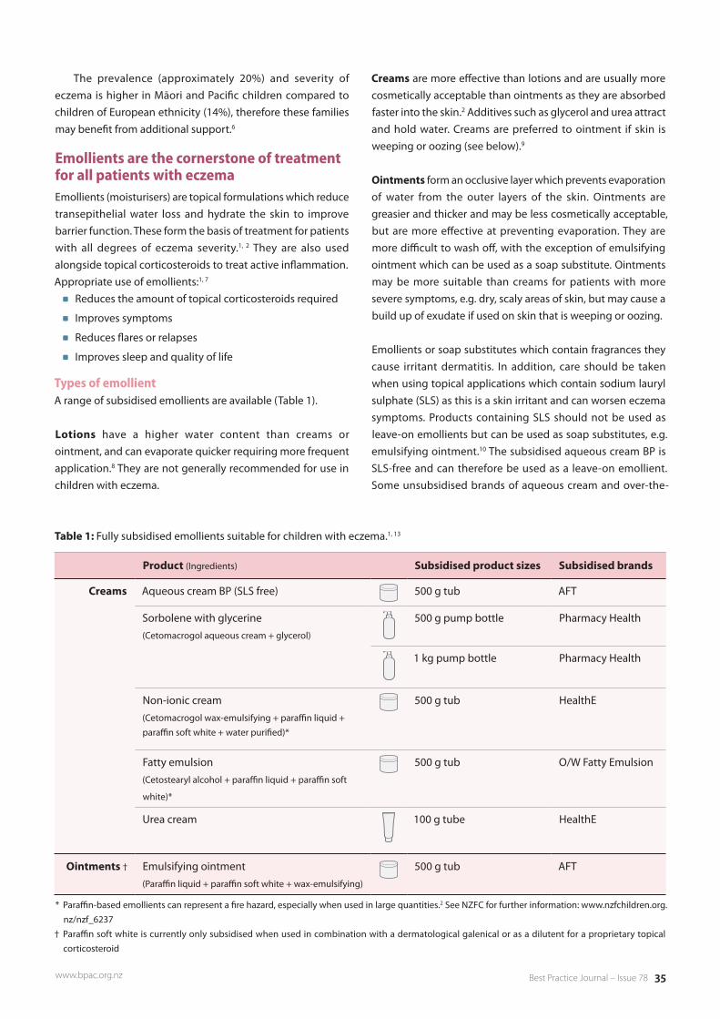
Best Practice Journal – Issue 78 35www.bpac.org.nz
The prevalence (approximately 20%) and severity of eczema is higher in Māori and Pacific children compared to children of European ethnicity (14%), therefore these families may benefit from additional support.6
Emollients are the cornerstone of treatment for all patients with eczemaEmollients (moisturisers) are topical formulations which reduce transepithelial water loss and hydrate the skin to improve barrier function. These form the basis of treatment for patients with all degrees of eczema severity.1, 2 They are also used alongside topical corticosteroids to treat active inflammation.Appropriate use of emollients:1, 7
Reduces the amount of topical corticosteroids required
Improves symptoms
Reduces flares or relapses
Improves sleep and quality of life
Types of emollientA range of subsidised emollients are available (Table 1).
lotions have a higher water content than creams or ointment, and can evaporate quicker requiring more frequent application.8 They are not generally recommended for use in children with eczema.
Creams are more effective than lotions and are usually more cosmetically acceptable than ointments as they are absorbed faster into the skin.2 Additives such as glycerol and urea attract and hold water. Creams are preferred to ointment if skin is weeping or oozing (see below).9
Ointments form an occlusive layer which prevents evaporation of water from the outer layers of the skin. Ointments are greasier and thicker and may be less cosmetically acceptable, but are more effective at preventing evaporation. They are more difficult to wash off, with the exception of emulsifying ointment which can be used as a soap substitute. Ointments may be more suitable than creams for patients with more severe symptoms, e.g. dry, scaly areas of skin, but may cause a build up of exudate if used on skin that is weeping or oozing.
Emollients or soap substitutes which contain fragrances they cause irritant dermatitis. In addition, care should be taken when using topical applications which contain sodium lauryl sulphate (SLS) as this is a skin irritant and can worsen eczema symptoms. Products containing SLS should not be used as leave-on emollients but can be used as soap substitutes, e.g. emulsifying ointment.10 The subsidised aqueous cream BP is SLS-free and can therefore be used as a leave-on emollient. Some unsubsidised brands of aqueous cream and over-the-
Table 1: Fully subsidised emollients suitable for children with eczema.1, 13
Product (Ingredients) Subsidised product sizes Subsidised brands
Creams Aqueous cream BP (SLS free) 500 g tub AFT
Sorbolene with glycerine(Cetomacrogol aqueous cream + glycerol)
500 g pump bottle Pharmacy Health
1 kg pump bottle Pharmacy Health
Non-ionic cream(Cetomacrogol wax-emulsifying + paraffin liquid + paraffin soft white + water purified)*
500 g tub HealthE
Fatty emulsion(Cetostearyl alcohol + paraffin liquid + paraffin soft
white)*
500 g tub O/W Fatty Emulsion
Urea cream 100 g tube HealthE
Ointments † Emulsifying ointment(Paraffin liquid + paraffin soft white + wax-emulsifying)
500 g tub AFT
* Paraffin-based emollients can represent a fire hazard, especially when used in large quantities.2 See NZFC for further information: www.nzfchildren.org.nz/nzf_6237
† Paraffin soft white is currently only subsidised when used in combination with a dermatological galenical or as a dilutent for a proprietary topical corticosteroid
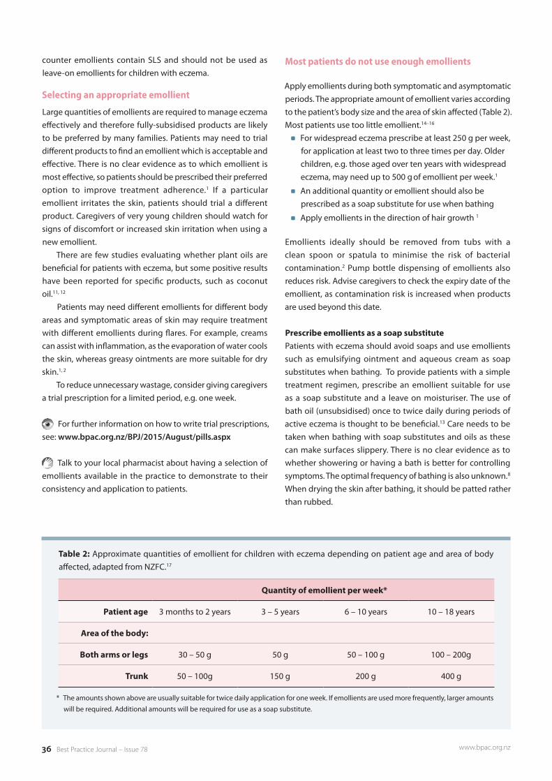
36 Best Practice Journal – Issue 78 www.bpac.org.nz
Table 2: Approximate quantities of emollient for children with eczema depending on patient age and area of body affected, adapted from NZFC.17
Quantity of emollient per week*
Patient age 3 months to 2 years 3 – 5 years 6 – 10 years 10 – 18 years
Area of the body:
both arms or legs 30 – 50 g 50 g 50 – 100 g 100 – 200g
Trunk 50 – 100g 150 g 200 g 400 g
* The amounts shown above are usually suitable for twice daily application for one week. If emollients are used more frequently, larger amounts will be required. Additional amounts will be required for use as a soap substitute.
counter emollients contain SLS and should not be used as leave-on emollients for children with eczema.
Selecting an appropriate emollient
Large quantities of emollients are required to manage eczema effectively and therefore fully-subsidised products are likely to be preferred by many families. Patients may need to trial different products to find an emollient which is acceptable and effective. There is no clear evidence as to which emollient is most effective, so patients should be prescribed their preferred option to improve treatment adherence.1 If a particular emollient irritates the skin, patients should trial a different product. Caregivers of very young children should watch for signs of discomfort or increased skin irritation when using a new emollient.
There are few studies evaluating whether plant oils are beneficial for patients with eczema, but some positive results have been reported for specific products, such as coconut oil.11, 12
Patients may need different emollients for different body areas and symptomatic areas of skin may require treatment with different emollients during flares. For example, creams can assist with inflammation, as the evaporation of water cools the skin, whereas greasy ointments are more suitable for dry skin.1, 2
To reduce unnecessary wastage, consider giving caregivers a trial prescription for a limited period, e.g. one week.
For further information on how to write trial prescriptions, see: www.bpac.org.nz/BPJ/2015/August/pills.aspx
Talk to your local pharmacist about having a selection of emollients available in the practice to demonstrate to their consistency and application to patients.
Most patients do not use enough emollients
Apply emollients during both symptomatic and asymptomatic periods. The appropriate amount of emollient varies according to the patient’s body size and the area of skin affected (Table 2). Most patients use too little emollient.14–16
For widespread eczema prescribe at least 250 g per week, for application at least two to three times per day. Older children, e.g. those aged over ten years with widespread eczema, may need up to 500 g of emollient per week.1
An additional quantity or emollient should also be prescribed as a soap substitute for use when bathing
Apply emollients in the direction of hair growth 1
Emollients ideally should be removed from tubs with a clean spoon or spatula to minimise the risk of bacterial contamination.2 Pump bottle dispensing of emollients also reduces risk. Advise caregivers to check the expiry date of the emollient, as contamination risk is increased when products are used beyond this date.
Prescribe emollients as a soap substitute Patients with eczema should avoid soaps and use emollients such as emulsifying ointment and aqueous cream as soap substitutes when bathing. To provide patients with a simple treatment regimen, prescribe an emollient suitable for use as a soap substitute and a leave on moisturiser. The use of bath oil (unsubsidised) once to twice daily during periods of active eczema is thought to be beneficial.13 Care needs to be taken when bathing with soap substitutes and oils as these can make surfaces slippery. There is no clear evidence as to whether showering or having a bath is better for controlling symptoms. The optimal frequency of bathing is also unknown.8 When drying the skin after bathing, it should be patted rather than rubbed.
Best Practice Journal – Issue 78 37www.bpac.org.nz
Maintenance treatment with topical corticosteroids can reduce the frequency of flares
Eczema is traditionally managed reactively, where topical corticosteroids are initiated during a flare and stopped when symptoms resolve. This approach is still appropriate for many patients.
Children with frequent flares, e.g. two per month, may benefit from a proactive approach, where topical corticosteroids are applied twice a week during periods of remission, i.e. between flares.1 This is often referred to as “weekend treatment”, however, treatment can occur on any two consecutive days in the week.1
For further information on weekend treatment, see: “Topical corticosteroids for childhood eczema: clearing up the confusion”, Page 44.
Use oral rather than topical antibiotics for infected eczemaAntibiotic treatment of infected eczema is not always necessary. If antibiotic treatment is needed, oral antibiotics are preferred over topical antibiotics due to increasing rates of fusidic acid resistance in New Zealand. Recently released data show that 21–46% of community acquired S. aureus infections were fusidic acid resistant in 2014.26
Studies suggest a watch and wait approach may be appropriate for patients with mild to moderately infected eczema; oral antibiotics can be reserved for patients with worsening or severe infection.27 A double-blind randomised controlled trial in primary care in the United Kingdom found that for children with mild to moderately infected eczema, the use of topical or oral antibiotics had no effect on symptom severity or made eczema symptoms worse. Children with severe infection were excluded from this study.
If antibiotic treatment is required, a suitable first-line oral regimen is:28
Flucloxacillin 12.5 mg/kg/dose, four times daily, for five days (maximum 500 mg/dose)
Flucloxacillin capsules can be prescribed for older children who are able to swallow them. Alternative oral antibiotic choices include erythromycin, co-trimoxazole or cephalexin.
Dosing regimens are available in the bpacnz antibiotic guide: www.bpac.org.nz/Supplement/2013/July/antibiotics-guide.aspx
For patients with recurrent infected eczema, the focus should be on managing the eczema effectively. Appropriate use of topical corticosteroids and emollients can improve the skin microbiota of people with eczema and reduce S. aureus skin colonisation.4, 5, 20, 29, 30
Emulsifying ointment cleans and moisturises the skin and can be made into an effective soap substitute with easy-to-use consistency by mixing three to four large spoonfuls with very hot water to achieve a creamy soap consistency. Once cool, this can be used in the shower or bath and more prepared as required. Instructions for preparation can be included on the prescription label.
Many patients find bleach baths beneficial, however, recent studies suggest that evidence is mixed as to whether bleach baths improve symptoms.18–21 Caregivers should avoid using fragranced bleach.
Information on how to prepare a bleach bath is available from: www.bpac.org.nz/BPJ/2015/April/eczema.aspx
Escalate treatment during flaresTopical corticosteroids should be applied on all areas of active eczema, no matter how severely inflamed, and stopped once the eczema has cleared (unless following “weekend treatment”, see below). Once daily application may be sufficient, especially if treatment is initiated when symptoms first develop.2 The potency of the topical corticosteroid should be appropriate for the area of the body being treated, the age of the patient and their symptom severity, e.g. mildly inflamed eczema on the hands will typically need a potent steroid for treatment to be effective.
For further information on assessing the severity of eczema symptoms, see: www.bpac.org.nz/BPJ/2015/April/eczema.aspx
Encourage caregivers to continue emollient use during flares. Either emollients or topical corticosteroids can be applied first with only a short interval needed between applications.1 The practice of waiting up to 45 minutes between applications is not evidence-based and can reduce adherence.22
Topical pimecrolimus (unsubsidised), a calcineurin inhibitor, is a second-line treatment option for patients with eczema if the use of topical corticosteroids is contraindicated or inappropriate, e.g. for long-term use on body areas such as the face, neck and groin.1 Pimecrolimus may be as effective as mild to moderate potency topical corticosteroids for the treatment of eczema, but is more likely to cause a burning sensation and pruritus.23 Concerns regarding a possible association between topical calcineurin inhibitor use and increased risk of lymphoma have been raised based on studies in animals, however, studies in humans have not established an increased risk of malignancy.24, 25
For further information on the use of topical pimecrolimus, see: www.bpac.org.nz/BPJ/2015/April/eczema.aspx

38 Best Practice Journal – Issue 78 www.bpac.org.nz
There is little evidence to suggest topical corticosteroids worsen the course of bacterial or viral skin infection, and they may improve skin barrier function.31 Topical corticosteroids can continue to be used on excoriated skin and eczema with bacterial or viral infection. However, topical corticosteroids should be stopped in patients with fungal infections as they may exacerbate the infection.2, 31
Antihistamines may benefit children with severe symptoms
Antihistamines are not routinely used children with eczema, but a bedtime dose of a sedating antihistamine can be trialled to aid sleep in children aged over two years during an eczema flare:2, 13 see NZFC, www.nzfc.org.nz, for dosing recommendations.
When to referChildren should be referred to secondary care if they:1, 2
Have ongoing symptoms despite appropriate topical corticosteroid use
Have recurrent skin infections
Have infected eczema which does not clear with antibiotic treatment
Show symptoms and signs of eczema herpeticum, e.g. fever and small, grouped, circular blisters with a central depression which become crusted and eroded
Have symptoms which substantially reduce their quality of life, such as eczema which results in frequent waking at night, reduces school attendance or which causes social problems
Reduce obstacles to adherence
Reducing obstacles to treatment adherence is a priority in eczema care. Caring for a child with eczema can be challenging for parents and caregivers as it requires daily attention to manage effectively.
Steps which can improve adherence include:32
Simple treatment regimens
Prescribe the simplest effective treatment regimen.32 Provide caregivers and older children with written instructions so they know what treatments to use when, on which parts of the body and in what quantities.
For examples of simple take-home care plans, see: www.starship.org.nz/media/269759/caring_for_your_child_s_eczema_june_2014.pdf
Improving understanding about the causes of eczema and treatment options
– Eczema education highlights the need for proactive management. Many families want to find a cause
of the child’s eczema, e.g. a food intolerance, with the expectation that once identified, the condition could be cured. This belief can lead families to focus on exclusion of potential triggers at the expense of controlling symptoms through frequent emollient and appropriate topical corticosteroid use.33 To reduce the risk of flares, patients with eczema require ongoing maintenance treatment even during asymptomatic phases; while avoiding triggers can help, proactive management with frequent emollient use is key.
– Consider an education session for parents and caregivers. Education sessions delivered by a general practitioner, practice nurse or pharmacist which cover the causes of eczema, application of emollients, and the appropriate use of topical corticosteroids can improve parents’ or caregiver’s knowledge and confidence about treating their child’s eczema.14, 34 Some DHBs have dedicated eczema nurses who can offer educational support.
– Dietary modifications are typically unnecessary. Allergy is not recognised as a major cause of childhood eczema and there is no evidence to support widespread use of dietary modifications or food exclusions.35 However, children with eczema are at higher risk of developing immediate hypersensitivity reactions to foods. The possibility of food allergy should be considered where young children have immediate reactions to a food, particularly if accompanied by urticaria, angiodema, colic, or vomiting. A short trial of food exclusions, supervised by a dietitian, could be considered in children with moderate to severe eczema and ongoing symptoms which do not improve with adherence to appropriate treatment.1 Discussions about the role of food allergy can also be used as a useful springboard to reinforce healthy eating messages.
Further education and information for parents: Instructional videos on the use of moisturisers,
bathing and topical corticosteroids: www.kidshealth.org.nz/eczema
Printable parent information sheets: www.starship.org.nz/for-health-professionals/new-zealand-child-and-youth-clinical-networks/child-and-youth-eczema-clinical-network/family-information-and-handouts/
Information on introducing solids for children with eczema: www.kidshealth.org.nz/food-recommendations-infants-risk-allergy

Best Practice Journal – Issue 78 39www.bpac.org.nz
Additional information is available from the Goodfellow Unit:
www.goodfellowunit.org/podcast/childhood-eczema-paul-jarrett
https://youtu.be/EMCV-LPvecM
Acknowledgement: Thank you to Dr Diana Purvis,
Dermatologist, Auckland DHB for expert review of this article.
References:1. National Institute for Health and Care Excellence (NICE). Atopic eczema in
under 12s: diagnosis and management. 2007. Available from: www.nice.org.
uk/guidance/cg57 (Accessed Dec, 2016).
2. Scottish Intercollegiate Guidelines Network (SIGN). Management of atopic
eczema in primary care. Edinburgh: SIGN 2011. Available from: www.sign.
ac.uk/pdf/sign125.pdf (Accessed Dec, 2016).
3. Margolis JS, Abuabara K, Bilker W, et al. Persistence of mild to moderate
atopic dermatitis. JAMA Dermatol 2014;150:593–600. doi:10.1001/
jamadermatol.2013.10271
4. Seite S, Flores GE, Henley JB, et al. Microbiome of affected and unaffected skin
of patients with atopic dermatitis before and after emollient treatment. J Drugs
Dermatol 2014;13:1365–72.
5. Gong JQ, Lin L, Lin T, et al. Skin colonization by Staphylococcus aureus in
patients with eczema and atopic dermatitis and relevant combined topical
therapy: a double-blind multicentre randomized controlled trial. Br J Dermatol
2006;155:680–7. doi:10.1111/j.1365-2133.2006.07410.x
6. Clayton T, Asher MI, Crane J, et al. Time trends, ethnicity and risk factors
for eczema in New Zealand children: ISAAC Phase Three. Asia Pac Allergy
2013;3:161–78. doi:10.5415/apallergy.2013.3.3.161
7. Moncrieff G, Cork M, Lawton S, et al. Use of emollients in dry-skin conditions:
consensus statement. Clin Exp Dermatol 2013;38:231–8. doi:10.1111/
ced.12104
8. Eichenfield LF, Tom WL, Berger TG, et al. Guidelines of care for the management
of atopic dermatitis: section 2. Management and treatment of atopic
dermatitis with topical therapies. J Am Acad Dermatol 2014;71:116–32.
doi:10.1016/j.jaad.2014.03.023
9. N. H. S. Choices. Emollients. 2016. Available from: www.nhs.uk/Conditions/
Emollients/Pages/Introduction.aspx (Accessed Dec, 2016).
10. Medicines and Healthcare products Regulatory Agency (MHRA). MHRA UK
Public Assessment Report. Aqueous cream: contains sodium lauryl sulfate
which may cause skin reactions, particularly in children with eczema.
2013. Available from: www.mhra.gov.uk/home/groups/s-par/documents/
websiteresources/con512958.pdf (Accessed Dec, 2016).
11. Evangelista MTP, Abad-Casintahan F, Lopez-Villafuerte L. The effect of topical
virgin coconut oil on SCORAD index, transepidermal water loss, and skin
capacitance in mild to moderate pediatric atopic dermatitis: a randomized,
double-blind, clinical trial. Int J Dermatol 2014;53:100–8. doi:10.1111/ijd.12339
12. Thandar Y, Gray A, Botha J, et al. Topical herbal medicines for atopic eczema: a
systematic review of randomized controlled trials. Br J Dermatol 2016; [Epub
ahead of print]. doi:10.1111/bjd.14840
13. Starship Children’s Health. Starship clinical guidelines. Eczema. 2016. Available
from: www.starship.org.nz/for-health-professionals/starship-clinical-
guidelines/e/eczema/ (Accessed Dec, 2016).
14. Mason JM, Carr J, Buckley C, et al. Improved emollient use reduces atopic
eczema symptoms and is cost neutral in infants: before-and-after evaluation
of a multifaceted educational support programme. BMC Dermatol 2013;13:7.
doi:10.1186/1471-5945-13-7
15. Lee JH, Jung KE, Lee YB, et al. Use of emollients in atopic dermatitis: a
questionnaire survey study. Ann Dermatol 2014;26:528–31. doi:10.5021/
ad.2014.26.4.528
16. Santer M, Muller I, Yardley L, et al. Parents’ and carers’ views about emollients
for childhood eczema: qualitative interview study. BMJ Open 2016;6:e011887.
doi:10.1136/bmjopen-2016-011887
17. New Zealand Formulary for Children. NZFC v53. 2016. Available from: www.
nzfchildren.org.nz/ (Accessed Nov, 2016).
18. Hon KL, Tsang YCK, Lee VWY, et al. Efficacy of sodium hypochlorite (bleach)
baths to reduce Staphylococcus aureus colonization in childhood onset
moderate-to-severe eczema: A randomized, placebo-controlled cross-over trial.
J Dermatolog Treat 2016;27:156–62. doi:10.3109/09546634.2015.1067669
19. Wong S, Ng TG, Baba R. Efficacy and safety of sodium hypochlorite (bleach)
baths in patients with moderate to severe atopic dermatitis in Malaysia. J
Dermatol 2013;40:874–80. doi:10.1111/1346-8138.12265
20. Gonzalez ME, Schaffer JV, Orlow SJ, et al. Cutaneous microbiome effects of
fluticasone propionate cream and adjunctive bleach baths in childhood
atopic dermatitis. J Am Acad Dermatol 2016;75:481–493.e8. doi:10.1016/j.
jaad.2016.04.066
21. Bath-Hextall FJ, Birnie AJ, Ravenscroft JC, et al. Interventions to reduce
Staphylococcus aureus in the management of atopic eczema: an updated
Cochrane review. Br J Dermatol 2010;163:12–26. doi:10.1111/j.1365-
2133.2010.09743.x
22. Smoker A, Voegeli D. Topical steroid or emollient — which to apply first? A
critical review of the science and debate. Dermatol Nurs (Lond) 2014;13:14–26.
23. Broeders JA, Ahmed Ali U, Fischer G. Systematic review and meta-analysis of
randomized clinical trials (RCTs) comparing topical calcineurin inhibitors with
topical corticosteroids for atopic dermatitis: A 15-year experience. J Am Acad
Dermatol 2016;75:410–419.e3. doi:10.1016/j.jaad.2016.02.1228
24. Siegfried EC, Jaworski JC, Kaiser JD, et al. Systematic review of published trials:
long-term safety of topical corticosteroids and topical calcineurin inhibitors
in pediatric patients with atopic dermatitis. BMC Pediatrics 2016;16:75.
doi:10.1186/s12887-016-0607-9
25. Segal AO, Ellis AK, Kim HL. CSACI position statement: safety of topical
calcineurin inhibitors in the management of atopic dermatitis in children and
adults. Allergy, Asthma & Clinical Immunology 2013;9:24. doi:10.1186/1710-
1492-9-24
26. Vogel A, Lennon D, Best E, et al. Where to from here? The treatment of impetigo
in children as resistance to fusidic acid emerges. N Z Med J 2016;129:77–83.
27. Francis NA, Ridd MJ, Thomas-Jones E, et al. A randomised placebo-controlled
trial of oral and topical antibiotics for children with clinically infected eczema
in the community: the ChildRen with Eczema, Antibiotic Management
(CREAM) study. Health Technol Assess 2016;20:i–xxiv, 1-84. doi:10.3310/
hta20190
28. Best Practice Advocacy Centre, bpacnz. Antibiotics Guide. Impetigo. 2016.
Available from: http://bpac.org.nz/Supplement/2013/July/antibiotics-guide.
aspx (Accessed Dec, 2016).
29. Hung S-H, Lin Y-T, Chu C-Y, et al. Staphylococcus colonization in atopic
dermatitis treated with fluticasone or tacrolimus with or without
antibiotics. Ann Allergy Asthma Immunol 2007;98:51–6. doi:10.1016/S1081-
1206(10)60859-9
30. Stalder JF, Fleury M, Sourisse M, et al. Local steroid therapy and bacterial skin
flora in atopic dermatitis. Br J Dermatol 1994;131:536–40.
31. Mooney E, Rademaker M, Dailey R, et al. Adverse effects of topical
corticosteroids in paediatric eczema: Australasian consensus statement.
Australas J Dermatol 2015;56:241–51. doi:10.1111/ajd.12313
32. Sokolova A, Smith SD. Factors contributing to poor treatment outcomes in
childhood atopic dermatitis. Australas J Dermatol 2015;56:252–7. doi:10.1111/
ajd.12331
33. Santer M, Burgess H, Yardley L, et al. Experiences of carers managing childhood
eczema and their views on its treatment: a qualitative study. Br J Gen Pract
2012;62:e261-267. doi:10.3399/bjgp12X636083
34. Cork MJ, Britton J, Butler L, et al. Comparison of parent knowledge, therapy
utilization and severity of atopic eczema before and after explanation and
demonstration of topical therapies by a specialist dermatology nurse. Br J
Dermatol 2003;149:582–9.
35. Preece K, Blincoe A, Grangaard E, et al. Paediatric non-IgE mediated food
allergy: guide for practitioners. N Z Med J 2016;129:78–88.
40 Best Practice Journal – Issue 78 www.bpac.org.nz
Topical corticosteroids for childhood eczema: clearing up the confusion
CHIlD HEAlTH DERMATOlOgy
What do prescribers need to know? Use the lowest potency topical corticosteroid needed to
control the patient’s symptoms
Be clear when prescribing where each product should, and should not, be used and avoid the term “use sparingly” and encourage appropriate use
Check that patients and caregivers can identify a flare and are able to respond with appropriate treatment
For patients with persistent eczema, short treatment “bursts”, e.g. three to five days, with higher potency corticosteroids may be preferable to longer courses of treatment with less potent corticosteroids; topical corticosteroids should be stepped down, e.g. from potent or moderate potency to mild potency, as the patients symptoms resolve
Include descriptions of potency in the prescription so that it is printed on the medicine label to reduce confusion
Navigating common concerns and confusion
Topical corticosteroids are one of the key medicines used in the management of childhood eczema. However, adherence is typically poor, often due to “corticosteroid phobia”.1 Common themes contribute to the reluctance of caregivers to use topical corticosteroids (Table 1). Addressing these concerns may improve treatment adherence and patient outcomes.
Provide clear information when prescribing and dispensing topical corticosteroids
Avoid “use sparingly”: encourage appropriate use
Advising patients to “use topical corticosteroids sparingly” creates confusion; patients and caregivers are prescribed a medicine but simultaneously warned against using it. This advice may cause patients to only use corticosteroids when symptoms are severe, resulting in inadequate use and poor symptom control. Caregivers should instead be encouraged to “use corticosteroids appropriately”, which will maximise the benefits of use and minimise adverse effects.
Best Practice Journal – Issue 78 41www.bpac.org.nz
Table 1: Caregiver misconceptions and concerns associated with the use of topical corticosteroids in childhood eczema and evidence-based responses.1–4
Misconception or concern what does the evidence say?
Topical corticosteroids should only be used for
severe symptoms
Topical corticosteroids can and should be used for all severities of eczema, including mild symptoms.*
Products have a range of potencies to treat patients with differing symptom severity. Treatment should be with mildest topical corticosteroid which is able to resolve the inflammation within a short period of time so that the patient is able to have days without using topical corticosteroids. Different potencies are required for different parts of the body depending on the thickness of the stratum corneum.
Regular use of topical corticosteroids causes
adverse effects such as skin thinning
Topical corticosteroids are unlikely to cause skin thinning or other long-term harm to children if used appropriately.
Skin thinning is one of the most frequently cited concerns reported by patients and caregivers, however, it is very unlikely to occur if patients and caregivers use topical corticosteroids appropriately.2, 3, 5 This consensus is based on research and clinical experience from Australia and New Zealand, including evaluations of children treated with potent topical corticosteroids.5, 6 Skin thinning is more likely to occur in areas with a thinner stratum corneum, such as the face and groin.
The percentage of a topical corticosteroid is its strength
The percentage value of different formulations of topical corticosteroids does not indicate their potency, e.g. hydrocortisone 1% is a weaker formulation than hydrocortisone butyrate 0.1%.
Corticosteroids are confused with anabolic steroids
Clarify the meaning of the word “steroid”.
Inform patients and caregivers that the label “steroids” is a classification used for a wide group of hormones and medicines with different functions, including corticosteroids and anabolic steroids.
Topical corticosteroids should not be applied to broken skin
The consensus of paediatric dermatologists in Australia and New Zealand is that topical corticosteroids can be applied to areas of eczema with broken skin.5
This concern possibly arose as topical corticosteroid absorption will be greater through broken skin. However, this can prevent patients having topical corticosteroids applied to areas of active eczema particularly when severely inflamed or excoriated. All skin with an active eczema flare will have reduced barrier function, and the best way to address this is through appropriate use of topical corticosteroids.
Topical corticosteroids are not “natural”
Corticosteroids mimic the effects of hormones produced by the adrenal glands, despite being “man-made”.
* For further information on symptom severity and recommended treatment escalation, see: www.bpac.org.nz/BPJ/2015/April/eczema.aspx
Patients should know:1. which corticosteroid to apply, i.e. using the right
potency and formulation 2. where on the body to apply it 3. when to apply it, i.e. when to start treatment and how
long to use it for4. How much to apply
Arrange to review the patient within two to four weeks of prescribing topical corticosteroids. This gives an opportunity to assess their response to treatment and reinforce education as well as allowing the patient and caregiver to focus on treating the eczema rather than watching for adverse effects.
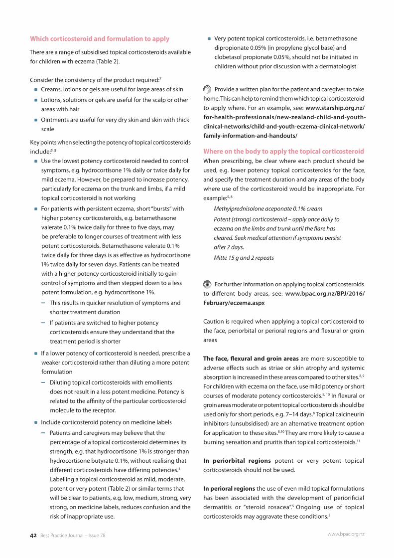
42 Best Practice Journal – Issue 78 www.bpac.org.nz
Which corticosteroid and formulation to apply
There are a range of subsidised topical corticosteroids available for children with eczema (Table 2).
Consider the consistency of the product required:7
Creams, lotions or gels are useful for large areas of skin
Lotions, solutions or gels are useful for the scalp or other areas with hair
Ointments are useful for very dry skin and skin with thick scale
Key points when selecting the potency of topical corticosteroids include:5, 8
Use the lowest potency corticosteroid needed to control symptoms, e.g. hydrocortisone 1% daily or twice daily for mild eczema. However, be prepared to increase potency, particularly for eczema on the trunk and limbs, if a mild topical corticosteroid is not working
For patients with persistent eczema, short “bursts” with higher potency corticosteroids, e.g. betamethasone valerate 0.1% twice daily for three to five days, may be preferable to longer courses of treatment with less potent corticosteroids. Betamethasone valerate 0.1% twice daily for three days is as effective as hydrocortisone 1% twice daily for seven days. Patients can be treated with a higher potency corticosteroid initially to gain control of symptoms and then stepped down to a less potent formulation, e.g. hydrocortisone 1%.
– This results in quicker resolution of symptoms and shorter treatment duration
– If patients are switched to higher potency corticosteroids ensure they understand that the treatment period is shorter
If a lower potency of corticosteroid is needed, prescribe a weaker corticosteroid rather than diluting a more potent formulation
– Diluting topical corticosteroids with emollients does not result in a less potent medicine. Potency is related to the affinity of the particular corticosteroid molecule to the receptor.
Include corticosteroid potency on medicine labels
– Patients and caregivers may believe that the percentage of a topical corticosteroid determines its strength, e.g. that hydrocortisone 1% is stronger than hydrocortisone butyrate 0.1%, without realising that different corticosteroids have differing potencies.4 Labelling a topical corticosteroid as mild, moderate, potent or very potent (Table 2) or similar terms that will be clear to patients, e.g. low, medium, strong, very strong, on medicine labels, reduces confusion and the risk of inappropriate use.
Very potent topical corticosteroids, i.e. betamethasone dipropionate 0.05% (in propylene glycol base) and clobetasol propionate 0.05%, should not be initiated in children without prior discussion with a dermatologist
Provide a written plan for the patient and caregiver to take home. This can help to remind them which topical corticosteroid to apply where. For an example, see: www.starship.org.nz/for-health-professionals/new-zealand-child-and-youth-clinical-networks/child-and-youth-eczema-clinical-network/family-information-and-handouts/
Where on the body to apply the topical corticosteroid When prescribing, be clear where each product should be used, e.g. lower potency topical corticosteroids for the face, and specify the treatment duration and any areas of the body where use of the corticosteroid would be inappropriate. For example:5, 8
Methylprednisolone aceponate 0.1% cream
Potent (strong) corticosteroid – apply once daily to eczema on the limbs and trunk until the flare has cleared. Seek medical attention if symptoms persist after 7 days.
Mitte 15 g and 2 repeats
For further information on applying topical corticosteroids to different body areas, see: www.bpac.org.nz/BPJ/2016/February/eczema.aspx
Caution is required when applying a topical corticosteroid to the face, periorbital or perioral regions and flexural or groin areas
The face, flexural and groin areas are more susceptible to adverse effects such as striae or skin atrophy and systemic absorption is increased in these areas compared to other sites.8, 9 For children with eczema on the face, use mild potency or short courses of moderate potency corticosteroids.8, 10 In flexural or groin areas moderate or potent topical corticosteroids should be used only for short periods, e.g. 7–14 days.8 Topical calcineurin inhibitors (unsubsidised) are an alternative treatment option for application to these sites.8,10 They are more likely to cause a burning sensation and pruritis than topical corticosteroids.11
In periorbital regions potent or very potent topical corticosteroids should not be used.
In perioral regions the use of even mild topical formulations has been associated with the development of periorificial dermatitis or “steroid rosacea”.5 Ongoing use of topical corticosteroids may aggravate these conditions.5
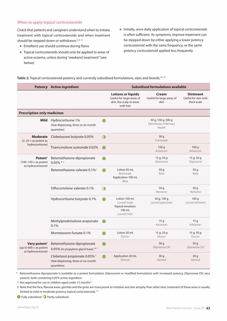
Best Practice Journal – Issue 78 43www.bpac.org.nz
Table 2: Topical corticosteroid potency and currently subsidised formulations, sizes and brands.16, 17
Potency Active ingredient Subsidised formulations available
lotions or liquidsUseful for large areas of skin, the scalp or areas
with hair
CreamUseful for large areas of
skin
OintmentUseful for skin with
thick scale
Prescription only medicines
Mild Hydrocortisone 1%(Stat dispensing, three or six month quantities)
30 g, 100 g, 500 gDermAssist, Pharmacy
Health
Moderate(2–25 × as potent as
hydrocortisone)
Clobetasone butyrate 0.05% 30 gEumovate
Triamcinolone acetonide 0.02% 100 gAristocort
100 gAristocort
Potent‡
(100–150 × as potent as hydrocortisone)
Betamethasone dipropionate 0.05% * †
15 g, 50 gDiprosone
15 g, 50 gDiprosone
Betamethasone valerate 0.1%† Lotion 50 mLBetnovate
Application 100 mLBeta
50 gBeta
50 gBeta
Diflucortolone valerate 0.1% 50 gNerisone
50 gNerisone
Hydrocortisone butyrate 0.1% Lotion 100 mLLocoid Scalp
Topical emulsion100 mL
Locoid Crelo
30 g, 100 gLocoid Lipocream
100 gLocoid ointment
Methylprednisolone aceponate 0.1%
15 gAdvantan
15 gAdvantan
Mometasone furoate 0.1% Lotion 30 mLElocon
15 g, 50 gElocon
15 g, 50 gElocon
Very potent‡
(up to 600 × as potent as hydrocortisone)
Betamethasone dipropionate 0.05% (in propylene glycol base) * †
30 gDiprosone OV
30 gDiprosone OV
Clobetasol propionate 0.05% †
(Stat dispensing, three or six month quantities)
Application 30 mLDermol
30 gDermol
30 gDermol
* Betamethasone dipropionate is available as a potent formulation (Diprosone) or modified formulation with increased potency (Diprosone OV; very potent), both containing 0.05% active ingredient
† Not approved for use in children aged under 12 months17
‡ Note that the face, flexural areas, genitals and the groin are more prone to irritation and skin atrophy than other sites; treatment of these areas is usually limited to mild or moderate potency topical corticosteroids.7, 9
Fully subsidised Partly subsidised
When to apply topical corticosteroids
Check that patients and caregivers understand when to initiate treatment with topical corticosteroids and when treatment should be stepped down or withdrawn:5, 8, 12
Emollient use should continue during flares
Topical corticosteroids should only be applied to areas of active eczema, unless during “weekend treatment” (see below)
Initially, once daily application of topical corticosteroids is often sufficient. As symptoms improve treatment can be stepped down by either applying a lower potency corticosteroid with the same frequency, or the same potency corticosteroid applied less frequently
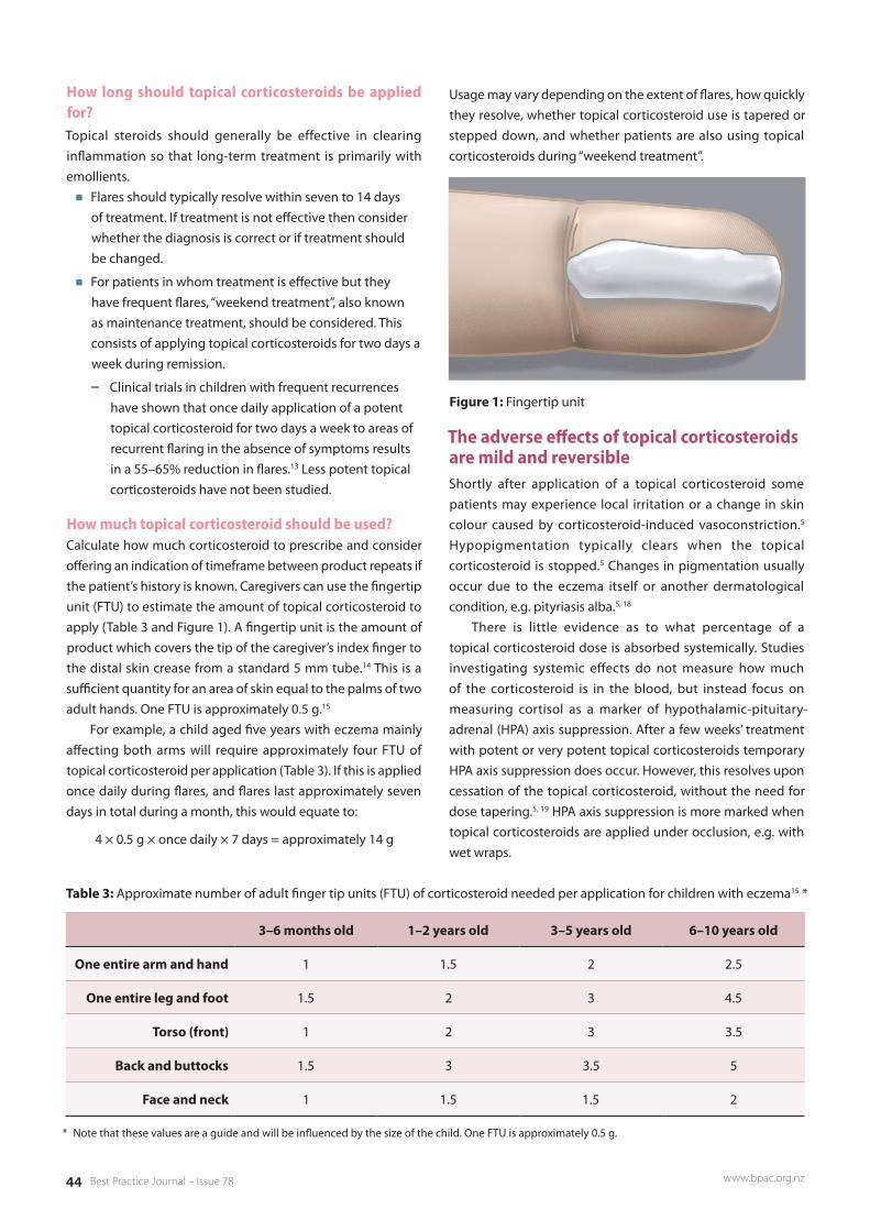
44 Best Practice Journal – Issue 78 www.bpac.org.nz
Usage may vary depending on the extent of flares, how quickly they resolve, whether topical corticosteroid use is tapered or stepped down, and whether patients are also using topical corticosteroids during “weekend treatment”.
Figure 1: Fingertip unit
The adverse effects of topical corticosteroids are mild and reversibleShortly after application of a topical corticosteroid some patients may experience local irritation or a change in skin colour caused by corticosteroid-induced vasoconstriction.5 Hypopigmentation typically clears when the topical corticosteroid is stopped.5 Changes in pigmentation usually occur due to the eczema itself or another dermatological condition, e.g. pityriasis alba.5, 18
There is little evidence as to what percentage of a topical corticosteroid dose is absorbed systemically. Studies investigating systemic effects do not measure how much of the corticosteroid is in the blood, but instead focus on measuring cortisol as a marker of hypothalamic-pituitary-adrenal (HPA) axis suppression. After a few weeks’ treatment with potent or very potent topical corticosteroids temporary HPA axis suppression does occur. However, this resolves upon cessation of the topical corticosteroid, without the need for dose tapering.5, 19 HPA axis suppression is more marked when topical corticosteroids are applied under occlusion, e.g. with wet wraps.
How long should topical corticosteroids be applied for?Topical steroids should generally be effective in clearing inflammation so that long-term treatment is primarily with emollients.
Flares should typically resolve within seven to 14 days of treatment. If treatment is not effective then consider whether the diagnosis is correct or if treatment should be changed.
For patients in whom treatment is effective but they have frequent flares, “weekend treatment”, also known as maintenance treatment, should be considered. This consists of applying topical corticosteroids for two days a week during remission.
– Clinical trials in children with frequent recurrences have shown that once daily application of a potent topical corticosteroid for two days a week to areas of recurrent flaring in the absence of symptoms results in a 55–65% reduction in flares.13 Less potent topical corticosteroids have not been studied.
How much topical corticosteroid should be used?Calculate how much corticosteroid to prescribe and consider offering an indication of timeframe between product repeats if the patient’s history is known. Caregivers can use the fingertip unit (FTU) to estimate the amount of topical corticosteroid to apply (Table 3 and Figure 1). A fingertip unit is the amount of product which covers the tip of the caregiver’s index finger to the distal skin crease from a standard 5 mm tube.14 This is a sufficient quantity for an area of skin equal to the palms of two adult hands. One FTU is approximately 0.5 g.15
For example, a child aged five years with eczema mainly affecting both arms will require approximately four FTU of topical corticosteroid per application (Table 3). If this is applied once daily during flares, and flares last approximately seven days in total during a month, this would equate to:
4 × 0.5 g × once daily × 7 days = approximately 14 g
Table 3: Approximate number of adult finger tip units (FTU) of corticosteroid needed per application for children with eczema15 *
3–6 months old 1–2 years old 3–5 years old 6–10 years old
One entire arm and hand 1 1.5 2 2.5
One entire leg and foot 1.5 2 3 4.5
Torso (front) 1 2 3 3.5
back and buttocks 1.5 3 3.5 5
Face and neck 1 1.5 1.5 2
* Note that these values are a guide and will be influenced by the size of the child. One FTU is approximately 0.5 g.
Best Practice Journal – Issue 78 45www.bpac.org.nz
Inappropriate or prolonged use may cause more serious adverse effects
More serious adverse effects include clinically significant HPA axis suppression, skin atrophy or striae or withdrawal symptoms upon stopping the corticosteroid, such as erythema and aggravation of cutaneous symptoms.5 These are rarely seen with normal prescribing patterns.
The risk of these adverse effects is increased:5, 20
With a higher potency of corticosteroid
With application to a greater area of skin or a larger quantity of application
When corticosteroids are applied under occlusion or to flexural or groin areas, which increases absorption
If patients are also taking oral or high-dose inhaled corticosteroids
When potent topical corticosteroids are applied to striae-prone areas, e.g. axillae or groin areas, during growth phases of puberty
If patients request repeat prescriptions earlier than expected consider whether they may be using a topical corticosteroid inappropriately; case reports of adverse effects typically involve patients who have used the product for longer than it was prescribed for.5, 20
Ask patients to bring their topical corticosteroids with them to appointments so you can more accurately assess the quantities used.
Patient information on the use of topical corticosteroids is available at:
www.kidshealth.org.nz/eczema-care-steroids: A video guide for how to apply topical corticosteroids
www.healthnavigator.org.nz/medicines/t/topical-steroids/: Information on topical corticosteroids, how to apply them and potential adverse effects
www.nhs.uk/Conditions/Corticosteroid-preparations-(topical): Information on what conditions topical corticosteroids are used to treat, different potencies and formulations of corticosteroids, how to use these medicines and potential adverse effects
Additional information for general practitioners on the use of topical corticosteroids is available from the Goodfellow Unit: www.goodfellowunit.org/podcast/topical-steroids-paul-jarrett
Acknowledgement: Thank you to Dr Diana Purvis,
Dermatologist, Auckland DHB for expert review of this article.
References:1. Sokolova A, Smith SD. Factors contributing to poor treatment outcomes in
childhood atopic dermatitis. Australas J Dermatol 2015;56:252–7. doi:10.1111/ajd.12331
2. Smith SD, Hong E, Fearns S, et al. Corticosteroid phobia and other confounders in the treatment of childhood atopic dermatitis explored using parent focus groups. Australas J Dermatol 2010;51:168–74. doi:10.1111/j.1440-0960.2010.00636.x
3. Kojima R, Fujiwara T, Matsuda A, et al. Factors associated with steroid phobia in caregivers of children with atopic dermatitis. Pediatr Dermatol 2013;30:29–35. doi:10.1111/j.1525-1470.2012.01808.x
4. Teasdale E, Muller I, Santer M. Carers’ views of topical-corticosteroid use in childhood eczema: a qualitative study of online discussion forums. Br J Dermatol 2016; [Epub ahead of print]. doi:10.1111/bjd.15130
5. Mooney E, Rademaker M, Dailey R, et al. Adverse effects of topical corticosteroids in paediatric eczema: Australasian consensus statement. Australas J Dermatol 2015;56:241–51. doi:10.1111/ajd.12313
6. Hong E, Smith S, Fischer G. Evaluation of the atrophogenic potential of topical corticosteroids in pediatric dermatology patients. Pediatr Dermatol 2011;28:393–6. doi:10.1111/j.1525-1470.2011.01445.x
7. National Institutes for Health and Care Excellence (NICE). Psoriasis: assessment and management. 2012. Available from: www.nice.org.uk/guidance/cg153 (Accessed Dec, 2016).
8. National Institute for Health and Care Excellence (NICE). Atopic eczema in under 12s: diagnosis and management. 2007. Available from: www.nice.org.uk/guidance/cg57 (Accessed Dec, 2016).
9. Scottish Intercollegiate Guidelines Network (SIGN). Guideline 121: Diagnosis and management of psoriasis and psoriatic arthritis in adults. 2010. Available from: www.sign.ac.uk/guidelines/fulltext/121/index.html (Accessed Dec, 2016).
10. Scottish Intercollegiate Guidelines Network (SIGN). Management of atopic eczema in primary care. Edinburgh: SIGN 2011. Available from: www.sign.ac.uk/pdf/sign125.pdf (Accessed Dec, 2016).
11. Broeders JA, Ahmed Ali U, Fischer G. Systematic review and meta-analysis of randomized clinical trials (RCTs) comparing topical calcineurin inhibitors with topical corticosteroids for atopic dermatitis: A 15-year experience. J Am Acad Dermatol 2016;75:410–419.e3. doi:10.1016/j.jaad.2016.02.1228
12. Darsow U, Wollenberg A, Simon D, et al. ETFAD/EADV eczema task force 2009 position paper on diagnosis and treatment of atopic dermatitis. J Eur Acad Dermatol Venereol 2010;24:317–28. doi:10.1111/j.1468-3083.2009.03415.x
13. Williams H c. Preventing eczema flares with topical corticosteroids or tacrolimus: which is best? Br J Dermatol 2011;164:231–3. doi:10.1111/j.1365-2133.2011.10205.x
14. Bewley A, Dermatology Working Group. Expert consensus: time for a change in the way we advise our patients to use topical corticosteroids. Br J Dermatol 2008;158:917–20. doi:10.1111/j.1365-2133.2008.08479.x
15. Long CC, Mills CM, Finlay AY. A practical guide to topical therapy in children. Br J Dermatol 1998;138:293–6.
16. PHARMAC. Pharmaceutical Schedule. Volume 23 Number 2. 2016. Available from: www.pharmac.govt.nz/tools-resources/pharmaceutical-schedule/ (Accessed Nov, 2016).
17. New Zealand Formulary for Children. NZFC v53. 2016. Available from: www.nzfchildren.org.nz/ (Accessed Nov, 2016).
18. Miazek N, Michalek I, Pawlowska-Kisiel M, et al. Pityriasis alba - common disease, enigmatic entity: up-to-date review of the literature. Pediatr Dermatol 2015;32:786–91. doi:10.1111/pde.12683
19. Levin E, Gupta R, Butler D, et al. Topical steroid risk analysis: differentiating between physiologic and pathologic adrenal suppression. J Dermatolog Treat 2014;25:501–6. doi:10.3109/09546634.2013.844314
20. Böckle BC, Jara D, Nindl W, et al. Adrenal insufficiency as a result of long-term misuse of topical corticosteroids. Dermatology (Basel) 2014;228:289–93.
doi:10.1159/000358427
46 Best Practice Journal – Issue 78 www.bpac.org.nz
KEy POINTS
Cordarone-X is the sole subsidised brand of amiodarone from 1 January, 2017
Regular monitoring is necessary for patients taking amiodarone as adverse effects are common and potentially serious, including pulmonary and hepatic toxicity and thyroid dysfunction; it is reported that up to 20% of patients withdraw from amiodarone treatment due to adverse effects
Pulmonary toxicity occurs in up to 5% of patients taking amiodarone and should be suspected in all patients who present with new or worsening pulmonary symptoms
For information on the brand change from Aratac to Cordarone-X, see: www.bpac.org.nz/2016/amiodarone.aspx
Amiodarone brand-change and a reminder on patient monitoring
MEDICINE INDIC ATIONS MEDICINES MANAgEMENT C ARDIOVASCul AR SySTEM
The pharmacology of amiodaroneAmiodarone is an anti-arrhythmic medicine.2 When initiated, a loading dose is required to reduce the time taken for amiodarone to have a clinical effect.4 Amiodarone has a half-life between 14 and 59 days in patients taking it long-term.3 This is due to lipid binding and accumulation in peripheral tissues acting as a reservoir.4 Amiodarone toxicity may occur due to deposition in the lungs, liver, heart, skin, corneal epithelium and peripheral nerves.4 Amiodarone contains iodine which can inhibit the peripheral conversion of thyroxine (T4) to triiodothyronine (T3) and patients may develop hyper- or hypothyroidism.5 All patients taking amiodarone long-term need to be monitored for the development of adverse effects.
Prescribing amiodarone
Amiodarone is typically initiated in secondary care. It is indicated for the treatment of patients with supraventricular or ventricular arrhythmias, atrial fibrillation and flutter, and tachyarrhythmias associated with Wolff-Parkinson-White syndrome.2
Amiodarone is contraindicated in patients with sinus bradycardia, sino-atrial heart block, and thyroid dysfunction, unless the patient is in cardiac arrest.2 In patients with severe
Best Practice Journal – Issue 78 47www.bpac.org.nz
conduction disturbances or sinus node disease, amiodarone should be avoided unless the patient has a pacemaker.2 Amiodarone is currently contraindicated in patients with a history of iodine hypersensitivity.2 There is, however, little evidence of cross-reactivity with other iodinated compounds such as radiocontrast media.6 Oral amiodarone is generally prescribed in adults as follows:2
A loading dose of 200 mg, three times daily, for one week
Reduced to 200 mg, twice daily, for a further week
A maintenance dose of 200 mg daily or the minimum required for arrhythmia control
When amiodarone is commenced, ensure the patient understands that the loading dose is taken only for the initiation period, so that high-dose amiodarone is not inadvertently continued long-term. Check that baseline monitoring (Table 1) has been completed; some baseline testing may need to be arranged in primary care. It is useful to set reminders for follow-up in the patient’s notes. The expected duration of treatment should also be entered in the notes, so that amiodarone is not continued in the community beyond what was intended in secondary care.
Further prescribing information for amiodarone is available from: www.nzf.org.nz/nzf_1090
A reminder of the importance of monitoringMonitoring of patients taking amiodarone is essential as use of the medicine is associated with pulmonary and hepatic toxicity and thyroid dysfunction (Table 1).3 The reported prevalence of adverse effects associated with amiodarone is 15% in the first year, and 50% of patients taking amiodarone long-term are expected to experience adverse effects.4
The importance of detecting adverse effects early
Pulmonary toxicity is a potentially serious adverse effect associated with the use of amiodarone.3 Rarely, acute toxicity occurs, with reported mortality rates as high as 50%.4 The risk of acute toxicity is elevated in the first hours to days following surgical procedures or pulmonary angiography.3 In one study, the cumulative incidence of pulmonary toxicity in patients taking amiodarone long-term was found to be 4% at one year, 8% at three years and 11% at five years.7 The pathophysiology of this process is not completely understood, although it does appear to be dose-related.4
Risk factors for amiodarone-induced pulmonary toxicity include:3
Daily doses greater than 400 mg
Treatment duration greater than two months
Male sex
Age over 60 years
Pre-existing lung disease
Amiodarone pulmonary toxicity can cause pneumonitis, interstitial lung disease or respiratory distress.4 Patients with amiodarone-induced pulmonary toxicity may present with:3
Non-productive cough
Dyspnoea
Fever
Pleuritic chest pain
Fatigue
Weight loss
Pulmonary toxicity should be suspected and investigated in all patients taking amiodarone who develop new or worsening pulmonary symptoms.3 Pulmonary toxicity is a diagnosis of exclusion and other conditions including heart failure, infectious pneumonia, pulmonary embolism and malignancy should also be considered.3 Infiltrates on chest X-ray and a reduction of more than 20% in the diffusing capacity of the lung for carbon monoxide (DLCO) is highly suggestive of amiodarone-induced pulmonary toxicity.3 Withdrawal of amiodarone may increase the risk of serious arrhythmias, so it is important to be as certain as possible of the diagnosis. Discussion with a cardiologist is recommended before withdrawing treatment, particularly for patients with life-threatening arrhythmias.
Managing patients with amiodarone-induced pulmonary toxicity Adverse pulmonary effects due to amiodarone are reversible in most patients upon withdrawal of the medicine.8 Due to the long half-life of amiodarone symptoms may be slow to resolve or even initially worsen following withdrawal.3 Oral corticosteroids are generally recommended for 4 – 12 months to help improve symptoms and prevent relapse.9 In patients with lung parenchyma involvement, treatment with corticosteroids is reportedly associated with earlier recovery and decreased parenchymal fibrosis.9 A slow withdrawal of corticosteroids over two to six months is recommended to prevent rebound toxicity.3
Changes in thyroid function may occurHyperthyroidism or hypothyroidism, may occur in patients taking amiodarone.2 Amiodarone can interfere with thyroid function due to iodine in the medicine blocking the conversion of T4 to T3. A 200 mg tablet of amiodarone will yield approximately 40 times the recommended 150 microgram daily intake of iodine in a patient in steady state metabolism.5, 10 Altered thyroid function tests in the first three months of treatment are common.4
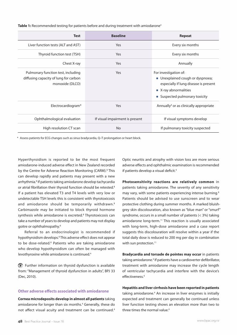
48 Best Practice Journal – Issue 78 www.bpac.org.nz
Hyperthyroidism is reported to be the most frequent amiodarone-induced adverse effect in New Zealand recorded by the Centre for Adverse Reaction Monitoring (CARM).8 This can develop rapidly and patients may present with a new arrhythmia.8 If patients taking amiodarone develop tachycardia or atrial fibrillation their thyroid function should be retested.8 If a patient has elevated T3 and T4 levels with very low or undetectable TSH levels this is consistent with thyrotoxicosis and amiodarone should be temporarily withdrawn.8 Carbimazole may be initiated to block thyroid hormone synthesis while amiodarone is excreted.8 Thyrotoxicosis can take a number of years to develop and patients may not display goitre or ophthalmopathy.8
Referral to an endocrinologist is recommended if hypothyroidism develops.8 This adverse effect does not appear to be dose-related.8 Patients who are taking amiodarone who develop hypothyroidism can often be managed with levothyroxine while amiodarone is continued.4
Further information on thyroid dysfunction is available from: “Management of thyroid dysfunction in adults”, BPJ 33 (Dec, 2010).
Other adverse effects associated with amiodarone
Cornea microdeposits develop in almost all patients taking amiodarone for longer than six months.4 Generally, these do not affect visual acuity and treatment can be continued.4
Optic neuritis and atrophy with vision loss are more serious adverse effects and ophthalmic examination is recommended if patients develop a visual deficit.4
Photosensitivity reactions are relatively common in patients taking amiodarone. The severity of any sensitivity may vary, with some patients experiencing intense burning.8 Patients should be advised to use sunscreen and to wear protective clothing during summer months. A marked bluish-grey skin discolouration, also known as “blue man” or “smurf” syndrome, occurs in a small number of patients (< 3%) taking amiodarone long-term.11 This reaction is usually associated with long-term, high-dose amiodarone and a case report suggests this discolouration will resolve within a year if the total daily dose is reduced to 200 mg per day in combination with sun protection.11
bradycardia and torsade de pointes may occur in patients taking amiodarone.4 If patients have a cardioverter defibrillator, treatment with amiodarone may increase the cycle length of ventricular tachycardia and interfere with the device’s effectiveness.4
Hepatitis and liver cirrhosis have been reported in patients taking amiodarone.4 An increase in liver enzymes is initially expected and treatment can generally be continued unless liver function testing shows an elevation more than two to three times the normal value.4
Table 1: Recommended testing for patients before and during treatment with amiodarone3
Test baseline Repeat
Liver function tests (ALT and AST) Yes Every six months
Thyroid function test (TSH) Yes Every six months
Chest X-ray Yes Annually
Pulmonary function test, including diffusing capacity of lung for carbon
monoxide (DLCO)
Yes For investigation of: Unexplained cough or dyspnoea;
especially if lung disease is present
X-ray abnormalities
Suspected pulmonary toxicity
Electrocardiogram* Yes Annually4 or as clinically appropriate
Ophthalmological evaluation If visual impairment is present If visual symptoms develop
High resolution CT scan No If pulmonary toxicity suspected
* Assess patients for ECG changes such as sinus bradycardia, Q–T prolongation or heart block.

Best Practice Journal – Issue 78 49www.bpac.org.nz
References1. Ministry of Health. Pharmaceutical Claims Collection. 2016.
2. New Zealand Formulary (NZF). NZF v48. 2016. Available from: www.nzf.org.nz
(Accessed Aug, 2016)
3. Medsafe. Amiodarone pulmonary toxicity - early recognition is vital. Prescr
Update;34:38–9.
4. Merino J L, Perez L. Treatment with amiodarone: how to avoid complications.
E-J Cardiol Pract 2011;10. Available from: www.escardio.org/Guidelines-
&-Education/Journals-and-publications/ESC-journals-family/E-journal-of-
Cardiology-Practice/Volume-10/Treatment-with-amiodarone-How-to-avoid-
complications (Accessed Sep, 2016)
5. Sanofi-Aventis New Zealand limited. Data sheet: CORDARONE X. 2015.
Available from: www.medsafe.govt.nz/profs/datasheet/c/CordaroneXtabinj.
6. Lakshmanadoss U, Lindsley J, Glick D, et al. Incidence of amiodarone
hypersensitivity in patients with previous allergy to iodine or iodinated
contrast agents. Pharmacotherapy 2012;32:618–22. doi:10.1002/j.1875-
9114.2012.01094.x
7. Yamada Y, Shiga T, Matsuda N, et al. Incidence and predictors of pulmonary
toxicity in Japanese patients receiving low-dose amiodarone. Circ J
2007;71:1610–6.
8. Waitemata DHB. Amiodarone - safe prescribing - keep an eye on it. 2013.
Available from: www.saferx.co.nz/full/amiodarone.pdf (Accessed Aug, 2016)
9. Nacca N, Bhamidipati CM, Yuhico LS, et al. Severe amiodarone induced
pulmonary toxicity. J Thorac Dis 2012;4:667–70. doi:10.3978/j.issn.2072-
1439.2012.06.08
10. Ministry of Health (MOH). Nutrient reference values for Australia and New
Zealand. 2005. Available from: www.nhmrc.gov.au/_files_nhmrc/publications/
attachments/n35.pdf? (Accessed Aug, 2016)
11. Jolly U, Klein G. Blue man syndrome. CMAJ 2016;188:604. doi:10.1503/
cmaj.150393
12. U.S. Food and Drug Administration. FDA Drug Safety Communication: Revised
dose limitation for Zocor (simvastatin) when taken with amiodarone. 2011.
Available from: www.fda.gov/Drugs/DrugSafety/ucm283137.htm (Accessed
Sep, 2016)
The potential for medicine interactions should be checked when initiating amiodarone or a new medicine in a patient already taking amiodarone.
The New Zealand Formulary (NZF) interaction checker is available from: www.nzf.org.nz/nzf_1090
Several interactions with amiodarone of note include:2
Amiodarone should generally be avoided in patients concurrently taking another medicine which may cause QT prolongation, e.g. citalopram, erythromycin, ondansetron, digoxin, or sofosbuvir or ledipasvir + sofosbuvir for the treatment of hepatitis C
Concurrent use of simvastatin or atorvastatin may increase the risk of myopathy; the dose of simvastatin should not exceed 20 mg per day in patients taking amiodarone.12 Pravastatin does not interact with amiodarone.
Interactions between beta blockers and amiodarone may increase cardiac depression and this combination should be prescribed with caution, although in some patients this may be beneficial. If a patient is taking these medicines their potassium levels should be monitored closely.
The anticoagulant effect of dabigatran can be potentiated by the concurrent use of amiodarone, although dabigatran does not appear to affect the pharmacokinetics of amiodarone.
Warfarin may have an increased anticoagulant effect in patients taking amiodarone. INR levels should be monitored weekly until a steady state is achieved.
Grapefruit juice inhibits the metabolism of amiodarone and regular consumption should be avoided in patients taking amiodarone
Amiodarone may interact with numerous medicines

50 Best Practice Journal – Issue 78 www.bpac.org.nz
Prescribing isotretinoin for patients with acne in primary care
DERMATOlOgy
What do prescribers need to know? Oral isotretinoin is a highly effective treatment for
patients with acne that is causing scarring or distress, or that persists following other treatments
Low-dose isotretinoin, e.g. 10 mg per day, is effective for most patients
Treatment should continue for at least three to four months after acne has cleared to reduce the risk of relapse
The time for acne clearance to occur is variable and some patients require a longer period of treatment
If pregnancy is possible, women should use effective contraception, i.e. two methods (refer below), for at least one month before beginning treatment, during treatment, and for at least one month after stopping treatment
Isotretinoin is an isomer of retinoic acid that has been used for the treatment of acne for over 30 years.1 Isotretinoin is recommended for patients with moderate acne that produces scarring or distress, or for acne that persists following other treatments.2
Treatment with isotretinoin results in decreased sebum production. This reduces acne, prevents scarring and may lead to a better quality of life and improved mental health in some patients.2 Isotretinoin is subsidised subject to Special Authority approval; vocationally registered general practitioners or nurse practitioners working in a relevant scope of practice are able to prescribe provided they have up-to-date knowledge of the safety issues.
A PHARMAC funded electronic decision support tool for isotretinoin prescribing is available from: www.bestpractice.net.nz/feat_mod_NatFunded.php#isotretinoin
Contraindications and cautions for isotretinoin use
Isotretinoin is highly teratogenic and is contraindicated in women if pregnancy is possible unless effective contraception is used (Table 1).1 Women who are breastfeeding should avoid
Best Practice Journal – Issue 78 51www.bpac.org.nz
using isotretinoin as should patients with hepatic impairment.1 Hyperlipidaemia is considered a relative contraindication. Patients with elevated lipids can usually be managed with close monitoring. Advise patients to avoid donating blood during treatment and for at least one month after treatment has finished.1
Prescribe low-dose isotretinoin to most patients with acne
The approved dose of isotretinoin is calculated according to the patient’s weight, i.e. 500 micrograms per kg, daily (in one to two divided doses) for two to four weeks, increased if necessary to 1 mg per kg, daily, for 16 – 24 weeks; maximum cumulative dose 150 mg per kg, per course.1 If a relapse occurs, treatment can be repeated if at least eight weeks have passed since the previous course.1
In practice, lower doses of isotretinoin are prescribed to the majority of patients in New Zealand (see: “Low-dose versus weight-based dosing of isotretinoin”), e.g. 10 mg per day until acne has cleared and for another three to four months thereafter.3 For many patients, a lower dose of 5 mg per day is likely to be effective, however, this formulation is not currently funded in New Zealand and 10 mg capsules should not be divided in half.3
To improve absorption and reduce fluctuations in systemic availability, isotretinoin should be taken with or just after food.4
low-dose isotretinoin is as effective as weight-based isotretinoinLow-dose regimens of isotretinoin are generally considered to be as effective as weight-based dosing regimens. A retrospective review of 1,453 patients treated with isotretinoin
over a six-year period concluded that continuing treatment for at least two months after acne had completely resolved was more important than the dosing regimen in determining treatment success.5 A low-dose treatment regimen may be better tolerated as the majority of the adverse effects of isotretinoin are dose-dependent.5
For additional information, see: www.goodfellowunit.org/gems/acne-low-dose-isotretinoin-10-mg-daily-effective-fewer-side-effects
less frequent dosing can reduce the risk of acne flaresParadoxically, flares of acne are frequently reported by patients after three to six weeks of treatment with higher doses of isotretinoin.6 Flares of acne are less common in patients taking low-dose isotretinoin.3 If a patient is likely to experience flares of acne (see below), consider prescribing 10 mg isotretinoin, two to three times per week, rather than daily. The frequency can then be increased to daily, if tolerated, after four weeks. Expert opinion is that flares of acne are more common in patients with a large number of macrocomedones (facial closed comedones larger than 2 – 3 mm in diameter) or very severe acne.
To decrease flares, expert opinion is that patients with particularly inflammatory acne can be prescribed trimethoprim, 300 mg per day, for the first six to eight weeks of treatment with isotretinoin.
The time taken for acne to clear variesPatients with acne that is likely to take longer to clear should be advised of this before starting isotretinoin so that treatment is not stopped if results do not occur as quickly as the patient expects. Experience suggests that females with acne limited to the face are likely to respond to isotretinoin treatment within
Table 1: Contraindications and cautions for treatment with isotretinoin.1
Contraindications Cautions
Pregnancy – highly teratogenic (see below)
Breastfeeding – present in breast milk
Hepatic impairment – further impairment may occur
Hyperlipidaemia – can usually be managed with close monitoring
Concurrent use of tetracyclines – due to the risk of intracranial hypertension
Hypervitaminosis A – although rarely seen
Visual disturbances including reduced night vision – pilots and commercial drivers may be subject to restrictions
Diabetes – elevations in fasting blood sugar levels may occur
Prior history of mental illness (see below)
Blood donation should be avoided during treatment and for at least one month after treatment has finished
Further information on isotretinoin is available from the New Zealand Formulary (NZF): www.nzf.org.nz/nzf_6452
52 Best Practice Journal – Issue 78 www.bpac.org.nz
three months. Males with acne on their trunk or back may take five to eight months to respond to treatment with isotretinoin. Patients with macrocomedonal acne may require as long as 12 to 18 months of treatment with low-dose isotretinoin. Additional risk factors for a slow response to treatment include: age under 14 years, age over 25 years in females, severe acne, current smoking and polycystic ovary syndrome.6
Further information and images of macrocomedonal acne are available from: www.dermnetnz.org/topics/comedonal-acne/
Preventing pregnancy in women taking isotretinoinTaking isotretinoin during key developmental periods is detrimental. If a woman who is pregnant takes isotretinoin during weeks six to ten of gestation major disruptions to organogenesis can occur.8 Isotretinoin adversely affects 25 – 40% of foetuses exposed during embryogenesis.9
Extreme care is required when prescribing to women of child-bearing age
Effective contraception must be used for at least one month before beginning treatment, during treatment, and for at least one month after stopping treatment.1 Barrier methods of contraception should not be used alone and as oral progesterone-only contraceptives need to be taken within a three-hour window these are not recommended.1 Ideally, two forms of contraception should be used, i.e. condoms and hormonal treatment.1
begin treatment with isotretinoin on day two or three of the woman’s menstrual cycle.1 Testing to exclude pregnancy, preferably a serum HCG test, is recommended up to three days prior to treatment, every month during treatment and five weeks after stopping treatment.1
The New Zealand recommendations for isotretinoin are based on early studies.5 To determine optimal dosing, patients were prescribed doses ranging from 0.1 mg per kg, daily, to 1.0 mg per kg, daily.2 The same degree of efficacy was reported across this range, however, higher daily weight-based doses were interpreted as having lower rates of relapse.2 This led to the approved dose of isotretinoin of 0.5 – 1 mg per kg, daily. These studies, however, only compared doses for a fixed time period.5
It is now known that lower doses of isotretinoin, given for longer periods of time, are similarly effective with a lower risk of adverse effects to the patient. This has led to patients taking lower daily doses of isotretinoin, i.e. off-label prescribing, for longer periods of time.5
Further information on the unapproved use of medicines is available from: www.bpac.org.nz/BPJ/2013/March/unapproved-medicines.aspx
Why is low-dose isotretinoin an unapproved regimen?
The adverse effects of isotretinoin
Retinoic acid is a metabolite of vitamin A which can cause similar adverse effects as excess vitamin A. These include dryness of the skin, lips (cheilitis – which may persist for months), eyes, nasal and pharyngeal mucosa.1, 2 Adverse effects are more likely to be experienced by patients taking higher doses of isotretinoin. A review of patients taking high-dose isotretinoin (> 0.75 mg per kg, daily) found that cheilitis (96%), eczema (16%) and tiredness (18%) were frequently reported but these were less common in patients taking low-dose isotretinoin (< 0.25 mg per kg, daily).10 Myalgia, arthralgia, changes in vision and headache may also be reported.1 Photosensitivity reactions can occur and patients taking isotretinoin should be advised to be “sun smart” and to avoid the use of sunbeds.1 The adverse effects of isotretinoin usually resolve completely once treatment is withdrawn.2
Changes in mood, depression and suicide have been reported in patients taking isotretinoin and monitoring patients’ mood is recommended (see below).2 At a population level, however, studies do not support an association between treatment with isotretinoin and adverse psychiatric effects.2
Conversely, there is evidence suggesting that for some patients with acne, treatment may lead to improvements in mood, memory and higher-level cognitive functions.2
There is mixed evidence of an association between the use of isotretinoin and inflammatory bowel disease (IBD). Several studies are reported to have shown a link between patients taking isotretinoin and subsequent development of IBD, in particular ulcerative colitis. However, more recent analyses have not confirmed this association.2
Monitoring hepatic function and serum lipids
Isotretinoin treatment can cause dose-related elevations in serum transaminases, cholesterol and triglycerides.2 ,4 Current monitoring recommendations (including an electronic
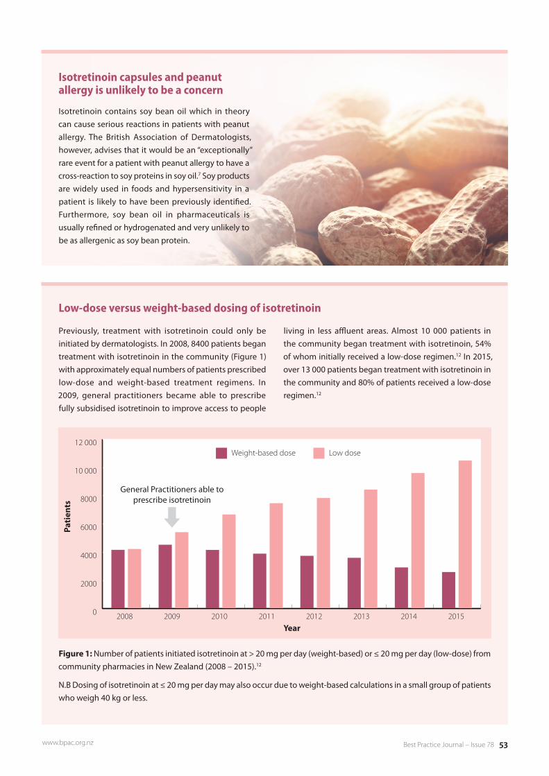
Best Practice Journal – Issue 78 53www.bpac.org.nz
Isotretinoin capsules and peanut allergy is unlikely to be a concern
Isotretinoin contains soy bean oil which in theory can cause serious reactions in patients with peanut allergy. The British Association of Dermatologists, however, advises that it would be an “exceptionally” rare event for a patient with peanut allergy to have a cross-reaction to soy proteins in soy oil.7 Soy products are widely used in foods and hypersensitivity in a patient is likely to have been previously identified. Furthermore, soy bean oil in pharmaceuticals is usually refined or hydrogenated and very unlikely to be as allergenic as soy bean protein.
0
1
2
3
4
2008 2009 2010 2011 2012 2013 2014 2015
0
2000
4000
6000
8000
10 000
12 000
2008 2009 2010 2011 2012 2013 2014 2015
Weight-based dose Low dose
General Practitioners able to prescribe isotretinoin
Year
Year
European
Asian
Māori
Paci�c Island
Pati
ents
Pati
ents
dis
pens
ed p
er 1
000
enro
lled
pati
ents
Previously, treatment with isotretinoin could only be initiated by dermatologists. In 2008, 8400 patients began treatment with isotretinoin in the community (Figure 1) with approximately equal numbers of patients prescribed low-dose and weight-based treatment regimens. In 2009, general practitioners became able to prescribe fully subsidised isotretinoin to improve access to people
living in less affluent areas. Almost 10 000 patients in the community began treatment with isotretinoin, 54% of whom initially received a low-dose regimen.12 In 2015, over 13 000 patients began treatment with isotretinoin in the community and 80% of patients received a low-dose regimen.12
Figure 1: Number of patients initiated isotretinoin at > 20 mg per day (weight-based) or ≤ 20 mg per day (low-dose) from community pharmacies in New Zealand (2008 – 2015).12
N.B Dosing of isotretinoin at ≤ 20 mg per day may also occur due to weight-based calculations in a small group of patients who weigh 40 kg or less.
Low-dose versus weight-based dosing of isotretinoin

0
1
2
3
4
2008 2009 2010 2011 2012 2013 2014 2015
0
2000
4000
6000
8000
10 000
12 000
2008 2009 2010 2011 2012 2013 2014 2015
Weight-based dose Low dose
General Practitioners able to prescribe isotretinoin
Year
Year
European
Asian
Māori
Paci�c Island
Pati
ents
Pati
ents
dis
pens
ed p
er 1
000
enro
lled
pati
ents
54 Best Practice Journal – Issue 78 www.bpac.org.nz
decision support tool) for patients taking isotretinoin include an assessment of hepatic function and serum lipids, before treatment is initiated, after starting treatment and every three months thereafter.1
In practice, however, there is likely to be little value in routinely monitoring hepatic function and serum lipids in otherwise healthy patients taking low-dose isotretinoin. In 2016, a systematic review of studies including patients taking high-dose isotretinoin (≥ 40 mg, daily or ≥ 0.5 mg per kg, daily) found that while treatment with isotretinoin was associated with changes in serum transaminases and lipids, especially triglycerides and total cholesterol, there was no evidence to support monthly testing in otherwise healthy patients.11
A pragmatic approach would be to ensure there is a recent assessment of the patient’s hepatic function and lipid profile and to monitor patients with risks factors, e.g. a history of either hepatic dysfunction or hyperlipidaemia. Isotretinoin
dosing should be reduced or treatment withdrawn in patients with persistently raised serum lipids, or transaminase,1 e.g. ALT greater than three times the upper limit of normal. Discussion with a dermatologist is recommended for patients with significantly elevated serum triglycerides; levels > 9 mmol/L have been associated with acute pancreatitis.1
Advise patients to report adverse changes in mood
When discussing isotretinoin treatment ask how the acne is affecting the patient’s mood and consider their prior mental health. Inform the patient that there have been reports of depression in patients taking isotretinoin and ask them to report any adverse changes in mood.
Additional information is available from: www.goodfellowunit.org/gems/depression-and-isotretinoin-another-reason-low-dose
There is no evidence that Māori or Pacific peoples have lower rates of acne.13 It is therefore reasonable to expect that Māori and Pacific peoples would be prescribed isotretinoin at rates similar to that of New Zealand Europeans. However, analysis of New Zealand prescribing data from 2008 to 2015 suggests that Māori and Pacific peoples may not be receiving equitable treatment for acne compared to New Zealand European and Asian
people (Figure 2). It is not known if these differences in prescribing are due to under treatment, disparities in access to care, personal attitudes to acne or a combination of factors. Clinicians in primary care can help to improve acne treatment rates among Māori and Pacific peoples by discussing acne with all patients who are affected and ensuring patients know that treatment with isotretinoin is available.
Figure 2: Number of patients dispensed isotretinoin, per 1000 enrolled patients, from community pharmacies in New Zealand by ethnicity (2008 – 2015).12
Māori and Pacific peoples with acne may be under treated

Best Practice Journal – Issue 78 55www.bpac.org.nz
bestpractice Acne including Isotretinoin
The bestpractice Acne Management module provides tools for the initial assessment of acne severity, context sensitive advice, treatment and management options.
The module features guidance on the safe prescribing of isotretinoin, including:
Contraindications, cautions and ■side effectsLaboratory testing ■requirements and schedulingPatient information ■Patient consent documents ■
More information is available at:
www.bestpractice.net.nz
bestpractice Decision Support is developed by BPAC Inc, which is separate from bpacnz.bpacnz bears no responsibility for bestpractice Decision Support or any use that is made of it.
bestpracticeDECISION SUPPORT FOR HEALTH PROFESSIONALS
ISOTRETINOIN
www.bestpractice.net.nz
Managing relapses of acne
Patients who experience a relapse of acne may be offered a second course of isotretinoin eight weeks after treatment is completed;1 this can be expected in approximately 22% of patients.5 Patients with risk factors for acne that is slow to clear are also more likely to experience a relapse as are those who discontinue treatment before their acne has cleared and those with excessive seborrhoea after treatment has finished.6 Extending the treatment period to four to six months after acne has cleared may reduce the risk of relapse for these patients.
Consider discussing patients with a dermatologist if they experience multiple relapses of acne despite being adherent to treatment.
Acknowledgement: Thank you to Associate Professor Marius Rademaker, Clinical Director of the Dermatology Unit,
Waikato, DHB, for expert review of this article.
A webinar on prescribing isotretinoin for acne in primary care presented by Associate Professor Rademaker is available from the Goodfellow Unit: www.goodfellowunit.org/webinars
References1. New Zealand Formulary (NZF). NZF v55. 2016. Available from: www.nzf.org.nz
(Accessed Dec, 2016)
2. Zaenglein AL, Pathy AL, Schlosser BJ, et al. Guidelines of care for the
management of acne vulgaris. J Am Acad Dermatol 2016;74:945–973.e33.
doi:10.1016/j.jaad.2015.12.037
3. Rademaker M, Wishart JM, Birchall NM. Isotretinoin 5 mg daily for low-grade
adult acne vulgaris--a placebo-controlled, randomized double-blind study. J
Eur Acad Dermatol Venereol JEADV 2014;28:747–54. doi:10.1111/jdv.12170
4. Mylan New Zealand Ltd. New Zealand data sheet: Isotane. 2015. Available
from: www.medsafe.govt.nz/profs/datasheet/i/isotanecap.pdf
5. Rademaker M. Making sense of the effects of the cumulative dose of
isotretinoin in acne vulgaris. Int J Dermatol 2016;55:518–23. doi:10.1111/
ijd.12942
6. Rademaker M. Isotretinoin: dose, duration and relapse. What does 30 years
of usage tell us? Australas J Dermatol 2013;54:157–62. doi:10.1111/j.1440-
0960.2012.00947.x
7. British Association of Dermatologists. Alitretinoin. 2015. Available from: www.
bad.org.uk/shared/get-file.ashx?id=3659&itemtype=document
8. Duester G. Retinoic acid synthesis and signaling during early organogenesis.
Cell 2008;134:921–31. doi:10.1016/j.cell.2008.09.002
9. Prevost N, English III JC. Isotretinoin: Update on Controversial Issues. J Pediatr
Adolesc Gynecol 2013;26:290–3. doi:10.1016/j.jpag.2013.05.007
10. Rademaker M. Adverse effects of isotretinoin: A retrospective review of
1743 patients started on isotretinoin. Australas J Dermatol 2010;51:248–53.
doi:10.1111/j.1440-0960.2010.00657.x
11. Lee YH, Scharnitz TP, Muscat J, et al. Laboratory Monitoring During Isotretinoin
Therapy for Acne: A Systematic Review and Meta-analysis. JAMA Dermatol
2016;152:35–44. doi:10.1001/jamadermatol.2015.3091
12. Ministry of Health. Pharmaceutical Claims Collection. 2016.
13. Moodie P, Jaine R, Arnold J, et al. Usage and equity of access to isotretinoin in
New Zealand by deprivation and ethnicity. N Z Med J 2011;124:34–43.
56 Best Practice Journal – Issue 78 www.bpac.org.nz
Oestradiol patches now fully subsidised: what is their place in the treatment of menopausal symptoms?
ENDOCRINOlOgy gyNAECOlOgy & uRINARy TR AC T
Transdermal oestradiol patches (25, 50 and 100 microgram Estradot brand patches) are now fully subsidised, without the need for Special Authority approval, as a form of menopausal hormone therapy (MHT) for the treatment of menopausal symptoms. From 1 January, 2017, a 75 microgram patch has also been available.1
Consider MHT when menopausal symptoms are affecting a woman’s quality of lifeDuring the menopausal transition, women have variations in menstrual cycle length and bleeding pattern. A woman is considered to be post-menopausal one year after her last menstrual period. Hormone replacement therapy for peri- or post-menopausal women is now referred to as menopausal hormone therapy (MHT) to differentiate it from hormone replacement for other endocrine conditions, e.g. growth hormone replacement.2 MHT regimens consist of transdermal or oral preparations of oestrogen, alone or in combination with progestogens.
MHT can reduce hot flushes associated with menopause.2–4 Vasomotor symptoms (hot flushes) continue to affect 45–55%
of women three to four years post-menopause, and subside on average after approximately eight years.5 MHT can reduce the severity of hot flushes and reduce their frequency by 75% compared with placebo.6 MHT also improves other symptoms of menopause, including sexual function, genitourinary symptoms, urinary tract infections, depressive symptoms and sleep disruption.2, 4, 7
women who primarily have vaginal symptoms of menopause, including vaginal dryness, irritation and painful intercourse can often be managed with topical (vaginal) treatments alone. Vaginal moisturisers and lubricants are appropriate as a first-line treatment.2 If symptoms are ongoing, a topical oestrogen can be trialled.2
lifestyle interventions that may be helpful for some women include stress reduction, weight management, smoking cessation and moderating caffeine and alcohol intake.6 Evidence is inconclusive regarding whether exercise reduces hot flushes; in some women it may help but in others it may aggravate hot flushes, and effects may differ according to the type of exercise.2, 3, 6, 14
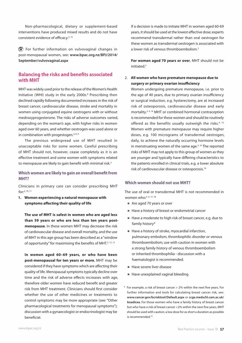
Best Practice Journal – Issue 78 57www.bpac.org.nz
Non-pharmacological, dietary or supplement-based interventions have produced mixed results and do not have consistent evidence of efficacy.2, 15
For further information on vulvovaginal changes in post-menopausal women, see: www.bpac.org.nz/BPJ/2014/September/vulvovaginal.aspx
Balancing the risks and benefits associated with MHT
MHT was widely used prior to the release of the Women’s Health Initiative (WHI) study in the early 2000s.8 Prescribing then declined rapidly following documented increases in the risk of breast cancer, cardiovascular disease, stroke and mortality in women using conjugated equine oestrogens with or without medroxyprogesterone. The risks of adverse outcomes varied, depending on the woman’s age, with higher risks in women aged over 60 years, and whether oestrogen was used alone or in combination with progestogen.4, 8, 9
The previous widespread use of MHT resulted in unacceptable risks for some women. Careful prescribing of MHT should not, however, cease completely as it is an effective treatment and some women with symptoms related to menopause are likely to gain benefit with minimal risk.3
Which women are likely to gain an overall benefit from MHT?
Clinicians in primary care can consider prescribing MHT for:2, 10, 11
1. women experiencing a natural menopause with symptoms affecting their quality of life
The use of MHT is safest in women who are aged less than 59 years or who are less than ten years post-menopause. In these women MHT may decrease the risk of cardiovascular disease and overall mortality, and the use of MHT in this age group has been described as a “window of opportunity” for maximising the benefits of MHT.3, 12, 13
In women aged 60–69 years, or who have been post-menopausal for ten years or more, MHT may be considered if they have symptoms which are affecting their quality of life. Menopausal symptoms typically decline over time and the risk of adverse effects increases with age, therefore older women have reduced benefit and greater risk from MHT treatment. Clinicians should first consider whether the use of other medicines or treatments to control symptoms may be more appropriate (see: “Other pharmacological treatments for menopausal symptoms”); discussion with a gynaecologist or endocrinologist may be beneficial.
If a decision is made to initiate MHT in women aged 60-69 years, it should be used at the lowest effective dose; experts recommend transdermal rather than oral oestrogen for these women as transdermal oestrogen is associated with a lower risk of venous thromboembolism.3
For women aged 70 years or over, MHT should not be initiated.3
2. All women who have premature menopause due to surgery or primary ovarian insufficiencyWomen undergoing premature menopause, i.e. prior to the age of 40 years, due to primary ovarian insufficiency or surgical induction, e.g. hysterectomy, are at increased risk of osteoporosis, cardiovascular disease and early mortality.3, 4, 16 MHT or combined hormonal contraception is recommended for these women and should be routinely offered as the benefits usually outweigh the risks.4, 16 Women with premature menopause may require higher doses, e.g. 100 micrograms of transdermal oestrogen, daily, to achieve the naturally occurring hormone levels in menstruating women of the same age.3, 16 The reported risks of MHT may not apply to this group of women as they are younger and typically have differing characteristics to the patients enrolled in clinical trials, e.g. a lower absolute risk of cardiovascular disease or osteoporosis.16
Which women should not use MHT?
The use of oral or transdermal MHT is not recommended in women who:2–4, 17, 18
Are aged 70 years or over
Have a history of breast or endometrial cancer
Have a moderate to high risk of breast cancer, e.g. due to family history*
Have a history of stroke, myocardial infarction, pulmonary embolism, thrombophilic disorder or venous thromboembolism; use with caution in women with a strong family history of venous thromboembolism or inherited thrombophilia - discussion with a haematologist is recommended.
Have severe liver disease
Have unexplained vaginal bleeding
* For example, a risk of breast cancer > 2% within the next five years. For further information and tools for calculating breast cancer risk, see: www.cancer.gov/bcrisktool/Default.aspx or ccge.medschl.cam.ac.uk/boadicea. For those women who have a family history of breast cancer but who have a risk of breast cancer <2% within the next five years, MHT should be used with caution; a low dose for as short a duration as possible is recommended.19
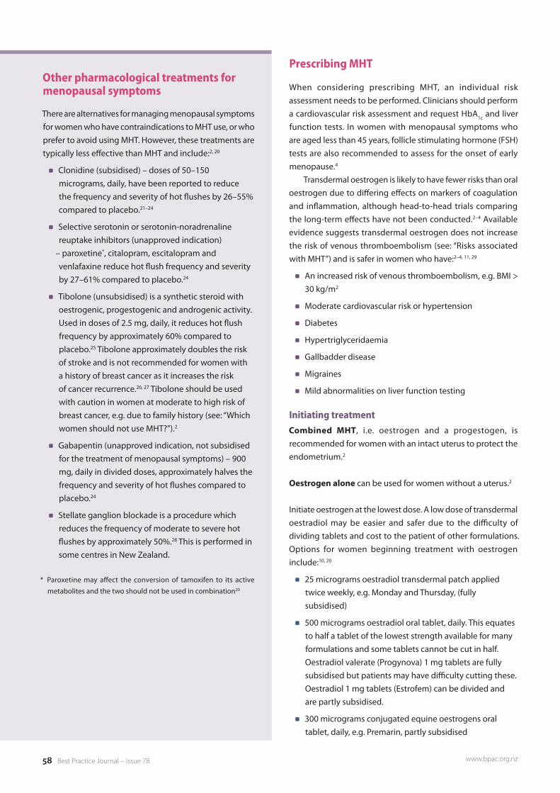
58 Best Practice Journal – Issue 78 www.bpac.org.nz
Prescribing MHT
When considering prescribing MHT, an individual risk assessment needs to be performed. Clinicians should perform a cardiovascular risk assessment and request HbA1c and liver function tests. In women with menopausal symptoms who are aged less than 45 years, follicle stimulating hormone (FSH) tests are also recommended to assess for the onset of early menopause.4
Transdermal oestrogen is likely to have fewer risks than oral oestrogen due to differing effects on markers of coagulation and inflammation, although head-to-head trials comparing the long-term effects have not been conducted.2–4 Available evidence suggests transdermal oestrogen does not increase the risk of venous thromboembolism (see: “Risks associated with MHT”) and is safer in women who have:2–4, 11, 29
An increased risk of venous thromboembolism, e.g. BMI > 30 kg/m2
Moderate cardiovascular risk or hypertension
Diabetes
Hypertriglyceridaemia
Gallbadder disease
Migraines
Mild abnormalities on liver function testing
Initiating treatment
Combined MHT, i.e. oestrogen and a progestogen, is recommended for women with an intact uterus to protect the endometrium.2
Oestrogen alone can be used for women without a uterus.2
Initiate oestrogen at the lowest dose. A low dose of transdermal oestradiol may be easier and safer due to the difficulty of dividing tablets and cost to the patient of other formulations. Options for women beginning treatment with oestrogen include:10, 20
25 micrograms oestradiol transdermal patch applied twice weekly, e.g. Monday and Thursday, (fully subsidised)
500 micrograms oestradiol oral tablet, daily. This equates to half a tablet of the lowest strength available for many formulations and some tablets cannot be cut in half. Oestradiol valerate (Progynova) 1 mg tablets are fully subsidised but patients may have difficulty cutting these. Oestradiol 1 mg tablets (Estrofem) can be divided and are partly subsidised.
300 micrograms conjugated equine oestrogens oral tablet, daily, e.g. Premarin, partly subsidised
Other pharmacological treatments for menopausal symptoms
There are alternatives for managing menopausal symptoms for women who have contraindications to MHT use, or who prefer to avoid using MHT. However, these treatments are typically less effective than MHT and include:2, 20
Clonidine (subsidised) – doses of 50–150 micrograms, daily, have been reported to reduce the frequency and severity of hot flushes by 26–55% compared to placebo.21–24
Selective serotonin or serotonin-noradrenaline reuptake inhibitors (unapproved indication)
– paroxetine*, citalopram, escitalopram and venlafaxine reduce hot flush frequency and severity by 27–61% compared to placebo.24
Tibolone (unsubsidised) is a synthetic steroid with oestrogenic, progestogenic and androgenic activity. Used in doses of 2.5 mg, daily, it reduces hot flush frequency by approximately 60% compared to placebo.25 Tibolone approximately doubles the risk of stroke and is not recommended for women with a history of breast cancer as it increases the risk of cancer recurrence.26, 27 Tibolone should be used with caution in women at moderate to high risk of breast cancer, e.g. due to family history (see: “Which women should not use MHT?”).2
Gabapentin (unapproved indication, not subsidised for the treatment of menopausal symptoms) – 900 mg, daily in divided doses, approximately halves the frequency and severity of hot flushes compared to placebo.24
Stellate ganglion blockade is a procedure which reduces the frequency of moderate to severe hot flushes by approximately 50%.28 This is performed in some centres in New Zealand.
* Paroxetine may affect the conversion of tamoxifen to its active metabolites and the two should not be used in combination20
Best Practice Journal – Issue 78 59www.bpac.org.nz
For women requiring combined oestrogen + progesterone MHT, progesterone can be prescribed as a separate formulation, an intrauterine levonorgestrel device (subsidised under Special Authority approval), which also provides contraception for women who may still be fertile, or a combination oestrogen + progesterone formulation (see: “Adding a progestogen to increase endometrial protection”).11, 20
For further information on prescribing the progesterone component of MHT, see the NZF: www.nzf.org.nz/nzf_3858
Adverse effects of MHT
Women taking MHT may experience breast tenderness, bloating, headache and urinary incontinence; a dose reduction can be trialled to see if adverse effects resolve.6 Women using transdermal patches may experience contact sensitisation.20
Women using cyclical MHT can be expected to have withdrawal bleeding. Unexpected vaginal bleeding, i.e. outside of expected withdrawal bleeds in women using cyclical MHT or at any time in women using continuous MHT, often occurs within three months of initiating MHT and then settles. Advise women to report any unexpected vaginal bleeding. Unexpected vaginal bleeding which begins or continues after six months of MHT use should be investigated to assess for endometrial cancer.11 Other findings associated with endometrial cancer include visible haematuria, thrombocytosis, low haemoglobin levels and high blood glucose levels.30 Patients should be referred to secondary care if endometrial cancer is suspected.
Monitoring and follow-up
Women should be followed up within one to three months of initiating MHT to assess treatment response and any adverse effects.2 Subsequent follow-up can occur every six to 12 months.2 Women using MHT should have a mammogram every two years.11
Treatment can be titrated up or down according to clinical response, e.g. 25 microgram oestradiol patch to a 50 microgram oestradiol patch, if symptom relief is not adequate.2, 6 Measurement of serum oestradiol levels is generally not necessary but may be useful to confirm absorption if symptoms persist after increasing doses.2
Duration of treatment
The appropriate duration of MHT is uncertain. Menopausal symptoms often return as MHT is withdrawn. For women who have undergone a natural menopause, the continued use of MHT treatment should be reviewed annually, taking into account the risks and benefits of continuing treatment.4, 15, 17 For women using oestrogen combined with progestogen treatment, the risk of breast cancer increases after seven years of use and progressively decreases after stopping treatment.3 It is
Adding a progestogen to increase endometrial protection
Combined MHT can be prescribed as follows: An oestrogen tablet or patch with a separate
formulation of progestogen. The use of separate formulations of oestrogen and progesterone provides greater dose control than combination formulations:
– Progesterone capsules (Utrogestan), unsubsidised, 100 mg daily or 200 mg for the last 12 days of a cycle.3, 20 The progesterone in these capsules is micronised; evidence suggests micronised progesterone has less effect on cardiovascular and breast cancer risk than other synthetic forms of progesterone.11
– Medroxyprogesterone acetate tablets (fully subsidised) – 2.5 mg medroxyprogesterone acetate, daily or 5–10 mg, daily for 10–14 days of a 28-day oestrogen MHT cycle20
– Norethisterone tablets (fully subsidised) – for a low dose of oestrogen, e.g. 25 micrograms oestradiol transdermal patch applied twice weekly, 500 micrograms oestradiol oral tablet, daily, or 300 micrograms conjugated equine oestrogens oral tablet, daily; two 350 microgram norethisterone tablets (Noriday – unapproved indication); for higher oestrogen doses, e.g. 2 mg oral oestradiol, one quarter of a 5 mg tablet (Primolut N); tablets can be easily divided into quarters, providing approximately the same 2:1 dose ratio of combined oestradiol + norethisterone formulations
An oestrogen tablet or patch with an intrauterine levonorgestrel device, which also provides contraception for women who may still become pregnant11, 20
A combination formulation of oestrogen and progestogen, e.g. conjugated equine oestrogens + medroxyprogesterone, oestradiol + norethisterone (partially subsidised)
60 Best Practice Journal – Issue 78 www.bpac.org.nz
Acknowledgement: Thank you to Dr Anna Fenton, Endocrinologist, Canterbury District Health Board, Immediate
Past President Australasian Menopause Society and Co-Editor-
in-chief Climacteric (Journal of the International Menopause
Society) for expert review of this article.
recommended that the risks and benefits of treatment be re-evaluated if continuing use beyond five years.17
For women who have undergone premature menopause, treatment should continue until they reach their early fifties, the average age of natural menopause, at which point a shared decision on withdrawal of treatment can be made.3, 4, 11
Consultation with a gynaecologist is recommended if treatment is to be continued long-term.
Withdrawing MHT
Up to 50% of women report the reappearance of vasomotor symptoms upon withdrawal of MHT; this is similar for women regardless of age or duration of use, suggesting that it is related to the withdrawal of treatment as opposed to the reappearance of underlying symptoms.6 Withdrawal symptoms may last for 12 months or longer.31 Women may be at greater risk of myocardial infarction if MHT is stopped before age 60 years compared to ceasing use after age 60 years, and women of any age may have a transiently increased risk of myocardial infarction and fracture after stopping MHT.10, 32
Withdrawing treatment can be done either by stopping abruptly or by tapering the dose over weeks to months. Women who stop abruptly can experience greater initial withdrawal symptoms, however, evidence suggests there are no differences in rates of withdrawal symptoms six months after stopping MHT.4, 6
Patient information and videos covering the symptoms of menopause, pharmacological and non-pharmacological management are available from the Australasian Menopause Society:
www.menopause.org.au/for-women/information-sheets
www.menopause.org.au/for-women/videos
References:1. PHARMAC. Decision to fully fund oestradiol transdermal patches without
restriction. 2016. Available from: www.pharmac.govt.nz/news/notification-
2016-06-10-oestradiol-patches/ (Accessed Oct, 2016).
2. Stuenkel CA, Davis SR, Gompel A, et al. Treatment of symptoms of the
menopause: an Endocrine Society clinical practice guideline. J Clin Endocrinol
Metab 2015;100:3975–4011. doi:10.1210/jc.2015-2236
3. Baber RJ, Panay N, Fenton A, et al. 2016 IMS Recommendations on women’s
midlife health and menopause hormone therapy. Climacteric 2016;19:109–50.
doi:10.3109/13697137.2015.1129166
4. National Institute for Health and Care Excellence (NICE). Menopause: diagnosis
and management. 2015. Available from: www.nice.org.uk/guidance/ng23
(Accessed Oct, 2016).
5. Politi MC, Schleinitz MD, Col NF. Revisiting the duration of vasomotor
symptoms of menopause: a meta-analysis. J Gen Intern Med 2008;23:1507–13.
doi:10.1007/s11606-008-0655-4
6. American College of Obstetricians and Gynecologists’ Committee on
Gynecologic Practice. ACOG Practice Bulletin No. 141: management of
menopausal symptoms. Obstet Gynecol 2014;123:202–16. doi:10.1097/01.
AOG.0000441353.20693.78
7. Martins WP, Lara LA, Ferriani RA, et al. Hormone therapy for female sexual
function during perimenopause and postmenopause: a Cochrane review.
Climacteric 2014;17:133–5. doi:10.3109/13697137.2013.828688
8. Rossouw JE, Anderson GL, Prentice RL, et al. Risks and benefits of estrogen
plus progestin in healthy postmenopausal women: principal results from the
Women’s Health Initiative randomized controlled trial. JAMA 2002;288:321–33.
9. Manson JE, Chlebowski RT, Stefanick ML, et al. Menopausal hormone
therapy and health outcomes during the intervention and extended
poststopping phases of the Women’s Health Initiative randomized trials. JAMA
2013;310:1353–68. doi:10.1001/jama.2013.278040
10. North American Menopause Society. The 2012 hormone therapy position
statement of the North American Menopause Society. Menopause
2012;19:257–71. doi:10.1097/gme.0b013e31824b970a
11. Royal Australian and New Zealand College of Obstetricians and Gynaecologists
(RANZCOG). Menopausal hormone therapy advice. Victoria, Australia:
RANZCOG 2015. Available from: www.ranzcog.edu.au/Statements-Guidelines/
(Accessed Oct, 2016).
12. Schierbeck LL, Rejnmark L, Tofteng CL, et al. Effect of hormone replacement
therapy on cardiovascular events in recently postmenopausal women:
randomised trial. BMJ 2012;345:e6409.
13. Boardman HMP, Hartley L, Eisinga A, et al. Hormone therapy for preventing
cardiovascular disease in post-menopausal women. Cochrane Database Syst
Rev 2015;3:CD002229. doi:10.1002/14651858.CD002229.pub4
14. Daley A, Stokes-Lampard H, Macarthur C. Exercise for vasomotor
menopausal symptoms. Cochrane Database Syst Rev 2011;5:CD006108.
doi:10.1002/14651858.CD006108.pub3
15. Goodman NF, Cobin RH, Ginzburg SB, et al. American Association of Clinical
Endocrinologists medical guidelines for clinical practice for the diagnosis and
treatment of menopause: executive summary of recommendations. Endocr
Pract 2011;17:949–54.

Best Practice Journal – Issue 78 61www.bpac.org.nz
16. Faubion SS, Kuhle CL, Shuster LT, et al. Long-term health consequences
of premature or early menopause and considerations for management.
Climacteric 2015;18:483–91. doi:10.3109/13697137.2015.1020484
17. Royal Australian and New Zealand College of Obstetricians and Gynaecologists
(RANZCOG). Management of the menopause. Victoria, Australia: RANZCOG
2014. Available from: www.ranzcog.edu.au/Statements-Guidelines/ (Accessed
Oct, 2016).
18. Royal Australian and New Zealand College of Obstetricians and Gynaecologists
(RANZCOG). Management of the menopause after breast cancer. Victoria,
Australia: RANZCOG 2014. Available from: www.ranzcog.edu.au/Statements-
Guidelines/ (Accessed Oct, 2016).
19. National Institutes for Health and Care Excellence (NICE). Familial breast cancer:
classification, care and managing breast cancer and related risks in people with
a family history of breast cancer. 2015. Available from: https://www.nice.org.
uk/guidance/CG164/ (Accessed Oct, 2016).
20. New Zealand Formulary (NZF). NZF v48. 2016. Available from: www.nzf.org.nz
(Accessed Oct, 2016)
21. Boekhout AH, Vincent AD, Dalesio OB, et al. Management of hot flashes in
patients who have breast cancer with venlafaxine and clonidine: a randomized,
double-blind, placebo-controlled trial. J Clin Oncol 2011;29:3862–8.
doi:10.1200/JCO.2010.33.1298
22. Loibl S, Schwedler K, von Minckwitz G, et al. Venlafaxine is superior to
clonidine as treatment of hot flashes in breast cancer patients - a double-blind,
randomized study. Ann Oncol 2007;18:689–93. doi:10.1093/annonc/mdl478
23. Buijs C, Mom CH, Willemse PHB, et al. Venlafaxine versus clonidine for the
treatment of hot flashes in breast cancer patients: a double-blind, randomized
cross-over study. Breast Cancer Res Treat 2009;115:573–80. doi:10.1007/
s10549-008-0138-7
24. Rada G, Capurro D, Pantoja T, et al. Non-hormonal interventions for hot
flushes in women with a history of breast cancer. Cochrane Database Syst Rev
2010;9:CD004923. doi:10.1002/14651858.CD004923.pub2
25. Formoso G, Perrone E, Maltoni S, et al. Short and long term effects of tibolone
in postmenopausal women. Cochrane Database Syst Rev 2012;2:CD008536.
doi:10.1002/14651858.CD008536.pub2
26. Kenemans P, Bundred NJ, Foidart J-M, et al. Safety and efficacy of tibolone in
breast-cancer patients with vasomotor symptoms: a double-blind, randomised,
non-inferiority trial. Lancet Oncol 2009;10:135–46. doi:10.1016/S1470-
2045(08)70341-3
27. Cummings SR, Ettinger B, Delmas PD, et al. The effects of tibolone in older
postmenopausal women. N Engl J Med 2008;359:697–708. doi:10.1056/
NEJMoa0800743
28. Walega DR, Rubin LH, Banuvar S, et al. Effects of stellate ganglion block on
vasomotor symptoms: findings from a randomized controlled clinical trial
in postmenopausal women. Menopause 2014;21:807–14. doi:10.1097/
GME.0000000000000194
29. Mohammed K, Abu Dabrh AM, Benkhadra K, et al. Oral vs transdermal estrogen
therapy and vascular events: a systematic review and meta-analysis. J Clin
Endocrinol Metab 2015;100:4012–20. doi:10.1210/jc.2015-2237
30. National Institutes for Health and Care Excellence (NICE). Suspected cancer:
recognition and referral. 2015. Available from: https://www.nice.org.uk/
guidance/ng12 (Accessed Oct, 2016).
31. Haimov-Kochman R, Barak-Glantz E, Arbel R, et al. Gradual discontinuation
of hormone therapy does not prevent the reappearance of climacteric
symptoms: a randomized prospective study. Menopause 2006;13:370–6.
doi:10.1097/01.gme.0000186663.36211.c0
32. Mikkola TS, Tuomikoski P, Lyytinen H, et al. Increased cardiovascular mortality
risk in women discontinuing postmenopausal hormone therapy. J Clin
Endocrinol Metab 2015;100:4588–94. doi:10.1210/jc.2015-1864
NEED MORE CPD?
www.bpac.org.nz/audits
Audits are endorsed by the RNZCgP as a CQI activity for allocation of MOPS credits for general Practitioners:
www.bpac.org.nz/quizzes
Interactive quizzes and case studies are based on material found on the bpacnz
website, and are also endorsed as a CPD activity:
www.bpac.org.nz/peergroup
Peer group discussions are based on material previously published by bpacnz. They include a brief summary along with discussion points to take to your next peer group meeting:
bpacnz is here to help you with your Continuing Professional Development requirements
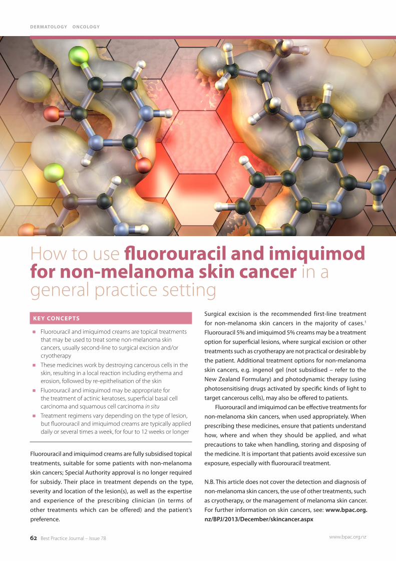
62 Best Practice Journal – Issue 78 www.bpac.org.nz
How to use fluorouracil and imiquimod for non-melanoma skin cancer in a general practice setting
DERMATOlOgy ONCOlOgy
KEy CONCEPTS
Fluorouracil and imiquimod creams are topical treatments that may be used to treat some non-melanoma skin cancers, usually second-line to surgical excision and/or cryotherapy
These medicines work by destroying cancerous cells in the skin, resulting in a local reaction including erythema and erosion, followed by re-epithelisation of the skin
Fluorouracil and imiquimod may be appropriate for the treatment of actinic keratoses, superficial basal cell carcinoma and squamous cell carcinoma in situ
Treatment regimens vary depending on the type of lesion, but fluorouracil and imiquimod creams are typically applied daily or several times a week, for four to 12 weeks or longer
Fluorouracil and imiquimod creams are fully subsidised topical treatments, suitable for some patients with non-melanoma skin cancers; Special Authority approval is no longer required for subsidy. Their place in treatment depends on the type, severity and location of the lesion(s), as well as the expertise and experience of the prescribing clinician (in terms of other treatments which can be offered) and the patient’s preference.
Surgical excision is the recommended first-line treatment for non-melanoma skin cancers in the majority of cases.1 Fluorouracil 5% and imiquimod 5% creams may be a treatment option for superficial lesions, where surgical excision or other treatments such as cryotherapy are not practical or desirable by the patient. Additional treatment options for non-melanoma skin cancers, e.g. ingenol gel (not subsidised – refer to the New Zealand Formulary) and photodynamic therapy (using photosensitising drugs activated by specific kinds of light to target cancerous cells), may also be offered to patients.
Fluorouracil and imiquimod can be effective treatments for non-melanoma skin cancers, when used appropriately. When prescribing these medicines, ensure that patients understand how, where and when they should be applied, and what precautions to take when handling, storing and disposing of the medicine. It is important that patients avoid excessive sun exposure, especially with fluorouracil treatment.
N.B. This article does not cover the detection and diagnosis of non-melanoma skin cancers, the use of other treatments, such as cryotherapy, or the management of melanoma skin cancer. For further information on skin cancers, see: www.bpac.org.nz/BPJ/2013/December/skincancer.aspx
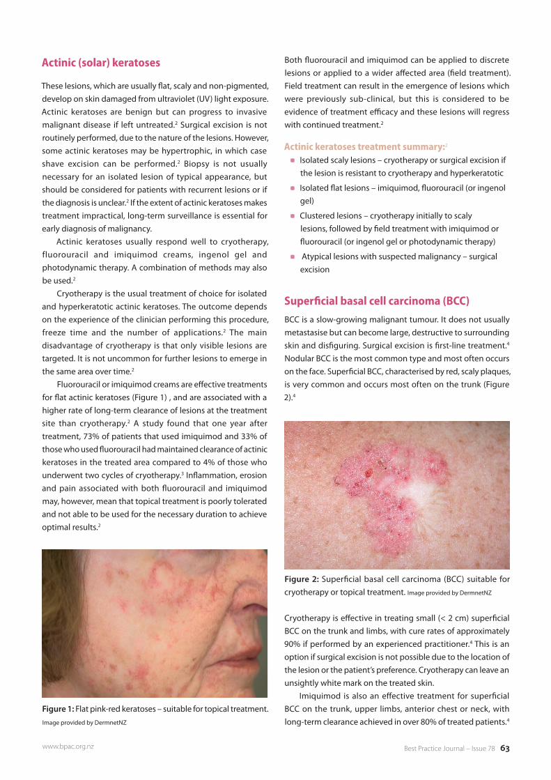
Best Practice Journal – Issue 78 63www.bpac.org.nz
Actinic (solar) keratoses
These lesions, which are usually flat, scaly and non-pigmented, develop on skin damaged from ultraviolet (UV) light exposure. Actinic keratoses are benign but can progress to invasive malignant disease if left untreated.2 Surgical excision is not routinely performed, due to the nature of the lesions. However, some actinic keratoses may be hypertrophic, in which case shave excision can be performed.2 Biopsy is not usually necessary for an isolated lesion of typical appearance, but should be considered for patients with recurrent lesions or if the diagnosis is unclear.2 If the extent of actinic keratoses makes treatment impractical, long-term surveillance is essential for early diagnosis of malignancy.
Actinic keratoses usually respond well to cryotherapy, fluorouracil and imiquimod creams, ingenol gel and photodynamic therapy. A combination of methods may also be used.2
Cryotherapy is the usual treatment of choice for isolated and hyperkeratotic actinic keratoses. The outcome depends on the experience of the clinician performing this procedure, freeze time and the number of applications.2 The main disadvantage of cryotherapy is that only visible lesions are targeted. It is not uncommon for further lesions to emerge in the same area over time.2
Fluorouracil or imiquimod creams are effective treatments for flat actinic keratoses (Figure 1) , and are associated with a higher rate of long-term clearance of lesions at the treatment site than cryotherapy.2 A study found that one year after treatment, 73% of patients that used imiquimod and 33% of those who used fluorouracil had maintained clearance of actinic keratoses in the treated area compared to 4% of those who underwent two cycles of cryotherapy.3 Inflammation, erosion and pain associated with both fluorouracil and imiquimod may, however, mean that topical treatment is poorly tolerated and not able to be used for the necessary duration to achieve optimal results.2
Figure 1: Flat pink-red keratoses – suitable for topical treatment. Image provided by DermnetNZ
Both fluorouracil and imiquimod can be applied to discrete lesions or applied to a wider affected area (field treatment). Field treatment can result in the emergence of lesions which were previously sub-clinical, but this is considered to be evidence of treatment efficacy and these lesions will regress with continued treatment.2
Actinic keratoses treatment summary:2
Isolated scaly lesions – cryotherapy or surgical excision if the lesion is resistant to cryotherapy and hyperkeratotic
Isolated flat lesions – imiquimod, fluorouracil (or ingenol gel)
Clustered lesions – cryotherapy initially to scaly lesions, followed by field treatment with imiquimod or fluorouracil (or ingenol gel or photodynamic therapy)
Atypical lesions with suspected malignancy – surgical excision
Superficial basal cell carcinoma (BCC)
BCC is a slow-growing malignant tumour. It does not usually metastasise but can become large, destructive to surrounding skin and disfiguring. Surgical excision is first-line treatment.4 Nodular BCC is the most common type and most often occurs on the face. Superficial BCC, characterised by red, scaly plaques, is very common and occurs most often on the trunk (Figure 2).4
Figure 2: Superficial basal cell carcinoma (BCC) suitable for cryotherapy or topical treatment. Image provided by DermnetNZ
Cryotherapy is effective in treating small (< 2 cm) superficial BCC on the trunk and limbs, with cure rates of approximately 90% if performed by an experienced practitioner.4 This is an option if surgical excision is not possible due to the location of the lesion or the patient’s preference. Cryotherapy can leave an unsightly white mark on the treated skin.
Imiquimod is also an effective treatment for superficial BCC on the trunk, upper limbs, anterior chest or neck, with long-term clearance achieved in over 80% of treated patients.4
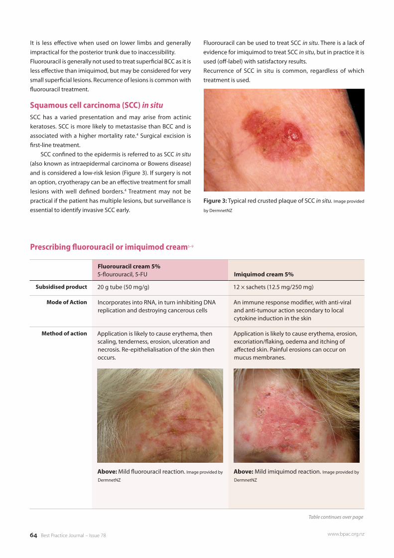
64 Best Practice Journal – Issue 78 www.bpac.org.nz
It is less effective when used on lower limbs and generally impractical for the posterior trunk due to inaccessibility.Fluorouracil is generally not used to treat superficial BCC as it is less effective than imiquimod, but may be considered for very small superficial lesions. Recurrence of lesions is common with fluorouracil treatment.
Squamous cell carcinoma (SCC) in situ SCC has a varied presentation and may arise from actinic keratoses. SCC is more likely to metastasise than BCC and is associated with a higher mortality rate.4 Surgical excision is first-line treatment.
SCC confined to the epidermis is referred to as SCC in situ (also known as intraepidermal carcinoma or Bowens disease) and is considered a low-risk lesion (Figure 3). If surgery is not an option, cryotherapy can be an effective treatment for small lesions with well defined borders.4 Treatment may not be practical if the patient has multiple lesions, but surveillance is essential to identify invasive SCC early.
Figure 3: Typical red crusted plaque of SCC in situ. Image provided
by DermnetNZ
Prescribing fluorouracil or imiquimod cream5–9
Fluorouracil cream 5% 5-flourouracil, 5-FU Imiquimod cream 5%
Subsidised product 20 g tube (50 mg/g) 12 × sachets (12.5 mg/250 mg)
Mode of Action Incorporates into RNA, in turn inhibiting DNA replication and destroying cancerous cells
An immune response modifier, with anti-viral and anti-tumour action secondary to local cytokine induction in the skin
Method of action Application is likely to cause erythema, then scaling, tenderness, erosion, ulceration and necrosis. Re-epithelialisation of the skin then occurs.
Above: Mild fluorouracil reaction. Image provided by
DermnetNZ
Application is likely to cause erythema, erosion, excoriation/flaking, oedema and itching of affected skin. Painful erosions can occur on mucus membranes.
Above: Mild imiquimod reaction. Image provided by
DermnetNZ
Fluorouracil can be used to treat SCC in situ. There is a lack of evidence for imiquimod to treat SCC in situ, but in practice it is used (off-label) with satisfactory results. Recurrence of SCC in situ is common, regardless of which treatment is used.
Table continues over page
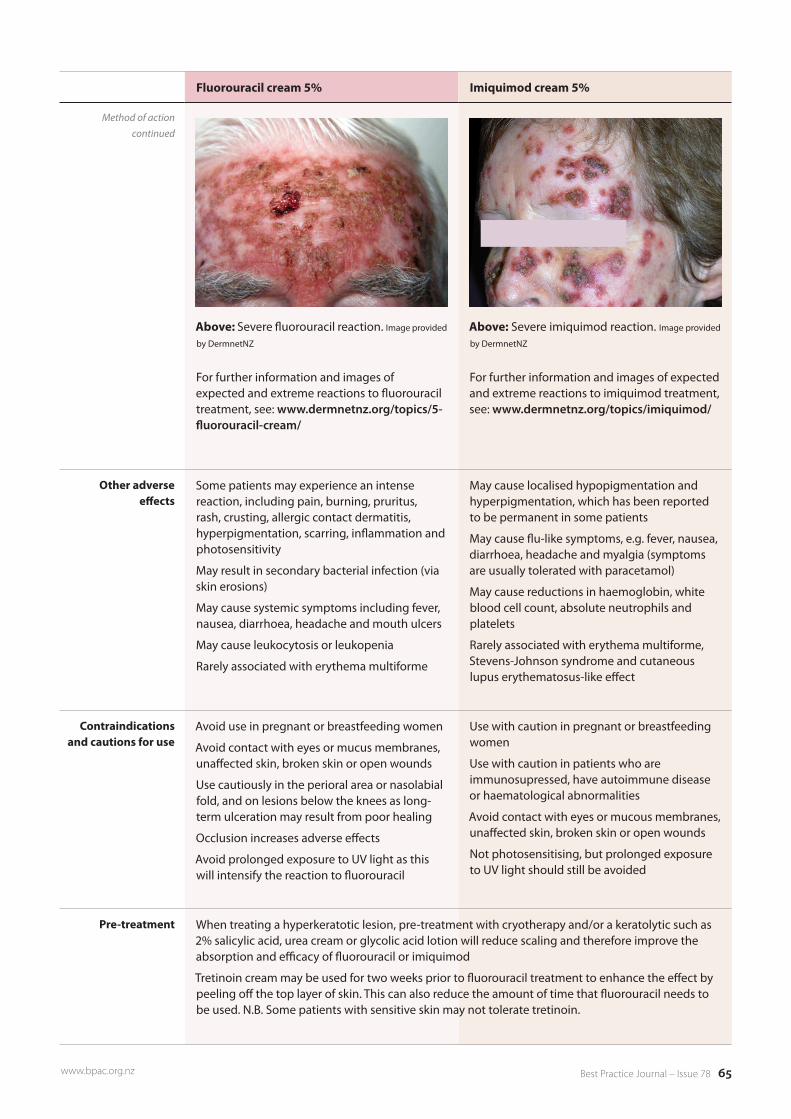
Best Practice Journal – Issue 78 65www.bpac.org.nz
Fluorouracil cream 5% Imiquimod cream 5%
Method of action continued
Above: Severe fluorouracil reaction. Image provided
by DermnetNZ
For further information and images of expected and extreme reactions to fluorouracil treatment, see: www.dermnetnz.org/topics/5-fluorouracil-cream/
Above: Severe imiquimod reaction. Image provided
by DermnetNZ
For further information and images of expected and extreme reactions to imiquimod treatment, see: www.dermnetnz.org/topics/imiquimod/
Other adverse effects
Some patients may experience an intense reaction, including pain, burning, pruritus, rash, crusting, allergic contact dermatitis, hyperpigmentation, scarring, inflammation and photosensitivity
May result in secondary bacterial infection (via skin erosions)
May cause systemic symptoms including fever, nausea, diarrhoea, headache and mouth ulcers
May cause leukocytosis or leukopenia
Rarely associated with erythema multiforme
May cause localised hypopigmentation and hyperpigmentation, which has been reported to be permanent in some patients
May cause flu-like symptoms, e.g. fever, nausea, diarrhoea, headache and myalgia (symptoms are usually tolerated with paracetamol)
May cause reductions in haemoglobin, white blood cell count, absolute neutrophils and platelets
Rarely associated with erythema multiforme, Stevens-Johnson syndrome and cutaneous lupus erythematosus-like effect
Contraindications and cautions for use
Avoid use in pregnant or breastfeeding women
Avoid contact with eyes or mucus membranes, unaffected skin, broken skin or open wounds
Use cautiously in the perioral area or nasolabial fold, and on lesions below the knees as long-term ulceration may result from poor healing
Occlusion increases adverse effects
Avoid prolonged exposure to UV light as this will intensify the reaction to fluorouracil
Use with caution in pregnant or breastfeeding women
Use with caution in patients who are immunosupressed, have autoimmune disease or haematological abnormalities
Avoid contact with eyes or mucous membranes, unaffected skin, broken skin or open wounds
Not photosensitising, but prolonged exposure to UV light should still be avoided
Pre-treatment When treating a hyperkeratotic lesion, pre-treatment with cryotherapy and/or a keratolytic such as 2% salicylic acid, urea cream or glycolic acid lotion will reduce scaling and therefore improve the absorption and efficacy of fluorouracil or imiquimod
Tretinoin cream may be used for two weeks prior to fluorouracil treatment to enhance the effect by peeling off the top layer of skin. This can also reduce the amount of time that fluorouracil needs to be used. N.B. Some patients with sensitive skin may not tolerate tretinoin.
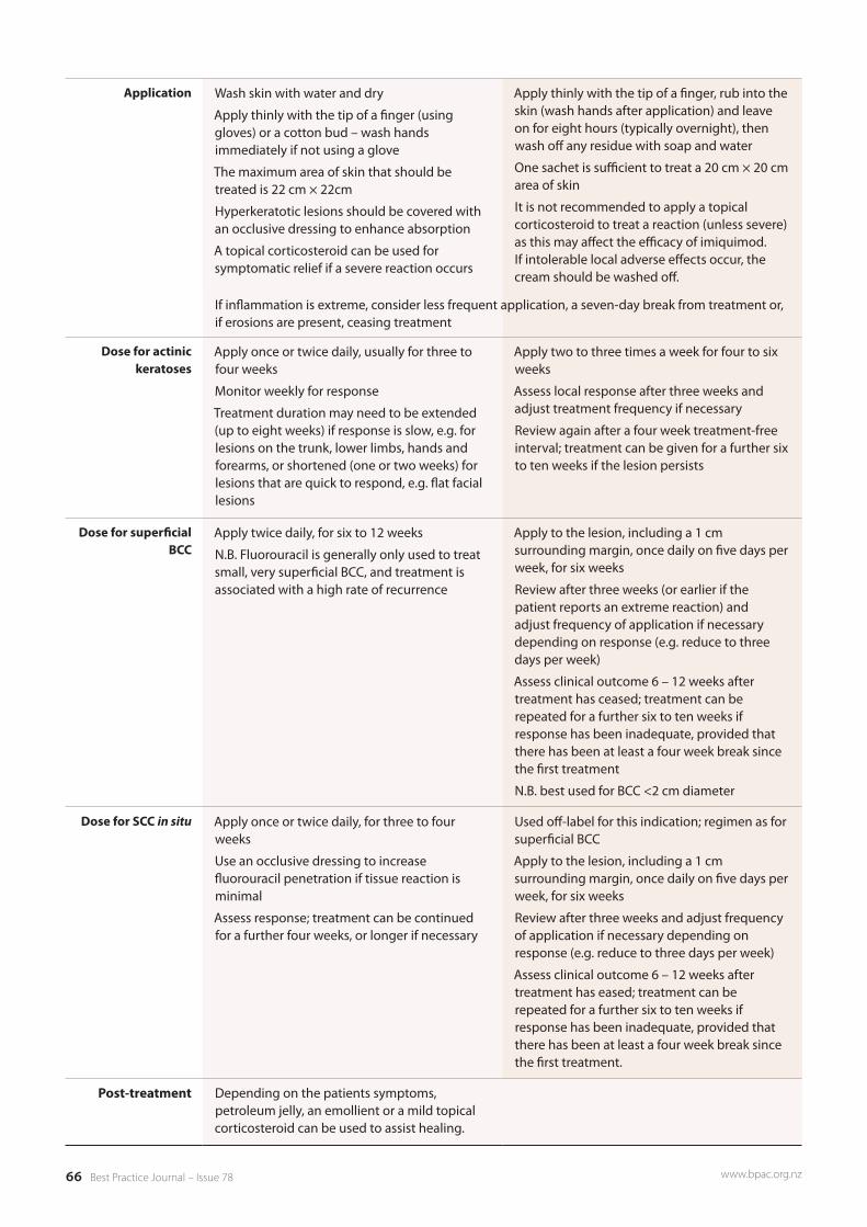
66 Best Practice Journal – Issue 78 www.bpac.org.nz
Application Wash skin with water and dry
Apply thinly with the tip of a finger (using gloves) or a cotton bud – wash hands immediately if not using a glove
The maximum area of skin that should be treated is 22 cm × 22cm
Hyperkeratotic lesions should be covered with an occlusive dressing to enhance absorption
A topical corticosteroid can be used for symptomatic relief if a severe reaction occurs
Apply thinly with the tip of a finger, rub into the skin (wash hands after application) and leave on for eight hours (typically overnight), then wash off any residue with soap and water
One sachet is sufficient to treat a 20 cm × 20 cm area of skin
It is not recommended to apply a topical corticosteroid to treat a reaction (unless severe) as this may affect the efficacy of imiquimod. If intolerable local adverse effects occur, the cream should be washed off.
If inflammation is extreme, consider less frequent application, a seven-day break from treatment or, if erosions are present, ceasing treatment
Dose for actinic keratoses
Apply once or twice daily, usually for three to four weeks
Monitor weekly for response
Treatment duration may need to be extended (up to eight weeks) if response is slow, e.g. for lesions on the trunk, lower limbs, hands and forearms, or shortened (one or two weeks) for lesions that are quick to respond, e.g. flat facial lesions
Apply two to three times a week for four to six weeks
Assess local response after three weeks and adjust treatment frequency if necessary
Review again after a four week treatment-free interval; treatment can be given for a further six to ten weeks if the lesion persists
Dose for superficial bCC
Apply twice daily, for six to 12 weeks
N.B. Fluorouracil is generally only used to treat small, very superficial BCC, and treatment is associated with a high rate of recurrence
Apply to the lesion, including a 1 cm surrounding margin, once daily on five days per week, for six weeks
Review after three weeks (or earlier if the patient reports an extreme reaction) and adjust frequency of application if necessary depending on response (e.g. reduce to three days per week)
Assess clinical outcome 6 – 12 weeks after treatment has ceased; treatment can be repeated for a further six to ten weeks if response has been inadequate, provided that there has been at least a four week break since the first treatment
N.B. best used for BCC <2 cm diameter
Dose for SCC in situ Apply once or twice daily, for three to four weeks
Use an occlusive dressing to increase fluorouracil penetration if tissue reaction is minimal
Assess response; treatment can be continued for a further four weeks, or longer if necessary
Used off-label for this indication; regimen as for superficial BCC
Apply to the lesion, including a 1 cm surrounding margin, once daily on five days per week, for six weeks
Review after three weeks and adjust frequency of application if necessary depending on response (e.g. reduce to three days per week)
Assess clinical outcome 6 – 12 weeks after treatment has eased; treatment can be repeated for a further six to ten weeks if response has been inadequate, provided that there has been at least a four week break since the first treatment.
Post-treatment Depending on the patients symptoms, petroleum jelly, an emollient or a mild topical corticosteroid can be used to assist healing.
Best Practice Journal – Issue 78 67www.bpac.org.nz
Acknowledgement: Thank you to Dr Amanda Oakley,
Honorary Associate Professor and Dermatologist, Waikato
District Health Board for expert review of this article.
Patient information on the use of fluorouracil and imiquimod is available from:
www.saferx.co.nz/Patient_info_fluorouracil.pdfwww.saferx.co.nz/imiquimod-patient-guide.pdf
References1. Newlands C, Currie R, Memon A, et al. Non-melanoma skin cancer:
United Kingdom National Multidisciplinary Guidelines. J Laryngol Otol
2016;130:S125–32. doi:10.1017/S0022215116000554
2. Poulin Y, Lynde CW, Barber K, et al. Non-melanoma Skin Cancer in Canada
Chapter 3: Management of Actinic Keratoses. J Cutan Med Surg 2015;19:227–
38. doi:10.1177/1203475415583414
3. Krawtchenko N, Roewert-Huber J, Ulrich M, et al. A randomised study of topical
5% imiquimod vs. topical 5-fluorouracil vs. cryosurgery in immunocompetent
patients with actinic keratoses: a comparison of clinical and histological
outcomes including 1-year follow-up. Br J Dermatol 2007;157 Suppl 2:34–40.
doi:10.1111/j.1365-2133.2007.08271.x
4. Zloty D, Guenther LC, Sapijaszko M, et al. Non-melanoma Skin Cancer in
Canada Chapter 4: Management of Basal Cell Carcinoma. J Cutan Med Surg
2015;19:239–48. doi:10.1177/1203475415586664
5. New Zealand Formulary (NZF). NZF v55. 2016. Available from: www.nzf.org.nz
(Accessed Dec, 2016)
6. Bausch & Lomb (NZ) Ltd. New Zealand data sheet: ALDARA. 2015. Available
from: www.medsafe.govt.nz/profs/datasheet/a/aldaracream.pdf
7. Bausch & Lomb (NZ) Ltd. Data sheet: EFUDIX. 2016. Available from: www.
medsafe.govt.nz/profs/datasheet/e/Efudixcr.pdf
8. DermNet New Zealand. Skin cancer. Available from: www.dermnetnz.org/
topics/skin-cancer/
9. bpacnz. Managing non-melanoma skin cancer in primary care. BPJ 57, 2013.
Available from: www.bpac.org.nz (Accessed Jan, 2017)

68 Best Practice Journal – Issue 78 www.bpac.org.nz
A recently published study from the University of Otago provides clarity as to which patients in general practice would benefit most from opportunistic investigation for abdominal aortic aneurysms (AAA).
Abdominal aortic aneurysms (AAA) are present in 5–10% of older men and 1–2% of older women1, 2 and cause the death of five men and two women per 100,000 annually.3 The rate of spontaneous AAA rupture increases with aneurysm size.4, 5 One study found aneurysms 5.0–5.9 cm had an annual rupture risk of 9.4%; the risk increased to 32.5% for aneurysms of 7.0 cm or more.4 Spontaneous AAA rupture is associated with a high mortality rate (80%), and emergency surgery following AAA rupture has a significantly higher mortality rate (30–65%) than elective AAA repair (3–10%).5–7
General practitioners can identify patients at risk of AAA. Early diagnosis allows patients to be offered surgery when the risk of spontaneous rupture outweighs the risk of surgery, usually when the AAA diameter is greater than 5.5 cm.7 AAA may be detected by palpation in patients with low or normal body mass, but it is usually detected by abdominal ultrasound.7
Targeted testing for abdominal aortic aneurysm
C ARDIOVASCulAR SySTEM RESEARCH uPDATES
Targeted testing for AAA typically focuses on males aged over 65 years. International studies and screening programmes targeting males of this age have been reported to reduce mortality due to AAA by approximately 40%.7, 8 Such programmes raise concerns, however, regarding potential overtreatment and health system capacity.6 Screening programmes have been criticised for excluding other at-risk groups, such as women, who constitute approximately 25% of those presenting with ruptured AAA.1 In New Zealand, targeted testing of males aged over 65 years may disadvantage Māori, as they experience rupture at a younger age, Māori women are equally affected and Māori appear to experience worse outcomes from AAA than non-Māori.6, 9, 10
A testing programme for AAAA recently published University of Otago study involving over 4000 men and women aged over 50 years from the Southern region tested participants for AAA using abdominal ultrasound.1 This study compared the effectiveness of identifying patients for AAA investigation based on cardiovascular risk. Study groups comprised:1
Patients attending the cardiology service for coronary angiography
RESEARCH uPDATE
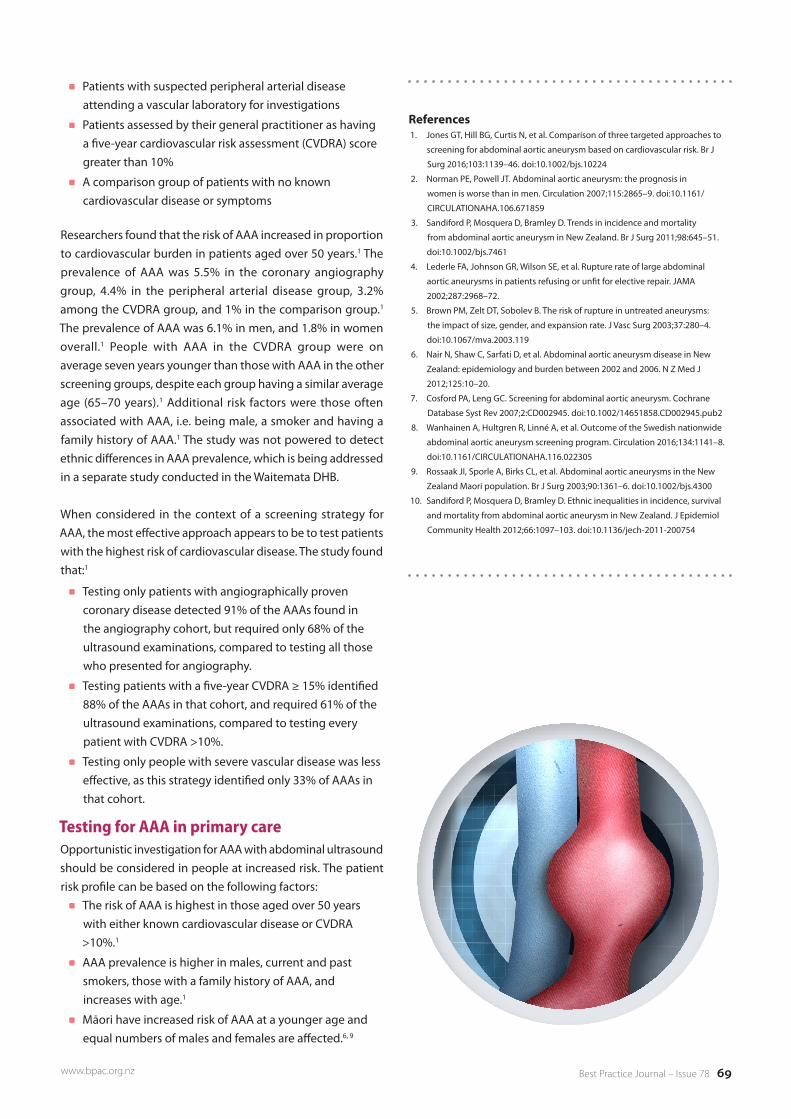
Best Practice Journal – Issue 78 69www.bpac.org.nz
Patients with suspected peripheral arterial disease attending a vascular laboratory for investigations
Patients assessed by their general practitioner as having a five-year cardiovascular risk assessment (CVDRA) score greater than 10%
A comparison group of patients with no known cardiovascular disease or symptoms
Researchers found that the risk of AAA increased in proportion to cardiovascular burden in patients aged over 50 years.1 The prevalence of AAA was 5.5% in the coronary angiography group, 4.4% in the peripheral arterial disease group, 3.2% among the CVDRA group, and 1% in the comparison group.1 The prevalence of AAA was 6.1% in men, and 1.8% in women overall.1 People with AAA in the CVDRA group were on average seven years younger than those with AAA in the other screening groups, despite each group having a similar average age (65–70 years).1 Additional risk factors were those often associated with AAA, i.e. being male, a smoker and having a family history of AAA.1 The study was not powered to detect ethnic differences in AAA prevalence, which is being addressed in a separate study conducted in the Waitemata DHB.
When considered in the context of a screening strategy for AAA, the most effective approach appears to be to test patients with the highest risk of cardiovascular disease. The study found that:1
Testing only patients with angiographically proven coronary disease detected 91% of the AAAs found in the angiography cohort, but required only 68% of the ultrasound examinations, compared to testing all those who presented for angiography.
Testing patients with a five-year CVDRA ≥ 15% identified 88% of the AAAs in that cohort, and required 61% of the ultrasound examinations, compared to testing every patient with CVDRA >10%.
Testing only people with severe vascular disease was less effective, as this strategy identified only 33% of AAAs in that cohort.
Testing for AAA in primary careOpportunistic investigation for AAA with abdominal ultrasound should be considered in people at increased risk. The patient risk profile can be based on the following factors:
The risk of AAA is highest in those aged over 50 years with either known cardiovascular disease or CVDRA >10%.1
AAA prevalence is higher in males, current and past smokers, those with a family history of AAA, and increases with age.1
Māori have increased risk of AAA at a younger age and equal numbers of males and females are affected.6, 9
References1. Jones GT, Hill BG, Curtis N, et al. Comparison of three targeted approaches to
screening for abdominal aortic aneurysm based on cardiovascular risk. Br J
Surg 2016;103:1139–46. doi:10.1002/bjs.10224
2. Norman PE, Powell JT. Abdominal aortic aneurysm: the prognosis in
women is worse than in men. Circulation 2007;115:2865–9. doi:10.1161/
CIRCULATIONAHA.106.671859
3. Sandiford P, Mosquera D, Bramley D. Trends in incidence and mortality
from abdominal aortic aneurysm in New Zealand. Br J Surg 2011;98:645–51.
doi:10.1002/bjs.7461
4. Lederle FA, Johnson GR, Wilson SE, et al. Rupture rate of large abdominal
aortic aneurysms in patients refusing or unfit for elective repair. JAMA
2002;287:2968–72.
5. Brown PM, Zelt DT, Sobolev B. The risk of rupture in untreated aneurysms:
the impact of size, gender, and expansion rate. J Vasc Surg 2003;37:280–4.
doi:10.1067/mva.2003.119
6. Nair N, Shaw C, Sarfati D, et al. Abdominal aortic aneurysm disease in New
Zealand: epidemiology and burden between 2002 and 2006. N Z Med J
2012;125:10–20.
7. Cosford PA, Leng GC. Screening for abdominal aortic aneurysm. Cochrane
Database Syst Rev 2007;2:CD002945. doi:10.1002/14651858.CD002945.pub2
8. Wanhainen A, Hultgren R, Linné A, et al. Outcome of the Swedish nationwide
abdominal aortic aneurysm screening program. Circulation 2016;134:1141–8.
doi:10.1161/CIRCULATIONAHA.116.022305
9. Rossaak JI, Sporle A, Birks CL, et al. Abdominal aortic aneurysms in the New
Zealand Maori population. Br J Surg 2003;90:1361–6. doi:10.1002/bjs.4300
10. Sandiford P, Mosquera D, Bramley D. Ethnic inequalities in incidence, survival
and mortality from abdominal aortic aneurysm in New Zealand. J Epidemiol
Community Health 2012;66:1097–103. doi:10.1136/jech-2011-200754

visit us at www.bpac.org.nzCall us on 03 477 5418 Email us at [email protected] Freefax us on 0800 27 22 69