Elastomeric electrospun scaffolds of poly(l-lactide-co-trimethylene carbonate) for myocardial tissue...
11
Elastomeric electrospun scaffolds of poly(L-lactide-co- trimethylene carbonate) for myocardial tissue engineering Shayanti Mukherjee • Chiara Gualandi • Maria Letizia Focarete • Rajeswari Ravichandran • Jayarama Reddy Venugopal • Michael Raghunath • Seeram Ramakrishna Received: 2 February 2011 / Accepted: 15 May 2011 / Published online: 27 May 2011 Ó Springer Science+Business Media, LLC 2011 Abstract In myocardial tissue engineering the use of synthetically bioengineered flexible patches implanted in the infarcted area is considered one of the promising strategy for cardiac repair. In this work the potentialities of a biomimetic electrospun scaffold made of a commercial copolymer of (L)-lactic acid with trimethylene carbonate (P(L)LA-co-TMC) are investigated in comparison to elec- trospun poly(L)lactic acid. The P(L)LA-co-TMC scaffold used in this work is a glassy rigid material at room tem- perature while it is a rubbery soft material at 37°C. Mechanical characterization results (tensile stress–strain and creep-recovery measurements) show that at 37°C electrospun P(L)LA-co-TMC displays an elastic modulus of around 20 MPa and the ability to completely recover up to 10% of deformation. Cell culture experiments show that P(L)LA-co-TMC scaffold promotes cardiomyocyte prolif- eration and efficiently preserve cell morphology, without hampering expression of sarcomeric alpha actinin marker, thus demonstrating its potentialities as synthetic biomate- rial for myocardial tissue engineering. 1 Introduction Myocardial infarction (MI) is the most common cause of death and disability in the developed countries. Current pharmacological and surgical therapies are unable to ade- quately prevent disease progression. Apart from heart transplantation, there is no standard procedure that can adequately prevent disease progression [1]. Owing to shortcomings of current methods, the search for new strategies to repair the injured myocardium continues. Biomaterial approaches intend to take advantage of the ability of an elastomeric biomaterial sheet to act as a flexible patch sized to fit the infarcted area, while enabling direct apposition of exogenously introduced cells. With this approach, cells would remain intact beneath the patch directly over the infarct site, preventing cell loss and pro- viding a more site-directed repair. Three essential bioma- terial criteria include: (i) long term elasticity that matches and responds to the dynamic working conditions of heart muscle; (ii) adaptable biodegradation, which would avoid the detrimental effects of a persisting foreign structure; and (iii) the ability to deliver and retain beating cells at the location of patch attachment [2]. Many efforts by the scientific community in the field of myocardial tissue engineering (MTE) are dedicated to identify materials possessing specific mechanical proper- ties [3]. First of all, it is desirable that the mechanical performances of bioengineered scaffolds match as much as S. Mukherjee M. Raghunath Division of Bioengineering, National University of Singapore, Singapore, Singapore C. Gualandi M. L. Focarete (&) Department of Chemistry ‘‘G. Ciamician’’ and National Consortium of Materials Science and Technology (INSTM, Bologna RU), University of Bologna, Bologna, Italy e-mail: [email protected] S. Mukherjee R. Ravichandran J. R. Venugopal (&) S. Ramakrishna HEM Laboratory, Nanoscience and Nanotechnology Initiative, National University of Singapore, Singapore, Singapore e-mail: [email protected] R. Ravichandran S. Ramakrishna Department of Mechanical Engineering, National University of Singapore, Singapore, Singapore M. Raghunath Department of Biochemistry, Yong Loo Lin School of Medicine, National University of Singapore, Singapore, Singapore 123 J Mater Sci: Mater Med (2011) 22:1689–1699 DOI 10.1007/s10856-011-4351-2
-
Upload
independent -
Category
Documents
-
view
3 -
download
0
Transcript of Elastomeric electrospun scaffolds of poly(l-lactide-co-trimethylene carbonate) for myocardial tissue...

Elastomeric electrospun scaffolds of poly(L-lactide-co-trimethylene carbonate) for myocardial tissue engineering
Shayanti Mukherjee • Chiara Gualandi • Maria Letizia Focarete •
Rajeswari Ravichandran • Jayarama Reddy Venugopal • Michael Raghunath •
Seeram Ramakrishna
Received: 2 February 2011 / Accepted: 15 May 2011 / Published online: 27 May 2011
� Springer Science+Business Media, LLC 2011
Abstract In myocardial tissue engineering the use of
synthetically bioengineered flexible patches implanted in
the infarcted area is considered one of the promising
strategy for cardiac repair. In this work the potentialities of
a biomimetic electrospun scaffold made of a commercial
copolymer of (L)-lactic acid with trimethylene carbonate
(P(L)LA-co-TMC) are investigated in comparison to elec-
trospun poly(L)lactic acid. The P(L)LA-co-TMC scaffold
used in this work is a glassy rigid material at room tem-
perature while it is a rubbery soft material at 37�C.
Mechanical characterization results (tensile stress–strain
and creep-recovery measurements) show that at 37�C
electrospun P(L)LA-co-TMC displays an elastic modulus
of around 20 MPa and the ability to completely recover up
to 10% of deformation. Cell culture experiments show that
P(L)LA-co-TMC scaffold promotes cardiomyocyte prolif-
eration and efficiently preserve cell morphology, without
hampering expression of sarcomeric alpha actinin marker,
thus demonstrating its potentialities as synthetic biomate-
rial for myocardial tissue engineering.
1 Introduction
Myocardial infarction (MI) is the most common cause of
death and disability in the developed countries. Current
pharmacological and surgical therapies are unable to ade-
quately prevent disease progression. Apart from heart
transplantation, there is no standard procedure that can
adequately prevent disease progression [1]. Owing to
shortcomings of current methods, the search for new
strategies to repair the injured myocardium continues.
Biomaterial approaches intend to take advantage of the
ability of an elastomeric biomaterial sheet to act as a
flexible patch sized to fit the infarcted area, while enabling
direct apposition of exogenously introduced cells. With this
approach, cells would remain intact beneath the patch
directly over the infarct site, preventing cell loss and pro-
viding a more site-directed repair. Three essential bioma-
terial criteria include: (i) long term elasticity that matches
and responds to the dynamic working conditions of heart
muscle; (ii) adaptable biodegradation, which would avoid
the detrimental effects of a persisting foreign structure; and
(iii) the ability to deliver and retain beating cells at the
location of patch attachment [2].
Many efforts by the scientific community in the field of
myocardial tissue engineering (MTE) are dedicated to
identify materials possessing specific mechanical proper-
ties [3]. First of all, it is desirable that the mechanical
performances of bioengineered scaffolds match as much as
S. Mukherjee � M. Raghunath
Division of Bioengineering, National University of Singapore,
Singapore, Singapore
C. Gualandi � M. L. Focarete (&)
Department of Chemistry ‘‘G. Ciamician’’ and National
Consortium of Materials Science and Technology (INSTM,
Bologna RU), University of Bologna, Bologna, Italy
e-mail: [email protected]
S. Mukherjee � R. Ravichandran � J. R. Venugopal (&) �S. Ramakrishna
HEM Laboratory, Nanoscience and Nanotechnology Initiative,
National University of Singapore, Singapore, Singapore
e-mail: [email protected]
R. Ravichandran � S. Ramakrishna
Department of Mechanical Engineering, National University of
Singapore, Singapore, Singapore
M. Raghunath
Department of Biochemistry, Yong Loo Lin School of Medicine,
National University of Singapore, Singapore, Singapore
123
J Mater Sci: Mater Med (2011) 22:1689–1699
DOI 10.1007/s10856-011-4351-2

possible those of the heart extracellular matrix in terms of
stiffness, since the scaffold should be flexible enough to
promote the contraction of the growing cells [4–6]. In
addition, since the myocardial tissue is subjected to cycli-
cal and constant deformation, materials are requested to
show elastomeric properties and possibly long-term
elasticity.
To date, both natural and synthetic polymers have been
proposed as candidates to fabricate three-dimensional (3D)
scaffolds for the application in MTE. Since the scaffold
should initially act as adhesive substrate for the cells,
natural polymers are commonly considered particularly
promising. Collagen [7–11], alginate [12–15] and gelatin
[16, 17] have been extensively employed in MTE. How-
ever, despite the successful results in terms of cell attach-
ment and proliferation, these highly hydrophilic polymers
are mainly employed in the form of gels, with limited
possibility of designing devices with specific 3D architec-
ture and mechanical properties.
Synthetic elastomeric scaffolds for MTE are desirable as
their mechanical conditioning regimens have shown to
promote tissue formation along with gradual stress transfer
from the degrading synthetic matrix to the newly formed
tissue [18, 19]. Synthetic aliphatic polyesters such as
polycaprolactone (PCL), poly(glycolic acid) (PGA),
poly(lactic acid) (PLA), and their copolymers are popular
materials used in the biomedical sector thanks to their
clinically confirmed biocompatibility associated with con-
trolled biodegradation rate. They can also be easily pro-
cessed into 3D structures with defined morphology and
thermo-mechanical properties. PGA and PLGA are among
the first synthetic biomaterials used for MTE [20–23].
However, the above cited polyesters are generally rigid
material under physiological conditions [24]. Biodegrad-
able polyurethanes are another class of synthetic polymers
used for MTE that, depending on their chemical structure
and macromolecular flexibility, can display an elastic
behavior [25–28]. In recent times, new materials for MTE,
possessing both suitable mechanical properties and the
ability to support cell growth, are developed by following
two main approaches: (i) the combination of natural and
synthetic polymers, where the natural component is
responsible of enhancing cell attachment while the syn-
thetic one acts as the supporting phase with proper
mechanical properties [29–31]; (ii) the synthesis of new
polymers specifically developed to possess mechanical
performances suitable for MTE [3, 32–37].
Co-polymerization is a well known effective approach
to synthesize new material with tuned physical properties
and controlled hydrophylicity, which in turns determines
the rate of hydrolytic degradation. In the context of soft
tissue engineering, random copolymers of lactic acid with
trimethylene carbonate (PLA-co-TMC) have been
described as materials possessing attractive thermal and
mechanical properties, as well as an interesting hydrolyti-
cal degradation behavior. Indeed, by varying the compo-
sition of the two co-monomers it is possible to change the
polymer thermal and mechanical properties, providing
materials that at room temperature range from hard and
brittle or moderately tough solids to rubbery and highly
elastic solids [38–40]. Porous scaffolds made of P(D,L)LA-
co-TMC copolymers, obtained by particulate leaching,
have been successfully employed by Pego et al. for cardiac
tissue engineering [41, 42]. Moreover, when P(D,L)LA-co-
TMC films were implanted in the back of rats only a mild
tissue reaction was observed [43].
In designing scaffolds for MTE, the selection of proper
polymeric material must be coupled with a suitable scaf-
fold structure that is capable of mimicking much of the
natural extracellular environment. The fibrillar structure of
extracellular matrix can be effectively addressed by using
electrospinning technique to fabricate micro- and nanofi-
brous biomimetic porous scaffolds, with interconnected
pore structure. Electrospun scaffolds made of synthetic
polymers, either coupled with natural polymers or as plain
materials, have been used for cardiac tissue engineering in
several studies [44–48].
In this study, the potentialities of electrospun P(L)LA-
co-TMC scaffolds for MTE are presented and discussed for
the first time, in comparison with scaffolds of the corre-
sponding homopolymer P(L)LA. A commercial P(L)LA-co-
TMC copolymer was processed into sub-micrometric fibers
by electrospinning and scaffold thermo-mechanical prop-
erties were investigated. The capability of the scaffold to
act as substrate for cardiomyocyte cell growth was assessed
in order to explore the feasibility of engineering a com-
patible heart patch from P(L)LA-co-TMC electrospun
fibrous scaffolds.
2 Materials and methods
2.1 Scaffold fabrication
Poly(L-lactide-co-trimethylene carbonate) (P(L)LA-co-
TMC, Resomer LT 706, LA:TMC 70:30 weight ratio,
inherent viscosity 1.2–1.6 dl/g) was purchased from
Boheringer Ingelheim, Germany. Poly(L-lactic acid) (P(L)LA,
Lacea H.100-E, Mw = 8.4 9 104 g/mol, PDI = 1.7) was
supplied by Mitsui Fine Chemicals (Dusseldorf, Germany).
Dichloromethane (DCM) and dimethylformamide (DMF)
were purchased by Sigma-Aldrich and were used without
any further purification. The electrospinning apparatus,
made in house, was composed of a high voltage power
supply (Spellman, SL 50 P 10/CE/230), a syringe pump
(KD Scientific 200 series), a glass syringe, a stainless-steel
1690 J Mater Sci: Mater Med (2011) 22:1689–1699
123

blunt-ended needle (inner diameter: 0.84 mm) connected
with the power supply electrode and a grounded aluminum
plate-type collector. The polymer solution was dispensed
through a Teflon tube to the needle that was vertically
placed on the collecting plate. P(L)LA-co-TMC solution
was prepared at room temperature (RT) by dissolving the
polymer in DCM:DMF = 70:30 v/v at a concentration of
12% w/v. P(L)LA-co-TMC electrospun scaffolds were
fabricated using the following conditions: applied volt-
age = 16 kV, needle to collector distance = 15 cm, solu-
tion flow rate = 0.01 ml/min, at RT and relative humidity
RH = 40–50%. P(L)LA scaffolds were fabricated from a
solution of P(L)LA in DCM:DMF = 65:35 v/v, at a con-
centration of 13% w/v, by using the following conditions:
applied voltage = 12 kV, needle to collector dis-
tance = 15 cm, solution flow rate = 0.015 ml/min, at RT
and RH = 40–50%. All the electrospun mats obtained
were kept under vacuum over P2O5 at RT overnight in
order to remove residual solvents.
2.2 Scaffold morphology
SEM observations were carried out by using a Philips 515
SEM at an accelerating voltage of 15 kV, on samples
sputter-coated with gold. The distribution of fiber diame-
ters was determined through the measurement of about 250
fibers by means of an acquisition and image analysis
software (EDAX Genesis) and the results were given as the
average diameter ± standard deviation.
2.3 Scaffold thermal characterization
Thermogravimetric analysis (TGA) measurements were
performed with a TA Instruments TGA2950 Thermo-
gravimetric Analyzer from RT to 600�C (heating
rate = 10�C/min, nitrogen purge gas). Differential Scan-
ning Calorimetry (DSC) measurements were carried out
using a TA Instruments Q100 DSC equipped with the
Liquid Nitrogen Cooling System accessory. DSC scans of
electrospun scaffolds were performed in helium atmo-
sphere from -50 to 180�C. A rate of 20�C/min was used
during heating scans whereas the cooling scans were per-
formed at a rate of 10�C/min. The glass transition tem-
perature (Tg) was taken at half-height of the glass transition
heat capacity step, while the crystallization temperature
(Tc) and the melting temperature (Tm) were taken at the
peak maximum of crystallization exotherm and melting
endotherm, respectively.
2.4 Scaffold mechanical characterization
Tensile stress–strain measurements and creep-recovery
tests were performed by using a Dynamic Mechanical
Thermal Analyzer (DMTA, TA Instruments Q800 series)
equipped with tension-film clamps. All the analyses were
performed on rectangular strips cut from electrospun mats
(width = 5 mm; gauge length = 10 mm). Mat thickness,
measured by microcaliper, was: P(L)LA: 23 ± 6 lm;
P(L)LA-co-TMC: 30 ± 1 lm (about 15 replicate mea-
surements were performed for each sample and results are
given as the average value ± standard deviation). Stress–
strain measurements were carried out both at RT and at
37�C. In the latter case the sample was heated to 37�C and
it was allowed to equilibrate for 5 min prior to beginning
the test. Tensile stress–strain measurements were carried
out by applying a preload force of 0.001 N and using a
cross-head speed of 5 mm/min. Tensile elastic modulus
was determined from the initial linear slope of the stress–
strain curve. Eight replicate specimens were run for each
sample and results were given as the average value ±
standard deviation. The creep-recovery measurements were
carried out at 37�C. The sample was instantaneously sub-
jected to a stress hold at a constant value for 2 s. After
removal of the applied stress the sample recovery was
followed for 2 min with no additional force other than the
preload force of 0.001 N. The recovery process was
monitored by plotting the strain as a function of time.
2.5 Cardiomyocyte isolation and culture
Cardiomyocytes were freshly isolated from adult New
Zealand rabbit hearts, as per institution guidelines. The left
ventricle of the heart was cleaned thoroughly with Phos-
phate Buffer Saline (PBS) (1st base) containing antibiotics
(Sigma-Aldrich) for removal of fat tissue and clots and it
was minced into fine pieces. The processed tissue was
treated with 5 mg of collagenase (Sigma-Aldrich) for
20 min at 37�C. Freshly isolated cardiac cells were cul-
tured in DMEM media supplemented with 10% FBS and
1% antibiotic and antimycotic solutions (Invitrogen Corp,
USA) in a 75 cm2 cell culture flask. Cells were incubated at
37�C in a humidified atmosphere containing 5% CO2 for
6 days and the culture medium was changed every 2 days.
2.6 Scaffold preparation and cell seeding
Each of the nanofibrous scaffolds was adhered to 15 mm
coverslip using silicon bioglue and was placed in 24-well
plate while being pressed with a stainless steel ring to
ensure complete contact of the scaffolds with wells. The
specimens were sterilized under UV light, washed three
times with PBS and subsequently immersed in complete
DMEM overnight before cell seeding. The trypsinised cells
from the culture flasks were centrifuged, counted by trypan
blue assay using a hemocytometer and seeded onto the
scaffolds at a density of 2 9 104 cells/well. These were
J Mater Sci: Mater Med (2011) 22:1689–1699 1691
123
allowed to adhere for 3 h before adding medium, which
was thereafter changed every 3 days.
2.7 Proliferation study
The adhesion and proliferation of cultured cardiomyocytes
on scaffolds and TCP used as control were determined
using the colorimetric MTS assay (CellTiter 96 AQueous
One solution, Promega, Madison, WI). The reduction of
yellow tetrazolium salt [3-(4,5-dimethylthiazol-2-yl)-5-(3-
carboxymethoxyphenyl)-2(4-sulfophenyl)-2H-tetrazolium]
in MTS to form purple formazan crystals by the dehydro-
genase enzymes secreted by mitochondria of metabolically
active cells forms the basis of this assay. The formazan dye
shows absorbance at 492 nm and the amount of formazan
crystals formed is directly proportional to the number of
cells. In order to process the samples for the MTS assay,
they were rinsed with PBS to remove unattached cells and
incubated with 20% MTS reagent in a serum free medium
for a period of 3 h at 37�C. Absorbance of the obtained dye
was measured at 490 nm using a spectrophotometric plate
reader (FLUOstar OPTIMA, BMG lab Technologies).
2.8 Cell morphology
Morphological study of in vitro cultured cardiomyocyte
cells on scaffolds was performed after 7 and 14 days of cell
culture by processing them for SEM analysis. The scaffolds
were rinsed twice with PBS and fixed in 3% glutaraldehyde
(Sigma-Aldrich) for 3 h. Thereafter, the scaffolds were
rinsed in deionized water and dehydrated with upgrading
concentrations of ethanol (50, 70, 90, 100%) twice for
10 min each. Final washing with 100% ethanol was fol-
lowed by treating the specimens with hexamethyldisilazane
(Fluka). The hexamethyldisilazane was air-dried by keep-
ing the samples in fume hood. Finally, the scaffolds were
sputter-coated with gold (JEOL JFC-1200 finecoater,
Japan) and observed by SEM (FEI-QUANTA 200F,
Netherland) at an accelerating voltage of 10 kV.
2.9 Expression of cardiac specific protein marker
Cells cultured on different substrates were fixed using
100% chilled methanol and permeabilized with 0.1% Tri-
ton X 100 buffer. The non specific binding sites were
blocked using 3% BSA. Thereafter they were stained with
anti sarcomeric alpha actinin antibody (Sigma-Aldrich) and
FITC (Sigma-Aldrich) as secondary antibody. The nucleus
was stained using 40,6-diamidino-2-phenylindole,DAPI
(Invitrogen). Finally, the samples were washed to remove
unbound antibodies and mounted using Vectashield (Vec-
tor Laboratories). These samples were scanned under Laser
Confocal Scanning Microscope (Olympus FV1000) and
checked for expression of alpha actinin using Argon laser.
2.10 Statistical analysis
All data are expressed as mean ± standard deviation and
were analyzed using Student’s t-test for the calculation of
significance level of the data. Differences were considered
statistically significant at P B 0.05.
3 Results and discussion
3.1 Scaffold fabrication and thermo-mechanical
characterization
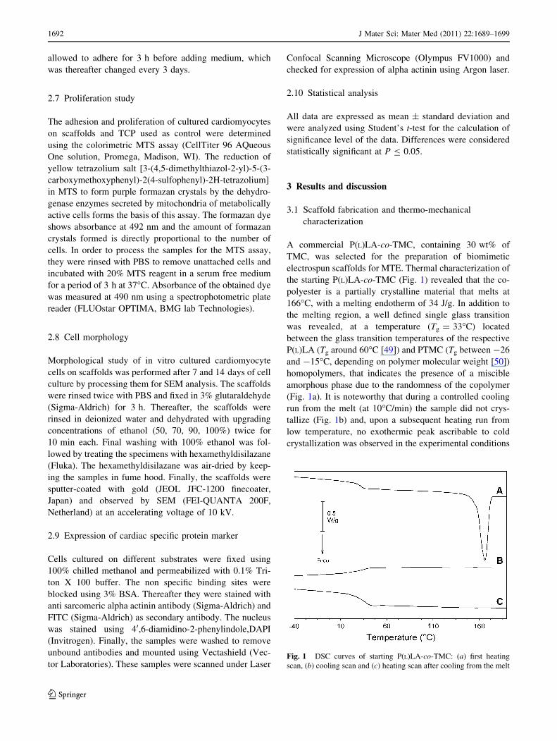
A commercial P(L)LA-co-TMC, containing 30 wt% of
TMC, was selected for the preparation of biomimetic
electrospun scaffolds for MTE. Thermal characterization of
the starting P(L)LA-co-TMC (Fig. 1) revealed that the co-
polyester is a partially crystalline material that melts at
166�C, with a melting endotherm of 34 J/g. In addition to
the melting region, a well defined single glass transition
was revealed, at a temperature (Tg = 33�C) located
between the glass transition temperatures of the respective
P(L)LA (Tg around 60�C [49]) and PTMC (Tg between -26
and -15�C, depending on polymer molecular weight [50])
homopolymers, that indicates the presence of a miscible
amorphous phase due to the randomness of the copolymer
(Fig. 1a). It is noteworthy that during a controlled cooling
run from the melt (at 10�C/min) the sample did not crys-
tallize (Fig. 1b) and, upon a subsequent heating run from
low temperature, no exothermic peak ascribable to cold
crystallization was observed in the experimental conditions
Fig. 1 DSC curves of starting P(L)LA-co-TMC: (a) first heating
scan, (b) cooling scan and (c) heating scan after cooling from the melt
1692 J Mater Sci: Mater Med (2011) 22:1689–1699
123

applied (Fig. 1c). This behavior is attributed to a slow
crystallization kinetics of P(L)LA-co-TMC. Results of
thermal characterization of the P(L)LA-co-TMC material
used in the present work are in line with earlier data on
TMC copolymers with a similar composition synthesized
by the group of Albertsson et al. [51].
Highly porous electrospun mats of P(L)LA-co-TMC and
P(L)LA, with micro-scale interstitial pores and a random
orientation of the sub-micrometric fibers were obtained, as
revealed by SEM analysis (Fig. 2) (P(L)LA-co-TMC mean
diameter distribution: 530 ± 110 nm; P(L)LA mean
diameter distribution: 610 ± 180 nm). Figure 2 shows that
a certain level of ‘fiber-fusion’ at the contact sites (arrows)
was originated during fiber deposition, for both polymers,
presumably due to the use of a high boiling point solvent
(DMF) during the spinning process. It is pointed out
however, that TGA analysis demonstrated the absence of
residual solvent in the desiccated electrospun scaffold (data
not shown).
Figure 3 shows the DSC curve of the as-spun P(L)LA-
co-TMC mat and of P(L)LA mat for comparison. The
calorimetric analysis revealed the presence of a Tg around
34�C for P(L)LA-co-TMC and around 60�C for P(L)LA.
DSC curves of both polymers show a cold crystallization
exotherm peak followed by a melting endotherm peak of
exactly the same entity (P(L)LA-co-TMC: DHc = DHm =
17 J/g; P(L)LA: DHc = DHm = 32 J/g). This result
demonstrates that completely amorphous P(L)LA-co-TMC
and P(L)LA mats are fabricated through electrospinning
since during the process polymer chains have little time to
organize in a crystal structure before the occurring of fiber
solidification. It is pointed out that, in the case of P(L)LA-
co-TMC polymer, a certain level of molecular orientation,
that is likely originated by the high elongational stretching
of the fibers, explains the occurrence of the cold
Fig. 2 SEM micrographs of electrospun mats of P(L)LA-co-TMC (a, b) and P(L)LA (c, d) at two different magnifications. Arrows point out
fiber-fusion points. Scale bar = 20 lm (a, c); scale bar = 2 lm (b, d)
Fig. 3 DSC curves of P(L)LA-co-TMC and P(L)LA electrospun mats
(first scan, heating rate: 20�C/min)
J Mater Sci: Mater Med (2011) 22:1689–1699 1693
123
crystallization phenomena during the DSC heating scan
which, on the contrary, did not occur for the starting
P(L)LA-co-TMC in the form of powder after cooling from
the melt, as previously mentioned.
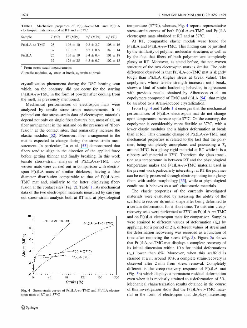
Mechanical performances of electrospun mats were
analyzed by tensile stress–strain measurements. It is
pointed out that stress–strain data of electrospun materials
depend not only on single fiber features but, most of all, on
fiber arrangement in the mat and on the presence of ‘fiber-
fusion’ at the contact sites, that remarkably increase the
elastic modulus [52]. Moreover, fiber arrangement in the
mat is expected to change during the stress–strain mea-
surement. In particular, Lu et al. [53] demonstrated that
fibers tend to align in the direction of the applied force
before getting thinner and finally breaking. In this work
tensile stress–strain analysis of P(L)LA-co-TMC non-
woven mats were carried out in comparison with electro-
spun P(L)LA mats of similar thickness, having a fiber
diameter distribution comparable to that of P(L)LA-co-
TMC mat and, similarly to the latter, displaying fiber-
fusion at the contact sites (Fig. 2). Table 1 lists mechanical
data of the two electrospun materials measured by carrying
out stress–strain analysis both at RT and at physiological
temperature (37�C), whereas, Fig. 4 reports representative
stress–strain curves of both P(L)LA-co-TMC and P(L)LA
electrospun mats obtained at RT and at 37�C.
At RT, comparable elastic moduli were found for
P(L)LA and P(L)LA-co-TMC. This finding can be justified
by the similarity of polymer molecular structures as well as
by the fact that fibers of both polymers are completely
glassy at RT. Moreover, as stated before, the non-woven
structure of the two electrospun mats is similar. The only
difference observed is that P(L)LA-co-TMC mat is slightly
tough than P(L)LA (higher stress at break value). The
copolymer, whose tensile strength increases until break,
shows a kind of strain hardening behavior, in agreement
with previous results obtained by Albertsson et al. on
copolymers composed of TMC and (L)LA [54], that might
be ascribed to a strain-induced crystallization.
From Fig. 4 and Table 1 it emerges that the mechanical
performances of P(L)LA electrospun mat do not change
upon temperature increase up to 37�C. On the contrary, the
copolymer is considerably more flexible at 37�C, with a
lower elastic modulus and a higher deformation at break
than at RT. This dramatic change of P(L)LA-co-TMC mat
mechanical properties is related to the fact that the poly-
mer, being completely amorphous and possessing a Tg
around 34�C, is a glassy rigid material at RT while it is a
rubbery soft material at 37�C. Therefore, the glass transi-
tion at a temperature in between RT and the physiological
temperature makes the P(L)LA-co-TMC material used in
the present work particularly interesting: at RT the polymer
can be easily processed through electrospinning into glassy
fibers with stable morphology [55], while at physiological
conditions it behaves as a soft elastomeric materials.
The elastic properties of the currently investigated
materials were evaluated by assessing the ability of the
scaffold to recover its initial shape after being deformed to
a certain deformation for a short time. To this aim creep-
recovery tests were performed at 37�C on P(L)LA-co-TMC
and on P(L)LA electrospun mats for comparison. Samples
were strained to different values of deformation (ein) by
applying, for a period of 2 s, different values of stress and
the deformation recovering was recorded as a function of
time after removing the stress (Fig. 5). Figure 5a shows
that P(L)LA-co-TMC mat displays a complete recovery of
its initial dimension within 10 s for initial deformations
(ein) lower than 6%. Moreover, when this scaffold is
strained at a ein around 10%, a complete strain-recovery is
observed after 2 min from stress removal. Completely
different is the creep-recovery response of P(L)LA mat
(Fig. 5b) which displays a permanent residual deformation
even when it is modestly strained to a deformation of 3%.
Mechanical characterization results obtained in the course
of this investigation show that the P(L)LA-co-TMC mate-
rial in the form of electrospun mat displays interesting
Table 1 Mechanical properties of P(L)LA-co-TMC and P(L)LA
electrospun mats measured at RT and at 37�C
Sample T (�C) Ea (MPa) rba (MPa) eb
a (%)
P(L)LA-co-TMC 25 108 ± 10 9.8 ± 2.7 108 ± 16
37 19 ± 5 8.2 ± 0.6 187 ± 14
P(L)LA 25 105 ± 19 3.4 ± 0.4 101 ± 18
37 126 ± 25 4.3 ± 0.7 102 ± 13
a From stress–strain measurements
E tensile modulus, rb stress at break, eb strain at break
Fig. 4 Stress-strain curves of P(L)LA-co-TMC and P(L)LA electro-
spun mats at RT and 37�C
1694 J Mater Sci: Mater Med (2011) 22:1689–1699
123
elastomeric behavior, thus it can be considered as a suitable
candidate for MTE applications.
3.2 Isolation of cardiomyocytes
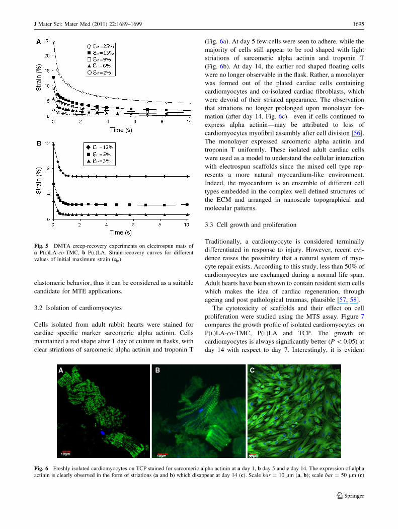
Cells isolated from adult rabbit hearts were stained for
cardiac specific marker sarcomeric alpha actinin. Cells
maintained a rod shape after 1 day of culture in flasks, with
clear striations of sarcomeric alpha actinin and troponin T
(Fig. 6a). At day 5 few cells were seen to adhere, while the
majority of cells still appear to be rod shaped with light
striations of sarcomeric alpha actinin and troponin T
(Fig. 6b). At day 14, the earlier rod shaped floating cells
were no longer observable in the flask. Rather, a monolayer
was formed out of the plated cardiac cells containing
cardiomyocytes and co-isolated cardiac fibroblasts, which
were devoid of their striated appearance. The observation
that striations no longer prolonged upon monolayer for-
mation (after day 14, Fig. 6c)—even if cells continued to
express alpha actinin—may be attributed to loss of
cardiomyocytes myofibril assembly after cell division [56].
The monolayer expressed sarcomeric alpha actinin and
troponin T uniformly. These isolated adult cardiac cells
were used as a model to understand the cellular interaction
with electrospun scaffolds since the mixed cell type rep-
resents a more natural myocardium-like environment.
Indeed, the myocardium is an ensemble of different cell
types embedded in the complex well defined structures of
the ECM and arranged in nanoscale topographical and
molecular patterns.
3.3 Cell growth and proliferation
Traditionally, a cardiomyocyte is considered terminally
differentiated in response to injury. However, recent evi-
dence raises the possibility that a natural system of myo-
cyte repair exists. According to this study, less than 50% of
cardiomyocytes are exchanged during a normal life span.
Adult hearts have been shown to contain resident stem cells
which makes the idea of cardiac regeneration, through
ageing and post pathological traumas, plausible [57, 58].
The cytotoxicity of scaffolds and their effect on cell
proliferation were studied using the MTS assay. Figure 7
compares the growth profile of isolated cardiomyocytes on
P(L)LA-co-TMC, P(L)LA and TCP. The growth of
cardiomyocytes is always significantly better (P \ 0.05) at
day 14 with respect to day 7. Interestingly, it is evident
Fig. 5 DMTA creep-recovery experiments on electrospun mats of
a P(L)LA-co-TMC, b P(L)LA. Strain-recovery curves for different
values of initial maximum strain (ein)
Fig. 6 Freshly isolated cardiomyocytes on TCP stained for sarcomeric alpha actinin at a day 1, b day 5 and c day 14. The expression of alpha
actinin is clearly observed in the form of striations (a and b) which disappear at day 14 (c). Scale bar = 10 lm (a, b); scale bar = 50 lm (c)
J Mater Sci: Mater Med (2011) 22:1689–1699 1695
123

from Fig. 7 that at both time points, the growth of
cardiomyocytes on P(L)LA-co-TMC is significantly higher
than the growth on both P(L)LA and TCP (P \ 0.05). Our
results indicate that incorporation of TMC units in P(L)LA
macromolecular chain has significant influence on cell
growth on nanofibrous scaffolds.
3.4 Cell-material interaction
The interaction of cells with substratum forms the basis of
tissue organization. Therefore, in order to explore the
potential of our scaffolds in promoting cell-to-cell inter-
action, we analyzed the different scaffolds at time intervals
of 7 and 14 days by means of SEM. Figure 8 depicts the
resultant morphological arrangement of cardiomyocytes on
P(L)LA-co-TMC and P(L)LA at different culture times. It is
evident from Fig. 8 that the cells start to stabilize and
align themselves by day 14 on P(L)LA-co-TMC, whereas
on P(L)LA cellular arrangement is rather random and
Fig. 7 Cell proliferation assay using MTS. Cardiomyocyte growth at
day 7 and day 14 on P(L)LA-co-TMC is significantly higher than cell
growth on both TCP and P(L)LA. Moreover, a statistically significant
difference in cell activity between day 7 and day 14 is observed for
P(L)LA-co-TMC. (*P \ 0.05)
Fig. 8 SEM images of cardiomyocytes grown onto P(L)LA (a,
b) and onto P(L)LA-co-TMC (c, d) after 7 (a, c) and 14 (b, d) days of
cell culture. Cardiomyocytes cultured onto P(L)LA-co-TMC show
better cell-to-cell connection indicating superior cellular interaction
compared to P(L)LA
1696 J Mater Sci: Mater Med (2011) 22:1689–1699
123
scattered. In particular, by day 14, P(L)LA-co-TMC shows
formation of cardiomyocyte monolayer interconnected by
intercellular junctions that are well aligned and guided in
evidently parallel fashion. The intercellular network on
P(L)LA-co-TMC was also observed to be superior to that
on TCP on day 14 (data not shown). These results indicate
that P(L)LA-co-TMC provides better adhesion at a nano-
scale level than P(L)LA.
From a clinical perspective, it is of utmost importance
that an implantable material integrates with the host organ
cells and this is initiated in the form of cell spreading. The
latter is also indicative of acceptance from the host. The
inability of a biomaterial to promote cell spreading and
integration limits its use at clinical level [59]. Lack of
suitable microenvironment in myocardium post MI is the
main cause of cell loss in cellular therapeutics. Our results
confirm that P(L)LA-co-TMC provides a suitable micro-
environment where cells can interact well with other cells
as well as with the nanofibrous scaffold.
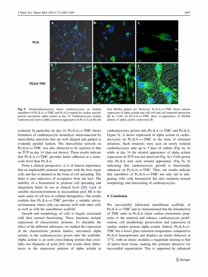
Growth and morphology of cells is largely associated
with their normal functioning. These functions include
expression of characteristic proteins. To elucidate the
effect of the different substrates, we studied the expression
of the characteristic protein marker, sarcomeric alpha
actinin, in the cardiomyocytes grown onto the scaffolds.
Alpha actinin is an actin cross-linking protein that cross-
links two filaments of actin [60]. Our results show differ-
ences in the expression patterns of alpha actinin in
cardiomyocytes grown onto P(L)LA-co-TMC and P(L)LA.
Figure 9c, d shows expression of alpha actinin in cardio-
myocytes on P(L)LA-co-TMC in the form of reformed
striations. Such striations were seen on newly isolated
cardiomyocytes only up to 5 days of culture (Fig. 6a, b)
while at day 14 the striated appearance of alpha actinin
expression on TCP was not observed (Fig. 6c). Cells grown
onto P(L)LA lack such striated appearance (Fig. 9a, b)
indicating that cardiomyocyte growth is functionally
enhanced on P(L)LA-co-TMC. Thus, our results indicate
that nanofibers of P(L)LA-co-TMC not only aid in inte-
grating cells with biomaterial but also maintain normal
morphology and functioning of cardiomyocytes.
4 Conclusion
We successfully fabricated nanofibrous scaffolds of
P(L)LA-co-TMC and we demonstrated that the introduction
of TMC units in P(L)LA chain confers elastomeric prop-
erties to the material and enhance cardiomyocyte prolif-
eration, cell morphology preservation and expression of
cardiac marker protein alpha actinin. Indeed, P(L)LA-co-
TMC has a lower glass transition temperature compared to
P(L)LA homopolymer and displays an elastic behavior at
37�C, with an elastic modulus a magnitude nearing to that
of native heart tissue, making this polymer attractive for
myocardial regeneration. This is supported by enhanced
Fig. 9 Immunofluorescence shows cardiomyocytes on random
nanofibers of P(L)LA-co-TMC and P(L)LA stained for cardiac specific
protein sarcomeric alpha actinin at day 14. Cardiomyocytes remain
scattered and express alpha actinin in aggregates on P(L)LA (a, b) and
lack fibrillar pattern (c). However, P(L)LA-co-TMC shows intense
expression of alpha actinin and cell–cell and cell–material interaction
(d, e). Cells on P(L)LA-co-TMC show re-appearance of fibrillar
pattern of alpha actinin expression (f)
J Mater Sci: Mater Med (2011) 22:1689–1699 1697
123

proliferation of cardiomyocytes isolated from adult heart
that preserve their ability to develop intercellular interac-
tions and impressively maintain morphology and expres-
sion of cardiac protein markers in the form of striations.
Thus, our results indicate that electrospun P(L)LA-co-TMC
performs excellently as a scaffold for cardiomyocytes, and
it is a highly potential synthetic biomaterial for MTE.
Acknowledgments The authors gratefully acknowledge the NRF-
Technion (Grant No.: R-398-001-063-592), Division of Bioengineer-
ing, Nanoscience and Nanotechnology Initiative (National University
of Singapore) and Italian Ministry of University and Research for the
financial support. C.G. is the recipient of a fellowship awarded from
the Spinner Consortium of Regione Emilia Romagna (Italy).
References
1. Kao RL, Browder W, Li C. Cellular cardiomyoplasty: What have
we learned? Asian Cardiov Thorac Ann. 2009;17:89–101.
2. Kong HJ, Kaigler D, Kim K, Mooney DJ. Controlling rigidity and
degradation of alginate hydrogels via molecular weight distri-
bution. Biomacromolecules. 2004;5:1720–7.
3. Serrano MC, Chung EJ, Ameer GA. Advances and applications
of biodegradable elastomers in regenerative medicine. Adv Func
Mat. 2010;20:192–208.
4. Omens JH. Stress and strain as regulators of myocardial growth.
Prog Biophys Mol Biol. 1998;69:559–72.
5. Nagueh SF, Shah G, Wu Y, Lahmers S. Altered titin expression,
myocardial stiffness, and left ventricular function in patients with
dilated cardiomyopathy. Circulation. 2004;110:155–62.
6. Mukherjee S, Venugopal J, Ravichandran R, Ramakrishna S,
Raghunath M. Multimodal biomaterial strategies for regeneration
of infarcted myocardium. J Mater Chem. 2010;20:8819–31.
7. Eschenhagen T, Didie M, Munzel F, Schubert P, Zimmerman
W-H. 3D engineered heart tissue for replacement therapy. Bas
Res Cardiol. 2002;97:146–52.
8. Chiu LLY, Radisic M, Vujak-Novakovic G. Bioactive scaffolds
for engineering vascularized cardiac tissues. Macromol Biosci.
2010;10:1286–301.
9. Kofidis T, Akhyari P, Boublik J, Theodorou P, Martin U, Ruh-
parwar A, Fischer S, Eschenhagen T, Kubis HP, Kraft T, Leyh R,
Haverich A. In vitro engineering of heart muscle: artificial
myocardial tissue. J Thor Cardiovasc Surg. 2002;124:63–9.
10. Kofidis T, de Bruin JL, Hoyt G, Ho Y, Tanaka M, Yamane T,
Lebl DR, Swijnenburg R-J, Chang C-P, Quertermous T, Robbins
RC. Myocardial restoration with embryonic stem cell bioartificial
tissue transplantation. J Heart Lung Transpl. 2005;24(6):737–44.
11. Zimmerman W-H, Fink C, Kralish D, Remmers U, Weil J,
Eschenhagen T. Three-dimensional engineered heart tissue from
neonatal rat cardiac myocytes. Biotechnol Bioeng. 2000;68:
106–14.
12. Amir G, Miller L, Shachar M, Feinberg MS, Holbova R, Cohen
S, Leor J. Evaluation of a peritoneal-generated cardiac patch in a
rat model of heterotopic heart transplantation. Cell Transpl.
2009;18:275–82.
13. Ayelet D, Shachar M, Leor J, Cohen S. Cardiac tissue engi-
neering-optimization of cardiac cell seeding and distribution in
3D porous alginate scaffolds. Biotechnol Bioeng. 2002;80:
305–12.
14. Leor J, Aboulafia-Etzion S, Ayelet D, Shapiro L, Barbash IM,
Battler A, Granot J, Cohen S. Bioengineered cardiac grafts: a new
approach to repair the infarcted myocardium. Circulation.
2000;102:56–61.
15. Sapir Y, Kryukov O, Cohen S. Integration of multiple cell–matrix
interactions into alginate scaffolds for promoting cardiac tissue
regeneration. Biomaterials. 2011;32:1838–47.
16. Akhyari P, Fedak PWM, Weisel RD, Lee T-YJ, Verma S, Mickle
DAG, Li R-K. Mechanical stretch regimen enhances the forma-
tion of bioengineered autologous cardiac muscle graft. Circula-
tion. 2002;106:137–42.
17. Iwakura A, Fujita M, Kataoka K, Tambara K, Sakakibara Y,
Komeda M, Tabada Y. Intramyocardial sustained delivery of
basic fibroblast growth factor improves angiogenesis and ven-
tricular function in a rat infarct model. Heart Vessel. 2003;
18:93–9.
18. Lutolf MP, Hubbell JA. Synthetic biomaterials as instructive
extracellular microenvironments for morphogenesis in tissue
engineering. Nat Biotechnol. 2005;23:47–55.
19. Williams DF. On the nature of biomaterials. Biomaterials.
2009;30:5897–909.
20. Bursac N, Papadaki M, Cohen RJ, Schoen FJ, Eisenberg SR,
Carrier R, Vujak-Novakovic G, Freed LE. Cardiac muscle tissue
engineering: toward an in vitro model for electrophysiological
studies. Am J Physiol. 1999;277:433–44.
21. Hosseinkhani H, Hosseinkhani M, Hattori S, Matsuoka R,
Kawaguchi N. Micro and nano-scale in vitro 3D culture system
for cardiac stem cells. J Biomed Mat Res. 2010;94A:1–8.
22. Ke Q, Yang Y, Rana JS, Yu C, Morgan JP, Yong-Fu X.
Embryonic stem cells cultured in biodegradable scaffold repair
infarcted myocardium in mice. Acta Physiol Sin. 2005;57:
673–81.
23. Kellar RS, Landeen LK, Shephered BR, Naughton GK, Ratcliffe
A, Williams SK. Scaffold-based three-dimensional human fibro-
blast culture provides a structural matrix that supports angio-
genesis in infarcted heart tissue. Circulation. 2001;104:2063–8.
24. Rabkin E, Schoen J. Cardiovascular tissue engineering. Cardio-
vasc Pathol. 2002;11:305–17.
25. Alperin C, Zandstra PW, Woodhouse KA. Polyurethane films
seeded with embryonic stem cell-derived cardiomyocytes for use
in cardiac tissue engineering applications. Biomaterials.
2005;26:7377–86.
26. Giraud M-N, Flueckiger R, Cook S, Ayuni E, Siepe M, Carrel T,
Tevaearai HT. Long-term evaluation of myoblast seeded patches
implanted on infarcted rat hearts. Artif Organs. 2010;34:
E184–92.
27. McDevitt TC, Woodhouse KA, Hauschka SD, Murry CE, Stayton
PS. Spatially organized layers of cardiomyocytes on biodegrad-
able polyurethane films for myocardial repair. J Biomed Mat Res.
2003;66A:586–95.
28. Siepe M, Giraud M-N, Liljensten E, Nydegger U, Menasche P,
Carrel T, Tevaearai HT. Construction of skeletal myoblast-based
polyurethane scaffolds for myocardial repair. Artif Organs.
2007;31:425–33.
29. Krupnick AS, Kreisel D, Engels FH, Szeto WY, Plappert T,
Pompa SH, Flake AW, Rosengard BR. A novel small animal
model of left ventricular tissue engineering. J Heart Lung
Transpl. 2002;21:233–43.
30. Ozawa T, Mickle DAG, Weisel RD, Koyama N, Ozawa S, Li
R-K. Optimal biomaterial for creation of autologous cardiac
grafts. Circulation. 2002;106:176–82.
31. Park H, Radisic M, Lim JO, Chang BH, Vujak-Novakovic G. A
novel composite scaffold for cardiac tissue engineering. In Vitro
Cell Dev Biol Anim. 2005;41:188–96.
32. Bat E, van Kooten TG, Feijen J, Grijpma DW. Resorbable elas-
tomeric networks prepared by photocrosslinking of high-molec-
ular-weight poly(trimethylene carbonate) with photoinitiators and
1698 J Mater Sci: Mater Med (2011) 22:1689–1699
123

poly(trimethylene carbonate) macromers as crosslinking aids.
Acta Biomater. 2011;7:1939–48.
33. Bruggeman JP, de Bruin B-J, Bettinger CJ, Langer R. Biode-
gradable poly(polyol sebacate) polymers. Biomaterials. 2008;29:
4726–35.
34. Chen Q-Z, Bismarck A, Hansen U, Junaid S, Tran MQ, Harding
SE, Ali NN, Boccaccini AR. Characterization of a soft elastomer
poly(glycerol sebacate) designed to match the mechanical
propeeties of myocardial tissue. Biomaterials. 2008;29:47–57.
35. Song Y, Kamphuis MMJ, Zhang Z, LMTh Sterk, Poot AA, Feijen
J, Grijpma DW. Flexible and elastic porous poly(trimethylene
carbonate) structures for use in vascular tissue engineering. Acta
Biomater. 2010;6:1269–77.
36. Wang Y, Ameer GA, Sheppard B, Langer R. A tough biode-
gradable elastomer. Nat Biotechnol. 2002;20:602–6.
37. You Z, Cao H, Gao J, Shin PH, Day BW, Wang Y. A func-
tionalizable polyester with free hydroxyl groups and tunable
physiochemical and biological properties. Biomaterials. 2010;31:
3129–38.
38. Buchholz B. Analysis and characterization of resorbable D,L-
lactide-trimethylene carbonate copolyesters. J Mat Sci: Mat Med.
1993;4:381–8.
39. Pego AP, Poot AA, Grijpma DW, Feijen J. Physical properties of
high molecular weight 1,3-trimethylene carbonate and D,L-lactide
copolymers. J Mater Sci: Mater Med. 2003;14:767–73.
40. Tyson T, Finne-Wistrand A, Albertsson A-C. Degradable porous
scaffolds from various L-lactide and trimethylene carbonate
copolymers obtained by a simple and effective method. Bio-
macromolecules. 2009;10:149–54.
41. Pego AP, Siebum B, Luyn V, Gallego XJ, Seijen YV, Poot AA,
Grijpma DW, Feijen J. Preparation of degradable porous struc-
tures based on 1,3-trimethylene carbonate and D,L-lactide
(co)polymers for heart tissue engineering. Tissue Eng. 2003;9:
981–94.
42. Pego AP, Poot AA, Grijpma DW, Feijen J. Biodegradable elas-
tomeric scaffolds for soft tissue engineering. J Control Release.
2003;87:69–79.
43. Pego AP, Van Luyn MJA, Brouwer LA, Van Wachem PB, Poot
AA, Grijpma DW, Feijen J. In vivo behavior of poly(1,3-tri-
methylene carbonate) and copolymers of 1,3-trimethylene car-
bonate with D,L-lactide or -caprolactone: degradation and tissue
response. J Biomed Mater Res. 2003;67A:1044–54.
44. Li M, Mondrinos MJ, Chen X, Gandhi MR, Ko FK, Lelkes PI.
Co-electrospun poly(lactide-co-glycolide), gelatin and elastin
blends for tissue engineering scaffolds. J Biomed Mater Res.
2006;79A:963–73.
45. Rockwood DN, REJr Akins, Parrag IC, Woodhouse KA, Rabolt
JF. Culture on electrospun polyurethane scaffolds decreases atrial
natriuretic peptide expression by cardiomyocytes in vitro. Bio-
materials. 2008;29:4783–91.
46. Shin M, Ishii O, Sueda T, Vacanti JP. Contractile cardiac grafts
using a novel nanofibrous mesh. Biomaterials. 2004;25:3717–23.
47. Stankus JJ, Guan J, Wagner WR. Fabrication of biodegradable
elastomeric scaffolds with sub-micron morphologies. J Biomed
Mater Res. 2004;70A:603–14.
48. Zong X, Bien H, Chung CY, Yin L, Fang D, Hsiao BS, Chu B,
Entcheva E. Electrospun fine-textured scaffolds for heart tissue
constructs. Biomaterials. 2005;26:5330–8.
49. Gupta B, Revagade N, Hilborn J. Poly(lactic acid) fiber: an
overview. Prog Polym Sci. 2007;32:455–82.
50. Zhu KJ, Hendren RW, Jensen K, Pitt CG. Synthesis, properties,
and biodegradation of poly(1,3-trimethylene carbonate). Macro-
molecules. 1991;24:1736–40.
51. Peter P, Malberg S, Albertsson AC. Design of resorbable porous
tubular copolyester scaffolds for use in nerve regeneration. Bio-
macromolecules. 2009;10:1259–64.
52. Wei X, Xia Z, Wong S-C, Baji A. Modelling of mechanical
peroperties of electrospun nanofibre network. Int J Exp Comput
Biomech. 2009;1:45–57.
53. Lu J-W, Zhang ZP, Ren XZ, Chen YZ, Yu J, Guo ZX. High-
elongation fiber mats by electrospinning of polyoxymethylene.
Macromolecules. 2008;41:3762–4.
54. Andronova N, Albertsson A-C. Resilient bioresorbable copoly-
mers based on trimethylene carbonate, L-lactide, and 1,5-dioxe-
pan-2-one. Biomacromolecules. 2006;7:1495.
55. Focarete ML, Gualandi C, Moroni L. Working with electrospun
scaffolds: some practical hints for tissue engineers. In: Haghi AK,
editor. Electrospun nanofibers research: recent developments,
Hauppauge. New York: Nova Science Publisher; 2009. p. 19–34.
56. Du A, Sanger JM, Linask KK, Sanger JW. Myofibrillogenesis in
the first cardiomyocytes formed from isolated quail precardiac
mesoderm. Dev Biol. 2003;257:382–94.
57. Messina E, De Angelis L, Frati G, Morrone S, Chimenti S, Fi-
ordaliso F, Salio M, Battaglia M, Latronico MV, Coletta M,
Vivarelli E, Frati L, Cossu G, Giacomello A. Isolation and
expansion of adult cardiac stem cells from human and murine
heart. Circ Res. 2004;95:911–21.
58. Bearzi C, Rota M, Hosoda T, Tillmanns J, Nacimbene A, De
Angelis A, Yasuzawa-Amano S, Trofimova I, Siggins RW, Le-
Capitaine N, Cascapera S, Beltrami AP, D’Alessandro DA, Zias
E, Quaini F, Urbanek K, Michler RE, Bolli R, Kajstura J, Leri A,
Anversa P. Human cardiac stem cells. Proc Natl Acad Sci USA.
2007;104:14068–73.
59. Martinez EC, Kofidis T. Myocardial tissue engineering: the quest
for the ideal myocardial substitute. Expert Rev Cardiovasc Ther.
2009;7:921–8.
60. Cabello N, Remelli R, Canela L, Soriguera A, Mallol J, Canela
EI, Robbins MJ, Lluis C, Franco R, McIlhinney RA, Ciruela F.
Actin-binding protein alpha-actinin-1 interacts with the metabo-
tropic glutamate receptor type 5b and modulates the cell surface
expression and function of the receptor. J Biol Chem. 2007;
282:12143–53.
J Mater Sci: Mater Med (2011) 22:1689–1699 1699
123