p53-cofactor JMY is a multifunctional actin nucleation factor
Upload
khangminh22Category
view
0download
0
Original Article
A Multifunctional LNA Oligonucleotide-BasedStrategy Blocks AR Expressionand Transactivation Activity in PCa CellsDaniela Castanotto,1,9 Xiaowei Zhang,1,9 Jacqueline Rüger,1 Jessica Alluin,2 Ritin Sharma,3 Patrick Pirrotte,3
Lars Joenson,4 Silvia Ioannou,5 Michael S. Nelson,6 Jonas Vikeså,4 Bo Rode Hansen,7 Troels Koch,8
Mads Aaboe Jensen,4 John J. Rossi,2 and Cy A. Stein1
1Department of Medical Oncology, City of Hope, 1500 East Duarte Road, Duarte, CA 91010, USA; 2Department of Molecular and Cellular Biology, City of Hope, 1500 East
Duarte Road, Duarte, CA 91010, USA; 3Collaborative Center for Translational Mass Spectrometry, Translational Genomics Research Institute, Phoenix, AZ, USA; 4Roche
Innovation Center Copenhagen A/S, Fremtidsvej 3, 2970 Hørsholm, Denmark; 5Science Department, Flintridge Preparatory School, 4543 Crown Avenue, La Cañada
Flintridge, CA 91011, USA; 6The Light Microscopy and Digital Imaging Core, Beckman Research Institute, City of Hope, 1500 East Duarte Road, Duarte CA 91010;7Genevant Sciences, 245 Main Street, Floor 2, Cambridge, MA 02142; 8Frederikskaj 10B, 2nd floor, 2450 Copenhagen SV, Denmark
Received 11 August 2020; accepted 21 October 2020;https://doi.org/10.1016/j.omtn.2020.10.032.9These authors contributed equally
Correspondence: Daniela Castanotto, Department of Medical Oncology, City ofHope, 1500 East Duarte Road, Duarte, CA 91010, USA.E-mail: [email protected]: Cy A. Stein, Department of Medical Oncology, City of Hope,1500 East Duarte Road, Duarte, CA 91010, USA.E-mail: [email protected]
The androgen receptor (AR) plays a critical role in the develop-ment of prostate cancer (PCa) through the activation ofandrogen-induced cellular proliferation genes. Thus, blockingAR-mediated transcriptional activation is expected to inhibitthe growth and spread of PCa. Using tailor-made splice-switch-ing locked nucleic acid (LNA) oligonucleotides (SSOs), we suc-cessfully redirected splicing of the AR precursor (pre-)mRNAand destabilized the transcripts via the introduction of prema-ture stop codons. Furthermore, the SSOs simultaneously favoredproduction of the AR45 mRNA in lieu of the full-length AR.AR45 is an AR isoform that can attenuate the activity of bothfull-length and oncogenic forms of AR by binding to their com-mon N-terminal domain (NTD), thereby blocking their transac-tivation potential. A large screen was subsequently used to iden-tify individual SSOs that could best perform this dual function.The selected SSOs powerfully silence AR expression and modu-late the expression of AR-responsive cellular genes. This bi-func-tional strategy that uses a single therapeutic molecule can be thebasis for novel PCa treatments. It might also be customized toother types of therapies that require the silencing of one geneand the simultaneous expression of a different isoform.
INTRODUCTIONProstate cancer (PCa) is the second most common cancer in menworldwide.1 In the United States, approximately 190,000 new PCacases will be diagnosed in 2020.2 Of these, an estimated 30,000 caseswill end in death, making PCa the secondmost deadly cancer inmen.2
The most important molecular driver in PCa is the androgen receptor(AR).3–5 The AR, found in the cytoplasm, responds to androgensignaling by translocating to the cell nucleus where it acts as a regu-lator of gene transcription.6 The AR is a key evolutionary gene thatplays a critical role in male sexual development and in the mainte-nance of the male phenotype throughout life. Hence, it is to be ex-pected that cells have evolved ways to maintain AR function.
Molecular TThis is an open access article under the CC BY-NC
The full-length AR (AR-FL) precursor (pre-)mRNA generally con-sists of eight exons (Exs). Exons 5–8 encode the ligand-bindingdomain (LBD) (i.e., the testosterone/dihydrotestosterone [DHT]binding site). Under selective pressure from some of the anti-andro-genic drugs administered to patients, the AR can undergo mutationsthat enable expression of shorter oncogenic forms. These oncogenicAR isoforms, including those lacking the LBD, remain active andcan function in place of the AR-FL. A common strategy, blockingthe AR transactivation function in later stage PCa, is known asandrogen deprivation therapy (ADT). This form of treatment relieson the use of drugs that block the binding of androgens to the LBDof the AR protein, which inhibits its translocation from the cytoplasmto the nucleus. At the outset, ADT generally results in tumor regres-sion, but, as mentioned above, hormone-independent AR isoformscan later surface and lead to castration-resistant PCa (CRPC).7–10
These CRPC-driving AR isoforms lack the LBD of the AR that is en-coded by the C-terminal exons of the AR pre-mRNA.11 The mostcommon CRPC-related AR isoform is ARv7, present in 20%–40%of patients who previously underwent ADT.12 In the ARv7 mRNA,exons 4–8, which include the DNA-binding domain, are splicedout.13 Therefore, those agents used for PCa therapy, e.g., enzaluta-mide (Xtandi), which bind to the LBD, are ineffective.12 Newergenerations of non-steroidal anti-androgens such as apalutamide (Er-leada)14 and darolutamide (Nubeqa),15 which were US Food andDrug Administration (FDA) approved in 2018 and 2019,
herapy: Nucleic Acids Vol. 23 March 2021 ª 2020 The Authors. 63-ND license (http://creativecommons.org/licenses/by-nc-nd/4.0/).

Molecular Therapy: Nucleic Acids
respectively,16,17 improve metastasis-free survival of CRPC patients.However, they have significant toxicity and do not provide a long-term, permanent solution.18
Although nearly all AR variants translocate to the nucleus and trans-activate survival genes, one alternatively spliced isoform, AR45,19
does not exhibit this oncogenic activity. AR45 contains the DNA-binding domain and LBD, but it lacks AF-1, the transactivating N-ter-minal domain (NTD) encoded in the first exon of the AR transcript.This is important because the AR activation function relies primarilyon this first activation domain within the N-terminal region (NTR),20
which contains transcriptional activation units 1 and 5 (TAU-1 andTAU-5). TAU-1 makes up almost the entire transactivation domainand is required to activate the AR-FL protein.21 TAU-5 is a smallerregion that is required and sufficient to activate AR isoforms thatlack the LBD.21 TAU-1 and TAU-5 are therefore necessary for ARtranscriptional activity.22,23 This explains why AR spliced isoformsthat lack the second transactivation domain (AF-2) and the LBD21
(e.g., ARv7) can maintain potent transactivation properties. The ARNTD has little homology with other steroid receptors and is the leastconserved of all AR domains; consequently, the NTD is difficult totarget with small molecular therapeutic entities. As long as thisNTD region is present and functional, the AR and all of its isoformswill continue to transactivate nuclear genes and promote tumor cellsurvival.
In this study, we performed a splice-switching oligonucleotide (SSO)24
screen to identify sequences that best drove production of AR proteinsfrom the AR-FL and its derived isoforms toward AR45. To alter thesplicing pattern, we used 16- to 20-mer phosphorothioate (PS) SSOs,each containing six to eight interspersed locked nucleic acid (LNA)moieties,25 and targeted them either at, upstream, or downstream ofthe 30 or 50 splice sites of the AR-FL. Binding sites for the SSOs onthe AR transcripts were chosen based on which SSO caused the mosteffective re-direction of splicing to selected alternative cryptic splicesites, and destabilized the resulting transcripts by triggeringnonsense-mediated decay (NMD).26 Preference was given to thoseSSOs that could also favor the production of the AR45 mRNA inlieu of the AR-FL mRNA. Using this strategy, we identified powerfulSSOs that silenced AR expression and modulated the expression ofAR-responsive genes. Our “multitask” design SSO could be the basisfor more effective PCa treatments, especially for patients with CRPC.
RESULTSSuppression of AR Gene Expression via an NMD Pathway
When the selective pressure from anti-androgenic drugs given to pa-tients leads to the expression of truncated AR isoforms, it is difficult tospecifically block their oncogenic activity with small molecules. Thisis because these short isoforms do not include the LBD, the targetedregion of small molecule-based drugs. Therefore, we focused ondeveloping a strategy that would be unaffected by the absence ofthe LBD. Strategies targeting the spliceosome have been used to alterAR-FL alternative splicing and to block production of these short iso-forms.27 However, the spliceosome is not specific to a single gene, so
64 Molecular Therapy: Nucleic Acids Vol. 23 March 2021
this approach may produce unacceptable cellular toxicity. Severalantisense oligonucleotides (ASOs) targeting the AR have also beenused with promising results, including one that is currently in a phaseI clinical trial.28,29 Nonetheless, the analysis of 914 samples from fiveindependent studies indicated that both RNaseH2A and RNaseH2Bare depleted in PCa.30 Downregulation of RNaseH2B, which isrequired for ASO activity, could present a significant challenge tothe use of ASOs.
When fully chemically modified, SSOs are highly stable in vivo andmay in fact be preferred for therapeutic purposes. Indeed, such chem-ical entities (e.g., eteplirsen) have demonstrated efficacy in clinical tri-als and have been FDA approved and commercialized.24,31 We de-signed the SSO-based strategy reported herein based on theassumption that if silencing takes advantage of a natural cellular pro-cess, it might be better tolerated by cells and more long-lasting. Wetherefore targeted the SSOs to canonical splice sites in the AR pre-mRNA transcript to block and/or redirect the splicing in a mannerthat would result in the inclusion of premature termination codons(PTCs). The introduction of PTCs upstream of an exon-exonjunction triggers NMD, which leads to translational repression anddegradation of the mRNA.32We performed proof-of-principle exper-iments using sequences encompassing either the 50 splice site (ss), the30 ss, or the polypyrimidine tract, an intronic splicing regulatorysequence (Figure 1; Figure S1).We specifically targeted the first intronof the AR with the goal of eliminating the NTD encoded by exon 1. Asmentioned above, the NTD domain can, in the absence of theremainder of the protein, function as a transcription activator. There-fore, any splicing alteration in the downstream introns/exons mightgenerate new, stable isoforms with transactivation activity. TheNTD is common to all AR variants (except for AR45), extendingthe targeting potential of our SSOs.
The first SSO targeting the canonical 30 ss of intron 1 (SSO753, Fig-ures 1A and 1B; Figures S1A and S1B) also spanned and blockedan adjacent 30 ss. This latter site, if utilized as an alternative splicesite, would create a translatable open reading frame (ORF). SSO753was also designed to base-pair almost entirely with exon 2 and toblock only a single nucleotide (G) of the 30 ss AG. Our intent herewas to generate an SSO with a dual function. These functions are(1) to arrest the splicing at the canonical AG (the proximal SSO/pre-mRNA duplex would likely obscure spliceosomal recognition ofthat AG), and (2) to fully base-pair with any mRNA that might orig-inate from escaped transcripts not bound by the SSO, thus blockingand/or reducing mRNA translation. Coincidentally, the last nucleo-tide of the first exon is also a G. Thus, the correctly spliced ARmRNA generates the exact reverse complement of SSO753 (Fig-ure S1B). Therefore, SSO753 can also target the AR transcripts inboth the nucleus, as an SSO, and the cytoplasm, where it would func-tion as a non-RNaseH active ASO (a.k.a. a steric blocking ASO;Figure S1B).
To determine the extent of gene silencing resulting from targetingexclusively intronic sequences, we designed an SSO (SSO815,

Figure 1. SSO-Based Strategies to Silence AR Gene Expression
(A) Schematic diagram of different splice-switching strategies. SSO749 blocks recognition of the endogenous translation start codon; SSO748 blocks the 50 ss of intron 1;
SSO815 blocks the polypyrimidine sequence upstream of the 30 ss of intron 1; and SSO753 and SSO752 block the canonical 30 ss at the intron 1-exon 2 junction and a
second alternative 30 ss within exon 2, respectively. (B) Initial SSO sequences employed in this work. (C) Representative western blot analysis showing AR protein expression
in two prostate cancer cell lines (LAPC-4 and LNCaP) transfected with 20 nM of each SSO as indicated in the figure. SSO-CNTR is a non-targeting LNA PS oligonucleotide,
used as a control for non-specific AR silencing. ENZ4176 is a potent AON targeting the AR mRNA, which was used as comparative control; a-tubulin is the loading control.
www.moleculartherapy.org
Figures 1A and 1B; Figure S1A) directed at the polypyrimidine tract, aregulatory sequence that plays a key role in the recognition and selec-tion of splice sites.33,34 The third SSO (SSO752, Figures 1A and 1B)was designed to block a potential alternative downstream 30 ss, which,if selected, could generate a translatable ORF. The fourth SSO(SSO748, Figures 1A and 1B) was designed to base-pair and blockrecognition of the 50 ss. The final SSO (SSO749, Figures 1A and 1B)was designed to base-pair with and block the translation start codonof the AR mRNA. SSO749 was included to compare the extent ofsilencing induced by directly interfering with translation (using theSSO as a steric blocker)35 to the extent of silencing resulting fromtranslational suppression due to SSO-mediated cellular NMD. The re-sults of these experiments indicate that the latter is a significantlymore effective mechanism to silence AR gene expression (Figure 1C)than is the former.
The bi-functional SSO753 exhibited the strongest silencing activity ofall SSOs (Figure 1C). In general, targeting the 30 ss (Figure 1C,SSO815, SSO753, SSO752) proved to be more effective than targetingthe 50 ss (Figure 1C, SSO748). However, blocking the canonical 30 sscould not prevent engagement of the 50 ss by the spliceosome and thepotential selection of an alternative intronic AG. This abnormalsplicing reaction would still result in the inclusion of PTCs (weconfirmed that no other ORF can form) and subsequent degradationof the AR RNA, but it would diminish/eliminate the likelihood ofgenerating AR45 transcripts.
AR45 is the only known AR isoform that does not contain exon 113
and does not exhibit oncogenic activity.19 In place of exon 1, AR45contains exon 1b, which encodes a 7-aa-long peptide sequence uniqueto this isoform.19 AR45 appears to form heterodimers with the NTDs
Molecular Therapy: Nucleic Acids Vol. 23 March 2021 65

Molecular Therapy: Nucleic Acids
of other AR isoforms, acting as an antagonist and blocking AR func-tion.19 It is possible that preventing the splicing reaction from occur-ring at the canonical 50 ss of the AR transcript could grant sufficienttime for the alternative 50 ss of exon 1b to splice and generate theAR45 transcript. This presents an opportunity to design a secondbi-functional SSO that would degrade the AR RNA transcripts (dueto the partial intron retention), while at the same time favoring pro-duction of the AR antagonist AR45. Ribosome binding to the canon-ical translation start site would encouter premature stop codons,hence triggering NMD. Whereas ribosome binding to the exon 1b in-ternal start site (prior to any transcript degradation), would allowtranslation to occur and AR45 expression. To test whether SSO748possessed this dual function, we performed a series of qRT-PCR reac-tions to analyze the AR pre-mRNA and mRNA transcripts resultingfrom SSO treatment of LNCaP cells (Figures S2A and S2B). The PCRprimers were designed to specifically amplify the AR-FL or the AR45transcripts. The data show that all SSO treatments resulted in adecrease of AR gene expression (Figure S2B). SSO748, the only SSOtargeting the 50 ss, was shown, in fact, to be a bi-functional oligonu-cleotide, as it could also generate AR45 transcripts (Figure S1B).
Optimization of SSO Function through Design Modeling and
Empirical Screens
After determining the feasibility of the strategy, we then optimized SSOpotency. The datawere consistent with the bi-functional SSOs being themost potent. Therefore, we selected those two targeted regions (the 50 ss[748 series of SSOs] and 30 ss [753 series of SSOs] of intron 1) to design ascreen of approximately 250 oligonucleotides. The sequence, length,and location of the LNAmodifications were varied based on a proprie-tary modeling prediction program (developed by Roche InnovationCenter, Copenhagen, Denmark). The resulting SSOs were used to treatdifferent PCa cell lines. Subsequently, their activity was assessed viaqRT-PCR and western analyses (examples of these screens are shownin Figures S3A, S3B, S4A, and S4B). The leading SSOs from the 753 se-rieswere chosenbasedon their ability to silenceARexpression (e.g., Fig-ures S3A and S3B), as they were not expected to generate AR45. Theleading SSOs from the 748 series, however, were selected based notonly on their ability to silence AR expression, but also to generateAR45 transcripts. We screened for SSOs that produced the strongestAR silencing and the best simultaneous AR45 expression. The latterwas established forbothAR45absolute expression andAR45expressionas a percentage of the remaining AR. For example, SSO363 was prefer-able to SSO362 (Figure S4B, left graph) because even though theydown-regulated AR expression to the same extent, SSO363 generated a higherexpressionofAR45 (Figure S4B, left graph).However, SSO372was pref-erable to SSO376 (Figure S4B, right graph), despite producing a loweroverall expression of AR45. This is because, when analyzed by the per-centage of residual AR transcripts, cells treated with SSO372 stilldemonstrated a greater AR45/AR-FL ratio. Moreover, AR silencingwas better achieved after SSO372 treatment (compare SSO372 toSSO376; Figure S4B, right graph).
Based on the above results, we chose the eight most active SSOs (Fig-ures S5; Figure 2). By combining the modeling program and the
66 Molecular Therapy: Nucleic Acids Vol. 23 March 2021
empirical screen, we were able to identify 50 ss-targeting SSOs (748 se-ries) that were as effective as the 753 series in silencing AR expression(Figures 2A and 2B; compare SSO407, SSO390, SSO410, and SSO395with SSO541, SSO480, SSO547, and SSO111) and as potent as an ASOcontrol (ENZ-4176, Figures 2A and 2B). SSOs belonging to the 748series also increased expression of AR45 mRNA (Figure 2B,SSO407, SSO390, SSO410, and SSO395). To determine whether thereduction in AR gene expression was reflected in the regulation ofAR-responsive promoters, we delivered the selected SSOs to LNCaPcells previously engineered to express an AR-responsive luciferasegene (see Materials and Methods). All SSOs treatments reduced lucif-erase expression, with SSO541 and SSO407 being the most effective(Figure 2C). Under our experimental conditions, none of the SSOsshowed any significant treatment-related toxicity in the PCa cell linesused for this work (Figures 3A and 3B and data not shown).
The potency of the eight most active SSOs was also assessed andconfirmed in LAPC-4 cells (Figures 4A [748 series] and 4B [753 series])and 22RV1 cells (Figures 4C [748 series] and 4D [753 series]). The22RV1 PCa line expresses ARv7,36 the most prominent AR mutationfound in later stage PCa patients. Notably, all selected SSOs wereable to diminish expression of the ARv7 isoform (Figures 4C and 4D).
Reduced AR Expression Is Reflected in the Downregulation of
AR-Responsive Cellular Genes
The most potent four (Figures 2A–2C and 4A–4D; SSO390, SSO407,SSO457, and SSO541) out of the eight previously selected SSOs werefurther tested in serial dilution experiments to identify the best overallSSO for the 753 and 748 series (to be carried forward for in vivostudies). SSO407 and SSO541 were effective at lower concentrations(Figure 5A). Furthermore, serial dilutions of SSO407 appeared toexhibit an inverse relationship between the expression of AR andAR45 (Figures 5B and 5C). These results were consistent with theappearance of an approximately 45-kDa molecular weight product,which increased as the AR-FL detection decreased (Figure S6A).
Finally, we corroborated the potency of the SSOs by determining thedegree to which two AR-responsive endogenous cellular genes, pros-tate-specific antigen (PSA) and transmembrane serine protease 2(TMPRSS2), are affected by SSO-driven AR silencing. All SSOsreduced expression of the AR-regulated PSA and TMPRSS2 genes,with the best outcomes obtained using SSO541 and SSO407 (Fig-ure 6). The reduced expression may reflect elimination of the AR-gene transactivation and a return to a more canonical endogenousexpression. Importantly, even a modest, but simultaneous decreasein gene expression of multiple AR-responsive genes could have a sig-nificant impact on blocking PCa progression.
SSO541 and SSO407 were then evaluated in a small in vivo pilotstudy, in which four groups of six NSG mice were subcutaneouslyengrafted with LNCaP tumors of comparable sizes. The mice weretreated with either the delivery medium (1� PBS) for the injections(vehicle), a non-targeting control oligonucleotide (SSO-control[CNTR]), or either SSO541 or SSO407 (Figure S6B). The treatment

Figure 2. SSO-Mediated AR Regulation
(A) Representative western blot analyses of lysates harvested from
LNCaP cells treated for 2 days with 20 nM of each specific SSO (as
indicated in the figure). a-Tubulin was used as the loading control.
The experiment was repeated several times with comparable re-
sults. (B) LNCaP cells were transfected with the AR-responsive
luciferase gene and 20 nM of each SSO (as indicated in the graph)
for 48 h; total RNA was then harvested for AR and AR45 mRNA
qRT-PCR. The non-treated cells and the SSO-CNTR-treated cells
were used as reference groups. Values were normalized to
GAPDH mRNA expression and expressed as the mean ± SD; n =
3. p values for changes in AR expression were as follows: ENZ-
4176, p = 0.002; SSO541, p = 0.007; SSO480, p = 0.009;
SSO457, p = 0.002; SSO111, p = 0.002; SSO407, p = 0.002;
SSO390, p = 0.008; SSO410, p = 0.02; SSO395, p = 0.007. p
values for changes in AR-45 expression were as follows: ENZ-
4176, p = 0.005; SSO541, p = 0.006; SSO480, p = 0.003;
SSO457, p = 0.01; SSO111, p = 0.004; SSO407, p = 0.008;
SSO390, p = 0.004; SSO410, p = 0.007; SSO395, p = 0.006. (C)
An LNCaP cell line expressing an AR-responsive luciferase gene
was transfected with 20 nM of each SSO (as indicated in the
graph). Cells were harvested 48 h after treatment and analyzed
using a Dual-Luciferase assay system. Any decrease of luciferase
expression represents an equivalent loss of AR nuclear trans-
activation activity, which in turn is caused by SSO-mediated AR
silencing. Therefore, the extent of luciferase silencing reflects the
degree of SSO potency. Values are expressed as the mean ± SD;
n = 3. p values for changes in luciferase units were: ENZ-4176, p =
0.007; SSO541, p = 0.01; SSO480, p = 0.007; SSO457, p =
0.0002; SSO111, p = 0.009; SSO407, p = 0.0002; SSO390, p =
0.017; SSO410, p = 0.02; SSO395, p = 0.038. The non-targeting
SSO-CNTR was used as a negative control, and ENZ4176 served
as the positive control. Samples SSO541, SSO480, SSO457, and
SSO111 target the 30 ss of intron 1. Samples SSO407, SSO390,
SSO410, and SSO395 target the 50 ss of intron 1.
www.moleculartherapy.org
Molecular Therapy: Nucleic Acids Vol. 23 March 2021 67

Figure 3. Apoptosis and Cytotoxicity (MTT) Assays of SSO-Transfected LNCaP Cells
(A and B) Flow cytometry (A) andMTT (B) assays were performed to determine potential SSO-driven cytotoxicity. An untreated sample (CNTR) and the non-targeting SSO-CNTR
were included as negative controls. A sample (CNTR-ATO) treated with 2mM arsenic trioxide to induce cell apoptosis, was included as positive control. The profiles shown for the
SSOs are typical of three independent experiments and are comparable to the controls, indicating no significant toxicity under the conditions of these experiments.
Molecular Therapy: Nucleic Acids
regimen consisted of three injections of limited SSO amounts(25 ng/g) during the first week. Mice were sacrificed after 3 weeks.The tumors were dissected from the animals and weighed. Theaverage of the tumor weights for each group (Figure S6B) is indic-ative of the extent to which SSO-mediated AR silencing was effec-tive in vivo.
DISCUSSIONThe AR is expressed early in PCa and continues to be expressedthroughout the course of the disease, driving the growth, metastaticcapability, and anti-apoptotic phenotype of PCa cells. One of themainstays of current clinical PCa therapy is, therefore, a reductionin the levels of plasma and intra-tumoral androgen.37 This strategy,known as ADT, is initially accomplished by drugs that act at the hy-pothalamic-pituitary-gonadal axis. These agents lower plasmatestosterone to castrate levels but do not lower intra-tumoral levelsof testosterone.37 On disease recurrence, patients are then oftentreated with oral hormonal agents such as abiraterone or enzaluta-mide.38 The former is a potent inhibitor of CYP17, a key enzyme inthe biosynthesis of testosterone from steroid precursors. The latter isa potent inhibitor of the binding of testosterone to the AR, and ofthe translocation of the AR to the cell nucleus.38 However, all pa-tients will eventually progress39 through these treatments. Patientsthat progress to CRPC inevitably become refractory to PCatherapies.
68 Molecular Therapy: Nucleic Acids Vol. 23 March 2021
ASO gapmers have previously been used to successfully silence ARexpression.28,29,40 Some excellent work using a PCa mouse model hasbeenperformedbyYamamotoet al.28 andDeVelascoet al.,29whodevel-oped an effectiveARASO (IONIS-AR-2.5Rx). ThisASO is currently be-ing tested in a phase I PCa clinical trial in combination with enzaluta-mide. However, another gapmer (ENZ-4176) previously developed byEnzon Pharmaceuticals, which had displayed strong efficacy in mousestudies,40 did not prove to be beneficial in a human PCa clinical trial.40
Perhaps these discrepancies are in part due to the gapmers’ central re-gion, which must allow recognition of the DNA/RNA duplex by RNa-seH2B, and as a result, cannot be chemically protected by anythingmore than PS linkages. Although new generations of gapmers haveincreased stability, systemic longevity in human blood and tissuesmay still be an issue. Furthermore, RNaseH2B, which is required forgapmer function, is downregulated in several cancers, including PCa.30
In this work, we developed anti-AR SSOs that have nuclease-resistantmodifications spaced throughout the molecule.41–43 Currently, thereare three SSO treatments that are FDA approved: eteplirsen,31,44 nusi-nersen,24,45,46 and golodirsen.47 Nuclease resistance was achieved withmorpholino48,49 (eteplirsen and golodirsen) and the 20-O-methoxyethyl(20-MOE)46 (nusinersen) PS modifications. We chose to use LNA PSmodifications as they increase the melting temperature (Tm) of eachmodified nucleotide/complement heterodimer by 2�C –8�C,25 stabiliz-ing the oligonucleotide and its interaction with the target.50–52 In
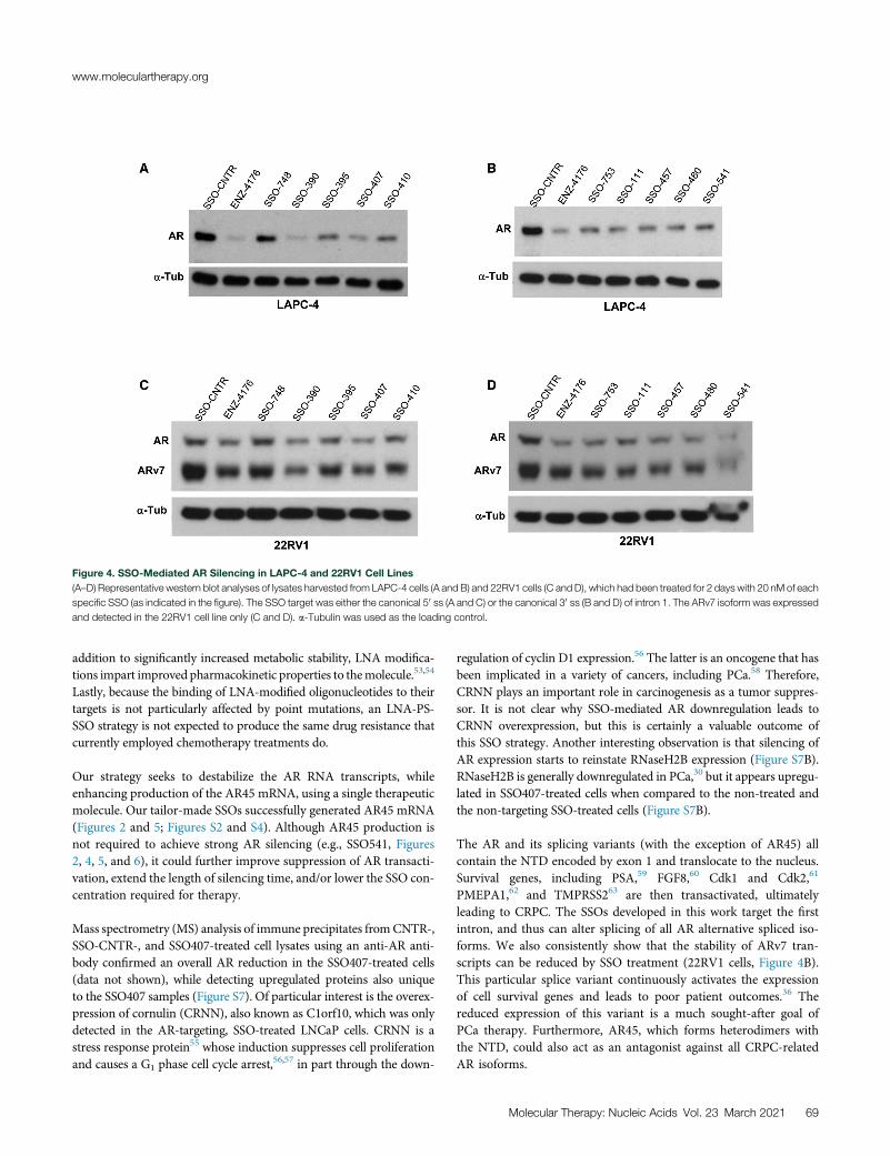
Figure 4. SSO-Mediated AR Silencing in LAPC-4 and 22RV1 Cell Lines
(A–D) Representative western blot analyses of lysates harvested from LAPC-4 cells (A and B) and 22RV1 cells (C and D), which had been treated for 2 days with 20 nM of each
specific SSO (as indicated in the figure). The SSO target was either the canonical 50 ss (A and C) or the canonical 30 ss (B and D) of intron 1. The ARv7 isoform was expressed
and detected in the 22RV1 cell line only (C and D). a-Tubulin was used as the loading control.
www.moleculartherapy.org
addition to significantly increased metabolic stability, LNA modifica-tions impart improved pharmacokinetic properties to themolecule.53,54
Lastly, because the binding of LNA-modified oligonucleotides to theirtargets is not particularly affected by point mutations, an LNA-PS-SSO strategy is not expected to produce the same drug resistance thatcurrently employed chemotherapy treatments do.
Our strategy seeks to destabilize the AR RNA transcripts, whileenhancing production of the AR45 mRNA, using a single therapeuticmolecule. Our tailor-made SSOs successfully generated AR45 mRNA(Figures 2 and 5; Figures S2 and S4). Although AR45 production isnot required to achieve strong AR silencing (e.g., SSO541, Figures2, 4, 5, and 6), it could further improve suppression of AR transacti-vation, extend the length of silencing time, and/or lower the SSO con-centration required for therapy.
Mass spectrometry (MS) analysis of immune precipitates fromCNTR-,SSO-CNTR-, and SSO407-treated cell lysates using an anti-AR anti-body confirmed an overall AR reduction in the SSO407-treated cells(data not shown), while detecting upregulated proteins also uniqueto the SSO407 samples (Figure S7). Of particular interest is the overex-pression of cornulin (CRNN), also known as C1orf10, which was onlydetected in the AR-targeting, SSO-treated LNCaP cells. CRNN is astress response protein55 whose induction suppresses cell proliferationand causes a G1 phase cell cycle arrest,
56,57 in part through the down-
regulation of cyclin D1 expression.56 The latter is an oncogene that hasbeen implicated in a variety of cancers, including PCa.58 Therefore,CRNN plays an important role in carcinogenesis as a tumor suppres-sor. It is not clear why SSO-mediated AR downregulation leads toCRNN overexpression, but this is certainly a valuable outcome ofthis SSO strategy. Another interesting observation is that silencing ofAR expression starts to reinstate RNaseH2B expression (Figure S7B).RNaseH2B is generally downregulated in PCa,30 but it appears upregu-lated in SSO407-treated cells when compared to the non-treated andthe non-targeting SSO-treated cells (Figure S7B).
The AR and its splicing variants (with the exception of AR45) allcontain the NTD encoded by exon 1 and translocate to the nucleus.Survival genes, including PSA,59 FGF8,60 Cdk1 and Cdk2,61
PMEPA1,62 and TMPRSS263 are then transactivated, ultimatelyleading to CRPC. The SSOs developed in this work target the firstintron, and thus can alter splicing of all AR alternative spliced iso-forms. We also consistently show that the stability of ARv7 tran-scripts can be reduced by SSO treatment (22RV1 cells, Figure 4B).This particular splice variant continuously activates the expressionof cell survival genes and leads to poor patient outcomes.36 Thereduced expression of this variant is a much sought-after goal ofPCa therapy. Furthermore, AR45, which forms heterodimers withthe NTD, could also act as an antagonist against all CRPC-relatedAR isoforms.
Molecular Therapy: Nucleic Acids Vol. 23 March 2021 69

Figure 5. SSO Concentration Dependence on AR
Gene Expression
(A and B) Representative western blot analysis of lysates
harvested from LNCaP cells treated for 2 days with 5, 10,
and 20 nM (A) and 2, 5, 10, and 20 nM (B) of each SSO, as
illustrated in the figure. Western blot analysis shows a
concentration-dependent SSO-mediated reduction of AR
protein expression, with SSO541 (targeting the 30 ss) andSSO407 (targeting the 50 ss) being the most potent at the
lower concentrations (compare SSO541 [10 nM] with
SSO390 [10 nM] and SSO407 [5 nM] with SSO457 [5 nM]).
a-Tubulin serves as the loading control. (C) LNCaP cells
were transfected with decreasing concentrations (20, 10,
5, and 2 nM) of SSO407 for 48 h. Total RNA was then
harvested for AR and AR45 mRNA qRT-PCR. The data
show an SSO-mediated concentration-dependent reduc-
tion of AR mRNA expression, which is mirrored by an in-
crease in AR45 expression. The experiments were done in
triplicates and all values were normalized to the internal
control (GAPDH). Non-treated cells (CNTR) were used as
reference group for the treatments, and ENZ4176 was
used as positive control. Both controls were transfected at
the highest concentration (20 nM).
Molecular Therapy: Nucleic Acids
Preliminary data suggest that SSO407, which can lead to the pro-duction of AR45, performs slightly better than SSO541 in vivo (Fig-ure S6B). In future experiments, a larger cohort of mice will betreated with SSO407 and SSO541 to confirm these observations.They will be evaluated alone or combined with As III and NH4
+,two FDA-approved drugs that we previously demonstrated to in-crease ASO and SSO potency.64,65 If proven effective in vivo, ourbi-functional AR SSO design could become the basis for a newtype of treatment of androgen-independent PCa. Importantly, thisstrategy can potentially be customized to other types of therapiesthat require the silencing of one gene and the simultaneous expres-sion of another, as long as these genes share a common pre-mRNAtranscript.
70 Molecular Therapy: Nucleic Acids Vol. 23 March 2021
MATERIALS AND METHODSCells, Culture Conditions, and Reagents
The PCa cell lines LNCaP, 22Rv1, and LAPC-4were cultured in RPMI 1640 (Media Tech/Cell-gro) supplemented with 10% fetal bovine serum(FBS) and 2 mM L-glutamine. LNCaP andLAPC-4 cells were cultured on dishes or platescoated with PBS containing 0.01% poly-lysine(Sigma-Aldrich, St. Louis, MO, USA) to facili-tate cell growth. All cell line cultures were main-tained at 37�C in a humidified 5% CO2
incubator.
One day prior to transfection, cells were seeded at60% confluence in RPMI 1640 containing 10%FBS. The SSOs were delivered at a final concen-tration of 5–20 nM using Lipofectamine 3000(Thermo Fisher Scientific, Waltham, MA, USA)
following the manufacturer’s recommendations. Cells were harvested48 h after SSO transfection for western blotting and qRT-PCR.
A list of all SSO sequences screened in this work is available uponrequest.
The primary antibodies used in this work were as follows: AR anti-body (441) from Santa Cruz Biotechnology, GAPDH antibody(D16H11) from Cell Signaling Technology (Boston, MA, USA),and anti-a-tubulin antibody from Sigma-Aldrich (St. Louis,MO, USA). Secondary antibodies against rabbit (NA934) andmouse (NA931) primary antibodies were purchased from GEHealthcare.

Figure 6. Levels of Expression of mRNA for AR-Regulated Genes after SSO
Treatment
Levels of prostate-specific antigen (PSA) mRNA and TMPRSS2 (TM) mRNA were
measured by qRT-PCR. LNCaP cells were transfected with 20 nM SSO and har-
vested for RNA isolation 48 h later. qRT-PCR was performed using the SYBR Green
assay. All samples were first normalized to GAPDH as an internal control and then to
the untreated (UNTR) samples. The non-targeting ON, SSO-CNTR, was used as a
negative control. Samples SSO541, SSO480, SSO457, and SSO111 targeted the
canonical 30 ss of intron 1. Samples SSO407, SSO390, SSO410, and SSO395
targeted the canonical 50 ss of intron 1. The experiment was performed three times
in triplicate. Statistical analysis was performed using an unpaired t test (PSA-
SSO541, p = 0.0003; PSA-SSO407, p = 0.0005; TM-SSO541, p < 0.0001; TM-
SSO407, p < 0.0001).
www.moleculartherapy.org
Western Analysis
Generally, cells were harvested 48 h after treatment by trypsin diges-tion following a washing step with PBS. Cell pellets were lysed usingcold radioimmunoprecipitation assay (RIPA) buffer containing pro-tease inhibitors followed by 2 s of sonication and a 5-min incubationon ice. After removal of cell debris by centrifugation at 4�C, proteinconcentrations were determined using the Pierce bicinchoninic acid(BCA) protein assay kit (Thermo Fisher Scientific, Waltham, MA,USA). Aliquots of cell extracts containing 25–50 mg of protein wereresolved by 4%–20% precast polyacrylamide gels purchased fromBio-Rad (Hercules, CA, USA) and imaged using chemiluminescence.The dilution of the various antibodies followed the manufacturers’ in-structions. The monoclonal mouse anti-human a-tubulin antibody(Sigma-Aldrich) was added at 4,000� dilution in Tris-buffered salinewith Tween 20 (TBST) containing 5% fat-free dry milk. Protein sig-nals on the blot were quantified with the ImageJ program and proteinexpression was normalized to the SSO control.
qRT-PCR
Total RNA from LN-CAP cells was extracted using RNA-STAT60(Tel-Test, Friendswood, TX, USA, or AMS Biotechnology, Abingdon,UK) according to the manufacturers’ recommendations. The RNAwas treated with Turbo DNase I (Ambion, Foster City, CA, USA)or digested with Ambion DNase I (Thermo Fisher Scientific, Wal-
tham, MA, USA) to eliminate residual DNA according to the manu-facturer’s instructions. First-strand cDNA was obtained by reversetranscribing 2 mg of total RNA with iScript reverse transcriptase(Bio-Rad, Hercules, CA, USA). Fifty nanograms of the resultingcDNA was analyzed via qRT-PCR using Power SYBR Green PCRmaster mix (Thermo Fisher Scientific, Waltham, MA, USA) orSsoAdvanced SYBR Green supermix (Bio-Rad, Hercules, CA, USA)with the corresponding specific primer sets. PCR was performed for40 cycles at an annealing temperature of 60�C for 45 s, followed bya melting curve analysis. mRNA levels were normalized to the levelof endogenous glyceraldehyde 3-phosphate dehydrogenase(GAPDH) mRNA, which served as an internal control. Values weredetermined from the calculated threshold cycle using the CFXMaestro software from Bio-Rad (Hercules, CA, USA). Each samplewas analyzed in triplicate.
The PCR primers were as follows: AR 45-56 (50-CCTGGCTTCCGCAACTTACAC-30, 50-GGACTTGTGCATGCGGTACTCA-30 and 50-CGCACAGGTACTTCTGTTTCC-30, 50-GGACTTGTGCATGCGGTACTCA-30), AR-V7 (50-CCATCTTGTCGTCTTCGGAAATGTTA-30, 50-TTTGAATGAGGCAAGTCAGCCTTTCT-30),12 AR45 P1 (50-CGAGCAGAGAGG GATTCCTCGGAGG-30,50-CCTGGCAGTCTCCAAACTGTGAAGCC-30), AR45 P2 (50-ATGATACTCTGGCTTCACAG-30, 50-GCGGCTCTTTTGAAGAAGAC-30), PSA (50-TCTGCGGCGGTGTTCTG-30, 50-GCCGACCCAGCAAGATCA-30), TSPRSS2 (50-GGACAGTGTGCACCTCAAAGAC-30, 50-TCCCACGAGGAAGGTCCC-30), GAPDH (50-AGGTGAAGGTCGGAGTCAAC-30, 50-ATCTCGCTCCTGGAAGATGG-30).
Dual-Luciferase Assay
LNCaP cells were transduced using the Cignal Lenti AR reporter(Luc) kit (catalog no. CLS-2020L, product no. 336851; QIAGEN,CA, USA) following the manufacturer’s recommendations. Thelentiviral reporter expresses an AR-responsive luciferase gene, whichreflects AR transactivation activity. Transduced cells were later trans-fected with 20 nM selected SSOs and harvested 48 h after treatment.Dual-Luciferase activities were determined by the Dual-Luciferaseassay systems as per the manufacturer’s protocol (Promega, Madison,WI, USA).
Flow Cytometry Analysis and Cell Viability
LNCaP cells were treated for 2 days with the various SSOs as indicatedin Figure 3. Cell apoptosis assays were performed with Annexin V-FITC Apoptosis Staining / Detection Kit (Abcam). Cells treated fortwo days with 2 mM Arsenic trioxide served as a positive controlfor apoptosis. Flow cytometry data were collected using a CyAnflow cytometer (Beckman Coulter, Brea, CA, USA) and analyzedwith FlowJo software (Tree Star, Ashland, OR, USA) to determinefluorescence intensity versus cell number. Cell growth and viabilityassays were performed with CellTiter 96 AQueous One solution(Promega, Madison, WI, USA) . For CellTiter 96 AQueous assays, cellswere seeded into 96-well plates and treated as indicated for 2 days.20 mL of CellTiter 96 AQueous reagent was added to each well and
Molecular Therapy: Nucleic Acids Vol. 23 March 2021 71

Molecular Therapy: Nucleic Acids
incubated at 37�C for 1–2 h prior to recording the absorbance at490 nm with a microplate reader.
Immunoprecipitation
LNCaP cell lines were seeded at about 50% confluency in 10-cmdishes. Approximately 24 h later, 40 nM SSOs (final concentration)were transfected using Lipofectamine 3000 (Thermo Fisher Scientific,Waltham, MA, USA) as recommended by the manufacturer. After 48h, the dishes were placed on ice, the media were removed, and the cellswere washed twice with 10 mL of cold PBS. Cells were lysed by adding600 mL of cold RIPA buffer (Bio-Rad, Hercules, CA, USA) includingprotease inhibitor (protease inhibitor tablets were purchased fromThermo Fisher Scientific, Waltham, MA, USA) directly to the cells.After about 10 min of incubation on ice, cells were scraped andcollected into tubes. Samples were frozen overnight to facilitate com-plete lysis. The next day, cell debris was removed by centrifugation at4�C and 13,000 rpm for 15 min (Eppendorf refrigerated centrifuge5418 R). The supernatant/lysate was then transferred to cold, freshtubes. Protein concentrations were determined using the PierceBCA protein assay kit (Thermo Fisher Scientific, Waltham, MA,USA). Immunoprecipitation was performed using the Dynabeadsco-immunoprecipitation (coIP) kit by Thermo Fisher Scientific (Wal-tham, MA, USA) as recommended by the manufacturer, with thefollowing adjustments.
1.5 mg of beads per sample was weighed and transferred into a micro-centrifuge tube. Beads were washed with 1 mL pf C1 buffer, the tubewas placed into a magnet, and the supernatant was removed onceclear. To covalently couple the antibody to the beads, 1 mL of C2buffer, 969 mL of C1 buffer, and 31 mL of anti-AR antibody (C termi-nus, EP670Y, Abcam, Cambridge, UK) were added to the beads (cor-responds to 3 mg of antibody per 1 mg of beads). The beads-antibodymix was incubated overnight at 37�C with rotation. The next day, thebeads were placed into the magnet and supernatant was removedonce clear. At room temperature, the beads were washed first with800 mL of HB buffer and then 800 mL of LB buffer, both containing0.05% Tween 20. After removal of the supernatant, two short washesand one 15-min wash with 800 mL of SB buffer followed. The 15-minwash was performed on a rotator at room temperature. Finally, thebeads were placed in the magnet, the supernatant was removed,and the beads were resuspended in 100 mL of SB buffer per milligramof beads.
No detergent was added to the extraction buffer. Cell lysate contain-ing 1 mg of protein was used per sample and the final elution was per-formed with 30 mL of EB buffer (Dynabeads kit, Thermo Fisher Sci-entific, Waltham, MA, USA).
Proteomics Sample Preparation
Eluates of control, scrambled oligonucleotide, and AR immunopre-cipitations were subjected to the single-pot, solid-phase-enhancedsample preparation (SP3) method for proteomics analysis as previ-ously described.66 Briefly, eluates were first denatured with 8 Murea, then reduced with 5 mM dithiothreitol (DTT), and finally alky-
72 Molecular Therapy: Nucleic Acids Vol. 23 March 2021
lated using 20 mM iodoacetamide (IAA), all in 50 mM HEPES buffer(pH 8). Unreacted IAA was quenched using 5 mM DTT. Sampleswere then subjected to SP3 clean-up using an equal mixture of rinsedSera-Mag beads (GE Healthcare, catalog nos. 45152105050250 and65152105050250) at a final concentration of 1 mg/mL. Following addi-tion of ethanol (final concentration 50%), samples were briefly incu-bated with shaking and then placed on a magnetic rack to remove thesupernatant. The beads were rinsed twice with 80% ethanol, and thesupernatant was discarded. Samples were then digested overnightwith trypsin (Trypsin Gold, MS grade, Promega) on a ThermoMixer(Eppendorf), at 37�C and 1,000 rpm. On the following day, the sam-ples were centrifuged at 20,000� g for 1 min. The peptide-containingsupernatant was transferred into fresh tubes and vacuum concen-trated (Eppendorf Vacufuge plus) to dryness.
Liquid Chromatography-Tandem MS (LC-MS/MS)
Dried peptides were reconstituted in 2% acetonitrile, 0.1% formic acidcontaining 25 fmol of Pierce peptide retention time calibration(PRTC)mixture. MS data were acquired on an FAIMS Pro (high-fieldasymmetric waveform ion mobility spectrometry) and advanced peakdetection (APD)-enabled Orbitrap Eclipse mass spectrometer(Thermo Fisher Scientific) coupled to an Ultimate 3000 ultra high-performance liquid chromatograph (UHPLC) system (ThermoFisher Scientific) running binary solvent system A (H2O, 0.1% formicacid) and B (acetonitrile, 0.1% formic acid). Peptides were injecteddirectly (5 mL, 18.3 min loading time) on a C18 analytical column(EasySpray ES801A, 50-mm inner diameter [ID] � 15 cm, 2-mm par-ticle size, 100-Å pore size) kept at 45�C, and separated at a flow rate of300 nL/min using a gradient of 75 min: 2% to 5% B in 5 min, 5% to19% B in 37 min, 19% to 30% B in 6 min, 30% to 90% B in 0.5 min,plateau at 90% B for 1 min, return to initial conditions in 11.5 min,and re-equilibration for 15 min. All MS data were acquired in data-dependent top speed mode with a 1-s duty cycle per FAIMS compen-sation voltage (CV). The FAIMS device was operated in standardmode with three CVs at �40, �60, and �80 V and the following pa-rameters per FAIMS CV: spray voltage of 2,000 V, ion transfer tubetemperature of 305�C, survey scan in the Orbitrap Eclipse mass spec-trometer at a resolution of 120,000, scan range of 375–1,575 m/z,standard automatic gain control (AGC) target, and auto maximumion injection time. Every parent scan was followed by a daughterscan using high-energy collisional dissociation (HCD) of top abun-dant peaks and detection in the ion trap with the following settings:quadrupole isolation mode enabled, isolation window at 1.6 m/z,standard AGC target with auto maximum ion injection time, andHCD collision energy of 32%. Dynamic exclusion was set to 60 s.
Peptide and Protein Identification
Mass spectra were queried against the human protein sequence data-base (SwissProt/UniProtKB 2017, 42,150 entries) using the Mascot67
search engine (version 2.6) in Proteome Discoverer 2.2.0.388 with thefollowing parameters: 10 ppm precursor mass tolerance, 0.6 Da frag-ment mass tolerance, tryptic peptide cleavage with up to two missedinternal cleavage sites, methionine oxidation as a dynamic modifica-tion, and cysteine carbamidomethylation as a static modification.

www.moleculartherapy.org
Peptide spectrum matches were filtered using Percolator (version3.0)68 to a 1% target false discovery rate (FDR). Intensity-based pro-tein abundance was calculated using the precursor ion quantifiernode, and abundances were normalized using the iterative rank ordernormalization (IRON) approach.69 Potential contaminants listed inthe CRAPome database70 with an average spectral count greaterthan 10 were not retained for differential analysis.
LNCaP Xenograft Mice
LNCaP cells were grown in the exponential phase and resuspended inserum-free, antibiotic-free media. Ten-week-old NSGmale mice wereeach injected with 5 million LNCaP cells mixed (1:1) with Matrigel(Corning Life Sciences, Corning, NY, USA), in a total volume of100 mL (Institutional Animal Care and Use Committee [IACUC]#18068). Inoculations were performed into the mouse flank and cal-ipers were used tomonitor tumor growth.Mice were divided into fourgroups: a vehicle control, SSO-CNTR, SSO407, and SSO541 andsorted at six per cage. Treatments started when tumors reached100 mm3. Three injections of SSOs, at a concentration of 25 ng/g ina saline solution, were given to the mice only during the first week.Tumor measurements were taken every other day for 3 additionalweeks. Mice were euthanized, tumors were removed, and theirweights were used to determine SSO antitumor activity.
SUPPLEMENTAL INFORMATIONSupplemental Information can be found online at https://doi.org/10.1016/j.omtn.2020.10.032.
ACKNOWLEDGMENTSWe are grateful to Dr. Jun Wu and Yuming Guo for their assistancewith the mouse studies. Research performed in the Mass Spectrom-etry & Proteomic and Animal Facility Cores was supported by the Na-tional Cancer Institute of the National Institutes of Health underaward number P30CA033572. This work was supported by fundingfrom Hoffmann Roche Innovation Center awarded to D.C. andC.A.S. The content is solely the responsibility of the authors anddoes not necessarily represent the official views of the National Insti-tutes of Health.
AUTHOR CONTRIBUTIONSConceptualization, D.C. and C.A.S.; Methodology, D.C., B.R.H., T.K.,P.P., R.S., and C.A.S.; Validation, X.Z., J.R., J.A., and L.J.; FormalAnalysis, D.C., J.A., J.R., P.P., and R.S.; Investigation, X.Z., J.R., J.A.,R.S., L.J., S.I., M.S.N., and J.V.; Resources, P.P., J.V., B.R.H., T.K.,M.A.J., and J.J.R.; Writing –Original Draft, D.C. and C.A.S.; Writing–Review & Editing, M.S.N. and S.I.; Visualization, X.Z., D.C., J.R., J.A.,R.S., and S.I.; Supervision, D.C., P.P., B.R.H., T.K., M.A.J., J.J.R., andC.A.S.; Funding Acquisition: D.C. and C.A.S.
DECLARATION OF INTERESTSThe authors declare no competing interests.
REFERENCES1. Rawla, P. (2019). Epidemiology of prostate cancer. World J. Oncol. 10, 63–89.
2. Siegel, R.L., Miller, K.D., and Jemal, A. (2020). Cancer statistics, 2020. CA Cancer J.Clin. 70, 7–30.
3. Sadi, M.V., Walsh, P.C., and Barrack, E.R. (1991). Immunohistochemical study ofandrogen receptors in metastatic prostate cancer. Comparison of receptor contentand response to hormonal therapy. Cancer 67, 3057–3064.
4. Ruizeveld de Winter, J.A., Janssen, P.J., Sleddens, H.M., Verleun-Mooijman, M.C.,Trapman, J., Brinkmann, A.O., Santerse, A.B., Schröder, F.H., and van der Kwast,T.H. (1994). Androgen receptor status in localized and locally progressive hormonerefractory human prostate cancer. Am. J. Pathol. 144, 735–746.
5. Attard, G., Swennenhuis, J.F., Olmos, D., Reid, A.H., Vickers, E., A’Hern, R., Levink,R., Coumans, F., Moreira, J., Riisnaes, R., et al. (2009). Characterization of ERG, ARand PTEN gene status in circulating tumor cells from patients with castration-resis-tant prostate cancer. Cancer Res. 69, 2912–2918.
6. Dai, C., Heemers, H., and Sharifi, N. (2017). Androgen signaling in prostate cancer.Cold Spring Harb. Perspect. Med. 7, a030452.
7. Hu, R., Dunn, T.A., Wei, S., Isharwal, S., Veltri, R.W., Humphreys, E., Han, M.,Partin, A.W., Vessella, R.L., Isaacs, W.B., et al. (2009). Ligand-independent androgenreceptor variants derived from splicing of cryptic exons signify hormone-refractoryprostate cancer. Cancer Res. 69, 16–22.
8. Pelekanou, V., Notas, G., Stathopoulos, E.N., Castanas, E., and Kampa, M. (2013).Androgen receptors in early and castration resistant prostate cancer: friend or foe?Hormones (Athens) 12, 224–235.
9. Sun, S., Sprenger, C.C.T., Vessella, R.L., Haugk, K., Soriano, K., Mostaghel, E.A., Page,S.T., Coleman, I.M., Nguyen, H.M., Sun, H., et al. (2010). Castration resistance in hu-man prostate cancer is conferred by a frequently occurring androgen receptor splicevariant. J. Clin. Invest. 120, 2715–2730.
10. Dehm, S.M., Schmidt, L.J., Heemers, H.V., Vessella, R.L., and Tindall, D.J. (2008).Splicing of a novel androgen receptor exon generates a constitutively active androgenreceptor that mediates prostate cancer therapy resistance. Cancer Res. 68, 5469–5477.
11. Litvinov, I.V., De Marzo, A.M., and Isaacs, J.T. (2003). Is the Achilles’ heel for pros-tate cancer therapy a gain of function in androgen receptor signaling? J. Clin.Endocrinol. Metab. 88, 2972–2982.
12. Antonarakis, E.S., Lu, C., Wang, H., Luber, B., Nakazawa, M., Roeser, J.C., Chen, Y.,Mohammad, T.A., Chen, Y., Fedor, H.L., et al. (2014). AR-V7 and resistance to en-zalutamide and abiraterone in prostate cancer. N. Engl. J. Med. 371, 1028–1038.
13. Lu, C., and Luo, J. (2013). Decoding the androgen receptor splice variants. Transl.Androl. Urol. 2, 178–186.
14. Clegg, N.J., Wongvipat, J., Joseph, J.D., Tran, C., Ouk, S., Dilhas, A., Chen, Y., Grillot,K., Bischoff, E.D., Cai, L., et al. (2012). ARN-509: a novel antiandrogen for prostatecancer treatment. Cancer Res. 72, 1494–1503.
15. Borgmann, H., Lallous, N., Ozistanbullu, D., Beraldi, E., Paul, N., Dalal, K., Fazli, L.,Haferkamp, A., Lejeune, P., Cherkasov, A., and Gleave, M.E. (2018). Moving towardsprecision urologic oncology: targeting enzalutamide-resistant prostate cancer andmutated forms of the androgen receptor using the novel inhibitor darolutamide(ODM-201). Eur. Urol. 73, 4–8.
16. Heidegger, I., Brandt, M.P., and Heck, M.M. (2020). Treatment of non-metastaticcastration resistant prostate cancer in 2020: what is the best? Urol. Oncol. 38,129–136.
17. Al-Salama, Z.T. (2018). Apalutamide: first global approval. Drugs 78, 699–705.
18. Bastos, D.A., and Antonarakis, E.S. (2019). Darolutamide for castration-resistantprostate cancer. OncoTargets Ther. 12, 8769–8777.
19. Ahrens-Fath, I., Politz, O., Geserick, C., and Haendler, B. (2005). Androgen receptorfunction is modulated by the tissue-specific AR45 variant. FEBS J. 272, 74–84.
20. Reid, J., Kelly, S.M., Watt, K., Price, N.C., and McEwan, I.J. (2002). Conformationalanalysis of the androgen receptor amino-terminal domain involved in transactiva-tion. Influence of structure-stabilizing solutes and protein-protein interactions.J. Biol. Chem. 277, 20079–20086.
21. Jenster, G., van der Korput, H.A., Trapman, J., and Brinkmann, A.O. (1995).Identification of two transcription activation units in the N-terminal domain of thehuman androgen receptor. J. Biol. Chem. 270, 7341–7346.
22. Jenster, G., van der Korput, H.A., van Vroonhoven, C., van der Kwast, T.H.,Trapman, J., and Brinkmann, A.O. (1991). Domains of the human androgen receptor
Molecular Therapy: Nucleic Acids Vol. 23 March 2021 73

Molecular Therapy: Nucleic Acids
involved in steroid binding, transcriptional activation, and subcellular localization.Mol. Endocrinol. 5, 1396–1404.
23. Simental, J.A., Sar, M., Lane, M.V., French, F.S., and Wilson, E.M. (1991).Transcriptional activation and nuclear targeting signals of the human androgen re-ceptor. J. Biol. Chem. 266, 510–518.
24. Rüger, J., Ioannou, S., Castanotto, D., and Stein, C.A. (2020). Oligonucleotides to the(gene) rescue: FDA approvals 2017–2019. Trends Pharmacol. Sci. 41, 27–41.
25. Jepsen, J.S., Sørensen, M.D., and Wengel, J. (2004). Locked nucleic acid: a potent nu-cleic acid analog in therapeutics and biotechnology. Oligonucleotides 14, 130–146.
26. Kurosaki, T., and Maquat, L.E. (2016). Nonsense-mediated mRNA decay in humansat a glance. J. Cell Sci. 129, 461–467.
27. Paschalis, A., Sharp, A., Welti, J.C., Neeb, A., Raj, G.V., Luo, J., Plymate, S.R., and deBono, J.S. (2018). Alternative splicing in prostate cancer. Nat. Rev. Clin. Oncol. 15,663–675.
28. Yamamoto, Y., Loriot, Y., Beraldi, E., Zhang, F., Wyatt, A.W., Al Nakouzi, N., Mo, F.,Zhou, T., Kim, Y., Monia, B.P., et al. (2015). Generation 2.5 antisense oligonucleotidestargeting the androgen receptor and its splice variants suppress enzalutamide-resis-tant prostate cancer cell growth. Clin. Cancer Res. 21, 1675–1687.
29. De Velasco, M.A., Kura, Y., Sakai, K., Hatanaka, Y., Davies, B.R., Campbell, H., Klein,S., Kim, Y., MacLeod, A.R., Sugimoto, K., et al. (2019). Targeting castration-resistantprostate cancer with androgen receptor antisense oligonucleotide therapy. JCI Insight4, e122688.
30. Wang, C., Wang, G., Feng, X., Shepherd, P., Zhang, J., Tang, M., Chen, Z., Srivastava,M., McLaughlin, M.E., Navone, N.M., et al. (2019). Genome-wide CRISPR screensreveal synthetic lethality of RNASEH2 deficiency and ATR inhibition. Oncogene38, 2451–2463.
31. Stein, C.A., and Castanotto, D. (2017). FDA-approved oligonucleotide therapies in2017. Mol. Ther. 25, 1069–1075.
32. Kurosaki, T., Popp, M.W., and Maquat, L.E. (2019). Quality and quantity control ofgene expression by nonsense-mediated mRNA decay. Nat. Rev. Mol. Cell Biol. 20,406–420.
33. Reed, R. (1989). The organization of 30 splice-site sequences in mammalian introns.Genes Dev. 3 (12B), 2113–2123.
34. Coolidge, C.J., Seely, R.J., and Patton, J.G. (1997). Functional analysis of the polypyr-imidine tract in pre-mRNA splicing. Nucleic Acids Res. 25, 888–896.
35. Lecosnier, S., Cordier, C., Simon, P., François, J.C., and Saison-Behmoaras, T.E.(2011). A steric blocker of translation elongation inhibits IGF-1R expression andcell transformation. FASEB J. 25, 2201–2210.
36. Qu, Y., Dai, B., Ye, D., Kong, Y., Chang, K., Jia, Z., Yang, X., Zhang, H., Zhu, Y., andShi, G. (2015). Constitutively active AR-V7 plays an essential role in the developmentand progression of castration-resistant prostate cancer. Sci. Rep. 5, 7654.
37. Villanueva, M.T. (2014). Prostate cancer: is androgen-deprivation therapy chosenwisely? Nat. Rev. Clin. Oncol. 11, 501.
38. Zhang, T., Zhu, J., George, D.J., and Armstrong, A.J. (2015). Enzalutamide versusabiraterone acetate for the treatment of men with metastatic castration-resistantprostate cancer. Expert Opin. Pharmacother. 16, 473–485.
39. Watson, P.A., Arora, V.K., and Sawyers, C.L. (2015). Emerging mechanisms of resis-tance to androgen receptor inhibitors in prostate cancer. Nat. Rev. Cancer 15,701–711.
40. Bianchini, D., Omlin, A., Pezaro, C., Lorente, D., Ferraldeschi, R., Mukherji, D.,Crespo, M., Figueiredo, I., Miranda, S., Riisnaes, R., et al. (2013). First-in-humanphase I study of EZN-4176, a locked nucleic acid antisense oligonucleotide to exon4 of the androgen receptor mRNA in patients with castration-resistant prostate can-cer. Br. J. Cancer 109, 2579–2586.
41. Dominski, Z., and Kole, R. (1993). Restoration of correct splicing in thalassemic pre-mRNA by antisense oligonucleotides. Proc. Natl. Acad. Sci. USA 90, 8673–8677.
42. Kole, R., Krainer, A.R., and Altman, S. (2012). RNA therapeutics: beyond RNA inter-ference and antisense oligonucleotides. Nat. Rev. Drug Discov. 11, 125–140.
43. Shimo, T., Tachibana, K., Saito, K., Yoshida, T., Tomita, E., Waki, R., Yamamoto, T.,Doi, T., Inoue, T., Kawakami, J., and Obika, S. (2014). Design and evaluation of
74 Molecular Therapy: Nucleic Acids Vol. 23 March 2021
locked nucleic acid-based splice-switching oligonucleotides in vitro. Nucleic AcidsRes. 42, 8174–8187.
44. Aartsma-Rus, A., Ferlini, A., Goemans, N., Pasmooij, A.M., Wells, D.J., Bushby, K.,Vroom, E., and Balabanov, P. (2014). Translational and regulatory challenges forexon skipping therapies. Hum. Gene Ther. 25, 885–892.
45. Hua, Y., Sahashi, K., Hung, G., Rigo, F., Passini, M.A., Bennett, C.F., and Krainer, A.R.(2010). Antisense correction of SMN2 splicing in the CNS rescues necrosis in a typeIII SMA mouse model. Genes Dev. 24, 1634–1644.
46. Hua, Y., Vickers, T.A., Okunola, H.L., Bennett, C.F., and Krainer, A.R. (2008).Antisense masking of an hnRNP A1/A2 intronic splicing silencer corrects SMN2splicing in transgenic mice. Am. J. Hum. Genet. 82, 834–848.
47. Heo, Y.A. (2020). Golodirsen: first approval. Drugs 80, 329–333.
48. Stein, C.A. (2016). Eteplirsen approved for Duchenne muscular dystrophy: the FDAfaces a difficult choice. Mol. Ther. 24, 1884–1885.
49. Summerton, J., andWeller, D. (1997). Morpholino antisense oligomers: design, prep-aration, and properties. Antisense Nucleic Acid Drug Dev. 7, 187–195.
50. Wengel, J., Petersen, M., Nielsen, K.E., Jensen, G.A., Håkansson, A.E., Kumar, R.,Sørensen, M.D., Rajwanshi, V.K., Bryld, T., and Jacobsen, J.P. (2001). LNA (lockednucleic acid) and the diastereoisomeric a-L-LNA: conformational tuning and high-affinity recognition of DNA/RNA targets. Nucleosides Nucleotides Nucleic Acids20, 389–396.
51. Kurreck, J., Wyszko, E., Gillen, C., and Erdmann, V.A. (2002). Design of antisenseoligonucleotides stabilized by locked nucleic acids. Nucleic Acids Res. 30, 1911–1918.
52. Castanotto, D., Lin, M., Kowolik, C., Wang, L., Ren, X.Q., Soifer, H.S., Koch, T.,Hansen, B.R., Oerum, H., Armstrong, B., et al. (2015). A cytoplasmic pathway forgapmer antisense oligonucleotide-mediated gene silencing in mammalian cells.Nucleic Acids Res. 43, 9350–9361.
53. Duschmalé, J., Hansen, H.F., Duschmalé, M., Koller, E., Albaek, N., Møller, M.R.,Jensen, K., Koch, T.,Wengel, J., and Bleicher, K. (2020). In vitro and in vivo propertiesof therapeutic oligonucleotides containing non-chiral 30 and 50 thiophosphate link-ages. Nucleic Acids Res. 48, 63–74.
54. Hagedorn, P.H., Persson, R., Funder, E.D., Albæk, N., Diemer, S.L., Hansen, D.J.,Møller, M.R., Papargyri, N., Christiansen, H., Hansen, B.R., et al. (2018). Locked nu-cleic acid: modality, diversity, and drug discovery. Drug Discov. Today 23, 101–114.
55. Yagui-Beltran, A., Craig, A.L., Lawrie, L., Thompson, D., Pospisilova, S., Johnston, D.,Kernohan, N., Hopwood, D., Dillon, J.F., and Hupp, T.R. (2001). The human oeso-phageal squamous epithelium exhibits a novel type of heat shock protein response.Eur. J. Biochem. 268, 5343–5355.
56. Imai, F.L., Uzawa, K., Nimura, Y., Moriya, T., Imai, M.A., Shiiba, M., Bukawa, H.,Yokoe, H., and Tanzawa, H. (2005). Chromosome 1 open reading frame 10(C1orf10) gene is frequently down-regulated and inhibits cell proliferation in oralsquamous cell carcinoma. Int. J. Biochem. Cell Biol. 37, 1641–1655.
57. Roblick, U.J., Bader, F.G., Hammarstedt, L., Habermann, J.K., Hellman, U., Becker, S.,Sundmacker, A., Gemoll, T., Zimmermann, K., Auer, G., and Munck-Wikland, E.(2008). Proteomic analysis of protein expression in human tonsillar cancer: differen-tially expressed proteins characterize human tonsillar cancer. Acta Oncol. 47, 1493–1501.
58. Kim, J.K., and Diehl, J.A. (2009). Nuclear cyclin D1: an oncogenic driver in humancancer. J. Cell. Physiol. 220, 292–296.
59. Borgoño, C.A., and Diamandis, E.P. (2004). The emerging roles of human tissue kal-likreins in cancer. Nat. Rev. Cancer 4, 876–890.
60. Gnanapragasam, V.J., Robson, C.N., Neal, D.E., and Leung, H.Y. (2002). Regulationof FGF8 expression by the androgen receptor in human prostate cancer. Oncogene21, 5069–5080.
61. Gregory, C.W., Hamil, K.G., Kim, D., Hall, S.H., Pretlow, T.G., Mohler, J.L., andFrench, F.S. (1998). Androgen receptor expression in androgen-independent prostatecancer is associated with increased expression of androgen-regulated genes. CancerRes. 58, 5718–5724.
62. Xu, L.L., Shi, Y., Petrovics, G., Sun, C., Makarem, M., Zhang, W., Sesterhenn, I.A.,McLeod, D.G., Sun, L., Moul, J.W., and Srivastava, S. (2003). PMEPA1, anandrogen-regulated NEDD4-binding protein, exhibits cell growth inhibitory
www.moleculartherapy.org
function and decreased expression during prostate cancer progression. Cancer Res.63, 4299–4304.
63. Lin, B., Ferguson, C., White, J.T., Wang, S., Vessella, R., True, L.D., Hood, L., andNelson, P.S. (1999). Prostate-localized and androgen-regulated expression of themembrane-bound serine protease TMPRSS2. Cancer Res. 59, 4180–4184.
64. Zhang, X., Castanotto, D., Liu, X., Shemi, A., and Stein, C.A. (2018). Ammonium andarsenic trioxide are potent facilitators of oligonucleotide function when delivered bygymnosis. Nucleic Acids Res. 46, 3612–3624.
65. Castanotto, D., Zhang, X., Alluin, J., Zhang, X., Rüger, J., Armstrong, B., Rossi, J.,Riggs, A., and Stein, C.A. (2018). A stress-induced response complex (SIRC) shuttlesmiRNAs, siRNAs, and oligonucleotides to the nucleus. Proc. Natl. Acad. Sci. USA115, E5756–E5765.
66. Hughes, C.S., Moggridge, S., Müller, T., Sorensen, P.H., Morin, G.B., and Krijgsveld, J.(2019). Single-pot, solid-phase-enhanced sample preparation for proteomics experi-ments. Nat. Protoc. 14, 68–85.
67. Perkins, D.N., Pappin, D.J., Creasy, D.M., and Cottrell, J.S. (1999). Probability-basedprotein identification by searching sequence databases using mass spectrometry data.Electrophoresis 20, 3551–3567.
68. Käll, L., Canterbury, J.D., Weston, J., Noble, W.S., and MacCoss, M.J. (2007). Semi-supervised learning for peptide identification from shotgun proteomics datasets. Nat.Methods 4, 923–925.
69. Welsh, E.A., Eschrich, S.A., Berglund, A.E., and Fenstermacher, D.A. (2013). Iterativerank-order normalization of gene expression microarray data. BMC Bioinformatics14, 153.
70. Mellacheruvu, D., Wright, Z., Couzens, A.L., Lambert, J.P., St-Denis, N.A., Li, T.,Miteva, Y.V., Hauri, S., Sardiu, M.E., Low, T.Y., et al. (2013). The CRAPome: acontaminant repository for affinity purification-mass spectrometry data. Nat.Methods 10, 730–736.
Molecular Therapy: Nucleic Acids Vol. 23 March 2021 75

OMTN, Volume 23
Supplemental Information
A Multifunctional LNA Oligonucleotide-Based
Strategy Blocks AR Expression
and Transactivation Activity in PCa Cells
Daniela Castanotto, Xiaowei Zhang, Jacqueline Rüger, Jessica Alluin, Ritin Sharma, PatrickPirrotte, Lars Joenson, Silvia Ioannou, Michael S. Nelson, Jonas Vikeså, Bo RodeHansen, Troels Koch, Mads Aaboe Jensen, John J. Rossi, and Cy A. Stein

1
Figure S1. SSO exploratory strategies. (A) Locations of the SSO749, SSO748, SSO815,
SSO753 and SSO752 targeting sites in the AR pre-mRNA transcripts are shown. The SSO
nucleotide sequences for SSO749 and SSO752 (which are show in Figure 1) are not included in
this figure. Binding of SSO815 or SSO753 to the AR pre-mRNA would block selection of the
canonical 3’ss in intron 1 and exon-1/exon2 splicing (depicted with a blue line). (B) SSO753 has
a fully complementary nucleotide sequence to the AR pre-mRNA and can target both the pre-

2
mRNA AR nuclear transcripts and the cytoplasmic spliced AR mRNA transcripts. Altering the
correct splicing pattern will introduce one or more premature stop codons and destabilize the AR
transcripts. Any transcripts refractory to the splice switch can be bound by the SSO in the
cytoplasm, which should result in a block of translation and protein expression.
Figure S2. qRT-PCR Employing Different Primer Sets Confirms Oligonucleotides Splice-
Switching Abilities. (A) Illustration of AR pre-mRNA and targeting SSOs 748, 815, and 753.
Below is a schematic representation of AR45 mRNA and the binding sites of the RT-qPCR primer
sets, which were used to detect full-length AR (blue), spliced AR45 mRNA and upstream intron 1
sequence (pink) and spliced AR45 mRNA (green). The reverse AR45 PCR primer of the former

3
(pink) was designed to base-pair across the spliced exon1B/exon2 junction. The forward AR45
PCR primer of the latter (green) was designed to fully base-pare with the exon 1B nucleotide
sequence. (B) qRT-PCR was performed using the different primer sets, as depicted above, to
determine the extent of SSO activity in silencing AR expression, thus favoring production of AR45
mRNA. In this experiment, SSO748 was the only SSO designed to block the selection of the
canonical 5’ss of intron 1 and to best promote exon 1B inclusion. Shown are relative levels of full-
length AR (blue) and AR-45 (pink and green) mRNA. The color reflects the primer set used for
the qPCR reactions. All samples were first normalized to the internal GAPDH control and then to
the irrelevant ON control (CNTR).

4
Figure S3. SSO library screen to assess the potency of each SSO in silencing AR expression.
LNCaP cells were transfected with 20 nM SSO and harvested for Western and qPCR analysis 48
hr after treatment. (A) Representative Western blot analysis was performed to detect AR protein
levels. α–Tubulin was used as loading control and ENZ4176 was the positive control. (B) qRT-
PCR was performed to detect AR mRNA levels. All samples were first normalized to the GAPDH
internal control and to the untreated LNCap cells (CNTR). Each experiment was performed in
triplicate.
Figure S4. SSO library screen to assess the potency of each SSO in silencing AR expression
while increasing expression of AR45. LNCaP cells were transfected with 20 nM SSO and

5
harvested for Western and qPCR analyses 48 hr after treatment. (A) Representative Western blot
analysis performed to detect AR protein levels. α–Tubulin was used as the loading control and
ENZ4176 was the positive control. (B) qRT-PCR was performed to detect AR and AR45 mRNA
levels. All samples were first normalized to the GAPDH internal control and to the untreated
LNCaP cells (CNTR). Each experiment was performed in triplicate.
Figure S5. Nucleotide sequences of the most effective eight SSOs. The nucleotide sequences of
the most effective four SSOs in the 753 series (left) and the most effective four SSOs in the 748
series (right) are shown at the top of the figure. The colors of the circles on the left of the
nomenclature correspond to the colors below, and are used to highlight the target binding site for
each of these SSOs in the AR pre-mRNA.

6
Figure S6. Preliminary evidence of AR45 protein expression in vitro and in vivo. (A)
Representative Western blot analysis showing concentration dependent (5 nM and 10 nM) SSO-
mediated reduction of AR protein expression. The appearance of a product with a MW of
approximately 45KD (highlighted by an arrow) seems to increase as AR expression decreases. No
AR45 specific antibody is commercially available to confirm the identity of this product (only 7
amino acid-long peptide sequence are unique to this isoform19). (B) A pilot study using a small
cohort of mice indicates that SSO407 may be more effective than SSO541 in vivo three weeks
after SSO treatments. Mice injected with saline solution (Vehicle) or a non-targeting SSO
(SSOCNTR) served as controls for this study.

7

8
Figure S7. Differentially expressed proteins in SSO407 treated IP samples. Lysate collected
from LNCaP cells untreated, treated with SSOCNTR or SSO407 were immune-precipitated with
an anti-AR C-terminus antibody and analyzed by Mass Spectrometry (MS). The analysis was
performed to identify proteins that were either increased (A) or decreased (B) more than 2 standard
deviations in the mean log2 ratio in SSO407-treated cells when compared to both controls
[untreated (CNTR) and non-targeting SSO-treated (SSOCNTR)]. The Venn diagram at the top
shows that 19 proteins were consistently increased and 18 proteins were consistently decreased
exclusively in SSO407 treated cells. The bar plot below shows the relative proteins expression
increase or decrease in SSO407-treated cells when compared to the untreated (CNTR, black bar)
or SSOCNTR (stripe bar). The bars are in decreasing order of their log2 ratio (SSO407 /controls).
19 proteins showed increased abundance while 18 show decreased abundance compared to the
control samples at the threshold of 2 standard deviations away from the mean.
Copyright © 2022 FDOKUMEN