
HAIR GROWTH HERO Natural Solutions For Healthy, Happy Hair Restoration

Compound Heterozygous Desmoplakin MutationsResult in a Phenotype with a Combination ofMyocardial, Skin, Hair, and Enamel AbnormalitiesMy G. Mahoney1, Sara Sadowski1, Donna Brennan1, Pekka Pikander2, Pekka Saukko2, James Wahl3,Heikki Aho4, Kristiina Heikinheimo5,6, Leena Bruckner-Tuderman7, Andrzej Fertala1, Juha Peltonen8,Jouni Uitto1 and Sirkku Peltonen9
Desmoplakin (DP) anchors the intermediate filament cytoskeleton to the desmosomal cadherins and therebyconfers structural stability to tissues. In this study, we present a patient with extensive mucocutaneous blisters,epidermolytic palmoplantar keratoderma, nail dystrophy, enamel dysplasia, and sparse woolly hair. The patientdied at the age of 14 years from undiagnosed cardiomyopathy. The skin showed hyperplasia and acantholysis inthe mid- and lower epidermal layers, whereas the heart showed extensive fibrosis and fibrofatty replacement inboth ventricles. Immunofluorescence microscopy showed a reduction in the C-terminal domain of DP in theskin and oral mucosa. Sequencing of the DP gene showed undescribed mutations in the maternal and paternalalleles. Both mutations affected exon 24 encoding the C-terminal domain. The paternal mutation, c.6310delA,leads to a premature stop codon. The maternal mutation, c.7964 C to A, results in a substitution of an asparticacid for a conserved alanine residue at amino acid 2655 (A2655D). Structural modeling indicated that thismutation changes the electrostatic potential of the mutated region of DP, possibly altering functions thatdepend on intermolecular interactions. To conclude, we describe a combination of DP mutation phenotypesaffecting the skin, heart, hair, and teeth. This patient case emphasizes the importance of heart examination ofpatients with desmosomal genodermatoses.
JID JOURNAL CLUB ARTICLE: For questions and answers about this article, please go to http://www.nature.com/jid/journalclub
Journal of Investigative Dermatology (2010) 130, 968–978; doi:10.1038/jid.2009.357; published online 19 November 2009
INTRODUCTIONDesmosomes are specialized intercellular adhesive junctionsfound in simple and stratified epithelium, and are vital for theintegrity and stability of these tissues (for review, see Getsioset al., 2004). Although desmosomes are traditionally
considered as epithelial junctions, they are also found inthe intercalated discs of cardiomyocytes in specialized hybridstructures of desmosomes and adherens junctions, called areacomposita. The cardiac desmosome has at least two proposedroles: supporting structural stability and regulating transcrip-tion of genes involved in adipogenesis and apoptosis (forreview, see Awad et al., 2008). In addition, desmosomes mayparticipate in maintaining proper electrical conductivitythrough regulation of gap junctions (Oxford et al., 2007;Saffitz et al., 2007). The desmosomal complex of proteinsincludes the armadillo repeat-containing proteins plakoglo-bin (PG) and plakophilins; the desmosomal cadherins,desmocollins 1–3 and, desmogleins (Dsg) 1–4; and theplakins, desmoplakin (DP), envoplakin, periplakin, plectin,bullous pemphigoid antigen 1, and microtubule actin cross-linking factor. DP is a major scaffolding component of thedesmosomal plaque and serves to anchor the cytoskeleton tothe plasma membrane (for review, see Jefferson et al., 2004).Similar to other plakin family proteins, DP is composed oftwo globular domains flanked by a central coiled-coil roddomain that facilitates homodimerization (Green et al.,1990). The N-terminal region binds to plakophilin 1 andPG and is required for desmosome assembly, whereas theC-terminal region, which contains the plakin repeat domains,
ORIGINAL ARTICLE
968 Journal of Investigative Dermatology (2010), Volume 130 & 2010 The Society for Investigative Dermatology
Received 14 April 2009; revised 15 September 2009; accepted 18 September2009; published online 19 November 2009
1Department of Dermatology and Cutaneous Biology, Thomas JeffersonUniversity, Philadelphia, Pennsylvania, USA; 2Department of ForensicMedicine, University of Turku, Turku, Finland; 3Department of Oral Biology,University of Nebraska Medical Center, Omaha, Nebraska, USA;4Department of Pathology, Turku University Central Hospital, Turku, Finland;5Department of Oral and Maxillofacial Surgery, University of Turku, Turku,Finland; 6Department of Oral Diseases, Turku University Central Hospital,Turku, Finland; 7Department of Dermatology, University Medical CenterFreiburg and Freiburg Institute for Advanced Studies, School of Life Sciences –Lifenet, Freiburg, Germany; 8Institute of Biomedicine, Department of CellBiology and Anatomy, University of Turku, Turku, Finland and 9Departmentof Dermatology, University of Turku and Turku University Central Hospital,Turku, Finland
Correspondence: Sirkku Peltonen, Department of Dermatology, University ofTurku, PL 52, Turku 20521, Finland. E-mail: [email protected]
Abbreviations: Ab, antibody; ARVC/D, arrhythmogenic right ventriculardilation/cardiomyopathy; DP, desmoplakin; Dsg, desmoglein;EP, electrostatic potential; IF, intermediate filament; PG, plakoglobin; PPK,palmoplantar keratoderma

facilitates interactions with intermediate filaments (IFs)(Stappenbeck and Green, 1992; Stappenbeck et al., 1993;Kouklis et al., 1994; Cowin and Burke, 1996; Kowalczyket al., 1997; Smith and Fuchs, 1998). DP exists in twoisoforms, DPI (predicted 310 kDa, reported 240–285 kDa)and DPII (predicted 238 kDa, reported 210–225 kDa), gener-ated by alternative splicing (Green et al., 1988; Virata et al.,1992), with DPII having a truncated rod domain. Both DPisoforms are expressed in all desmosome-containing tissueswith the exception of the heart, in which only DPI protein isexpressed; although DPII mRNA has been detected byreverse transcriptase-PCR (Uzumcu et al., 2006).
Heritable mutations in the DP gene have recently beenreported to cause cardiomyopathies and keratodermas (forreview, see Lai-Cheong et al., 2007). Dominant mutations ofDP can result in arrhythmogenic right ventricular dilation/cardiomyopathy (ARVC/D) without skin involvement (Ram-pazzo et al., 2002; Bauce et al., 2005; Norman et al., 2005)or striate palmoplantar keratoderma (PPK) without heartinvolvement (Armstrong et al., 1999; Whittock et al., 1999).ARVC/D is a condition defined by extensive myocardialfibrosis in the right ventricle in which arrhythmias arise. PPKpatients develop extensive hyperplasia and hyperkeratosis inthe palms of the hands and soles of the feet.
In exons coding for the DP C-terminal tail, a total of sevendifferent homozygous or compound heterozygous recessivemutations have been identified (Table 1). The mutation7901delG in the DP gene causes a frameshift and subsequenttruncation of the DP protein with partial loss of the keratin-binding domain (Norgett et al., 2000). The phenotypeincludes dilated left ventricular cardiomyopathy, woolly hair,and striate PPK, but no skin fragility. The homozygousmutation p.G2375R in exon 24 caused ARVC/D, woolly hair,dry skin, and acantholytic skin lesions in palms, soles, andknees (Alcalai et al., 2003). The mutation p.R2366C, together
with a nonsense mutation in the N-terminal W domain,resulted in acantholytic disease, combined with woolly hairand PPK, but no signs of cardiac involvement (Whittock et al.,2002). Compound heterozygous recessive mutationsc.6079C4T and c.6370delTT together led to neonatallylethal acantholytic disease. Thus, the C-terminal tail isessential in the functioning of DP, but the genotype–pheno-type correlations with DP mutations are poorly understood.
The C-terminal domain of DP interacts with the IFcytoskeleton, and the crystal structure of subdomains B andC has been determined (Choi et al., 2002). The C-terminaldomain is composed of three homologous subdomains,designated A, B, and C, flanked by intervening sequencesof varying lengths (Green et al., 1990; Choi et al., 2002).It has been proposed that these distinct subdomains andflanking sequences mediate binding to different IF proteins(Stappenbeck and Green, 1992; Stappenbeck et al., 1993;Kouklis et al., 1994; Kowalczyk et al., 1997; Meng et al., 1997;Fontao et al., 2003). In the heart, DP interacts with the IF proteindesmin at the intercalated disc (Lapouge et al., 2006). Thus,mutations affecting the region coding for the C-terminal domaincould lead to a disturbed interaction between the desmosomalplaque and the desmin IF cytoskeleton, thereby disrupting thestructural integrity of the intercalated disc. Mutations in theregions encoding the DP C-terminal tail show a genotype–phe-notype correlation in terms of cardiac involvement; however,the clinical consequences of mutations affecting other regions ofthe DP gene have been more unpredictable.
In this study, we report a compound heterozygous patientwith a missense mutation and a single-nucleotide deletion inthe DP gene affecting both DPI and DPII isoforms. Theresulting clinical manifestations in diverse tissues highlightthe significance of the C-terminal tail of this desmosomalplaque protein and its role in cell adhesion and survival.These previously unreported DP mutations also add to a
Table 1. Desmoplakin gene mutations affecting the C-terminal domain of desmoplakin
Clinical symptoms Inheritance Exon(s) Sequence changes Amino acid change Reference
Woolly hair/PPK/left ventricular
cardiomyopathy
Recessive 24/24 7901delG
7901delG
Frameshift and PTC Norgett et al. (2000)
Woolly hair, skin fragility Recessive 15/24 1990C4T
7096C4T
Q664X/R2366C Whittock et al. (2002)
ARVC/D, woolly hair, dry skin, acantholysis in palms,
soles, and knees
Recessive 24 7402GoC G2375R Alcalai et al. (2003)
ARVC/D Dominant Intron 3
splicing
Skipping of
exon 4
PTC Bauce et al. (2005)
Lethal acantholytic EB Recessive 24/24 6079C4T
6370delTT
R1934X/PTC Jonkman et al. (2005)
ARVC/D Dominant 24 8501G4A R2834H Yang et al. (2006)
ARVC/D Dominant 24 7516G4A R2339Q Yu et al. (2008)
Woolly hair/PPK/biventricular
cardiomyopathy
Recessive 18/23 2516del4/
3971del4
Frameshift and PTC Tanaka et al. (2009)
Woolly hair/PPK/mucocutaneous
blistering/biventricular cardiomyopathy
Recessive 24/24 7964C4A
6310delA
A2655D/PTC This study
Abbreviations: ARVC/D, arrhythmogenic right ventricular dilation/cardiomyopathy; PPK, palmoplantar keratoderma; PTC, premature termination codon.
www.jidonline.org 969
MG Mahoney et al.Desmoplakin Gene Mutations and Phenotype
genotype–phenotype correlation in this group of inheritedectodermal dysplasia disorders.
RESULTSClinical features
Our female patient was born of Finnish descent in 1994 tononconsanguineous parents. The parents and two siblingsshowed no skin or cardiac symptoms. The pregnancy anddelivery were reported as normal, but skin fragility becameapparent on postnatal day 1. Superficial blisters and skinerosions, 0.5–1.5 cm in diameter, appeared within 12 hoursafter birth on the patient’s buttocks, back, abdomen, thighs,legs, and ears. Mucosal blistering in the mouth andesophagus was noted a week after birth and persisted for ayear. During her first 2–3 years, she had persistent blisteringin various skin areas, including the scalp and face. Theblisters healed without scarring or milia formation. At the ageof 6–8 years, the blistering tendency slowly diminished andblisters only developed after severe mechanical stress. Hergrowth was delayed compared with a standard deviation of�3 during the first 2 years, but by puberty she had reached aheight with a standard deviation of only �0.5. By the age of14 years, she had reached a height of 164 cm, which is theaverage female height in Finland.
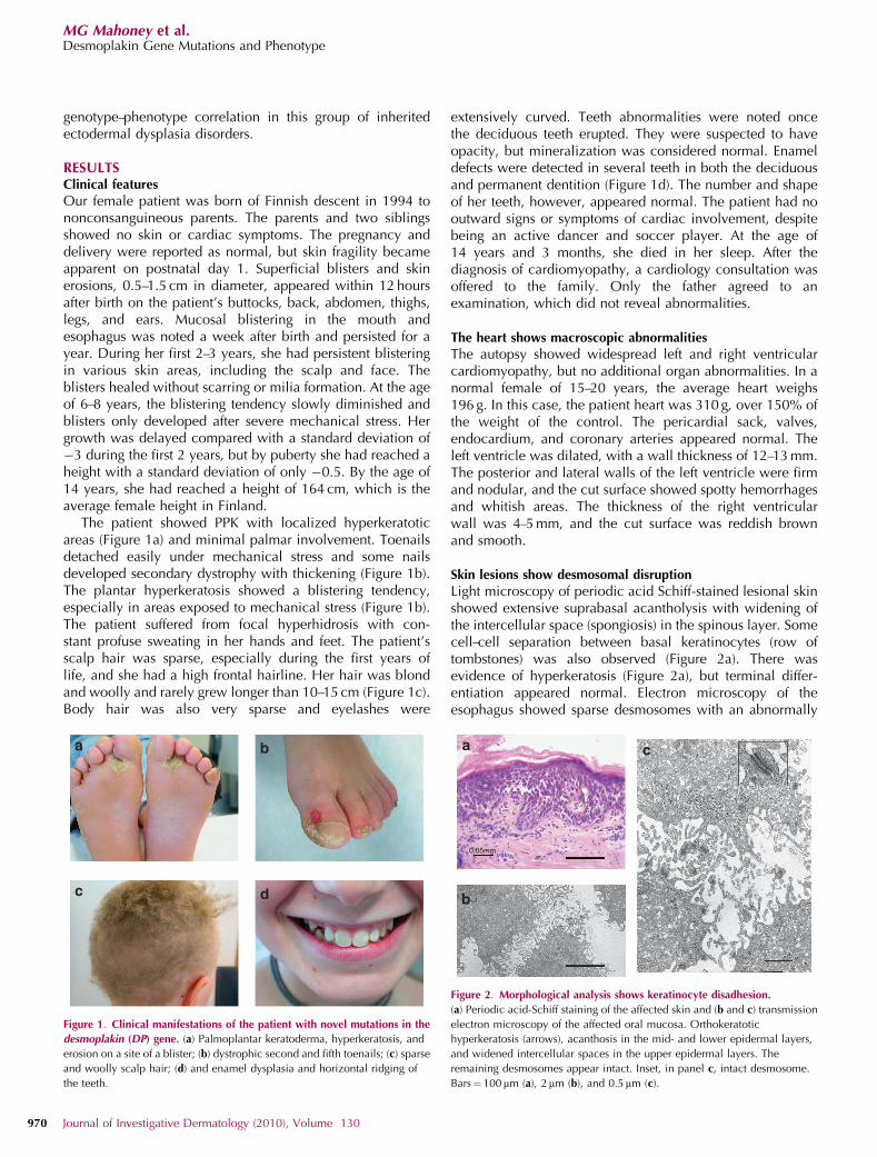
The patient showed PPK with localized hyperkeratoticareas (Figure 1a) and minimal palmar involvement. Toenailsdetached easily under mechanical stress and some nailsdeveloped secondary dystrophy with thickening (Figure 1b).The plantar hyperkeratosis showed a blistering tendency,especially in areas exposed to mechanical stress (Figure 1b).The patient suffered from focal hyperhidrosis with con-stant profuse sweating in her hands and feet. The patient’sscalp hair was sparse, especially during the first years oflife, and she had a high frontal hairline. Her hair was blondand woolly and rarely grew longer than 10–15 cm (Figure 1c).Body hair was also very sparse and eyelashes were
extensively curved. Teeth abnormalities were noted oncethe deciduous teeth erupted. They were suspected to haveopacity, but mineralization was considered normal. Enameldefects were detected in several teeth in both the deciduousand permanent dentition (Figure 1d). The number and shapeof her teeth, however, appeared normal. The patient had nooutward signs or symptoms of cardiac involvement, despitebeing an active dancer and soccer player. At the age of14 years and 3 months, she died in her sleep. After thediagnosis of cardiomyopathy, a cardiology consultation wasoffered to the family. Only the father agreed to anexamination, which did not reveal abnormalities.
The heart shows macroscopic abnormalities
The autopsy showed widespread left and right ventricularcardiomyopathy, but no additional organ abnormalities. In anormal female of 15–20 years, the average heart weighs196 g. In this case, the patient heart was 310 g, over 150% ofthe weight of the control. The pericardial sack, valves,endocardium, and coronary arteries appeared normal. Theleft ventricle was dilated, with a wall thickness of 12–13 mm.The posterior and lateral walls of the left ventricle were firmand nodular, and the cut surface showed spotty hemorrhagesand whitish areas. The thickness of the right ventricularwall was 4–5 mm, and the cut surface was reddish brownand smooth.
Skin lesions show desmosomal disruption
Light microscopy of periodic acid Schiff-stained lesional skinshowed extensive suprabasal acantholysis with widening ofthe intercellular space (spongiosis) in the spinous layer. Somecell–cell separation between basal keratinocytes (row oftombstones) was also observed (Figure 2a). There wasevidence of hyperkeratosis (Figure 2a), but terminal differ-entiation appeared normal. Electron microscopy of theesophagus showed sparse desmosomes with an abnormally
Figure 1. Clinical manifestations of the patient with novel mutations in the
desmoplakin (DP) gene. (a) Palmoplantar keratoderma, hyperkeratosis, and
erosion on a site of a blister; (b) dystrophic second and fifth toenails; (c) sparse
and woolly scalp hair; (d) and enamel dysplasia and horizontal ridging of
the teeth.
0.05mm
Figure 2. Morphological analysis shows keratinocyte disadhesion.
(a) Periodic acid-Schiff staining of the affected skin and (b and c) transmission
electron microscopy of the affected oral mucosa. Orthokeratotic
hyperkeratosis (arrows), acanthosis in the mid- and lower epidermal layers,
and widened intercellular spaces in the upper epidermal layers. The
remaining desmosomes appear intact. Inset, in panel c, intact desmosome.
Bars¼ 100 mm (a), 2mm (b), and 0.5mm (c).
970 Journal of Investigative Dermatology (2010), Volume 130
MG Mahoney et al.Desmoplakin Gene Mutations and Phenotype

thin inner plaque. The diameter of the affected desmosomeswas 110.6±44.7 nm (n¼ 11), essentially considered normalfor desmosomes in the spinous cell layer (Figure 2b and c).However, IFs were rarely observed interacting with thedesmosomal plaques. Furthermore, the plasma membraneappeared undulated and stretched, resulting in the rupture ofthe inter-desmosome plasma membrane, leaving intactdesmosomes despite extensive acantholysis (Figure 2b and c).
Immunohistochemistry of skin and oral mucosa suggestsaberrant DP expression
Paraffin-embedded skin and cryosections of oral mucosawere available for analyses. The expression and localizationof DP and other desmosomal proteins were examined byimmunohistochemistry using a panel of antibodies (Abs)recognizing different epitopes (Figure 3a). A schematicpresentation of the structure of DP and the sites recognizedby the different Abs is shown in Figure 3a. DP mix was acommercially available mixture of three different monoclonalAbs with uncharacterized antigenic sites. Staining with DPmix showed an intense immunosignal at cell–cell borders inthe epidermis of the patient and of healthy control persons(Figure 3b). In contrast, Ab 10F6 detecting the epitopeslocated in the C-terminal subdomain A showed a markedlyreduced immunoreaction in the patient’s epidermis (Figure3b). Abs 2C8 and 2A5, specific for the C-terminal subdomainA and for parts of subdomains B and C, respectively, wereused for studying the cryosections of oral mucosa (Figure 3b).The patient’s sample showed a substantially decreasedintensity compared with normal oral mucosa, and widenedintercellular gaps as a sign of disrupted cell–cell adhesion(Figure 3b). Analysis of the patient’s skin with Abs againstdesmosomal cadherin Dsg1 and the desmosomal plaqueprotein PG showed staining patterns and intensity similar tothose seen in control skin (Figure 3c). A slight decrease in theadherens junction protein b-catenin was noted in the affectedskin, probably because of formalin fixation and paraffinembedding. Oral mucosa, examined in frozen sections,showed a normal staining intensity for b-catenin (Figure 3c).
Heart shows severe myocardial abnormalities
Masson’s trichrome staining of paraffin-embedded hearttissue showed abnormalities in both the right (Figure 4a)and left (Figure 4b) ventricular walls. Fibrosis could be seenin all parts of the myocardium, but in focal areas of bothventricles, the fibrotic tissue had largely replaced myocytes.Fatty replacement of myocytes could be detected focally(Figure 4c). A higher magnification showed shrunkenmyocytes, which had cytoplasmic vacuoles and pycnoticnuclei, suggesting ongoing apoptosis (Figure 4d). Very fewinflammatory cells were present. Immunostaining for con-nexin 43 showed a clear reduction and even disappearanceof protein in both ventricles in areas affected by cardiomyo-pathy (Figure 4f and g). A normal ordered pattern of connexin43 gap junctions was seen in apparently normal cardiomyo-cytes (Figure 4e).
The expression and localization of desmosomal compo-nents in the patient’s heart tissue were examined using Abs to
DP, PG, and Dsg2 (Figure 4h), and the labeling pattern wascompared with that of normal heart (Figure 4h, top panel).The results did not show significant differences in theexpression levels of DP, PG, and Dsg2, whereas the interca-lated disks in the patient’s heart were arranged less regularlycompared with that of the control (Figure 4h, lower panel).
Sequencing of the DP gene reveals two mutations
As the structural and protein data suggested a possiblemutation in the DP gene, all exons of the DP gene weresequenced to identify potential pathogenic mutations. Twomutations were identified in exon 24 of the DP gene(GenBank accession no. m77830) (Figure 5a). The paternalmutation c.6310delA is a deletion of an A at nucleotideposition 6310 in the DP gene, which causes a frameshift andcreates a downstream STOP codon resulting in a prematurelytruncated protein (amino acids 1–2,103þ12 missense)(Figure 5a). The frameshift changed the wild-type aminoacids from TVSVSEAIKKNL to QYLFQKPSRKI, ending atamino acid 2115, followed by a STOP codon. The maternalmutation, c.7964 C to A, results in a substitution of anaspartic acid for an alanine at amino acid 2655 in the DPprotein (p.A2655D). A survey of 100 alleles in unrelatedcontrol individuals showed the presence of alanine exclu-sively at position 2655, indicating that this amino-acidchange is not a common polymorphism. Genetic screeningshowed that both surviving siblings are carriers of thepaternal mutation (Figure 5b).
The structural domains of DP include the N-terminaldesmosome-binding domain (amino acids: 1–1056), thecentral coiled-coil rod oligomerization domain (amino acids:1057–1945), and the C-terminal IF-binding domain (aminoacids: 1946–2871) (Figure 5c). The C-terminal domain isfurther divided into three functional subdomains, A, B, and C,each comprising 4.5 copies of the plakin repeat domain. Thematernal amino-acid substitution mutation is localized to thebeginning of the C subdomain (Figure 5c). The paternalmutation (c.6310delA) is located within the genomic DNAsequence encoding for the A subdomain. This results in atruncated protein product, in which 144 amino acids of the Asubdomain are present and the remainder of the C-terminaltail are missing.
As mentioned above, the predicted size of the twoalternative spliced DP isoforms are of 310 and 238 kDa,with a difference of 70 kDa. However, western blot analysisoften detects a doublet of 250 and 210 kDa with a differenceof only 40 kDa. In this study, we used the DP mix Ab and bywestern blot analysis detected a single 250 kDa band innormal human skin. In addition to the 250-kDa band, wedetected several bands ranging in size from 180 to 205 kDa inthe paternal skin (Figure 5d). We reprobed the samemembrane with the Ab 10F6 raised against DP subdomainA and showed both 250 and 210 kDa bands in normal andpaternal skin (Figure 5d). We did not detect any truncatedproducts from paternal skin using this Ab, possibly because oflow Ab sensitivity. It is not clear why the Ab DP mix onlydetected 250 kDa DPI and not 210 kDa DPII. Nevertheless,the presence of lower bands suggests that the paternal
www.jidonline.org 971
MG Mahoney et al.Desmoplakin Gene Mutations and Phenotype

2A510F6 & 2C8
DP (2A5)DP (2C8) DP (Mix) DP (10F6)
Con
trol
Oral mucosaEpidermis
Pat
ient
Dsg1 (27B2) PG (11E4) β-catenin (6F9) β-catenin (6F9)
Con
trol
Pat
ient
SkinOral mucosa
A B C
Figure 3. Immunohistochemical staining of the epidermis and oral epithelium using a panel of desmoplakin (DP)-specific antibodies (Abs). (a) Schematic diagram
of DP depicts regions of antibody recognition. (b) All antibodies showed cell surface staining throughout the normal adult epidermis (upper panel). In patient’s skin,
the multiclonal epitope antibody (DP mix) shows significant but disrupted staining of DP in the skin, whereas the C-terminal antibody 10F6 does not detect
appreciable levels of DP. Two different antibodies (2C8 and 2A8) detecting parts of the C-terminal domain of DP show clear reduction in patient’s oral mucosa
compared with normal oral epithelium. Typical cell–cell border staining of desmoglein 1 (Dsg1) and plakoglobin (PG) (c) is observed in both control and patient’s
skin. Staining for b-catenin in formalin-fixed, paraffin-embedded patient’s skin is reduced when compared with that of control epidermis (possibly because of
formalin fixation). However, b-catenin expression does not seem to be altered in patient’s oral mucosa when compared with that of the control section. Bar¼ 50mm.
972 Journal of Investigative Dermatology (2010), Volume 130
MG Mahoney et al.Desmoplakin Gene Mutations and Phenotype

Right ventricle Left ventricle
Right ventricleNormal heart
PG (11E4)DP (Mix) Dsg2 (Rb5)
Nor
mal
hea
rtP
atie
nt h
eart
Left ventricle
0.5mm
0.05
mm
0.02mm
0.5mm
Figure 4. Widespread right and left ventricular cardiomyopathy. Masson’s trichrome staining of the (a) right and (b–d) left ventricles showing loss of cardiomyocytes
(pink/red) with replacement by fibrous tissue (blue/gray/green). Degenerating cardiomyocytes embedded in extensive deposits of collagenous connective tissue
and fat tissue (c), with dense ‘‘pycnotic’’ nuclei, suggestive of apoptosis (arrows in d), as compared with the slender and spindle-shaped nuclei in normal cells (arrowheads
in d). Connexin 43 containing gap junctions in normal heart (e) and in the patient’s right ventricle (f). (g) In the patient’s left ventricle, the expression of connexin
43 was clearly reduced. Immunostaining of normal heart (h, top panels) and patient’s myocardial sample (h, lower panels) for desmosomal proteins using antibodies
to desmoplakin (DP) (DP mix), PG (11E4), and desmoglein 2 (Dsg2) (Rb5) shows disorganization of intercalated disks in the patient’s sample, whereas the expression levels
of desmosomal components do not show significant differences. Blue: DAPI-stained nuclei. Bars¼0.5mm (a and b), 50mm (c), 20mm (d), 100mm (e–g), and 25mm (h).
www.jidonline.org 973
MG Mahoney et al.Desmoplakin Gene Mutations and Phenotype

mutation leads to truncated protein products. As such, aproduct would not be detected if mediated by nonsense-mediated RNA decay.
DP mutations predict changes in the DP protein
In an attempt to understand the mechanisms contributing tothe cell adhesion defects observed in the patient skin andheart, we observed more closely the mutated DP C-terminaldomain harboring the mutation A2655D. Sequence analysisshowed that the A2655D mutation site is adjacent to the foldregion, which has been reported to be a binding site for IFproteins (Choi et al., 2002). A structural analysis of this regionshowed no major changes (data not shown), but an analysis
of the physicochemical properties of the molecular surfacesshowed that the A2655D substitution decreased the electro-static potential (EP) of a region encompassing the mutationsite (Figure 5e). In particular, the EP values (expressed inkcal mol�1) for the region encompassing the A2566 residueranged from (þ )227.9 to (�)247.7. In contrast, the corre-sponding EP values for the analogous region encompassingthe D2655 site ranged from (þ )216.9 to (�)269.4. Althoughthese changes are relatively small, clinical features suggestthat this slight alteration in EP may critically impair DPbinding to the IFs contributing to the multiorgan phenotype.At the same time, the lipophilic potential of the mutant site ofthe protein was not altered.
Exon 24 Exon 24
6310delA A2655D
Pat
ient
Pat
erna
lM
ater
nal
Con
trol
c.6310delA
c.6310delAc.6310delA c.6310delAp.A2655D
p.A2655D
Plakin
6310delA
Maternal
DP mix
Ab 10F6
PaternalA2655D
Rod PRD
B CA
Nor
mal
ski
n
Pat
erna
l ski
n
Nor
mal
ski
n
Pat
erna
l ski
n
223 –223 –
kDaAla-2655
Asp-2655
kDa
230
109 –
83 –
205196180
(+)
(–)
Figure 5. Novel compound heterozygous mutations in desmoplakin (DP) gene. Nucleotide sequencing of PCR-amplified genomic DNA of the patient, father,
mother, and control corresponding to exon 24 of the DP gene (a). Arrows indicate the positions of identified mutations. Pedigree of the family with novel
compound heterozygous mutations (b). Father and all three offspring harbor a c.6310delA mutation, whereas only the deceased proband inherited the maternal
mutation, p.A2655D (b). A schematic structure of DP showing three functional domains: plakophilin/plakoglobin-binding domain (plakin), central rod
domain, and intermediate filament-binding plakin repeat domain (PRD). Arrows indicate mutation sites. Predicted paternal and maternal DP proteins based
on sequence analysis; the sites of mutations indicated in red (c). Western analysis of normal human skin and paternal skin using two different DP antibodies
shows classical DP doublets and truncation of DP (d). Molecular analysis of the electrostatic potential (EP) of the surface of the wild-type (WT) and mutant
fragments (e). The EP range for WT is (þ )227.9/(�)247.7, whereas that of the mutant is (þ )216.9/(�)269.4. Color-coded panel indicates differences in the
EP and lipophilic potential (LP): red, highly positive EP; blue, highly negative EP; brown, highly hydrophobic; and blue, highly hydrophilic.
974 Journal of Investigative Dermatology (2010), Volume 130
MG Mahoney et al.Desmoplakin Gene Mutations and Phenotype

DISCUSSIONIn this study, we report a patient who inherited two mutationsin the C-terminal domain of the DP gene. Together, thesepreviously undescribed mutations contributed to a fatalsyndrome, including all characteristics thus far describedfor DP mutations: woolly hair, blistering skin disease,PPK, and left and right ventricular cardiomyopathy, whichled to the demise of the patient at the age of 14 years.In addition, she had dental anomalies. At the proteinlevel, the expression of the C-terminal domain of DP wasfound decreased in the epidermis and oral mucosa, which isin accordance with the putative consequences of themutations at the protein level. In the heart, the levels of PGand Dsg2 were similar to those in normal myocardium.Reduced levels of PG at intercalated disks have recently beenreported to be specific for ARVC/D (Asimaki et al., 2009),suggesting that our PG Ab recognized epitopes that were notdetected by the previous study or that the heart disease of ourpatient cannot be classified as ARVC/D. Interestingly, theCarvajal syndrome is characterized by PPK, woolly hair, andleft ventricular cardiomyopathy, and is caused by mutationsresulting in the ablation of subdomain C of DP (Carvajal-Huerta, 1998). Furthermore, similar to our patient, skinphenotypes are typically restricted to regions of mechanicalstress. Furthermore, homozygous missense mutations insubdomain B are reported in cases of Carvajal-like syndrome(Alcalai et al., 2003) in which PPK, woolly hair, blisters, andARVC/D are reported. Our reported paternal mutation resultsin the loss of much of the C-terminus, whereas the maternalmutation is postulated to reduce the C-terminal bindingfunction. Taken together with the biventricular involvementobserved in this patient, our data suggest similarities to thesesyndromes, and may account for the normal levels of PGobserved in our patient.
The consequences of the combination of the two muta-tions may affect several known functions of desmosomes,including cell adhesion, differentiation, morphogenesis, andadhesion-independent signaling. Defects in cell–cell adhe-sion have been shown to perturb the integrity of theepidermis, as shown by acantholysis, and detachment ofnails under mechanical stress. Correspondingly, myocardialinjury could be explained by a mechanistic model in whichthe DP–desmin interaction is perturbed, resulting in de-creased cardiomyocyte adhesion. A recent model suggeststhat myocardial fibrosis results from alterations in thedesmosome-mediated Wnt/b-catenin signaling pathway andapoptosis (Garcia-Gras et al., 2006; Martin et al., 2009; forreview, see Awad et al., 2008). The results of this study alsosuggest that DP has a role in keratinocyte differentiation, asshown by the development of palmoplantar hyperkeratosis,particularly in response to mechanical stress. Similarly, DP issuggested to participate in the morphogenesis of hair follicles,as evidenced by the presence of woolly hair and a distinctgrowth pattern on scalp hair. Enamel opacity was suspectedin the deciduous teeth of the patient, and later, severalpermanent teeth showed yellowish spots. Enamel is producedby ameloblasts, specialized epithelial cells with desmo-somes, having roles in the production, resorption, and
degradation of the enamel matrix (Matthiessen and Rømert,1980; Sasaki et al., 1984). We speculate that defectivedesmosomal adhesion may have either affected amelogenesisor predisposed the patient’s teeth to damage from other toxicfactors such as antibiotics.
Our patient had inherited two different mutations of the DPgene. The maternal mutation translates into a protein with anamino-acid substitution (A2655D) in subdomain C of the C-terminal domain of the DP. The paternal mutation, also in theregion encoding the C-terminal domain, is a single-nucleotidedeletion that results in a premature stop codon in subdomainA. Thus, the paternal allele reported here translates into atruncated DP protein product lacking much of the C-terminaltail. As these mutations occur downstream of the predictedalternative splice site that generates DPII mRNA, both DPI andDPII isoforms are predicted to possess the correspondingchanges at the protein level. Before this study, only four pairsof mutations affecting the C-terminal domain of DP have beenreported in literature (Lai-Cheong et al., 2007). The twohomozygous recessive pairs resulted in phenotypes, whichincluded cardiomyopathies (Norgett et al., 2000; Alcalai et al.,2003) together with woolly hair and PPK. The remaining twocompound heterozygous mutation pairs each include amutation upstream of the ABC domain; cardiac involvementwas not reported in either case (Whittock et al., 2002;Jonkman et al., 2005). In the paper by Whittock et al.(2002), the authors describe two patients with severemucocutaneous findings without cardiac symptoms by theages of 4 and 17 years. The younger patient had bothmutations upstream of the C-terminal domain, whereas theolder patient had a missense mutation in exon 24, similar tothe present case. It is not known whether she developedcardiac symptoms later. However, this comparison empha-sizes the critical role of the conserved alanine 2655, whichwas mutated in the maternal allele in our subject.
Although the phenotype–genotype correlations of DPmutations are not well understood, the present and previousstudies suggest that the truncation of the C-terminal domainpredisposes to cardiomyopathy. The C-terminal domain ofDP has been shown to interact with the IF cytoskeleton. Onthe basis of these observations, the DP mutations identified inthis patient are predicted to disrupt the interactions betweenDP and the IF cytoskeleton both in the skin and heart. As bothmutations are located in exon 24, 30 of the last exon–exonborder, the mRNAs would not likely be subjected tononsense-mediated mRNA decay (Nagy and Maquat,1998). Indeed, we detected truncated DP protein productsin the paternal skin (Figure 5d).
The maternal mutation is an amino-acid substitution(A2655D) in subdomain C of the DP and in the presence ofthe paternal allele is unable to confer a normal desmosomefunction. Alanine 2655 is a highly conserved amino acid(Table 2) in a groove lined with basic residues in subdomainC. This conserved groove is speculated to be an importantrecognition site for binding to IFs, including cardiac vimentinand desmin (Choi et al., 2002). Although this mutation doesnot significantly alter the three-dimensional structure of DP,structural modeling indicates changes in the EP of the
www.jidonline.org 975
MG Mahoney et al.Desmoplakin Gene Mutations and Phenotype

mutated region. The change may seem subtle but the clinicalphenotypes suggest that it alters IF-binding functions thatdepend on electrostatically driven intermolecular interactions(Choi et al., 2002). These results show the importance of thecorrect amino-acid sequence of the plakin repeat domain forthe DP functions and suggest potentially devastating effects ofmutations that occur in this specific region.
We believe that identification of more DP mutations andadditional structural/functional analyses are needed beforewe can accurately predict the clinical manifestations of agiven mutation. Meanwhile, if a child with skin symptoms ofdesmosomal genodermatosis is examined, the possibility ofcardiac involvement should be considered, even in theabsence of overt heart-related symptoms.
MATERIALS AND METHODSTissue material and histology
Medical records and archival tissue material were used with
permission from the Turku University Central Hospital and the
Department of Forensic Medicine (University of Turku, Finland),
Department of Dermatology (University Medical Center Freiburg,
Germany), and Freiburg Institute for Advanced Studies, School of
Life Sciences (Lifenet, Freiburg, Germany). Family members
provided informed consent for the collection and analysis of DNA
material, and approval for publication of data. Material was
collected according to the Declaration of Helsinki Principles.
Control tissue slides were obtained from BioChain Institute
(Hayward, CA). Unless otherwise indicated, all chemicals were
from Sigma (St Louis, MO) or Fisher Scientific (Waltham, MA). For
histology, tissues were fixed overnight at room temperature in 10%
formalin solution. Tissues were processed for paraffin embedding,
sectioned (4mm), mounted on glass slides, and stained with
hematoxylin and eosin periodic acid Schiff or Masson’s trichrome.
For transmission electron microscopy, skin and esophagus
biopsies were fixed in 3% glutaraldehyde in 0.1 M Millonig
phosphate buffer, pH 7.4, at 4 1C for 4 hours, and post-fixed with
1% OsO4 0.1 M in veronal acetate buffer. Samples were dehydrated
in a graded ethanol series and embedded in Epon 812, Shell
Chemical Corp., Cleveland, Ohio. Ultrathin sections were cut on
coated copper grids and stained with uranyl acetate and lead citrate.
The sections were examined using a Jeol, Tokyo, Japan. 1200SX
electron microscope and photographed.
AbsAbs used for immunostaining of the skin, esophagus, and heart
sections were the following: DP mix, a ‘‘cocktail’’ of mAbs DP-2.15,
DP-2.17, and DP-2.20 purchased from American Research Products
(Belmont, MA); Abs 2C8 and 10F6 were generated using recombi-
nant 6x His-tagged DP amino acids 1,960–2,151, and 2A5 was
raised against amino acids 2,343–2,822 (Johnson et al., 1993).
Abs 6F9 (b-catenin), 27B2 (Dsg1), and 11E4 (PG) were from our
laboratory (Wahl, 2002). Ab for connexin 43 was purchased from
Cell Signaling Technology (cat. no. 3512, Beverly, MA). Ab dilutions
for immunofluorescence using OCT frozen tissues were 1:100 for
6F9, 1:100 for 27B2, 1:800 for 11E4, 1:800 for 2C8, 1:800 for 2A5,
and 1:800 for 10F6. Ab dilutions for immunostaining on paraffin
tissues were 1:20 for 6F9, 1:4 for 27B2, 1:20 for 11E4, 1:2 for 10F6,
1:2 for DP mix, and 1:50 for connexin 43. Alexa Fluor-conjugated
secondary Abs (488 and 594 nm) were purchased from Invitrogen (at
dilution of 1:400; Eugene, OR).
Immunohistochemistry and western analysis
For immunostaining, we prepared OCT-fixed tissue sections (5 mm)
or formalin-fixed and paraffin-embedded tissue sections (5 mm) as
previously described (Mahoney et al., 2006). Briefly, OCT sections
were fixed in methanol (�20 1C) for 15 minutes, permeabilized
with 1% Triton-X-100 in PBS for 5 minutes, and incubated in
blocking buffer (5% normal goat serum, 1% BSA, and 0.02% TX-100
in PBS) for 1 hour at room temperature. Tissues were incubated
overnight with primary Abs at 4 1C and with secondary Abs for
1 hour at room temperature. For formalin-fixed, paraffin-embedded
sections, tissues were deparaffinized in 100% xylene (5 minutes;
twice), 100% ethanol (5 minutes; twice), 95% ethanol (5 minutes;
twice), 75% ethanol (2 minutes), 50% ethanol (2 minutes), and
H2O (2 minutes). Antigen retrieval was performed by the micro-
wave method with sodium citrate buffer (pH 6.0), followed
by digestion with trypsin (Sigma) for 10 minutes at 42 1C. Tissues
were incubated in primary and secondary Abs in blocking buffer.
Nuclei were stained with DAPI (100 ng ml�1) before being mounted
for viewing. All images were acquired using a Hamamatsu
monochromatic digital camera (Phase 3 Imaging Systems, Glen
Mills, PA; C4742-95). Direct immunofluorescence with FITC-
conjugated Abs to human IgG, IgA, IgM, and complement C3
and C1q was performed twice as part of the diagnostic procedure.
The results were first nonspecific and the second time showed
negative results.
For western analysis, DP was extracted in 9 M urea in lysis buffer
from normal skin and a punch biopsy from the proband’s father.
Proteins were separated in 5% SDS–PAGE gel and immunoblotted
with DP mix Ab at a dilution of 1:100.
DNA extraction
DNA was extracted from 100 normal human controls and living
relatives of the patient through buccal swabs (Cell Projects, Kent,
UK). The patient’s genomic DNA was extracted from tissue
previously fixed and mounted in a paraffin block. A 5� 5 mm2 slice
of tissue was extracted and incubated with xylene for 10 minutes.
The xylene was removed by three subsequent incubations in
100, 80, and 70% ethanol and the sample was air-dried. The
pellet was suspended in TE buffer, pH 8.0 (Tris-HCl 50 mM,
EDTA 1 mM) and digested with Proteinase K (1 mg ml�1) at 60 1C.
Table 2. Conservation of the alanine residue at aminoacid 2655 between species
Species Sequence
2655
k
Human IVDSITGQRLLEAQACTGGII
Mouse IVDSITGQRLLEAQACTGGII
Rat IVDSITGQRLLEAQACTGGII
Chimpanzee IVDSITGQRLLEAQACTGGII
Horse IVDSITGQRLLEAQACTGGII
Chicken IVDSITGQRLLEAQACTGGII
976 Journal of Investigative Dermatology (2010), Volume 130
MG Mahoney et al.Desmoplakin Gene Mutations and Phenotype

Genomic DNA was further purified by standard phenol chloroform
extraction.
PCR and sequencing
Conditions and primers for generating PCR products spanning all
exons of the coding regions and flanking intronic sequences of DP
have been described previously (Whittock et al., 1999). Additional
primers were designed to screen for mutation 6310delA: forward—
50-AATCAGCCCAGAATCCACAG-30, and reverse—50-TATCTCGGG
GATCATTCAGG-30. PCR was carried out using Qiagen Taq poly-
merase and Q buffer (Qiagen, Valencia, CA), according to the manu-
facturer’s instructions. The patient sample and two controls (normal
human DNA and distilled water) were PCR amplified simulta-
neously. PCRs contained 200 ng DNA as template and 100 ng of
each primer in a final volume of 50 ml. Cycling conditions for all
primer pairs were 94 1C for 5 minutes, followed by 41 cycles of
denaturation at 94 1C for 1 minute; annealing temperature for a par-
ticular primer pair (range: 55–60 1C) for 1 minute; extension at 72 1C
for 1 minute; and finally at 72 1C for 5 minutes. The PCR products of
the patient and control samples were sequenced forward and reverse
two times. In all, 100 control alleles were studied to rule out the
possibility that the mutation might be a frequent polymorphism.
DNA sequencing was performed using an ABI Prism 377 or ABI
3100 automated sequencer (Perkin-Elmer-Cetus, Foster City, CA).
Structural modeling of the A2655D mutation site
A computer model of the region spanning the A2655D mutation site
was generated by the Sybyl7.3 program (Tripos, St Louis, MO) on the
basis of coordinates from the Protein Data Base (1lm5). Molecular
surfaces were generated using the MOLCAD module of the SYBYL
program (Tripos Inc, St Louis, MO) (Connolly, 1983). The same
module was used to calculate the EP (physical unit: kcal mol�1) of
the analyzed molecular surfaces (Warwicker and Watson, 1982).
CONFLICT OF INTERESTThe authors state no conflict of interest.
ACKNOWLEDGMENTSWe thank the family members of the patient for their cooperation andparticipation in our study. This research was supported by grants fromthe National Institutes of Health to My G. Mahoney (AR055251), James K.Wahl III (DE016905), Andrzej Fertala (AR049537-06), and Jouni Uitto(P01AR38923); an Epidermolysis Bullosa Network grant from the GermanMinistry for Education and Research to Leena Bruckner-Tuderman; a grantfrom the Finnish Academy to Juha Peltonen; and grants from the Finnish MedicalFoundation and the Southwest Finland Hospital District to Sirkku Peltonen.
REFERENCES
Alcalai R, Metzger S, Rosenheck S et al. (2003) A recessive mutation indesmoplakin causes arrhythmogenic right ventricular dysplasia, skindisorder, and woolly hair. J Am Coll Cardiol 42:319–27
Armstrong DK, McKenna KE, Purkis P et al. (1999) Haploinsufficiency ofdesmoplakin causes a striate subtype of palmoplantar keratoderma. HumMol Genet 8:143–8
Asimaki A, Tandri H, Huang H et al. (2009) A new diagnostic test forarrhythmogenic right ventricular cardiomyopathy. N Engl J Med 360:1075–84
Awad MM, Calkins H, Judge DP (2008) Mechanisms of disease: moleculargenetics of arrhythmogenic right ventricular dysplasia/cardiomyopathy.Nat Clin Pract Cardiovasc Med 5:258–67
Bauce B, Basso C, Rampazzo A et al. (2005) Clinical profile of four familieswith arrhythmogenic right ventricular cardiomyopathy caused bydominant desmoplakin mutations. Eur Heart J 26:1666–75
Carvajal-Huerta L (1998) Epidermolytic palmoplantar keratoderma withwoolly hair and dilated cardiomyopathy. J Am Acad Dermatol39:418–21
Choi HJ, Park-Snyder S, Pascoe LT et al. (2002) Structures of two intermediatefilament-binding fragments of desmoplakin reveal a unique repeat motifstructure. Nat Struct Biol 9:612–20
Connolly ML (1983) Solvent-accessible surfaces of proteins and nucleic acids.Science 221:709–13
Cowin P, Burke B (1996) Cytoskeleton–membrane interactions. Curr OpinCell Biol 8:56–65
Fontao L, Favre B, Riou S et al. (2003) Interaction of the bullous pemphigoidantigen 1 (BP230) and desmoplakin with intermediate filaments ismediated by distinct sequences within their COOH terminus. Mol BiolCell 14:1978–92
Garcia-Gras E, Lombardi R, Giocondo MJ et al. (2006) Suppression ofcanonical Wnt/beta-catenin signaling by nuclear plakoglobin recapitu-lates phenotype of arrhythmogenic right ventricular cardiomyopathy.J Clin Invest 116:2012–21
Getsios S, Huen AC, Green KJ (2004) Working out the strength and flexibilityof desmosomes. Nat Rev Mol Cell Biol 5:271–81
Green KJ, Goldman RD, Chisholm RL (1988) Isolation of cDNAs encodingdesmosomal plaque proteins: evidence that bovine desmoplakins I and IIare derived from two mRNAs and a single gene. Proc Natl Acad Sci USA85:2613–7
Green KJ, Parry DA, Steinert PM et al. (1990) Structure of the humandesmoplakins. Implications for function in the desmosomal plaque. J BiolChem 265:2603–12
Jefferson JJ, Leung CL, Liem RK (2004) Plakins: goliaths that link cell junctionsand the cytoskeleton. Nat Rev Mol Cell Biol 5:542–53
Johnson KR, Lewis JE, Li D et al. (1993) P- and E-cadherin are in separatecomplexes in cells expressing both cadherins. Exp Cell Res 207:252–60
Jonkman MF, Pasmooij AM, Pasmans SG et al. (2005) Loss of desmoplakin tailcauses lethal acantholytic epidermolysis bullosa. Am J Hum Genet77:653–60
Kouklis PD, Hutton E, Fuchs E (1994) Making a connection: direct bindingbetween keratin intermediate filaments and desmosomal proteins. J CellBiol 127:1049–60
Kowalczyk AP, Bornslaeger EA, Borgwardt JE et al. (1997) The amino-terminaldomain of desmoplakin binds to plakoglobin and clusters desmosomalcadherin–plakoglobin complexes. J Cell Biol 139:773–84
Lai-Cheong JE, Arita K, McGrath JA (2007) Genetic diseases of junctions.J Invest Dermatol 127:2713–25
Lapouge K, Fontao L, Champliaud MF et al. (2006) New insights into themolecular basis of desmoplakin- and desmin-related cardiomyopathies.J Cell Sci 119:4974–85
Mahoney MG, Hu Y, Brennan D et al. (2006) Delineation of diversifieddesmoglein distribution in stratified squamous epithelia: implication indiseases. Exp Dermatol 15:101–9
Martin ED, Moriarty MA, Byrnes L et al. (2009) Plakoglobin has bothstructural and signalling roles in zebrafish development. Dev Biol327:83–96
Matthiessen ME, Rømert P (1980) Ultrastructure of the human enamel organ.I. External enamel epithelium, stellate reticulum, and stratum inter-medium. Cell Tissue Res 205:361–70
Meng JJ, Bornslaeger EA, Green KJ et al. (1997) Two-hybrid analysisreveals fundamental differences in direct interactions between desmo-plakin and cell type-specific intermediate filaments. J Biol Chem272:21495–503
Nagy E, Maquat LE (1998) A rule for termination-codon position withingintrol-containing genes: when nonsense affects RNA abundance. TrendsBiochem Sci 23:198–9
Norgett EE, Hatsell SJ, Carvajal-Huerta L et al. (2000) Recessive mutation indesmoplakin disrupts desmoplakin-intermediate filament interactions
www.jidonline.org 977
MG Mahoney et al.Desmoplakin Gene Mutations and Phenotype

and causes dilated cardiomyopathy, woolly hair and keratoderma. HumMol Genet 9:2761–6
Norman M, Simpson M, Mogensen J et al. (2005) Novel mutation indesmoplakin causes arrhythmogenic left ventricular cardiomyopathy.Circulation 112:636–42
Oxford EM, Musa H, Maass K et al. (2007) Connexin43 remodeling caused byinhibition of plakophilin-2 expression in cardiac cells. Circ Res101:703–11
Rampazzo A, Nava A, Malacrida S et al. (2002) Mutation in human desmoplakinbinding domain to plakoglobin causes a dominant form of arrhythmogenicright ventricular cardiomyopathy. Am J Hum Genet 71:1200–6
Saffitz JE, Hames KY, Kanno S (2007) Remodeling of gap junctions in ischemicand nonischemic forms of heart disease. J Membr Biol 218:65–71
Sasaki T, Segawa K, Takiguchi R et al. (1984) Intercellular junctions in thecells of the human enamel organ as revealed by freeze-fracture. ArchOral Biol 29:275–86
Smith EA, Fuchs E (1998) Defining the interactions between intermediatefilaments and desmosomes. J Cell Biol 141:1229–41
Stappenbeck TS, Bornslaeger EA, Corcoran CM et al. (1993) Functionalanalysis of desmoplakin domains: specification of the interaction withkeratin versus vimentin intermediate filament networks. J Cell Biol123:691–705
Stappenbeck TS, Green KJ (1992) The desmoplakin carboxyl terminuscoaligns with and specifically disrupts intermediate filament networkswhen expressed in cultured cells. J Cell Biol 116:1197–209
Tanaka A, Lai-Cheong JE, Cafe ME et al. (2009) Novel truncating mutationsin PKP1 and DSP cause similar skin phenotypes in two Brazilian familes.Br J Dermatol 160:692–7
Uzumcu A, Norgett EE, Dindar A et al. (2006) Loss of desmoplakin isoform Icauses early onset cardiomyopathy and heart failure in a Naxos-likesyndrome. J Med Genet 43:e5
Virata ML, Wagner RM, Parry DA et al. (1992) Molecular structure of thehuman desmoplakin I and II amino terminus. Proc Natl Acad Sci USA89:544–8
Wahl JK III (2002) Generation of monoclonal antibodies specific fordesmoglein family members. Hybrid Hybridomics 21:37–44
Warwicker J, Watson HC (1982) Calculation of the electric potential in theactive site cleft due to alpha-helix dipoles. J Mol Biol 157:671–9
Whittock NV, Ashton GH, Dopping-Hepenstal PJ et al. (1999) Striatepalmoplantar keratoderma resulting from desmoplakin haploinsuffi-ciency. J Invest Dermatol 113:940–6
Whittock NV, Wan H, Morley SM et al. (2002) Compound heterozygosity fornon-sense and mis-sense mutations in desmoplakin underlies skinfragility/woolly hair syndrome. J Invest Dermatol 118:232–8
Yang Z, Bowles NE, Scherer SE et al. (2006) Desmosomal dysfunction due tomutations in desmoplakin causes arrhythmogenic right ventriculardysplasia/cardiomyopathy. Circ Res 99:646–55
Yu CC, Yu CH, Hsueh CH et al. (2008) Arrhythmogenic right ventriculardysplasia: clinical characteristics and identification of novel desmosomegene mutations. J Formos Med Assoc 107:548–58
978 Journal of Investigative Dermatology (2010), Volume 130
MG Mahoney et al.Desmoplakin Gene Mutations and Phenotype
Copyright © 2022 FDOKUMEN