TEM characterization of a silorane composite bonded to enamel/dentin
9
dental materials 26 ( 2 0 1 0 ) 524–532 available at www.sciencedirect.com journal homepage: www.intl.elsevierhealth.com/journals/dema TEM characterization of a silorane composite bonded to enamel/dentin Atsushi Mine a , Jan De Munck a , Annelies Van Ende a , Marcio Vivan Cardoso a , Takuo Kuboki b , Yasuhiro Yoshida c , Bart Van Meerbeek a,∗ a Leuven BIOMAT Research Cluster, Department of Conservative Dentistry, School of Dentistry, Oral Pathology and Maxillo-Facial Surgery, Catholic University of Leuven, Kapucijnenvoer 7, B-3000 Leuven, Belgium b Department of Oral and Maxillofacial Rehabilitation, Okayama University Graduate School of Medicine, Dentistry and Pharmaceutical Science, Okayama, Japan c Department of Biomaterials, Okayama University Graduate School of Medicine, Dentistry and Pharmaceutical Science, Okayama, Japan article info Article history: Received 25 June 2009 Accepted 26 January 2010 Keywords: Adhesion Low-shrinking composite Silorane Self-etch adhesive Nano-interaction abstract Objectives. The low-shrinking composite composed of combined siloxane–oxirane technol- ogy (Filtek Silorane, 3M ESPE, Seefeld, Germany) required the development of a specific adhesive (Silorane System Adhesive, 3M ESPE), in particular because of the high hydropho- bicity of the silorane composite. The purpose of this study was to characterize the interfacial ultra-structure at enamel and dentin using transmission electron microscopy (TEM). Methods. Non-demineralized/demineralized 70–90 nm sections were prepared following common TEM specimen processing procedures. Results. TEM revealed a typical twofold build-up of the adhesive resin, resulting in a total adhesive layer thickness of 10–20 m. At bur-cut enamel, a tight interface without distinct dissolution of hydroxyapatite was observed. At bur-cut dentin, a relatively thin hybrid layer of maximum a few hundreds of nanometer was formed without clear surface deminer- alization. No clear resin tags were formed. At fractured dentin, the interaction appeared very superficial (100–200 nm). Distinct resin tags were formed due to the absence of smear plugs. Silver-nitrate infiltration showed a varying pattern of both spot- and cluster-like appearance of nano-leakage. Traces of Ag were typically detected along some part of the enamel–adhesive interface and/or between the two adhesive resin layers. Substantially more Ag-infiltration was observed along the dentin–adhesive interface of bur-cut dentin, as compared to that of fractured dentin. Conclusions. The nano-interaction of Silorane System Adhesive should be attributed to its relatively high pH of 2.7. The obtained tight interface at both enamel and dentin indicates that the two-step self-etch adhesive effectively bridged the hydrophilic tooth substrate with the hydrophobic silorane composite. © 2010 Academy of Dental Materials. Published by Elsevier Ltd. All rights reserved. ∗ Corresponding author. Tel.: +32 16 33 75 87; fax: +32 16 33 27 52. E-mail address: [email protected] (B. Van Meerbeek). 0109-5641/$ – see front matter © 2010 Academy of Dental Materials.Published by Elsevier Ltd. All rights reserved. doi:10.1016/j.dental.2010.01.010
-
Upload
independent -
Category
Documents
-
view
8 -
download
0
Transcript of TEM characterization of a silorane composite bonded to enamel/dentin
d e n t a l m a t e r i a l s 2 6 ( 2 0 1 0 ) 524–532
avai lab le at www.sc iencedi rec t .com
journa l homepage: www. int l .e lsev ierhea l th .com/ journa ls /dema
TEM characterization of a silorane composite bondedto enamel/dentin
Atsushi Minea, Jan De Muncka, Annelies Van Endea, Marcio Vivan Cardosoa, TakuoKubokib, Yasuhiro Yoshidac, Bart Van Meerbeeka,∗
a Leuven BIOMAT Research Cluster, Department of Conservative Dentistry, School of Dentistry, Oral Pathology andMaxillo-Facial Surgery, Catholic University of Leuven, Kapucijnenvoer 7, B-3000 Leuven, Belgiumb Department of Oral and Maxillofacial Rehabilitation, Okayama University Graduate School of Medicine,Dentistry and Pharmaceutical Science, Okayama, Japanc Department of Biomaterials, Okayama University Graduate School of Medicine, Dentistry and Pharmaceutical Science,Okayama, Japan
a r t i c l e i n f o
Article history:
Received 25 June 2009
Accepted 26 January 2010
Keywords:
Adhesion
Low-shrinking composite
Silorane
Self-etch adhesive
Nano-interaction
a b s t r a c t
Objectives. The low-shrinking composite composed of combined siloxane–oxirane technol-
ogy (Filtek Silorane, 3M ESPE, Seefeld, Germany) required the development of a specific
adhesive (Silorane System Adhesive, 3M ESPE), in particular because of the high hydropho-
bicity of the silorane composite. The purpose of this study was to characterize the interfacial
ultra-structure at enamel and dentin using transmission electron microscopy (TEM).
Methods. Non-demineralized/demineralized 70–90 nm sections were prepared following
common TEM specimen processing procedures.
Results. TEM revealed a typical twofold build-up of the adhesive resin, resulting in a total
adhesive layer thickness of 10–20 �m. At bur-cut enamel, a tight interface without distinct
dissolution of hydroxyapatite was observed. At bur-cut dentin, a relatively thin hybrid layer
of maximum a few hundreds of nanometer was formed without clear surface deminer-
alization. No clear resin tags were formed. At fractured dentin, the interaction appeared
very superficial (100–200 nm). Distinct resin tags were formed due to the absence of smear
plugs. Silver-nitrate infiltration showed a varying pattern of both spot- and cluster-like
appearance of nano-leakage. Traces of Ag were typically detected along some part of the
enamel–adhesive interface and/or between the two adhesive resin layers. Substantially
more Ag-infiltration was observed along the dentin–adhesive interface of bur-cut dentin,
as compared to that of fractured dentin.
Conclusions. The nano-interaction of Silorane System Adhesive should be attributed to its
relatively high pH of 2.7. The obtained tight interface at both enamel and dentin indicates
that the two-step self-etch adhesive effectively bridged the hydrophilic tooth substrate with
the hydrophobic silorane composite.
© 2010 Academy of Dental Materials.
Published by Elsevier Ltd. All rights reserved.
∗ Corresponding author. Tel.: +32 16 33 75 87; fax: +32 16 33 27 52.E-mail address: [email protected] (B. Van Meerbeek).
0109-5641/$ – see front matter © 2010 Academy of Dental Materials.Published by Elsevier Ltd. All rights reserved.doi:10.1016/j.dental.2010.01.010
2 6
1
RoGtcpsaacarr
ditasSat
d e n t a l m a t e r i a l s
. Introduction
ecently, a new class of low-shrinking composites basedn silorane technology (Filtek Silorane, 3M ESPE, Seefeld,ermany) was introduced. The silorane resin replaces
he conventionally used methacrylate resin matrix withinonventional dental composites, thereby providing lowerolymerization shrinkage [1–3] as well as better hydrolytictability [4,5]. Since long, reduced polymerization shrink-ge is a highly desired property of composites in order tovoid primary clinically problems that are typically asso-iated with polymerization shrinkage stress, such as therere adhesive-tooth de-bonding, post-operative sensitivity,estoration marginal discoloration and defects, caries recur-ence, enamel cracks, etc. [6].
As the resin matrix of the silorane composite significantlyiffers from that of conventional methacrylate-based compos-
tes, a new adhesive needed to be designed and developedo enable bonding of the silorane composite to tooth enamelnd dentin. Filtek Silorane therefore comes with a two-step
elf-etch adhesive, called Silorane System Adhesive (3M ESPE,SA). It still possesses features of conventional methacrylatedhesives, especially with regard to its bonding mechanism toooth tissue, while adaptation was needed, especially to makeTable 1 – Materials used.
Materials Compo
Silorane System Adhesive – Self-Etch PrimerLot. 292319pH = 2.73M ESPE, Seefeld, Germany
15–25% 2-hydroxyeth15–25% bisphenol a ddimethacrylate (BIS-10–15% ethanol; 5–15acid–methacryloxy–htreated silica; 5–10%dimethacrylate; <5%itaconic acid; <5% (dimethacrylate; <3% dlphosphine oxide
Silorane System Adhesive – BondLot. 2922743M ESPE
70–80% substituted dsilane treated silica;dimethacrylate (TEGacid–methacryloxy–hdl-camphorquinone;dimethacrylate
FiltekTM SiloraneLot. 203905, Shade A33M ESPE
5–15% 3,4-epoxycyclopolymethylsiloxane;bis-3,4-epoxycyclohephenylmethylsilane;quartz;10–20% yttriucamphorquinone
( 2 0 1 0 ) 524–532 525
it compatible with the highly hydrophobic silorane matrix.The adhesive somewhat differs from a typical two-step self-etch adhesive, since it involves the application of two resinsolutions, of which the first one (Silorane System Adhesive– Self-etch Primer or SSA-Primer) is rather hydrophilic tobond to tooth tissue and the second solution (Silorane Sys-tem Adhesive – Bond or SSA-Bond) is on the contrary quitehydrophobic in order to adequately bridge the hydrophilictooth substrate with the hydrophobic silorane composite. Forthis reason, each resin solution needs to be light-cured sepa-rately.
The purpose of this study was to characterize ultra-morphologically the interface complex of the low-shrinkingsilorane composite Filtek Silorane (3M ESPE) bonded to enameland dentin using the two-step self-etch adhesive SiloraneSystem Adhesive. For this, transmission electron microscopy(TEM) is the method of choice because of its high structuralresolution and relatively low artifact incidence.
2. Materials and methods
Six non-carious human third molars were stored in 0.5% chlo-ramine solution at 4 ◦C and used within one month afterextraction. The teeth were randomly divided into 3 groups
sition (wt%) Application
yl methacrylate (HEMA);iglycidyl ether
GMA); 10–15% water;% phosphoricexylesters; 8–12% silane
1,6-hexanediolcopolymer of acrylic andmethylamino) ethyl-camphorquinone; <3%
(1) Shake the bottle briefly beforedosing so that the primer becomesless viscous.(2) Place one drop of primer into thedosing well, then close the dosingwell to protect the primer from lightand prevent the evaporation of thesolvent.(3) Apply the primer to the entiresurface of the cavity and massageover the entire area for 15 s.(4) Use a gentle stream of air untilthe primer is spread to an even filmand does not move any longer.(5) Cure the primer for 10 s.
imethacrylate; 5–10%5–10% triethylene glycolDMA); <5% phosphoricexylesters; <3%<3% 1,6-hexanediol
(1) Shake bottle briefly beforedosing so that the bond becomesless viscous.(2) Place one drop of bond in thedosing well and close the dosingwell to protect the bond from light.(3) Apply the bond to the entire areaof the cavity.(4) Use a gentle stream of air untilthe bond is spread to an even filmand does not move any longer.(5) Cure the bond for 10 s.
hexylethylcyclo-5–15%xylethyl-50–70% silanizedmfluoride;
(1) The thickness of the individualincrements must not exceed2.5 mm.(2) Cure the filling material for 40 s.
l s 2
526 d e n t a l m a t e r i a(‘bur-cut enamel’, ‘bur-cut dentin’ or ‘fractured dentin’; seebelow). All teeth were mounted in gypsum blocks in order toease manipulation. For the enamel specimens, lingual andbuccal enamel was flattened using a medium-grit (100 �m)diamond bur (842, Komet, Lemgo, Germany) in a water-cooled high-speed contra-angle handpiece mounted in theMicroSpecimen Former (The University of Iowa, Iowa, IA,USA). For bur-cut dentin specimens, the occlusal third of thecrown was removed at the level of mid-coronal dentin usinga slow-speed diamond saw (Isomet 1000, Buehler, lake Bluff,IL, USA), after which a standard smear layer was producedby the medium-grit (100 �m) diamond bur, which was alsoused to prepare the enamel specimens. From the last twoteeth, a shallow 1–2 mm deep groove was cut circumferen-tially around the tooth at the level of mid-coronal dentin,after which the coronal part was fractured using a forceps toproduce a fractured dentin surface free of smear debris. Alldentin surfaces were carefully verified for absence of enameland/or pulp tissue using a stereo-microscope (Wild M5A, WildHeerbrugg AG, Heerbrugg, Switzerland). The adhesive Silorane
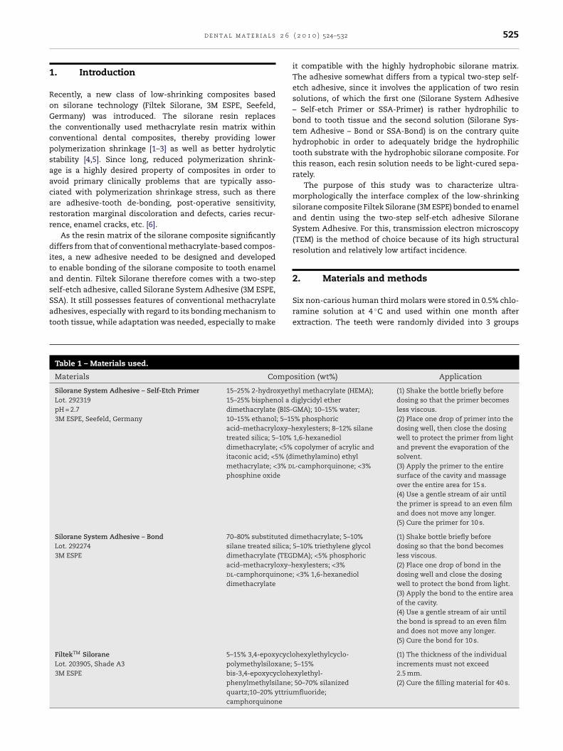
System Adhesive (3M ESPE, SSA) with the silorane compositeFiltek Silorane (3M ESPE) was then applied, strictly accordingto the manufacturer’s instructions (Table 1). To appraise theultra-morphological structure of the silorane composite, TEMFig. 1 – TEM photomicrographs of Filtek Silorane and Silorane Syrevealed irregularly polyhedral-shaped filler and appeared less losilanized quartz: more electron-lucent, polyhedral-shaped particyttriumfluoride: irregular, electron-opaque particles with rounderounded edges in three size-ranges. L, large: 1 �m or larger; M, mrevealed spherical filler particles. L, large: 1 �m or larger; M, medhomogenous distribution of the filler within both resin layers. Thdistinct layers with different filler content.
6 ( 2 0 1 0 ) 524–532
specimens of two conventional micro-hybrid resin compos-ites (Clearfil AP-X, Kuraray, Okayama, Japan; Filtek Z100, 3MESPE) were prepared using a silicon mold. Light-curing wasperformed using an Optilux 500 (Demetron/Kerr, Danbury, CT,USA) device with a light output not less than 600 mW/cm2.After bonding procedures, specimens were stored for 1 day intap water at 37 ◦C.
The specimens were processed for TEM according to theprocedure described in detail before by Van Meerbeek et al. [7].Non-demineralized and lab-demineralized ultra-thin sectionswere cut (Ultracut UCT, Leica, Vienna, Austria) and exam-ined unstained and positively stained (3% uranyl acetate for12 min/lead citrate for 13 min) using TEM (JEM-1200EX II, JEOL,Tokyo, Japan). After observation of the resin–enamel inter-face sections, the TEM grids were additionally exposed for 5 sto 0.1N HCl, and subsequently carefully rinsed with distilledwater to remove all dissolved mineral components follow-ing a method described before by Hanning et al. [8]. Doingso, the same spots could be imaged by TEM before and afterdecalcification. In order to reveal potential porous zones in
the interface complex, additional specimens were immersedin a 50 wt% ammoniacal silver nitrate solution according toa nanoleakage-detection protocol previously described by Tayet al. [9].stem Adhesive (bonded to dentin). (a) Filtek Siloraneaded than both conventional composites; see (c) and (d). Q,
les with sharp edges (size ranging from 0.05 to 5 �m). YF,d edges. (b) Clearfil AP-X revealed filler particles withedium: about 0.1 �m; N, nano: nanofiller. (c) Filtek Z100ium: about 0.1 �m; N, nano: nanofiller. (d) SSA revealed ane separately cured SSA-Primer and SSA-Bond did form two
2 6
3
T(h(aSavrcomnbaod(tp
tnp
FtdHst
d e n t a l m a t e r i a l s
. Results
he ultra-structure of the silorane composite (Filtek Silorane)Fig. 1a) differed clearly from the two conventional micro-ybrid composites Clearfil AP-X (Fig. 1b) and Filtek Z100
Fig. 1c). At first, the filler loading of the silorane compositeppeared lower than that of both conventional composites.econdly, the filler particles within the silorane compositere typically irregularly (polyhedral) shaped, while the con-entional composites contain filler particles that are moreounded off (Clearfil AP-X) or even spherical (Filtek Z100). Bothonventional composites obviously contain three size-rangesf filler particles: large with a diameter of 1 �m or larger,edium with a diameter of about 0.1 �m and small with a
ano-sized diameter. The silorane composite lacks nanofiller,ut also clear ranges of different sizes could not be observed;ctually, any filler size between 0.05 and 5 �m could bebserved. Although not different in size, two filler kinds wereetected to be present within the silorane composite, namely
1) more electron-lucent, irregularly polyhedral-shaped par-icles with sharp edges and (2) irregular, electron-opaquearticles with rounded edges.
The interaction of SSA with enamel/dentin and withhe silorane composite was free of voids in all sections;o de-bonding at any interface occurred during specimen-reparation/imaging. The total thickness of the adhesive resin
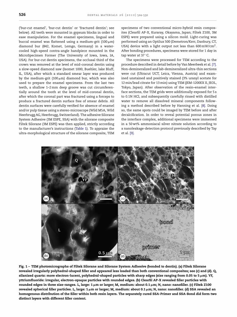
ig. 2 – TEM photomicrographs of the interface of Silorane Systeight interface. (b) Corresponding HCl-decalcified section of (a). Aeep was produced, though this varied widely from region to regigher magnification of (a). Visible micro-mechanical interlockinurface. (d) Corresponding HCl-decalcified section of (c). The intehan 1 �m with a huge variety. Black dotted line, bottom of inter
( 2 0 1 0 ) 524–532 527
varied regionally from about 10 to 20 �m. Within the adhesivelayer, two distinct layers with different nanofiller concentra-tion could be distinguished, corresponding to the separateapplication/light-curing of the higher silica-filled SSA-Primerand the lower silica-filled SSA-Bond (Fig. 1d). As initially theamount of filler in SSA-Primer and SSA-Bond is relatively com-parable (Table 1: 8–12% and 5–10%, respectively), the observedfiller-loading difference is probably due to a concentratingeffect in SSA-Primer during solvent removal by air-thinning.
SSA adhesive tightly interacted with bur-cut enamel (Fig. 2).The interaction zone was, however, very thin with mostly onlytiny micro-tags observable and without distinct dissolutionof hydroxyapatite. Lab-demineralization with HCl discloseda so-called inter-crystallite nano-retention pattern [8] of lessthan 1 �m deep, though this widely varied from region toregion. SSA adhesive also tightly interacted with bur-cutdentin (Fig. 3). A relatively thin interaction layer of maximum afew hundred nanometers was formed without distinct surfacedemineralization. The orifices of the dentinal tubules werefilled with irregular material, most likely representing smearplugs that were partially impregnated with resin, but withoutformation of ‘true’ resin tags. At fractured dentin, the actualinteraction of SSA adhesive without smear-layer interference
revealed a very thin, 100–200 nm interaction zone withoutclear signs of surface demineralization (Fig. 4). Distinct resintags were formed due to the absence of smear plugs. Silver-nitrate infiltration showed a varying pattern of both spot- andm Adhesive bonded to enamel. (a) Overview, showing an inter-crystallite nano-retention pattern of less than 1 �mion. Black dotted line, bottom of intercrystal network. (c)g was limited to the present roughness of the enamelr-crystallite nano-retention was detected to a depth of lesscrystal network.
528 d e n t a l m a t e r i a l s 2 6 ( 2 0 1 0 ) 524–532
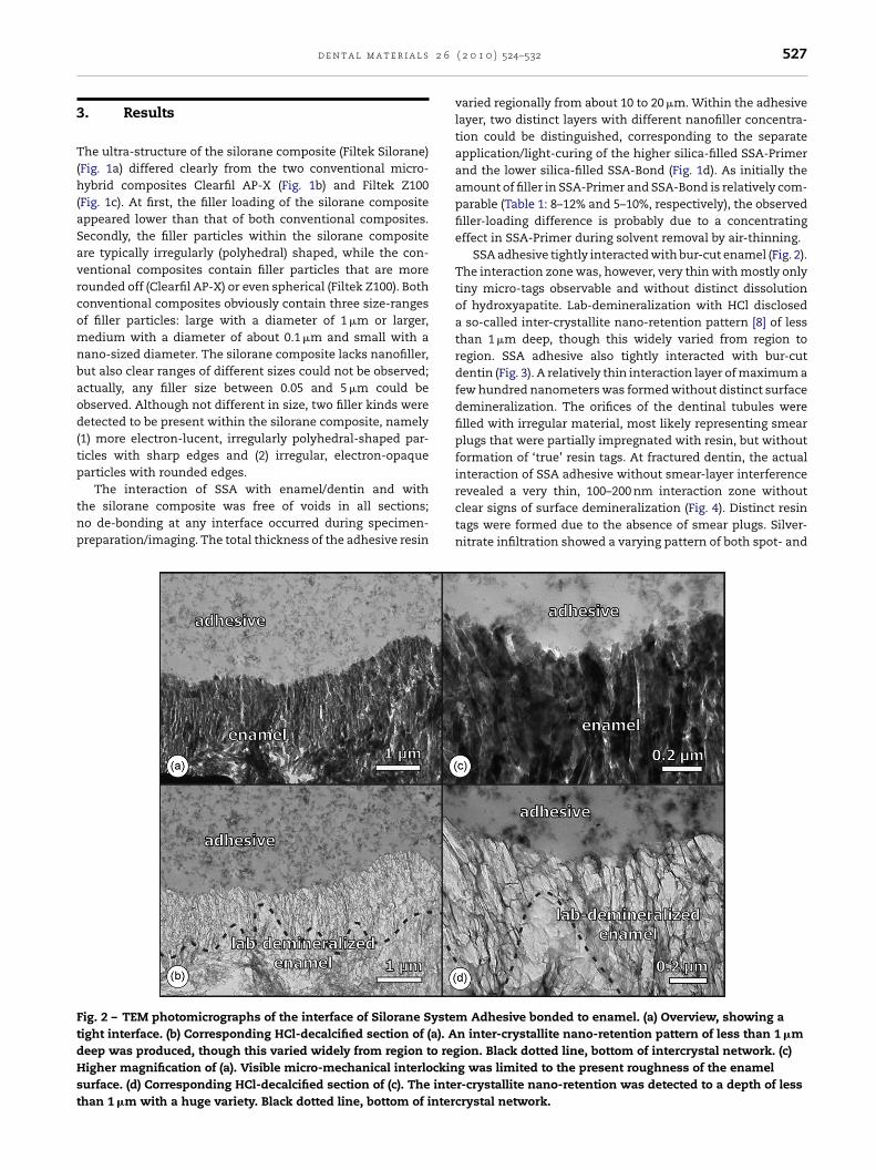
Fig. 3 – TEM photomicrographs of the interface of Silorane System Adhesive bonded to bur-cut dentin. (a)Non-demineralized, unstained section. The actual hybrid layer is difficult to detect, as the interaction with the bur smearlayer is somewhat unclear. (b) Non-demineralized, unstained section; higher magnification of the interface between SSAadhesive and dentin. Even at this magnification, it is very difficult to distinguish a distinct hybrid layer (hand pointer).Hydroxyapatite crystals are scattered along the interaction zone (arrow). (c) Non-demineralized, UA/LC stained section. Theresin-infiltrated smear layer, containing residual hydroxyapatite, becomes more evident upon heavy-metal staining (handpointer). Arrow head = hydroxyapatite. (d) Demineralized, UA/LC stained section. The interaction zone reacted heavily with
ollaggna
the heavy-metal staining solution. A very shallow zone of cFig. 4d), can be observed immediately below the resin-impre
cluster-like appearance of nano-leakage (Fig. 5), but this variedwidely with the region. Substantially more silver-infiltrationwas observed along the adhesive–dentin interface at bur-cutdentin than at fractured dentin. Noteworthy is also the consid-erable amount of Ag detected along the SSA-Primer/SSA-Bondinterface, along with a spot-like infiltration pattern in SSA-Primer, but not in SSA-Bond (Fig. 5a).
4. Discussion
The key-advantage of Filtek Silorane is low polymerizationshrinkage, which is attributed to the new siloxane–oxiranetechnology [1–3]. Indeed, TEM ultra-structural imagingrevealed a filler-matrix organization that significantly dif-fered from that of the two conventional methacrylate-basedcomposites evaluated (Clearfil AP-X, Filtek Z100) with regardto filler loading, distribution and composition (Fig. 1).Both methacrylate-based composites apparently employ a
‘smarter’ filler distribution, with filler sizes ranging from about1 �m to nanofiller, increasing the filler volume and decreasingthe matrix volume, to the direct benefit of enhanced mechani-cal properties and reduced polymerization shrinkage. Becauseen exposure, representing the hybrid layer (see alsoted smear.
of its low-shrinking nature, the silorane composite does notneed this ‘smart’ filler loading to decrease the shrinkage. Alsoin terms of mechanical properties as flexural strength andhardness, this reduced filler loading seemed no disadvantage[5].
SSA requires separate light-curing of the firstly appliedSSA-Primer and the secondly applied SSA-Bond, and conse-quently differs from typical two-step self-etch adhesives, ofwhich the successive application of a primer and adhesiveresin is finalized by only one light-curing step [10] (Table 1).Therefore, SSA’s bond to tooth enamel/dentin was establishedin the first application step, similarly as achieved by one-stepself-etch adhesives. One could argue that the name givento this first bottle does not fully meet the definition of a‘conventional’ primer. As ‘adhesion-promoter’, a primer isaimed to promote the infiltration of the subsequently applied,actual ‘adhesive resin’ or ‘bonding resin’, while the sepa-rately light-cured SSA-Primer can actually be regarded as aone-step self-etch adhesive. Considering its interaction with
enamel/dentin is limited to a few hundreds of nanometers,the bonding mechanism involves a form of ‘nano-interaction’,like it was disclosed before for so-called ‘ultra-mild’ self-etch adhesives [11,12]. This shallow interaction should be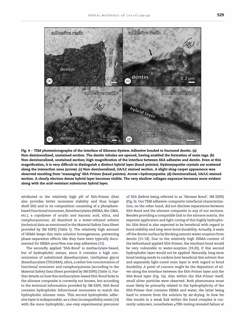
d e n t a l m a t e r i a l s 2 6 ( 2 0 1 0 ) 524–532 529
Fig. 4 – TEM photomicrographs of the interface of Silorane System Adhesive bonded to fractured dentin. (a)Non-demineralized, unstained section. The dentin tubules are opened, having enabled the formation of resin tags. (b)Non-demineralized, unstained section; high magnification of the interface between SSA adhesive and dentin. Even at thismagnification, it is very difficult to distinguish a distinct hybrid layer (hand pointer). Hydroxyapatite crystals are scatteredalong the interaction zone (arrow). (c) Non-demineralized, UA/LC stained section. A slight shag-carpet appearance wasobserved resulting from ‘massaging’ SSA-Primer (hand pointer). Arrow = hydroxyapatite. (d) Demineralized, UA/LC staineds e. Tha
aasbec(popm
bcdfMtttchsw
ection. A clearly electron dense hybrid layer becomes visibllong with the acid-resistant submicron hybrid layer.
ttributed to the relatively high pH of SSA-Primer (thatlso provides better monomer stability and thus longerhelf life) and to its composition consisting of a phosphate-ased functional monomer, dimethacrylates (HEMA, Bis-GMA,tc.), a copolymer of acrylic and itaconic acid, silica, andamphorquinone, all dissolved in a water–ethanol solventtechnical data as mentioned in the Material Safety Data Sheetrovided by 3M ESPE) (Table 1). The relatively high amountf HEMA keeps this resin solution homogeneous, preventinghase-separation effects like they have been typically docu-ented for HEMA-poor/free one-step adhesives [13].The secondly applied ‘SSA-Bond’ is methacrylate-based,
ut of hydrophobic nature, since it contains a high con-entration of substituted dimethacrylate, triethylene glycolimethacrylate (TEGDMA), silica, a rather low concentration ofunctional monomer and camphorquinone (according to the
aterial Safety Data Sheet provided by 3M ESPE) (Table 1). Fur-her details on how this methacrylate-based SSA-Bond links tohe silorane composite is currently not known, but accordingo the technical information provided by 3M ESPE, SSA-Bond
ontains hydrophobic bifunctional monomers to match theydrophobic silorane resin. This second hydrophobic adhe-ive layer is indispensable, as a clear incompatibility exists [14]ith the more hydrophilic, one-step experimental precursore very shallow collagen exposure becomes more evident
of SSA (before being referred to as ‘Hermes Bond’, 3M ESPE)(Fig. 6). Our TEM adhesive–composite interfacial characteriza-tion, on the other hand, did not disclose separations betweenSSA-Bond and the silorane composite in any of our sections.Besides providing a compatible link to the silorane matrix, theseparate application and light-curing of this highly hydropho-bic SSA-Bond is also expected to be beneficial with regard tobond stability and long-term bond durability. Actually, it sealsoff the dentin surface by blocking osmotic water-sorption fromdentin [15–18]. Due to the relatively high HEMA-content ofthe beforehand applied SSA-Primer, the resultant bond wouldbe very vulnerable to water-sorption [19,20], if this secondhydrophobic layer would not be applied. Naturally, long-termbond testing needs to confirm how beneficial this solvent-freeand separately light-cured resin layer is with regard to bonddurability. A point of concern might be the deposition of sil-ver along the interface between the SSA-Primer layer and theSSA-Bond layer (Fig. 5a). Also within the SSA-Primer itself,small silver particles were observed. Both phenomena mustmost likely be primarily related to the hydrophylicity of the
SSA-Primer that contains HEMA and water, the latter beinghard to remove from the solution by air-drying. In how farthis results in a weak link within the bond complex is cur-rently unknown, nonetheless �TBS-testing revealed failure at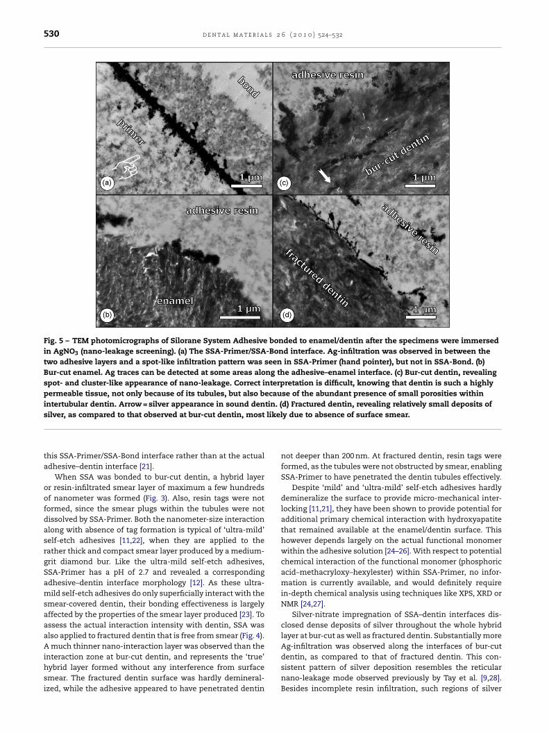
530 d e n t a l m a t e r i a l s 2 6 ( 2 0 1 0 ) 524–532
Fig. 5 – TEM photomicrographs of Silorane System Adhesive bonded to enamel/dentin after the specimens were immersedin AgNO3 (nano-leakage screening). (a) The SSA-Primer/SSA-Bond interface. Ag-infiltration was observed in between thetwo adhesive layers and a spot-like infiltration pattern was seen in SSA-Primer (hand pointer), but not in SSA-Bond. (b)Bur-cut enamel. Ag traces can be detected at some areas along the adhesive–enamel interface. (c) Bur-cut dentin, revealingspot- and cluster-like appearance of nano-leakage. Correct interpretation is difficult, knowing that dentin is such a highlypermeable tissue, not only because of its tubules, but also because of the abundant presence of small porosities withinintertubular dentin. Arrow = silver appearance in sound dentin. (d) Fractured dentin, revealing relatively small deposits of
likel
silver, as compared to that observed at bur-cut dentin, mostthis SSA-Primer/SSA-Bond interface rather than at the actualadhesive–dentin interface [21].
When SSA was bonded to bur-cut dentin, a hybrid layeror resin-infiltrated smear layer of maximum a few hundredsof nanometer was formed (Fig. 3). Also, resin tags were notformed, since the smear plugs within the tubules were notdissolved by SSA-Primer. Both the nanometer-size interactionalong with absence of tag formation is typical of ‘ultra-mild’self-etch adhesives [11,22], when they are applied to therather thick and compact smear layer produced by a medium-grit diamond bur. Like the ultra-mild self-etch adhesives,SSA-Primer has a pH of 2.7 and revealed a correspondingadhesive–dentin interface morphology [12]. As these ultra-mild self-etch adhesives do only superficially interact with thesmear-covered dentin, their bonding effectiveness is largelyaffected by the properties of the smear layer produced [23]. Toassess the actual interaction intensity with dentin, SSA wasalso applied to fractured dentin that is free from smear (Fig. 4).A much thinner nano-interaction layer was observed than the
interaction zone at bur-cut dentin, and represents the ‘true’hybrid layer formed without any interference from surfacesmear. The fractured dentin surface was hardly demineral-ized, while the adhesive appeared to have penetrated dentiny due to absence of surface smear.
not deeper than 200 nm. At fractured dentin, resin tags wereformed, as the tubules were not obstructed by smear, enablingSSA-Primer to have penetrated the dentin tubules effectively.
Despite ‘mild’ and ‘ultra-mild’ self-etch adhesives hardlydemineralize the surface to provide micro-mechanical inter-locking [11,21], they have been shown to provide potential foradditional primary chemical interaction with hydroxyapatitethat remained available at the enamel/dentin surface. Thishowever depends largely on the actual functional monomerwithin the adhesive solution [24–26]. With respect to potentialchemical interaction of the functional monomer (phosphoricacid–methacryloxy–hexylester) within SSA-Primer, no infor-mation is currently available, and would definitely requirein-depth chemical analysis using techniques like XPS, XRD orNMR [24,27].
Silver-nitrate impregnation of SSA–dentin interfaces dis-closed dense deposits of silver throughout the whole hybridlayer at bur-cut as well as fractured dentin. Substantially moreAg-infiltration was observed along the interfaces of bur-cut
dentin, as compared to that of fractured dentin. This con-sistent pattern of silver deposition resembles the reticularnano-leakage mode observed previously by Tay et al. [9,28].Besides incomplete resin infiltration, such regions of silver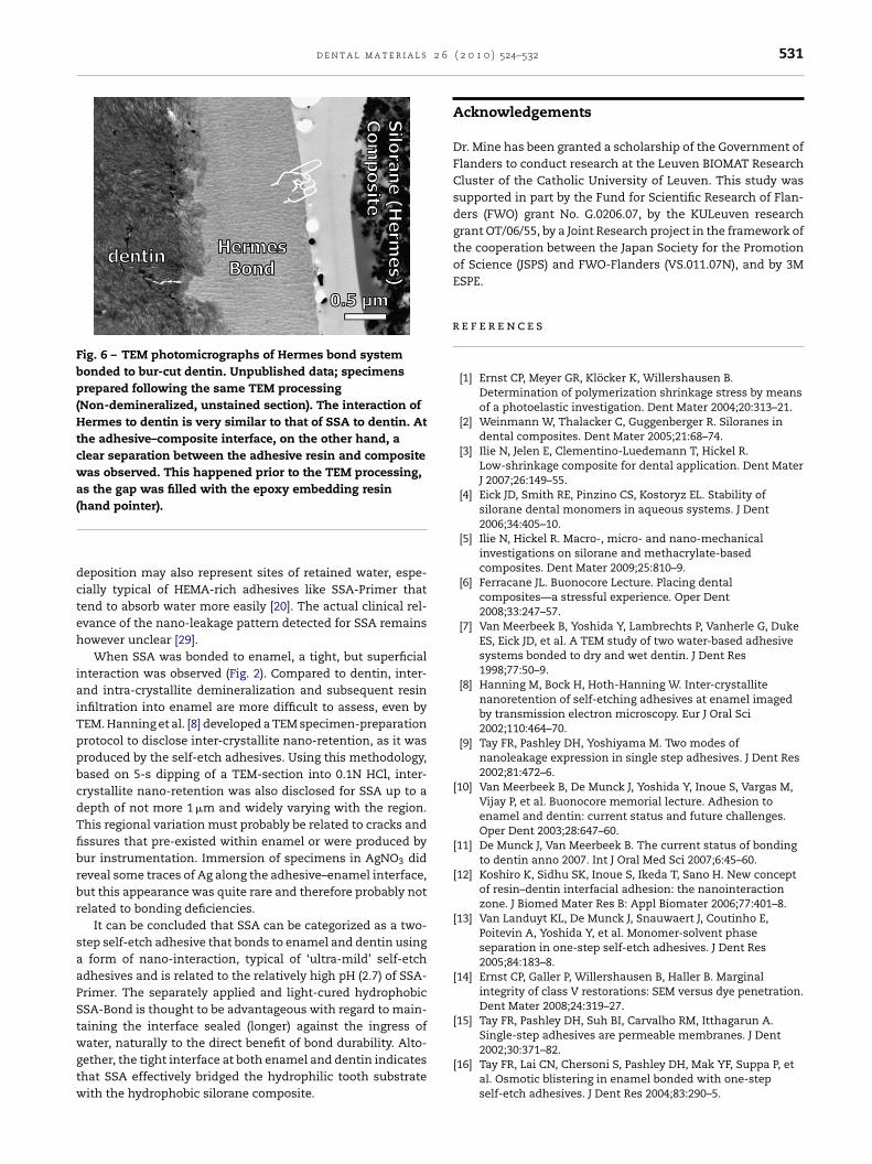
d e n t a l m a t e r i a l s 2 6
Fig. 6 – TEM photomicrographs of Hermes bond systembonded to bur-cut dentin. Unpublished data; specimensprepared following the same TEM processing(Non-demineralized, unstained section). The interaction ofHermes to dentin is very similar to that of SSA to dentin. Atthe adhesive–composite interface, on the other hand, aclear separation between the adhesive resin and compositewas observed. This happened prior to the TEM processing,as the gap was filled with the epoxy embedding resin(
dcteh
iaiTppbcdTfibrbr
saaPStwgtw
r
hand pointer).
eposition may also represent sites of retained water, espe-ially typical of HEMA-rich adhesives like SSA-Primer thatend to absorb water more easily [20]. The actual clinical rel-vance of the nano-leakage pattern detected for SSA remainsowever unclear [29].
When SSA was bonded to enamel, a tight, but superficialnteraction was observed (Fig. 2). Compared to dentin, inter-nd intra-crystallite demineralization and subsequent resinnfiltration into enamel are more difficult to assess, even byEM. Hanning et al. [8] developed a TEM specimen-preparationrotocol to disclose inter-crystallite nano-retention, as it wasroduced by the self-etch adhesives. Using this methodology,ased on 5-s dipping of a TEM-section into 0.1N HCl, inter-rystallite nano-retention was also disclosed for SSA up to aepth of not more 1 �m and widely varying with the region.his regional variation must probably be related to cracks andssures that pre-existed within enamel or were produced byur instrumentation. Immersion of specimens in AgNO3 dideveal some traces of Ag along the adhesive–enamel interface,ut this appearance was quite rare and therefore probably notelated to bonding deficiencies.
It can be concluded that SSA can be categorized as a two-tep self-etch adhesive that bonds to enamel and dentin using
form of nano-interaction, typical of ‘ultra-mild’ self-etchdhesives and is related to the relatively high pH (2.7) of SSA-rimer. The separately applied and light-cured hydrophobicSA-Bond is thought to be advantageous with regard to main-aining the interface sealed (longer) against the ingress of
ater, naturally to the direct benefit of bond durability. Alto-ether, the tight interface at both enamel and dentin indicateshat SSA effectively bridged the hydrophilic tooth substrateith the hydrophobic silorane composite.
( 2 0 1 0 ) 524–532 531
Acknowledgements
Dr. Mine has been granted a scholarship of the Government ofFlanders to conduct research at the Leuven BIOMAT ResearchCluster of the Catholic University of Leuven. This study wassupported in part by the Fund for Scientific Research of Flan-ders (FWO) grant No. G.0206.07, by the KULeuven researchgrant OT/06/55, by a Joint Research project in the framework ofthe cooperation between the Japan Society for the Promotionof Science (JSPS) and FWO-Flanders (VS.011.07N), and by 3MESPE.
e f e r e n c e s
[1] Ernst CP, Meyer GR, Klöcker K, Willershausen B.Determination of polymerization shrinkage stress by meansof a photoelastic investigation. Dent Mater 2004;20:313–21.
[2] Weinmann W, Thalacker C, Guggenberger R. Siloranes indental composites. Dent Mater 2005;21:68–74.
[3] Ilie N, Jelen E, Clementino-Luedemann T, Hickel R.Low-shrinkage composite for dental application. Dent MaterJ 2007;26:149–55.
[4] Eick JD, Smith RE, Pinzino CS, Kostoryz EL. Stability ofsilorane dental monomers in aqueous systems. J Dent2006;34:405–10.
[5] Ilie N, Hickel R. Macro-, micro- and nano-mechanicalinvestigations on silorane and methacrylate-basedcomposites. Dent Mater 2009;25:810–9.
[6] Ferracane JL. Buonocore Lecture. Placing dentalcomposites—a stressful experience. Oper Dent2008;33:247–57.
[7] Van Meerbeek B, Yoshida Y, Lambrechts P, Vanherle G, DukeES, Eick JD, et al. A TEM study of two water-based adhesivesystems bonded to dry and wet dentin. J Dent Res1998;77:50–9.
[8] Hanning M, Bock H, Hoth-Hanning W. Inter-crystallitenanoretention of self-etching adhesives at enamel imagedby transmission electron microscopy. Eur J Oral Sci2002;110:464–70.
[9] Tay FR, Pashley DH, Yoshiyama M. Two modes ofnanoleakage expression in single step adhesives. J Dent Res2002;81:472–6.
[10] Van Meerbeek B, De Munck J, Yoshida Y, Inoue S, Vargas M,Vijay P, et al. Buonocore memorial lecture. Adhesion toenamel and dentin: current status and future challenges.Oper Dent 2003;28:647–60.
[11] De Munck J, Van Meerbeek B. The current status of bondingto dentin anno 2007. Int J Oral Med Sci 2007;6:45–60.
[12] Koshiro K, Sidhu SK, Inoue S, Ikeda T, Sano H. New conceptof resin–dentin interfacial adhesion: the nanointeractionzone. J Biomed Mater Res B: Appl Biomater 2006;77:401–8.
[13] Van Landuyt KL, De Munck J, Snauwaert J, Coutinho E,Poitevin A, Yoshida Y, et al. Monomer-solvent phaseseparation in one-step self-etch adhesives. J Dent Res2005;84:183–8.
[14] Ernst CP, Galler P, Willershausen B, Haller B. Marginalintegrity of class V restorations: SEM versus dye penetration.Dent Mater 2008;24:319–27.
[15] Tay FR, Pashley DH, Suh BI, Carvalho RM, Itthagarun A.
Single-step adhesives are permeable membranes. J Dent2002;30:371–82.[16] Tay FR, Lai CN, Chersoni S, Pashley DH, Mak YF, Suppa P, etal. Osmotic blistering in enamel bonded with one-stepself-etch adhesives. J Dent Res 2004;83:290–5.
l s 2
532 d e n t a l m a t e r i a[17] Van Landuyt KL, Snauwaert J, De Munck J, Coutinho E,Poitevin A, Yoshida Y, et al. Origin of interfacial dropletswith one-step adhesives. J Dent Res 2007;86:739–44.
[18] Reis A, Albuquerque M, Pegoraro M, Mattei G, Bauer JR,Grande RH, et al. Can the durability of one-step self-etchadhesives be improved by double application or by an extralayer of hydrophobic resin? J Dent 2008;36:309–15.
[19] Tay FR, King NM, Chan KM, Pashley DH. How cannanoleakage occur in self-etching adhesive systems thatdemineralize and infiltrate simultaneously? J Adhes Dent2002;4:255–69.
[20] Van Landuyt KL, Snauwaert J, Peumans M, De Munck J,Lambrechts P, Van Meerbeek B. The role of HEMA in one-stepself-etch adhesives. Dent Mater 2008;24:1412–9.
[21] Van Ende A, De Munck J, Mine A, Lambrechts P, VanMeerbeek B. Does a low-shrinking composite induce lessstress at the adhesive interface? Dent Mater 2010;26:215–22.
[22] Sarr M, Kane WA, Vreven J, Mine A, Van Landuyt KL,Peumans M, Lambrechts P, Van Meerbeek B, De Munck J.Micro-tensile bond strength and interfacial characterizationof eleven contemporary adhesives bonded to bur-cut dentin.Oper Dent 2010;35:94–104.
6 ( 2 0 1 0 ) 524–532
[23] Ermis RB, De Munck J, Cardoso MV, Coutinho E, Van LanduytKL, Poitevin A, et al. Bond strength of self-etch adhesives todentin prepared with three different diamond burs. DentMater 2008;24:978–85.
[24] Yoshida Y, Van Meerbeek B, Nakayama Y, Snauwaert J,Hellemans L, Lambrechts P, et al. Evidence of chemicalbonding at biomaterial–hard tissue interfaces. J Dent Res2000;79:709–14.
[25] Yoshida Y, Nagakane K, Fukuda R, Nakayama Y, Okazaki M,Shintani H, et al. Comparative study on adhesiveperformance of functional monomers. J Dent Res2004;83:454–8.
[26] Yoshioka M, Yoshida Y, Inoue S, Lambrechts P, Vanherle G,Nomura Y, et al. Adhesion/decalcification mechanisms ofacid interactions with human hard tissues. J Biomed MaterRes 2002;59:56–62.
[27] Fukegawa D, Hayakawa S, Yoshida Y, Suzuki K, Osaka A, VanMeerbeek B. Chemical interaction of phosphoric acid ester
with hydroxyapatite. J Dent Res 2006;85:941–4.[28] Tay FR, Pashley DH. Water treeing–a potential mechanismfor degradation of dentin adhesives. Am J Dent 2003;16:6–12.
[29] Van Meerbeek B. The “Myth” of nanoleakage. J Adhes Dent2007;9:491–2.