Comparative Analysis of Root Canal Dentin Removal ... - MDPI
Upload
independentCategory
view
3download
0
Bond strength of adhesives to dentin contaminated with smoker’ssaliva
Lilliam M. Pinzon,Department of Preventive and Restorative Dentistry, 707 Parnassus Avenue, Room 3212,University of California, San Francisco, CA 94143-0758, USA, Tel. +1-415-476-2048; Fax+1-415-476-0858
Makoto Oguri,Tokuyama Dental Corporation, Tsukuba, Japan
Kathy O’Keefe,Department of Restorative Dentistry and Biomaterials, University of Texas Dental Branch atHouston, Houston, TX, USA
Vladimir Dusevish,Electron Microscopy Laboratory, University of Missouri, Kansas City, MO, USA
Paulette Spencer,Department of Mechanical Engineering, University of Kansas, Lawrence, KS, USA
John M. Powers, andDepartment of Restorative Dentistry and Biomaterials, University of Texas Dental Branch atHouston, Houston, TX, USA
Grayson W. MarshallDivision of Biomaterials and Bioengineering and Department of Preventive and Restorative DentalSciences, University of California, San Francisco, CA, USALilliam M. Pinzon: [email protected]
AbstractThe purpose of this study was to determine the effects of contamination with smoker’s and non-smoker’s saliva on the bond strength of resin composite to superficial dentin using different adhesivesystems. The interfacial structure between the resin and dentin was evaluated for each treatment usingenvironmental scanning electron microscopy (ESEM). Freshly extracted human molars were groundwith 600-grit SiC paper to expose the superficial dentin. Adhesives [One-Up-Bond-F-Plus (OUFP)and Adper-Prompt-L-Pop (APLP)] and resin composite (TPH-Spectrum) were bonded to the dentin(n = 8/group, 180 total specimens) under five surface conditions: control (adhesive applied followingmanufacturers’ instructions); saliva, then 5-s air dry, then adhesive; adhesive, saliva, 5-s air dry;adhesive, saliva, 5-s water rinse, 5-s air dry (ASW group); and adhesive, saliva, 5-s water rinse, 5-sair dry, reapply adhesive (ASWA group). After storage in water at 37°C for 24 h, the specimens weredebonded under tension at a speed of 0.5 mm/min. ESEM photomicrographs of the dentin/adhesiveinterfaces were taken. Mean bond strength ranged from 8.1 to 24.1 MPa. Fisher’s protected leastsignificant difference (P = 0.05) intervals for critical adhesive, saliva, and surface conditiondifferences were 1.3, 1.3, and 2.1 MPa, respectively. There were no significant differences in bondstrength to dentin between contamination by smoker’s and non-smoker’s saliva, but bond strengthswere significantly different between adhesive systems, with OUFP twice as strong as APLP under
Correspondence to: Lilliam M. Pinzon, [email protected].
NIH Public AccessAuthor ManuscriptOdontology. Author manuscript; available in PMC 2010 August 31.
Published in final edited form as:Odontology. 2010 February ; 98(1): 37–43. doi:10.1007/s10266-009-0109-4.
NIH
-PA Author Manuscript
NIH
-PA Author Manuscript
NIH
-PA Author Manuscript

almost all conditions. After adhesive application and contamination with either smoker’s ornonsmoker’s saliva followed by washing and reapplication of the adhesive (ASWA group), the bondstrength of both adhesive systems was the same as that of the control group.
KeywordsBond strength; Saliva; Smokers; SEM
IntroductionSeveral kinds of contamination can affect the structural and chemical properties of dentalrestorative materials. Oral fluids such as saliva, blood, and crevicular fluid can cause chemicalincompatibility with dental materials.1
Saliva is composed of 99.4% water and 0.6% solids, including macromolecules such as proteins(26% mucins), glycoproteins, sugar, and amylase; inorganic particles such as calcium, sodium,and chloride; and organic particles such as urea, amino acids, fatty acids, and free glucose.2,3 Saliva is an essential fluid in the mouth that serves as a barrier against desiccation andenvironmental injury. Furthermore, the different components of saliva interact to modulate pHand provide buffering, cleanse the oral tissues in general, prevent demineralization of the teeth,and serve as an antibacterial agent.4,5
Several studies have reported the effects of different contaminants found or used commonlyin the oral cavity on the bond strength of dentin and enamel. For example, the effects of water,saliva, astringents, plasma, handpiece lubricant, zinc oxide–eugenol cement, and non-eugenol–zinc oxide cement on bond strength have been studied.6–11
Several studies have also reported that saliva contamination of enamel and dentin did not affectthe bond strength of single-bottle (total-etch) adhesive systems.12 However, studies haveshown decreases in bond strength with contaminants such as astringents, blood, and saliva withdifferent self-etching adhesive systems and have suggested ways to correct and improve thebond strength in cases of contamination.7–9 Some self-etching adhesive systems seem to bemore compatible with the oral environment for reasons such as low moisture sensitivity andresistance to saliva contamination.12–15
This low sensitivity may be caused by the use of a water-based solvent in the primer. In theoral cavity, it is difficult to obtain dry conditions because as soon as the patient breathes thereis moisture in the environment. It is important to study contamination factors such as salivabecause some studies have reported that the proteins present in saliva are the main factorsresponsible for the improper formation of the dentin–adhesive interface.3,15
One billion men and a quarter of a billion of women worldwide consume some kind of tobacco.16 A cigarette contains at least 5000 chemicals that produce many changes in the oralenvironment, such as teeth pigmentation and inflammatory and degenerative illnesses.Furthermore, smoking causes cancer and is the main cause of death worldwide.17–19 Tobaccomay also affect the chemical interaction between oral biomaterials and the teeth. Thus,smoker’s saliva may affect restorative procedures and properties of restorations such as bondstrength and interfacial microstructure. In this study, we worked with smoker’s saliva but wedid not specifically work with smoker’s teeth because we wanted to focus specifically on theeffects of saliva. Future studies should be done with teeth of documented smokers, because wethink that frequent use of tobacco probably affects the oral tissues and teeth substrates as well.
Pinzon et al. Page 2
Odontology. Author manuscript; available in PMC 2010 August 31.
NIH
-PA Author Manuscript
NIH
-PA Author Manuscript
NIH
-PA Author Manuscript
The purpose of this study was to measure the bond strength of resin composite to saliva-contaminated dentin and to analyze the interfacial microstructure between smoker’s andnonsmoker’s saliva by using environmental scanning electron microscopy (ESEM).Contaminated teeth were bonded with one of two self-etching adhesive systems and we alsoevaluated two methods of correcting the saliva contamination. The null hypothesis was thatthere were no differences in the bond strength or the microstructure of the dentin adhesiveinterface between contamination with smoker’s and that with nonsmoker’s saliva.
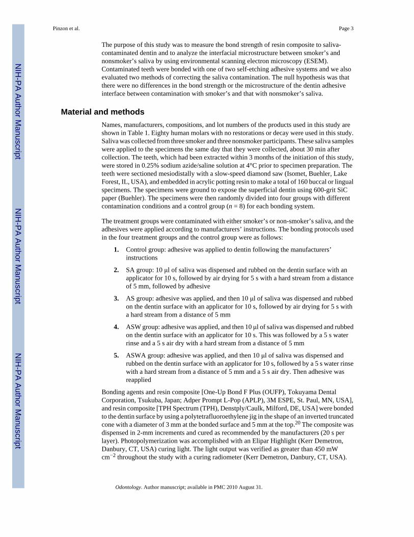
Material and methodsNames, manufacturers, compositions, and lot numbers of the products used in this study areshown in Table 1. Eighty human molars with no restorations or decay were used in this study.Saliva was collected from three smoker and three nonsmoker participants. These saliva sampleswere applied to the specimens the same day that they were collected, about 30 min aftercollection. The teeth, which had been extracted within 3 months of the initiation of this study,were stored in 0.25% sodium azide/saline solution at 4°C prior to specimen preparation. Theteeth were sectioned mesiodistally with a slow-speed diamond saw (Isomet, Buehler, LakeForest, IL, USA), and embedded in acrylic potting resin to make a total of 160 buccal or lingualspecimens. The specimens were ground to expose the superficial dentin using 600-grit SiCpaper (Buehler). The specimens were then randomly divided into four groups with differentcontamination conditions and a control group (n = 8) for each bonding system.
The treatment groups were contaminated with either smoker’s or non-smoker’s saliva, and theadhesives were applied according to manufacturers’ instructions. The bonding protocols usedin the four treatment groups and the control group were as follows:
1. Control group: adhesive was applied to dentin following the manufacturers’instructions
2. SA group: 10 μl of saliva was dispensed and rubbed on the dentin surface with anapplicator for 10 s, followed by air drying for 5 s with a hard stream from a distanceof 5 mm, followed by adhesive
3. AS group: adhesive was applied, and then 10 μl of saliva was dispensed and rubbedon the dentin surface with an applicator for 10 s, followed by air drying for 5 s witha hard stream from a distance of 5 mm
4. ASW group: adhesive was applied, and then 10 μl of saliva was dispensed and rubbedon the dentin surface with an applicator for 10 s. This was followed by a 5 s waterrinse and a 5 s air dry with a hard stream from a distance of 5 mm
5. ASWA group: adhesive was applied, and then 10 μl of saliva was dispensed andrubbed on the dentin surface with an applicator for 10 s, followed by a 5 s water rinsewith a hard stream from a distance of 5 mm and a 5 s air dry. Then adhesive wasreapplied
Bonding agents and resin composite [One-Up Bond F Plus (OUFP), Tokuyama DentalCorporation, Tsukuba, Japan; Adper Prompt L-Pop (APLP), 3M ESPE, St. Paul, MN, USA],and resin composite [TPH Spectrum (TPH), Denstply/Caulk, Milford, DE, USA] were bondedto the dentin surface by using a polytetrafluoroethylene jig in the shape of an inverted truncatedcone with a diameter of 3 mm at the bonded surface and 5 mm at the top.20 The composite wasdispensed in 2-mm increments and cured as recommended by the manufacturers (20 s perlayer). Photopolymerization was accomplished with an Elipar Highlight (Kerr Demetron,Danbury, CT, USA) curing light. The light output was verified as greater than 450 mWcm−2 throughout the study with a curing radiometer (Kerr Demetron, Danbury, CT, USA).
Pinzon et al. Page 3
Odontology. Author manuscript; available in PMC 2010 August 31.
NIH
-PA Author Manuscript
NIH
-PA Author Manuscript
NIH
-PA Author Manuscript

The adhesion protocol followed for the APLP self-etching adhesive system was as follows:
• After preparation, lightly dry to remove excess water.
• Apply adhesive with a rubbing motion for 15 s.
• Gently but thoroughly air-dry to remove the aqueous solvent.
• Apply a second coat (no waiting time for the second layer).
• Gently but thoroughly air-dry to remove the aqueous solvent.
• Light cure for 10 s.
The adhesion protocol followed for the OUFP self-etching adhesive system was:
• Rinse the prepared cavity. If no cavity preparation has been made, clean the toothsurface with a rubber cup and a fluoride-free cleaning paste; rinse thoroughly withwater.
• Remove water by blowing air to keep the cavity in a dry, or in a moist condition butwithout visible puddles or a shiny appearance.
• Using the provided dispenser, place one drop of bonding agent A and one drop ofbonding agent B into the mixing well.
• Mix thoroughly and evenly until the mixed bonding agent turns completely and evenlypink.
• Apply the mixed bonding agent to the cavity, both the enamel and dentin, with thedisposable applicator tip.
• After applying the mixed bonding agent, keep rubbing the mixed bonding agent onthe surface for at least 10 s.
• OUFP does not require air blowing because it contains no volatile solvent such asethanol or acetone.
• Light irradiate the mixed bonding agent for at least 10 s. keeping the curing light tipwithin about 2 mm from the cavity surface.
After the specimens were bonded, they were stored in water in an incubator at 37°C for 24 h.The specimens were then debonded under tension using a universal testing machine (Model4465, Instron, Canton, MA, USA) at a crosshead speed of 0.5 mm/min. Bond strength meanswere calculated and compared using three-way analysis of variance (ANOVA; StatView, SASInstitute, Cary, NC, USA). Fisher’s protected least significant difference (PLSD) post hoc test(StatView, SAS Institute) was used to determine significant differences at the 0.05 level.Failure modes were observed under 10 × magnification and recorded.
For the SEM analysis, 20 additional extracted human molars with the same characteristics asthose used for the bonding were ground with 600-grit SiC paper (Buehler) on the occlusalsurface to expose the dentin.
These specimens were randomly divided into ten groups of two specimens per group and thentreated and bonded as described above. The specimens were sectioned perpendicular to theadhesive interface with a slow-speed diamond saw (Isomet, Buehler) to produce two slices,each 2–3 mm thick. One slice was acid-etched with 5 N HCl for 30 s, followed by 5% NaOClfor 30 min, and then rinsed thoroughly with distillated water. The other slice was fracturedperpendicularly to the interface. Each slice was then dehydrated with 33% ethanol for 30 min,67% ethanol for 30 min, 85% ethanol for 30 min, and 100% ethanol for 1 h.
Pinzon et al. Page 4
Odontology. Author manuscript; available in PMC 2010 August 31.
NIH
-PA Author Manuscript
NIH
-PA Author Manuscript
NIH
-PA Author Manuscript

The specimens were left overnight to dry and then mounted on 12-mm aluminum stubs andsputter-coated with approximately 20 nm of gold-palladium alloy. The specimens were viewedat three levels of magnification (1000×, 2500×, and 5000×) and tilt angles in a Field EmissionPhilips XL30 SEM at high vacuum and 10 kV. Analyses of the dentin–adhesive interfacecharacteristics were based on at least 20 images taken along the length of the interface.
ResultsBond strength
Means (SD) of bond strength (MPa) are shown in Table 2. Results of the three-way ANOVAof the data are shown in Table 3. Fisher’s PLSD intervals (P < 0.05) for critical adhesive, saliva,and surface condition differences were 1.3, 1.3, and 2.1 MPa, respectively. Failure sites weremainly adhesive.
When contaminated with nonsmoker’s saliva, specimens in the SA and ASW groups bondedwith OUFP showed significant decreases in bond strength compared with the control group,and those in the AS group showed nonsignificant differences. When contaminated with smokersaliva, specimens in the ASWA group bonded with OUFP showed a significant increase inbond strength compared with the control group, and the other groups showed nonsignificantdifferences.
When contaminated with nonsmoker saliva, specimens in the AS and ASW groups bondedwith APLP showed significant decreases in bond strength, and those in the ASWA groupshowed significantly increased bond strength, compared with the control, whereas those in theSA group showed no significant difference. When contaminated with smoker saliva, specimensin the ASWA group bonded with APLP showed a significant increase in bond strengthcompared with the control. The SA, AS, and ASW groups showed no significant differences.Bond strength of OUFP was greater than that of APLP under all conditions.
The fractured specimens evaluated by SEM for in the OUFP control group (Fig. 1) showedsome resin tags, no visible hybrid layer, and an adhesive layer about 13 μm thick. The APLPcontrol group (Fig. 2) showed some resin tags with a hybrid layer about 5 μm thick.
The OUFP nonsmoker’s saliva ASWA group (Fig. 3) showed short resin tags in poor contactwith the subjacent dentin substrate. There was no visible hybrid layer, and the adhesive layerwas 50 μm thick.
The APLP nonsmoker’s saliva ASWA group (Fig. 4) showed numerous long, fractured resintags, a visible hybrid layer about 5 μm thick, and an adhesive layer about 14 μm thick. TheOUFP smoker’s saliva ASWA group (Fig. 5) showed some short resin tags with no infiltrationinto the dentin substrate and no hybrid layer and an adhesive layer 94 μm thick. The APLPsmoker’s saliva ASWA group (Fig. 6) showed long resin tags directed vertically andhorizontally all over the interface, no hybrid layer, and a thick adhesive layer. We thus observedSEM differences among the groups reported and also between adhesive systems. The SEMmicrographs of the different interfaces (OUFP) exhibited resin tags with morphologicalfeatures consistent with low monomer/polymer conversion.21
DiscussionThe oral cavity should be adequately irrigated with saliva for reasons such as immunologicaldefense and antioxidant and enzymatic functions. Saliva also protects the mucosa and promoteshealing.22 A rubber dam is a useful tool for isolating the teeth during operative and endodontictreatments. First described in 1864,23,24 a rubber dam provides benefits during dental
Pinzon et al. Page 5
Odontology. Author manuscript; available in PMC 2010 August 31.
NIH
-PA Author Manuscript
NIH
-PA Author Manuscript
NIH
-PA Author Manuscript

treatment; it was developed to isolate the teeth that will be restored from saliva, but not alldentists use it properly. Various researchers have discussed the potential detrimental effect ofsaliva on the bonding of some materials to the tooth surface for more than 40 years.
We found no differences in bond strength or interfacial structure between contamination withsmoker’s and that with nonsmoker’s saliva. In addition, neither APLP or OUFP showed asignificant decrease in bond strength under contaminated conditions. This result was somewhatsurprising because saliva contamination has been reported to decrease the quality of the dentin–adhesive interface with some adhesive systems.12,25–28 However, self-etching systems arereported to not be sensitive to moisture and thus resistant to contamination with saliva.13–15
The self-etching adhesive systems used in this study might be less sensitive to moisture becausethey use a water-based solvent. Furthermore, different salivas were applied to the specimens,but the specimens were not stored for a long period in the different salivas. Different resultsmight be observed if smokers’ teeth were used as well as smoker’s saliva or if the teeth werestored for a long period of time before testing with nonsmoker’s and smoker’s saliva.
There were significant differences between the adhesive systems in bond strength and in thedentin–adhesive interface microstructure. Several studies have suggested that 17 MPa is anacceptable bond strength when measuring the quality of bonding agents.29,30 We recordedacceptable bond strengths (17 MPa) for OUFP under different conditions and types of saliva.However, the SEM micrographs showed a very thin hybrid layer, and in some cases it wasimpossible to visualize any hybrid layer. Also, the resin tags were very thin and short. It isimportant to note that OUFP had a very compact interface structure strikingly different fromthat of APLP, and resulted in bond strengths that were twice as high. In fact, OUFP did notexhibit any hybrid layer under the SEM, whereas APLP exhibited a hybrid layer about 5 μmthick except under ASWA conditions.
With APLP, bond strengths did not differ under the different conditions but they were wellbelow values thought to be acceptable.29 Under SEM analysis, the APLP specimens showeda dentin–adhesive structure with long resin tags and a thick hybrid layer. This microstructuremay be due to the acidity of the monomer present in the primer of the adhesive system. Althoughthis acidity provided an interface with numerous resin tags and a thick hybrid layer, the interfacewas relatively weak. The hybrid layer is created by the penetration and polymerization ofadhesive monomers after removal of the smear layer and superficial demineralization of thedentin in total etch systems.31 The main goal of a self-etching adhesive system is to infiltratethe resin monomer into the smear layer as well as to demineralize and infiltrate the dentin,simultaneously forming a hybrid layer.32 Self-etching adhesive systems are water based andmixed with acidic monomers, such as carboxylic acid or hydroxyethyl methacrylate andphosphate ester, and a co-solvent that can be ethanol or acetone.33
The hybrid layer is an important part of the bonding mechanism in total etch adhesive systems.30,31 Most self-etching adhesive systems yield a thin hybrid layer,34 but the bond strength insome cases is higher than with total etch systems.35 These and other reported results lead usto conclude that hybrid layer thickness is not necessarily correlated with bonding strength todentin.36–40 The results of this study indicate that high bond strengths were not related to thepresence of a hybrid layer, since OUFP, a self-etching adhesive system, did not exhibit a hybridlayer. Furthermore, we expected that the pellicle created by the smoker’s saliva would bedifferent in protein composition or quantity than that created by nonsmoker’s saliva. If thiswere true, then the surface chemistry of the substrate would potentially be different. If theseconditions were met, then the surfaces to which the adhesives were bonded would present withdifferences. The bond strength measurements did not support these expected differences in thesurface chemistry.
Pinzon et al. Page 6
Odontology. Author manuscript; available in PMC 2010 August 31.
NIH
-PA Author Manuscript
NIH
-PA Author Manuscript
NIH
-PA Author Manuscript

Furthermore, the SEM evaluation showed tag fractures in the dentin–adhesive interfacestructure that might have been caused by inhibition of polymerization by some components ofthe saliva such as O2 or H2O. This finding is consistent with other reports in the literature thatwater has a detrimental effect on the monomer/polymer conversion of adhesives. 41–45
ConclusionDifferences in bond strength between contamination with smoker’s and nonsmoker’s salivawere not observed. Contamination with saliva reduced the bond strength in some treatmentgroups, but after contamination with saliva followed by washing and reapplication of adhesive(ASWA treatment), the bond strengths were the same as in the control group with both adhesivesystems. There was a nearly twofold difference in bond strength between the adhesive systems,but only the APLP specimens exhibited hybrid layers. OUFP specimens exhibited resin tagswith morphological features consistent with low monomer/polymer conversion.
AcknowledgmentsWe acknowledge UMKC-CRIS, Tokuyama Dental Corporation, and also NIH T32 DE015355-01 for supporting thisstudy.
References1. Powers JM, O’Keefe KL, Pinzon LM. Factors affecting in vitro bond strength of bonding agents to
human dentin. Odontology 2003;91:1–6. [PubMed: 14505182]2. Thornton, DJ.; Davies, JR.; Carlstedt, I.; Sheehan, JK. In air way mucus: basic mechanisms and clinical
perspectives. Rogers, DF.; Lethem, MI., editors. Basel, Switzerland: Birkhauser; 1997. p. 41-66.3. Eiriksson SO, Pereira PN, Swift EJ Jr, Heymann HO, Sigurdsson A. Effects of saliva contamination
of resin-resin bond strength. Dent Mater 2004;20:37–44. [PubMed: 14698772]4. Ellison, SA. Handbook of physiology, section 6, alimentary canal. Baltimore: Williams and Wilkins;
1967. Proteins and glycoproteins of saliva.5. Gupta R, Jentoft N, Jamieson AM, Blackwell J. Structural analysis of purified human tracheobronchial
mucins. Biopoly 1990;29:347–55.6. Matos AB, Oliveira DC, Vieira SN, Netto NG, Powers JM. Influence of oil contamination on in vitro
bond strength of bonding agents to dental substrates. Am J Dent 2008;21:101–4. [PubMed: 18578177]7. Itoh T, Fukushima T, Inoue Y, Arita S, Miyazaki K. Effect of water, saliva and blood contamination
on bonding of metal brackets with a 4-META/MMA/TBB resin to etched enamel. Am J Dent1999;12:299–304. [PubMed: 10850251]
8. Benderli Y, Gokce K, Buyukgokcesu S. In vitro shear bond strength of adhesive to normal andfluoridated enamel under various contaminated conditions. Quintessence Int 1999;30:570–5.[PubMed: 10635272]
9. Yoo HM, Pereira PN. Effect of blood contamination with 1-step self-etching adhesives on microtensilebond strength to dentin. Oper Dent 2006;31:660–5. [PubMed: 17153973]
10. Van Schalkwyk JH, Botha FS, van der Vyver PJ, de Wet FA, Botha SJ. Effect of biologicalcontamination on dentin bond strength of adhesive resins. J S Afr Dent Assoc 2003;58:143–7.
11. Bishara SE, Oonsombat C, Ajlouni R, Denehy G. The effect of saliva contamination on shear bondstrength of orthodontic brackets when using a self-etch primer. Angle Orthod 2002;72:554–7.[PubMed: 12518947]
12. O’Keefe KL, Pinzon LM, Rivera B, Powers JM. Bond strength of composite to astringent-contaminated dentin using self-etching adhesives. Am J Dent 2005;18:168–72. [PubMed: 16158807]
13. Park JW, Lee KC. The influence of salivary contamination on shear bond strength of dentin adhesivesystems. Oper Dent 2004;29:437–442. [PubMed: 15279484]
14. Yoo HM, Oh TS, Pereira PN. Effect of saliva contamination on the microshear bond strength of oneself-etching adhesive systems to dentin. Oper Dent 2006;31:127–34. [PubMed: 16536204]
Pinzon et al. Page 7
Odontology. Author manuscript; available in PMC 2010 August 31.
NIH
-PA Author Manuscript
NIH
-PA Author Manuscript
NIH
-PA Author Manuscript

15. Sattabanasuk V, Shimada Y, Tagami J. Effects of saliva contamination on dentin bond strength usingall-in-one adhesives. J Adhes Dent 2006;8:311–8. [PubMed: 17080879]
16. Irani S, Pinzon LM, Powers JM. Effect of saliva contamination on bond strength of self-etchingadhesives to dentin. J Dent Res 2003;82 AADR Abstract #571.
17. Pinzon LM, O’Keefe KL, Powers JM. Adhesion of composite with self-etching primer to saliva-contaminated moist and dry dentin. Trans Acad Dent Mater 2002;16 Abstract #P2.
18. Schaneveldt S, Foley TF. Bond strength comparison of moisture-insensitive primers. Am J OrthodDentofacial Orthop 2002;122:267–73. [PubMed: 12226607]
19. Hitmi L, Attai JP, Degrange M. Influence of the time-point of salivary contamination on dentin shearbond strength of 3 dentin adhesive systems. J Adhes Dent 1999;1:219–32. [PubMed: 11725670]
20. Sasco AJ, Secretan MB, Straif K. Tobacco smoking and cancer: a brief review of recentepidemiological evidence. Lung Cancer 2004;2:S3–9. [PubMed: 15552776]
21. Zappacosta B, Persichilli S, De Sole P, Mordente A, Giardina B. Effect of smoking one cigarette onantioxidant metabolites in the saliva of healthy smokers. Arch Oral Biol 1999;44:485–8. [PubMed:10401526]
22. Stewart, BW.; Kleihues, P. World cancer report. Lyon, France: IAR-CPress; 2003.23. Guerin, MR.; Jenskins, RA.; Tomkins, BA. Mainstream and side-stream cigarette smoke. In: Eisberg,
M., editor. The chemistry of environmental tobacco smoke: composition and measurement. Chelsea,MI: Lewis Publisher; 1992.
24. Wang Y, Spencer P. Continuing etching of an all-in-one adhesive in wet dentin tubules. J Dent Res2005;84:350–4. [PubMed: 15790742]
25. Barakat MM, Powers JM. In vitro bond strength of cements to treated teeth. Aust Dent J 1986;31:415–9. [PubMed: 3551900]
26. Nagler RM, Klein I, Zarzhevsky N, Drigues N, Reznick AZ. Characterization of the differentiatedantioxidant profile of human saliva. Free Radic Biol Med 2002;32:268–77. [PubMed: 11827752]
27. Eick JD, Gwinnett AJ, Pashley DH, Robinson SJ. Current concepts on adhesion to dentin. Crit RevOral Biol Med 1997;8:306–35. [PubMed: 9260046]
28. Barnum SC. History of the discovery of the dam. Can J Dent Sci 1877;4:88–9.29. Ireland L. The rubber dam: its advantages and application. Texas Dent J 1962;80:6–15.30. Davidson CL, de Gee AJ. Relaxation of polymerization contraction stresses by flow in dental
composites. J Dent Res 1984;63:146–8. [PubMed: 6229557]31. Nakabayashi N, Kojima K, Masuhara E. The promotion of adhesion by the infiltration of monomers
into tooth substrates. J Biomed Mater Res 1982;16:265–73. [PubMed: 7085687]32. Nakabayashi N. Dentinal bonding mechanisms. Quintessence Int 1991;22:73–4. [PubMed: 2068252]33. Haller B. Recent developments in dentin bonding. Am J Dent 2000;13:44–50. [PubMed: 11763902]34. Vaidyanathan TK, Vaidyanathan J. Recent advances in the theory and mechanism of adhesive resin
bonding to dentin: a critical review. J Biomed Mater Res B Appl Biomater 2009;88:558–78.[PubMed: 18975378]
35. Luz MA, Arana-Chavez VE, Netto NG. Scanning electron microscopy examination of 3 differentadhesive systems. Quintessence Int 2005;36:687–94. [PubMed: 16163871]
36. Strydom C. Self-etching adhesives: review of adhesion to tooth structure part I. SADJ 2004;59:413,415–7. [PubMed: 15696735]
37. Pashley DH, Ciucchi B, Sano H, Homer JA. Permeability of dentin to adhesive agents. QuintessenceInt 1993;24:618–31. [PubMed: 8272500]
38. Sano H, Takatsu T, Ciucchi B, Horner JA, Matthews WG, Pashley DH. Nanoleakage: leakage withinthe hybrid layer. Oper Dent 1995;20:18–25. [PubMed: 8700762]
39. Spencer P, Swafford JR. Unprotected protein at the dentin adhesive interface. Quintessence Int1999;30:5001–7.
40. Spencer P, Wang Y, Walker MP, Wieliczka DM, Swafford JR. Interfacial chemistry of the dentin/adhesive bond. J Dent Res 2000;79:1458–63. [PubMed: 11005728]
41. Wang Y, Spencer P. Quantifying adhesive penetration in adhesive/dentin interface using confocalRaman microspectroscopy. J Biomed Mater Res 2002;59:46–55. [PubMed: 11745536]
Pinzon et al. Page 8
Odontology. Author manuscript; available in PMC 2010 August 31.
NIH
-PA Author Manuscript
NIH
-PA Author Manuscript
NIH
-PA Author Manuscript

42. Spencer P, Wang Y, Katz JL, Misra A. Physicochemical interactions at the dentin/adhesive interfaceusing FTIR chemical imaging. J Biomed Optics 2005;10:031104.
43. Wang Y, Spencer P, Hager C, Bohaty B. Comparison of interfacial characteristics of adhesive bondingto superficial versus deep dentin using SEM and staining techniques. J Dent 2006;34:26–34.[PubMed: 15907359]
44. Wang Y, Spencer P, Yao X, Ye Q. Effect of Co-initiator and water on the photoreactivity andphotopolymerization of HEMA/camphoroquinone-based reactant mixtures. J Biomed Mater Res A2006;78:721–8. [PubMed: 16739171]
45. Ye Q, Spencer P, Wang Y, Misra A. Relationship of solvent to the photopolymerization process,properties and structure in model dentin adhesives. J Biomed Mater Res A 2007;80:342–50.[PubMed: 17001655]
Pinzon et al. Page 9
Odontology. Author manuscript; available in PMC 2010 August 31.
NIH
-PA Author Manuscript
NIH
-PA Author Manuscript
NIH
-PA Author Manuscript

Fig. 1.One Up Bond F Plus (OUFP) control group fracture, 5000× magnification
Pinzon et al. Page 10
Odontology. Author manuscript; available in PMC 2010 August 31.
NIH
-PA Author Manuscript
NIH
-PA Author Manuscript
NIH
-PA Author Manuscript

Fig. 2.Adper Prompt L Pop (APLP) control group fracture, 5000× magnification
Pinzon et al. Page 11
Odontology. Author manuscript; available in PMC 2010 August 31.
NIH
-PA Author Manuscript
NIH
-PA Author Manuscript
NIH
-PA Author Manuscript

Fig. 3.OUFP/nonsmoker’s saliva/water/air/OUFP (ASWA) treatment, 5000 × magnification
Pinzon et al. Page 12
Odontology. Author manuscript; available in PMC 2010 August 31.
NIH
-PA Author Manuscript
NIH
-PA Author Manuscript
NIH
-PA Author Manuscript

Fig. 4.APLP with ASWA treatment, 5000× magnification
Pinzon et al. Page 13
Odontology. Author manuscript; available in PMC 2010 August 31.
NIH
-PA Author Manuscript
NIH
-PA Author Manuscript
NIH
-PA Author Manuscript

Fig. 5.OUFP with ASWA treatment, 2500× magnification
Pinzon et al. Page 14
Odontology. Author manuscript; available in PMC 2010 August 31.
NIH
-PA Author Manuscript
NIH
-PA Author Manuscript
NIH
-PA Author Manuscript

Fig. 6.APLP with ASWA treatment, 2500× magnification
Pinzon et al. Page 15
Odontology. Author manuscript; available in PMC 2010 August 31.
NIH
-PA Author Manuscript
NIH
-PA Author Manuscript
NIH
-PA Author Manuscript

NIH
-PA Author Manuscript
NIH
-PA Author Manuscript
NIH
-PA Author Manuscript
Pinzon et al. Page 16
Table 1
Products, manufacturers, and batch numbers of products
Product Manufacturer Chemical composition Component Lot. No.
One-Up Bond F Plus Tokuyama Dental Corporation MAC-10, photoinitiator, methacryloylalkylacid phosphate, multifunctional methacrylicmonomer
Bonding A MS-13
MMA, HEMA, water, F-deliverablemicrofiller (fluoroaluminosilicate glass),photoinitiator
Bonding B MS-13
Adper Prompt L-Pop 3M ESPE Methacrylated phosphoric esters, bis-GMA,initiators based on camphoroquinone,stabilizer
Liquid A 176296 (BOX 178281)
Water, HEMA, polyalkenoic acid, stabilizer Liquid B
TPH Spectrum Dentsply Caulk Bis-GMA-adduct (adduct of 2,2-bis[4-2-hydroxy-3-methacryloyloxpropoxy)-phenyl] propane with hexamethylenediisocyanate), ethoxylated bisphenol-A-dimethacrylate (bis-EMA, 2,2-bis[4-(2-methacryloyloxyethoxy)-phenyl]propane,)and triethylene glycol dimethacrylate.Barium aluminoboro silicate glass filler,colloidal silica
A2 0306102
MMA, methyl methacrylate; MAC-10, 11-methacryloxy-1,1-un-decanedicarboxylic acid; HEMA, 2-hydeoxyethyl methacrylate; bis-EMA,bisphenol-A-dimethacrylate; bis-GMA, bisphenol A glycidyl methacrylate
Odontology. Author manuscript; available in PMC 2010 August 31.

NIH
-PA Author Manuscript
NIH
-PA Author Manuscript
NIH
-PA Author Manuscript
Pinzon et al. Page 17
Tabl
e 2
Tens
ile b
ond
stre
ngth
(MPa
) and
failu
re m
odes
One
Up
Bon
d F
Plus
Adp
er P
rom
pt L
Pop
Saliv
aC
ontr
olSA
AS
ASW
ASW
AC
ontr
olSA
AS
ASW
ASW
A
Non
smok
er’s
21.7
(4.4
)19
.6 (5
.5)
23.1
(5.6
)15
.3 (5
.6)
20.8
(4.1
)10
.8 (2
.2)
9.3
(3.7
)8.
1 (2
.1)
8.3
(2.3
)14
.5 (3
.7)
A (8
5%)
A (8
0%)
A (7
9%)
A (1
00%
)A
(60%
)A
(100
%)
A (9
5%)
A (1
00%
)A
(100
%)
A (9
0%)
C-C
R (1
5%)
C-C
R (2
0%)
C-C
R (2
1%)
C-C
R (4
0%)
C-C
R (5
%)
C-C
R (1
0%)
Smok
er’s
21.7
(4.4
)22
.2 (4
.5)
20.1
(5.5
)23
.1 (4
.4)
24.1
(5.0
)10
.8 (2
.2)
10.4
(4.8
)9.
3 (3
.0)
9.2
(3.5
)13
.4 (4
.1)
A (8
5%)
A (6
1%)
A (9
2%)
A (9
4%)
A (6
7%)
A (1
00%
)A
(100
%)
A (1
00%
)A
(100
%)
A (8
0%)
C-C
R (1
5%)
C-C
R (3
9%)
C-C
R (8
%)
C-C
R (6
%)
C-C
R (3
3%)
C-C
R (2
0%)
Tens
ile b
ond
stre
ngth
val
ues a
re m
eans
(SD
), n
= 8
Fish
er’s
pro
tect
ed le
ast s
igni
fican
t diff
eren
ces (
P =
0.05
) int
erva
ls fo
r crit
ical
adh
esiv
e, sa
liva,
and
surf
ace
cond
ition
diff
eren
ces w
ere
1.3,
1.3
, and
2.1
MPa
, res
pect
ivel
y
SA, s
aliv
a/ai
r dry
/adh
esiv
e; A
S, a
dhes
ive/
saliv
a/ai
r dry
; ASW
, adh
esiv
e/sa
liva/
wat
er ri
nse/
air d
ry; A
SWA
, adh
esiv
e/sa
liva/
wat
er ri
nse/
air d
ry/a
dhes
ive;
A, a
dhes
ive
failu
re; C
-CR
, coh
esiv
e re
sin
in c
ompo
site
resi
n
Odontology. Author manuscript; available in PMC 2010 August 31.

NIH
-PA Author Manuscript
NIH
-PA Author Manuscript
NIH
-PA Author Manuscript
Pinzon et al. Page 18
Tabl
e 3
Ana
lysi
s of v
aria
nce
of b
ond
stre
ngth
DF
Sum
of s
quar
esM
ean
squa
reF
valu
eP
valu
eL
ambd
aPo
wer
Bon
d ag
ent
146
34.6
5546
34.6
5526
5.37
4<0
.001
265.
374
1.00
0
Smok
er1
66.1
1366
.113
3.78
60.
0537
3.78
60.
475
Con
ditio
n4
316.
258
79.0
654.
527
0.00
1818
.108
0.94
6
Bon
d ag
ent *
smok
er1
29.8
5129
.851
1.70
90.
1932
1.70
90.
240
Bon
d ag
ent *
con
ditio
n4
78.7
1519
.679
1.12
70.
3464
4.50
70.
339
Smok
er *
con
ditio
n4
128.
576
32.1
441.
841
0.12
447.
362
0.54
0
Bon
d ag
ent *
smok
er *
con
ditio
n4
143.
160
35.7
902.
049
0.09
088.
197
0.59
4
Res
idua
l10
2445
.051
17.4
65
Odontology. Author manuscript; available in PMC 2010 August 31.
Copyright © 2022 FDOKUMEN