Saliva microbiota carry caries-specific functional gene signatures
11
Saliva Microbiota Carry Caries-Specific Functional Gene Signatures Fang Yang 1. , Kang Ning 2. , Xingzhi Chang 2 , Xiao Yuan 1 , Qichao Tu 3 , Tong Yuan 3 , Ye Deng 3 , Christopher L. Hemme 3 , Joy Van Nostrand 3 , Xinping Cui 4 , Zhili He 3 , Zhenggang Chen 1 , Dawei Guo 1 , Jiangbo Yu 1 , Yue Zhang 1 , Jizhong Zhou 3 *, Jian Xu 2 * 1 Oral Research Center, Qingdao Municipal Hospital, Qingdao, Shandong, China, 2 Shandong Key Laboratory of Energy Genetics, CAS Key Laboratory of Biofuels and BioEnergy Genome Center, Qingdao Institute of Bioenergy and Bioprocess Technology, Chinese Academy of Sciences, Qingdao, Shandong, China, 3 Institute for Environmental Genomics and Department of Botany and Microbiology, University of Oklahoma, Norman, Oklahoma, United States of America, 4 Department of Statistics, University of California, Riverside, California, United States of America Abstract Human saliva microbiota is phylogenetically divergent among host individuals yet their roles in health and disease are poorly appreciated. We employed a microbial functional gene microarray, HuMiChip 1.0, to reconstruct the global functional profiles of human saliva microbiota from ten healthy and ten caries-active adults. Saliva microbiota in the pilot population featured a vast diversity of functional genes. No significant distinction in gene number or diversity indices was observed between healthy and caries-active microbiota. However, co-presence network analysis of functional genes revealed that caries-active microbiota was more divergent in non-core genes than healthy microbiota, despite both groups exhibited a similar degree of conservation at their respective core genes. Furthermore, functional gene structure of saliva microbiota could potentially distinguish caries-active patients from healthy hosts. Microbial functions such as Diaminopimelate epimerase, Prephenate dehydrogenase, Pyruvate-formate lyase and N-acetylmuramoyl-L-alanine amidase were significantly linked to caries. Therefore, saliva microbiota carried disease-associated functional signatures, which could be potentially exploited for caries diagnosis. Citation: Yang F, Ning K, Chang X, Yuan X, Tu Q, et al. (2014) Saliva Microbiota Carry Caries-Specific Functional Gene Signatures. PLoS ONE 9(2): e76458. doi:10.1371/journal.pone.0076458 Editor: Sudha Chaturvedi, Wadsworth Center, United States of America Received April 6, 2013; Accepted August 26, 2013; Published February 12, 2014 Copyright: ß 2014 Yang et al. This is an open-access article distributed under the terms of the Creative Commons Attribution License, which permits unrestricted use, distribution, and reproduction in any medium, provided the original author and source are credited. Funding: This work was supported in part by grants 2011IM030100 and 2009AA02Z310 from Ministry of Science and Technology of China, grants 31271410 and 31300424 from National Science Foundation of China, and grants 2011-WSZD029, KJZD-12-16-nsh and VII2013W002 from Qingdao. The funders had no role in study design, data collection and analysis, decision to publish, or preparation of the manuscript Competing Interests: The authors have declared that no competing interests exist. * E-mail: [email protected] (JZ); [email protected] (JX) . These authors contributed equally to this work. Introduction Caries is the most common infectious disease throughout the world [1]. Lesions and cavities on tooth surfaces, caused by caries activity, result in infection and pain and can lead to decay and even the loss of tooth structure. Furthermore, once started, the destruction process is usually irreversible. Therefore, preventive measures against caries, as well as the prognosis and early diagnosis, are of particular clinical significance. Human saliva is home to numerous microorganisms [2,3,4,5,6]. Evidences have recently emerged from our group and others that the organismal structure of saliva microbiota is highly individu- alized among human hosts [3,4,5,6,7,8] and that changes in organismal structure are linked to caries [9], gingivitis [10] and periodontitis [11]. However, the functional characteristics of saliva microbiota are not well understood [12] and the potential roles of saliva microbiota in health and diseases remain elusive, as (i) organismal lineages do not necessarily correlate with functional activities; (ii) many organisms in a given microbiota are either novel or uncultured; (iii) the degree of microbial functional divergence among host individuals is presently unknown. Here we reported the global functional profiles of human saliva microbiota associated with dental caries and health. Saliva samples from ten healthy (‘‘H’’) and ten caries-active (‘‘C’’) hosts were analyzed using HuMiChip 1.0, a new generation of Geochip targeting microbial metabolism in human and mouse microbiota, based on a modified pipeline in the well validated GeoChip3.0 [13]. Our results showed that the functional gene structure of saliva microbiota is able to distinguish caries-active patients from healthy hosts, suggesting that the structure and selected microbial functional gene markers can be potentially exploited for caries diagnosis and perturbation. Thus saliva can serve as a sensitive and non-invasive venue for simultaneously tracking the host, microbial and environmental attributes whose interactions under- lie health and disease. Materials and Methods Study design All human host volunteers (nearly 700 individuals) were from an oral health census on the undergraduates from the east campus of Sun Yat-sen University, Guangzhou, China, in September, 2009 [9]. After oral health survey, ‘‘healthy’’ individuals (DMFT = 0) PLOS ONE | www.plosone.org 1 February 2014 | Volume 9 | Issue 2 | e76458
Transcript of Saliva microbiota carry caries-specific functional gene signatures
Saliva Microbiota Carry Caries-Specific Functional GeneSignaturesFang Yang1., Kang Ning2., Xingzhi Chang2, Xiao Yuan1, Qichao Tu3, Tong Yuan3, Ye Deng3,
Christopher L. Hemme3, Joy Van Nostrand3, Xinping Cui4, Zhili He3, Zhenggang Chen1, Dawei Guo1,
Jiangbo Yu1, Yue Zhang1, Jizhong Zhou3*, Jian Xu2*
1 Oral Research Center, Qingdao Municipal Hospital, Qingdao, Shandong, China, 2 Shandong Key Laboratory of Energy Genetics, CAS Key Laboratory of Biofuels and
BioEnergy Genome Center, Qingdao Institute of Bioenergy and Bioprocess Technology, Chinese Academy of Sciences, Qingdao, Shandong, China, 3 Institute for
Environmental Genomics and Department of Botany and Microbiology, University of Oklahoma, Norman, Oklahoma, United States of America, 4 Department of Statistics,
University of California, Riverside, California, United States of America
Abstract
Human saliva microbiota is phylogenetically divergent among host individuals yet their roles in health and disease arepoorly appreciated. We employed a microbial functional gene microarray, HuMiChip 1.0, to reconstruct the globalfunctional profiles of human saliva microbiota from ten healthy and ten caries-active adults. Saliva microbiota in the pilotpopulation featured a vast diversity of functional genes. No significant distinction in gene number or diversity indices wasobserved between healthy and caries-active microbiota. However, co-presence network analysis of functional genesrevealed that caries-active microbiota was more divergent in non-core genes than healthy microbiota, despite both groupsexhibited a similar degree of conservation at their respective core genes. Furthermore, functional gene structure of salivamicrobiota could potentially distinguish caries-active patients from healthy hosts. Microbial functions such asDiaminopimelate epimerase, Prephenate dehydrogenase, Pyruvate-formate lyase and N-acetylmuramoyl-L-alanine amidasewere significantly linked to caries. Therefore, saliva microbiota carried disease-associated functional signatures, which couldbe potentially exploited for caries diagnosis.
Citation: Yang F, Ning K, Chang X, Yuan X, Tu Q, et al. (2014) Saliva Microbiota Carry Caries-Specific Functional Gene Signatures. PLoS ONE 9(2): e76458.doi:10.1371/journal.pone.0076458
Editor: Sudha Chaturvedi, Wadsworth Center, United States of America
Received April 6, 2013; Accepted August 26, 2013; Published February 12, 2014
Copyright: � 2014 Yang et al. This is an open-access article distributed under the terms of the Creative Commons Attribution License, which permitsunrestricted use, distribution, and reproduction in any medium, provided the original author and source are credited.
Funding: This work was supported in part by grants 2011IM030100 and 2009AA02Z310 from Ministry of Science and Technology of China, grants 31271410 and31300424 from National Science Foundation of China, and grants 2011-WSZD029, KJZD-12-16-nsh and VII2013W002 from Qingdao. The funders had no role instudy design, data collection and analysis, decision to publish, or preparation of the manuscript
Competing Interests: The authors have declared that no competing interests exist.
* E-mail: [email protected] (JZ); [email protected] (JX)
. These authors contributed equally to this work.
Introduction
Caries is the most common infectious disease throughout the
world [1]. Lesions and cavities on tooth surfaces, caused by caries
activity, result in infection and pain and can lead to decay and
even the loss of tooth structure. Furthermore, once started, the
destruction process is usually irreversible. Therefore, preventive
measures against caries, as well as the prognosis and early
diagnosis, are of particular clinical significance.
Human saliva is home to numerous microorganisms [2,3,4,5,6].
Evidences have recently emerged from our group and others that
the organismal structure of saliva microbiota is highly individu-
alized among human hosts [3,4,5,6,7,8] and that changes in
organismal structure are linked to caries [9], gingivitis [10] and
periodontitis [11]. However, the functional characteristics of saliva
microbiota are not well understood [12] and the potential roles of
saliva microbiota in health and diseases remain elusive, as (i)
organismal lineages do not necessarily correlate with functional
activities; (ii) many organisms in a given microbiota are either
novel or uncultured; (iii) the degree of microbial functional
divergence among host individuals is presently unknown.
Here we reported the global functional profiles of human saliva
microbiota associated with dental caries and health. Saliva samples
from ten healthy (‘‘H’’) and ten caries-active (‘‘C’’) hosts were
analyzed using HuMiChip 1.0, a new generation of Geochip
targeting microbial metabolism in human and mouse microbiota,
based on a modified pipeline in the well validated GeoChip3.0
[13]. Our results showed that the functional gene structure of
saliva microbiota is able to distinguish caries-active patients from
healthy hosts, suggesting that the structure and selected microbial
functional gene markers can be potentially exploited for caries
diagnosis and perturbation. Thus saliva can serve as a sensitive
and non-invasive venue for simultaneously tracking the host,
microbial and environmental attributes whose interactions under-
lie health and disease.
Materials and Methods
Study designAll human host volunteers (nearly 700 individuals) were from an
oral health census on the undergraduates from the east campus of
Sun Yat-sen University, Guangzhou, China, in September, 2009
[9]. After oral health survey, ‘‘healthy’’ individuals (DMFT = 0)
PLOS ONE | www.plosone.org 1 February 2014 | Volume 9 | Issue 2 | e76458
and ‘‘caries-active’’ subjects (DMFT§6) were chosen for saliva
sample collection (Materials S1). All volunteers provided written
informed consent in accordance with the sampling protocol with
approval of the ethical committee of the Guanghua Stomatological
Hospital, Sun Yat-sen University. They were all unrelated
individuals of both genders, aged between 18 and 23 years and
shared a relatively homogeneous college-campus living environ-
ment. All reported no antibiotics intake for the preceding at least
six months and no smoking or tobacco used. All were asked to
avoid eating or drinking for 1 h before oral sampling. Those with
other oral (for example, periodontitis or halitosis) or systematic
diseases were excluded. To decipher the functional landscape of
saliva microbiota, 20 saliva samples (including ten from the
‘‘healthy’’ group and ten from the ‘‘caries-active’’ group) were
randomly selected for HuMiChip analysis (Table 1).
Sample collection and processingTwo milliliters of saliva were collected from each human-host
individual into a tube containing an equal volume of lysis buffer
(50 mM Tris, pH 8.0, 50 mM EDTA, 50 mM sucrose, 100 mM
NaCl and 1% SDS) [10]. Samples were stored at 280uC before
high-salt DNA extraction [14]. Thirty microliters of proteinase K
(20 mg/mL, Sigma, USA) and 150 mL of 10% SDS were added to
2 mL of the saliva extraction buffer mixture, which was then
incubated overnight at 53uC in a shaking water bath. After
addition of 400 mL 5 M NaCl and 10 min incubation on ice, the
mixture was equally distributed into two 2-mL centrifuge tubes
and centrifuged for 10 min at 13,000 rpm in an Eppendorf 5415D
centrifuge. The supernatant from each tube was transferred to a
new tube, where 800 mL isopropanol was added. The tubes were
then incubated for 10 min at room temperature and centrifuged
for 15 min at 13,000 rpm. The supernatants were discarded and
then the DNA pellets were washed once with 500 mL 70%
ethanol, dried and dissolved in 30 mL double-distilled water.
Concentrations of the resulted total DNA were measured by
Nanovue (GE, USA). DNA purity was determined by A260/A280,
with the inclusion criteria of above 1.8. DNA integrity was verified
via agarose gel electrophoresis after ethidium bromide staining
under ultraviolet light. DNA Samples were stored at 220uCbefore further processing.
HuMiChip analysis of saliva microbiota functionA functional gene microarray (HuMiChip1.0) was developed to
interrogate microbial metabolism in human and mouse microbiota
(details in Materials S1). The design of HuMiChip employed a
modified pipeline as that in the well validated GeoChip 3.0
[15,16,17]. In total, 36,056 probes targeting 139 functional genes
families were included in HuMiChip 1.0, covering 50,007 coding
sequences from 322 draft/finished bacterial genomes and 27
shotgun metagenome datasets from various human body sites. The
microarrays were synthesized and manufactured by NimbleGen.
HuMiChip analysis was performed for totally 20 saliva
microbiota that include ten healthy and ten caries-active ones
(Table 1). Microarray sample preparation, hybridization, and
scaling were performed as previously described [16]. We used
minimal signal intensity of 1000 and SNR (Signal to Noise Ratio)
cutoff of 2 for positive callings of the presence of a protein. Raw
data obtained from microarray image analysis was uploaded to
microarray data manager for preprocessing and analysis (http://
ieg2.ou.edu/NimbleGen). Functional gene diversity (e.g., Shan-
non-Weaver index), detrended correspondence analysis (DCA)
and permutation t-tests were performed using R (version 2.9.1).
Permutation t-tests were performed based on host dental health-
state. All statistical tests were two-sided, with asterisks denoting
statistical significance (NS: not significant; *: p,0.1; **: p,0.05;
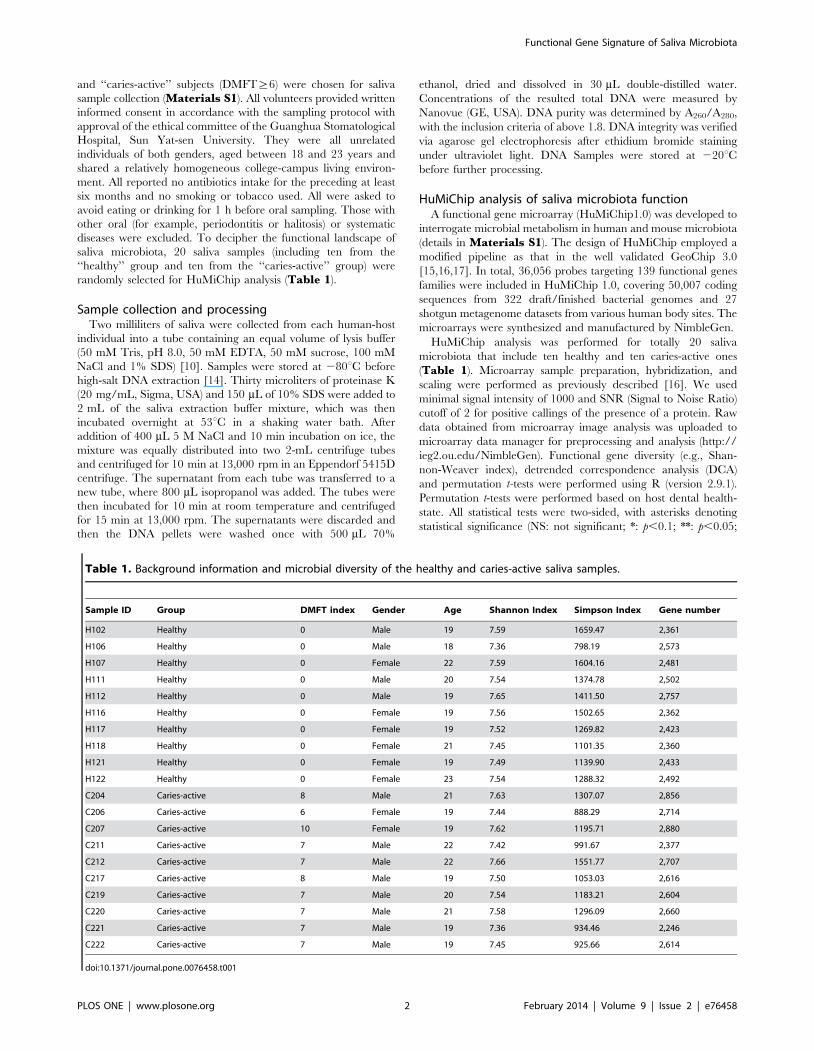
Table 1. Background information and microbial diversity of the healthy and caries-active saliva samples.
Sample ID Group DMFT index Gender Age Shannon Index Simpson Index Gene number
H102 Healthy 0 Male 19 7.59 1659.47 2,361
H106 Healthy 0 Male 18 7.36 798.19 2,573
H107 Healthy 0 Female 22 7.59 1604.16 2,481
H111 Healthy 0 Male 20 7.54 1374.78 2,502
H112 Healthy 0 Male 19 7.65 1411.50 2,757
H116 Healthy 0 Female 19 7.56 1502.65 2,362
H117 Healthy 0 Female 19 7.52 1269.82 2,423
H118 Healthy 0 Female 21 7.45 1101.35 2,360
H121 Healthy 0 Female 19 7.49 1139.90 2,433
H122 Healthy 0 Female 23 7.54 1288.32 2,492
C204 Caries-active 8 Male 21 7.63 1307.07 2,856
C206 Caries-active 6 Female 19 7.44 888.29 2,714
C207 Caries-active 10 Female 19 7.62 1195.71 2,880
C211 Caries-active 7 Male 22 7.42 991.67 2,377
C212 Caries-active 7 Male 22 7.66 1551.77 2,707
C217 Caries-active 8 Male 19 7.50 1053.03 2,616
C219 Caries-active 7 Male 20 7.54 1183.21 2,604
C220 Caries-active 7 Male 21 7.58 1296.09 2,660
C221 Caries-active 7 Male 19 7.36 934.46 2,246
C222 Caries-active 7 Male 19 7.45 925.66 2,614
doi:10.1371/journal.pone.0076458.t001
Functional Gene Signature of Saliva Microbiota
PLOS ONE | www.plosone.org 2 February 2014 | Volume 9 | Issue 2 | e76458
***: p,0.01). Array data were deposited at the Gene Expression
Omnibus with accession numbers GSE49875.
Statistical analysis in network reconstruction andbiomarker detection
The 3,656 functional genes with hybridization signals on
HuMiChip were grouped into ‘‘complete-presence proteins’’
(‘‘core’’, i.e., those present in all the 20 saliva microbiota) and
‘‘partial-presence proteins’’ (‘‘non-core’’, i.e., those missing in at
least one saliva microbiota). The ‘‘complete-presence proteins’’
were represented as normalized values according to their signal
intensity, while the ‘‘partial-presence proteins’’ as binary values
(either 1 or 0). The network of core functional genes was built
based on the normalized values of ‘‘all-presence genes’’ for the H
and C Groups respectively. The network of non-core functional
genes was based on the binary values of ‘‘partial-presence genes’’
on specific metabolic pathways that include Carbon-associated
Pathway (including ‘Complex carbohydrates’ and ‘Feeder pathways to
glycolysis’ and ‘Respiration’), AA-associated Pathway (including
‘‘Amino acid transport and metabolism’’ and ‘‘Amino acid synthesis’’) and
Nitrogen-associated Pathway (‘‘Nitrogen Metabolism’’).
To identify those markers that reliably distinguish caries
microbiota, the ten healthy samples were grouped into training
(seven samples) and testing (three samples) by all possible
combinations (thus 120 different groupings). Based on the binary
presence profiles of non-core genes, bootstrapping method was
used to randomly select a grouping and then used two steps
(‘‘feature selection’’ and ‘‘classification’’) to identify biomarkers
based on triplet feature selection. Features with the highest
discrimination power on the training data were selected and then
employed to ‘‘predict’’ the presence profiles of the testing data
(Figure S1; Materials S1). The biomarkers (each represented as
a triplet-feature set of microbial genes) identified after each of the
two steps were then subjected to manual inspections before
retrieving the final list of biomarkers.
Results
Functional gene diversity in healthy and caries-activesaliva microbiota
To interrogate microbial metabolisms in human and mouse
microbiota, we developed a functional gene microarray (HuMi-
Chip1.0) based on our well validated GeoChip 3.0 platform
[15,16,17]. HuMiChip 1.0 contains 36,056 oligonucleotide probes
targeting 139 functional genes families and covering 50,007 coding
sequences from 322 draft/finished bacterial genomes and 27
shotgun metagenome datasets from various human body sites
(Table S1; Materials S1). For a pilot-population of 20 human
adults (whose organismal structure were decoded [9]) that
included ten healthy (H Group) and ten caries-active (C Group)
(Table 1), metabolic functions of saliva microbiota were analyzed
via hybridizing the saliva DNA to the microarray. In total, 3,685
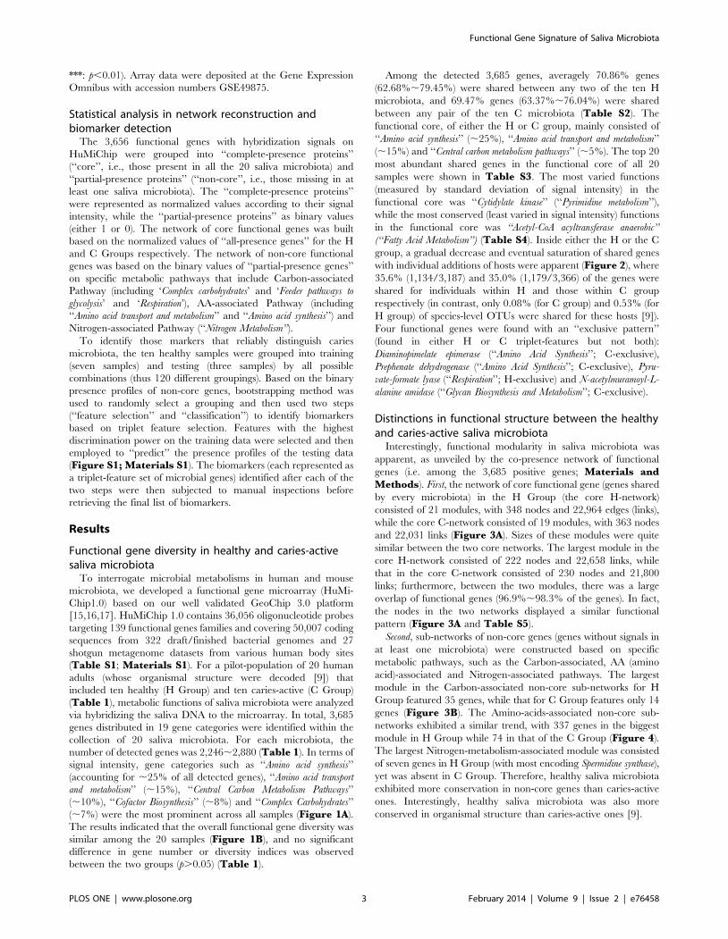
genes distributed in 19 gene categories were identified within the
collection of 20 saliva microbiota. For each microbiota, the
number of detected genes was 2,246,2,880 (Table 1). In terms of
signal intensity, gene categories such as ‘‘Amino acid synthesis’’
(accounting for ,25% of all detected genes), ‘‘Amino acid transport
and metabolism’’ (,15%), ‘‘Central Carbon Metabolism Pathways’’
(,10%), ‘‘Cofactor Biosynthesis’’ (,8%) and ‘‘Complex Carbohydrates’’
(,7%) were the most prominent across all samples (Figure 1A).
The results indicated that the overall functional gene diversity was
similar among the 20 samples (Figure 1B), and no significant
difference in gene number or diversity indices was observed
between the two groups (p.0.05) (Table 1).
Among the detected 3,685 genes, averagely 70.86% genes
(62.68%,79.45%) were shared between any two of the ten H
microbiota, and 69.47% genes (63.37%,76.04%) were shared
between any pair of the ten C microbiota (Table S2). The
functional core, of either the H or C group, mainly consisted of
‘‘Amino acid synthesis’’ (,25%), ‘‘Amino acid transport and metabolism’’
(,15%) and ‘‘Central carbon metabolism pathways’’ (,5%). The top 20
most abundant shared genes in the functional core of all 20
samples were shown in Table S3. The most varied functions
(measured by standard deviation of signal intensity) in the
functional core was ‘‘Cytidylate kinase’’ (‘‘Pyrimidine metabolism’’),
while the most conserved (least varied in signal intensity) functions
in the functional core was ‘‘Acetyl-CoA acyltransferase anaerobic’’
(‘‘Fatty Acid Metabolism’’) (Table S4). Inside either the H or the C
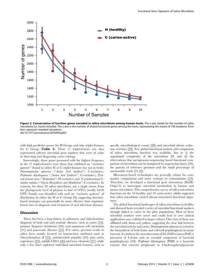
group, a gradual decrease and eventual saturation of shared genes
with individual additions of hosts were apparent (Figure 2), where
35.6% (1,134/3,187) and 35.0% (1,179/3,366) of the genes were
shared for individuals within H and those within C group
respectively (in contrast, only 0.08% (for C group) and 0.53% (for
H group) of species-level OTUs were shared for these hosts [9]).
Four functional genes were found with an ‘‘exclusive pattern’’
(found in either H or C triplet-features but not both):
Diaminopimelate epimerase (‘‘Amino Acid Synthesis’’; C-exclusive),
Prephenate dehydrogenase (‘‘Amino Acid Synthesis’’; C-exclusive), Pyru-
vate-formate lyase (‘‘Respiration’’; H-exclusive) and N-acetylmuramoyl-L-
alanine amidase (‘‘Glycan Biosynthesis and Metabolism’’; C-exclusive).
Distinctions in functional structure between the healthyand caries-active saliva microbiota
Interestingly, functional modularity in saliva microbiota was
apparent, as unveiled by the co-presence network of functional
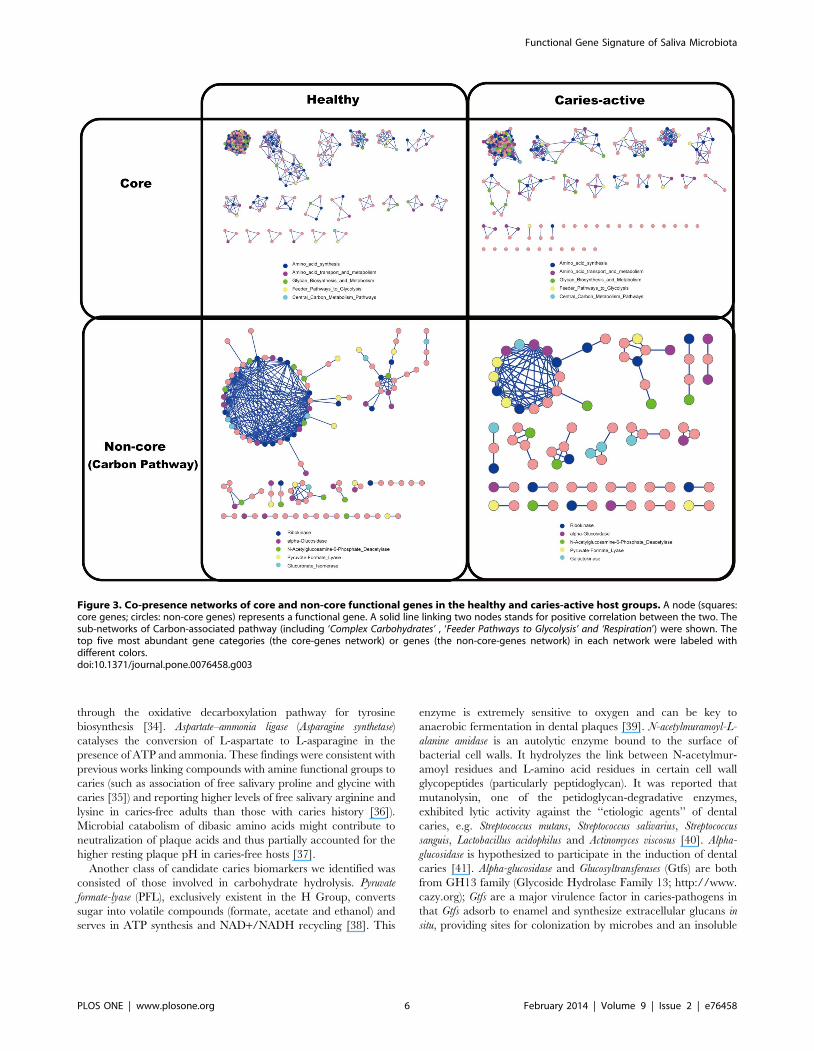
genes (i.e. among the 3,685 positive genes; Materials andMethods). First, the network of core functional gene (genes shared
by every microbiota) in the H Group (the core H-network)
consisted of 21 modules, with 348 nodes and 22,964 edges (links),
while the core C-network consisted of 19 modules, with 363 nodes
and 22,031 links (Figure 3A). Sizes of these modules were quite
similar between the two core networks. The largest module in the
core H-network consisted of 222 nodes and 22,658 links, while
that in the core C-network consisted of 230 nodes and 21,800
links; furthermore, between the two modules, there was a large
overlap of functional genes (96.9%,98.3% of the genes). In fact,
the nodes in the two networks displayed a similar functional
pattern (Figure 3A and Table S5).
Second, sub-networks of non-core genes (genes without signals in
at least one microbiota) were constructed based on specific
metabolic pathways, such as the Carbon-associated, AA (amino
acid)-associated and Nitrogen-associated pathways. The largest
module in the Carbon-associated non-core sub-networks for H
Group featured 35 genes, while that for C Group features only 14
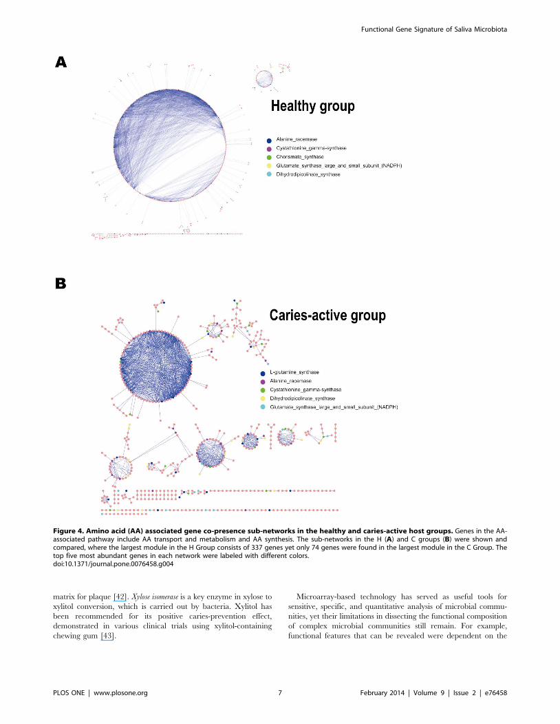
genes (Figure 3B). The Amino-acids-associated non-core sub-
networks exhibited a similar trend, with 337 genes in the biggest
module in H Group while 74 in that of the C Group (Figure 4).
The largest Nitrogen-metabolism-associated module was consisted
of seven genes in H Group (with most encoding Spermidine synthase),
yet was absent in C Group. Therefore, healthy saliva microbiota
exhibited more conservation in non-core genes than caries-active
ones. Interestingly, healthy saliva microbiota was also more
conserved in organismal structure than caries-active ones [9].
Functional Gene Signature of Saliva Microbiota
PLOS ONE | www.plosone.org 3 February 2014 | Volume 9 | Issue 2 | e76458
Functional gene markers of saliva microbiota that werelinked to caries
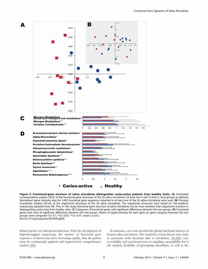
Although the overall functional gene diversity of saliva
microbial communities remained unchanged between the C and
H groups, their composition and structure were significantly
different as demonstrated by dissimilarity analysis (MRPP with p,
0.01) and detrended correspondence analysis (DCA) from all 3,685
detected genes on HuMiChip 1.0 (Figure 5A), indicating a
significant link between the host disease state and saliva microbiota
functioning. We have previously demonstrated a high degree of
divergence in organismal structure and a minimal organismal core
in human saliva microbiota among host individuals [9]. Our data
here showed that functional-gene structure of saliva microbiota
was able to distinguish the caries state from the healthy state of
human hosts. Thus a function-based strategy via HuMiChip
appears to be more effective than an organism-based strategy via
16S amplicon sequencing in our case (Figure 5B). Therefore,
functional gene structure of saliva microbiota can potentially be a
more reliable predictor of caries than established risk factors such
as Streptococcus mutans [18].
To understand the link between functional-gene structure of
saliva microbiota to caries-state, signal intensities of genes and
gene categories detected by HuMiChip were compared between
the two groups of hosts. Significant differences were detected for
gene categories of Complex carbohydrates, Nitrogen metabolisms and
Amino acid transport and metabolism (Figure 5C), and for functional
genes such as Xylose isomerase, N-acetylmuramoyl-L-alanine amidase,
Alpha-glucosidase, etc (Figure 5D). Through a ‘‘feature selection’’
strategy (Materials and Methods) based on the 2,822 non-core
functional genes, 1,247 triplet features were selected whose
accuracy was at least 80% each among all possible permutations
(Table S6). Among them, eight triplet-features were identified
Figure 1. Functional patterns of the ten healthy and ten caries-active human saliva microbiota. (A) Relative abundance of the functionalgenes among the 19 gene categories on HuMiChip 1.0. (B) Relative diversity of the functional genes among the 19 gene categories on HuMiChip 1.0.doi:10.1371/journal.pone.0076458.g001
Functional Gene Signature of Saliva Microbiota
PLOS ONE | www.plosone.org 4 February 2014 | Volume 9 | Issue 2 | e76458
with high predictive power for H Group, and nine triplet-features
for C Group (Table 2). These 17 triplet-feature sets thus
represented salivary microbial gene markers that were of value
in dissecting and diagnosing caries etiology.
Interestingly, those genes presented with the highest frequency
in the 17 triplet-features were those that exhibited an ‘‘exclusive
pattern’’ (found in either H or C triplet-features but not in both):
Diaminopimelate epimerase (‘‘Amino Acid Synthesis’’; C-exclusive),
Prephenate dehydrogenase (‘‘Amino Acid Synthesis’’; C-exclusive), Pyru-
vate-formate lyase (‘‘Respiration’’; H-exclusive) and N-acetylmuramoyl-L-
alanine amidase (‘‘Glycan Biosynthesis and Metabolism’’; C-exclusive). In
contrast, for these 20 saliva microbiota, not a single taxon, from
the phylogenetic level of phylum to that of OTUs (totally 9,628
OTU found), was identified with such an ‘‘exclusive pattern’’ of
distribution in either the H or C Group [9], suggesting function-
based strategies can potentially be more effective than organism-
based ones in diagnosis and treatment of oral infectious diseases.
Discussion
There has been a long history in sialometry and sialochemistry
diagnosis of both oral and systemic diseases, such as caries [19],
primary Sjogren’s Syndrome [20], oral squamous cell carcinoma
[21] and pancreatic diseases [22]. For caries, previous works in
saliva have mainly focused on human-host attributes such as
Glucosyltransferase B [23], antimicrobial peptides [24], past caries
experience [25], soluble CD14 [26] and trace elements [27], while
only a few have exploited individual microbial features, such as
specific microbiological counts [28] and microbial nitrate reduc-
tase activities [29]. Few global functional analysis and comparison
of saliva microbiota function was available, due to (i) the
organismal complexity of the microbiota [9] and (ii) the
observations that metagenome-sequencing based functional com-
parison of microbiota can be hampered by sequencing biases [30],
the paucity of reference genomes and the small percentage of
annotatable reads [31,32].
Microarray-based technologies are generally robust for com-
munity comparisons and more resistant to contaminants [33].
Therefore, we developed a functional gene microarray (HuMi-
Chip1.0) to interrogate microbial metabolism in human and
mouse microbiota. This comprehensive survey of saliva microbiota
functions on the 10 healthy and 10 caries-active adults suggested
that saliva microbiota carried disease-associated functional signa-
tures.
The global functional landscapes of saliva microbiota in healthy
and diseased hosts revealed a series of microbial functional markers
strongly linked to caries in the pilot populations. Most of these
microbial markers were novel and could lead to new clinical
applications once validated in larger cohorts. One class of them was
affiliated with Amino acid synthesis, suggesting the close link between
the microbial activity and caries. Diaminopimelate epimerase is central to
the biosynthesis of both lysine and cell-wall peptidoglycan in many
bacteria. It catalyzes the stereoinversion of LL-DAP to meso-DAP, a
precursor of L-lysine and an essential component of bacterial
peptidoglycans [34]. Prephenate dehydrogenase (PDH) is a bacterial
enzyme that converts prephenate to 4-hydroxyphenylpyruvate
Figure 2. Conservation of function genes encoded in saliva microbiota among human hosts. The x-axis stands for the number of salivamicrobiota (i.e. hosts) included. The y-axis is the number of shared functional genes among the hosts, representing the means of 100 iterations. Errorbars represent standard deviations.doi:10.1371/journal.pone.0076458.g002
Functional Gene Signature of Saliva Microbiota
PLOS ONE | www.plosone.org 5 February 2014 | Volume 9 | Issue 2 | e76458
through the oxidative decarboxylation pathway for tyrosine
biosynthesis [34]. Aspartate–ammonia ligase (Asparagine synthetase)
catalyses the conversion of L-aspartate to L-asparagine in the
presence of ATP and ammonia. These findings were consistent with
previous works linking compounds with amine functional groups to
caries (such as association of free salivary proline and glycine with
caries [35]) and reporting higher levels of free salivary arginine and
lysine in caries-free adults than those with caries history [36]).
Microbial catabolism of dibasic amino acids might contribute to
neutralization of plaque acids and thus partially accounted for the
higher resting plaque pH in caries-free hosts [37].
Another class of candidate caries biomarkers we identified was
consisted of those involved in carbohydrate hydrolysis. Pyruvate
formate-lyase (PFL), exclusively existent in the H Group, converts
sugar into volatile compounds (formate, acetate and ethanol) and
serves in ATP synthesis and NAD+/NADH recycling [38]. This
enzyme is extremely sensitive to oxygen and can be key to
anaerobic fermentation in dental plaques [39]. N-acetylmuramoyl-L-
alanine amidase is an autolytic enzyme bound to the surface of
bacterial cell walls. It hydrolyzes the link between N-acetylmur-
amoyl residues and L-amino acid residues in certain cell wall
glycopeptides (particularly peptidoglycan). It was reported that
mutanolysin, one of the petidoglycan-degradative enzymes,
exhibited lytic activity against the ‘‘etiologic agents’’ of dental
caries, e.g. Streptococcus mutans, Streptococcus salivarius, Streptococcus
sanguis, Lactobacillus acidophilus and Actinomyces viscosus [40]. Alpha-
glucosidase is hypothesized to participate in the induction of dental
caries [41]. Alpha-glucosidase and Glucosyltransferases (Gtfs) are both
from GH13 family (Glycoside Hydrolase Family 13; http://www.
cazy.org); Gtfs are a major virulence factor in caries-pathogens in
that Gtfs adsorb to enamel and synthesize extracellular glucans in
situ, providing sites for colonization by microbes and an insoluble
Figure 3. Co-presence networks of core and non-core functional genes in the healthy and caries-active host groups. A node (squares:core genes; circles: non-core genes) represents a functional gene. A solid line linking two nodes stands for positive correlation between the two. Thesub-networks of Carbon-associated pathway (including ‘Complex Carbohydrates’ , ‘Feeder Pathways to Glycolysis’ and ‘Respiration’) were shown. Thetop five most abundant gene categories (the core-genes network) or genes (the non-core-genes network) in each network were labeled withdifferent colors.doi:10.1371/journal.pone.0076458.g003
Functional Gene Signature of Saliva Microbiota
PLOS ONE | www.plosone.org 6 February 2014 | Volume 9 | Issue 2 | e76458
matrix for plaque [42]. Xylose isomerase is a key enzyme in xylose to
xylitol conversion, which is carried out by bacteria. Xylitol has
been recommended for its positive caries-prevention effect,
demonstrated in various clinical trials using xylitol-containing
chewing gum [43].
Microarray-based technology has served as useful tools for
sensitive, specific, and quantitative analysis of microbial commu-
nities, yet their limitations in dissecting the functional composition
of complex microbial communities still remain. For example,
functional features that can be revealed were dependent on the
Figure 4. Amino acid (AA) associated gene co-presence sub-networks in the healthy and caries-active host groups. Genes in the AA-associated pathway include AA transport and metabolism and AA synthesis. The sub-networks in the H (A) and C groups (B) were shown andcompared, where the largest module in the H Group consists of 337 genes yet only 74 genes were found in the largest module in the C Group. Thetop five most abundant genes in each network were labeled with different colors.doi:10.1371/journal.pone.0076458.g004
Functional Gene Signature of Saliva Microbiota
PLOS ONE | www.plosone.org 7 February 2014 | Volume 9 | Issue 2 | e76458
defined probe sets with known functions. With the development of
high-throughput sequencing, the number of functional gene
sequences of interest has been increasing rapidly, thus the probes
must be continuously updated and improved for comprehensive
analysis [44].
In summary, our work unveiled the global functional features of
human saliva microbiota. The sensitivity to host disease state, links
to systematic body functions (due to circulation; [45,46]), easy
accessibility and non-invasiveness in sampling, susceptibility for in
situ analysis, feasibility of genotyping microbiota, as well as the
Figure 5. Functional-gene structure of saliva microbiota distinguishes caries-active patients from healthy hosts. (A) Detrendedcorrespondence analysis (DCA) of the functional-gene structures of the 20 saliva microbiota (10 from the H and 10 from C host-groups) as defined.Normalized signal intensity data for 3,685 functional gene sequences detected in at least one of the 20 saliva microbiota were used. (B) PrincipalCoordinate Analysis (PCoA) of the organismal structures of the 20 saliva microbiota. The organismal structures were based on 16S-ampliconsequencing (adapted from [9]). Thus in this study functional-gene structure of saliva microbiota can be more sensitive than organismal structure indistinguishing caries state from healthy state. (C) Categories of functional genes with significant differences between the two groups. (D) Functionalgenes that were of significant differences between the two groups. Means of signal intensity for each gene (or gene category) between the twogroups were compared (*p,0.1; **p,0.05, ***p,0.01; mean6s.e.m.).doi:10.1371/journal.pone.0076458.g005
Functional Gene Signature of Saliva Microbiota
PLOS ONE | www.plosone.org 8 February 2014 | Volume 9 | Issue 2 | e76458
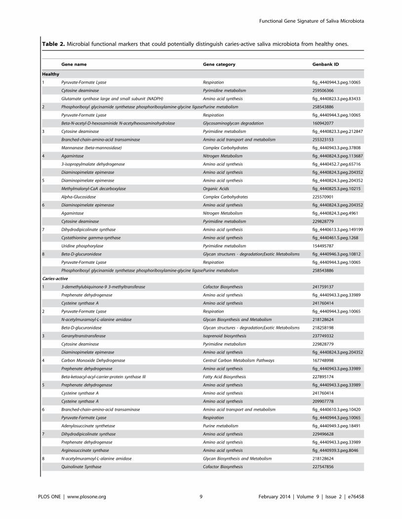
Table 2. Microbial functional markers that could potentially distinguish caries-active saliva microbiota from healthy ones.
Gene name Gene category Genbank ID
Healthy
1 Pyruvate-Formate Lyase Respiration fig_4440944.3.peg.10065
Cytosine deaminase Pyrimidine metabolism 259506366
Glutamate synthase large and small subunit (NADPH) Amino acid synthesis fig_4440823.3.peg.83433
2 Phosphoribosyl glycinamide synthetase phosphoribosylamine-glycine ligasePurine metabolism 258543886
Pyruvate-Formate Lyase Respiration fig_4440944.3.peg.10065
Beta-N-acetyl-D-hexosaminide N-acetylhexosaminohydrolase Glycosaminoglycan degradation 160942077
3 Cytosine deaminase Pyrimidine metabolism fig_4440823.3.peg.212847
Branched-chain-amino-acid transaminase Amino acid transport and metabolism 255323153
Mannanase (beta-mannosidase) Complex Carbohydrates fig_4440943.3.peg.37808
4 Agamintase Nitrogen Metabolism fig_4440824.3.peg.113687
3-isopropylmalate dehydrogenase Amino acid synthesis fig_4440452.7.peg.65716
Diaminopimelate epimerase Amino acid synthesis fig_4440824.3.peg.204352
5 Diaminopimelate epimerase Amino acid synthesis fig_4440824.3.peg.204352
Methylmalonyl-CaA decarboxylase Organic Acids fig_4440825.3.peg.10215
Alpha-Glucosidase Complex Carbohydrates 225570901
6 Diaminopimelate epimerase Amino acid synthesis fig_4440824.3.peg.204352
Agamintase Nitrogen Metabolism fig_4440824.3.peg.4961
Cytosine deaminase Pyrimidine metabolism 229828779
7 Dihydrodipicolinate synthase Amino acid synthesis fig_4440613.3.peg.149199
Cystathionine gamma-synthase Amino acid synthesis fig_4440461.5.peg.1268
Uridine phosphorylase Pyrimidine metabolism 154495787
8 Beta-D-glucuronidase Glycan structures - degradation;Exotic Metabolisms fig_4440946.3.peg.10812
Pyruvate-Formate Lyase Respiration fig_4440944.3.peg.10065
Phosphoribosyl glycinamide synthetase phosphoribosylamine-glycine ligasePurine metabolism 258543886
Caries-active
1 3-demethylubiquinone-9 3-methyltransferase Cofactor Biosynthesis 241759137
Prephenate dehydrogenase Amino acid synthesis fig_4440943.3.peg.33989
Cysteine synthase A Amino acid synthesis 241760414
2 Pyruvate-Formate Lyase Respiration fig_4440944.3.peg.10065
N-acetylmuramoyl-L-alanine amidase Glycan Biosynthesis and Metabolism 218128624
Beta-D-glucuronidase Glycan structures - degradation;Exotic Metabolisms 218258198
3 Geranyltranstransferase Isoprenoid biosynthesis 237749332
Cytosine deaminase Pyrimidine metabolism 229828779
Diaminopimelate epimerase Amino acid synthesis fig_4440824.3.peg.204352
4 Carbon Monoxide Dehydrogenase Central Carbon Metabolism Pathways 167748998
Prephenate dehydrogenase Amino acid synthesis fig_4440943.3.peg.33989
Beta-ketoacyl-acyl-carrier-protein synthase III Fatty Acid Biosynthesis 227895174
5 Prephenate dehydrogenase Amino acid synthesis fig_4440943.3.peg.33989
Cysteine synthase A Amino acid synthesis 241760414
Cysteine synthase A Amino acid synthesis 209907778
6 Branched-chain-amino-acid transaminase Amino acid transport and metabolism fig_4440610.3.peg.10420
Pyruvate-Formate Lyase Respiration fig_4440944.3.peg.10065
Adenylosuccinate synthetase Purine metabolism fig_4440949.3.peg.18491
7 Dihydrodipicolinate synthase Amino acid synthesis 229496628
Prephenate dehydrogenase Amino acid synthesis fig_4440943.3.peg.33989
Arginosuccinate synthase Amino acid synthesis fig_4440939.3.peg.8046
8 N-acetylmuramoyl-L-alanine amidase Glycan Biosynthesis and Metabolism 218128624
Quinolinate Synthase Cofactor Biosynthesis 227547856
Functional Gene Signature of Saliva Microbiota
PLOS ONE | www.plosone.org 9 February 2014 | Volume 9 | Issue 2 | e76458
extensive clinical knowledge base [47] and accumulating saliva-
omics data (e.g. the salivary proteome [48]) suggest saliva as an
advantageous venue and valuable research model for tracking
intricate interactions that underlie oral and even systemic diseases.
Supporting Information
Figure S1 Computational strategy for selecting the functional-
gene markers associated with caries in saliva microbiota.
(TIF)
Table S1 Probes and coding sequences on HuMiChip 1.0.
(DOCX)
Table S2 The percentages of microbial genes detected by
HuMiChip 1.0 that are shared between any pair of microbiota
from the 20 saliva microbiota.
(DOCX)
Table S3 The top 20 most abundant genes in the functional core
of the 20 saliva microbiota.
(DOCX)
Table S4 The most conserved and variable genes (in signal
intensity) in the functional cores of the 20 saliva microbiota.
(DOCX)
Table S5 Distribution of the functional-core genes in the healthy
and caries-active microbiota. Genes in both H and C groups were
shown.
(DOCX)
Table S6 Triplet feature set with high prediction power for
healthy and caries states of the hosts.
(DOCX)
Materials S1 Supplementary materials.
(DOC)
Author Contributions
Conceived and designed the experiments: FY JX. Performed the
experiments: FY XY ZC DG JY YZ. Analyzed the data: KN XZC QT
TY YD CH JVN XPC ZH. Contributed reagents/materials/analysis tools:
JZ. Wrote the paper: FY JX.
References
1. Qi X (2008) The Third National Sampling Epidemiological Survey on Oral
Health.
2. Kanasi E, Johansson I, Lu SC, Kressin NR, Nunn ME, et al. (2010) Microbial
risk markers for childhood caries in pediatricians’ offices. J Dent Res 89: 378–
383.
3. Keijser BJ, Zaura E, Huse SM, van der Vossen JM, Schuren FH, et al. (2008)
Pyrosequencing analysis of the oral microflora of healthy adults. J Dent Res 87:
1016–1020.
4. Lazarevic V, Whiteson K, Huse S, Hernandez D, Farinelli L, et al. (2009)
Metagenomic study of the oral microbiota by Illumina high-throughput
sequencing. J Microbiol Methods 79: 266–271.
5. Nasidze I, Quinque D, Li J, Li M, Tang K, et al. (2009) Comparative analysis of
human saliva microbiome diversity by barcoded pyrosequencing and cloning
approaches. Anal Biochem 391: 64–68.
6. Zaura E, Keijser BJ, Huse SM, Crielaard W (2009) Defining the healthy ‘‘core
microbiome’’ of oral microbial communities. BMC Microbiol 9: 259.
7. Lazarevic V, Whiteson K, Hernandez D, Francois P, Schrenzel J (2010) Study of
inter- and intra-individual variations in the salivary microbiota. BMC Genomics
11: 523.
8. Ling Z, Kong J, Jia P, Wei C, Wang Y, et al. (2010) Analysis of oral microbiota
in children with dental caries by PCR-DGGE and barcoded pyrosequencing.
Microb Ecol 60: 677–690.
9. Yang F, Zeng X, Ning K, Liu KL, Lo CC, et al. (2012) Saliva microbiomes
distinguish caries-active from healthy human populations. ISME J 6: 1–10.
10. Huang S, Yang F, Zeng X, Chen J, Li R, et al. (2011) Preliminary
characterization of the oral microbiota of Chinese adults with and without
gingivitis. BMC Oral Health 11: 33.
11. Griffen AL, Beall CJ, Campbell JH, Firestone ND, Kumar PS, et al. (2011)
Distinct and complex bacterial profiles in human periodontitis and health
revealed by 16S pyrosequencing. ISME J. 6.1176–1185.
12. (2012) Structure, function and diversity of the healthy human microbiome.
Nature 486: 207–214.
13. He Z, Deng Y, Van Nostrand JD, Tu Q, Xu M, et al. (2010) GeoChip 3.0 as a
high-throughput tool for analyzing microbial community composition, structure
and functional activity. ISME J 4: 1167–1179.
14. Quinque D, Kittler R, Kayser M, Stoneking M, Nasidze I (2006) Evaluation of
saliva as a source of human DNA for population and association studies. AnalBiochem 353: 272–277.
15. He Z, Deng Y, Van Nostrand JD, Tu Q, Xu M, et al. (2010) GeoChip 3.0 as a
high-throughput tool for analyzing microbial community composition, structureand functional activity. ISME J 4: 1167–1179.
16. Hazen TC, Dubinsky EA, DeSantis TZ, Andersen GL, Piceno YM, et al. (2010)
Deep-sea oil plume enriches indigenous oil-degrading bacteria. Science 330:204–208.
17. Lu Z, Deng Y, Van Nostrand JD, He Z, Voordeckers J, et al. (2012) Microbial
gene functions enriched in the Deepwater Horizon deep-sea oil plume. ISME J
6: 451–460.
18. Jiang Q, Yu M, Min Z, Yi A, Chen D, et al. (2012) AP-PCR detection ofStreptococcus mutans and Streptococcus sobrinus in caries-free and caries-active
subjects. Mol Cell Biochem.
19. Thomadaki K, Helmerhorst EJ, Tian N, Sun X, Siqueira WL, et al. (2011)Whole-saliva proteolysis and its impact on salivary diagnostics. J Dent Res 90:
1325–1330.
20. Baldini C, Giusti L, Ciregia F, Da Valle Y, Giacomelli C, et al. (2011) Proteomicanalysis of saliva: a unique tool to distinguish primary Sjogren’s syndrome from
secondary Sjogren’s syndrome and other sicca syndromes. Arthritis Res Ther 13:
R194.
21. Brinkmann O, Kastratovic DA, Dimitrijevic MV, Konstantinovic VS, JelovacDB, et al. (2011) Oral squamous cell carcinoma detection by salivary biomarkers
in a Serbian population. Oral Oncol 47: 51–55.
22. Farrell JJ, Zhang L, Zhou H, Chia D, Elashoff D, et al. (2011) Variations of oralmicrobiota are associated with pancreatic diseases including pancreatic cancer.
Gut.
23. Vacca Smith AM, Scott-Anne KM, Whelehan MT, Berkowitz RJ, Feng C, et al.
(2007) Salivary glucosyltransferase B as a possible marker for caries activity.Caries Res 41: 445–450.
24. Tao R, Jurevic RJ, Coulton KK, Tsutsui MT, Roberts MC, et al. (2005) Salivary
antimicrobial peptide expression and dental caries experience in children.Antimicrob Agents Chemother 49: 3883–3888.
25. Zhang Q, Bian Z, Fan M, van Palenstein Helderman WH (2007) Salivary
mutans streptococci counts as indicators in caries risk assessment in 6–7-year-oldChinese children. J Dent 35: 177–180.
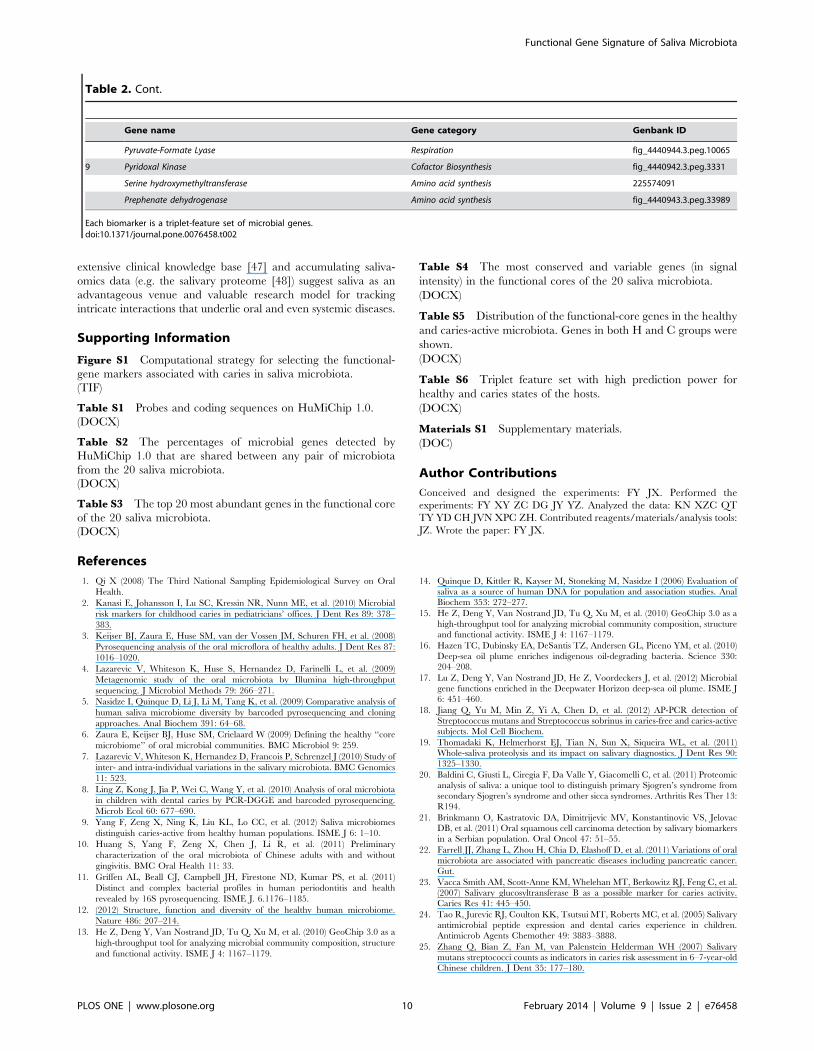
Table 2. Cont.
Gene name Gene category Genbank ID
Pyruvate-Formate Lyase Respiration fig_4440944.3.peg.10065
9 Pyridoxal Kinase Cofactor Biosynthesis fig_4440942.3.peg.3331
Serine hydroxymethyltransferase Amino acid synthesis 225574091
Prephenate dehydrogenase Amino acid synthesis fig_4440943.3.peg.33989
Each biomarker is a triplet-feature set of microbial genes.doi:10.1371/journal.pone.0076458.t002
Functional Gene Signature of Saliva Microbiota
PLOS ONE | www.plosone.org 10 February 2014 | Volume 9 | Issue 2 | e76458
26. Bergandi L, Defabianis P, Re F, Preti G, Aldieri E, et al. (2007) Absence of
soluble CD14 in saliva of young patients with dental caries. Eur J Oral Sci 115:93–96.
27. Zahir S, Sarkar S (2006) Study of trace elements in mixed saliva of caries free
and caries active children. J Indian Soc Pedod Prev Dent 24: 27–29.28. Martinez-Pabon MC, Ramirez-Puerta BS, Escobar-Paucar GM, Franco-Cortes
AM (2010) Physicochemical salivary properties, Lactobacillus, mutans strepto-cocci counts and early childhood caries in preschool children of Colombia. Acta
Odontol Latinoam 23: 249–256.
29. Doel JJ, Hector MP, Amirtham CV, Al-Anzan LA, Benjamin N, et al. (2004)Protective effect of salivary nitrate and microbial nitrate reductase activity
against caries. Eur J Oral Sci 112: 424–428.30. Morgan JL, Darling AE, Eisen JA (2010) Metagenomic sequencing of an in
vitro-simulated microbial community. PLoS One 5: e10209.31. Fodor AA, DeSantis TZ, Wylie KM, Badger JH, Ye Y, et al. (2012) The ‘‘most
wanted’’ taxa from the human microbiome for whole genome sequencing. PLoS
One 7: e41294.32. Xie G, Chain PS, Lo CC, Liu KL, Gans J, et al. (2010) Community and gene
composition of a human dental plaque microbiota obtained by metagenomicsequencing. Mol Oral Microbiol 25: 391–405.
33. He Z, Van Nostrand JD, Zhou J (2012) Applications of functional gene
microarrays for profiling microbial communities. Curr Opin Biotechnol.34. Ku HK, Do NH, Song JS, Choi S, Yeon SH, et al. (2011) Crystal structure of
prephenate dehydrogenase from Streptococcus mutans. Int J Biol Macromol 49:761–766.
35. Fonteles CS, Guerra MH, Ribeiro TR, Mendonca DN, de Carvalho CB, et al.(2009) Association of free amino acids with caries experience and mutans
streptococci levels in whole saliva of children with early childhood caries. Arch
Oral Biol 54: 80–85.36. Van Nieuw Amerongen A, Bolscher JG, Veerman EC (2004) Salivary proteins:
protective and diagnostic value in cariology? Caries Res 38: 247–253.37. Van Wuyckhuyse BC, Perinpanayagam HE, Bevacqua D, Raubertas RF,
Billings RJ, et al. (1995) Association of free arginine and lysine concentrations in
human parotid saliva with caries experience. J Dent Res 74: 686–690.
38. Yamamoto Y, Sato Y, Takahashi-Abbe S, Takahashi N, Kizaki H (2000)
Characterization of the Streptococcus mutans pyruvate formate-lyase (PFL)-
activating enzyme gene by complementary reconstitution of the In vitro PFL-
reactivating system. Infect Immun 68: 4773–4777.
39. Takahashi-Abbe S, Abe K, Takahashi N (2003) Biochemical and functional
properties of a pyruvate formate-lyase (PFL)-activating system in Streptococcus
mutans. Oral Microbiol Immunol 18: 293–297.
40. Thanyasrisung P, Komatsuzawa H, Yoshimura G, Fujiwara T, Yamada S, et al.
(2009) Automutanolysin disrupts clinical isolates of cariogenic streptococci in
biofilms and planktonic cells. Oral Microbiol Immunol 24: 451–455.
41. Mormann JE, Schmid R, Muhlemann HR (1983) Effect of alpha-amylase and
alpha-glucosidase inhibitors on caries incidence and plaque accumulation in rats.
Caries Res 17: 353–356.
42. Bowen WH, Koo H (2011) Biology of Streptococcus mutans-derived
glucosyltransferases: role in extracellular matrix formation of cariogenic biofilms.
Caries Res 45: 69–86.
43. Lif Holgerson P, Stecksen-Blicks C, Sjostrom I, Twetman S (2005) Effect of
xylitol-containing chewing gums on interdental plaque-pH in habitual xylitol
consumers. Acta Odontol Scand 63: 233–238.
44. He Z, Deng Y, Zhou J (2012) Development of functional gene microarrays for
microbial community analysis. Curr Opin Biotechnol 23: 49–55.
45. Mirzaii-Dizgah I, Riahi E (2011) Serum and saliva levels of cathepsin L in
patients with acute coronary syndrome. J Contemp Dent Pract 12: 114–119.
46. Xiao H, Zhang L, Zhou H, Lee JM, Garon EB, et al. (2011) Proteomic analysis
of human saliva from lung cancer patients using two-dimensional difference gel
electrophoresis and mass spectrometry. Mol Cell Proteomics.
47. Baum BJ, Yates JR 3rd, Srivastava S, Wong DT, Melvin JE (2011) Scientific
frontiers: emerging technologies for salivary diagnostics. Adv Dent Res 23: 360–
368.
48. Denny P, Hagen FK, Hardt M, Liao L, Yan W, et al. (2008) The proteomes of
human parotid and submandibular/sublingual gland salivas collected as the
ductal secretions. J Proteome Res 7: 1994–2006.
Functional Gene Signature of Saliva Microbiota
PLOS ONE | www.plosone.org 11 February 2014 | Volume 9 | Issue 2 | e76458