part 5. Preventive and treatment planning for dental caries
10
Oral diagnosis and treatment planning: part 5. Preventive and treatment planning for dental caries K. Yip 1 and R. Smales 2 VERIFIABLE CPD PAPER in the United Kingdom, patients who have regular dental care, including operative dentistry when necessary, are just as likely to require emergency dental treatment as those who visit a dentist infrequently. For all these reasons, the practice of preven- tive and minimal intervention dentistry or minimally invasive dentistry offers many advantages to patients and practitioners. There are many changes that have affected, and continue to affect, the practice of dentistry with increasing rapidity and with increasing demands on practitioners. FACTORS AFFECTING OPERATIVE DENTAL TREATMENT Changes in the population and dental workforce composition and numbers There are increasing numbers of elderly per- sons who want to retain their remaining dentitions, which are often periodontally involved, extensively worn, heavily restored and of poor appearance. Many of these elderly persons have deteriorating health and finances, and also sometimes unreal- istic expectations of what dental treatment can accomplish. Imbalances between the demand for dental treatment and the dental workforce have resulted in the closing and then the later re-opening of many dental schools and colleges, largely in response to political pressure from the dental profes- sion. However, changes in dental workforce composition and numbers seldom mirror INTRODUCTION Although operative dentistry can retain teeth and also restore function and aesthet- ics to improve the well-being of patients, it can be uncomfortable, time-consuming, repetitive and costly. But, as was shown The practice of operative dentistry continues to evolve, to reflect the many changes occurring in society and in dental diseases and conditions. However, the belief that all questionable and early carious lesions should be restored still persists. This belief is largely based upon the concept that the removal of all carious tissue followed by meticulous restoration of the tooth is the treatment of choice for dental caries. Yet restorations are not permanent and do not cure caries, as the causes remain. On the other hand, preventive measures can remove or partially remove the causes, thereby reducing the risks for future caries recurrence at the same site or elsewhere in the mouth. the ongoing optimum current population and economic requirements. Changes in dental diseases and conditions Over many years, there has been a sus- tained world-wide decrease in coronal den- tal caries in younger persons, but this trend has been reversed in several countries over the last decade in both children and young adults. The prevalence and severity of root surface caries, tooth wear, pulp pathoses and periodontal disease also increase markedly in elderly persons who have retained their natural teeth. Deterioration in their general health is often accom- panied by a severe deterioration in oral health. There has been a marked increase in the prevalence and severity of tooth erosion in younger persons in particular, affecting approximately 30% of children and adolescents. The reasons for these increases in dental disease may be related to a greatly increased consumption of hid- den refined sugars and acids in various foods and beverages, an increased use of xerogenic medications and illicit drugs, a decreased fluoridated water exposure, and the natural effects of retaining more teeth into old age. Changes in patients’ access to information and expectations Increased access to the internet and the production of ‘reality television body 1 Adjunct Professor, School of Dentistry, Charles Sturt University, Orange, New South Wales 2800, Australia; ,2* Visiting Research Fellow, School of Den‑ tistry, Faculty of Health Sciences, The University of Adelaide, Adelaide, South Australia 5005, Australia Correspondence to: Roger J. Smales Email: [email protected] Accepted 7 June 2012 DOI: 10.1038/sj.bdj.2012.774 © British Dental Journal 2012; 213: 211-220 • Highlights changes that have affected, and continue to affect, the practice of dentistry in relation to caries. • Stresses that the various preventive measures available will not be successful without the understanding and cooperation of a motivated patient. • Informs that studies of restoration failures have concluded that one in three restorations present at any one time is considered to be ‘unsatisfactory’. IN BRIEF PRACTICE Part 1. Introduction to oral diagnosis and treatment planning Part 2. Dental caries and assessment of risk Part 3. Periodontal disease and assessment of risk Part 4. Non-carious tooth surface loss and assessment of risk Part 5. Preventive and treatment planning for dental caries Part 6. Preventive and treatment planning for periodontal disease Part 7. Treatment planning for missing teeth Part 8. Reviews and maintenance of restorations *This series represents chapters 1, 7, 8, 9, 14, 15, 16 and 19 from the BDJ book A Clinical Guide to Oral Diagnosis and Treatment Planning, edited by Roger Smales and Kevin Yip. All other chapters are published in the complete clinical guide available from the BDJ Books online shop. ORAL DIAGNOSIS AND TREATMENT PLANNING* BRITISH DENTAL JOURNAL VOLUME 213 NO. 5 SEP 8 2012 211 © 2012 Macmillan Publishers Limited. All rights reserved.
-
Upload
khangminh22 -
Category
Documents
-
view
4 -
download
0
Transcript of part 5. Preventive and treatment planning for dental caries

Oral diagnosis and treatment planning: part 5. Preventive and treatment planning for dental cariesK. Yip1 and R. Smales2
VERIFIABLE CPD PAPER
in the United Kingdom, patients who have regular dental care, including operative dentistry when necessary, are just as likely to require emergency dental treatment as those who visit a dentist infrequently. For all these reasons, the practice of preven-tive and minimal intervention dentistry or minimally invasive dentistry offers many advantages to patients and practitioners.
There are many changes that have affected, and continue to affect, the practice of dentistry with increasing rapidity and with increasing demands on practitioners.
FACTORS AFFECTING OPERATIVE DENTAL TREATMENT
Changes in the population and dental workforce composition and numbersThere are increasing numbers of elderly per-sons who want to retain their remaining dentitions, which are often periodontally involved, extensively worn, heavily restored and of poor appearance. Many of these elderly persons have deteriorating health and finances, and also sometimes unreal-istic expectations of what dental treatment can accomplish. Imbalances between the demand for dental treatment and the dental workforce have resulted in the closing and then the later re-opening of many dental schools and colleges, largely in response to political pressure from the dental profes-sion. However, changes in dental workforce composition and numbers seldom mirror
INTRODUCTION
Although operative dentistry can retain teeth and also restore function and aesthet-ics to improve the well-being of patients, it can be uncomfortable, time-consuming, repetitive and costly. But, as was shown
The practice of operative dentistry continues to evolve, to reflect the many changes occurring in society and in dental diseases and conditions. However, the belief that all questionable and early carious lesions should be restored still persists. This belief is largely based upon the concept that the removal of all carious tissue followed by meticulous restoration of the tooth is the treatment of choice for dental caries. Yet restorations are not permanent and do not cure caries, as the causes remain. On the other hand, preventive measures can remove or partially remove the causes, thereby reducing the risks for future caries recurrence at the same site or elsewhere in the mouth.
the ongoing optimum current population and economic requirements.
Changes in dental diseases and conditions
Over many years, there has been a sus-tained world-wide decrease in coronal den-tal caries in younger persons, but this trend has been reversed in several countries over the last decade in both children and young adults. The prevalence and severity of root surface caries, tooth wear, pulp pathoses and periodontal disease also increase markedly in elderly persons who have retained their natural teeth. Deterioration in their general health is often accom-panied by a severe deterioration in oral health. There has been a marked increase in the prevalence and severity of tooth erosion in younger persons in particular, affecting approximately 30% of children and adolescents. The reasons for these increases in dental disease may be related to a greatly increased consumption of hid-den refined sugars and acids in various foods and beverages, an increased use of xerogenic medications and illicit drugs, a decreased fluoridated water exposure, and the natural effects of retaining more teeth into old age.
Changes in patients’ access to information and expectations
Increased access to the internet and the production of ‘reality television body
1Adjunct Professor, School of Dentistry, Charles Sturt University, Orange, New South Wales 2800, Australia;,2*Visiting Research Fellow, School of Den‑tistry, Faculty of Health Sciences, The University of Adelaide, Adelaide, South Australia 5005, Australia Correspondence to: Roger J. Smales Email: [email protected]
Accepted 7 June 2012 DOI: 10.1038/sj.bdj.2012.774 ©British Dental Journal 2012; 213: 211-220
• Highlights changes that have affected, and continue to affect, the practice of dentistry in relation to caries.
• Stresses that the various preventive measures available will not be successful without the understanding and cooperation of a motivated patient.
• Informs that studies of restoration failures have concluded that one in three restorations present at any one time is considered to be ‘unsatisfactory’.
I N B R I E F
PRA
CTICE
Part 1. Introduction to oral diagnosis and treatment planning
Part 2. Dental caries and assessment of risk
Part 3. Periodontal disease and assessment of risk
Part 4. Non-carious tooth surface loss and assessment of risk
Part 5. Preventive and treatment planning for dental caries
Part 6. Preventive and treatment planning for periodontal disease
Part 7. Treatment planning for missing teeth
Part 8. Reviews and maintenance of restorations
*This series represents chapters 1, 7, 8, 9, 14, 15, 16 and 19 from the BDJ book A Clinical Guide to Oral Diagnosis and Treatment Planning, edited by Roger Smales and Kevin Yip. All other chapters are published in the complete clinical guide available from the BDJ Books online shop.
ORAL DIAGNOSIS AND TREATMENT PLANNING*
BRITISH DENTAL JOURNAL VOLUME 213 NO. 5 SEP 8 2012 211
© 2012 Macmillan Publishers Limited. All rights reserved.

PRACTICE
makeover’ programmes have lead to an emphasis on improved dental appear-ance as part of social and employment expectations, and even as the basis for character judgements. There are increased expectations by some patients (of cos-metic procedures in particular) that may be unrealistic. The risks of iatrogenic damage, maintenance problems, failure and litigation increase with the costs and complexity of dental treatments. The cost of professional dental indemnity insur-ance has increased rapidly in recent years, and the need for documented case records and fully informed patient con-sent before treatment should be obvi-ous. Patients with persistent unrealistic expectations should not be accepted for dental treatment.
Changes in diagnostic and treatment methods
Increasingly expensive equipment is being purchased by practitioners for dental diagnosis and treatments, and for patient education and practice manage-ment. The increased practice costs have not been matched by the benefits paid by third parties for dental treatments, despite the increased insurance premiums paid by patients. There is an increased need for diagnostic methods that are more accurate and predictive of early dental disease for individuals, and for evidence-based infor-mation on the cost-effectiveness of differ-ent treatment alternatives.
Changes in dental treatments provided
In recent years, there has been an increase in adult endodontic, periodontic, ortho-dontic, dental implant, cosmetic and complex restorative treatments provided in dental practice. Though restorative dentistry still accounts for approximately 75-80% of the work and income in gen-eral practice, the types of procedures and the materials used have changed. There are fewer amalgams, gold castings and removable partial dentures, but more posterior resin composites and poly-mers, metal-ceramics and high-strength all-ceramics, fixed partial dentures and dental implants – all of which are more costly and many of which are finan-cially less cost-effective than alternative restorative treatments.
Changes in dental treatment philosophyAlong with the other changes occurring, there has been an increasing emphasis over the last two decades on preventive and on minimal intervention dentistry or mini-mally invasive dentistry (MID). This must involve patients in active participation and responsibility for their oral health. It also involves practitioners in increased patient education, clinical decision-making and treatment options, and monitoring of oral conditions. MID in its widest sense may be divided into three components:•Non-intervention: for example,
monitoring of unerupted impacted teeth, posterior tooth extraction spaces, caries in the primary dentition, and less-than-ideal restorations
•Prevention: for example, control of dental plaque microorganisms and erosive acids, use of fluorides and casein-derived remineralisation pastes, pit and fissure sealants, occlusal splints and sports mouthguards, control of tongue thrusting and digit sucking habits when indicated, and extraction of selected grossly carious posterior molar teeth in appropriate circumstances during the mixed dentition
•Preservation: for example, assisted remineralisation of tooth structure (topical fluorides, casein-derived remineralising pastes, saliva stimulation), use of pit and fissure sealants, small adhesive resin composite and glass-ionomer restorations, restoration refurbishments and repairs rather than replacements, overlaying or shoeing of weakened cusps, indirect pulp capping, tooth bleaching and ‘no-preparation’ veneers, and non-surgical endodontic and periodontal pocket therapy.
However, despite the obvious advan-tages of MID for the preservation of sound tooth structure and the reduction of often unnecessary invasive dentistry, concerns have been expressed by dental practition-ers. These concerns usually involve:•Problems with the early diagnosis and
individual risk assessment of dental diseases and conditions
• Inadequate patient and/or parental motivation and cooperation: ‘patients
want quick fixes subsidised by their health insurance fund’
•The reluctance by health funds to pay for some non-invasive procedures, and their concerns about possible fraudulent claims
•That using less-invasive procedures ‘will result in earning less income’
•The higher costs of additional diagnostic and micro-dentistry operative equipment
•The lack of controlled clinical trials, to prove the long-term success and cost-effectiveness of some MID procedures and restorative treatments in general practices.
Initial findings after two years from the intense non-invasive management of den-tal caries in many patients from multiple private practices have shown the positive efficacy of a preventive programme as compared with standard care restorative dental treatment.
PREVENTION OF PRIMARY CARIESMeasures taken to prevent the develop-ment of primary caries could include:•Fluoridation of domestic water supplies
at approximately 0.5+ ppm•Bacterial biofilm control, which could
involve: plaque removal by correct tooth brushing, use of dental floss, woodsticks, etc; chemical therapy by use of 0.2% chlorhexidine, essential oils, mouthrinses and baking soda; establishment of an adequate saliva flow
•Dietary counselling, which may include: avoiding sticky between-meal snacks; reducing refined sugar consumption – may be ‘hidden’ sugars; using non-cariogenic sugar-substitute sweeteners such as xylitol; increasing the consumption of non-cariogenic foods; finishing the meal with a detergent type of food such as cheese
•Fluoride toothpastes, mouthrinses, varnishes, gels and foams, and casein-derived remineralisation pastes (CPP-ACP)
•Placing resin-based/glass-ionomer pit and fissure sealants.
The various measures will not be success-ful without the understanding and coopera-tion of a motivated patient and/or parent/
212 BRITISH DENTAL JOURNAL VOLUME 213 NO. 5 SEP 8 2012
© 2012 Macmillan Publishers Limited. All rights reserved.

PRACTICE
guardian. Information should be provided and repeated at subsequent appointments in limited ‘bites’, a single key item at a time, using simple lay terms. Supplemental printed information should be appropriately written and illustrated for the age and level of understanding of the patient/parent. Not all persons can easily read and understand English, and an interpreter also may be required in some instances when English is not the native language.
PREVENTION OF SECONDARY CARIES (RECURRENT CARIES)
Measures taken to prevent the develop-ment of secondary caries could include:•Ensuring adequate removal of active
primary caries in the first instance•Reduction of plaque accumulation
adjacent to the restoration margins: correct proximal contouring of the contact area of the restoration;
smooth cavity/restoration margins; cavo-surface margin angles of 70º or more for amalgam and glass-ionomer restorations; prevention of gross marginal leakage - isolation of materials from moisture and: for amalgams: condense well on insertion, burnish margins during setting; for composites: oblique enamel rods at margins, plus acid-etch technique; for conventional glass-ionomers: protect initially with varnish/bonding agent; for luting cements: avoid weak, soluble materials
•Ensure that cavity margins are accessible and supragingival when possible
• Incorporate fluorides into appropriate restorative materials.
The management of dental caries and questionable restorations was discussed in
an article by Anusavice;1 based on infor-mation from this article, some of the ques-tions that may be asked include:•What is the treatment recommendation
concerning radiographic radiolucencies detected at the enamel-dentine junction or slightly into the dentine?
•What is the estimated mean time for progression of proximal caries through enamel and through dentine in permanent teeth in low-to-moderate risk patients?
•What are the problems in deciding whether to replace amalgams having faulty margins?
•What are the problems associated with replacing restorations?
•How does one assess if caries is active?•What methods are available for
controlling caries progression, and in which patients should they be used?
•When should pit and fissure sealants be used?
•When should preventive-resin restorations be used?
•At what stage in caries progression should restorations be considered?
•What is the difference between the surgical model and the medical model for the management of caries?
MANAGEMENT OF SMOOTH SURFACE LESIONS
The management of all dental carious lesions (smooth surface, pit and fissure, and root surface) depends on the extent of the lesion, which may be diagnosed and classified according to the simplified international caries detection and assess-ment system (ICDAS). Codes 1 and 2 for enamel caries and Code 1 for root caries record the visual changes in intact sur-faces caused by demineralisation, which are amenable to repair by remineralisation (Table 2, Part 2).2
Smooth surface carious lesions, includ-ing root surface lesions, usually progress relatively slowly, in enamel in particular, and require a different management from pit and fissure lesions. While the applica-tion of comprehensive preventive measures would cause any and all active carious lesions to become arrested, this compre-hensive approach is usually only required for high-risk individuals. Even small changes in dietary habits and a moderate increase in the use of topical fluorides and
Table 1 Survival of proximal tunnel and slot restorations in permanent teeth
Study Duration (yr) Success (%)
Proximal tunnel restorations (GIC):
Holst and Brännström, 1998 3.0 85
Pilbro et al., 1999, 2001 3.0 69
Strand et al., 2000 4.5 57
Lumley and Fisher, 1995 5.0 75
Nicolaisen et al., 2000 5.0 35
Hasselrot, 1998 7 39
Proximal slot restorations:
Kreulen et al., 1995 2.2 100
Kreulen et al., 1998 5.0 100
Lumley and Fisher, 1995 5.0 100
Nordbo et al., 1998 (saucer) 7.2 69
Table 2 Diagnostic and treatment planning considerations for occlusal pits and fissures
Clinical signs
Explorer No Yes Yes Yes
Discolouration* No No Yes Yes
Softness No No No Yes
DIAGNOdent 0‑13† 0‑13† 14‑20† >21†
Radiolucency dentine No No No Yes
Diagnosis Sound Sound Questionable Carious
Treatment options No treatment, or sealant Sealant
Sealant, or fissure restoration
Preventive resin restoration, or conventional restoration
*Opaque white, undermining enamel demineralisation. †Recommended cut‑off limits (Lussi et al., 20013). Adapted in 2009 from the paper: McConnachie I. The preventive resin restoration: a conservative alternative. J Can Dent Assoc, 1992; 58: 197–200 with permission.
BRITISH DENTAL JOURNAL VOLUME 213 NO. 5 SEP 8 2012 213
© 2012 Macmillan Publishers Limited. All rights reserved.

PRACTICE
casein-derived remineralisation pastes, or additional attention to plaque removal, may be sufficient to tip the balance towards remineralisation and arrest.
The decision to remineralise and arrest carious lesions
When one of the conditions to restore carious lesions does not apply, the most professional approach to specific lesions is as follows:•Record the site of the smooth surface
carious lesion•Show its presence to the patient•Teach the patient personalised
preventive measures that are relevant to the particular lesion and patient
•Tell the patient ‘theneedforfurthertreatment,andthusadditionalexpense,isinyourhands,foritisyouwhowillapplythepreventivemeasures’
•Reassess the lesion at recall intervals and act as necessary, using additional radiographs when indicated.
Use a hand mirror to show the patient any visible carious lesion sites and encour-age him or her to pay particular attention to oral hygiene at these sites, and to apply fluoride toothpaste regularly to the lesions. Depending on the age of the patient and the extent of active caries, fluoride tooth-pastes containing from 1,000-5,000 ppm fluoride can be used. The high-strength toothpastes and gels/foams usually are only available on prescription. For non-cavitated proximal surface lesions, inter-dental flossing or the use of extra fine dental woodsticks (Oral-B) is advisable. When sufficient gingival embrasure space is present, then interdental bristle brushes of various diameters can be used to con-vey fluoride toothpastes or casein-derived remineralisation pastes to the lesion sites. Fluoride varnishes (5% NaF), fluoride gels and foams (1.1% NaF) in trays, and fluoride mouthrinses (0.05% NaF daily, 0.2% NaF weekly) also are effective for managing active root surface caries in adults (Fig. 1).
However, despite their increasing usage for many clinical purposes, most com-mercial varnishes are not well accepted by patients because of having bitter tastes, opaque colour changes and rough surfaces. Acceptable products include VarnishAmerica Original/White (Medical Products Labs) and Duraflor Halo White
Varnish (Medicom, Inc.). Most preventive measures need to be given time to demon-strate their effect, so it is essential that their use is coupled where possible with a non-operative approach at the planning stage.
The decision to restore carious lesions
Recently, several clinical methods have been introduced either to infiltrate incipi-ent white spot lesions using an extremely low viscosity resin (Icon, and Icon-proximal, DMG), or to seal the surfaces of even more advanced but non-cavitated proximal surface lesions using a bonded thin polyurethane tape (Ivoclar Vivadent). Independent controlled clinical trials are required to evaluate these procedures in general practice.
It is necessary to have well-defined criteria for deciding when to attempt to arrest and when to restore a carious lesion. There are several conditions that usually indicate when a restoration is required. These include:•The tooth is sensitive to cold, heat,
sweetness, etc•Chewing is impaired because of
sensitivity•The coronal or radicular lesion can
be judged definitely to have extended well into the dentine
•The pulp is endangered•Previous attempts to arrest the lesion
have failed and there is evidence over several months or years that the lesion is progressing, and cavitation has occurred
•Tooth drifting is likely to occur through loss of a proximal contact area
•Periodontal tissue health is adversely affected because of food impaction
•The appearance is not acceptable to the patient.
The Simplified ICDAS Code 4 for cor-onal caries and Code 2 for root caries record visual and softening changes that are present in dentine, usually associated with frank cavitation that is not amenable to repair by remineralisation. Code 3 for coronal caries may be restricted to enamel breakdown without dentine involvement.2
When a restoration is required, then the size and form of the cavity prepara-tion should be largely determined by the extent of the carious lesion, rather than
by preconceived ‘extension for prevention’ outline forms.
Proximal ‘tunnel’ and proximal ‘slot’ restorations
The proximal tunnel restoration pro-vides access to proximal caries through a small occlusal cavity preparation, so as
Fig. 1 Root surface caries, and softened root surfaces caused by tooth erosion, will remineralise using fluoride varnish and/or casein-derived remineralisation pastes
Fig. 2 An extracted premolar tooth showing a small proximal surface cavity
Fig. 3 A tunnel preparation was restored through the occlusal access opening of the tooth in Fig. 2. Note the poorly finished proximal cavity margins and the incomplete marginal adaptation of the cement
214 BRITISH DENTAL JOURNAL VOLUME 213 NO. 5 SEP 8 2012
© 2012 Macmillan Publishers Limited. All rights reserved.

PRACTICE
to preserve the adjacent overlying intact marginal ridge. The tunnel may be either closed (‘blind’), or open when the proxi-mal enamel is perforated. Glass-ionomer cement (GIC) is the usual restorative mate-rial placed. The cement may be overlaid with resin composite to reduce occlusal wear of the cement. Many clinical trials
have found high failure rates, for closed tunnel preparations and for molar teeth in particular, from fractured marginal ridges, and residual and recurrent caries (Figs 2 and 3). The success of the technique is very operator dependent. Survival estimates for proximal tunnel and slot restorations are shown in Table 1. Small proximal slot or minibox preparations have recorded good clinical success rates, using either amal-gam or resin composite restorative materi-als (Figs 4 and 5). Failures from residual and recurrent caries were far lower than for tunnel restorations. Both slot and tunnel preparations remove similar amounts of tooth structure. Care must be taken during slot preparations not to damage the proxi-mal surfaces of the adjacent teeth. Such damage may occur in 60-70% of proxi-mal preparations in general and will result in more frequent caries of the damaged surfaces. The adjacent proximal surfaces should be protected when using rotary cut-ting instruments or, alternatively, oscillat-ing diamond-coated abrasive micro-tips (KaVo) may be used when preparing the proximal slot or minibox.
MANAGEMENT OF PIT AND FISSURE LESIONS
The overall reduction in the incidence of dental caries has largely involved smooth tooth surfaces, with relatively less effect on the incidence of pit and fissure caries which now dominates in industrialised countries.
The decision to remineralise and arrest carious lesions
While pit and fissure lesions may become arrested, it is difficult to prove that such arrest has occurred and that active car-ies progression has ceased. One method of monitoring suggested for small question-able lesions, is by measuring the laser light reflectance from pits and fissures with the DIAGNOdent (KaVo), and the Spectra Caries Detection Aid (Air Techniques). The measurements must be made under spe-cific conditions to avoid possible spurious results. Incorrect high readings may be caused by organic material in the occlusal fissures, by fluorescent prophylaxis pastes, resin-based materials, and white or opaque and hypomineralised enamel.
Repeated topical applications of fluorides and other agents to occlusal pits
and fissures appear to have had limited cost-effective benefits in preventing long-term caries progression at these sites. This situation contrasts with early smooth sur-face lesions that are more amenable to visual or radiographic assessments over time, and for which such preventive treat-ments are more effective and can be more readily monitored.
The decision to prevent and restore carious lesions
The practical alternatives to managing questionable carious pits and fissures are:•To apply a resin-based or a glass-
ionomer pit and fissure sealant, to seal the site from the oral environment. (May be used both as a preventive and a therapeutic procedure in selected fissures in selected patients)
•To widen the fissure (fissurotomy) using a very thin tapering diamond point or tungsten carbide bur and to restore the narrow V-shaped preparation, which is largely within enamel, with a flowable resin composite as a ‘fissure filling’ or fissure restoration
•To remove the fissure entirely and to restore the relatively large cavity preparation in the conventional manner with a posterior resin composite
•To use a combination of techniques, as when placing a preventive resin restoration (PRR). Any carious lesion found is removed and the small cavity restored with a resin composite, then the restoration and the contiguous apparently sound pits and fissures are covered with a sealant. Alternatively, the cavity is restored using a flowable resin composite that is also extended over the adjacent pits and fissures.
Possible treatment options for occlusal pits and fissures are shown in Table 2.
Pit and fissure sealing as a preven-tive and a therapeutic procedure
Resin-based sealants have been developed and used over approximately 40 years to provide a solution to the problem of pre-venting caries in pits and fissures. Several studies have found that after five years, sealed permanent molar teeth in children had approximately 50% fewer pit and fissure restorations placed than in the
Fig. 4 Small proximal slot amalgam restorations after three years
Fig. 5 A proximal slot resin composite restoration in the mesial of the first molar
Fig. 6 A lightly filled resin-based occlusal pit and fissure sealant in the first molar
Fig. 7 After two years, part of the sealant shown in Fig. 6. Placement of sealants is considered
BRITISH DENTAL JOURNAL VOLUME 213 NO. 5 SEP 8 2012 215
© 2012 Macmillan Publishers Limited. All rights reserved.

PRACTICE
corresponding unsealed teeth. The previ-ously sealed teeth had less extensive resto-rations, and sealants delayed the need for restorations, but were less effective with time. Sealant losses are 5-10% per year for occlusal surfaces, and 30% per year for buccal and palatal pits in permanent molars (Figs 6 and 7). Inadequate sealant retention is associated with aprismatic fis-sure enamel, and with fissure debris that may occlude more than half of the fis-sure volume in adults, leading to poor acid etching and sealant penetration. In adults, fissure fillings are preferable to sealants for heavily stained pits and fissures.
Placement of sealants is considered to be cost-effective in selected posterior teeth in selected patients. These situations include newly erupted teeth and teeth with habitually plaque-covered fissures, and where patients have mental disabilities or severe anxiety problems that would make any future restorative treatment difficult. Sealant use should be seen as an adjunct to a total caries preventive programme that also includes optimum fluoride and rem-ineralisation paste treatments, adequate salivary flow, restricted frequency of sticky refined carbohydrate intake, and regular patient recalls. When moisture control is difficult to achieve, as in children with partially-erupted molar teeth, then fast-setting glass-ionomer cement should be used as the sealant material.
Sealants have an advantage over restor-ative methods in that cavity preparation is not required and the tooth remains intact. However, it is essential that the fissures be cleaned of all debris, using air polishing, air abrasion, lasers or fissurotomy burs. Conventional occlusal amalgam restora-tions in particular weaken a tooth, whereas sealants do not. Therefore, it is difficult to accept a clinical decision to wait until obvious dental caries develops with frank cavitation, and then to place a much larger restoration. However, old non-maintained worn and stained resin-based sealants in non-smokers are associated with a very high caries risk, and may perhaps require large replacement restorations. Thus, occlusal sealants require frequent recall examinations, resealings when needed, and radiographs to detect possible caries extending into dentine.
The avoidance of a restoration, with its attendant drawbacks, also points to
a therapeutic role for fissure sealants for established small non-cavitated carious pit and fissure lesions in erupted teeth. However, this role would only seem appro-priate in ituations where the unfilled or lightly-filled resin-based sealant is afforded some protection from direct occlusal con-tact with the opposing tooth, to reduce the potential for sealant wear and marginal leakage. A more appropriate treatment method when small non-cavitated carious fissure lesions are present, especially in adults, is the placement of either a narrow V-shaped fissure filling, or a PRR.
Preventive resin restoration (PRR)Many investigators have studied so-called ‘preventive resin restorations’ (sealant-res-torations) in occlusal tooth surfaces. The PRR is a variant of pit and fissure sealing. An exploratory excavation of the carious pit or fissure is made with a small round bur. The carious tissue is removed, but no attempt is made to extend the cavity to include contiguous fissures considered to be non-carious. An etched-retained flowable resin composite may then be used to fill the cavity, and to seal over the remaining adjacent fissures. Some clinicians prefer to use a resin-based pit and fissure sealant material for the latter purpose. Conventional and resin-modified glass-ionomer cements may be placed in the cavities as alternative restorative mate-rials; (conventional glass-ionomer cement is used to fill the cavity preparation and contiguous fissures in the ‘press-finger’ atraumatic restorative treatment (ART) method). Clearly, the PRR offers increased diagnostic safety with respect to caries at the base of the fissure.
The argument that the PRR is a par-ticularly good treatment method, in that it guards against a missed diagnosis of dental caries, is countered by that it will inevitably also be responsible for occa-sional operative treatments when no lesion exists. However, by comparison with much larger conventional cavity preparations, a small cavity preparation commenced in enamel for a PRR can easily be stopped and repaired when a misdiagnosis is discovered.
Indications•For minimal discrete occlusal pit and
fissure caries, where the remaining
tooth substance is shown on radiographs to be free of carious lesions
•Where a blunt explorer catches in a pit and fissure
•Where deep pits and fissures are present•Where the prognosis using only a
fissure sealant is uncertain.
Contraindications•Presence of large occlusal surface
carious lesions•Presence of interproximal carious
lesions•Situations in which caries is
uncontrolled.
Advantages•Minimal removal of tooth structure•Better aesthetics than when using
amalgam•Local anaesthesia not always required•Prevention of caries in adjacent pits
and fissures•Restoration can be completed in one
visit.
Disadvantages•Absolute need for moisture control for
resin-based composites•Strict adherence to the principles of
the acid-etch technique• Inability of resin-based materials to
release adequate fluoride ions (cf. glass-ionomers) or corrosion products (cf. amalgams) if microleakage occurs
•Takes longer to carry out compared with an amalgam restoration.
Clinical procedure•Administer local anaesthesia if
necessary• Isolate the tooth with rubber dam or
cotton rolls•Remove the caries with a small round
diamond point or tungsten carbide bur•Provide pulpal protection if necessary,
using a glass-ionomer cement•Etch entire occlusal surface using a
phosphoric acid gel, or use a self-etch adhesive
•Place a dentine bonding agent•Place a posterior resin composite into
the cavity preparation•Apply sealant over the restoration and
adjacent previously-etched fissures•Equilibrate the occlusion (adjust using
articulating foil and selective grinding).
216 BRITISH DENTAL JOURNAL VOLUME 213 NO. 5 SEP 8 2012
© 2012 Macmillan Publishers Limited. All rights reserved.

PRACTICE
The PRR has been shown to be a suc-cessful treatment method, although hav-ing widely varying survivals, as shown in Table 3. Approximately 80% of resto-rations survived for five years. The most common cause of failure was wear of the resin sealant, which could be compensated for by the addition of more material at a recall visit. Wear may be reduced by ini-tially using a filled resin-based sealant or a flowable resin composite. The advan-tage of the PRR in the saving of sound
tooth structure and the possible preven-tion of future molar tooth fractures is a major consideration for the selection of this restorative technique.
THE SELECTION OF RESTORATIVE MATERIALS
There is a fairly large range of generic materials available for direct placement and indirect placement dental restorations, with numerous and constantly chang-ing branded products marketed, often
with no controlled clinical trials having been undertaken. The American Dental Association (www.ada.org) has published useful information for dental practitioners and for the public, comparing the advan-tages and disadvantages, and appropriate uses of commonly used materials: com-parison of direct restorative dental materi-als and comparison of indirect restorative dental materials.
From many studies, the usual longevi-ties or survivals of restorations are shown in Table 4. The selection of a particular generic type and brand of restorative material, and the particular type of resto-ration to be placed for a particular patient, is dependent on many factors. However, the final selection decision should be based largely on the most cost-effective evidence available from long-term clinical studies undertaken in general dental practices. Although relatively few in number, such studies suggest that amalgam is the pre-ferred cost-effective material for larger posterior restorations and resin composite or porcelain veneers for larger anterior restorations. Where posterior full veneer crowns are indicated, particularly in high-stress situations, then gold and cer-amo-metal materials are preferred. Large posterior resin composite restorations and all-ceramic crowns should be restricted largely to critical aesthetic situations. The amount of tooth tissue loss involved in the initial tooth preparation, and poten-tially in subsequent crown and restoration replacements, should be an important fac-tor involved in the selection of a particular restorative material and procedure.
THE SUCCESS AND FAILURE OF OPERATIVE DENTISTRY
Studies concerned with the prevalence of restoration failures have concluded that one in three restorations present at any one time is considered to be ‘unsatisfac-tory’. Therefore, it is no surprise to find that restorations are often not very durable in general dental practices. Indeed, the aver-age survival times of routine intra-coronal restorations in adults have been stated in many studies to be only approximately 5-10 years. However, it is important to dis-tinguish between restoration replacement rates and restoration failure rates in gen-eral dental practices. The former rates may well be much higher than the latter for
Table 3 Survival of preventive resin restorations in permanent teeth
Study Duration (years) Success*(%)
Simonsen and Stallard, 1977 1.0 100
Walker et al., 1990 1.3† 82
Houpt et al., 1982 1.5 91
Walls et al., 1988 2.0 97
Simonsen and Jensen, 1979 2.5 96
Raadal, 1978 2.5 84
Simonsen, 1980 3.0 99
Houpt et al., 1984 3.0 77
Houpt et al., 1985 4.0 64
Welbury et al., 1990 5.0 95
Houpt et al., 1988 6.5 68
Simonsen and Landy, 1984 7.0 90
Houpt et al., 1994 9.0 75
Mertz‑Fairhurst et al., 1998 10.0 87
*Complete retention and caries free. †Average duration. (Adapted from Ripa and Wolff, 1992; Tyas et al., 2000)
Table 4 Longevities of restorations in general dental practice
Restoration Range (years)
Amalgam 8‑12
Anterior resin composite 7‑10
Posterior resin composite 5‑8
Glass‑ionomer cement (GIC) 5‑8
Resin pit and fissure sealant 5‑7
Resin‑bonded bridgework (fixed partial dentures) 6‑8
Conventional crown and bridgework (fixed partial dentures) 15‑18
Ceramic inlay/onlay 10‑16
Cast gold inlay/onlay 12‑15
Anterior porcelain veneer 12‑18
Cast and metal‑ceramic crowns 15‑20
NB: Extended restoration longevities have been shown in clinical studies from dental schools and private patient dental practices
BRITISH DENTAL JOURNAL VOLUME 213 NO. 5 SEP 8 2012 217
© 2012 Macmillan Publishers Limited. All rights reserved.

PRACTICE
reasons largely unrelated to strictly defined criteria for restoration replacement.
As restorations are placed and replaced, the cavities become larger and the teeth become weaker (Figs 8-11). Dentists have an urge to ‘freshen up’ the margins of cav-ity preparations, irrespective of their qual-ity. This insidious removal of tooth tissue partly explains why an increased provision of crowns has been reported, providing further evidence that restorative treatment generates further increasingly complex and costly restorative treatments. In vivo studies have shown that replacement res-torations frequently contained the same errors as their predecessors. However, the clinicians involved were generally una-ware of the faults in their new restorations, for they assessed them all as being sat-isfactory, with approximately 90% being excellent in all respects.
ASSESSING RESTORATIONSWhen examining the margins of restora-tions, a ‘catch’ by a sharp dental explorer is often erroneously considered to provide grounds for their replacement even when secondary caries is not detected. Up to 40% of decisions to replace restorations have been reported to be for the reason that the existing restorations are of dubi-ous integrity, though without associated caries or suspected caries. Other studies have provided more detailed information regarding the dubious integrity, stating poor marginal adaptation and anatomic form, isthmus and tooth fractures, and discoloration as specific reasons for replacing restorations.
It is important to recognise the signifi-cance of discoloration of tooth-coloured restorations, as some patients are particu-larly sensitive to even a minor mismatch in colour between the tooth and the restora-tion. It is necessary to differentiate between marginal discoloration, surface discolora-tion and body discolouration of a restora-tion. Marginal discolouration is a sign of microleakage, where the tooth/restoration interface is not adequately sealed. However, such discolouration may be very limited and not penetrating. Surface discoloura-tion may be due to the intake of particu-lar foods, drinks or medicines, and simple polishing of the restoration may solve or improve this situation. Body discolouration is primarily a material defect, provided the
restoration originally matched the colour of the tooth. In some instances, vital tooth bleaching will result in colour mismatches appearing. The patient’s assessment of the condition is often decisive in deciding the need for treatment.
In deciding whether or not to replace a restoration, it is necessary to weigh the advantages against the disadvantages. The argument that the dental pulp might be damaged if a suspect restoration is not replaced should be seen in proper perspec-tive against the disadvantages.
The decision to replace or to repair existing restorations
Restorations should only be replaced or repaired when there is actual evidence of adverse biological, functional or aesthetic (appearance as judged by the patient)
effects directly related to the restoration. Evidence of restoration deterioration alone does not constitute an adequate reason for restoration replacement or repair (Figs 12 and 13).
Many restorations finally fail (often many years later), for reasons completely unrelated to the deterioration of either their margins, surface texture, surface contour or colour match. There is scant evidence available that justifies ‘replace-ment for preventive reasons’ on the basis of restoration deterioration. The follow-ing criteria have been stated for restoration replacement or repair:•Caries adjacent to the restoration
extends into the dentine, and is active•The restoration has failed for
biological, functional or aesthetic reasons
Fig. 8 Preoperative occlusal view of an extensively restored maxillary arch
Fig. 11 Postoperative occlusal view following the replacement of several amalgam restorations
Fig. 9 Postoperative occlusal view showing the much larger replacement amalgam restorations
Fig. 12 Despite the exposed surface voidsand associated staining, the resin composite restorations remain functionally and aesthetically adequate
Fig. 10 Preoperative occlusal view of the opposing restored mandibular arch
Fig. 13 The presence of marginal fractures and staining are insufficient reasons for replacing the amalgam restoration. The appearance can be improved by refurbishment
218 BRITISH DENTAL JOURNAL VOLUME 213 NO. 5 SEP 8 2012
© 2012 Macmillan Publishers Limited. All rights reserved.

PRACTICE
•The restoration has appeared to cause an allergic response. (Mild responses should be observed for several weeks to determine if the reaction is self-limiting)
•When the patient requests the removal of the restoration because of undue psychological stress associated with it. (The consequences of restoration removal and replacement should be explained to and accepted by the patient).
RESTORATION REPLACEMENTSThe complete replacement of restora-tions usually results in less sound tooth structure:•Removal of resin composites is usually
slow and incomplete, and results in approximately 30% increased loss of sound tooth structure
•Removal of amalgams and glass-ionomers is usually faster and more complete, and results in little increased loss of sound tooth structure
•Preparations for indirect restorations are more destructive of sound tooth
structure than for direct placement restorations
•Larger preparations are associated with reduced restoration longevities.
Restoration repairsRelatively more existing amalgams are repaired, or simply refurbished, than are replaced. However, despite the increased loss of sound tooth structure that occurs during the replacement of resin compos-ites, relatively few are repaired or even refurbished. Dentists are often reluc-tant to repair existing restorations for several reasons:•Concerns about residual caries and
weakened restorations and tooth structure
•Beliefs that repairs are an inferior treatment, which may have been instilled by their teachers
•Lack of clinical information on the relative cost-effectiveness of repairs
•Less economic inducement.
The relative advantages and disadvan-tages of existing restoration repairs are shown in Table 5. Before existing resto-rations are repaired, or dislodged indi-rect restorations are re-cemented, the functional significance of the tooth and its prognosis should be determined. The
Table 5 Relative advantages and disadvantages of restoration repairs
Advantages Disadvantages
Less sound tooth structure destroyed Inadequate access for caries removal and placement of repair material
Less potential pulp damage Potential damage to adjacent approximal tooth surfaces and gingival tissues
Less pain and discomfort May compromise strength of remaining tooth and restoration, and restoration retention
Extended longevity of restoration May be problems with shade matching
Table 6 Preventive and progressive restorative treatment and material options
Clinical condition Treatment options Preventive-restorative material options
Questionable occlusal fissure caries. Probably sound
Oral hygiene instruction, fluorides, seal pits and fissures Fluoride varnish, resin‑based sealant or glass‑ionomer cement sealant
Questionable proximal caries. Probably sound Hygiene instruction, fluorides, observe and evaluate Topical fluoride gel, casein‑derived remineralisation pastes
‘White spots’ Protect against further demineralisation, observe Fluorides, chlorhexidine, casein‑derived pastes and/or sealant
Arrested occlusal fissure and smooth surface caries
No treatment, protect with sealant, or restore if unaes‑thetic, or if a cavity exists
Sealant and/or fluorides, glass‑ionomer cement, resin composite or amalgam restoration
Incipient active occlusal fissure caries Diet control, oral hygiene, fluorides and/or chlorhexidine and radiographic monitoring, preventive resin restoration
Fluoride varnish, chlorhexidine and/or fissure filling/preventive resin restoration
Non‑cavitated active proximal caries up to one‑third through dentine
Diet control, oral hygiene, fluorides and/or chlorhexidine and radiographic monitoring
Fluoride gel, chlorhexidine, casein‑derived remineralisa‑tion pastes and/or resin‑based sealant
Moderate to extensive active proximal caries one‑third or more through dentine
Restore, proximal surface cavity is probably present Glass‑ionomer, resin composite or amalgam restoration
Defective restoration Seal margins, refurbish, repair or replace if cannot be repaired
Materials used depend on whether restoration is sealed, repaired or replaced
Failed restoration ‑ because of biological, functional, or aesthetic problems
Restore and protect with onlay or crown if tooth struc‑ture loss is likely to result in fracture
Anterior: composite, glass‑ionomer, ceramo‑metal crown (CMC) or all‑ceramic crown. Posterior: amalgam, composite, cast‑metal inlay/onlay/ crown, CMC or all‑ceramic crown
Endodontically treated tooth Restore with plastic material Anterior: CMC or all‑ceramic crown. Posterior: cast‑metal onlay/crown, CMC or all‑ceramic crown
Fractured tooth Endodontic treatment and restore. Extract if non‑restorable
Amalgam or composite restoration. Anterior: CMC or all‑ceramic crown. Posterior: cast‑metal onlay/crown, CMC or all‑ceramic crown
Unacceptable appearance Vital bleach if feasible, restore with resin composite, CMC or all‑ceramic crown
Anterior: composite restoration, ceramic veneer, all‑ceramic crown. Posterior: ceramic or resin composite inlay/onlay, CMC or all‑ceramic crown
Anusavice K. Protocol for conservative treatment decisions. JADA 1995;126; Table 2 Copyright© 1995 American Dental Association. All rights reserved. Adapted 2012 with permission of the American Dental Association
BRITISH DENTAL JOURNAL VOLUME 213 NO. 5 SEP 8 2012 219
© 2012 Macmillan Publishers Limited. All rights reserved.
PRACTICE
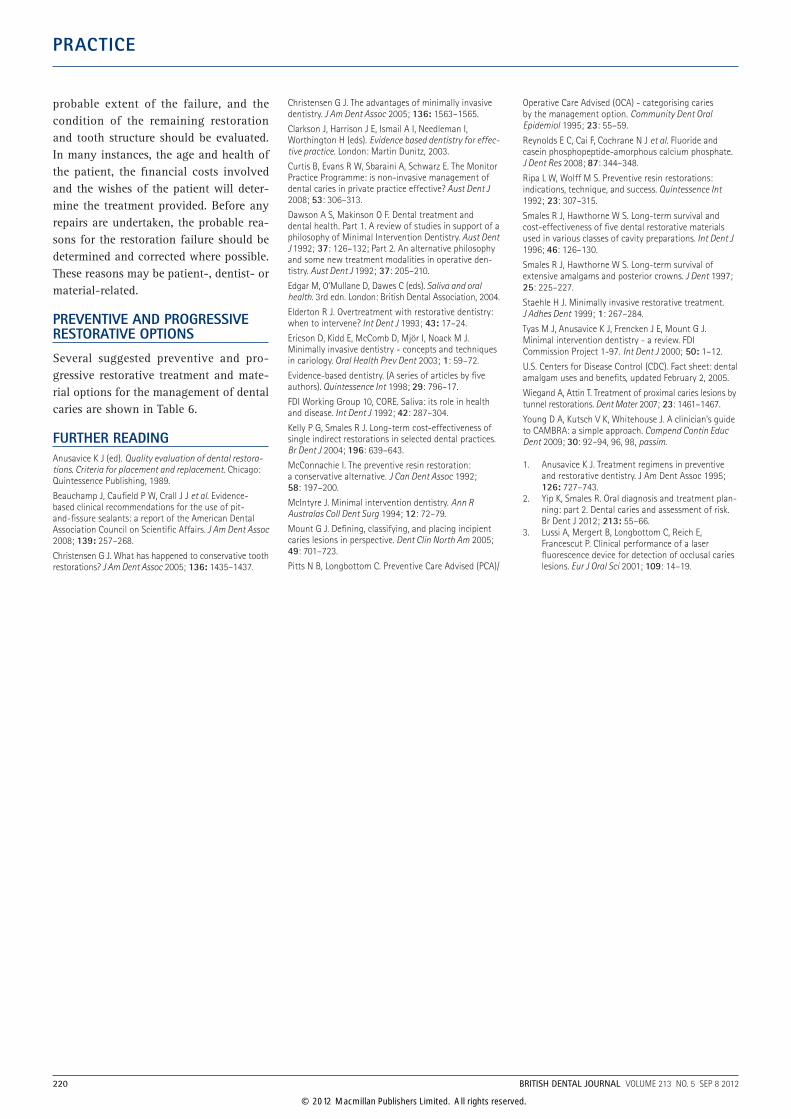
probable extent of the failure, and the condition of the remaining restoration and tooth structure should be evaluated. In many instances, the age and health of the patient, the financial costs involved and the wishes of the patient will deter-mine the treatment provided. Before any repairs are undertaken, the probable rea-sons for the restoration failure should be determined and corrected where possible. These reasons may be patient-, dentist- or material-related.
PREVENTIVE AND PROGRESSIVE RESTORATIVE OPTIONS
Several suggested preventive and pro-gressive restorative treatment and mate-rial options for the management of dental caries are shown in Table 6.
FURTHER READINGAnusavice K J (ed). Quality evaluation of dental restora-tions. Criteria for placement and replacement. Chicago: Quintessence Publishing, 1989.
Beauchamp J, Caufield P W, Crall J J et al. Evidence‑based clinical recommendations for the use of pit‑and‑fissure sealants: a report of the American Dental Association Council on Scientific Affairs. J Am Dent Assoc 2008; 139: 257–268.
Christensen G J. What has happened to conservative tooth restorations? J Am Dent Assoc 2005; 136: 1435–1437.
Christensen G J. The advantages of minimally invasive dentistry. J Am Dent Assoc 2005; 136: 1563–1565.
Clarkson J, Harrison J E, Ismail A I, Needleman I, Worthington H (eds). Evidence based dentistry for effec-tive practice. London: Martin Dunitz, 2003.
Curtis B, Evans R W, Sbaraini A, Schwarz E. The Monitor Practice Programme: is non‑invasive management of dental caries in private practice effective? Aust Dent J 2008; 53: 306–313.
Dawson A S, Makinson O F. Dental treatment and dental health. Part 1. A review of studies in support of a philosophy of Minimal Intervention Dentistry. Aust Dent J 1992; 37: 126–132; Part 2. An alternative philosophy and some new treatment modalities in operative den‑tistry. Aust Dent J 1992; 37: 205–210.
Edgar M, O’Mullane D, Dawes C (eds). Saliva and oral health. 3rd edn. London: British Dental Association, 2004.
Elderton R J. Overtreatment with restorative dentistry: when to intervene? Int Dent J 1993; 43: 17–24.
Ericson D, Kidd E, McComb D, Mjör I, Noack M J. Minimally invasive dentistry ‑ concepts and techniques in cariology. Oral Health Prev Dent 2003; 1: 59–72.
Evidence‑based dentistry. (A series of articles by five authors). Quintessence Int 1998; 29: 796–17.
FDI Working Group 10, CORE. Saliva: its role in health and disease. Int Dent J 1992; 42: 287–304.
Kelly P G, Smales R J. Long‑term cost‑effectiveness of single indirect restorations in selected dental practices. Br Dent J 2004; 196: 639–643.
McConnachie I. The preventive resin restoration: a conservative alternative. J Can Dent Assoc 1992; 58: 197–200.
McIntyre J. Minimal intervention dentistry. Ann R Australas Coll Dent Surg 1994; 12: 72–79.
Mount G J. Defining, classifying, and placing incipient caries lesions in perspective. Dent Clin North Am 2005; 49: 701–723.
Pitts N B, Longbottom C. Preventive Care Advised (PCA)/
Operative Care Advised (OCA) ‑ categorising caries by the management option. Community Dent Oral Epidemiol 1995; 23: 55–59.
Reynolds E C, Cai F, Cochrane N J et al. Fluoride and casein phosphopeptide‑amorphous calcium phosphate. J Dent Res 2008; 87: 344–348.
Ripa L W, Wolff M S. Preventive resin restorations: indications, technique, and success. Quintessence Int 1992; 23: 307–315.
Smales R J, Hawthorne W S. Long‑term survival and cost‑effectiveness of five dental restorative materials used in various classes of cavity preparations. Int Dent J 1996; 46: 126–130.
Smales R J, Hawthorne W S. Long‑term survival of extensive amalgams and posterior crowns. J Dent 1997; 25: 225–227.
Staehle H J. Minimally invasive restorative treatment. J Adhes Dent 1999; 1: 267–284.
Tyas M J, Anusavice K J, Frencken J E, Mount G J. Minimal intervention dentistry ‑ a review. FDI Commission Project 1‑97. Int Dent J 2000; 50: 1–12.
U.S. Centers for Disease Control (CDC). Fact sheet: dental amalgam uses and benefits, updated February 2, 2005.
Wiegand A, Attin T. Treatment of proximal caries lesions by tunnel restorations. Dent Mater 2007; 23: 1461–1467.
Young D A, Kutsch V K, Whitehouse J. A clinician’s guide to CAMBRA: a simple approach. Compend Contin Educ Dent 2009; 30: 92–94, 96, 98, passim.
1. Anusavice K J. Treatment regimens in preventive and restorative dentistry. J Am Dent Assoc 1995; 126: 727–743.
2. Yip K, Smales R. Oral diagnosis and treatment plan‑ning: part 2. Dental caries and assessment of risk. Br Dent J 2012; 213: 55–66.
3. Lussi A, Mergert B, Longbottom C, Reich E, Francescut P. Clinical performance of a laser fluorescence device for detection of occlusal caries lesions. Eur J Oral Sci 2001; 109: 14–19.
220 BRITISH DENTAL JOURNAL VOLUME 213 NO. 5 SEP 8 2012
© 2012 Macmillan Publishers Limited. All rights reserved.