PRH/HHex inhibits the migration of breast and prostate epithelial cells through direct...
9
ORIGINAL ARTICLE PRH/HHex inhibits the migration of breast and prostate epithelial cells through direct transcriptional regulation of Endoglin RM Kershaw 1,3 , YH Siddiqui 2,3 , D Roberts 1 , P-S Jayaraman 1,4 and K Gaston 2,4 PRH/HHex (proline-rich homeodomain protein) is a transcription factor that controls cell proliferation and cell differentiation in a variety of tissues. Aberrant subcellular localisation of PRH is associated with breast cancer and thyroid cancer. Further, in blast crisis chronic myeloid leukaemia, and a subset of acute myeloid leukaemias, PRH is aberrantly localised and its activity is downregulated. Here we show that PRH is involved in the regulation of cell migration and cancer cell invasion. We show for the first time that PRH is expressed in prostate cells and that a decrease in PRH protein levels increases the migration of normal prostate epithelial cells. We show that a decrease in PRH protein levels also increases the migration of normal breast epithelial cells. Conversely, PRH overexpression inhibits cell migration and cell invasion by PC3 and DU145 prostate cancer cells and MDA-MB-231 breast cancer cells. Previous work has shown that the transforming growth factor-b co-receptor Endoglin inhibits the migration of prostate and breast cancer cells. Here we show that PRH can bind to the Endoglin promoter in immortalised prostate and breast cells. PRH overexpression in these cells results in increased Endoglin protein expression, whereas PRH knockdown results in decreased Endoglin protein expression. Moreover, we demonstrate that Endoglin overexpression abrogates the increased migration shown by PRH knockdown cells. Our data suggest that PRH controls the migration of multiple epithelial cell lineages in part at least through the direct transcriptional regulation of Endoglin. We discuss these results in terms of the functions of PRH in normal cells and the mislocalisation of PRH seen in multiple cancer cell types. Oncogene advance online publication, 18 November 2013; doi:10.1038/onc.2013.496 Keywords: HHex; PRH; cell migration; invasion; breast cancer; prostate cancer INTRODUCTION The transcription factor PRH (proline-rich homeodomain/HHex) is essential for formation of the vertebrate body axis and the development of most organs including the heart, thyroid, pancreas, vasculature and haematopoietic compartment. 1 PRH can activate or repress transcription of its target genes and it can also control gene expression at the post-transcriptional level via a protein–protein interaction with eIF4E. 2–5 In the mouse, retroviral expression of PRH leads to a T-cell leukaemia. 6 Moreover, in human T-cell leukaemias associated with the aberrant expression of the LMO2 oncogene, elevated PRH expression is necessary for development of the disease. 7,8 However, more generally, it is the disruption of PRH activity that is associated with a variety of diseases states. 9–12 Aberrant subcellular localisation of PRH with loss of nuclear PRH is associated with blast crisis chronic myeloid leukaemia and some subtypes of acute myeloid leukaemia. 12 Further, in one human acute myeloid leukaemia the only characterised genetic change is a fusion of the prh gene with the nucleoporin gene Nup98 and this is thought to decrease the activity of endogenous PRH. 11 We have shown that in a chronic myeloid leukaemia cell line, BCR-ABL activity indirectly results in the downregulation of PRH transcriptional repression activity and the de-repression of several PRH target genes including Vegfa, Vegfr-1 and Vegfr-2. 13 The repression of these genes in chronic myeloid leukaemia K562 cells by PRH results in decreased vascular endothelial growth factor (VEGF) autocrine signalling and decreased cell survival. 13 Conversely, inactivation or downregulation of PRH and the consequent de-repression of these genes results in increased cell survival. Significantly, decreased nuclear PRH protein is also associated with breast and thyroid tumours. 9,10 Endoglin is a transforming growth factor (TGF)-b co-receptor that modulates TGF-b-dependent cellular responses. 14,15 Most studies on Endoglin have focused on its pro-angiogenic role in endothelial cells, its involvement in vascular remodelling and its role as a marker of the tumour vasculature, but it also has a direct role in tumorigenesis. 16 Several studies have concluded that decreased Endoglin expression is associated with prostate cancer cell migration. For example, Endoglin expression was found to be lower in multiple cancer cell lines than in immortalised normal prostate cells and downregulation of Endoglin expression was shown to increase prostate cancer cell migration and invasion. 17 More recent data have shown that decreased Endoglin levels in prostate cancer cells result in increased metastasis and increased tumour size. 18 The inhibition of prostate cell migration by Endoglin occurs through the activation of TGF-b co-receptor signalling and the consequent phosphorylation of Smad1, as well as through a Smad1-independent pathway. 19,20 Endoglin has also been shown to inhibit invasion and colony formation by oesophageal epithelial cells 21 and to suppress cancer formation by skin epithelial cells. 22 Moreover, Endoglin inhibits the migration and invasion of breast tumour cells in vivo by modulating cytoskeletal remodelling rather than through TGFb co-receptor 1 Division of Immunity and Infection, School of Medicine, University of Birmingham, Edgbaston, Birmingham, UK and 2 School of Biochemistry, University Walk, University of Bristol, Bristol, UK. Correspondence: Dr K Gaston, School of Biochemistry, University of Bristol, University Walk, University of Bristol, Bristol B17 8QB, UK. E-mail: [email protected] 3 These authors contributed equally to this work. 4 These authors contributed equally to this work. Received 15 May 2013; revised 19 September 2013; accepted 11 October 2013 Oncogene (2013), 1–9 & 2013 Macmillan Publishers Limited All rights reserved 0950-9232/13 www.nature.com/onc
Transcript of PRH/HHex inhibits the migration of breast and prostate epithelial cells through direct...
ORIGINAL ARTICLE
PRH/HHex inhibits the migration of breast and prostate epithelialcells through direct transcriptional regulation of EndoglinRM Kershaw1,3, YH Siddiqui2,3, D Roberts1, P-S Jayaraman1,4 and K Gaston2,4
PRH/HHex (proline-rich homeodomain protein) is a transcription factor that controls cell proliferation and cell differentiation in avariety of tissues. Aberrant subcellular localisation of PRH is associated with breast cancer and thyroid cancer. Further, in blast crisischronic myeloid leukaemia, and a subset of acute myeloid leukaemias, PRH is aberrantly localised and its activity is downregulated.Here we show that PRH is involved in the regulation of cell migration and cancer cell invasion. We show for the first time that PRHis expressed in prostate cells and that a decrease in PRH protein levels increases the migration of normal prostate epithelial cells.We show that a decrease in PRH protein levels also increases the migration of normal breast epithelial cells. Conversely, PRHoverexpression inhibits cell migration and cell invasion by PC3 and DU145 prostate cancer cells and MDA-MB-231 breast cancercells. Previous work has shown that the transforming growth factor-b co-receptor Endoglin inhibits the migration of prostate andbreast cancer cells. Here we show that PRH can bind to the Endoglin promoter in immortalised prostate and breast cells. PRHoverexpression in these cells results in increased Endoglin protein expression, whereas PRH knockdown results in decreasedEndoglin protein expression. Moreover, we demonstrate that Endoglin overexpression abrogates the increased migration shown byPRH knockdown cells. Our data suggest that PRH controls the migration of multiple epithelial cell lineages in part at least throughthe direct transcriptional regulation of Endoglin. We discuss these results in terms of the functions of PRH in normal cells and themislocalisation of PRH seen in multiple cancer cell types.
Oncogene advance online publication, 18 November 2013; doi:10.1038/onc.2013.496
Keywords: HHex; PRH; cell migration; invasion; breast cancer; prostate cancer
INTRODUCTIONThe transcription factor PRH (proline-rich homeodomain/HHex) isessential for formation of the vertebrate body axis and thedevelopment of most organs including the heart, thyroid, pancreas,vasculature and haematopoietic compartment.1 PRH can activate orrepress transcription of its target genes and it can also control geneexpression at the post-transcriptional level via a protein–proteininteraction with eIF4E.2–5 In the mouse, retroviral expression of PRHleads to a T-cell leukaemia.6 Moreover, in human T-cell leukaemiasassociated with the aberrant expression of the LMO2 oncogene,elevated PRH expression is necessary for development of thedisease.7,8 However, more generally, it is the disruption of PRHactivity that is associated with a variety of diseases states.9–12
Aberrant subcellular localisation of PRH with loss of nuclear PRH isassociated with blast crisis chronic myeloid leukaemia and somesubtypes of acute myeloid leukaemia.12 Further, in one humanacute myeloid leukaemia the only characterised genetic change is afusion of the prh gene with the nucleoporin gene Nup98 and this isthought to decrease the activity of endogenous PRH.11 We haveshown that in a chronic myeloid leukaemia cell line, BCR-ABLactivity indirectly results in the downregulation of PRHtranscriptional repression activity and the de-repression of severalPRH target genes including Vegfa, Vegfr-1 and Vegfr-2.13 Therepression of these genes in chronic myeloid leukaemia K562 cellsby PRH results in decreased vascular endothelial growth factor(VEGF) autocrine signalling and decreased cell survival.13
Conversely, inactivation or downregulation of PRH and theconsequent de-repression of these genes results in increased cellsurvival. Significantly, decreased nuclear PRH protein is alsoassociated with breast and thyroid tumours.9,10
Endoglin is a transforming growth factor (TGF)-b co-receptorthat modulates TGF-b-dependent cellular responses.14,15 Moststudies on Endoglin have focused on its pro-angiogenic role inendothelial cells, its involvement in vascular remodelling and itsrole as a marker of the tumour vasculature, but it also has a directrole in tumorigenesis.16 Several studies have concluded thatdecreased Endoglin expression is associated with prostate cancercell migration. For example, Endoglin expression was found to belower in multiple cancer cell lines than in immortalised normalprostate cells and downregulation of Endoglin expression wasshown to increase prostate cancer cell migration and invasion.17
More recent data have shown that decreased Endoglin levels inprostate cancer cells result in increased metastasis and increasedtumour size.18 The inhibition of prostate cell migration byEndoglin occurs through the activation of TGF-b co-receptorsignalling and the consequent phosphorylation of Smad1, as wellas through a Smad1-independent pathway.19,20 Endoglin has alsobeen shown to inhibit invasion and colony formation byoesophageal epithelial cells21 and to suppress cancer formationby skin epithelial cells.22 Moreover, Endoglin inhibits the migrationand invasion of breast tumour cells in vivo by modulatingcytoskeletal remodelling rather than through TGFb co-receptor
1Division of Immunity and Infection, School of Medicine, University of Birmingham, Edgbaston, Birmingham, UK and 2School of Biochemistry, University Walk, University of Bristol,Bristol, UK. Correspondence: Dr K Gaston, School of Biochemistry, University of Bristol, University Walk, University of Bristol, Bristol B17 8QB, UK.E-mail: [email protected] authors contributed equally to this work.4These authors contributed equally to this work.Received 15 May 2013; revised 19 September 2013; accepted 11 October 2013
Oncogene (2013), 1–9& 2013 Macmillan Publishers Limited All rights reserved 0950-9232/13
www.nature.com/onc
modulation and low Endoglin expression correlates with poorprognosis in a panel of invasive breast tumours.23
Interestingly, PRH overexpression has been demonstrated toinhibit angiogenesis but to increase Endoglin mRNA and proteinlevels in human endothelial cells.24 However, it is not knownwhether Endoglin is a direct target for transcriptional regulation byPRH in these cells. It is also unknown whether the regulation ofEndoglin by PRH occurs in other cell types and whether this couldbe important in tumorigenesis. Here we show that PRH regulatesthe migration of normal prostate and breast epithelial cells andthat PRH overexpression inhibits migration and invasion ofprostate and breast tumour cells. Our findings suggest that inprostate cancer cells and breast cancer cells the regulation ofEndoglin by PRH may be attenuated resulting in increased cellmigration and cell invasion.
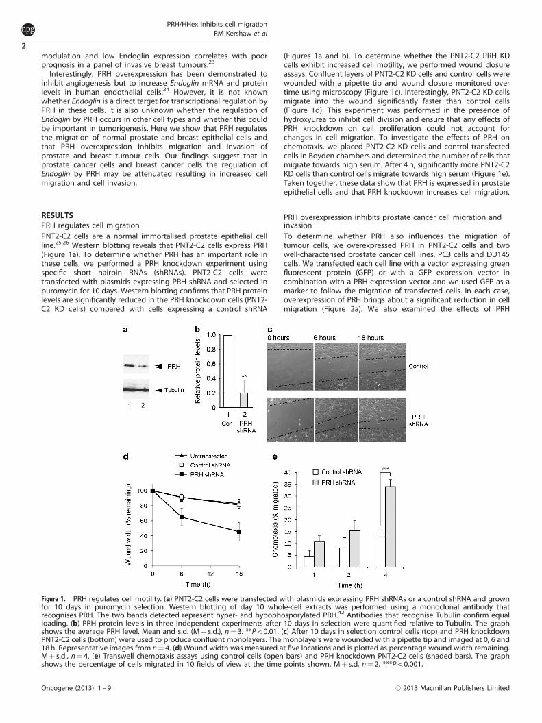
RESULTSPRH regulates cell migrationPNT2-C2 cells are a normal immortalised prostate epithelial cellline.25,26 Western blotting reveals that PNT2-C2 cells express PRH(Figure 1a). To determine whether PRH has an important role inthese cells, we performed a PRH knockdown experiment usingspecific short hairpin RNAs (shRNAs). PNT2-C2 cells weretransfected with plasmids expressing PRH shRNA and selected inpuromycin for 10 days. Western blotting confirms that PRH proteinlevels are significantly reduced in the PRH knockdown cells (PNT2-C2 KD cells) compared with cells expressing a control shRNA
(Figures 1a and b). To determine whether the PNT2-C2 PRH KDcells exhibit increased cell motility, we performed wound closureassays. Confluent layers of PNT2-C2 KD cells and control cells werewounded with a pipette tip and wound closure monitored overtime using microscopy (Figure 1c). Interestingly, PNT2-C2 KD cellsmigrate into the wound significantly faster than control cells(Figure 1d). This experiment was performed in the presence ofhydroxyurea to inhibit cell division and ensure that any effects ofPRH knockdown on cell proliferation could not account forchanges in cell migration. To investigate the effects of PRH onchemotaxis, we placed PNT2-C2 KD cells and control transfectedcells in Boyden chambers and determined the number of cells thatmigrate towards high serum. After 4 h, significantly more PNT2-C2KD cells than control cells migrate towards high serum (Figure 1e).Taken together, these data show that PRH is expressed in prostateepithelial cells and that PRH knockdown increases cell migration.
PRH overexpression inhibits prostate cancer cell migration andinvasionTo determine whether PRH also influences the migration oftumour cells, we overexpressed PRH in PNT2-C2 cells and twowell-characterised prostate cancer cell lines, PC3 cells and DU145cells. We transfected each cell line with a vector expressing greenfluorescent protein (GFP) or with a GFP expression vector incombination with a PRH expression vector and we used GFP as amarker to follow the migration of transfected cells. In each case,overexpression of PRH brings about a significant reduction in cellmigration (Figure 2a). We also examined the effects of PRH
Figure 1. PRH regulates cell motility. (a) PNT2-C2 cells were transfected with plasmids expressing PRH shRNAs or a control shRNA and grownfor 10 days in puromycin selection. Western blotting of day 10 whole-cell extracts was performed using a monoclonal antibody thatrecognises PRH. The two bands detected represent hyper- and hypophosporylated PRH.42 Antibodies that recognise Tubulin confirm equalloading. (b) PRH protein levels in three independent experiments after 10 days in selection were quantified relative to Tubulin. The graphshows the average PRH level. Mean and s.d. (Mþ s.d.), n¼ 3. **Po0.01. (c) After 10 days in selection control cells (top) and PRH knockdownPNT2-C2 cells (bottom) were used to produce confluent monolayers. The monolayers were wounded with a pipette tip and imaged at 0, 6 and18 h. Representative images from n¼ 4. (d) Wound width was measured at five locations and is plotted as percentage wound width remaining.Mþ s.d., n¼ 4. (e) Transwell chemotaxis assays using control cells (open bars) and PRH knockdown PNT2-C2 cells (shaded bars). The graphshows the percentage of cells migrated in 10 fields of view at the time points shown. Mþ s.d. n¼ 2. ***Po0.001.
PRH/HHex inhibits cell migrationRM Kershaw et al
2
Oncogene (2013) 1 – 9 & 2013 Macmillan Publishers Limited
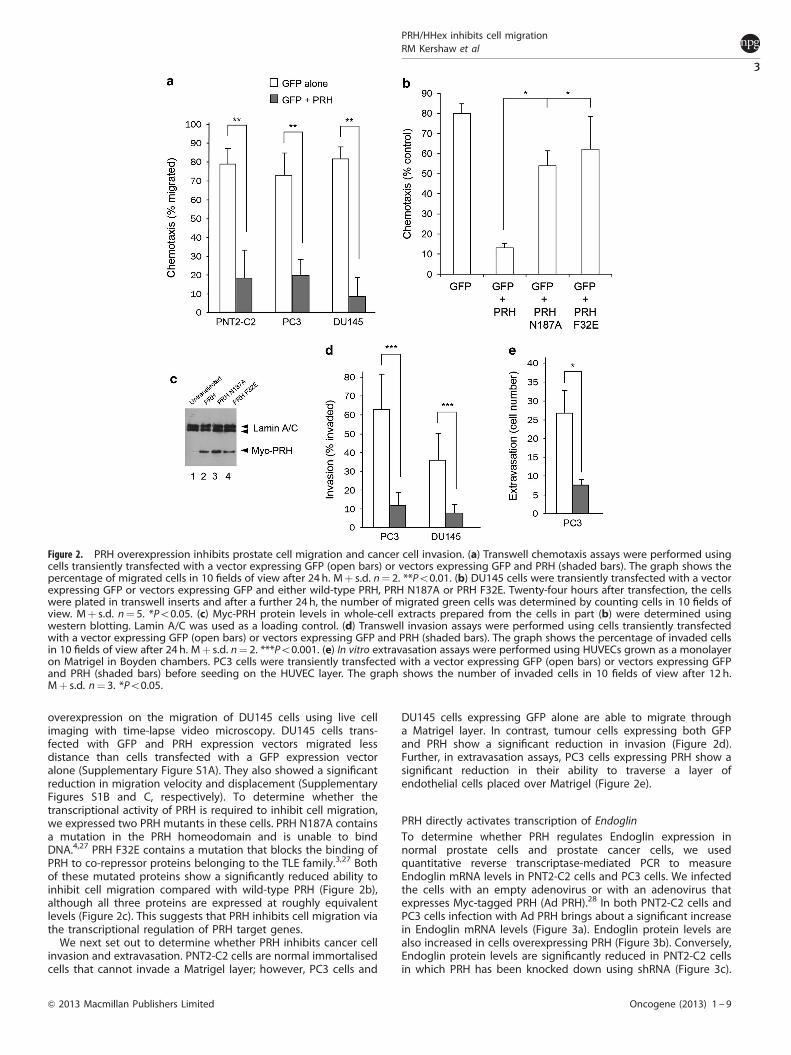
overexpression on the migration of DU145 cells using live cellimaging with time-lapse video microscopy. DU145 cells trans-fected with GFP and PRH expression vectors migrated lessdistance than cells transfected with a GFP expression vectoralone (Supplementary Figure S1A). They also showed a significantreduction in migration velocity and displacement (SupplementaryFigures S1B and C, respectively). To determine whether thetranscriptional activity of PRH is required to inhibit cell migration,we expressed two PRH mutants in these cells. PRH N187A containsa mutation in the PRH homeodomain and is unable to bindDNA.4,27 PRH F32E contains a mutation that blocks the binding ofPRH to co-repressor proteins belonging to the TLE family.3,27 Bothof these mutated proteins show a significantly reduced ability toinhibit cell migration compared with wild-type PRH (Figure 2b),although all three proteins are expressed at roughly equivalentlevels (Figure 2c). This suggests that PRH inhibits cell migration viathe transcriptional regulation of PRH target genes.
We next set out to determine whether PRH inhibits cancer cellinvasion and extravasation. PNT2-C2 cells are normal immortalisedcells that cannot invade a Matrigel layer; however, PC3 cells and
DU145 cells expressing GFP alone are able to migrate througha Matrigel layer. In contrast, tumour cells expressing both GFPand PRH show a significant reduction in invasion (Figure 2d).Further, in extravasation assays, PC3 cells expressing PRH show asignificant reduction in their ability to traverse a layer ofendothelial cells placed over Matrigel (Figure 2e).
PRH directly activates transcription of EndoglinTo determine whether PRH regulates Endoglin expression innormal prostate cells and prostate cancer cells, we usedquantitative reverse transcriptase-mediated PCR to measureEndoglin mRNA levels in PNT2-C2 cells and PC3 cells. We infectedthe cells with an empty adenovirus or with an adenovirus thatexpresses Myc-tagged PRH (Ad PRH).28 In both PNT2-C2 cells andPC3 cells infection with Ad PRH brings about a significant increasein Endoglin mRNA levels (Figure 3a). Endoglin protein levels arealso increased in cells overexpressing PRH (Figure 3b). Conversely,Endoglin protein levels are significantly reduced in PNT2-C2 cellsin which PRH has been knocked down using shRNA (Figure 3c).
Figure 2. PRH overexpression inhibits prostate cell migration and cancer cell invasion. (a) Transwell chemotaxis assays were performed usingcells transiently transfected with a vector expressing GFP (open bars) or vectors expressing GFP and PRH (shaded bars). The graph shows thepercentage of migrated cells in 10 fields of view after 24 h. Mþ s.d. n¼ 2. **Po0.01. (b) DU145 cells were transiently transfected with a vectorexpressing GFP or vectors expressing GFP and either wild-type PRH, PRH N187A or PRH F32E. Twenty-four hours after transfection, the cellswere plated in transwell inserts and after a further 24 h, the number of migrated green cells was determined by counting cells in 10 fields ofview. Mþ s.d. n¼ 5. *Po0.05. (c) Myc-PRH protein levels in whole-cell extracts prepared from the cells in part (b) were determined usingwestern blotting. Lamin A/C was used as a loading control. (d) Transwell invasion assays were performed using cells transiently transfectedwith a vector expressing GFP (open bars) or vectors expressing GFP and PRH (shaded bars). The graph shows the percentage of invaded cellsin 10 fields of view after 24 h. Mþ s.d. n¼ 2. ***Po0.001. (e) In vitro extravasation assays were performed using HUVECs grown as a monolayeron Matrigel in Boyden chambers. PC3 cells were transiently transfected with a vector expressing GFP (open bars) or vectors expressing GFPand PRH (shaded bars) before seeding on the HUVEC layer. The graph shows the number of invaded cells in 10 fields of view after 12 h.Mþ s.d. n¼ 3. *Po0.05.
PRH/HHex inhibits cell migrationRM Kershaw et al
3
& 2013 Macmillan Publishers Limited Oncogene (2013) 1 – 9
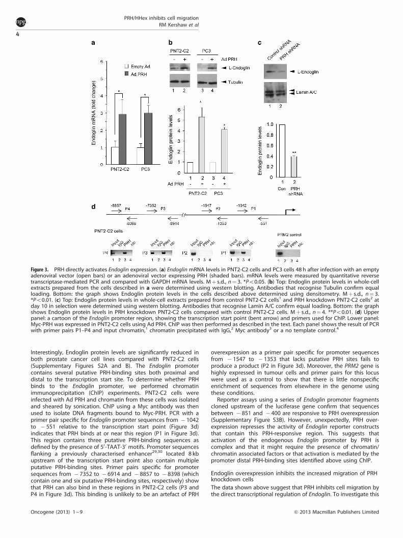
Interestingly, Endoglin protein levels are significantly reduced inboth prostate cancer cell lines compared with PNT2-C2 cells(Supplementary Figures S2A and B). The Endoglin promotercontains several putative PRH-binding sites both proximal anddistal to the transcription start site. To determine whether PRHbinds to the Endoglin promoter, we performed chromatinimmunoprecipitation (ChIP) experiments. PNT2-C2 cells wereinfected with Ad PRH and chromatin from these cells was isolatedand sheared by sonication. ChIP using a Myc antibody was thenused to isolate DNA fragments bound to Myc-PRH. PCR with aprimer pair specific for Endoglin promoter sequences from � 1042to � 551 relative to the transcription start point (Figure 3d)indicates that PRH binds at or near this region (P1 in Figure 3d).This region contains three putative PRH-binding sequences asdefined by the presence of 50-TAAT-30 motifs. Promoter sequencesflanking a previously characterised enhancer29,30 located 8 kbupstream of the transcription start point also contain multipleputative PRH-binding sites. Primer pairs specific for promotersequences from � 7352 to � 6914 and � 8857 to � 8398 (whichcontain one and six putative PRH-binding sites, respectively) showthat PRH can also bind in these regions in PNT2-C2 cells (P3 andP4 in Figure 3d). This binding is unlikely to be an artefact of PRH
overexpression as a primer pair specific for promoter sequencesfrom � 1547 to � 1353 that lacks putative PRH sites fails toproduce a product (P2 in Figure 3d). Moreover, the PRM2 gene ishighly expressed in tumour cells and primer pairs for this locuswere used as a control to show that there is little nonspecificenrichment of sequences from elsewhere in the genome usingthese conditions.
Reporter assays using a series of Endoglin promoter fragmentscloned upstream of the luciferase gene confirm that sequencesbetween � 851 and � 400 are responsive to PRH overexpression(Supplementary Figure S3B). However, unexpectedly, PRH over-expression represses the activity of Endoglin reporter constructsthat contain this PRH-responsive region. This suggests thatactivation of the endogenous Endoglin promoter by PRH iscomplex and that it might require the presence of chromatin/chromatin associated factors or that activation is mediated by thepromoter distal PRH-binding sites identified above using ChIP.
Endoglin overexpression inhibits the increased migration of PRHknockdown cellsThe data shown above suggest that PRH inhibits cell migration bythe direct transcriptional regulation of Endoglin. To investigate this
Figure 3. PRH directly activates Endoglin expression. (a) Endoglin mRNA levels in PNT2-C2 cells and PC3 cells 48 h after infection with an emptyadenoviral vector (open bars) or an adenoviral vector expressing PRH (shaded bars). mRNA levels were measured by quantitative reversetranscriptase-mediated PCR and compared with GAPDH mRNA levels. Mþ s.d., n¼ 3. *Po0.05. (b) Top: Endoglin protein levels in whole-cellextracts prepared from the cells described in a were determined using western blotting. Antibodies that recognise Tubulin confirm equalloading. Bottom: the graph shows Endoglin protein levels in the cells described above determined using densitometry. Mþ s.d., n¼ 3.*Po0.01. (c) Top: Endoglin protein levels in whole-cell extracts prepared from control PNT2-C2 cells1 and PRH knockdown PNT2-C2 cells2 atday 10 in selection were determined using western blotting. Antibodies that recognise Lamin A/C confirm equal loading. Bottom: the graphshows Endoglin protein levels in PRH knockdown PNT2-C2 cells compared with control PNT2-C2 cells. Mþ s.d., n¼ 4. **Po0.01. (d) Upperpanel: a cartoon of the Endoglin promoter region, showing the transcription start point (bent arrow) and primers used for ChIP. Lower panel:Myc-PRH was expressed in PNT2-C2 cells using Ad PRH. ChIP was then performed as described in the text. Each panel shows the result of PCRwith primer pairs P1–P4 and input chromatin,1 chromatin precipitated with IgG,2 Myc antibody3 or a no template control.4
PRH/HHex inhibits cell migrationRM Kershaw et al
4
Oncogene (2013) 1 – 9 & 2013 Macmillan Publishers Limited
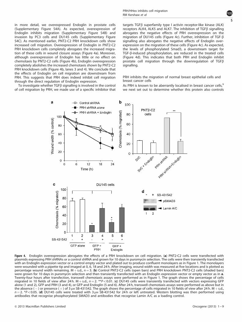
in more detail, we overexpressed Endoglin in prostate cells(Supplementary Figure S4A). As expected, overexpression ofEndoglin inhibits migration (Supplementary Figure S4B) andinvasion by PC3 cells and DU145 cells (Supplementary FigureS4C). As mentioned earlier, PNT2-C2 PRH knockdown cells showincreased cell migration. Overexpression of Endoglin in PNT2-C2PRH knockdown cells completely abrogates the increased migra-tion of these cells in wound closure assays (Figure 4a). Moreover,although overexpression of Endoglin has little or no effect onchemotaxis by PNT2-C2 cells (Figure 4b), Endoglin overexpressioncompletely abolishes the increased chemotaxis shown by PNT2-C2PRH knockdown cells (Figure 4b, lanes 3 and 4). We conclude thatthe effects of Endoglin on cell migration are downstream fromPRH. This suggests that PRH does indeed inhibit cell migrationthrough the direct regulation of Endoglin expression.
To investigate whether TGFb signalling is involved in the controlof cell migration by PRH, we made use of a specific inhibitor that
targets TGFb superfamily type I activin receptor-like kinase (ALK)receptors ALK4, ALK5 and ALK7. The inhibition of TGFb signallingabrogates the negative effects of PRH overexpression on themigration of DU145 cells (Figure 4c). Further, inhibition of TGF-bsignalling also abrogates the negative effects of Endoglin over-expression on the migration of these cells (Figure 4c). As expected,the levels of phosphorylated Smad3, a downstream target forTGF-b-induced phosphorylation, are reduced in the treated cells(Figure 4d). This indicates that both PRH and Endoglin inhibitprostate cell migration through the downregulation of TGFbsignalling.
PRH inhibits the migration of normal breast epithelial cells andbreast cancer cellsAs PRH is known to be aberrantly localised in breast cancer cells,9
we next set out to determine whether this protein also controls
Figure 4. Endoglin overexpression abrogates the effects of a PRH knockdown on cell migration. (a) PNT2-C2 cells were transfected withplasmids expressing PRH shRNAs or a control shRNA and grown for 10 days in puromycin selection. The cells were then transiently transfectedwith an Endoglin expression vector or a control empty vector and plated out to produce confluent monolayers as in Figure 1. The monolayerswere wounded with a pipette tip and imaged at 0, 6, 18 and 24h. After imaging, wound width was measured at five locations and is plotted aspercentage wound width remaining. Mþ s.d., n¼ 3. (b) Control PNT2-C2 cells (open bars) and PRH knockdown PNT2-C2 cells (shaded bars)were grown for 10 days in puromycin selection and then transiently transfected with an Endoglin expression vector or empty vector as in a.Twenty-four hours after transfection, transwell chemotaxis assays were performed as in Figure 1. The graph shows the percentage of cellsmigrated in 10 fields of view after 24 h. Mþ s.d., n¼ 2. **Po0.01. (c) DU145 cells were transiently transfected with vectors expressing GFPalone (1 and 2), GFP and PRH (3 and 4), or GFP and Endoglin (5 and 6). After 24 h, transwell chemotaxis assays were performed as above but inthe absence (� ) or presence (þ ) of 3 mM SB-431542. The graph shows the percentage of cells migrated in 10 fields of view after 24 h. Mþ s.d.,n¼ 2. *Po0.05. (d) DU145 cells were treated with 3mM SB-431542 for 24 h or left untreated. Western blotting was then performed usingantibodies that recognise phosphorylated SMAD3 and antibodies that recognise Lamin A/C as a loading control.
PRH/HHex inhibits cell migrationRM Kershaw et al
5
& 2013 Macmillan Publishers Limited Oncogene (2013) 1 – 9
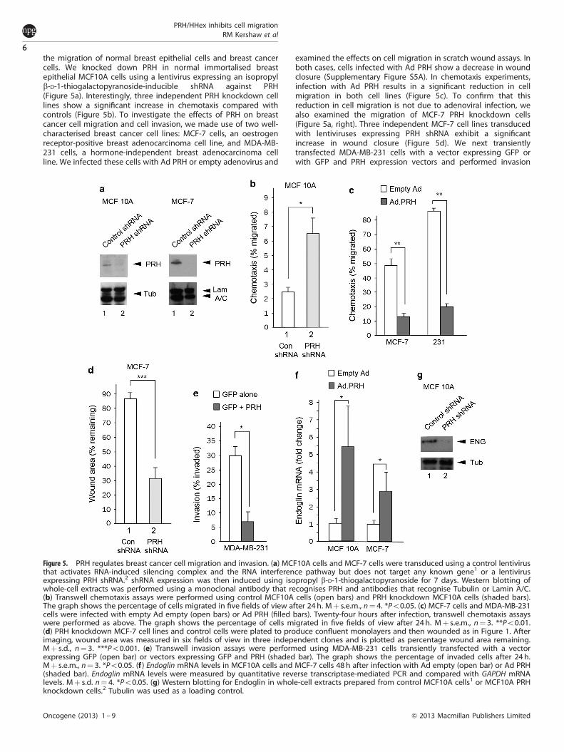
the migration of normal breast epithelial cells and breast cancercells. We knocked down PRH in normal immortalised breastepithelial MCF10A cells using a lentivirus expressing an isopropylb-D-1-thiogalactopyranoside-inducible shRNA against PRH(Figure 5a). Interestingly, three independent PRH knockdown celllines show a significant increase in chemotaxis compared withcontrols (Figure 5b). To investigate the effects of PRH on breastcancer cell migration and cell invasion, we made use of two well-characterised breast cancer cell lines: MCF-7 cells, an oestrogenreceptor-positive breast adenocarcinoma cell line, and MDA-MB-231 cells, a hormone-independent breast adenocarcinoma cellline. We infected these cells with Ad PRH or empty adenovirus and
examined the effects on cell migration in scratch wound assays. Inboth cases, cells infected with Ad PRH show a decrease in woundclosure (Supplementary Figure S5A). In chemotaxis experiments,infection with Ad PRH results in a significant reduction in cellmigration in both cell lines (Figure 5c). To confirm that thisreduction in cell migration is not due to adenoviral infection, wealso examined the migration of MCF-7 PRH knockdown cells(Figure 5a, right). Three independent MCF-7 cell lines transducedwith lentiviruses expressing PRH shRNA exhibit a significantincrease in wound closure (Figure 5d). We next transientlytransfected MDA-MB-231 cells with a vector expressing GFP orwith GFP and PRH expression vectors and performed invasion
Figure 5. PRH regulates breast cancer cell migration and invasion. (a) MCF10A cells and MCF-7 cells were transduced using a control lentivirusthat activates RNA-induced silencing complex and the RNA interference pathway but does not target any known gene1 or a lentivirusexpressing PRH shRNA.2 shRNA expression was then induced using isopropyl b-D-1-thiogalactopyranoside for 7 days. Western blotting ofwhole-cell extracts was performed using a monoclonal antibody that recognises PRH and antibodies that recognise Tubulin or Lamin A/C.(b) Transwell chemotaxis assays were performed using control MCF10A cells (open bars) and PRH knockdown MCF10A cells (shaded bars).The graph shows the percentage of cells migrated in five fields of view after 24 h. Mþ s.e.m., n¼ 4. *Po0.05. (c) MCF-7 cells and MDA-MB-231cells were infected with empty Ad empty (open bars) or Ad PRH (filled bars). Twenty-four hours after infection, transwell chemotaxis assayswere performed as above. The graph shows the percentage of cells migrated in five fields of view after 24 h. Mþ s.e.m., n¼ 3. **Po0.01.(d) PRH knockdown MCF-7 cell lines and control cells were plated to produce confluent monolayers and then wounded as in Figure 1. Afterimaging, wound area was measured in six fields of view in three independent clones and is plotted as percentage wound area remaining.Mþ s.d., n¼ 3. ***Po0.001. (e) Transwell invasion assays were performed using MDA-MB-231 cells transiently transfected with a vectorexpressing GFP (open bar) or vectors expressing GFP and PRH (shaded bar). The graph shows the percentage of invaded cells after 24 h.Mþ s.e.m., n¼ 3. *Po0.05. (f ) Endoglin mRNA levels in MCF10A cells and MCF-7 cells 48 h after infection with Ad empty (open bar) or Ad PRH(shaded bar). Endoglin mRNA levels were measured by quantitative reverse transcriptase-mediated PCR and compared with GAPDH mRNAlevels. Mþ s.d. n¼ 4. *Po0.05. (g) Western blotting for Endoglin in whole-cell extracts prepared from control MCF10A cells1 or MCF10A PRHknockdown cells.2 Tubulin was used as a loading control.
PRH/HHex inhibits cell migrationRM Kershaw et al
6
Oncogene (2013) 1 – 9 & 2013 Macmillan Publishers Limited
assays. As in PC3 cells and DU145 cells, overexpression of PRHdramatically reduces the ability of MDA-MB-231 cells to invadeMatrigel (Figure 5e).
Similar to the results seen in prostate cells, Endoglin proteinlevels are higher in normal breast MCF10A cells than in eitherMCF-7 cells or MDA-MB-231 cells (Supplementary Figures S5Band C). To examine whether the ability of PRH to influence themigration of MCF10A cells and breast cancer cells also involvesthe regulation of Endoglin, we measured Endoglin mRNA levels inMCF-7 and MCF10A cells overexpressing PRH. In both cases, PRHoverexpression results in a significant increase in Endoglin mRNA(Figure 5f). Moreover, Endoglin protein levels are decreased in PRHknockdown MCF10A cells (Figure 5g). The effects of PRH onEndoglin mRNA and protein expression appear to be direct, as ChIPof Myc-tagged PRH pulls down Endoglin promoter sequences inboth MCF-7 and MCF10A cells (Supplementary Figure S5D). Thesedata suggest that PRH inhibits the migration of normal breast cellsand breast cancer cells via transcriptional regulation of Endoglinexpression.
DISCUSSIONThe ability of PRH/HHex to control cell proliferation and celldifferentiation in multiple cell types is well documented. PRH cancontrol cell proliferation via the post-transcriptional regulation ofcyclin D mRNA transport.2 In addition, our previous work hasshown that in leukaemic cells, PRH directly represses multiplegenes that encode proteins involved in VEGF signalling includingVegfa and Vegfr-1.31 In these cells, VEGF acts as an autocrinegrowth factor and the transcriptional regulation of these VEGF-signalling genes by PRH controls cell survival. The extent to whichthese modes of cell survival control operate in other cell types isnot known, although in breast cancer MCF-7 cells, PRH regulatesthe transcription of Vegf receptor genes and a PRH knockdownincreases cell growth.31 In endothelial cells, PRH overexpressionalso controls VEGF-signalling genes and alters cell migration andinvasion.24 In breast and thyroid cancer cells, PRH subcellularlocalisation is altered resulting in the cytoplasmic and nucleolaraccumulation of PRH.9,10 However, the ability of PRH to regulateother aspects of cancer cell behaviour relevant to tumorigenesis,including cell motility and cell invasion, has not been investigated.
Here we have shown that PRH knockdown increases themotility of immortalised prostate and breast epithelial cells andincreases chemotaxis by these cells. Conversely, PRH overexpres-sion decreases cell migration by prostate and breast cancer cellsand inhibits the ability of these cells to invade a Matrigel layer.Moreover, PRH overexpression decreases the ability of prostatecancer cells to travel though a layer of endothelial cells inextravasation assays. These findings suggest that the aberrantlocalisation of PRH seen in breast cancer cells and thyroid cancercells could contribute to their increased migration and cellinvasion as well as having effects on cell proliferation. Moreover,these results show that PRH activity is important in the control ofthese aspects of cell behaviour in normal epithelial cells, and theysuggest that PRH may also be aberrantly localised or otherwisemisregulated in prostate cancer cells.
Knockout of PRH in embryoid bodies results in downregulationof Endoglin mRNA,32 and PRH overexpression has been shown toupregulate Endoglin mRNA and protein levels in endothelialcells.24 We have shown that PRH associates directly with theEndoglin promoter and upregulates transcription of Endoglin innormal prostate and breast epithelial cells and prostate and breastcancer cell lines. PRH binds to DNA sequences within the Endoglinproximal promoter located around 500 bp upstream of thetranscription start point and upstream of a number of importanttranscription factor-binding sites including sites for regulation bySp1, HIF-1, Smad proteins, KLF6 and ETS family members.30,33–35
PRH also binds to sequences in the distal promoter near a
previously characterised enhancer29,30 around 8 kb upstream ofthe transcription start point. These regions both contain multiplecopies of the core PRH-binding site 50-TAAT-30. Other PRH targetgenes including Goosecoid, Vegfr-1 and Vegfa also contain arraysof PRH core binding sites and at these promoters PRH also bindsover extensive regions.36,37 This suggests that PRH oligomers bindto these promoters in a similar manner in order to controlpromoter architecture. However, at other promoters, PRH binds inconjunction with other transcription factors and in these casesextensive direct contacts between PRH and the DNA may not berequired.1 Surprisingly, although the promoter proximal bindingsites in the Endoglin promoter confer responsiveness to PRHoverexpression in luciferase assays, PRH appears to repressEndoglin transcription when bound at these sites. Presumably,the promoter distal PRH-binding sites are responsible for Endoglintranscription activation by PRH. Alternatively, the presence ofchromatin/chromatin associated factors may be important foractivation of the Endoglin promoter by PRH just as they are forrepression by PRH at the Vegfr-1 promoter.31 Further experimentswill be required to determine the mechanisms that PRH uses inorder to activate transcription at this promoter.
Importantly, the increased cell migration shown by PRHknockdown cells is completely abolished by Endoglin over-expression. We conclude that in both breast and prostate cells,PRH regulates Endoglin expression and that the effects of PRH oncell migration are in part at least through the control of this targetgene. Endoglin has been shown to inhibit cell migration, cellinvasion and tumour growth by prostate cancer cells and cellmigration and invasion of breast cancer cells.18,23 However, it hasbeen reported that Endoglin expression is higher in metastaticcancer epithelial cells and prostatic intraepithelial neoplasiacompared normal prostate cells.38 Moreover, in highly metastaticbreast cancer cells, increased Endoglin expression is associatedwith invasion and metastasis.39 It would seem likely that theeffects of Endoglin overexpression may depend on the cell typeand/or the overexpression level as well as interplay between thetumour and the stroma and inputs from multiple signallingpathways. However, we have observed decreased Endoglinexpression in prostate tumour cells and breast tumour cellsrelative to immortalised cells. Henry et al.23 also reporteddecreased Endoglin expression in most breast tumour cell linesrelative to immortalised cells and detected Endoglin expression inonly a minority of primary tumours. We expect that the regulationof Endoglin by PRH is disrupted in breast cancer cells. Furtherexperiments will be required to determine whether PRH activity inprostate cancer cells is disrupted by changes in PRH localisation ashas been observed in breast cancer cells9 or by some othermechanism. However, it would seem probable that in both ofthese cancer types and in other cancer cells in which PRH isaberrantly localised, the loss of PRH activity may contribute toincreased cell invasion, increased cancer metastasis andultimately, decreased patient survival in part at least through adecrease in Endoglin expression. Loss of PRH activity would alsobe expected to de-repress VEGF-signalling genes resulting inincreased cell survival and/or increased neoangiogenesis. Thiscombination of outcomes arising from the loss of PRH activitysuggests that this protein has a critical role in tumourigenesis.
MATERIALS AND METHODSExpression vectors and reporterspMUG1-Myc-PRH expresses human PRH tagged with the Myc9E10epitope.3 pMUG1-Myc-PRH N187A and pMUG1-Myc-PRH F32E expressmutated PRH proteins that fail to bind DNA and TLE, respectively.3 Theplasmids shRNAPRH49 and shRNAPRH51 and control shRNA plasmid wereobtained from Origene (Rockville, MD, USA). The Endoglin expressionvector pcDNA3.1-Endoglin (long isoform) was a gift from Professor ClareIsacke. The recombinant adenoviral construct expressing Myc-PRH has
PRH/HHex inhibits cell migrationRM Kershaw et al
7
& 2013 Macmillan Publishers Limited Oncogene (2013) 1 – 9

been described previously.28 pCD105(� 2450/þ 350), pCD105(� 851/þ 350) and pCD105(� 400/þ 350) containing Endoglin promotersequences cloned upstream of the firefly luciferase gene were a giftfrom Professor Carmelo Bernabeu.34,35 pRL-CMV expressing Renillaluciferase was purchased from Promega (Madison, WI, USA). Lentiviralconstructs expressing isopropyl b-D-1-thiogalactopyranoside-inducible PRHshRNA or a control shRNA were obtained from Sigma (Gillingham, UK).
Cell culturePNT2-C2, PC3, DU145, MCF-7 and MDA-MB-231 cells were cultured inRoswell Park Memorial Institute medium-1640 supplemented with 10%fetal bovine serum, 2 mM L-glutamine and 1% penicillin/streptomycin. MCF10A cells were cultured in Dulbecco’s Modified Eagle Medium:F12 (Sigma)supplemented with 5% horse serum heat-inactivated (Sigma), 20 ng/mlepidermal growth factor (Peprotech, Rocky Hill, NJ, USA) 0.5 mg/mlhydrocortisone (Sigma), 100 ng/ml cholera toxin (Sigma), 10mg/ml insulin(Sigma) and 1% penicillin/streptomycin. Human umbilical vein endothelialcells were cultured in dulbecco’s modified eagle medium:F12 supplemen-ted with 5 ng/ml epidermal growth factor (Peprotech), 10 ng/ml basicfibroblast growth factor, 20mg/ml heparin, 1 mg/ml hydrocortisone, 250 ng/ml insulin, 1% penicillin/streptomycin and 2% fetal calf serum. All cellswere maintained in a humidified atmosphere at 37 1C and 5% CO2.
Transient transfectionPNT2-C2 cells, PC3 cells and DU145 cells were transfected using TransIT(Mirus, Madison, WI, USA). MCF-7 cells and MDA-MB-231 cells weretransfected using Lipofectamine 2000 (Invitrogen, Grand Island, NY, USA).PC3 cells were transfected for luciferase assays using electroporation(250 V, 975mF).
Luciferase assaysPC3 cells were transiently transfected with reporter plasmids and eitherpMUG1-Myc-PRH or empty pMUG1 vector. After 24 h at 37 1C and 5% CO2,the cells were lysed and assayed for luciferase activity using a dualluciferase assay system (Promega) and a Berthold Technologies (Wildbad,Germany) luminometer. Renilla luciferase activity was used as an internalcontrol for transfection efficiency.
PRH knockdownKnockdown of PRH in PNT2-C2 cells was performed as describedpreviously.31 Knockdown of PRH in MCF-7 and MCF 10A cells wasperformed using an isopropyl b-D-1-thiogalactopyranoside-inducible PRHshRNA lentiviral construct (TRCN0000274008 Sigma). Cells were infected withthe PRH shRNA lentiviral construct or a control scrambled shRNA lentivirus(SHC332) and after 48 h transduced cells were selected using puromycin.Stably transduced cell lines were grown in the presence of 1 mM isopropylb-D-1-thiogalactopyranoside for 7 days to induce shRNA expression.
Cell migration assaysCell monolayers on microscope coverslips were produced plating cellsinfected with Ad PRH or empty adenovirus (multiplicity of infection 50) andincubating the cells for 24 h at 37 1C and 5% CO2. After the addition of 1 mM
hydroxyurea (Sigma) to inhibit cell division, a wound was created using aP1000 pipette tip. Pictures were taken using a Leica DMIRBE microscope(Leica, Wetzlar, Germany) with Hamamatsu (Hamamatsu City, Japan)charge-coupled device camera or a AMG EVOS XL CORE AMEX 1200, andthe width and area of the wound was quantified using ImageJ software.40
Chemotaxis assays were performed by seeding cells onto 8 mm Boydenchambers (Greiner Bio-One, Kremsmunster, Austria) in Roswell ParkMemorial Institute medium with 2% fetal bovine serum. The chamberswere placed into 24-well plates containing Roswell Park Memorial Institutemedium with 10% fetal bovine serum to create a serum gradient. In someexperiments, cells were seeded in the presence of 3 mM ALK 4/5/7 kinaseinhibitor SB-431542 (Sigma) or dimethyl sulfoxide. At the time pointsindicated, the cells were fixed with 4% paraformaldehyde (Fisher,Loughborough, UK) and stained with 2 mg/ml bisbenzimide (Sigma). Cellson the top and bottom of the membrane were counted using a Leica Q550inverted epifluorescence microcope or Zeiss axioplan 2 (Zeiss, Gottingen,Germany).
Invasion assays were performed as above except that 50% Matrigel (BDBiosciences, Billeria, MA, USA) was added to the Boyden chambers and leftat 37 1C for 1 h to solidify before seeding the cells. Extravasation assayswere performed as described by Ma and Wang41 except that PC3 cellswere dissociated using Cell Dissociation Solution (Sigma) before seedingon the human umbilical vein endothelial cell layer.
Quantitative reverse transcriptase-mediated PCRRNA was purified 48 h after infection as described previously.31,42
Quantitative PCR was performed in triplicate with Endoglin and GAPDH(glyceraldehyde-3-phosphate dehydrogenase) primers (shown below).Data were analysed using Rotorgene 6 software (Corbett Research(Mortlake, Sidney, Australia); Rotorgene RG-3000) with GAPDH mRNA asan internal control.
Endoglin 50-GCCGTGCTGGGCATCACCTT-30 , 50-CGCTTGCTGGGGGAACCTGG-30 , annealing at 60 1C.
GAPDH 50-TGATGACATCAAGAAGGTGGTGAAG-30 , 50-TCCTTGGAGGCCATGT GGGCCAT-30 , annealing at 55 1C.
Western blottingWhole-cell extracts were prepared using TRIS-EDTA-SDS (TES) buffer (1%SDS, 2 mM EDTA, 20 mM Tris–HCl pH 7.4) as described previously.27 A rabbitpolyclonal antibody was used to detect Endoglin (Abcam, Cambridge, UK).An in house PRH monoclonal antibody was produced by Abmart(Arlington, MA, USA). Lamin A/C and tubulin antibodies were from SantaCruz (Dallas, TX, USA). Densitometry was performed using Quantity One 4.6software (Bio-Rad, Hemel Hempstead, UK). Phosphorylated Smad3 (Ser423/425) was detected using a rabbit monoclonal antibody (Cell SignalingTechnology, Danvers, MA, USA).
Chromatin immunoprecipitationCells were infected with Ad PRH or control virus (multiplicity of infection50) and incubated for 24 h at 37 1C and 5% CO2 before fixation in 1%formaldehyde for 8 min at 21 1C. Glycine was added to a finalconcentration of 125 mM, and then the cells were pelleted by microcen-trifugation at 4 1C, resuspended in 130ml lysis buffer (50 mM Tris–Cl pH 8.0,10 mM EDTA, 1% SDS, 1 mM phenylmethylsulfonyl fluoride and proteaseinhibitor cocktail (Roche, Welwyn Garden City, UK)) and sonicated in aBiorupter (Diagenode, Denville, NJ, USA) for 20 min on high power at 4 1C.Dynabeads protein A magnetic beads (Invitrogen) were incubated withnormal mouse IgG (Santa Cruz) or Myc9B11 (Cell Signalling Technology) inRIPA buffer (10 mM Tris–Cl pH 8.0, 140 mM NaCl, 1% v/v Triton X-100, 1 mM
EDTA, 0.5 mM EGTA, 0.1% w/v SDS, 0.1% sodium deoxycholate) for 2 h at4 1C on a rotary wheel. Chromatin lysates were incubated withantibody:bead complexes overnight at 4 1C on a rotary wheel. Beads werewashed three times with 1 ml RIPA buffer, 1 ml RIPA containing 500 mM
NaCl, 1 ml RIPA containing 0.5% NP-40 and twice with 1 ml TE buffer(10 mM Tris–Cl pH 8.0, 1 mM EDTA pH 8.0) and resuspended in elutionbuffer (20 mM Tris–Cl pH 7.5, 50 mM NaCl, 5 mM EDTA, 1% SDS). Afterdigestion with proteinase K for 2 h at 68 1C DNA was obtained by phenolchloroform isoamyl alcohol extraction and precipitated before resuspen-sion in TE. Binding was analysed by PCR (95 1C for 1 min, 60 1C for 1 min,72 1C for 1 min) using the following Endoglin primer pairs: P1 50-CAGGAAGGCATCGTGCCCCA-30 , 50-TCACCGACAAAACACAGCTCCA-30 ; P2:50-CTCTGCCAGCGTCCTTCTGCTC-30 , 50-AGGGTGCCAGACTAAGC AAAGCAAC-30 ;P3 5’-GGGTTGCCATGGTGGGAATATA-30 , 50-TATGGGTGTT GGGGGCATTC-30 ;P4 50–AGCTAATAGCCCGTGTGCAA-30 , 50-AGGGGGAGAG TGGTCCTAGA-30 .The prm2 gene was used as a negative control: prm2 primers 50–TGTACAGGCAGCAGTTGCATGG-30 , 50-CTCCTTCGAGAGCAGTGTCTGC-30 (annealingtemperature 62 1C, 33 cycles).
CONFLICT OF INTERESTThe authors declare no conflict of interest.
ACKNOWLEDGEMENTSWe are grateful to Professor Norman J Maitland (University of York) for PNT2-C2 cellsand useful discussions, Professor Kate Nobes (University of Bristol) for access to livecell imaging, Professor Clare Isacke (Institute of Cancer Research) for the Endoglinexpression vector, Professor Harry Mellor (University of Bristol) and Professor RoyBicknell (University of Birmingham) for HUVECs and useful discussions, and ProfessorCarmelo Bernabeu (Centro de Investigaciones Biologicas, Madrid) for Endoglin-luciferase reporter constructs. We are grateful to Laura A V Rodriguez and Xin Yang
PRH/HHex inhibits cell migrationRM Kershaw et al
8
Oncogene (2013) 1 – 9 & 2013 Macmillan Publishers Limited
for technical assistance. This work was funded by a Breast Cancer Campaign projectgrant to PSJ and KG. YHS is grateful to the University of Bristol for a PhD Scholarshipand to the Charles Wallace Pakistan Trust for additional support. DR is grateful to theMRC for a PhD studentship.
REFERENCES1 Soufi A, Jayaraman PS. PRH/Hex: an oligomeric transcription factor and multi-
functional regulator of cell fate. Biochem J 2008; 412: 399–413.2 Topisirovic I, Culjkovic B, Cohen N, Perez JM, Skrabanek L, Borden KL. The proline-
rich homeodomain protein, PRH, is a tissue-specific inhibitor of eIF4E-dependentcyclin D1 mRNA transport and growth. EMBO J 2003; 22: 689–703.
3 Swingler TE, Bess KL, Yao J, Stifani S, Jayaraman PS. The proline-rich home-odomain protein recruits members of the Groucho/Transducin-like enhancer ofsplit protein family to co-repress transcription in hematopoietic cells. J Biol Chem2004; 279: 34938–34947.
4 Guiral M, Bess K, Goodwin G, Jayaraman PS. PRH represses transcription inhematopoietic cells by at least two independent mechanisms. J Biol Chem 2001;276: 2961–2970.
5 Denson LA, Karpen SJ, Bogue CW, Jacobs HC. Divergent homeobox gene hexregulates promoter of the Na(þ )-dependent bile acid cotransporter. Am J PhysiolGastrointest Liver Physiol 2000; 279: G347–G355.
6 George A, Morse 3rd HC, Justice MJ. The homeobox gene Hex induces T-cell-derived lymphomas when overexpressed in hematopoietic precursor cells.Oncogene 2003; 22: 6764–6773.
7 McCormack MP, Young LF, Vasudevan S, de Graaf CA, Codrington R, Rabbitts THet al. The Lmo2 oncogene initiates leukemia in mice by inducing thymocyte self-renewal. Science 2010; 327: 879–883.
8 Oram SH, Thoms JA, Pridans C, Janes ME, Kinston SJ, Anand S et al. A previouslyunrecognized promoter of LMO2 forms part of a transcriptional regulatory circuitmediating LMO2 expression in a subset of T-acute lymphoblastic leukaemiapatients. Oncogene 2010; 29: 5796–5808.
9 Puppin C, Puglisi F, Pellizzari L, Manfioletti G, Pestrin M, Pandolfi M et al. HEXexpression and localization in normal mammary gland and breast carcinoma. BMCCancer 2006; 6: 192.
10 D’Elia AV, Tell G, Russo D, Arturi F, Puglisi F, Manfioletti G et al. Expression andlocalization of the homeodomain-containing protein HEX in human thyroidtumors. J Clin Endocrinol Metab 2002; 87: 1376–1383.
11 Jankovic D, Gorello P, Liu T, Ehret S, La SR, Desjobert C et al. Leukemogenicmechanisms and targets of a NUP98/HHEX fusion in acute myeloid leukemia.Blood 2008; 111: 5672–5682.
12 Topisirovic I, Guzman ML, McConnell MJ, Licht JD, Culjkovic B, Neering SJ et al.Aberrant eukaryotic translation initiation factor 4E-dependent mRNA transportimpedes hematopoietic differentiation and contributes to leukemogenesis. MolCell Biol 2003; 23: 8992–9002.
13 Noy P, Gaston K, Jayaraman PS. Dasatinib inhibits leukaemic cell survival bydecreasing PRH/Hhex phosphorylation resulting in increased repression of VEGFsignalling genes. Leuk Res 2012; 36: 1434–1437.
14 Guerrero-Esteo M, Lastres P, Letamendia A, Perez-Alvarez MJ, Langa C, Lopez LAet al. Endoglin overexpression modulates cellular morphology, migration, andadhesion of mouse fibroblasts. Eur J Cell Biol 1999; 78: 614–623.
15 Quintanilla M, Ramirez JR, Perez-Gomez E, Romero D, Velasco B, Letarte M et al.Expression of the TGF-beta coreceptor endoglin in epidermal keratinocytes andits dual role in multistage mouse skin carcinogenesis. Oncogene 2003; 22:5976–5985.
16 Bernabeu C, Conley BA, Vary CP. Novel biochemical pathways of endoglin invascular cell physiology. J Cell Biochem 2007; 102: 1375–1388.
17 Liu Y, Jovanovic B, Pins M, Lee C, Bergan RC. Over expression of endoglin inhuman prostate cancer suppresses cell detachment, migration and invasion.Oncogene 2002; 21: 8272–8281.
18 Lakshman M, Huang X, Ananthanarayanan V, Jovanovic B, Liu Y, Craft CS et al.Endoglin suppresses human prostate cancer metastasis. Clin Exp Metastasis 2011;28: 39–53.
19 Craft CS, Romero D, Vary CP, Bergan RC. Endoglin inhibits prostate cancer motilityvia activation of the ALK2-Smad1 pathway. Oncogene 2007; 26: 7240–7250.
20 Romero D, Terzic A, Conley BA, Craft CS, Jovanovic B, Bergan RC et al. Endoglinphosphorylation by ALK2 contributes to the regulation of prostate cancer cellmigration. Carcinogenesis 2010; 31: 359–366.
21 Wong VC, Chan PL, Bernabeu C, Law S, Wang LD, Li JL et al. Identification of aninvasion and tumor-suppressing gene, Endoglin (ENG), silenced by both epige-netic inactivation and allelic loss in esophageal squamous cell carcinoma. Int JCancer 2008; 123: 2816–2823.
22 Perez-Gomez E, Villa-Morales M, Santos J, Fernandez-Piqueras J, Gamallo C, DotorJ et al. A role for endoglin as a suppressor of malignancy during mouse skincarcinogenesis. Cancer Res 2007; 67: 10268–10277.
23 Henry LA, Johnson DA, Sarrio D, Lee S, Quinlan PR, Crook T et al. Endoglinexpression in breast tumor cells suppresses invasion and metastasis and corre-lates with improved clinical outcome. Oncogene 2011; 30: 1046–1058.
24 Nakagawa T, Abe M, Yamazaki T, Miyashita H, Niwa H, Kokubun S et al. HEX acts asa negative regulator of angiogenesis by modulating the expression of angio-genesis-related gene in endothelial cells in vitro. Arterioscler Thromb Vasc Biol2003; 23: 231–237.
25 Berthon P, Cussenot O, Hopwood L, Leduc A, Maitland N. Functional expression ofsv40 in normal human prostatic epithelial and fibroblastic cells—differentiationpattern of nontumorigenic cell-lines. Int J Oncol 1995; 6: 333–343.
26 Lang SH, Sharrard RM, Stark M, Villette JM, Maitland NJ. Prostate epithelial celllines form spheroids with evidence of glandular differentiation in three-dimen-sional Matrigel cultures. Br J Cancer 2001; 85: 590–599.
27 Desjobert C, Noy P, Swingler T, Williams H, Gaston K, Jayaraman PS. The PRH/Hexrepressor protein causes nuclear retention of Groucho/TLE co-repressors. BiochemJ 2009; 417: 121–132.
28 Soufi A, Smith C, Clarke AR, Gaston K, Jayaraman PS. Oligomerisation of thedevelopmental regulator proline rich homeodomain (PRH/Hex) is mediated by anovel proline-rich dimerisation domain. J Mol Biol 2006; 358: 943–962.
29 Pimanda JE, Chan WY, Donaldson IJ, Bowen M, Green AR, Gottgens B. Endoglinexpression in the endothelium is regulated by Fli-1, Erg, and Elf-1 acting on thepromoter and a � 8-kb enhancer. Blood 2006; 107: 4737–4745.
30 Pimanda JE, Chan WY, Wilson NK, Smith AM, Kinston S, Knezevic K et al. Endoglinexpression in blood and endothelium is differentially regulated by modularassembly of the Ets/Gata hemangioblast code. Blood 2008; 112: 4512–4522.
31 Noy P, Williams H, Sawasdichai A, Gaston K, Jayaraman PS. PRH/HHex controls cellsurvival through coordinate transcriptional regulation of VEGF signalling. Mol CellBiol 2010; 30: 2120–2134.
32 Guo Y, Chan R, Ramsey H, Li W, Xie X, Shelley WC et al. The homeoprotein Hex isrequired for hemangioblast differentiation. Blood 2003; 102: 2428–2435.
33 Botella LM, Sanchez-Elsner T, Rius C, Corbi A, Bernabeu C. Identification of acritical Sp1 site within the endoglin promoter and its involvement in the trans-forming growth factor-beta stimulation. J Biol Chem 2001; 276: 34486–34494.
34 Sanchez-Elsner T, Botella LM, Velasco B, Langa C, Bernabeu C. Endoglinexpression is regulated by transcriptional cooperation between the hypoxiaand transforming growth factor-beta pathways. J Biol Chem 2002; 277:43799–43808.
35 Botella LM, Sanchez-Elsner T, Sanz-Rodriguez F, Kojima S, Shimada J, Guerrero-Esteo M et al. Transcriptional activation of endoglin and transforming growthfactor-beta signaling components by cooperative interaction between Sp1 andKLF6: their potential role in the response to vascular injury. Blood 2002; 100:4001–4010.
36 Williams H, Jayaraman PS, Gaston K. DNA wrapping and distortion by an oligo-meric homeodomain protein. J Mol Biol 2008; 383: 10–23.
37 Noy P, Sawasdichai A, Jayaraman PS, Gaston K. Protein kinase CK2 inactivatesPRH/Hhex using multiple mechanisms to de-repress VEGF-signalling genes andpromote cell survival. Nucleic Acids Res 2012; 40: 9008–9020.
38 Kassouf W, Ismail HR, Aprikian AG, Chevalier S. Whole-mount prostate sectionsreveal differential endoglin expression in stromal, epithelial, and endothelial cellswith the development of prostate cancer. Prostate Cancer Prostatic Dis 2004; 7:105–110.
39 Oxmann D, Held-Feindt J, Stark AM, Hattermann K, Yoneda T, Mentlein R.Endoglin expression in metastatic breast cancer cells enhances their invasivephenotype. Oncogene 2008; 27: 3567–3575.
40 Schneider CA, Rasband WS, Eliceiri KW. NIH Image to ImageJ: 25 years of imageanalysis. Nat Methods 2012; 9: 671–675.
41 Ma C, Wang XF. In vitro assays for the extracellular matrix protein-regulatedextravasation process. CSH Protocols 2008; pdb prot5034.
42 Soufi A, Noy P, Buckle M, Sawasdichai A, Gaston K, Jayaraman PS. CK2 phos-phorylation of the PRH/Hex homeodomain functions as a reversible switch forDNA binding. Nucleic Acids Res 2009; 37: 3288–3300.
Supplementary Information accompanies this paper on the Oncogene website (http://www.nature.com/onc)
PRH/HHex inhibits cell migrationRM Kershaw et al
9
& 2013 Macmillan Publishers Limited Oncogene (2013) 1 – 9