Endothelial transcytosis of myeloperoxidase confers specificity to vascular ECM proteins as targets...
12
Introduction Nitration of free and protein-associated tyrosine residues to 3-nitrotyrosine (NO 2 Tyr) is considered a hall- mark of inflammatory tissue injury and has been detect- ed in multiple species, organ systems, and cell types dur- ing both acute and chronic inflammation (1). Nitrotyrosine formation not only is a mechanistically revealing footprint of oxidative injury but is also criti- cally linked to altered protein structure and function during inflammatory conditions. For example, the selec- tive cellular incorporation of NO 2 Tyr into the extreme carboxyl terminus of the cytoskeletal protein α-tubulin impaired cell morphology, monolayer barrier function, and distribution of associated motor proteins (2). Also, nitration of Tyr 67 in cytochrome c profoundly affects its redox-related properties (3); nitration of SERCA2a Ca 2+ ATPase Tyr 294 and Tyr 295 inhibits the enzyme (4); nitration of Tyr 34 in concert with dityrosine formation accounts for inactivation of human Mn superoxide dis- mutase in rejecting transplanted kidneys (5, 6); and The Journal of Clinical Investigation | December 2001 | Volume 108 | Number 12 1759 Endothelial transcytosis of myeloperoxidase confers specificity to vascular ECM proteins as targets of tyrosine nitration Stephan Baldus, 1,2 Jason P. Eiserich, 1,2 Alireza Mani, 3 Laura Castro, 1,2 Mario Figueroa, 1,2 Phillip Chumley, 1,2 Wenxin Ma, 2,4 Albert Tousson, 5 C. Roger White, 2,4 Daniel C. Bullard, 6 Marie-Luise Brennan, 7 Aldons J. Lusis, 8 Kevin P. Moore, 3 and Bruce A. Freeman 1,2,9 1 Department of Anesthesiology, and 2 The Center for Free Radical Biology, University of Alabama at Birmingham, Birmingham, Alabama, USA 3 Center for Hepatology, Department of Medicine, Royal Free and University College Medical School, London, United Kingdom 4 Department of Medicine, Vascular Biology and Hypertension Program, and 5 The UAB Imaging Facility, University of Alabama at Birmingham, Birmingham, Alabama, USA 6 Department of Genomics and Pathobiology, University of Alabama at Birmingham, Birmingham, Alabama, USA 7 Department of Cell Biology, Cleveland Clinic Foundation, Cleveland, Ohio, USA 8 Department of Medicine, University of California at Los Angeles, Los Angeles, California, USA 9 Department of Biochemistry and Molecular Genetics, University of Alabama at Birmingham, Birmingham, Alabama, USA Address correspondence to: Bruce Freeman, Department of Anesthesiology, 946 Tinsley Harrison Tower, 619 19th Street South, The University of Alabama at Birmingham, Birmingham, Alabama 35233, USA. Phone: (205) 934-4234; Fax: (205) 934-7437; E-mail: [email protected]. Stephan Baldus’ present address is: Department of Cardiology, University Hospital Eppendorf, Hamburg, Germany. Jason P. Eiserich’s present address is: Department of Internal Medicine, Division of Nephrology, University of California at Davis, Davis, California, USA. Stephan Baldus and Jason P. Eiserich contributed equally to this work. Received for publication February 26, 2001, and accepted in revised form August 31, 2001. Nitrotyrosine formation is a hallmark of vascular inflammation, with polymorphonuclear neu- trophil–derived (PMN-derived) and monocyte-derived myeloperoxidase (MPO) being shown to catalyze this posttranslational protein modification via oxidation of nitrite (NO 2 – ) to nitrogen dioxide (NO 2 • ). Herein, we show that MPO concentrates in the subendothelial matrix of vascular tissues by a transcy- totic mechanism and serves as a catalyst of ECM protein tyrosine nitration. Purified MPO and MPO released by intraluminal degranulation of activated human PMNs avidly bound to aortic endothelial cell glycosaminoglycans in both cell monolayer and isolated vessel models. Cell-bound MPO rapidly transcytosed intact endothelium and colocalized abluminally with the ECM protein fibronectin. In the presence of the substrates hydrogen peroxide (H 2 O 2 ) and NO 2 – , cell and vessel wall–associated MPO cat- alyzed nitration of ECM protein tyrosine residues, with fibronectin identified as a major target protein. Both heparin and the low–molecular weight heparin enoxaparin significantly inhibited MPO binding and protein nitrotyrosine (NO 2 Tyr) formation in both cultured endothelial cells and rat aortic tissues. MPO –/– mice treated with intraperitoneal zymosan had lower hepatic NO 2 Tyr/tyrosine ratios than did zymosan-treated wild-type mice. These data indicate that MPO significantly contributes to NO 2 Tyr for- mation in vivo. Moreover, transcytosis of MPO, occurring independently of leukocyte emigration, con- fers specificity to nitration of vascular matrix proteins. J. Clin. Invest. 108:1759–1770 (2001). DOI:10.1172/JCI200112617.
-
Upload
independent -
Category
Documents
-
view
0 -
download
0
Transcript of Endothelial transcytosis of myeloperoxidase confers specificity to vascular ECM proteins as targets...

IntroductionNitration of free and protein-associated tyrosineresidues to 3-nitrotyrosine (NO2Tyr) is considered a hall-mark of inflammatory tissue injury and has been detect-ed in multiple species, organ systems, and cell types dur-ing both acute and chronic inflammation (1).Nitrotyrosine formation not only is a mechanisticallyrevealing footprint of oxidative injury but is also criti-cally linked to altered protein structure and functionduring inflammatory conditions. For example, the selec-
tive cellular incorporation of NO2Tyr into the extremecarboxyl terminus of the cytoskeletal protein α-tubulinimpaired cell morphology, monolayer barrier function,and distribution of associated motor proteins (2). Also,nitration of Tyr 67 in cytochrome c profoundly affectsits redox-related properties (3); nitration of SERCA2aCa2+ATPase Tyr 294 and Tyr 295 inhibits the enzyme (4);nitration of Tyr 34 in concert with dityrosine formationaccounts for inactivation of human Mn superoxide dis-mutase in rejecting transplanted kidneys (5, 6); and
The Journal of Clinical Investigation | December 2001 | Volume 108 | Number 12 1759
Endothelial transcytosis of myeloperoxidase confersspecificity to vascular ECM proteins as targets of tyrosine nitration
Stephan Baldus,1,2 Jason P. Eiserich,1,2 Alireza Mani,3 Laura Castro,1,2 Mario Figueroa,1,2
Phillip Chumley,1,2 Wenxin Ma,2,4 Albert Tousson,5 C. Roger White,2,4 Daniel C. Bullard,6
Marie-Luise Brennan,7 Aldons J. Lusis,8 Kevin P. Moore,3 and Bruce A. Freeman1,2,9
1Department of Anesthesiology, and2The Center for Free Radical Biology, University of Alabama at Birmingham, Birmingham, Alabama, USA3Center for Hepatology, Department of Medicine, Royal Free and University College Medical School, London, United Kingdom
4Department of Medicine, Vascular Biology and Hypertension Program, and5The UAB Imaging Facility, University of Alabama at Birmingham, Birmingham, Alabama, USA6Department of Genomics and Pathobiology, University of Alabama at Birmingham, Birmingham, Alabama, USA7Department of Cell Biology, Cleveland Clinic Foundation, Cleveland, Ohio, USA8Department of Medicine, University of California at Los Angeles, Los Angeles, California, USA9Department of Biochemistry and Molecular Genetics, University of Alabama at Birmingham, Birmingham, Alabama, USA
Address correspondence to: Bruce Freeman, Department of Anesthesiology, 946 Tinsley Harrison Tower, 619 19th Street South, The University of Alabama at Birmingham, Birmingham, Alabama 35233, USA. Phone: (205) 934-4234; Fax: (205) 934-7437; E-mail: [email protected].
Stephan Baldus’ present address is: Department of Cardiology, University Hospital Eppendorf, Hamburg, Germany.
Jason P. Eiserich’s present address is: Department of Internal Medicine, Division of Nephrology, University of California at Davis, Davis, California, USA.
Stephan Baldus and Jason P. Eiserich contributed equally to this work.
Received for publication February 26, 2001, and accepted in revised form August 31, 2001.
Nitrotyrosine formation is a hallmark of vascular inflammation, with polymorphonuclear neu-trophil–derived (PMN-derived) and monocyte-derived myeloperoxidase (MPO) being shown to catalyzethis posttranslational protein modification via oxidation of nitrite (NO2
–) to nitrogen dioxide (NO2•).
Herein, we show that MPO concentrates in the subendothelial matrix of vascular tissues by a transcy-totic mechanism and serves as a catalyst of ECM protein tyrosine nitration. Purified MPO and MPOreleased by intraluminal degranulation of activated human PMNs avidly bound to aortic endothelialcell glycosaminoglycans in both cell monolayer and isolated vessel models. Cell-bound MPO rapidlytranscytosed intact endothelium and colocalized abluminally with the ECM protein fibronectin. In thepresence of the substrates hydrogen peroxide (H2O2) and NO2
–, cell and vessel wall–associated MPO cat-alyzed nitration of ECM protein tyrosine residues, with fibronectin identified as a major target protein.Both heparin and the low–molecular weight heparin enoxaparin significantly inhibited MPO bindingand protein nitrotyrosine (NO2Tyr) formation in both cultured endothelial cells and rat aortic tissues.MPO–/– mice treated with intraperitoneal zymosan had lower hepatic NO2Tyr/tyrosine ratios than didzymosan-treated wild-type mice. These data indicate that MPO significantly contributes to NO2Tyr for-mation in vivo. Moreover, transcytosis of MPO, occurring independently of leukocyte emigration, con-fers specificity to nitration of vascular matrix proteins.
J. Clin. Invest. 108:1759–1770 (2001). DOI:10.1172/JCI200112617.
finally, nitration of Tyr 161, 164, and 166 reduces sur-factant protein A–dependent lipid aggregation (7). Thereversibility of NO2Tyr formation implies that tyrosinenitration not only represents a marker of reactive nitro-gen species formation and loss of protein function butcan potentially evoke protein conformational changesthat mimic or impact on cell signaling events such asadenylation and tyrosine phoshorylation (8, 9).
Despite evidence for the prevalence of this proteinmodification under inflammatory conditions, themechanisms underlying tyrosine nitration in vivoremain poorly understood. Presently, NO2Tyr forma-tion is most frequently cited as a footprint of perox-ynitrite (ONOO–) formation and reactivity. Peroxyni-trite, the product of •NO and O2
•– reaction, andnitrosoperoxocarbonate (ONOOCO2
–), the product ofONOO– reaction with CO2, are both recognized oxi-dizing and nitrating species (10, 11).
However, myeloperoxidase (MPO), a heme proteinabundantly expressed in polymorphonuclear neu-trophils (PMNs) and monocytes, is also a catalyst ofNO2Tyr formation via nitrite oxidation to the potentnitrating species nitrogen dioxide (•NO2) (12, 13–15).In addition, MPO is considered a general index ofinflammation, with increased tissue MPO activitythought to reflect neutrophil and monocyte extravasa-tion. Importantly, there is often increased free MPOobserved in the plasma of patients during inflamma-tory conditions (16, 17). Thus, the intraluminal releaseof this highly cationic protein may facilitate electro-static interactions with the negatively charged endothe-lial plasma membrane, thereby favoring neutrophil-independent binding of MPO with the vascular intima(18, 19). Independently, the endothelium and thesubendothelial space have been identified as a pre-dominant site for NO2Tyr formation during tissueinflammatory reactions (20–22), suggesting that MPOmay catalyze this oxidative protein modification.
Herein, it is demonstrated that MPO significantlycontributes to NO2Tyr formation in vivo. We identifya novel process of MPO extravasation that occurs inde-pendent of neutrophil diapedesis, thus leading to thedeposition of MPO within the vascular ECM. Fromthis, a high degree of spatial codistribution of MPOand NO2Tyr formation occurs, suggesting that thisfocalization of MPO confers specificity upon ECM pro-teins as targets of tyrosine nitration. In aggregate, thesefindings reveal that MPO catalysis provides an impor-tant enzymatic pathway for protein tyrosine nitrationduring vascular inflammation.
MethodsMaterials. Purified MPO derived from human PMNs, xan-thine oxidase (XO), rabbit polyclonal antisera againstMPO, and rat fibronectin were obtained from Cal-biochem Inc. (La Jolla, California, USA). Monoclonalanti-fibronectin (clone E3E) was purchased from Chemi-con International (Temecula, California, USA). Heparin(porcine intestinal mucosa) was from Polysciences Inc.
(Warrington, Pennsylvania, USA), enoxaparin from Aven-tis Pharmaceuticals (Parsippany, New Jersey, USA). Gly-cosaminoglycan lyases were from Seikagaku Corp.(Tokyo, Japan). Mouse monoclonal anti–3-nitrotyrosine(clone 1A6) and ONOO– were a gift from Joe Beckmanand Alvaro Estevez (University of Alabama at Birming-ham). Sheep polyclonal anti-rat vWF was from CedarlaneLaboratories Ltd. (Hornby, Ontario, Canada). Secondaryfluorescent antibodies Alexa 488 goat anti-rabbit IgGconjugate, Alexa 594 goat anti-mouse IgG conjugate,Alexa 594 donkey anti-sheep IgG conjugate, and 4,6-diamino-2-phenylindole (DAPI) were from MolecularProbes Inc. (Eugene, Oregon, USA). Lymphocyte separa-tion medium was obtained from Organon Teknika(Durham, North Carolina, USA), TRITC-labeled dextran(4,400 Da) and zymosan from Sigma Chemical Co. (St.Louis, Missouri, USA), Transwell cell culture inserts fromBecton Dickinson and Co. (Franklin Lakes, New Jersey,USA), chambered cell culture slides (Permanox) fromNalge NUNC International (Rochester, New York, USA),gradient gels from Bio-Rad Laboratories (Hercules, Cali-fornia, USA), and enhanced chemiluminescence (FemtoSuperSignal) for Western blot analysis from Pierce Chem-ical Co. (Rockford, Illinois, USA).
MPO activity assay. MPO activity in cell lysates wasdetermined by adding an aliquot of cell lysate to 43mM NaH2PO4 (pH 5.4), 1.2 mM tetramethylbenzidine,and 100 µM H2O2. Absorbance kinetics were assessedspectrophotometrically at 655 nm.
MPO binding studies. Confluent bovine aortic endothe-lial cells (BAECs; passages 4–9) seeded in 9-cm2 disheswere exposed to MPO (2–13 nM) in HBSS (pH 7.4) forvarious periods of time. Cells were scraped in lysis buffer(100 mM NaH2PO4 buffer containing 0.01% Triton X-100 [pH 5.5], aprotinin [10 µg⋅ml–1], leupeptin [10µg⋅ml–1], and pepstatin A [1 µg⋅ml–1]). In some cases, cellswere trypsinized until completely detached (8 minutes),trypsin inhibitor (1 mg trypsin inhibitor⋅1.8 mg trypsin–1)added, cells centrifuged for 10 minutes at 400 g, and thepellet resuspended in lysis buffer. In other experiments,BAECs were pretreated with heparin, enoxaparin, orchondroitin sulfate (all 150 µg·ml–1) for 45 minutes. Cellswere washed and exposed to MPO (13 nM) for 2 hoursand scraped in lysis buffer before further processing forDNA quantification, MPO activity, and immunoblot-ting. In some instances, cells were pretreated with hepari-nase, heparitinase, or chondroitinase (all 8 mU·ml–1) for45 minutes at 37°C, and then cells were washed andexposed to MPO at 4°C for 2 hours before harvesting inlysis buffer and subsequent DNA content and MPOactivity determination. BAECs were also exposed to MPO(13 nM, 1 µg·ml–1) for 2 hours in concert with increasingdoses of xanthine oxidase (XO, 0–100 µg·ml–1) that waspurified prior to use as described previously (23). Cell pro-tein for Western blotting was quantitated by bicin-choninic acid assay (24) and the DNA content of celllysates determined as described previously (25). In otherexperiments, endothelial cells were grown on Transwellfilters and then exposed to MPO (13–130 nM) in HBSS
1760 The Journal of Clinical Investigation | December 2001 | Volume 108 | Number 12
containing TRITC-labeled dextran (4,400 Da, 100 µM).After 2 hours, MPO activity and TRITC fluorescence(excitation wavelength 557 nm, emission wavelength 576nm) in the basolateral compartment were analyzed.
Localization of protein tyrosine nitration in culturedendothelial cells. BAECs were grown to confluence inchambered slides and exposed to MPO (13 nM; 1 µg)for 2 hours. In some cases, cell MPO incubation wasfollowed by NO2
– (100 µM) and H2O2 (50 µM) expo-sure prior to fixation and processing for immunocyto-chemistry. To evaluate the effect of ONOO–, confluentcells were grown in chambered slides and placed on arocker platform, and ONOO– was infused for 10 min-utes at 10 µM·min–1 in PBS (pH 7.4) for a cumulativeexposure of 100 µM ONOO–. Cells were processed forimmunohistochemistry as described below.
ECM isolation. ECM-rich fractions were isolated byrinsing cells with HBSS, placing culture dishes on ice,and extracting cell and membrane elements with 0.5%sodium desoxycholate in 10 mM Tris-HCl, 150 mMNaCl, pH 8.0. Desoxycholate-resistant ECM was firstrinsed with 10 mM Tris (pH 8.0) and then harvested byadding 17 µl/cm2 reducing buffer (1 M Tris-HCl, pH6.8, 4% SDS, 10% β-mercaptoethanol, 23% glycerol) andheated to 95°C. The samples were then sonicated,boiled at 95°C for 10 minutes, and processed for West-ern blotting as described below.
Purified fibronectin Western blot analysis. Soluble humanplasma fibronectin and the 30-, 45-, and 70-kDa frag-ments of fibronectin (all 100 µg·ml–1) were incubatedwith MPO (26 nM), H2O2 (50 µM), and NO2
– (100 µM)for 90 minutes at 37°C. In some cases, fibronectin waspreincubated with enoxaparin (150 µg·ml–1) for 45minutes. Proteins were separated by SDS-PAGE on7.5% gels for fibronectin and on 10% gels forfibronectin fragments.
Cell lysate and ECM fraction Western blot analysis. ForMPO detection, cell and ECM proteins were separatedby SDS-PAGE on 10% gels. For fibronectin and NO2Tyrdetection, proteins of ECM-rich fractions were sepa-rated by SDS-PAGE on 4–20% gradient gels. Loading ofequal amounts of ECM proteins was affirmed byCoomassie blue staining. Proteins were transferred tonitrocellulose membranes and incubated in blockingbuffer (PBS containing 0.05% Tween and 3% nonfat drymilk) overnight. Primary antibody concentrationswere: rabbit polyclonal anti-MPO, 1:1,000; rabbit poly-clonal anti-NO2Tyr, 1:1,000; and mouse monoclonalanti-fibronectin, 1:1,000. Horseradish peroxidase–con-jugated goat anti-rabbit IgG was used as secondaryantibody (1:150,000 for anti-MPO and anti-fibronectin, and 1:300,000 for anti-NO2Tyr) followedby enhanced chemiluminescence for detection.
Immunocytochemistry. Cell monolayers were fixed in 4%paraformaldehyde. Rat aortic segments were embeddedin OCT compound, frozen, and fixed in 4%paraformaldehyde. Following fixation, cell monolayersand tissues were permeabilized using 0.1% Triton X-100in PBS and blocked for 2 hours in 10% goat serum in
PBS. Primary antibody incubations were for 12 hours at4°C in 10% goat serum at the following dilutions: poly-clonal rabbit anti-MPO, 1:500 for cell cultures and1:100 for tissues; monoclonal mouse anti-fibronectin,1:300); monoclonal mouse anti-nitrotyrosine, 1:200;and polyclonal rabbit anti-rat fibronectin, 1:200. Exper-iments with polyclonal rabbit anti-vWF (1:50) were per-formed in 10% BSA. Secondary antibodies were Alexa488–conjugated goat anti-rabbit, Alexa 594–conjugat-ed goat anti-rabbit, Alexa 594–conjugated goat anti-mouse, and Alexa 594–conjugated goat anti-sheep (all1:80). Nuclei were stained with DAPI (1 µg·ml–1). Imageswere acquired on a Leitz orthoplan microscope (LeicaInc., Wetzlar, Germany) or a Leica DMIRBE invertedepifluorescence-Nomarski microscope with Leica TCSNT Laser Confocal optics (Leica Inc.).
Human PMN studies. Blood was drawn from healthyhuman volunteers after written informed consent, andisolation of PMNs was performed as described previ-ously (12). The PMNs were suspended either in PBScontaining MgCl2 (0.5 mM), CaCl2 (1 mM), and glu-cose (1 mg·ml–1), pH 7.4, or in 100% autologous serumsupplemented with nitrite (200 µM). Neutrophils wereactivated with 12-D-tetradecanoylphorbol-13-acetate(TPA; 50 ng·ml–1) or FMLP (1 µM) at the time of instil-lation into the vessel.
Vascular tissue models. Descending thoracic aorta wasexcised from rats and adventitial tissue was removedbefore the vessel was cut into rings measuring 3–4 mmin diameter. Aortic rings were incubated in HBSS and insome cases pretreated with enoxaparin (150 µg·ml–1) for45 minutes. The rings were washed thoroughly and thenexposed to MPO (65 nM) for 90 minutes. In all but theenoxaparin-pretreated rings, MPO was removed, and therings were washed and finally exposed to hydrogen per-oxide (50 µM) and NO2
– (100 µM) in HBSS for 120 min-utes at 37°C. In additional experiments, thoracicbranches and the orifice of the aorta were cauterized and200 µl of buffer containing activated PMNs (2 × 10⁶
PMNs/aorta) was instilled. The aorta was placed inHBSS and kept on a rocker platform at 37°C for 4 hours.In some instances, PMNs (2 × 10⁶ PMNs/aorta) wereactivated and instilled into rat aorta in 100% autologousserum containing nitrite (200 µM), incubated for 3hours at 37°C, and freshly isolated PMNs were instilledinto the aortic lumen for 3 hours as described above. Thetissue was processed for immunohistochemical analysisof MPO as described above.
MPO-deficient mouse model of inflammation. Gene-tar-geted MPO-null (MPO–/–) mutant mice were generat-ed as previously described (26). Both MPO–/– and con-trol mice (MPO+/+) were from a mixed 129/Sv ×C57BL/6 background. Mice were treated withzymosan (5 mg/ml in isotonic NaCl) boiled at 95°Cfor 30 minutes prior to intraperitoneal injection(0.25 mg/g body weight). After 96 hours, mice wereanesthetized and the liver was perfused in situ withNaCl via the portal vein. For quantitative NO2Tyranalysis by gas chromatography/mass spectroscopy,
The Journal of Clinical Investigation | December 2001 | Volume 108 | Number 12 1761
tissues were flash-frozen in liquid nitrogen, stored at–80°C, and further processed as described below.
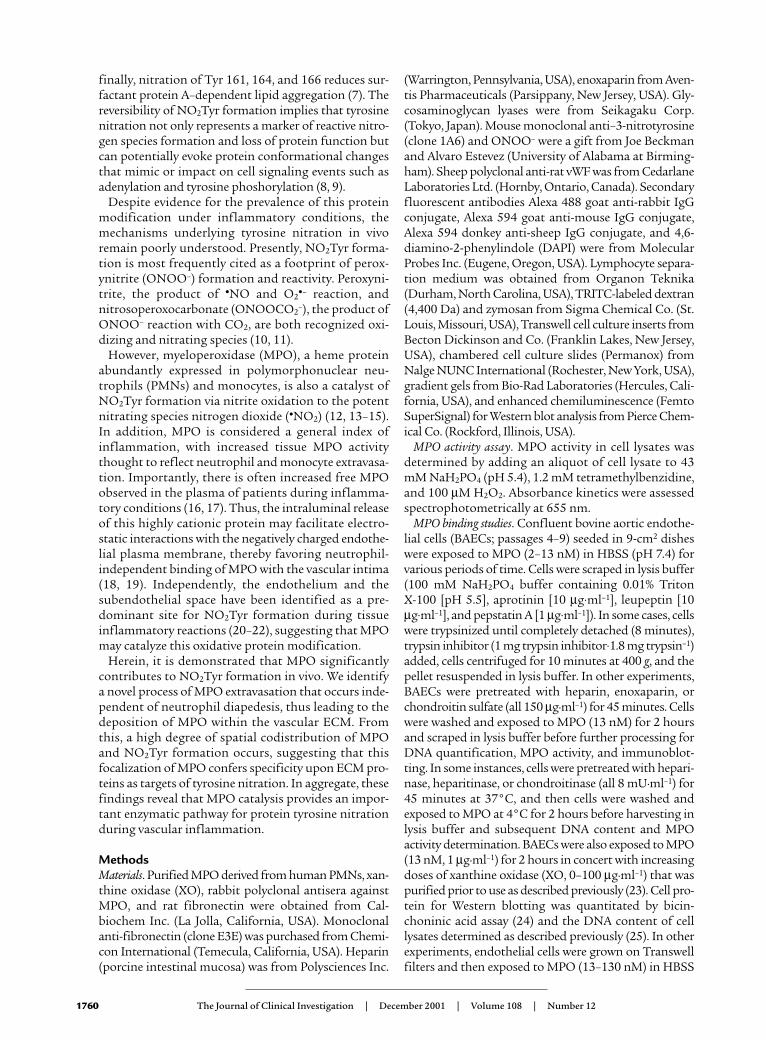
Quantification of tissue nitrotyrosine content. Nitrotyro-sine and tyrosine content in liver proteins was meas-ured by gas chromatography/mass spectrometry as pre-viously described (27). In brief, liver tissue washomogenized in a mixture of saline (2 ml) and chloro-form/methanol (2:1) (13 ml) on ice, and the proteinprecipitate (middle layer) was isolated by centrifuga-tion at 2000 g for 30 minutes at 4°C. Followinglyophilization of precipitated proteins, 1–1.5 mg of tis-
sue was hydrolyzed for 24 hours at 120°Cin 1 ml 4 M sodium hydroxide followingthe addition of 20 ng 13C9-nitrotyrosineand 10 µg of D4-tyrosine as stable isotopicinternal standards. These conditions pre-vent the nitration of tyrosine that occursduring acidic hydrolysis conditions andpermit identification of potential artifac-tual nitration reactions. Following solidphase extraction, nitrotyrosine and tyro-sine were quantitated by gas chromatogra-phy/negative ion chemical ionization massspectrometry. Results are expressed as aratio of nitrotyrosine to tyrosine (ng/mg).
Statistics. All data represent the mean ±SD. Statistical analyses were performedusing ANOVA with Tukey’s post hocanalysis on Systat 7.0 software (SPSS Sci-ence, Chicago, Illinois, USA). Results withP < 0.05 were considered significant.
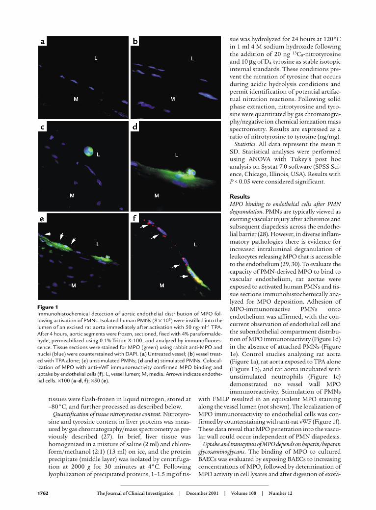
ResultsMPO binding to endothelial cells after PMNdegranulation. PMNs are typically viewed asexerting vascular injury after adherence andsubsequent diapedesis across the endothe-lial barrier (28). However, in diverse inflam-matory pathologies there is evidence forincreased intraluminal degranulation ofleukocytes releasing MPO that is accessibleto the endothelium (29, 30). To evaluate thecapacity of PMN-derived MPO to bind tovascular endothelium, rat aortae wereexposed to activated human PMNs and tis-sue sections immunohistochemically ana-lyzed for MPO deposition. Adhesion ofMPO-immunoreactive PMNs ontoendothelium was affirmed, with the con-current observation of endothelial cell andthe subendothelial compartment distribu-tion of MPO immunoreactivity (Figure 1d)in the absence of attached PMNs (Figure1e). Control studies analyzing rat aorta(Figure 1a), rat aorta exposed to TPA alone(Figure 1b), and rat aorta incubated withunstimulated neutrophils (Figure 1c)demonstrated no vessel wall MPOimmunoreactivity. Stimulation of PMNs
with FMLP resulted in an equivalent MPO stainingalong the vessel lumen (not shown). The localization ofMPO immunoreactivity to endothelial cells was con-firmed by counterstaining with anti-rat vWF (Figure 1f).These data reveal that MPO penetration into the vascu-lar wall could occur independent of PMN diapedesis.
Uptake and transcytosis of MPO depends on heparin/heparanglycosaminoglycans. The binding of MPO to culturedBAECs was evaluated by exposing BAECs to increasingconcentrations of MPO, followed by determination ofMPO activity in cell lysates and after digestion of exofa-
1762 The Journal of Clinical Investigation | December 2001 | Volume 108 | Number 12
Figure 1Immunohistochemical detection of aortic endothelial distribution of MPO fol-lowing activation of PMNs. Isolated human PMNs (8 × 10⁵) were instilled into thelumen of an excised rat aorta immediately after activation with 50 ng·ml–1 TPA.After 4 hours, aortic segments were frozen, sectioned, fixed with 4% paraformalde-hyde, permeabilized using 0.1% Triton X-100, and analyzed by immunofluores-cence. Tissue sections were stained for MPO (green) using rabbit anti-MPO andnuclei (blue) were counterstained with DAPI. (a) Untreated vessel; (b) vessel treat-ed with TPA alone; (c) unstimulated PMNs; (d and e) stimulated PMNs. Colocal-ization of MPO with anti-vWF immunoreactivity confirmed MPO binding anduptake by endothelial cells (f). L, vessel lumen; M, media. Arrows indicate endothe-lial cells. ×100 (a–d, f); ×50 (e).

cial cell proteins with trypsin. The MPO activity of celllysates increased dose-dependently, and the appearanceof MPO activity in a trypsin-resistant compartment sug-gested intracellular MPO deposition (Figure 2a). In addi-tion, MPO bound to BAECs after degranulation of acti-vated human neutrophils was enzymatically active, andendothelial cell–associated MPO activity was increasedto an even greater extent when the respiratory burst ofPMNs was inhibited by pretreatment with diphenyliodo-nium, which prevented the partial inactivation of MPOby PMN-derived oxidants (not shown). To test whetherMPO uptake by endothelial cells was energy-dependent,BAECs were exposed to MPO at 4°C, and trypsin-resist-ant and total cell-associated MPO activity was deter-mined (Figure 2b). Trypsin-resistant MPO activity at
4°C was reduced by more than 80%, suggesting energy-dependent internalization of the enzyme. The fact thattotal MPO activity was not decreased as much may be aresult of enhanced cell surface binding under 4°C con-ditions, as well as a result of cold-induced retraction ofthe cytoskeleton directly exposing the subendothelialmatrix to MPO. To evaluate the nature of MPO bindingto cell surface glycosaminoglycans, cells were pretreatedwith heparin and the low–molecular weight heparinenoxaparin prior to addition of MPO. Cell-associatedMPO activity and MPO protein content were signifi-cantly reduced, whereas pretreatment with chondroitinsulfate did not significantly alter cell MPO binding (Fig-ure 2c). Enoxaparin, a low–molecular weight heparin,added to cells after MPO exposure reduced cell-associat-
The Journal of Clinical Investigation | December 2001 | Volume 108 | Number 12 1763
Figure 2Characterization of the binding and uptake of MPO by cultured endothelial cells. (a) Characterization of cell MPO association. BAECs wereincubated with increasing concentrations of MPO (2–13 nM) for 2 hours at 37°C and harvested by scraping (total) or after exposure totrypsin (trypsin-resistant). Cell-associated MPO activity was about 10% of total MPO activity added (not shown). (b) The influence of tem-perature on cellular MPO binding and transport. BAECs exposed to MPO (13 nM) at 37°C and 4°C were harvested at different time pointsand MPO activity was determined. †P < 0.05 for MPO activity in total cell lysates at 37°C versus 4°C; *P < 0.05 for MPO activity in trypsin-resistant compartments at 37°C versus 4°C. (c) The influence of glycosaminoglycans on cell MPO binding. BAECs were pretreated withchondroitin sulfate (Chondr, 150 µg·ml–1), heparin (Hep, 150 µg·ml–1), or the low–molecular weight heparin enoxaparin (Enox, 150 µg·ml–1)for 45 minutes, washed with HBSS, and then exposed to MPO (13 nM) at 37°C. After washing again, cell-associated MPO was assessed byenzyme activity analysis and cellular MPO protein content by immunoblotting. “MPO hc ” denotes the immunoreactivity of the heavy chain(59 kDa) of MPO. (d) The effect of endoglycosidases on MPO binding. BAECs were pretreated with heparitinase, heparinase, and chon-droitinase (all 8 mU·ml–1) for 2 hours at 37°C, washed, and exposed to MPO (13 nM) at 4°C for 2 hours. Cell-associated MPO enzymeactivity was then determined. *P < 0.05 for MPO alone versus pretreatment with heparin and enoxaparin (c) and versus heparitinase andheparinase pretreatment (d). (e) Binding competition analysis of MPO and xanthine oxidase (XO). BAECs were incubated with MPO (1µg·ml–1) and increasing concentrations of XO (0–100 µg·ml–1, equivalent to 0–100 mU·ml–1) for 2 hours at 37°C. Cells were harvested asabove and MPO enzyme activity determined. Values represent mean ± SD.

ed MPO activity by 78% ± 3% (not shown). To furthercharacterize the nature of the putative glycosaminogly-can-dependent binding of MPO, BAECs were pretreatedwith endoglycosidases prior to MPO exposure. Hepar-itinase or heparinase pretreatment of endothelial cellsreduced cell-associated MPO activity by 63% ± 7% and
56% ± 8%, respectively, whereas chondroitinase did notsignificantly inhibit cell association of MPO (Figure 2d).Previous studies have revealed that binding of the O2
•-and H2O2-generating enzyme xanthine oxidase (XO) tovascular endothelial cells is predominantly chondroitinsulfate–dependent (23). To evaluate the potential bind-
1764 The Journal of Clinical Investigation | December 2001 | Volume 108 | Number 12
Figure 3Transcytosis of MPO by endothelial cells. (a) Intracellular localization and accumulation of MPO at the ECM. Cultured BAECs were grown toconfluence and exposed to MPO (13 nM) for 2 hours. Cells were processed as described in Methods and incubated with mouse monoclonalanti–α-tubulin (red) and rabbit polyclonal anti-MPO (green). Laser confocal microscopy revealed intracellular MPO immunoreactivity within 2minutes after MPO exposure and a high degree of MPO deposition at the subcellular matrix. Time = 0 minutes represents BAECs not exposed toMPO but stained with anti-MPO. ×63. (b) Colocalization of MPO and fibronectin (FN). BAECs treated as described for a were incubated withmouse monoclonal anti-fibronectin (red) and rabbit polyclonal anti-MPO (green). The nuclei (blue) were counterstained with DAPI. For assess-ing codistribution of MPO and fibronectin, images were merged (yellow). The control panel (Ctr) shows MPO-untreated cells stained with anti-MPO. The lower panel represents side-on views. ×63 (control, ×50). (c) Barrier function of endothelial cell monolayers exposed to MPO. BAECswere seeded on Transwell filters and exposed to increasing concentrations of MPO (13–130 nM) in the presence of TRITC-labeled dextran (4,400Da) for 2 hours. MPO activity and TRITC fluorescence were assessed in the basolateral chamber. Treatment of the cells with H2O2 (100 µM) servedas a control for increased permeability of TRITC-dextran. *P > 0.05 versus TRITC-labeled dextran alone. RFU, relative fluorescence units.
ing interactions of MPO and XO, the impact of increas-ing XO concentrations on endothelial cell MPO bindingwas determined. Increases in cell-associated MPO activ-ity were not inhibited by XO concentrations both equal-ing and exceeding those found in the circulation duringpathophysiological events (Figure 2e) (23).
MPO accumulates at the basolateral cell surface and colocal-izes with the ECM protein fibronectin. Using laser confocalmicroscopy we further localized the site of endothelialMPO deposition, by revealing intracellular MPO distri-bution within minutes after MPO exposure and astrong gradient between apical and basolateral MPOimmunoreactivity throughout the ECM after 2 hours(Figure 3a). Here, MPO was demonstrated to colocalizewith the ECM protein fibronectin (Figure 3b). Additionof MPO (130 nM) to BAECs in the presence of 100%human serum led to both intracellular and suben-dothelial distribution of MPO in the subendothelialmatrix (not shown). To test whether MPO influencesendothelial cell barrier function, thereby further facili-tating its subendothelial accumulation, we exposedendothelial cells grown on transwell filters to increasingdoses of MPO in the presence of TRITC-labeled dextran.While MPO activity increased dose-dependently in thebasolateral compartment, the transendothelial flux ofdextran remained unchanged (Figure 3c). In contrast,exposure of BAECs to H2O2 (100 µM) resulted in a sig-nificant increase in TRITC-dextran permeability. Inconcert with demonstrating the energy-dependence ofMPO transport (Figure 2b), the cell internalization ofMPO the enzyme (Figure 3a), and a diffuse distributionof MPO throughout the subendothelial matrix that was
in a diffuse manner not concentrated in cell junctionalregions (Figure 3, a and b) are all supporting evidencethat the mechanism by which MPO traverses endothe-lial cells is via transcytosis.
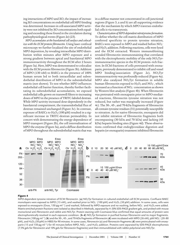
Characterization of MPO-dependent nitrotyrosine formation.To define whether the cell matrix distribution of MPOconferred specificity to protein tyrosine nitration,BAECs were exposed to MPO and washed before NO2
–
and H2O2 addition. Following reactions, cells were lysedand the ECM extracted. Western immunoblottingrevealed fibronectin immunostaining that correlatedwith the electrophoretic mobility of the only NO2Tyr-immunoreactive species in the ECM protein–rich frac-tion. In ECM fractions of cells pretreated with enoxa-parin, previously demonstrated to inhibit cell and vesselMPO binding/association (Figure 2c), NO2Tyrimmunoreactivity was profoundly reduced (Figure 4a).MPO also catalyzed NO2Tyr formation in solublehuman fibronectin exposed to H2O2 and NO2
–, whichincreased as a function of NO2
– concentration as shownby Western blot analysis (Figure 4b). When fibronectinwas pretreated with enoxaparin prior to MPO-mediat-ed reactions, fibronectin tyrosine nitration was notreduced, but rather was marginally increased (Figure4c). The 30-, 45-, and 70-kDa fragments of fibronectinall contain tyrosine residues (31) potentially susceptibleto nitration. As for native fibronectin, enoxaparin didnot inhibit nitration of fibronectin fragments bothencompassing (30 kDa and 70 kDa) and lacking (45kDa) heparin binding sites (Figure 4d). These observa-tions confirmed that endoglycosidase digestion andheparin (or enoxaparin) treatment inhibited fibronectin
The Journal of Clinical Investigation | December 2001 | Volume 108 | Number 12 1765
Figure 4MPO-dependent tyrosine nitration of ECM fibronectin. (a) NO2Tyr formation in cultured endothelial cell ECM proteins. Confluent BAECmonolayers were exposed to MPO (13 nM), and washed prior to NO2
– (100 µM) and H2O2 (50 µM) addition. In some cases, cells wereexposed to enoxaparin (Enox, 150 µg·ml–1) and washed, followed by MPO exposure and no washing, before NO2
– and H2O2 were added.Matrix-enriched protein fractions were isolated as described in Methods, separated by 4–20% SDS-PAGE gradient gels, and probed with mousemonoclonal anti-fibronectin and rabbit anti-NO2Tyr. Protein staining with Coomassie blue confirmed that equal amounts of protein wereelectrophoretically resolved in each exposure condition. (b–d) NO2Tyr formation in purified human fibronectin and its major fragments.Fibronectin (100 µg·ml–1) (b) and the 30-, 45-, and 70-kDa fragments of fibronectin (d) were incubated with MPO (26 nM), µM NO2
– (20–80µM), and H2O2 (50 µM) in HBSS for 90 minutes. In some cases, fibronectin (c) and fibronectin fragments (d) were preincubated with enoxa-parin (15 and 150 µg·ml–1) for 45 minutes before MPO, NO2
–, and H2O2 addition. Proteins were separated by SDS-PAGE electrophoresis(7.5% gels for fibronectin and 10% gels for fibronectin fragments) and then immunoblotted with rabbit polyclonal anti-NO2Tyr.
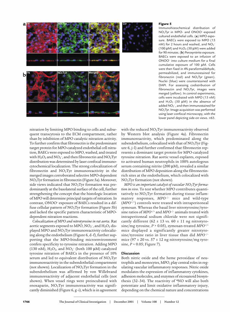
nitration by limiting MPO binding to cells and subse-quent transcytosis to the ECM compartment, ratherthan by inhibition of MPO catalytic nitration activity.To further confirm that fibronectin is the predominanttarget protein for MPO-catalyzed endothelial cell nitra-tion, BAECs were exposed to MPO, washed, and treatedwith H2O2 and NO2
–, and then fibronectin and NO2Tyrdistribution was determined by laser confocal immuno-cytochemical localization. The strong colocalization offibronectin and NO2Tyr immunoreactivity in themerged images corroborated selective MPO-dependentNO2Tyr formation in fibronectin (Figure 5a). Moreover,side views indicated that NO2Tyr formation was pre-dominantly at the basolateral surface of the cell, furtherstrengthening the concept that the histologic locationof MPO will determine principal targets of nitration. Incontrast, ONOO– exposure of BAECs resulted in a dif-fuse cellular pattern of NO2Tyr formation (Figure 5b)and lacked the specific pattern characteristic of MPO-dependent nitration reactions.
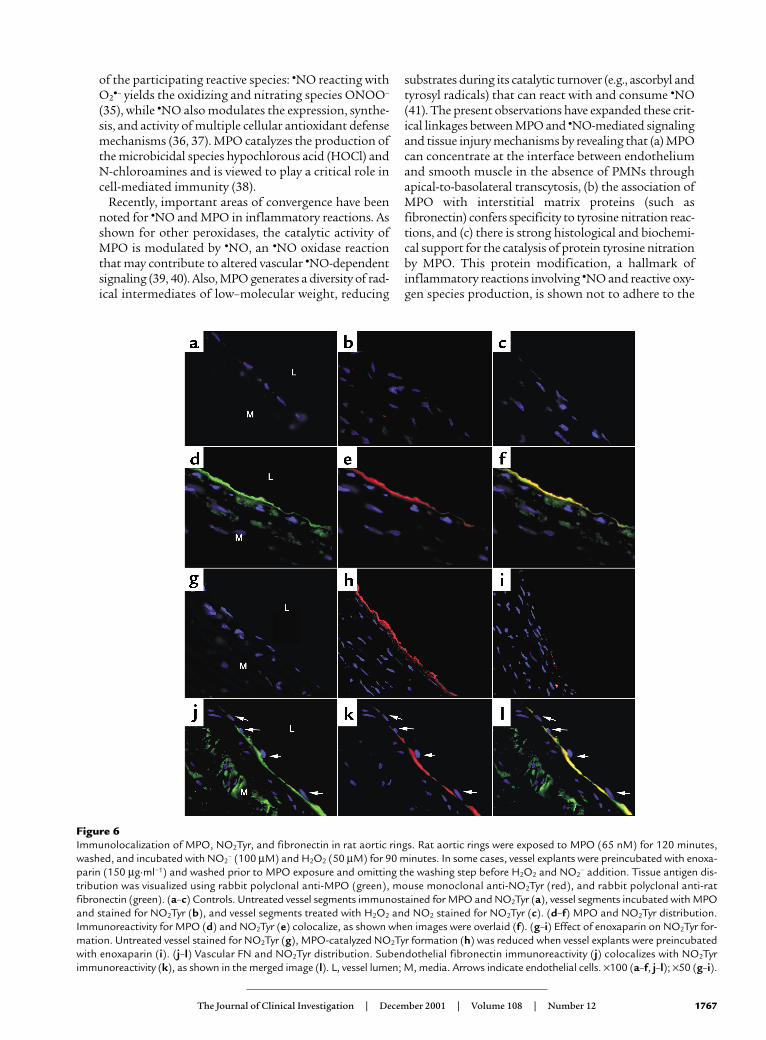
Colocalization of MPO and nitrotyrosine in rat aorta. Rataortic segments exposed to MPO, NO2
–, and H2O2 dis-played MPO and NO2Tyr immunoreactivity colocaliz-ing along the endothelium (Figure 6, d–f), further sup-porting that the MPO-binding microenvironmentconfers specificity to tyrosine nitration. Adding MPO(130 nM), H2O2, and NO2
– (both 100 µM) catalyzedtyrosine nitration of BAECs in the presence of 10%serum and led to equivalent distribution of NO2Tyrimmunoreactivity in the subendothelial compartment(not shown). Localization of NO2Tyr formation in thesubendothelium was affirmed by von Willebrandimmunoreactivity of adjacent endothelial cells (notshown). When vessel rings were preincubated withenoxaparin, NO2Tyr immunoreactivity was signifi-cantly diminished (Figure 6, g–i), which is in agreement
with the reduced NO2Tyr immunoreactivity observedby Western blot analysis (Figure 4a). Fibronectinimmunoreactivity, which predominated along thesubendothelium, colocalized with that of NO2Tyr (Fig-ure 6, j–l) and further confirmed that fibronectin rep-resents a dominant target protein for MPO-catalyzedtyrosine nitration. Rat aortic vessel explants, exposedto activated human neutrophils in 100% autologousserum containing nitrite (200 µM), revealed a similardistribution of MPO deposition along the fibronectin-rich sites at the endothelium, which colocalized withNO2Tyr formation (not shown).
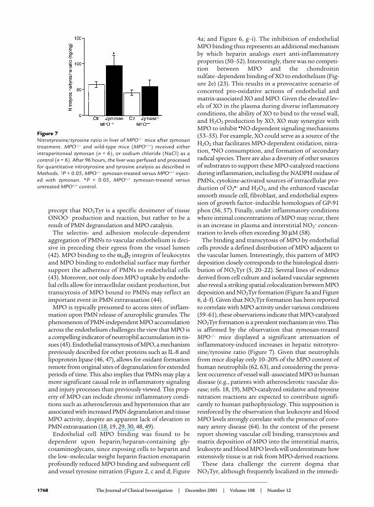
MPO is an important catalyst of vascular NO2Tyr forma-tion in vivo. To test whether MPO contributes quanti-tatively to NO2Tyr formation during tissue inflam-matory responses, MPO–/– mice and wild-type(MPO+/+) controls were treated with intraperitonealzymosan. Whereas the basal liver nitrotyrosine/tyro-sine ratios of MPO+/+ and MPO–/– animals treated withintraperitoneal sodium chloride were not signifi-cantly different (62 ± 13 vs. 60 ± 11 ng nitrotyro-sine/mg tyrosine, P > 0.05), zymosan-treated MPO+/+
mice displayed a significantly greater nitrotyro-sine/tyrosine ratio in liver tissue than did MPO–/–
mice (97 ± 20 vs. 57 ± 12 ng nitrotyrosine/mg tyro-sine, P < 0.05; Figure 7).
DiscussionBoth nitric oxide and the heme peroxidase of neu-trophils and monocytes, MPO, play central roles in reg-ulating vascular inflammatory responses. Nitric oxidemodulates the expression of inflammatory cytokines,adhesion molecules, and enzymes of eicosanoid biosyn-thesis (32–34). The reactivity of •NO will also bothpotentiate and limit oxidative inflammatory injury,depending on the chemical nature and concentrations
1766 The Journal of Clinical Investigation | December 2001 | Volume 108 | Number 12
Figure 5Immunohistochemical distribution ofNO2Tyr in MPO- and ONOO–-exposedcultured endothelial cells. (a) MPO expo-sure. BAECs were exposed to MPO (13nM) for 2 hours and washed, and NO2
–
(100 µM) and H2O2 (50 µM) were addedfor 90 minutes. (b) Peroxynitrite exposure.BAECs were exposed to an infusion ofONOO– into culture medium for a finalcumulative exposure of 100 µM. Cellswere then fixed in 4% paraformaldehyde,permeabilized, and immunostained forfibronectin (red) and NO2Tyr (green).Nuclei (blue) were counterstained withDAPI. For assessing codistribution offibronectin and NO2Tyr, images weremerged (yellow). In control experiments,cells were incubated with MPO (13 nM)and H2O2 (50 µM) in the absence ofadded NO2
–, and then immunostained forNO2Tyr. Image acquisition was performedusing laser confocal microscopy, with thelower panel depicting side-on views. ×63.
of the participating reactive species: •NO reacting withO2
•– yields the oxidizing and nitrating species ONOO–
(35), while •NO also modulates the expression, synthe-sis, and activity of multiple cellular antioxidant defensemechanisms (36, 37). MPO catalyzes the production ofthe microbicidal species hypochlorous acid (HOCl) andN-chloroamines and is viewed to play a critical role incell-mediated immunity (38).
Recently, important areas of convergence have beennoted for •NO and MPO in inflammatory reactions. Asshown for other peroxidases, the catalytic activity ofMPO is modulated by •NO, an •NO oxidase reactionthat may contribute to altered vascular •NO-dependentsignaling (39, 40). Also, MPO generates a diversity of rad-ical intermediates of low–molecular weight, reducing
substrates during its catalytic turnover (e.g., ascorbyl andtyrosyl radicals) that can react with and consume •NO(41). The present observations have expanded these crit-ical linkages between MPO and •NO-mediated signalingand tissue injury mechanisms by revealing that (a) MPOcan concentrate at the interface between endotheliumand smooth muscle in the absence of PMNs throughapical-to-basolateral transcytosis, (b) the association ofMPO with interstitial matrix proteins (such asfibronectin) confers specificity to tyrosine nitration reac-tions, and (c) there is strong histological and biochemi-cal support for the catalysis of protein tyrosine nitrationby MPO. This protein modification, a hallmark ofinflammatory reactions involving •NO and reactive oxy-gen species production, is shown not to adhere to the
The Journal of Clinical Investigation | December 2001 | Volume 108 | Number 12 1767
Figure 6Immunolocalization of MPO, NO2Tyr, and fibronectin in rat aortic rings. Rat aortic rings were exposed to MPO (65 nM) for 120 minutes,washed, and incubated with NO2
– (100 µM) and H2O2 (50 µM) for 90 minutes. In some cases, vessel explants were preincubated with enoxa-parin (150 µg·ml–1) and washed prior to MPO exposure and omitting the washing step before H2O2 and NO2
– addition. Tissue antigen dis-tribution was visualized using rabbit polyclonal anti-MPO (green), mouse monoclonal anti-NO2Tyr (red), and rabbit polyclonal anti-ratfibronectin (green). (a–c) Controls. Untreated vessel segments immunostained for MPO and NO2Tyr (a), vessel segments incubated with MPOand stained for NO2Tyr (b), and vessel segments treated with H2O2 and NO2 stained for NO2Tyr (c). (d–f) MPO and NO2Tyr distribution.Immunoreactivity for MPO (d) and NO2Tyr (e) colocalize, as shown when images were overlaid (f). (g–i) Effect of enoxaparin on NO2Tyr for-mation. Untreated vessel stained for NO2Tyr (g), MPO-catalyzed NO2Tyr formation (h) was reduced when vessel explants were preincubatedwith enoxaparin (i). (j–l) Vascular FN and NO2Tyr distribution. Subendothelial fibronectin immunoreactivity (j) colocalizes with NO2Tyrimmunoreactivity (k), as shown in the merged image (l). L, vessel lumen; M, media. Arrows indicate endothelial cells. ×100 (a–f, j–l); ×50 (g–i).
precept that NO2Tyr is a specific dosimeter of tissueONOO– production and reaction, but rather to be aresult of PMN degranulation and MPO catalysis.
The selectin- and adhesion molecule–dependentaggregation of PMNs to vascular endothelium is deci-sive in preceding their egress from the vessel lumen(42). MPO binding to the αMβ2 integrin of leukocytesand MPO binding to endothelial surface may furthersupport the adherence of PMNs to endothelial cells(43). Moreover, not only does MPO uptake by endothe-lial cells allow for intracellular oxidant production, buttranscytosis of MPO bound to PMNs may reflect animportant event in PMN extravasation (44).
MPO is typically presumed to access sites of inflam-mation upon PMN release of azurophilic granules. Thephenomenon of PMN-independent MPO accumulationacross the endothelium challenges the view that MPO isa compelling indicator of neutrophil accumulation in tis-sues (45). Endothelial transcytosis of MPO, a mechanismpreviously described for other proteins such as IL-8 andlipoprotein lipase (46, 47), allows for oxidant formationremote from original sites of degranulation for extendedperiods of time. This also implies that PMNs may play amore significant causal role in inflammatory signalingand injury processes than previously viewed. This prop-erty of MPO can include chronic inflammatory condi-tions such as atherosclerosis and hypertension that areassociated with increased PMN degranulation and tissueMPO activity, despite an apparent lack of elevation inPMN extravasation (18, 19, 29, 30, 48, 49).
Endothelial cell MPO binding was found to bedependent upon heparin/heparan-containing gly-cosaminoglycans, since exposing cells to heparin andthe low–molecular weight heparin fraction enoxaparinprofoundly reduced MPO binding and subsequent celland vessel tyrosine nitration (Figure 2, c and d; Figure
4a; and Figure 6, g–i). The inhibition of endothelialMPO binding thus represents an additional mechanismby which heparin analogs exert anti-inflammatoryproperties (50–52). Interestingly, there was no competi-tion between MPO and the chondroitinsulfate–dependent binding of XO to endothelium (Fig-ure 2e) (23). This results in a provocative scenario ofconcerted pro-oxidative actions of endothelial andmatrix-associated XO and MPO. Given the elevated lev-els of XO in the plasma during diverse inflammatoryconditions, the ability of XO to bind to the vessel wall,and H2O2 production by XO, XO may synergize withMPO to inhibit •NO-dependent signaling mechanisms(53–55). For example, XO could serve as a source of theH2O2 that facilitates MPO-dependent oxidation, nitra-tion, •NO consumption, and formation of secondaryradical species. There are also a diversity of other sourcesof substrates to support these MPO-catalyzed reactionsduring inflammation, including the NADPH oxidase ofPMNs, cytokine-activated sources of intracellular pro-duction of O2
•– and H2O2, and the enhanced vascularsmooth muscle cell, fibroblast, and endothelial expres-sion of growth factor–inducible homologues of GP-91phox (56, 57). Finally, under inflammatory conditionswhere intimal concentrations of MPO may occur, thereis an increase in plasma and interstitial NO2
– concen-tration to levels often exceeding 30 µM (58).
The binding and transcytosis of MPO by endothelialcells provide a defined distribution of MPO adjacent tothe vascular lumen. Interestingly, this pattern of MPOdeposition closely corresponds to the histological distri-bution of NO2Tyr (5, 20–22). Several lines of evidencederived from cell culture and isolated vascular segmentsalso reveal a striking spatial colocalization between MPOdeposition and NO2Tyr formation (Figure 5a and Figure6, d–f). Given that NO2Tyr formation has been reportedto correlate with MPO activity under various conditions(59–61), these observations indicate that MPO-catalyzedNO2Tyr formation is a prevalent mechanism in vivo. Thisis affirmed by the observation that zymosan-treatedMPO–/– mice displayed a significant attenuation ofinflammatory-induced increases in hepatic nitrotyro-sine/tyrosine ratio (Figure 7). Given that neutrophilsfrom mice display only 10–20% of the MPO content ofhuman neutrophils (62, 63), and considering the preva-lent occurrence of vessel wall–associated MPO in humandisease (e.g., patients with atherosclerotic vascular dis-ease; refs. 18, 19), MPO-catalyzed oxidative and tyrosinenitration reactions are expected to contribute signifi-cantly to human pathophysiology. This supposition isreinforced by the observation that leukocyte and bloodMPO levels strongly correlate with the presence of coro-nary artery disease (64). In the context of the presentreport showing vascular cell binding, transcytosis andmatrix deposition of MPO into the interstitial matrix,leukocyte and blood MPO levels will underestimate howextensively tissue is at risk from MPO-derived reactions.
These data challenge the current dogma thatNO2Tyr, although frequently localized in the immedi-
1768 The Journal of Clinical Investigation | December 2001 | Volume 108 | Number 12
Figure 7Nitrotyrosine/tyrosine ratio in liver of MPO–/– mice after zymosantreatment. MPO–/– and wild-type mice (MPO+/+) received eitherintraperitoneal zymosan (n = 6), or sodium chloride (NaCl) as acontrol (n = 6). After 96 hours, the liver was perfused and processedfor quantitative nitrotyrosine and tyrosine analysis as described inMethods. †P < 0.05, MPO–/– zymosan-treated versus MPO+/+ inject-ed with zymosan. *P < 0.05, MPO+/+ zymosan-treated versusuntreated MPO+/+ control.
ate vicinity of neutrophils, is a specific marker forONOO– production and reactivity (65). Enzymaticallycatalyzed tyrosine nitration by MPO that is sequesteredin defined tissue compartments represents a more con-trolled and specific process, compared with the diffu-sion controlled nitration reactions that are observedwith ONOO– (66). MPO has also been proposed to cat-alyze ONOO––dependent tyrosine nitration (67). Thismay represent another mechanism linking NO2Tyr for-mation and MPO deposition. The biochemical occur-rence and significance of this pathway in vivo remainsundefined but, if relevant, still underscores the centralrole of peroxidases in catalyzing tyrosine nitration.
The ECM protein fibronectin proved to be the pre-dominant protein target for MPO-dependent NO2Tyrformation. The impact of heparin and enoxaparin oninhibiting MPO-dependent NO2Tyr formation indi-cates that cell binding and transcytosis of MPO is of piv-otal importance for fibronectin tyrosine nitration, aconcept supported by the similar inhibitory effect ofheparin on MPO-dependent cell protein oxidation (68).In contrast, heparin coincubated directly withfibronectin did not inhibit MPO-dependent nitration(Figure 4, c and d), mirroring in vitro observations thatheparin-lipoprotein complex formation did not inhib-it MPO-dependent lipid oxidation (69). This supportsthe view that the predominant nitrating species formedby MPO, •NO2, has a limited diffusion distance andreactivity capable of readily mediating fibronectin nitra-tion in a glycosaminoglycan-containing microenviron-ment (e.g., the cell matrix). The local formation of both•NO2 and tyrosyl radicals may provide one explanationfor the highly specific nitration patterns displayed byMPO; that is, the ability of •NO2 to promote tyrosinenitration is facilitated by simultaneous tyrosyl radicalformation, as the reaction between •NO2 and tyrosylradical occurs at a nearly diffusion-limited rate (109
M–1s–1). Fibronectin critically affects cellular signalingevents via integrin and/or growth factor ligation, there-by orchestrating the adherence of neutrophils, as well asthe growth and migration of SMC and fibroblasts(70–73). Oxidation and/or nitration of fibronectin hasrecently been shown to impair fibroblast migration (74).Therefore, “inflamed fibronectin,” which has long beenassociated with actions of PMN-derived or -activatedproteases (75), may also be due to oxidative and nitra-tive modification of fibronectin that results in alteredprotein function during inflammatory conditions (76).
In summary, MPO is revealed to be an importantmediator for NO2Tyr formation in vivo. Endothelialtranscytosis of MPO resulted in MPO accumulation inthe ECM and proved to be elemental for fibronectinnitration that can occur remote from the site of leuko-cyte degranulation. In addition to serving as a media-tor of host defense and oxidative inflammatory tissueinjury, MPO is identified as a NO2
– oxidase whose closeproximity to specific binding sites such as fibronectinwill facilitate and confer specificity to nitration reac-tions within the vasculature.
AcknowledgmentsThis work was supported by grants from the NIH (toB.A. Freeman and C.R. White), the Deutsche Herz-stiftung (to S. Baldus), and the Max PlanckGesellschaft (to S. Baldus).
1. Ischiropoulos, H. 1998. Biological tyrosine nitration: a pathophysiolog-ical function of nitric oxide and reactive oxygen species. Arch. Biochem.Biophys. 356:1–11.
2. Eiserich, J.P., et al. 1999. Microtubule dysfunction by posttranslationalnitrotyrosination of α-tubulin: a nitric oxide-dependent mechanism ofcellular injury. Proc. Natl. Acad. Sci. USA. 96:6365–6370.
3. Cassina, A.M., et al. 2000. Cytochrome c nitration by peroxynitrite. J. Biol.Chem. 275:21409–21415.
4. Viner, R.I., Ferrington, D.A., Williams, T.D., Bigelow, D.J., and Schone-ich, C. 1999. Protein modification during biological aging: selective tyro-sine nitration of the SERCA2a isoform of the sarcoplasmic reticulumCa2+-ATPase in skeletal muscle. Biochem. J. 340:657–669.
5. MacMillan-Crow, L.A., Crow, J.P., Kerby, J.D., Beckman, J.S., and Thomp-son, J.A. 1996. Nitration and inactivation of manganese superoxide dis-mutase in chronic rejection of human renal allografts. Proc. Natl. Acad.Sci. USA. 93:11853–11858.
6. Yamakura, F., Taka, H., Fujimura, T., and Murayama K. 1998. Inactiva-tion of human manganese-superoxide dismutase by peroxynitrite iscaused by exclusive nitration of tyrosine 34 to 3-nitrotyrosine. J. Biol.Chem. 273:14085–14089.
7. Zhu, S., Basiouny, K.F., Crow, J.P., and Matalon, S. 2000. Carbon dioxideenhances nitration of surfactant protein A by activated alveolarmacrophages. Am. J. Physiol. Lung Cell. Mol. Physiol. 278:L1025–L1031.
8. Kamisaki, Y., et al. 1998. An activity in rat tissues that modifies nitroty-rosine-containing proteins. Proc. Natl. Acad. Sci. USA. 95:11584–11589.
9. Berlett, B.S., Levine, R.L., and Stadtman, E.R. 1998. Carbon dioxide stim-ulates peroxynitrite-mediated nitration of tyrosine residues and inhibitsoxidation of methionine residues of glutamine synthetase: both modi-fications mimic effects of adenylylation. Proc. Natl. Acad. Sci. USA.95:2784–2789.
10. Beckman, J.S., and Koppenol, W.H. 1996. Nitric oxide, superoxide, andperoxynitrite: the good, the bad, and ugly. Am. J. Physiol.271:C1424–C1437.
11. Radi, R., Denicola, A., and Freeman, B.A. 1999. Peroxynitrite reactionswith carbon dioxide-bicarbonate. Methods Enzymol. 301:353–367.
12. Eiserich, J.P., et al. 1998. Formation of nitric oxide-derived inflammato-ry oxidants by myeloperoxidase in neutrophils. Nature. 391:393–397.
13. van der Vliet, A., Eiserich, J.P., Halliwell, B., and Cross, C.E. 1997. For-mation of reactive nitrogen species during peroxidase-catalyzed oxida-tion of nitrite. A potential additional mechanism of nitric oxide-depend-ent toxicity. J. Biol. Chem. 272:7617–7625.
14. Hazen, S.L., et al. 1999. Formation of nitric oxide-derived oxidants bymyeloperoxidase in monocytes: pathways for monocyte-mediated pro-tein nitration and lipid peroxidation in vivo. Circ. Res. 85:950–958.
15. van Dalen, C.J., Winterbourn, C.C., Senthilmohan, R., and Kettle, A.J.2000. Nitrite as a substrate and inhibitor of myeloperoxidase. Implica-tions for nitration and hypochlorous acid production at sites of inflam-mation. J. Biol. Chem. 275:11638–11644.
16. Biasucci, L.M., et al. 1996. Intracellular neutrophil myeloperoxidase isreduced in unstable angina and acute myocardial infarction, but itsreduction is not related to ischemia. J. Am. Coll. Cardiol. 27:611–616.
17. Deby-Dupont, G., Deby, C., and Lamy, M. 1999. Neutrophil myeloper-oxidase revisited: its role in health and disease. Intensivmedizin-und-Not-fallmedizin. 36:500–513.
18. Daugherty, A., Dunn, J.L., Rateri, D.L., and Heinecke, J.W. 1994.Myeloperoxidase, a catalyst for lipoprotein oxidation, is expressed inhuman atherosclerotic lesions. J. Clin. Invest. 94:437–444.
19. Malle, E., et al. 2000. Immunohistochemical evidence for the myeloper-oxidase/H2O2/halide system in human atherosclerotic lesions: colocal-ization of myeloperoxidase and hypochlorite-modified proteins. Eur. J.Biochem. 267:4495–4503.
20. Albertini, M., et al. 2000. Role of poly-(ADP-ribose) synthetase inlipopolysaccharide-induced vascular failure and acute lung injury inpigs. J. Crit. Care. 15:73–83.
21. Beckman, J.S., et al. 1994. Extensive nitration of protein tyrosines inhuman atherosclerosis detected by immunohistochemistry. Biol. Chem.Hoppe Seyler. 375:81–88.
22. Hirabayashi, H., Takizawa, S., Fukuyama, N., Nakazawa, H., and Shino-hara, Y. 2000. Nitrotyrosine generation via inducible nitric oxide syn-thase in vascular wall in focal ischemia-reperfusion. Brain Res.852:319–325.
23. Houston, M., et al. 1999. Binding of xanthine oxidase to vascularendothelium. Kinetic characterization and oxidative impairment ofnitric oxide-dependent signaling. J. Biol. Chem. 274:4985–4994.
The Journal of Clinical Investigation | December 2001 | Volume 108 | Number 12 1769
24. Redinbaugh, M.G., and Turley, R.B. 1986. Adaptation of the bicin-choninic acid protein assay for use with microtiter plates and sucrosegradient fractions. Anal. Biochem. 153:267–271.
25. Fiszer-Szafarz, B., Szafarz, D., and Guevara de Murillo, A. 1981. A gen-eral, fast, and sensitive micromethod for DNA determination applica-tion to rat and mouse liver, rat hepatoma, human leukocytes, chickenfibroblasts, and yeast cells. Anal. Biochem. 110:165–170.
26. Brennan, M.-L., et al. 2001. Increased atherosclerosis in myeloperoxidase-deficient mice. J. Clin. Invest. 107:419–430.
27. Frost, M.T., Halliwell, B., and Moore, K.P. 2000. Analysis of free and pro-tein-bound nitrotyrosine in human plasma by a gas chromatogra-phy/mass spectrometry method that avoids nitration artifacts. Biochem.J. 345:453–458.
28. Brown, E. 1997. Neutrophil adhesion and the therapy of inflammation.Semin. Hematol. 34:319–326.
29. Mohacsi, A., Kozlovszky, B., Kiss, I., Seres, I., and Fulop, T. 1996. Neu-trophils obtained from obliterative atherosclerotic patients exhibitenhanced resting respiratory burst and increased degranulation inresponse to various stimuli. Biochim. Biophys. Acta. 1316:210–216.
30. Shen, K., DeLano, F.A., Zweifach, B.W., and Schmid-Schonbein, G.W.1995. Circulating leukocyte counts, activation, and degranulation inDahl hypertensive rats. Circ. Res. 76:276–283.
31. Kornblihtt, A.R., Umezawa, K., Vibe-Pedersen, K., and Baralle, F.E. 1985.Primary structure of human fibronectin: differential splicing may gen-erate at least 10 polypeptides from a single gene. EMBO J. 4:1755–1759.
32. Furuke, K., et al. 1999. Human NK cells express endothelial nitric oxidesynthase, and nitric oxide protects them from activation-induced celldeath by regulating expression of TNF-alpha. J. Immunol.163:1473–1480.
33. De Caterina, R., et al. 1995. Nitric oxide decreases cytokine-inducedendothelial activation. Nitric oxide selectively reduces endothelialexpression of adhesion molecules and proinflammatory cytokines. J.Clin. Invest. 96:60–68.
34. Salvemini, D., Currie, M.G., and Mollace, V. 1996. Nitric oxide-mediat-ed cyclooxygenase activation. A key event in the antiplatelet effects ofnitrovasodilators. J. Clin. Invest. 97:2562–2568.
35. Beckman, J.S., Beckman, T.W., Chen, J., Marshall, P.A., and Freeman, B.A.1990. Apparent hydroxyl radical production by peroxynitrite: implica-tions for endothelial injury from nitric oxide and superoxide. Proc. Natl.Acad. Sci. USA. 87:1620–1624.
36. Darley Usmar, M., Patel, R.P., O’Donell, V.B., and Freeman, B.A. 2000.Antioxidant actions of nitric oxide. In Nitric oxide. Biology and pathobiolo-gy. L.J. Ignarro, editor. Academic Press Inc. San Diego, California, USA.265–276.
37. Deneke, S.M., and Fanburg, B.L. 1989. Regulation of cellular glu-tathione. Am. J. Physiol. 257:L163–L173.
38. Klebanoff, S.J. 1999. Myeloperoxidase. Proc. Assoc. Am. Physicians.111:383–389.
39. Glover, R.E., Koshkin, V., Dunford, H.B., and Mason, R.P. 1999. The reac-tion rates of NO with horseradish peroxidase compounds I and II. NitricOxide. 3:439–444.
40. Abu-Soud, H.M., and Hazen, S.L. 2000. Nitric oxide is a physiologicalsubstrate for mammalian peroxidases. J. Biol. Chem. 275:37524–37532.
41. Marquez, L.A., and Dunford, H.B. 1995. Kinetics of oxidation of tyrosineand dityrosine by myeloperoxidase compounds I and II. Implications forlipoprotein peroxidation studies. J. Biol. Chem. 270:30434–30440.
42. Lefer, A.M. 1999. Role of the beta2-integrins and immunoglobulinsuperfamily members in myocardial ischemia-reperfusion. Ann. Thorac.Surg. 68:1920–1923.
43. Johansson, M.W., Patarroyo, M., Oberg, F., Siegbahn, A., and Nilsson, K.1997. Myeloperoxidase mediates cell adhesion via the alpha M beta 2integrin (Mac-1, CD11b/CD18). J. Cell Sci. 110:1133–1139.
44. Yang, J.J., et al. 2001. Internalization of proteinase 3 is concomitant withendothelial cell apoptosis and internalization of myeloperoxidase withgeneration of intracellular oxidants. Am. J. Pathol. 158:581–592.
45. Matsuo, Y., et al. 1994. Correlation between myeloperoxidase-quantifiedneutrophil accumulation and ischemic brain injury in the rat. Effects ofneutrophil depletion. Stroke. 25:1469–1475.
46. Middleton, J., et al. 1997. Transcytosis and surface presentation of IL-8by venular endothelial cells. Cell. 91:385–395.
47. Obunike, J.C., et al. 2001. Transcytosis of lipoprotein lipase across cul-tured endothelial cells requires both heparan sulfate proteoglycans andthe very low density lipoprotein receptor. J. Biol. Chem. 276:8934–8941.
48. Shen, K., et al. 1995. Properties of circulating leukocytes in sponta-neously hypertensive rats. Biochem. Cell Biol. 73:491–500.
49. Arndt, H., Smith, C.W., and Granger, D.N. 1993. Leukocyte-endothelialcell adhesion in spontaneously hypertensive and normotensive rats.Hypertension. 21:667–673.
50. Park, J.L., et al. 1999. N-acetylheparin pretreatment reduces infarct sizein the rabbit. Pharmacology. 58:120–131.
51. Salas, A., et al. 2000. Heparin attenuates TNF-alpha induced inflamma-tory response through a CD11b dependent mechanism. Gut. 47:88–96.
52. Darien, B.J., et al. 1998. Low molecular weight heparin prevents the pul-monary hemodynamic and pathomorphologic effects of endotoxin in aporcine acute lung injury model. Shock. 9:274–281.
53. White, C.R., et al. 1996. Circulating plasma xanthine oxidase contributesto vascular dysfunction in hypercholesterolemic rabbits. Proc. Natl. Acad.Sci. USA. 93:8745–8749.
54. Mugge, A., et al. 1991. Chronic treatment with polyethylene-glycolatedsuperoxide dismutase partially restores endothelium-dependent vascu-lar relaxations in cholesterol-fed rabbits. Circ. Res. 69:1293–1300.
55. Fridovich, I. 1970. Quantitative aspects of the production of superoxideanion radical by milk xanthine oxidase. J. Biol. Chem. 245:4053–4057.
56. Suh, Y.A., et al. 1999. Cell transformation by the superoxide-generatingoxidase Mox1. Nature. 401:79–82.
57. Geiszt, M., Kopp, J.B., Varnai, P., and Leto, T.L. 2000. Identification ofrenox, an NAD(P)H oxidase in kidney. Proc. Natl. Acad. Sci. USA.97:8010–8014.
58. Torre, D., et al. 1996. Serum concentrations of nitrite in patients withHIV-1 infection. J. Clin. Pathol. 49:574–576.
59. Lamb, N.J., Gutteridge, J.M., Baker, C., Evans, T.W., and Quinlan, G.J.1999. Oxidative damage to proteins of bronchoalveolar lavage fluid inpatients with acute respiratory distress syndrome: evidence for neu-trophil-mediated hydroxylation, nitration, and chlorination. Crit. CareMed. 27:1738–1744.
60. Van der Vliet, A., et al. 2000. Myeloperoxidase and protein oxidation incystic fibrosis. Am. J. Physiol. Lung Cell. Mol. Physiol. 279:L537–L546.
61. Cuzzocrea, S., Costantino, G., Mazzon, E., and Caputi, A.P. 1999. Pro-tective effect of N-acetylcysteine on multiple organ failure induced byzymosan in the rat. Crit. Care Med. 27:1524–1532.
62. Podrez, E.A., Abu-Soud, H., and Hazen, S.L. 2000. Myeloperoxidase-gen-erated oxidants and atherosclerosis. Free Radic. Biol. Med. 28:1717–1725.
63. Rausch, P.G., and Moore, T.G. 1975. Granule enzymes of polymor-phonuclear neutrophils: a phylogenetic comparison. Blood. 46:913–919.
64. Zhang, R., et al. 2001. Association between myeloperoxidase levels andrisk of coronary artery disease. JAMA. 286:2136–2142.
65. Thom, S.R., Fisher, D., and Manevich, Y. 2001. Roles for platelet-acti-vating factor and •NO-derived oxidants causing neutrophil adherenceafter CO poisoning. Am. J. Physiol. Heart Circ. Physiol. 281:H923–H930.
66. Souza, J.M., Daikhin, E., Yudkoff, M., Raman, C.S., and Ischiropoulos,H. 1999. Factors determining the selectivity of protein tyrosine nitration.Arch. Biochem. Biophys. 371:169–178.
67. Sampson, J.B., Ye, Y., Rosen, H., and Beckman, J.S. 1998. Myeloperoxi-dase and horseradish peroxidase catalyze tyrosine nitration in proteinsfrom nitrite and hydrogen peroxide. Arch. Biochem. Biophys. 356:207–213.
68. Daphna, E.M., Michaela, S., Eynat, P., Irit, A., and Rimon, S. 1998. Asso-ciation of myeloperoxidase with heparin: oxidative inactivation of pro-teins on the surface of endothelial cells by the bound enzyme. Mol. Cell.Biochem. 183:55–61.
69. Upritchard, J.E., and Sutherland, W.H. 1999. Oxidation of heparin-treat-ed low density lipoprotein by peroxidases. Atherosclerosis. 146:211–219.
70. Clark, E.A., King, W.G., Brugge, J.S., Symons, M., and Hynes, R.O. 1998.Integrin-mediated signals regulated by members of the rho family ofGTPases. J. Cell Biol. 142:573–586.
71. Hocking, D.C., Sottile, J., and Langenbach, K.J. 2000. Stimulation of inte-grin-mediated cell contractility by fibronectin polymerization. J. Biol.Chem. 275:10673–10682.
72. Proctor, R.A. 1987. Fibronectin: an enhancer of phagocyte function. Rev.Infect. Dis. 4:S412–S419.
73. Magnusson, M.K., and Mosher, D.F. 1998. Fibronectin: structure, assem-bly, and cardiovascular implications. Arterioscler. Thromb. Vasc. Biol.18:1363–1370.
74. Sato, E., Koyama, S., Camhi, S.L., Nelson, D.K., and Robbins, R.A. 2001.Reactive oxygen and nitrogen metabolites modulate fibronectin-inducedfibroblast migration in vitro. Free Radic. Biol. Med. 30:22–29.
75. Vercellotti, G.M., McCarthy, J., Furcht, L.T., Jacob, H.S., and Moldow,C.F. 1983. Inflamed fibronectin: an altered fibronectin enhances neu-trophil adhesion. Blood. 62:1063–1069.
76. Vissers, M.C., and Thomas, C. 1997. Hypochlorous acid disrupts the adhe-sive properties of subendothelial matrix. Free Radic. Biol. Med. 23:401–411.
1770 The Journal of Clinical Investigation | December 2001 | Volume 108 | Number 12