Endocrine-Disrupting Effects of Bisphenol A on the ... - MDPI

Blood Vessel Maturation and Response to Vascular-Disrupting
Therapy in Single Vascular Endothelial Growth Factor-A
Isoform–Producing Tumors
Gillian M. Tozer,1Simon Akerman,
1Neil A. Cross,
1Paul R. Barber,
2Meit A. Bjorndahl,
1
Olga Greco,1Sheila Harris,
1Sally A. Hill,
2Davina J. Honess,
2Christopher R. Ireson,
2
Katie L. Pettyjohn,1Vivien E. Prise,
2Constantino C. Reyes-Aldasoro,
1
Christiana Ruhrberg,3David T. Shima,
3and Chryso Kanthou
1
1Cancer Research UK Tumour Microcirculation Group, Academic Unit of Surgical Oncology, School of Medicine and Biomedical Sciences,University of Sheffield, Sheffield, United Kingdom; 2Gray Cancer Institute, Mount Vernon Hospital, Northwood, Middlesex,United Kingdom; and 3Institute of Ophthalmology, University College London, London, United Kingdom
Abstract
Tubulin-binding vascular-disrupting agents (VDA) are cur-rently in clinical trials for cancer therapy but the factorsthat influence tumor susceptibility to these agents are poorlyunderstood. We evaluated the consequences of modifyingtumor vascular morphology and function on vascular andtherapeutic response to combretastatin-A4 3-O-phosphate(CA-4-P), which was chosen as a model VDA. Mouse fibro-sarcoma cell lines that are capable of expressing all vascularendothelial growth factor (VEGF) isoforms (control) or onlysingle isoforms of VEGF (VEGF120, VEGF164, or VEGF188)were developed under endogenous VEGF promoter control.Once tumors were established, VEGF isoform expression didnot affect growth or blood flow rate. However, VEGF188 wasuniquely associated with tumor vascular maturity, resistanceto hemorrhage, and resistance to CA-4-P. Pericyte stainingwas much greater in VEGF188 and control tumors than inVEGF120 and VEGF164 tumors. Vascular volume was highestin VEGF120 and control tumors (CD31 staining) but total vas-cular length was highest in VEGF188 tumors, reflecting verynarrow vessels forming complex vascular networks. I.v.administered 40 kDa FITC-dextran leaked slowly from thevasculature of VEGF188 tumors compared with VEGF120tumors. Intravital microscopy measurements of vascularlength and RBC velocity showed that CA-4-P produced signi-ficantly more vascular damage in VEGF120 and VEGF164tumors than in VEGF188 and control tumors. Importantly,this translated into a similar differential in therapeuticresponse, as determined by tumor growth delay. Results implydifferences in signaling pathways between VEGF isoformsand suggest that VEGF isoforms might be useful in vascular-disrupting cancer therapy to predict tumor susceptibility toVDAs. [Cancer Res 2008;68(7):2301–11]
Introduction
Disodium combretastatin-A4 3-O-phosphate (CA-4-P) is thelead compound of a group of tubulin-binding agents that act asvascular-disrupting agents (VDA) in solid tumors (1, 2). CA-4-P isactive in human cancers (3) and is currently in phase II clinicaltrials in combination with conventional treatments (4).4 VDAscause a catastrophic collapse of blood flow to solid tumors andthus form a conceptually distinct group from the antiangiogenicagents (5).
Despite the progress of VDAs to clinical trials, and someevidence for improved patient survival upon adding VDAtreatment to conventional chemotherapy (6), the reasons for thesusceptibility of the tumor vasculature to VDAs remain unclear.Tumor blood vessels are generally considered to be immature.Thus, genetic modification of tumor cells to modulate expressionof key molecules involved in vascular maturation provides apotential route for investigating the factors that influence theresponse of the tumor vasculature to VDAs.
Vascular endothelial growth factor A (VEGF-A or simply VEGF)is a key stimulator of tumorigenic angiogenesis (7) and acts as amitogen, chemoattractant, survival factor, vasodilator, and perme-ability factor (8). Human and mouse VEGF mRNA is transcribedfrom eight exons and alternatively spliced to give rise to a numberof isoforms of the protein product (9), the most prevalentconsisting of 121, 165, and 189 amino acids in the human and120, 164, and 188 in the mouse (Fig. 1A). VEGF120/121 lacks theheparin-binding site and is readily diffusible, whereas VEGF188/189is tightly bound to proteoglycans in the extracellular matrix or onthe cell surface and VEGF164/165 has intermediate properties.These isoforms are found in most normal tissues and have affinityfor the VEGF receptors, FLT1 (VEGFR1) and KDR (VEGFR2; refs.10, 11). VEGF121 and VEGF165 are the most prevalent forms inhuman cancers (12, 13).
Using Cre/Lox technology, mice expressing only single isoformsof VEGF, known as Vegfa120/120, Vegfa164/164 , and Vegfa188/188 mice,have been developed (14, 15). Their analysis revealed differentroles for the individual VEGF isoforms in vascular growth andpatterning during development. In particular, VEGF164 was suffi-cient for developmental angiogenesis and neonatal growth, andVEGF120 supported endothelial cell proliferation and vasculogenesis;however, angiogenic vessel branching was impaired and vessels
Note: Supplementary data for this article are available at Cancer Research Online(http://cancerres.aacrjournals.org/).
G.M. Tozer and S. Akerman contributed equally to this work.Requests for reprints: Gillian M. Tozer, Cancer Research UK Tumour
Microcirculation Group, Academic Unit of Surgical Oncology, K Floor, School ofMedicine and Biomedical Sciences, University of Sheffield, Beech Hill Road, S10 2JFSheffield, United Kingdom. Phone: 44-114271-2423; Fax: 44-114271-3314; E-mail: [email protected].
I2008 American Association for Cancer Research.doi:10.1158/0008-5472.CAN-07-2011 4 http://www.oxigene.com/
www.aacrjournals.org 2301 Cancer Res 2008; 68: (7). April 1, 2008
Research Article
Research. on February 19, 2016. © 2008 American Association for Cancercancerres.aacrjournals.org Downloaded from

were hemorrhagic (16). VEGF188 was essential for the formation ofa normal cardiovasculature in 50% of mice (14, 15).
In the current study, we used the single VEGF isoform-expressingmice and the counterpart wild-type mice to develop mousefibrosarcoma cell lines from embryo fibroblasts. We reasoned thatthe different VEGF isoforms would give rise to tumors with verydifferent vascular characteristics that could be used to investigatesusceptibility to CA-4-P. The method of producing the tumorlines was substantially different from previous studies in whichoverexpression of individual VEGF isoforms was used (13, 17),ensuring that VEGF gene transcription was under the control of theendogenous VEGF promoter and thus susceptible to environmental
control. We gained new insights into the differential effects ofVEGF isoforms in tumors by using intravital microscopy and tumoruptake of i.v. administered markers to measure functional vascularend points, in addition to measuring vascular morphology andtumor growth, with and without CA-4-P.
Materials and Methods
Animal experiments were conducted in accordance with the United
Kingdom Animals (Scientific Procedures) Act 1986 and with local ethical
approval. CA-4-P was kindly provided by Professor G.R. Pettit (Arizona State
University, Tempe, AZ).
Figure 1. VEGF isoform expression. A, exon structure ofmurine VEGF isoforms. B, VEGF isoform–specific reversetranscription-PCR of cDNA extracted from control andsingle VEGF isoform–expressing tumor cell lines grownin vitro and solid tumor extracts from all four tumor typesgrown subcutaneously in SCID mice. Although bandscorresponding to VEGF188, VEGF164, and VEGF120are evident in control cell lines (B), only the relevantisoform is seen in each isoform-specific line. Alternativeisoforms are just visible in each isoform-specific line whengrown in vivo (B). Standards of known sizes were run inparallel (number of base pairs shown in the vertical bar).C, total VEGF protein level in conditioned medium from thedifferent cell lines in culture and from tumor extracts,measured by ELISA. Columns, mean for five repeat cellculture experiments and for tumors (n = 5) in each groupfor tumor extracts; bars, SE. ANOVA showed nosignificant differences between groups in the cell cultureexperiments. *, P < 0.05, significant differences betweengroups for extracts of solid tumors (ANOVA followed byTukey-Kramer HSD test). D, cell-conditioned media wereconcentrated 50-fold and analyzed by Western blottingusing a VEGF antibody that recognizes all isoforms ofVEGF-A. Mouse recombinant VEGF120 (r120 ) andVEGF164 (r164 ) migrated similarly to the mediumconditioned by the single isoform–expressing tumor cells.
Cancer Research
Cancer Res 2008; 68: (7). April 1, 2008 2302 www.aacrjournals.org
Research. on February 19, 2016. © 2008 American Association for Cancercancerres.aacrjournals.org Downloaded from

Tumor cell lines. Primary mouse embryo fibroblasts expressing singleisoforms of VEGF (120, 164, or 188) only or all isoforms (Fig. 1A) were
isolated from 13.5 dpc embryos produced by heterozygous breeding pairs of
single VEGF isoform-expressing mice on a Swiss background. Fibroblast
cultures were genotyped, as described (18), to identify wild-type samples andthose homozygous for the Vegfa120, Vegfa164 , or Vegfa188 allele. Fibroblasts
were immortalized and oncogenically transformed by retroviral trans-
duction with SV40 and HRAS (h-ras; refs. 19, 20). The resulting fibrosarcoma
cell lines were maintained in high glucose DMEM (Invitrogen) mediumcontaining L-glutamine, FCS, and the antibiotics G-418 and puromycin.
Apoptosis. Apoptotic cell death in response to CA-4-P was evaluated
using the cell death detection ELISAplus kit (Roche Diagnostics), as pre-
viously described (21). Cells were plated at 104 cells/well in 24-well plates,allowed to adhere for 24 h and then exposed to CA-4-P overnight before
analysis.
Subcutaneous tumor transplantation. Fibrosarcoma cells expressingonly VEGF120, VEGF164, or VEGF188 tumor cells or all three isoforms
(control tumor cells) were injected s.c. (1 � 106 cells in 0.05 mL) into the
rear dorsum of female severe combined immunodeficiency (SCID) mice
(8–12 weeks old, 20–25 g).VEGF-A mRNA and protein analysis. Excised tumors (6–8 mm
diameter) were collected into RNAlater (Ambion). RNA and protein were
isolated from tumor samples using the Mirvana PARIS kit (Ambion) and
from cell lines in culture using the Cells-to-cDNA11 kit, according tothe manufacturer’s instructions. VEGF isoforms were amplified using the
following PCR primers ( forward, 5¶CAGGCTGCTGTAACGATGAA3¶; and
reverse, 5¶CTTTCCGGTGAGAGGTCTGG3¶). Approximately 20% of eachPCR reaction, together with appropriate controls, was then run on 2%
agarose gels containing 1 Ag/mL of ethidium bromide and products
visualized under UV illumination.
For quantification of VEGF protein in culture supernatants, 2 � 106 cellswere plated in T-175 flasks, and at 24 h, the medium was replaced with
20 mL of fresh medium. Cells were incubated for a further 48 h and then
treated with 0.3 mmol/L of suramin for 3 h to release surface matrix-bound
VEGF where medium was collected. VEGF was quantified in the cell me-dium and tumor extracts using the Quantikine Immunoassay mouse VEGF
ELISA kit (R&D systems), according to the manufacturer’s instructions.
An antibody recognizing all VEGF-A isoforms (p20, Santa Cruz Biotech-nology) was used for Western blotting analysis. Cells were plated as above,
but at 24 h, the medium was replaced with medium without serum.
Conditioned media was collected 48 h later and concentrated 50-fold
(Amicon). Equal amounts of proteins were separated on NuPAGE Novexgels (Invitrogen) and transferred to nitrocellulose membranes. Immunore-
active bands were detected by enhanced chemiluminescence.
Subcutaneous tumor growth. Subcutaneous tumors were measured
using calipers and tumor volume (V ) was calculated from V = 0.52 � d1 �d2 � d3, where d1, d2, and d3 are the three orthogonal tumor diameters.
Untreated tumor growth curves were fitted to ‘‘Gompertz’’ curves [V = exp
(a � [b � (exp [�c � (t � d)])])], where a, b, c , and d are fitted variables,
and t is time after transplant.Tumors were treated when they reached 5 to 6 mm in diameter. CA-4-P
(50 mg/kg i.p., 10 mL/kg in saline) or saline alone, was given once a day for
10 days, with a 2-day gap between the fifth and sixth doses.Tumor histology and immunohistochemistry. Subcutaneous tumors
(6–8 mm diameter) were formalin- or zinc-fixed, paraffin-embedded, and
stained to identify endothelial cells (rat anti-mouse CD31 monoclonal
antibody; BD PharMingen, Int.) and pericytes [mouse anti-mouse a-smooth
muscle actin (a-sma) monoclonal antibody, Sigma-Aldrich; or rabbit anti-
mouse desmin polyclonal antibody, Abcam; or rabbit anti-mouse angio-
poietin-1 (ANG1) polyclonal antibody, Chemicon]. Antibody binding was
visualized using 3,3¶-diaminobenzidine (DakoCytomation) and signal
amplification was achieved via the avidin-biotin complex/horseradish
peroxidase system (CD31, a-sma, ANG1) or the EnVision system (desmin).
Sections were counterstained with hematoxylin.
Mounted sections stained for CD31 and a-sma were quantified according
to a random points scoring system (22). One hundred and twenty high-power regions of interest were counted per tumor to give vascular volume
as a percentage of tumor volume for CD31 staining. The percentage ofnecrosis was measured from H&E sections by the same method or by
delineation of necrotic regions in MATLAB (The Mathworks, Inc.). Desmin
and ANG1 staining were assessed qualitatively.
Double staining immunofluorescence employed the CD31 monoclonalantibody described above and a FITC-conjugated monoclonal anti-mouse
a-sma antibody (Sigma-Aldrich). Sections were incubated with a goat
anti-rat Alexa-555 Red (Invitrogen) antibody. Sections were mounted in
4¶,6-diamidino-2-phenylindole Vectashield (Vector Laboratories).Window chamber surgery and tumor implantation. SCID mice
(12–16 weeks old, 28–32 g) were anesthetized using fentanyl-fluanisone and
midazolam i.p., as described previously (23). Briefly, an aluminum window
chamber (total weight, f2 g), holding two parallel glass windows, wasimplanted into a dorsal skin flap. A tumor fragment (f0.5 mm in diameter)
from a donor animal was implanted onto the exposed panniculus muscle
before closing the chamber, allowing a depth of f200 Am for tumor growth.Animals were given a s.c. injection of dextrose saline (1 mL) and an i.p.
injection of buprenorphine (0.1 mL, Vetergesic) to aid recovery and then
kept in a warm room (28–30jC), until the day of the experiment.
Intravital microscopy. Donor RBC, acquired via cardiac puncture fromdonor mice were labeled with the fluorescent membrane dye, DiI
(Invitrogen), for the measurement of RBC velocity in the tumor vasculature,
as described previously (24–26). Tumors, at 3 to 5 mm in diameter, were
used for treatment f6 to 10 days after surgery.Treated animals received either CA-4-P (30 mg/kg i.v. at a concentration
of 3 mg/mL in 0.9% NaCl) or the same volume of 0.9% NaCl. Transmitted
light images and video sequences (25 frames/s) were captured at variousmagnifications and time points up to 24 h after treatment. For the
assessment of tumor vascularization in untreated tumors, imaging was
performed once per day for several days after tumor transplantation.
Average vessel length, total vascular length, and fractal dimensions wereacquired from transmitted light images using in-house–developed software,
as described previously (23, 27). A single vessel was defined as a vascular
length with no visible branches. RBC velocities were calculated from �20
objective video sequences, using epifluorescence illumination, as describedpreviously (26).
Macromolecular vascular leakage. FITC-labeled dextran (FITC-dex-
tran; 40 kDa, 0.013 mol FITC/mol dextran; Sigma-Aldrich) was used as amacromolecular marker for determining tumor blood vessel barrier
function. Following extensive washing to remove low-molecular weight
contaminants, FITC-dextran (20 mg/mL) was made up in PBS and admi-
nistered i.v. to awake, restrained mice at 0.05 mL per mouse. Multiphotonfluorescence microscopy, based on a modified Bio-Rad MRC 1024MP work-
station, was used for imaging, as described previously (23). Images were
captured every 4 min, at a working excitation wavelength of 890 nm.
Emission wavelength was 530 to 540 nm. The three-dimensional dataconsisted of 512 � 512 � 11 voxels each at a volume of 2.6 � 2.6 � 4.5 Am3.
Acquired images were processed, as described previously (28). Changes
in image fluorescence intensity over time were used as an index of tumor
vascular leakage of FITC-dextran. In addition, images from the first time
point were analyzed for ‘‘contiguity’’ to provide a quantitative measure of
vascular features in the different tumor lines. First, three-dimensional
images were segmented into intravascular and extravascular classes, based
on image intensities, as described previously (28). Then, each voxel within
the intravascular class was interrogated to determine the fraction of
neighboring voxels that were designated as being in the same class (29). The
average of this fraction, derived from analysis of all the intravascular voxels
from a particular tumor image, was defined as the contiguity of that tumor’s
vasculature. A highly contiguous vasculature suggests blood vessels that are
well connected, with large diameters and extending into a large fraction of
the tumor volume (diffuse).Statistics. Statistical analysis was carried out using JMP Statistics
version 5.1 for the Apple Macintosh (SAS Institute, Inc.). The Student’s t test
for unpaired data was used to test for significant differences between two
groups. ANOVA followed by the Tukey-Kramer honest significancedifference (HSD) test was used to test for significant differences between
more than two groups. Time courses of intravital microscopy and tumor
VEGF Isoforms and Tumor Response to Vascular Disruption
www.aacrjournals.org 2303 Cancer Res 2008; 68: (7). April 1, 2008
Research. on February 19, 2016. © 2008 American Association for Cancercancerres.aacrjournals.org Downloaded from

growth data were fitted to a multivariate model (MANOVA) with repeated
measures, and differences in responses of the different lines were tested for
significance using an approximate F test. In all cases, differences betweengroups were described as significant if the probability corresponding to the
appropriate statistic was <0.05.
Results
VEGF isoform expression. Fibrosarcoma cell lines expressingonly single isoforms of VEGF were generated from primary mouseembryo fibroblasts isolated from isoform-specific mice. Figure 1Bshows that the isoform-specific cell lines expressed the expectedVEGF isoform transcripts, when grown in vitro or as solid tumorsin vivo . Some faint bands corresponding to alternative VEGFisoforms were apparent for each of the isoform-specific tumor lineswhen grown in vivo . These most likely derive from infiltratingnormal mouse cells, as we have observed moderate macrophageinfiltration in all four tumor types (data not shown). Control tumor
cell lines predominantly expressed the VEGF164 and VEGF120mRNA (Fig. 1B). Sequencing of the lower band of the doublet thatis apparent just below VEGF164 in control tumor cell lines revealeda mixed sequence, which corresponded to VEGF164 and VEGF120(data not shown), suggesting that these two isoforms could forma heteroduplex under these experimental conditions (30).
Total VEGF-A protein production, measured by ELISA, inconditioned medium from the cells, was not significantlydifferent across the lines, although there was a tendency forthe highest production in control cells (Fig. 1C). VEGF levels incell lysates were very low by comparison (data not shown). Fortumors in vivo , total VEGF levels, measured by ELISA, werehighest in the VEGF164 line (Fig. 1C), despite equivalentquantitative gene expression levels across the lines (seeSupplementary Fig. S1). This confirms a previous report of highVEGF165 protein levels following transfection of MCF-7 tumorcells with VEGF121, VEGF165, or VEGF189 (31). Hypoxia, a
Figure 2. Immunohistochemical analysisof vascular wall maturity in tumorsgrown subcutaneously in SCID mice.A, near-adjacent sections from all fourtumor types were stained for CD31and a-sma (brown ). Arrows, the sameblood vessel in each pair of images.VEGF188-expressing and control tumorsexpressed more a-sma than the othertumor types, some but not all of which wereassociated with CD31-positive bloodvessels. B, quantitative analysis of CD31and a-sma staining for the whole group,stained as in A. Columns, mean for tumorsin each group (n = 8); bars, SE. *, P < 0.05,significant differences between groups(ANOVA followed by Tukey-Kramer HSDtest). C, double immunofluorescencestaining for CD31 (red) and a-sma(green ) with nuclei stained with4¶,6-diamidino-2-phenylindole (blue ). Inset,the area indicated at higher magnification,illustrating the spiraling morphology ofpericytes. D, desmin staining and Ang1staining (brown ) for VEGF120- andVEGF188-expressing tumors. Arrows,examples of vessels staining positively fordesmin and Ang1.
Cancer Research
Cancer Res 2008; 68: (7). April 1, 2008 2304 www.aacrjournals.org
Research. on February 19, 2016. © 2008 American Association for Cancercancerres.aacrjournals.org Downloaded from
common feature of the tumor microenvironment, was reportedto affect all isoforms of VEGF-A in a similar manner. However,repeated oxygen fluctuations have been reported to specificallyincrease VEGF164 in the retina (32) and this may be relevant toour results.
Western analysis confirmed that the cell lines produced signi-ficant quantities of the appropriate VEGF isoform, and no others,when cultured in vitro . However, protein expression of theVEGF164 isoform was predominant and VEGF188 expression waslow and below detection limits in the control cell line (Fig. 1D).
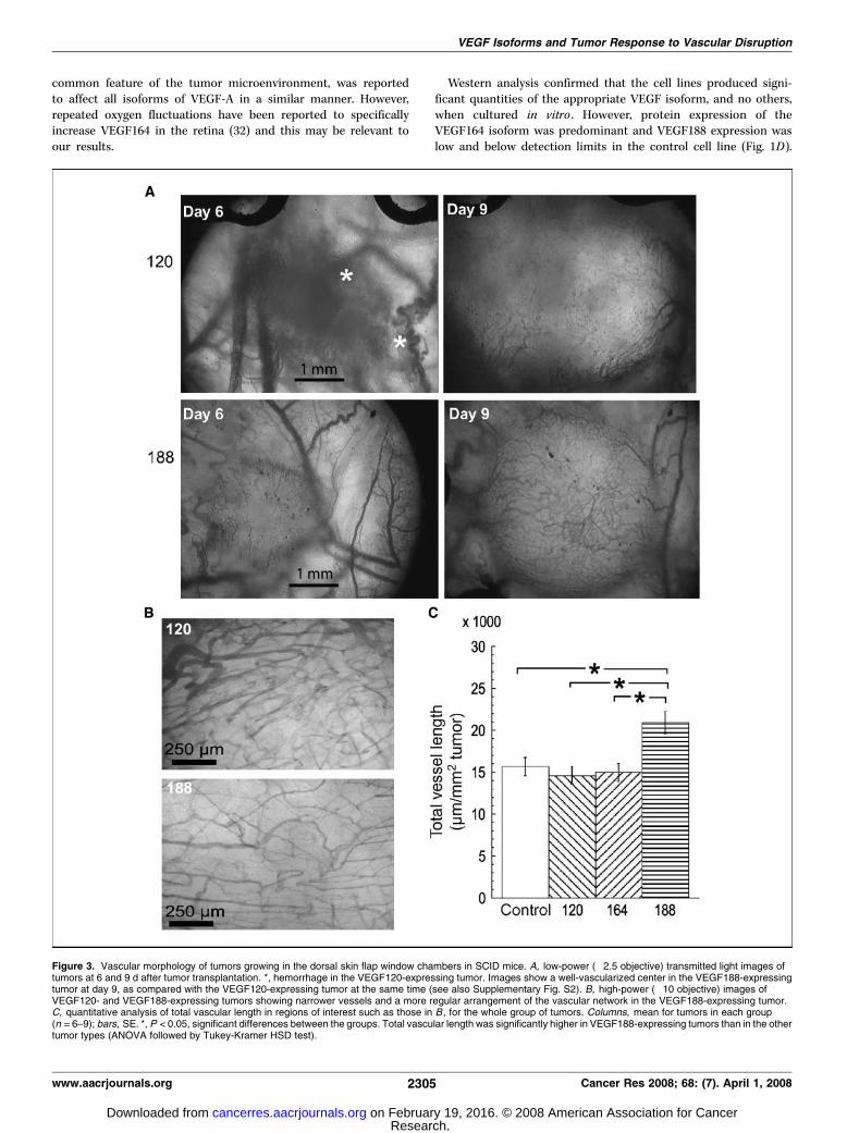
Figure 3. Vascular morphology of tumors growing in the dorsal skin flap window chambers in SCID mice. A, low-power (�2.5 objective) transmitted light images oftumors at 6 and 9 d after tumor transplantation. *, hemorrhage in the VEGF120-expressing tumor. Images show a well-vascularized center in the VEGF188-expressingtumor at day 9, as compared with the VEGF120-expressing tumor at the same time (see also Supplementary Fig. S2). B, high-power (�10 objective) images ofVEGF120- and VEGF188-expressing tumors showing narrower vessels and a more regular arrangement of the vascular network in the VEGF188-expressing tumor.C, quantitative analysis of total vascular length in regions of interest such as those in B , for the whole group of tumors. Columns, mean for tumors in each group(n = 6–9); bars, SE. *, P < 0.05, significant differences between the groups. Total vascular length was significantly higher in VEGF188-expressing tumors than in the othertumor types (ANOVA followed by Tukey-Kramer HSD test).
VEGF Isoforms and Tumor Response to Vascular Disruption
www.aacrjournals.org 2305 Cancer Res 2008; 68: (7). April 1, 2008
Research. on February 19, 2016. © 2008 American Association for Cancercancerres.aacrjournals.org Downloaded from

Attempts to carry out Western analysis on solid tumor extractswere not successful, probably because levels were below thesensitivity limits of the assay.Vascular morphology. Established subcutaneous tumors were
examined for vascularity and maturity of their vessel walls usingimmunohistochemistry. CD31 staining in Fig. 2A shows thatall tumor types were well-vascularized. However, a-sma stainingshows that control and VEGF188-expressing tumors were betterstabilized with pericytes than VEGF120- or VEGF164-expressingtumors. Although this staining was apparent in blood vessel walls,there was also substantial staining in a subset of extravascular cells.Quantification of staining (Fig. 2B) showed that control andVEGF120 tumors were the most vascular (CD31), whereas controland VEGF188 tumors had the most a-sma staining. These diffe-rences were statistically significant (see legend for details). Furtherinvestigations were carried out in the VEGF120-expressing (mostdiffusible) and the VEGF188-expressing (nondiffusible) tumors.Figure 2C shows double-staining for CD31 and a-sma, which con-firmed the localization of a-sma–positive cells (pericytes) in thevasculature of VEGF188 tumors and very little staining in theVEGF120 tumors. The spiraling morphology of the a-sma stainingin the VEGF188 tumor indicates close contact between pericytesand endothelial cells. Figure 2D shows that desmin- and ANG1-
positive cells were also preferentially found in VEGF188 tumorsrather than in VEGF120 tumors. The staining patterns were alsohighly suggestive of vascular localization. Taken together, theseresults indicate the association of VEGF120 with sustainedexpansion of the tumor vasculature, whereas VEGF188 is uniquelyassociated with maturation of the blood vessel wall.
Vascularization of single VEGF isoform–expressing tumors wasfurther analyzed by intravital microscopy. Figure 3A and Supple-mentary Fig. S2 illustrate the vascularization of VEGF120- andVEGF188-expressing tumors at early and late phases of growth inthe window chamber. Initial vascularization of the VEGF120-expressing tumors was typified by dilated preexisting normal bloodvessels, which were prone to hemorrhage and indistinct tumormargins (Fig. 3A). In contrast, preexisting normal vessels werenarrower in the VEGF188 implants, there was very little or nohemorrhage and the tumor margins were distinct. As tumors grew,hemorrhage within the VEGF120-expressing tumors tended toresolve but vascularization was often limited to the periphery ofthe tumor mass. In contrast, VEGF188-expressing tumors werevascularized throughout the tumor mass (Fig. 3A). Control andVEGF164-expressing tumors had intermediate properties.
Quantitative morphologic analysis of tumor blood vessels wascarried out on two peri-peripheral regions of interest within each
Figure 4. In vivo multiphotonfluorescence imaging of VEGF120- andVEGF188-expressing tumors growingin window chambers in SCID mice.A, maximum projection images from twotumors at different times (midpoint of thetime frame for image acquisition) followingi.v. injection of 40 kDa of FITC-dextran.Extensive leakage of FITC-dextranfrom the tumor vasculature into theinterstitium could be seen with time in theVEGF120-expressing tumor but not inthe VEGF188-expressing tumor. B, imagecontiguity (columns, mean; bars, SE) atthe first time point after injection ofFITC-dextran (2.5 min) for the whole tumorgroup (n = 5–7 tumors in each group).Contiguity was significantly higher inVEGF120 tumors than in VEGF188 tumors(*, P = 0.0005; Student’s t test for unpaireddata). C, the kinetics of FITC-dextranleakage for the whole tumor group.Intensities were averaged gray levels inwhole three-dimensional images of eachtumor, normalized to the first time point(mean for n = 5–7 tumors in each group;bars, 1 SE). The curves for thetwo groups were significantly different(P < 0.05, MANOVA with repeatedmeasures).
Cancer Research
Cancer Res 2008; 68: (7). April 1, 2008 2306 www.aacrjournals.org
Research. on February 19, 2016. © 2008 American Association for Cancercancerres.aacrjournals.org Downloaded from

tumor, using a �10 objective, once the tumors became optimallyvascularized. The vascular patterning in the VEGF188 tumors stoodout from the others in that the blood vessels were arranged moreregularly and individual vessels were uniformly narrow. Bloodvessels in the other tumor types were irregular in width butgenerally wider and very chaotic in their arrangement. Typicalregions of interest in a VEGF120- and VEGF188–expressing tumorare shown in Fig. 3B . Quantitative analysis revealed no significantdifference between the tumor lines in terms of mean vascularlengths or fractal dimensions (data not shown). However, the total
vascular length was higher in the VEGF188 tumors than in any ofthe other tumor types (Fig. 3C).Vascular function. Leakage of i.v. injected 40 kDa fluorescent
dextran was monitored in VEGF120 and VEGF188 tumors growingin window chambers, using multiphoton fluorescence microscopy(Fig. 4). Figure 4A shows typical images with diffuse vascularizationand rapid leakage in a VEGF120 tumor compared with a VEGF188tumor. Contiguity data for the whole group (Fig. 4B), is consistentwith the diffuse nature of the vasculature in VEGF120 tumors,wider diameter vessels, and leakiness. Figure 4C shows the kinetics
Figure 5. Response of the different tumor types growing in window chambers in SCID mice to a single dose of 30 mg/kg i.v. CA-4-P. A, typical low-power(�2.5 objective) images of VEGF120- and VEGF188-expressing tumors after CA-4-P treatment. There was a reduction in the visible vasculature at 1 h after treatmentin both tumor types and extensive avascular regions in VEGF120- but not VEGF188-expressing tumors by 24 h (*). Supplementary Fig. S3 shows that the responseof VEGF164 tumors was similar to VEGF120 tumors and the response of control tumors was similar to VEGF188 tumors. B and C , quantitative analysis ofvascular length and red cell velocity for all tumor types. Points, mean for tumors in each group (n = 4–6); bars, SE. *, P < 0.05, curves for VEGF120- andVEGF164-expressing tumors were significantly different from those for VEGF188-expressing and control tumors over these regions (MANOVA with repeated measures).
VEGF Isoforms and Tumor Response to Vascular Disruption
www.aacrjournals.org 2307 Cancer Res 2008; 68: (7). April 1, 2008
Research. on February 19, 2016. © 2008 American Association for Cancercancerres.aacrjournals.org Downloaded from

of fluorescence intensity in tumors with time after injection ofFITC-dextran. There was a significant difference between theVEGF120 and VEGF188 tumors, with average intensity decreasingwith time in the VEGF188 tumors and maintained or increasing inVEGF120 tumors. This difference was consistent with more rapid
leakage of FITC-dextran from the intravascular to extravascularspace in the VEGF120 tumors, which maintained average imageintensity in the face of vascular clearance of the marker.Response to CA-4-P. Vascular response to CA-4-P was deter-
mined in tumors growing in window chambers. Figure 5A showstypical images from VEGF120- and VEGF188-expressing tumors at0, 1, and 24 h following treatment with a moderate single dose ofCA-4-P (30 mg/kg i.v.). Response in both tumor types was typifiedby a substantial loss of the visible vasculature, primarily atthe tumor center, at 1 h after treatment. Subsequently, VEGF120-expressing tumors progressed to almost complete avascularity andopacity of the central tumor region at 24 h. The vascular mor-phology was substantially less affected in the VEGF188 tumors,with very little avascularity by 24 h (Fig. 5A).
Quantitation of the vascular damage in window chamber tumorsfollowing CA-4-P treatment is shown in Fig. 5B and C for all thetumor types. There is a clear distinction in response between theVEGF120 and VEGF164 tumors on the one hand, and the wild-typeand VEGF188 tumors on the other, confirming the observationsshown in Fig. 5A and Supplementary Fig. S3, which gives additionalimages from all tumor types. The distinction in response of thedifferent tumor types was clearly apparent from both a morpho-logic end point (total vessel length; Fig. 5B) and a functional endpoint (red cell velocity; Fig. 5C).
In order to determine whether these differences in early vascularresponse to CA-4-P translated into differences in therapeuticresponse, a repeat dosing schedule of CA-4-P was administered tomice bearing subcutaneous tumors (Fig. 6A). VEGF120 andVEGF164tumors were, again, the most sensitive. Interestingly, althoughthe control and VEGF188 tumors seemed to be completely resistantto the effects of CA-4-P in the first week of treatment, there wassome growth retardation during the second week. In a separateexperiment, VEGF120-expressing tumors were significantly morenecrotic than VEGF188-expressing tumors at 24 h after a single100 mg/kg dose of CA-4-P (79.5 F 9.3% versus 42.8 F 9.3% of tumorvolume, respectively, P < 0.05 for Student’s t test). However, afterfive doses of 50 mg/kg CA-4-P, as in Fig. 6A , the necrosis levels in thetwo tumor types were very similar (34.3 F 7.2% and 41.1 F 9.2%,respectively), suggesting that the responsiveness of the 188 tumors inthe second week was due to increased sensitivity of the vasculature.Vascular volume in viable regions of VEGF120-expressing tumors, asmeasured by CD31 staining, significantly increased from 7.2 F 0.5%to 11.0 F 0.6% of tumor volume after a week’s treatment (P < 0.05for Student’s t test). A similar trend in the VEGF188-expressingtumors was only borderline significant. Figure 6A also shows thatuntreated VEGF120- and VEGF164-expressing tumors reached ameasurable size f4 days before the VEGF188 and control tumors,but then, all lines grew at the same rate, consistent with comparableblood flow rates (Supplementary Fig. S4). CA-4-P (100 Amol/L)increased apoptosis in all the cell lines growing in vitro , with nosignificant difference in induction rates between the different celllines (Fig. 6B).
Discussion
We have shown that mouse fibrosarcoma cells, which expressonly single isoforms of VEGF, under the control of the endogenousVEGF promoter, produce tumors with very different vasculariza-tion patterns, vessel wall structure, and barrier function. Themethod used for production of the tumor lines ensured normaloverall VEGF levels. Expression of VEGF188, even at very low levels,
Figure 6. A, tumor volume growth curves for all tumor types growingsubcutaneously in SCID mice F CA-4-P treatment [10 fractions of 50 mg/kgof CA-4-P i.p. or equal volumes of saline, at times indicated (arrows )].Untreated tumor growth curves (solid lines ) were fitted to Gompertz curves,as described in the text. The lag phase in tumor growth was shorter for VEGF120and VEGF164 tumors than for control and VEGF188 tumors. However,at 18 d, untreated tumor growth rate was 73.4 F 7.8, 86.2 F 8.0,86.3 F 18.3, 59.2 F 5.4 mm3/d for control, VEGF120-, VEGF164-, andVEGF188-expressing tumors, respectively. These differences were notsignificant (P > 0.05, ANOVA), which was consistent with similar blood flowrates in the four cell lines (see Supplementary Fig. S4). VEGF120- andVEGF164-expressing tumors responded better to CA-4-P treatment (brokenlines) than VEGF188-expressing and control tumors. Points, mean for tumorsin each group, representing combined data from two separate experiments(n = 9–11); bars, SE. *, P < 0.05, significant differences were obtained betweenuntreated and treated curves over the regions indicated (MANOVA withrepeated measures). B, apoptosis induced by CA-4-P (100 Amol/L) in singleisoform–expressing tumor cells and control cells in vitro . Results are plotted asrates relative to those in untreated cells. Columns, mean for seven repeatexperiments; bars, SE. There were no significant differences across the groups(P > 0.05, ANOVA).
Cancer Research
Cancer Res 2008; 68: (7). April 1, 2008 2308 www.aacrjournals.org
Research. on February 19, 2016. © 2008 American Association for Cancercancerres.aacrjournals.org Downloaded from

was uniquely associated with extensive recruitment of vascularsupport cells (pericytes) and tumor resistance to the VDA CA-4-P.Overall, our results support the view that VEGF188 expressionresults in a different type of vascular bed, which renders the tumorvasculature less susceptible to CA-4-P.
Our data support previous findings that VEGF120/121 is asso-ciated with hemorrhage and vasodilation around tumors (33, 34).They are in agreement with Grunstein et al. (17) who found thatVEGF120 and VEGF164 were associated with rapid initiation offibrosarcoma growth. In contrast with our findings, Grunstein et al.found poor tumor growth associated with a fibrosarcoma cellline expressing only VEGF188. This may be due to high levels ofVEGF188 in their overexpressing tumor line, leading to overvascu-larization of tumors. Indeed, Grunstein et al. found particularlypoor vascular filling of contrast agent in their VEGF188-expressingtumors. In both studies, endogenous VEGF production fromnormal cells such as macrophages might be expected to contributeto vascular development. We found moderate macrophage invasionin all tumor types. However, our reverse transcription-PCR resultsshowed that host-derived expression of VEGF isoforms was verylow in all tumors and this was consistent with substantialdifferences in the vascular characteristics we observed. Quantita-tive differences in expression levels may explain why some authorshave found that tumor cells transfected to overexpress VEGF188/189 are nontumorigenic (13), whereas others are tumorigenic(34–36). Interestingly, selective down-regulation of VEGF189expression in a non–small cell lung cancer (35) and pancreaticcancer cell line (36) significantly reduced their xenotransplant-ability, suggesting that VEGF188/189 does play an important role intumorigenesis.
We found striking differences in the maturation status of thevascular wall in the different VEGF isoform–producing tumors,which related to response to CA-4-P. In particular, the presence ofVEGF188 resulted in tumor vascular recruitment of mural cells.This is consistent with the observations that mice selectivelyexpressing VEGF188 recruit mural cells normally to the developingretinal venules and capillaries, whereas mice selectively expressingVEGF120 show major defects in mural cell recruitment (15). Inaddition, we showed that ANG1 was expressed in a subset ofvessels in VEGF188 tumors and that barrier function in theVEGF188 tumors was more effective than in VEGF120 tumors,consistent with the observed structural differences. The processesinvolved in mural cell recruitment are complex and poorlyunderstood but undoubtedly involve growth factors in additionto VEGF, such as platelet-derived growth factor B (PDGFB; ref. 37).Nevertheless, the current result clearly shows a key role forVEGF188 in mural cell recruitment in tumors that is associatedwith a functional effect. In addition, positive staining for a-smawas found in a subset of extravascular cells, presumably tumor cellsor fibroblasts, of the VEGF188-expressing and control tumors butnot in the VEGF120- or VEGF164-expressing tumors. This suggestsa novel role for VEGF188/189 in pericyte differentiation.
Vascular network development was highly influenced bydifferential expression of VEGF isoforms, with VEGF120 beingassociated with a high vascular volume in established subcutane-ous tumors and VEGF188 being associated with regular, narrowvessels and a high vascular length per tumor volume. In tumorsgrowing in window chambers, which are subjected to high tissuepressure induced by the restraining glass, VEGF120 and VEGF164were associated with a failure to effectively vascularize the centerof tumors. This is likely to be due to the fragility of blood vessels,
suggested by their lack of mural cells. Expression of VEGF164 wasassociated with a relatively low vascular volume (similar toVEGF188) but rapid initiation of tumor growth (similar toVEGF120). This suggests a highly proliferating phenotype forVEGF120- and VEGF164-expressing tumor cells, at the expense ofvascular maturation. This is consistent with our recent in vitrodata, showing higher proliferation rates for VEGF120- or VEGF164-expressing tumor cells than VEGF188-expressing or control cells(ref. 20; Supplementary Table S1). Any differences between thevasculature in VEGF120- and VEGF164-expressing tumors weresubtle. However, the difference in vascular volume and the quali-tative observation of less hemorrhage in the VEGF164 tumors doessuggest some differences in their control of vascular maturation, anobservation that warrants further investigation.
Several explanations for the different effects of individual VEGFisoforms on tumor vascular morphology and function can beenvisaged. In the developing brain of Vegfa188/188 mice, there is anincreased extension of endothelial filopodia and vascular branchformation, compared with brains of Vegfa120/120 mice (16), and thismight also apply to tumor angiogenesis. There is also evidencefor differential activation of VEGF receptors by the differentisoforms. First, VEGF164/165 (and probably VEGF188/189) but notVEGF120/121, can bind to the accessory receptor, neuropilin-1(NRP1; ref. 38). However, impaired signaling through the NRP-1receptor could not account for all vascular branching defects inVegfa120/120 mouse embryos (16) and would not explain the signi-ficant differences between VEGF164- and VEGF188-expressingtumors. Heparan sulfate proteoglycans on the cell surface have alsobeen implicated in modifying VEGF signaling, partly by controllingthe distribution and bioavailability of secreted VEGF (39, 40).Other possibilities, which remain poorly understood, include thetranslocation of intracellular VEGF188/189 to the nucleus (31) andthe cross-talk of VEGF signaling pathways with integrins (41).
In interpreting our data, we cannot discount the possibility thatan adaptive response to the depletion of VEGF120 or VEGF164,resulting in increased expression of alternative vascular-relatedgrowth factors in the VEGF188-expressing cell line, could explainour results. Notwithstanding potential affinity issues of the VEGFantibody used in the ELISA kit for VEGF188, VEGF protein expres-sion in the VEGF188 line in vivo seems to be relatively low (Fig. 1C),highlighting this possibility. However, real-time PCR data (Supple-mentary Fig. S1) did not reveal any alternative candidate genesfor explaining our results. Transformation significantly loweredVEGFR2, NRP1, TIE2, Ang2 , and Hey2 gene expression across all thelines, but only Ang2 and PDGFB differed in their expression levelsbetween the VEGF188-expressing tumor cells and the other celltypes. Increased Ang2 and lowered PDGFB levels, as found inthe VEGF188-expressing line, are unlikely explanations for theobserved increase in pericyte recruitment of this line in vivobecause Ang2 is normally associated with vascular immaturity andPDGFB is a well-known chemoattractant for pericytes. In addition,administration of VEGF receptor kinase inhibitors in vitro andin vivo and a pan-isoform neutralizing antibody for VEGF in vitro ,equalized both tumor cell proliferation in vitro and vascularmorphology in vivo , across the different cell lines, suggesting thatbaseline differences in these variables were VEGF-induced(Supplementary Fig. S5; Supplementary Table S1).
Differential expression of VEGF isoforms was clearly associatedwith outcome following treatment with the VDA, CA-4-P, both interms of initial vascular response and tumor growth response to arepeated dosing schedule. A modest growth response, as observed
VEGF Isoforms and Tumor Response to Vascular Disruption
www.aacrjournals.org 2309 Cancer Res 2008; 68: (7). April 1, 2008
Research. on February 19, 2016. © 2008 American Association for Cancercancerres.aacrjournals.org Downloaded from

here even for the VEGF120-expressing tumors, is not unusual forVDAs and could mask significant cell-killing effects. This is thoughtto be due primarily to poor clearance of dead cells because of bloodflow reduction (42). The presence of VEGF188, even at low levelsin the control tumors, conferred resistance to treatment. Althoughspeculative, it is possible that very tight association of VEGF188with the cell surface minimizes the amount of protein necessaryfor a given outcome, as well as negatively feeding back on VEGF188production. Necrosis data showed that VEGF188 tumors sufferedvascular damage in the first week of treatment despite the lackof growth response. This may have been sufficient to sensitizethe tumors to continued treatment. CD31 staining indicated anincrease in vascularization of re-growing viable tumor regions afterthe first week of treatment, which could relate to homing of bonemarrow–derived vascular progenitor cells following CA-4-P treat-ment, as previously reported (43). This increased vascularizationmay also relate to increased sensitivity to CA-4-P in the secondweek of treatment, although potential mechanisms require furtherinvestigation. Finally, the larger size of tumors at the start oftreatment in the second week may have contributed to increasedsensitivity (44).
Previously, we hypothesized that high vascular permeability andthe immaturity of the vascular wall were major factors associatedwith the sensitivity of tumor vasculature to CA-4-P and similaragents (45, 46). The current results substantiate this hypothesis,
and furthermore, suggest that VEGF188 expression is uniquelypredictive of treatment outcome. Interestingly, several studies havesuggested that VEGF189 expression in human cancers is associatedwith tumor progression and poor outcome from conventionaltreatments (47–50).
In conclusion, the main VEGF isoforms, under the control ofthe endogenous VEGF promoter, have very different effects onvascularization of fibrosarcomas in an animal model. Inparticular, VEGF188 and/or associated gene expression plays acrucial role in tumor vascular maturation and conferringresistance to the VDA, CA-4-P. Therefore, VEGF isoformexpression may be useful for predicting tumor susceptibility tovascular-disrupting cancer therapy.
Acknowledgments
Received 5/30/2007; revised 12/3/2007; accepted 2/7/2008.Grant support: Cancer Research UK Programme grant C1276/A3307.The costs of publication of this article were defrayed in part by the payment of page
charges. This article must therefore be hereby marked advertisement in accordancewith 18 U.S.C. Section 1734 solely to indicate this fact.
We gratefully acknowledge Dr. Gabi Dachs, Dr. Andrew Steele, and Claudia Corallifor their roles in developing the tumor lines used in this study; Finuala Hylands, IanWilson, Frances Daley, and Matthew Fisher for their technical support; Professor BorisVojnovic and Dr. Simon Ameer-Beg for their expertise and support with multiphoton-fluorescence microscopy; the Gray Cancer Institute, University of Sheffield and CancerResearch UK London Institute for care of the animals; and Professor Bob Pettit forsupplying the CA-4-P.
References
1. Tozer GM, Kanthou C, Baguley BC. Disrupting tumourblood vessels. Nat Rev Cancer 2005;5:423–35.
2. Chaplin DJ, Horsman MR, Siemann DW. Currentdevelopment status of small-molecule vascular disrupt-ing agents. Curr Opin Investig Drugs 2006;7:522–8.
3. Galbraith SM, Maxwell RJ, Lodge MA, et al.Combretastatin A4 phosphate has tumor antivascularactivity in rat and man as demonstrated by dynamicmagnetic resonance imaging. J Clin Oncol 2003;21:2831–42.
4. Young SL, Chaplin DJ. Combretastatin A4 phosphate:background and current clinical status. Expert OpinInvestig Drugs 2004;13:1171–82.
5. Horsman MR, Siemann DW. Pathophysiologic effectsof vascular-targeting agents and the implications forcombination with conventional therapies. Cancer Res2006;66:11520–39.
6. von Pawel J, Reck M, McKeage M. Update on survivalin phase Ib/II study of DMXAA (AS1404) combined withcarboplatin and paclitaxel in non-small cell lung cancer(NSCLC). Proceedings of the 18th EORTC-NCI-AACRSymposium on Molecular Targets and Cancer Thera-peutics; 2006 Nov 7–10; Prague, Czech Republic.
7. Grunstein J, Roberts WG, Mathieu-Costello O, Hana-han D, Johnson RS. Tumor-derived expression ofvascular endothelial growth factor is a critical factorin tumor expansion and vascular function. Cancer Res1999;59:1592–8.
8. Ferrara N. Vascular endothelial growth factor: basicscience and clinical progress. Endocr Rev 2004;25:581–611.
9. Tischer E, Mitchell R, Hartman T, et al. The humangene for vascular endothelial growth factor. Multipleprotein forms are encoded through alternative exonsplicing. J Biol Chem 1991;266:11947–54.
10. Yamazaki Y, Morita T. Molecular and functionaldiversity of vascular endothelial growth factors. MolDivers 2006;10:515–27.
11. Ferrara N, Gerber H-P, LeCouter J. The biology ofVEGF and its receptors. Nat Med 2003;9:669–76.
12. Stimpfl M, Tong D, Fasching B, et al. Vascularendothelial growth factor splice variants and their
prognostic value in breast and ovarian cancer. ClinCancer Res 2002;8:2253–9.
13. Yu JL, Rak JW, Klement G, Kerbel RS. Vascularendothelial growth factor isoform expression as adeterminant of blood vessel patterning in humanmelanoma xenografts. Cancer Res 2002;62:1838–46.
14. Carmeliet P, Ng Y-S, Nuyens D, et al. Impairedmyocardial angiogenesis and ischemic cardiomyopathyin mice lacking the vascular endothelial growth factorisoforms VEGF164 VEGF188. Nat Med 1999;5:495–502.
15. Stalmans I, Ng YS, Rohan R, et al. Arteriolar andvenular patterning in retinas of mice selectivelyexpressing VEGF isoforms. J Clin Invest 2002;109:327–36.
16. Ruhrberg C, Gerhardt H, Golding M, et al. Spatiallyrestricted patterning cues provided by heparin-bindingVEGF-A control blood vessel branching morphogenesis.Genes Dev 2002;16:2684–98.
17. Grunstein J, Masbad JJ, Hickey R, Giordano F,Johnson RS. Isoforms of vascular endothelial growthfactor act in a coordinate fashion to recruit and expandtumor vasculature. Mol Cell Biol 2000;20:7282–91.
18. Vieira JM, Schwarz Q, Ruhrberg C. Selective require-ments for NRP1 ligands during neurovascular pattern-ing. Development 2007;134:1833–43.
19. Parada LF, Land H, Weinberg RA, Wolf D, Rotter V.Cooperation between gene encoding p53 tumourantigen and ras in cellular transformation. Nature1984;312:649–51.
20. Greco O, Coralli C, Dachs GU, et al. Role of VEGF intumour response to vascular disrupting agents. Micro-circulation 2005;12:681.
21. Kanthou C, Greco O, Stratford A, et al. The tubulin-binding agent combretastatin A-4-phosphate arrestsendothelial cells in mitosis and induces mitotic celldeath. Am J Pathol 2004;165:1401–11.
22. Chalkley HW. Method for quantitative morphologicanalysis of tissues. J Natl Cancer Inst 1943;4:47–53.
23. Tozer GM, Ameer-Beg SM, Baker J, et al. Intravitalimaging of tumour vascular networks using multi-photon fluorescence microscopy. Adv Drug Deliv Rev2005;57:135–52.
24. Unthank J, Lash J, Nixon J, Sidner R, Bohlen H.Evaluation of carbocyanine-labeled erythrocytes for
microvascular measurements. Microvascular Res 1993;45:193–210.
25. Kimura K, Braun RD, Ong ET, et al. Fluctuations inred cell flux in tumor microvessels can lead to transienthypoxia and reoxygenation in tumor parenchyma.Cancer Res 1996;56:5522–8.
26. Tozer GM, Prise VE, Wilson J, et al. Mechanismsassociated with tumor vascular shut-down induced bycombretastatin A-4 phosphate: intravital microscopyand measurement of vascular permeability. Cancer Res2001;61:6413–22.
27. Barber PR, Vojnovic B, Ameer-Beg SM, Hodgkiss RJ,Tozer GM, Wilson J. Semi-automated software for thethree-dimensional delineation of complex vascular net-works. J Microsc 2003;211:54–62.
28. Reyes-Aldasoro CC, Wilson I, Prise VE, et al.Estimation of apparent tumor vascular permeabilityfrom multiphoton fluorescence microscopic images ofP22 rat sarcomas in vivo . Microcirculation 2008;15:65–79.
29. Theiler JP, Gisler G. A contiguity-enhanced k-meansclustering algorithm for unsupervised multispectralimage segmentation. Proceedings SPIE 1997;3159:108–18.
30. Eckhart L, Ban J, Ballaun C, Weninger W, TschachlerE. Reverse transcription-polymerase chain reactionproducts of alternatively spliced mRNAs form DNAheteroduplexes and heteroduplex complexes. J BiolChem 1999;274:2613–5.
31. Zhang HT, Scott PA, Morbidelli L, et al. The 121amino acid isoform of vascular endothelial growthfactor is more strongly tumorigenic than other splicevariants in vivo . Br J Cancer 2000;83:63–8.
32. McColm JR, Geisen P, Hartnett ME. VEGF isoformsand their expression after a single episode of hypoxiaor repeated fluctuations between hyperoxia andhypoxia: relevance to clinical ROP. Mol Vis 2004;10:512–20.
33. Guo P, Xu L, Pan S, et al. Vascular endothelial growthfactor isoforms display distinct activities in promotingtumor angiogenesis at different anatomic sites. CancerRes 2001;61:8569–77.
34. Kusters B, de Waal RM, Wesseling P, et al. Differentialeffects of vascular endothelial growth factor A isoforms
Cancer Research
Cancer Res 2008; 68: (7). April 1, 2008 2310 www.aacrjournals.org
Research. on February 19, 2016. © 2008 American Association for Cancercancerres.aacrjournals.org Downloaded from

in a mouse brain metastasis model of human melano-ma. Cancer Res 2003;63:5408–13.
35. Oshika Y, Nakamura M, Tokunaga T, et al. Ribozymeapproach to downregulate vascular endothelial growthfactor (VEGF) 189 expression in non-small cell lungcancer (NSCLC). Eur J Cancer 2000;36:2390–6.
36. Tokunaga T, Abe Y, Tsuchida T, et al. Ribozymemediated cleavage of cell-associated isoform of vascu-lar endothelial growth factor inhibits liver metastasisof a pancreatic cancer cell line. Int J Oncol 2002;21:1027–32.
37. Benjamin LE, Hemo I, Keshet E. A plasticity windowfor blood vessel remodelling is defined by pericytecoverage of the preformed endothelial network and isregulated by PDGF-B and VEGF. Development 1998;125:1591–8.
38. Soker S, Takashima S, Miao HQ, Neufeld G,Klagsbrun M. Neuropilin-1 is expressed by endothelialand tumor cells as an isoform-specific receptor forvascular endothelial growth factor. Cell 1998;92:735–45.
39. Gitay-Goren H, Soker S, Vlodavsky I, Neufeld G. Thebinding of vascular endothelial growth factor to its
receptors is dependent on cell surface-associatedheparin-like molecules. J Biol Chem 1992;267:6093–8.
40. Lee S, Jilani SM, Nikolova GV, Carpizo D, Iruela-Arispe ML. Processing of VEGF-A by matrix metal-loproteinases regulates bioavailability and vascularpatterning in tumors. J Cell Biol 2005;169:681–91.
41. Hutchings H, Ortega N, Plouet J. Extracellular matrix-bound vascular endothelial growth factor promotesendothelial cell adhesion, migration, and survivalthrough integrin ligation. FASEB J 2003;17:1520–2.
42. ChaplinDJ, Pettit GR, Hill SA. Anti-vascular approachesto solid tumour therapy: evaluation of combretastatin A4phosphate. Anticancer Res 1999;19:189–96.
43. Shaked Y, Ciarrocchi A, Franco M, et al. Therapy-induced acute recruitment of circulating endothelialprogenitor cells to tumors. Science 2006;313:1785–7.
44. Landuyt W, Verdoes O, Darius DO, et al. Vasculartargeting of solid tumours: a major ‘inverse’ volume-response relationship following combretastatin A-4phosphate treatment of rat rhabdomyosarcomas. Eur JCancer 2000;36:1833–43.
45. Beauregard DA, Hill SA, Chaplin DJ, Brindle KM. Thesusceptibility of tumors to the antivascular drug
combretastatin A4 phosphate correlates with vascularpermeability. Cancer Res 2001;61:6811–5.
46. Tozer GM, Kanthou C, Parkins CS, Hill SA. Thebiology of the combretastatins as tumour vasculartargeting agents. Int J Exp Pathol 2002;83:21–38.
47. Yuan A, Yu CJ, Kuo SH, et al. Vascular endothelialgrowth factor 189 mRNA isoform expression specificallycorrelates with tumor angiogenesis, patient survival,and postoperative relapse in non-small-cell lung cancer.J Clin Oncol 2001;19:432–41.
48. Nakamura M, Abe Y, Tokunaga T. Pathologicalsignificance of vascular endothelial growth factor Aisoform expression in human cancer. Pathol Int2002;52:331–9.
49. Cressey R, Wattananupong O, Lertprasertsuke N,Vinitketkumnuen U. Alteration of protein expressionpattern of vascular endothelial growth factor (VEGF)from soluble to cell-associated isoform during tumouri-genesis. BMC Cancer 2005;5:128.
50. Jacobsen J, Grankvist K, Rasmuson T, Ljungberg B.Different isoform patterns for vascular endothelialgrowth factor between clear cell and papillary renalcell carcinoma. BJU Int 2006;97:1102–8.
VEGF Isoforms and Tumor Response to Vascular Disruption
www.aacrjournals.org 2311 Cancer Res 2008; 68: (7). April 1, 2008
Research. on February 19, 2016. © 2008 American Association for Cancercancerres.aacrjournals.org Downloaded from

2008;68:2301-2311. Cancer Res Gillian M. Tozer, Simon Akerman, Neil A. Cross, et al.
Producing Tumors−Growth Factor-A Isoform Vascular-Disrupting Therapy in Single Vascular Endothelial
Blood Vessel Maturation and Response to
Updated version
http://cancerres.aacrjournals.org/content/68/7/2301
Access the most recent version of this article at:
Material
Supplementary
http://cancerres.aacrjournals.org/content/suppl/2008/03/24/68.7.2301.DC1.html
Access the most recent supplemental material at:
Cited articles
http://cancerres.aacrjournals.org/content/68/7/2301.full.html#ref-list-1
This article cites 49 articles, 22 of which you can access for free at:
Citing articles
http://cancerres.aacrjournals.org/content/68/7/2301.full.html#related-urls
This article has been cited by 5 HighWire-hosted articles. Access the articles at:
E-mail alerts related to this article or journal.Sign up to receive free email-alerts
Subscriptions
Reprints and
To order reprints of this article or to subscribe to the journal, contact the AACR Publications
Permissions
To request permission to re-use all or part of this article, contact the AACR Publications
Research. on February 19, 2016. © 2008 American Association for Cancercancerres.aacrjournals.org Downloaded from
Copyright © 2022 FDOKUMEN