The Nk Problem in Power Grids: New Models, Formulations, and Numerical Experiments
Upload
independentCategory
view
4download
0
RESEARCH ARTICLE
Human Induced Pluripotent Stem Cells AreTargets for Allogeneic and AutologousNatural Killer (NK) Cells and Killing Is PartlyMediated by the Activating NK ReceptorDNAM-1Vanessa Kruse1☯, Carina Hamann2,3☯, Sebastian Monecke2,3, Lukas Cyganek1,3,Leslie Elsner2, Daniela Hübscher1,3, Lutz Walter4, Katrin Streckfuss-Bömeke1,3,Kaomei Guan1,3‡*, Ralf Dressel2,3‡*
1 Department of Cardiology and Pneumology, University Medical Center Göttingen, Göttingen, Germany,2 Department of Cellular and Molecular Immunology, University Medical Center Göttingen, Göttingen,Germany, 3 DZHK (German Center for Cardiovascular Research), Partner site Göttingen, Germany,4 Primate Genetics Laboratory, German Primate Center, Göttingen, Germany
☯ These authors contributed equally to this work.‡ These authors also contributed equally to this work.* [email protected] (RD); [email protected] (KG)
AbstractHuman induced pluripotent stem cells (hiPSCs) could be used to generate autologous cells
for therapeutic purposes, which are expected to be tolerated by the recipient. However,
iPSC-derived grafts are at risk of giving rise to teratomas in the host, if residuals of tumori-
genic cells are not rejected by the recipient. We have analyzed the susceptibility of hiPSC
lines to allogeneic and autologous natural killer (NK) cells. IL-2-activated, in contrast to rest-
ing NK cells killed hiPSC lines efficiently (P=1.69x10-39). Notably, the specific lysis of the in-
dividual hiPSC lines by IL-2-activated NK cells was significantly different (P=1.72x10⁻⁶)
and ranged between 46 % and 64 % in 51Cr-release assays when compared to K562 cells.
The hiPSC lines were killed by both allogeneic and autologous NK cells although autolo-
gous NK cells were less efficient (P=8.63x10⁻⁶). Killing was partly dependent on the activat-
ing NK receptor DNAM-1 (P=8.22x10⁻⁷). The DNAM-1 ligands CD112 and CD155 as well
as the NKG2D ligands MICA and MICB were expressed on the hiPSC lines. Low amounts
of human leukocyte antigen (HLA) class I proteins, which serve as ligands for inhibitory
and activating NK receptors were also detected. Thus, the susceptibility to NK cell killing
appears to constitute a common feature of hiPSCs. Therefore, NK cells might reduce the
risk of teratoma formation even after autologous transplantations of pluripotent stem cell-
derived grafts that contain traces of pluripotent cells.
PLOS ONE | DOI:10.1371/journal.pone.0125544 May 7, 2015 1 / 27
OPEN ACCESS
Citation: Kruse V, Hamann C, Monecke S, CyganekL, Elsner L, Hübscher D, et al. (2015) HumanInduced Pluripotent Stem Cells Are Targets forAllogeneic and Autologous Natural Killer (NK) Cellsand Killing Is Partly Mediated by the Activating NKReceptor DNAM-1. PLoS ONE 10(5): e0125544.doi:10.1371/journal.pone.0125544
Academic Editor: Niklas K Björkström, KarolinskaInstitutet, SWEDEN
Received: October 21, 2014
Accepted: March 25, 2015
Published: May 7, 2015
Copyright: © 2015 Kruse et al. This is an openaccess article distributed under the terms of theCreative Commons Attribution License, which permitsunrestricted use, distribution, and reproduction in anymedium, provided the original author and source arecredited.
Data Availability Statement: All relevant data arewithin the paper and its Supporting Information files.
Funding: This work was supported by the DeutscheForschungsgemeinschaft (SFB 1002, TP A04 and TPC05) and the Bundesministerium für Bildung undForschung (DZHK and 01GN0819). In addition, theauthors acknowledge support by the Open AccessPublication Funds of the Göttingen University. Thefunders had no role in study design, data collectionand analysis, decision to publish, or preparation ofthe manuscript.

IntroductionPluripotent stem cells hold great promises for regenerative medicine. They might become asource of cells and tissues for new cell replacement therapies, e.g. to treat heart failure or Par-kinson’s disease. However, the use of human embryonic stem cells (ESCs) for the developmentof new transplantation therapies is restricted due to ethical concerns. Induced pluripotent stemcells (iPSCs) obtained by reprogramming of somatic cells by a set of defined pluripotency fac-tors might overcome this problem [1–3]. In addition, iPSCs have the advantage that they canpotentially be used not only in allogeneic but also in autologous settings. Autologous iPSC-derived grafts would substantially reduce the risk of immune rejection, but they might on theother hand be at higher risk of teratoma formation than allogeneic transplants if residuals ofpluripotent cells remain in the grafts after in vitro differentiation into the desired cell type ortissue [4]. This assumption is based on results showing that murine ESCs and iPSCs give riseto teratomas in immunocompetent syngeneic but usually not in immunocompetent allogeneicmice [4–9].
In mice, several immune effector mechanisms appear to be important for the rejection of plu-ripotent stem cell-derived teratomas in allogeneic hosts, including T cells [10, 11] and the comple-ment system [12]. We have shown previously that murine pluripotent stem cells, including ESCsand iPSCs, are targets for allogeneic and syngeneic NK cells [5, 13]. NK cells can delay teratomagrowth after transplantation of pluripotent stem cells although they are not sufficient to suppressteratomas completely, if the stem cells are injected in high enough numbers [4, 13, 14]. Notably,murine NK cells also impaired the growth of human ESCs (hESCs) in a xenograft model [15].Recently, a porcine iPSC line was reported to be killed by swine leukocyte antigen (SLA)-matchedas well as SLA-mismatched NK cells [16]. Notably, these iPSCs failed to form teratomas in SLA-matched hosts although they formed tumors in immunodeficient mice [16]. Thus, NK cellsmight contribute to rejection of MHC-matched iPSCs also in a large animal-model.
On NK cells, inhibitory and activating receptors are expressed and the balance of activatingand inhibitory signals determines their activation against target cells [17]. Inhibitory receptorson human NK cells include killer cell immunoglobulin-like receptors (KIRs), which recognizecertain allelic groups of the classical peptide-presenting HLA-A, HLA-B, and HLA-C class Imolecules and the CD94-NKG2A receptor, which recognizes the non-classical class I moleculeHLA-E that presents preferentially leader peptides derived from other HLA class I molecules.The KIR receptor 2DL4 recognizes HLA-G, a further non-classical class I molecule, which ismainly expressed in the placenta [18] but was reported to be expressed also in hESCs [19].Lack of HLA class I molecules can trigger the cytotoxic activity of NK cells. The ‘missing self’concept describes this way of NK cell activation [20]. Activating receptors of human NK cellsinclude natural-killer group 2, member D (NKG2D), which recognize the MHC class I chain-related proteins (MIC)A and MICB and the UL16-binding proteins (ULBP1-6). The activatingDNAX accessory molecule (DNAM)-1 recognizes CD155 (the poliovirus receptor) and CD112(Nectin-2). The activating receptor 2B4 interacts with CD48. Activating KIRs recognize certainallotypes of HLA class I molecules and can therefore play a role in the rejection of allogeneictargets. A further group of activating receptors are the natural cytotoxicity receptors (NCRs)NKp30, NKp44, and NKp46, for which a number of different ligands have been described dur-ing the last years although several of them are debated [17]. Many ligands of activating NK re-ceptors such as MICA and MICB are not expressed by normal cells but become induced bycellular or genotoxic stress in virus-infected or malignant cells. The ‘stress-induced self’ con-cept refers to this way of NK cell activation [21].
Murine pluripotent stem cells lack MHC class I molecules [9, 13, 22, 23] at least at a level de-tectable by flow cytometry [24]. In mice, MHC class I molecules interact with inhibitory Ly49
Human iPSCs Are Targets for NK Cells
PLOS ONE | DOI:10.1371/journal.pone.0125544 May 7, 2015 2 / 27
Competing Interests: The authors have declaredthat no competing interests exist.

receptors on NK cells, a family of receptors that functionally replace KIRs in rodents. Thus, the‘missing self’ can contribute to the killing of mouse ESCs and iPSCs by NK cells. In addition, wefound ligands for the activating NK receptors NKG2D and DNAM-1 on murine pluripotent stemcells and demonstrated that killing of these cells by NK cells depends in part on NKG2D [5, 13].
Human ESCs have been described to express low but in flow cytometry detectable amountsof HLA class I molecules [25–27]. They were also reported to express low amounts of ligandsof the activating NK receptor NKp46 [26] and the NKG2D ligands MICA and MICB [28]. Sim-ilarly, human iPSCs (hiPSCs) were recently shown to express low amounts of HLA class I mol-ecules [29, 30] and NKp46 ligands [29]. In accord with this phenotype, NK cells can kill hESCsat least at a moderate level [26, 31]. In contrast, the hESC line H9 was reported not to inducedegranulation and interferon-γ release in NK cells [32, 33]. However, resting NK cells requirestimulation by cytokines to acquire full cytotoxicity against many targets [34] and such stimu-lation is likely provided by a pro-inflammatory milieu after transplantation. Therefore, we in-vestigated in this study the lysis of hiPSC lines by allogeneic and autologous NK cells activatedby low doses of interleukin-2 (IL-2) and analyzed the expression of ligands of activating and in-hibitory NK receptors. The results showed that hiPSC were targets for both allogeneic and au-tologous IL-2-activated NK cells.
Materials and Methods
Generation and culture of hiPSC linesThree hiPSC lines (D1-iPSC4, D2-iPSC1 and D3-iPSC3) used in this study were generatedfrom hair keratinocytes of three healthy donors (donor 1, 2 and 3), respectively. The genera-tion of D1-iPSC4 (also named as Kera4-iPS4) and D2-iPSC1 (also named as Kera2-iPS1) lineswas described in our previous study [35]. The D3-iPSC3 line was derived from a 30-year-oldhealthy male by using the STEMCCA system [36], which is a humanized excisable lentivirussystem containing the four reprogramming factors OCT4, SOX2, KLF4, and c-MYC in a single‘stem cell cassette’ (pHAGE2-EF1aFull-hOct4-F2AhKlf4-IRES-hSox2-P2A-hcMyc-W-loxP).All three hiPSC lines were characterized for their pluripotency within this study as describedpreviously [35]. A further hiPSC line (D6-iPSC2) used for some experiments has been charac-terized previously and was named FB2-iPS2 [35]. The ethics committee of the University Medi-cal Center Göttingen has approved the study (Az 21/1/11). The participants have given theirwritten informed consent to participate in this study. The ethics committee has approved theconsent procedure. The hiPSC lines were expanded on mitomycin C-inactivated mouse embry-onic fibroblasts (MEFs) as described previously [35]. To avoid contamination with MEFs, thecells were transferred to Matrigel-coated (BD Biosciences, Heidelberg, Germany) dishes andcultured in MEF-conditioned medium three to four days before being used for experiments.The cell lines were used at passages 31 to 46 (D1-iPSC4), 31 to 44 (D2-iPSC1), 13 to 23(D3-iPSC3), and 25 to 29 (D6-iPSC2).
Effector cells and 51Cr-release assayPeripheral blood mononuclear cells (PBMCs) were obtained from healthy donors by centrifuga-tion on Biocoll separating solution (Biochrom, Berlin, Germany) as described previously [34]. NKcells were isolated from PBMCs by magnetic-activated cell sorting (MACS) using negative selec-tion kits (NK cell isolation kit II, Miltenyi Biotec, Bergisch-Gladbach, Germany) and either useddirectly or cultured for four days with 200 U/ml human IL-2 (Proleukin, Chiron, Amsterdam,Netherlands). Unseparated PBMCs were cultured for four days with 200 U/ml IL-2 to obtain lym-phokine-activated killer (LAK) cells. K562 cells served as reference cell line in 51Cr-release assaysand were cultured as described previously [34]. Target cells were labeled by incubating 1 x 10⁶
Human iPSCs Are Targets for NK Cells
PLOS ONE | DOI:10.1371/journal.pone.0125544 May 7, 2015 3 / 27

cells in 200 μl Dulbecco’s modified Eagle medium (DMEM) containing 100 μl fetal calf serum(FCS) and 50 μCi Na2
51CrO4 (CrRA8, Hartmann Analytic, Braunschweig, Germany) for 1 h at37°C and washed three times with DMEM. Effector cells were added to 5 x 10³ 51Cr-labeled targetcells in triplicates at various ratios in 200 μl DMEMwith 10% FCS per well of round-bottomedmicrotiter plates. In some experiments, monoclonal antibodies (mAbs) were added at a concentra-tion of 10 μg/ml (anti-NKG2D, anti-DNAM-1, anti-ICAM-1 and mouse IgG1 as isotype controlor anti-HLA class I W6/32 HL [37] and as control the non-binding variant W6/32 HK [38]).Spontaneous release was determined by incubation of target cells in the absence of effector cells.In blocking experiments, the respective mAbs were added also to the wells, which served to deter-mine the spontaneous release. The microtiter plates were centrifuged for 5 min at 40 x g, incubatedat 37°C for 4 h, and then centrifuged again. Supernatants and Triton X-100-lysed sediments wereseparately taken to determine radioactivity in each well using a MicroBeta2 counter (PerkinElmerLife Sciences, Köln, Germany). Percentage of specific lysis was calculated by subtracting the spon-taneous 51Cr release.
Quantitative polymerase chain reaction (qPCR)Total RNA was extracted from cell lines, treated with DNase I to avoid contamination withgenomic DNA, and used for cDNA synthesis as described previously [24]. The analyzed genesand the primer pairs used are given in S1 Table. Amplification reactions were carried out in96-well plates in 20 μl reaction volumes with the Power SYBR green PCR master mix (AppliedBiosystems, Foster City, USA). The PCR reaction plates were preheated for 2 min at 50°Cand for 10 min at 95°C followed by 40 cycles of denaturation (15 s at 95°C) and amplification(1 min at 60°C). All reactions were performed in technical triplicates using an ABI 7500 RealTime PCR System. For the data analysis, the ABI 7000 system SDS software (Applied Biosys-tems) was used. To determine primer amplification efficiencies, each primer pair was testedin serial cDNA dilutions (1:5, 1:25, 1:125, 1:625) and a standard curve was plotted showingthe cycle threshold (ct)-values over the logarithm of cDNA amount to calculate the slope (m)of the standard curve. Primer amplification efficiencies (E) were calculated with the formulaE = 10(-1/m). Expression of housekeeping genes (GAPDH, HPRT and ACTB) was determinedin parallel to the genes of interest. The internal control gene stability measure M of each house-keeping gene was calculated to select the most appropriate reference gene [39]. GAPDH hadthe smallest M value (M = 0.04) compared to HPRT (M = 0.08) and ACTB (M = 0.12) and wastherefore used to normalize variations in cDNA concentration in different samples. The rela-tive amount of transcripts was expressed as Δct value (ct of the gene of interest minus ct ofGAPDH). The formula given by Pfaffl and colleagues [40] was used to calculate the mRNA ex-pression difference to a calibrator. PBMCs and K562 cells were used as calibrators of gene ex-pression in the hiPSC lines.
Flow cytometryFlow cytometry was performed with a FACSCalibur flow cytometer and CellQuestPro software(BD Biosciences). Cell surface expression of molecules of interest on hiPSCs was tested on pro-pidium iodide negative cells with the mAbs shown in Table 1. In addition, we used recombinanthuman IgG1 Fc chimeric NK receptor proteins to detect ligands of NKG2D (1299-NK), NKp30(1849-NK), NKp44 (2249-NK), and NKp46 (1850-NK) all purchased from R&D Systems, Wies-baden, Germany. The hiPSC lines were tested by flow cytometry in parallel to the 51Cr-releaseand CD107a degranulation assays so that the reported results characterize directly the stem cellsthat were functionally tested. For K562 cells data from additional experiments were included inthe analysis. We used 0.5 μg of the mAbs or the recombinant receptors to stain 1 x 10⁶ cells in
Human iPSCs Are Targets for NK Cells
PLOS ONE | DOI:10.1371/journal.pone.0125544 May 7, 2015 4 / 27

Table 1. Antibodies used in the study.
Antigen Isotype Clone Label1 Supplier
2B4 (CD244) mouse IgG1 C1.7 PE BioLegend, Fell, Germany
CD3 mouse IgG2a HIT3a FITC BioLegend
CD16 mouse IgG1 3G8 PE/Cy5
BioLegend
CD56 mouse IgG1 HCD56 PE BioLegend
CD56 mouse IgG1 HCD56 APC BioLegend
CD94 mouse IgG1 HP-3D9 FITC Becton Dickinson, Heidelberg, Germany
CD107a (LAMP-1) mouse IgG1 H4A3 FITC BioLegend
CD112 mouse IgG1 TX31 - BioLegend
CD155 mouse IgG1 Skll.4 - BioLegend
CD158 (KIR2DL1/S1/S3/S5) mouse IgG2b HP-MA4 PE BioLegend
CD158b mouse IgG2b CH-L PE Becton Dickinson
CD158b (KIR2DL2/L3, NKAT2) mouse IgG2a DX27 PE BioLegend
CD158d (KIR2DL4) mouse IgG1 33 PE BioLegend
CD158e/k (KIR3DL1/DL2) mouse IgG1 5133 PE Miltenyi, Bergisch Gladbach, Germany
CD158e1/e2 (KIR) recombinant humanIgG1
PE Miltenyi
CD158i (KIR2DS4) mouse IgG1 JJC11.6 PE Miltenyi
CD158f (KIR2DL5) mouse IgG1 UP-R1 PE BioLegend
DNAM-1 (CD226) mouse IgG1 11A8 PE BioLegend
DNAM-1 (CD226) mouse IgG1 11A8 - BioLegend
HLA-A, B, C (HLA class I) mouse IgG2a W6/32 HL - hybridoma supernatant (in house production)
HLA-A, B, C (HLA class I) non-bindingvariant
mouse IgG2a W6/32 HK - hybridoma supernatant (in house production)
HLA-DR (HLA class II) mouse IgG2a L243 - hybridoma supernatant (in house production)
HLA-E mouse IgG1 3D12 - BioLegend
ICAM-1 (CD54) mouse IgG1 HA58 - BioLegend
MICA mouse IgG1 AMO1 - Bamomab, Gräfelfing, Germany
MICB mouse IgG2a BMO2 - Bamomab
NKG2A (CD159a) mouse IgG2a 131411 PE R&D Systems, Wiesbaden, Germany
NKG2C (CD159c) REA205 APC Miltenyi
NKG2D (CD314) mouse IgG1 149810 PE R&D Systems
NKG2D (CD314) mouse IgG1 149810 - R&D Systems
NKp30 (CD337) mouse IgG1 P30-15 PE BioLegend
NKp44 (336) mouse IgG1 P44-8 APC BioLegend
NKp46 (CD335) mouse IgG1 9E2 PE BioLegend
ULBP1 mouse IgG2a AUMO2 - Bamomab
ULBP2 mouse IgG1 BUMO1 - Bamomab
ULBP3 mouse IgG1 CUMO3 - Bamomab
mouse IgG goat IgG polyclonal (155-095-062)
FITC Jackson Laboratories, via Dianova, Hamburg,Germany
human IgG goat IgG polyclonal (109-095-098)
FITC Jackson Laboratories
isotype control mouse IgG1 PPV-06 FITC Immunotools, Friesoythe, Germany
isotype control mouse IgG1 PPV-06 PE Immunotools
isotype control mouse IgG1 MOPC-21 PE/Cy5
BioLegend
isotype control mouse IgG2a MOPC-173 FITC Immunotools
isotype control mouse IgG2a MOPC-173 PE Immunotools
isotype control mouse IgG2b PLPV219 PE Immunotools
1APC: allophycocyanin, Cy5: Cyanine 5, FITC: fluorescein isothiocyanate, PE: phycoerythrin.
doi:10.1371/journal.pone.0125544.t001
Human iPSCs Are Targets for NK Cells
PLOS ONE | DOI:10.1371/journal.pone.0125544 May 7, 2015 5 / 27

100 μl phosphate-buffered saline (PBS). A fluorescein isothiocyanate (FITC)-conjugated goatanti-mouse IgG Ab (155-095-062, Jackson Laboratories, via Dianova, Hamburg, Germany)served as secondary reagent for the mAbs and a FITC-conjugated goat anti-human IgG Ab(109-095-098, Jackson Laboratories) as secondary reagent for the recombinant receptor mole-cules (1 μl/100 μl PBS). Staining with secondary Abs alone served as controls. NK cells werecharacterized using mAbs against the NK cell markers indicated. The respective antibodies andisotype controls are listed in Table 1. All stainings were performed at 4°C in the dark.
CD107a degranulation assayNK cells were cultured for four days with 200 U/ml human IL-2 and then incubated at a ratioof 4:1 with the respective target cells for 2 h at 37°C. During the last 30 min a FITC-conjugatedmAb against CD107a or the respective isotype control (mouse IgG1, clone PPV 06) was addedtogether with an allophycocyanin (APC)-conjugated anti-CD56 mAb. Moreover, phycoery-thrin (PE)-conjugated mAbs against certain NK cell receptors were added (anti-NKG2D, anti-NKG2A, anti-DNAM-1, or a mixture of anti-KIR mAbs). Antibodies were used at a concentra-tion of 10 μg/ml for 4 x 10⁵ NK cells. Afterwards, the cells were washed with ice-cold PBS andanalyzed by flow cytometry.
KIR genotypingThe KIR genotyping was done as described by Uhrberg et al. [41].
StatisticsThe data are presented as means ± standard error of the mean (SEM). They were analyzed withthe WinStat software (R. Fitch Software, Bad Krozingen, Germany). After testing for normaldistribution, analysis of variance (ANOVA) was performed for data consisting of more thantwo groups followed by Student-Newmann-Keuls post hoc test. Two-way-ANOVA was usedto analyze the lysis of different target cell lines by cytotoxic effector cells (adjusted for the effec-tor:target ratio). The non-parametric Kruskal-Wallis (H) test was used if the data were not nor-mally distributed. All results reported to be significant after ANOVA were also significant aftera non-parametric analysis using the H test. For two group comparisons, Student’s t-test or thenon-parametric Mann-Whitney (U) test was utilized. Two-sided P-values of<0.05 were con-sidered as statistically significant. Bonferroni-Holm corrections for multiple testing were per-formed when appropriate.
Results
Pluripotency characterization of human iPSC linesThe hiPSC lines selected for this study (D1-iPSC4, D2-iPSC1, and D3-iPSC3) were maintainedfor more than 20 passages without any obvious phenotypic changes. They showed the typicalhuman pluripotent stem cell morphology and were positive for alkaline phosphatase (Fig 1A).RT-PCR analyses showed the activation of endogenous pluripotency genes (OCT4, NANOG,LIN28, and SOX2) in all three analyzed hiPSC lines compared to their parental keratinocytes(Fig 1B). In addition, all three iPSC lines were positive for human pluripotent stem cell markersNANOG, OCT4, SSEA4, and TRA-1-60 as demonstrated by immunocytochemical staining (Fig1C). Thus, the hiPSC lines expressed the typical pluripotency markers. Upon spontaneous differ-entiation via embryoid body (EB) formation, all three iPSC lines differentiated into derivativesof three embryonic germ layers in vitro, as detected by expression of genes specific for endo-derm, albumin (ALB), for mesoderm, α-myosin heavy chain (α-MHC), and for neuroectoderm,
Human iPSCs Are Targets for NK Cells
PLOS ONE | DOI:10.1371/journal.pone.0125544 May 7, 2015 6 / 27

Fig 1. The hiPSC lines showed pluripotent characteristics. (A) The three hiPSC lines with typicalmorphology for human pluripotent stem cells expressed alkaline phosphatase. Scale bars: 100 μm. (B) Thethree hiPSC lines expressed endogenous pluripotency markersOCT4, NANOG, LIN28, and SOX2 at themRNA level as shown by RT-PCR.GAPDH is included as loading control. HES3 cells and MEFs wereincluded as positive and negative control, respectively. (C) Immunofluorescence staining of the hiPSC lineswith Abs against pluripotency markers NANOG, OCT4, SSEA4, and TRA1-60. The cells were counterstainedwith DAPI (blue). Scale bar: 100 μm. (D) Germ layer-specific genes ALB, α-MHC, and TH were expressed in
Human iPSCs Are Targets for NK Cells
PLOS ONE | DOI:10.1371/journal.pone.0125544 May 7, 2015 7 / 27

tyrosine hydroxylase (TH) (Fig 1D). The hiPSC line D6-iPSC2 that was used in some experi-ments was also maintained for more than 20 passages without any phenotypic changes and hasbeen extensively characterized elsewhere [35].
Killing of hiPSCs by autologous and allogeneic NK cellsThree hiPSC lines (D1-iPSC4, D2-iPSC1, and D3-iPSC3) were used as targets for purified IL-2-activated NK cells from the three donors of the keratinocytes (donors 1, 2 and 3) and two fur-ther unrelated blood donors (donors 4 and 5) in 51Cr-release assays. Thus, each hiPSC line wasa target for autologous and allogeneic NK cells obtained from four different unrelated donorsand each combination was tested in two to four independent replications (S1 Fig). The NK cellsensitive cell line K562 was always included in the experiments as reference target cell line. Wenoticed that the NK cells from the five different donors varied in their efficacy to kill K562 cells(Fig 2A) (P = 1.08x10⁻⁸, 2-way-ANOVA adjusted for E:T ratio). The cytotoxic activity of NKcells of donors 1 and 3 was relatively low and significantly different from the other donors.Therefore, the killing of K562 cells at the highest effector to target ratio (16:1) was set for someevaluations to 100% and the relative lysis of the other target cell lines and at the various effectorto target ratios was calculated in every experiment accordingly (S2 Fig, see also right panels inS1 Fig). The relative lysis of K562 by NK cells from the various donors (panel A in S2 Fig) wasnot significantly different due to this adjustment (P = 0.1690, H test).
The further analysis of these data indicated that the hiPSC lines were killed by NK cellsfrom the five donors although to a lesser degree than K562 cells (Fig 2B and panel B in S2 Fig).Over all experiments, the three hiPSC lines differed significantly from each other in their sus-ceptibility to NK cell-mediated lysis (specific lysis: P = 1.72x10⁻⁶, 2-way-ANOVA adjusted forE:T ratio, relative lysis: P = 1.27x10⁻⁵, H test). The D1-iPSC4 cells were most susceptible andthe D2-iSPC1 cells most resistant to NK cells. NK cells from the different donors varied also intheir activity against the three hiPSC lines (Fig 2C) (specific lysis: P = 7.25x10⁻²⁵, 2-way-ANOVA adjusted for E:T ratio) forming two groups of either relatively inefficient (donors 1and 3) or relatively efficient killers (donors 2, 4, and 5). This effect remained stable after adjust-ment for differences in the efficacy to kill K562 cells (panel C in S2 Fig) (relative lysis:P = 8.15x10⁻⁸, 2-way-ANOVA adjusted for E:T ratio), suggesting that the ability of NK cellsfrom these two donors to kill hiPSCs was particularly low. The hiPSC lines were in generalkilled by both autologous and allogeneic NK cells, however, allogeneic NK cells were more effi-cient than autologous NK cells (Fig 2D and panel D in S2 Fig) (specific lysis: P = 8.63x10⁻⁶, rel-ative lysis: P = 9.98x10⁻⁵, H test). These differences remained significant even when the resultsobtained with NK cells of the donors 4 and 5, for which we had no autologous iPSC lines andwhich killed all hiPSC lines efficiently, were removed from the analysis (data not shown).
Next, we analyzed the results obtained with allogeneic and autologous NK cells separately.The three hiPSC lines differed clearly in their susceptibility to allogeneic NK cells (panel A in S3Fig) (specific lysis: P = 4.34x10⁻⁷, relative lysis: P = 2.12x10⁻⁸, 2-way-ANOVA adjusted for E:Tratio) and D2-iPSC1 cells were most resistant. However, the allogeneic NK cells varied also intheir cytotoxic activity against the three hiPSC lines (panel B, left part in S3 Fig) (specific lysis:P = 4.64x10⁻¹⁵, 2-way-ANOVA adjusted for E:T ratio). This effect remained again stable afteradjustment for differences in efficacy to kill K562 cells (panel B, right part in S3 Fig) (relativelysis: P = 2.12x10⁻⁸, 2-way-ANOVA adjusted for E:T ratio). The efficacy of killing by autologous
a developmentally controlled manner during EB differentiation.GAPDH is included as loading control.Analyses were performed at different stages (days 5, 15, or 25) during differentiation of EBs after plating atday 8 (d8). Marker, 100 bp DNAmarker.
doi:10.1371/journal.pone.0125544.g001
Human iPSCs Are Targets for NK Cells
PLOS ONE | DOI:10.1371/journal.pone.0125544 May 7, 2015 8 / 27

Fig 2. Human iPSC lines were killed by purified and IL-2-activated NK cells of various donors butallogeneic effector cells were more efficient than autologous NK cells. (A) NK cells were stimulated forfour days with IL-2 (200 U/ml) and used as effector cells against the reference target cell line K562 in 51Cr-release assays. Each individual test was done in triplicates. The means of specific lysis and the standarderror of the mean (SEM) at different effector:target (E:T) ratios (16:1 to 0.25:1) are shown to summarize theseexperiments. The numbers of individual experiments (n) are indicated in the figure. (B) A summary of meansof specific lysis and the SEM of K562 and three hiPSC lines by IL-2-activated NK cells from five donors (1 to5) is shown. (C) A summary of means of specific lysis and the SEM of the three hiPSC lines (D1-iPSC4,D2-iPSC1, D3-iPSC3) by NK cells of five different donors is shown. (D) A summary of means of specific lysisand the SEM of the three hiPSC lines (D1-iPSC4, D2-iPSC1, D3-iPSC3) by allogeneic (allo) and autologous(auto) NK cells is shown.
doi:10.1371/journal.pone.0125544.g002
Human iPSCs Are Targets for NK Cells
PLOS ONE | DOI:10.1371/journal.pone.0125544 May 7, 2015 9 / 27

NK cells was also different for the three hiPSC lines (panel C in S3 Fig) (specific lysis:P = 0.0024, relative lysis: P = 0.0116, 2-way-ANOVA adjusted for E:T ratio).
The hiPSC line D1-iPSC4 showed a trend towards a higher susceptibility to allogeneic thanautologous NK cells (Fig 3A) (specific lysis: P = 0.0683, ANOVA, P = 0.0326, H test) althoughNK cells of donor 1 surprisingly killed the autologous D1-iPSC4 cells even better than allogene-ic hiPSCs (Fig 3B) (specific lysis: P = 0.0345, ANOVA). NK cells of donor 1 almost failed to killthe allogeneic D3-iPSC3 line (panel A in S1 Fig). The D2-iPSC1 cells were killed similarly but
Fig 3. Individual hiPSCs varied in their susceptibility to allogeneic and autologous NK cells and NK cells of individual donors varied in theiractivity against allogeneic and autologous hiPSCs. (A) A summary of means of specific lysis and the SEM of D1-iPSC4 cells by allogeneic (allo) andautologous (auto) NK cells is shown. The numbers of individual experiments (n) are indicated in the figure. (B) A summary of means of specific lysis and theSEM of the autologous hiPSC line D1-iPSC4 and the two allogeneic hiPSC lines (D2-iPSC1, D3-iPSC3) by NK cells of donor 1 is shown. (C) A summary ofmeans of specific lysis and the SEM of D2-iPSC1 cells by allogeneic (allo) and autologous (auto) NK cells is shown. (D) A summary of means of specific lysisand the SEM of the autologous hiPSC line D2-iPSC1 and the two allogeneic hiPSC lines (D1-iPSC4, D3-iPSC3) by NK cells of donor 2 is shown. (E) Asummary of means of specific lysis and the SEM of D3-iPSC3 by allogeneic (allo) and autologous (auto) NK cells is shown. (F) A summary of means ofspecific lysis and the SEM of the autologous hiPSC line D3-iPSC3 and the two allogeneic hiPSC lines (D1-iPSC4, D2-iPSC1) by NK cells of donor 3is shown.
doi:10.1371/journal.pone.0125544.g003
Human iPSCs Are Targets for NK Cells
PLOS ONE | DOI:10.1371/journal.pone.0125544 May 7, 2015 10 / 27

at a low level by autologous and allogeneic NK cells (Fig 3C) (specific lysis: P = 0.5688, ANOVA).NK cells of donor 2 killed allogeneic hiPSCs much better than the autologous D2-iPSC1 cells (Fig3D) (specific lysis: P = 3.80x10⁻⁶, ANOVA). D2-iPSC1 cells appeared to be generally more resis-tant to NK cells than the other hiPSC lines (see also Fig 2B and panel B in S2 and panel A in S3Figs). Line D3-iPSC3 was killed poorly by autologous NK cells in contrast to allogeneic NK cells(Fig 3E) (specific lysis: P = 7.58x10⁻⁶, ANOVA). However, NK cells of donor 3 showed in generalrather low cytotoxic activity although there was a trend towards a more efficient killing of alloge-neic hiPSC lines (Fig 3F) (specific lysis: P = 0.0539, ANOVA, P = 0.0145, H test).
To exclude differences in the percentage of NK cells among the effector cells of the differentdonors after 4 days of stimulation with IL-2, we determined the proportion of cells expressingCD16, CD56, and CD94 and found no difference when tested by ANOVA (data not shown). Inparallel to these experiments with purified and IL-2-activated NK cells, we also used unseparat-ed IL-2-activated PBMCs as effector cells and obtained in principle identical results (data notshown).
Killing of hiPSCs by IL-2-activated allogeneic NK cells compared toresting NK cellsIn the next set of experiments, we compared the specific lysis of hiPSC lines by resting and IL-2-activated allogeneic NK cells. Three hiPSC lines (D1-iPSC4, D2-iPSC1, and D6-iPSC2) wereused as targets for freshly purified resting (day 0) and IL-2-activated NK cells (day 4) fromthree unrelated blood donors (donors 4, 5, and 7) in 51Cr-release assays. Thus, each hiPSC linewas a target for resting and IL-2-activated NK cells obtained from three different allogeneic do-nors and each combination was tested in three independent replications (S4 Fig). The hiPSClines were again efficiently killed by IL-2-activated NK cells but hardly by resting NK cells (E:Tratios 4:1 to 0.25:1, P = 1.69x10-39, H test). K562 cells, in contrast, were killed by both restingand activated NK cells (Fig 4A and 4C). At a very low level of killing, the hiPSC lines differedin their susceptibility to resting NK cells and D6-iPSC2 cells were most resistant (Fig 4A, leftpanel) (P = 1.17x10⁻⁵, H test). This cell line was also slightly more resistant to IL-2-activatedNK cells than the other two hiPSC lines (Fig 4A, right panel) (P = 3.50x10⁻⁷, 2-way- ANOVAadjusted for E:T ratio). Resting NK cells from the three donors varied slightly in their (low) ac-tivity against the hiPSC lines and NK cells of donor 4 were more active than those of donors 5and 7 (Fig 4B, left panel) (P = 0.0143, H test). The activity of the IL-2-activated NK cells againstthe hiPSC lines was similar in this set of experiments (Fig 4B, right panel). However, killing ofK562 cells by resting (P = 1.58x10⁻⁵) and IL-2-activated NK cells (P = 0.0003, 2-way-ANOVAadjusted for E:T ratio) differed slightly for the three donors (Fig 4C).
The NK cells used in these experiments were characterized in more detail by a set of NK cellmarkers (S5 Fig). Fresh NK cells of the three donors varied significantly in the proportion ofNKG2C+, KIR+, NKp46+, and NKp30+ NK cells (P<0.05, ANOVA, after Bonferroni-Holmcorrection). The stimulation with IL-2 for 4 days led to an increase of CD94+ NK cells(P<0.05, ANOVA, after Bonferroni-Holm correction). The donors were also genotyped fortheir KIR loci (S2 Table) and their NK cells were tested for reactivity with a panel of anti-KIRmAbs (S6 Fig).
Characterization of NK cell receptors involved in killing of hiPSC linesTo determine which NK cells might be involved in the killing of hiPSC lines, we performedCD107a degranulation assays in parallel to the 51Cr-release assays with IL-2-activated NK cells.NK cells became positive for the degranulation marker CD107a upon exposure to hiPSC lines(Fig 5A). However, in these assays we did not observe significant differences between the three
Human iPSCs Are Targets for NK Cells
PLOS ONE | DOI:10.1371/journal.pone.0125544 May 7, 2015 11 / 27

hiPSC lines or the NK cells of the three blood donors (Fig 5B). We determined the expressionof NKG2D, NKG2A, DNAM-1 and KIR on all NK cells, on CD107a- NK cells, and onCD107a+ NK cells exposed to the hiPSC lines as illustrated in Fig 5A for KIR. The CD107a+
NK cells were enriched for NKG2D+, NKG2A+, and slightly for KIR+ NK cells (Fig 5C, leftpanel). Moreover, the CD107a+ NK cells expressed more DNAM-1 and slightly more NKG2Dand KIR molecules as determined by increased mean fluorescence intensities (MFI) values (Fig5C, right panel). When the NK cells were exposed to K562 cells, the NK cell receptor expres-sion pattern of CD107a+ NK cells was very similar (S7 Fig). Thus, we observed an enrichmentof NK cells expressing the activating receptor NKG2D, the inhibitory receptor NKG2A and theKIR family, which contains inhibitory and activating members. For those parameters, whichwere significantly different between CD107a- and CD107a+ NK cells exposed to hiPSCs, we
Fig 4. Resting NK cells largely fail to kill hiPSC lines in contrast to K562 cells. (A) A summary of means of specific lysis and the SEM of K562 and threehiPSC lines by freshly isolated resting (day 0, left panel) and IL-2-activated NK cells (day 4, right panel) from the three donors 4, 5, and 7 is shown. Thenumbers of individual experiments (n) are indicated in the figure. (B) A summary of means of specific lysis and the SEM of the three hiPSC lines (D1-iPSC4,D2-iPSC1, D6-iPSC2) by resting and IL-2-activated NK cells of three NK cell donors is shown. (C) A summary of means of specific lysis and the SEM of K562cells by resting and IL-2-activated NK cells of three NK cell donors is shown. The E:T ratio ranges from 16:1 to 0.25:1 for resting NK cells and from 4:1 to0.06:1 for IL-2-activated NK cells.
doi:10.1371/journal.pone.0125544.g004
Human iPSCs Are Targets for NK Cells
PLOS ONE | DOI:10.1371/journal.pone.0125544 May 7, 2015 12 / 27

calculated the difference and performed further analyses. The enrichment of NKG2D+, NKG2A+,and KIR+ NK cells in the CD107a+ NK cell population was different between the donors but notbetween NK cells exposed to the three hiPSC lines (panels A, C, and E in S8 Fig). The increase ofDNAM-1 and KIR but not NKG2D expression intensity on CD107a+ NK cells (panels B, D, andF in S8 Fig) was also different between the donors. None of these parameters was significantly af-fected by the hiPSC line to which the NK cells were exposed. The individual results for each donorand each target cell line are shown in S9 Fig. In summary, the composition of the NK cell popula-tion reacting against hiPSC lines appeared to depend more on the NK cell donors than on thehiPSC lines. However, an overrepresentation of NK cells expressing specific receptors among theCD107a+ NK cells is of course not necessarily indicating that the respective receptor contributesto killing of the targets.
To further clarify which NK cell receptors were indeed important for the killing of hiPSClines, we performed inhibition experiments in the 51Cr-release assays with IL-2-activated NK
Fig 5. NK cells degranulating in response to hiPSC lines are enriched for several NK cell receptors. (A) The degranulation of IL-2-activated NK cells ofdonor 7 in response to D6-iPSC2 cells is shown. Without contact to hiPSCs only 2.9% of the NK cells expressed the degranulation marker CD107a. After co-culture with target cells for 2 h 25.2% of the CD56+ NK cells expressed CD107a at the plasmamembrane. 46.7% of the CD107a- NK cells were KIR positive.Among the CD107a+ NK cells 53.0% were KIR positive. The KIR staining was performed with a mixture of all anti-KIR mAbs indicated in Table 1 to cover allKIR molecules. (B) A summary of means and the SEM of CD107a+ NK cells of donors 4, 5, and 7 after exposure to three hiPSC lines (D1-iPSC4, D2-iPSC1,D6-iPSC2) and K562 cells is shown (n = 3). (C) In the left panel a summary of means and the SEM of NKG2D+, NKG2A+, DNAM-1+, and KIR+ cells among allNK cells exposed to the hiPSCs as well as CD107a- and CD107a+ NK cells is shown. In the right panel a summary of means and the SEM of the MFI ofNKG2D, NKG2A, DNAM-1, and KIR on all NK cells exposed to the hiPSCs as well as CD107a- and CD107a+ NK cells is shown. Significant differencesbetween CD107a- and CD107a+ NK cells are indicated (n = 26, *** P<0.001, ** P<0.01, t-test after Bonferroni-Holm correction).
doi:10.1371/journal.pone.0125544.g005
Human iPSCs Are Targets for NK Cells
PLOS ONE | DOI:10.1371/journal.pone.0125544 May 7, 2015 13 / 27
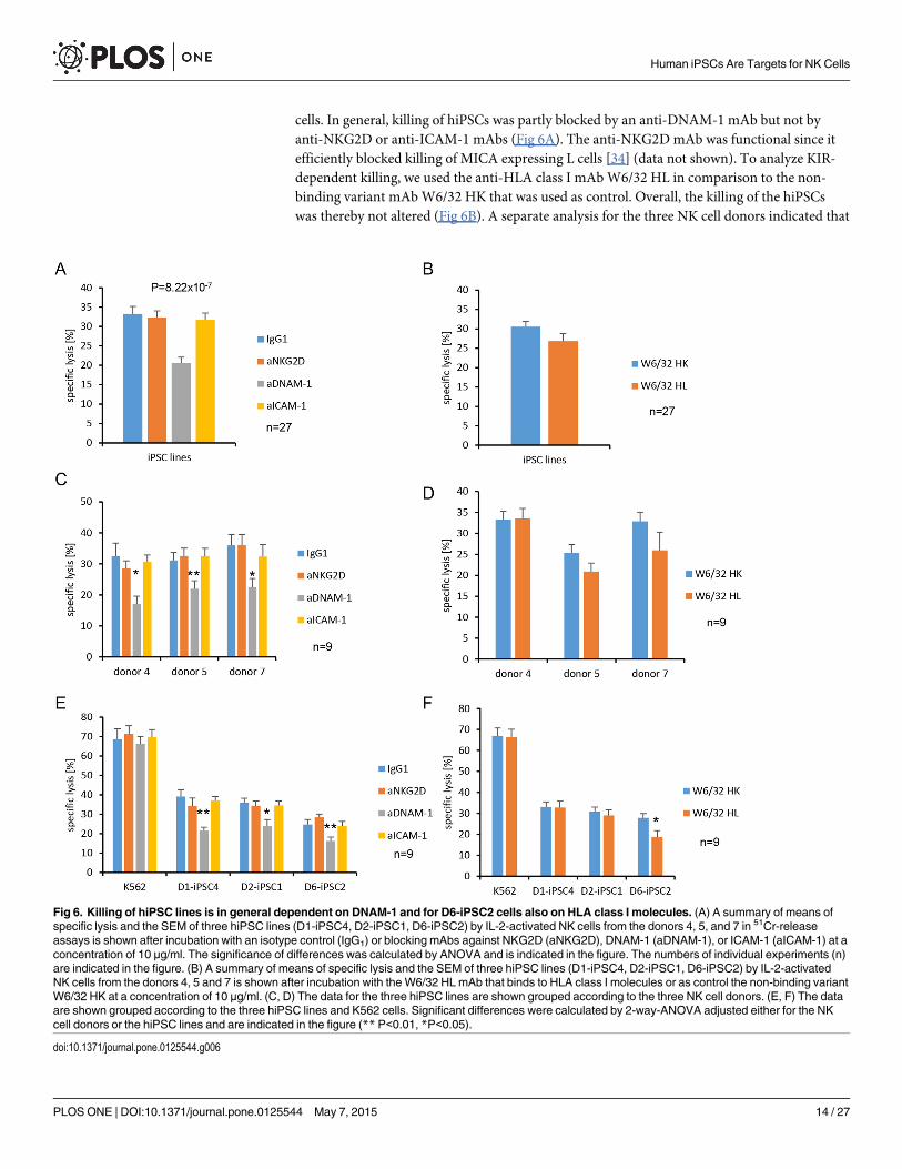
cells. In general, killing of hiPSCs was partly blocked by an anti-DNAM-1 mAb but not byanti-NKG2D or anti-ICAM-1 mAbs (Fig 6A). The anti-NKG2DmAb was functional since itefficiently blocked killing of MICA expressing L cells [34] (data not shown). To analyze KIR-dependent killing, we used the anti-HLA class I mAbW6/32 HL in comparison to the non-binding variant mAbW6/32 HK that was used as control. Overall, the killing of the hiPSCswas thereby not altered (Fig 6B). A separate analysis for the three NK cell donors indicated that
Fig 6. Killing of hiPSC lines is in general dependent on DNAM-1 and for D6-iPSC2 cells also on HLA class I molecules. (A) A summary of means ofspecific lysis and the SEM of three hiPSC lines (D1-iPSC4, D2-iPSC1, D6-iPSC2) by IL-2-activated NK cells from the donors 4, 5, and 7 in 51Cr-releaseassays is shown after incubation with an isotype control (IgG1) or blocking mAbs against NKG2D (aNKG2D), DNAM-1 (aDNAM-1), or ICAM-1 (aICAM-1) at aconcentration of 10 μg/ml. The significance of differences was calculated by ANOVA and is indicated in the figure. The numbers of individual experiments (n)are indicated in the figure. (B) A summary of means of specific lysis and the SEM of three hiPSC lines (D1-iPSC4, D2-iPSC1, D6-iPSC2) by IL-2-activatedNK cells from the donors 4, 5 and 7 is shown after incubation with the W6/32 HLmAb that binds to HLA class I molecules or as control the non-binding variantW6/32 HK at a concentration of 10 μg/ml. (C, D) The data for the three hiPSC lines are shown grouped according to the three NK cell donors. (E, F) The dataare shown grouped according to the three hiPSC lines and K562 cells. Significant differences were calculated by 2-way-ANOVA adjusted either for the NKcell donors or the hiPSC lines and are indicated in the figure (** P<0.01, *P<0.05).
doi:10.1371/journal.pone.0125544.g006
Human iPSCs Are Targets for NK Cells
PLOS ONE | DOI:10.1371/journal.pone.0125544 May 7, 2015 14 / 27

killing of hiPSCs was DNAM-1-dependent for all donors (Fig 6C) and all hiPSC lines (Fig 6E).However, it was not significantly altered by the anti-HLA class I mAbW6/32 HL for any donor(Fig 6D). This mAb blocked partly killing of the D6-iPSC2 line (Fig 6F). An individual analysisfor each donor and each hiPSC line (S10 Fig) revealed that the killing of D6-iPSC2 cells by NKcells of donor 7 was significantly inhibited by the anti-HLA class I mAbW6/32 HL (panel Fin S10 Fig). The killing of D6-iPSC2 cells by NK cells of donor 5 appeared also to be reducedbut only at borderline significance (panel D in S10 Fig) (P = 0.09, t-test). This does not provebut suggests that this cell line was killed partly via activating KIRs by NK cells of donor 7 anddonor 5, which might have recognized specific HLA class I molecules expressed by this cellline. More individual NK cell reactions against hiPSC lines could exist. NK cells of donor 4,e. g., appeared to kill D1-iPSC4 cells partly in an NKG2D-dependent manner (panel A in S10Fig) although only at borderline significance (P = 0.09, t-test). In summary, DNAM-1 was themost important NK cell receptor for killing of hiPSC lines but on an individual level other re-ceptor ligand pairs appeared to contribute.
Expression of ligands of activating and inhibitory NK cell receptors inhiPSCsWe then analyzed the gene expression of ligands for activating and inhibitory NK cell receptorsin three hiPSC lines by qPCR. Important activating NK receptors are NKG2D, DNAM-1, and2B4. We analyzed the mRNA expression of the NKG2D ligandsMICA,MICB, ULBP1, ULBP2,the DNAM-1 ligands CD112 (PVRL2), CD155 (PVR), and the 2B4 ligand CD48 (Fig 7A). Thehighest expression levels were found for CD112,MICB, andMICA. ULPB2mRNA was not de-tected in the hiPSC lines analyzed (ct>30). ICAM1 encodes an adhesion molecule that hasbeen described to be important for killing of mouse ESCs by NK cells [42] and low amounts ofthis mRNA were detected in the hiPSC lines (Fig 7A). The expression of classical (HLA-A,HLA-B,HLA-C) and non-classical (HLA-E,HLA-F, HLA-G) HLA class I genes was tested to-gether with B2M, encoding the non-polymorphic β2-microglobulin that associates with theHLA-class-I α-chain, and the HLA-class-II gene DRA (Fig 7A). HLA-A, HLA-B, HLA-C,HLA-E, and HLA-G function as ligands for inhibitory NK receptors.HLA-B,HLA-E, andHLA-CmRNAs were more abundant in the hiPSC lines thanHLA-AmRNA. HLA-F, HLA-G,andHLA-DR (DRA) mRNAs were not detected (ct>30). Since antigen processing is requiredto obtain normal expression levels of classical class I molecules, we tested the expression of im-portant components of the antigen processing machinery (Fig 7A). The genes of the chaper-ones calnexin (CANX), calreticulin (CALR), and Erp57 (PDIA3) were strongly expressed in thehiPSC lines, in contrast to the ‘transporter associated with antigen processing’ genes TAP1,TAP2, and the TAP-binding protein (TAPBP) gene as well as genes encoding the specific sub-units of the immunoproteasome LMP2 (PSMB9) and LMP7 (PSMB8). TheHLA-A, HLA-B,HLA-C, HLA-E, B2M, TAP1, TAP2, and TAPBPmRNAs were much less abundant in hiPSCsthan in PBMCs (Fig 7B), suggesting that the expression of TAP1, TAP2 and TAPBP (Tapasin)might limit the HLA-class-I expression in hiPSC cells. When the gene expression pattern wascompared to K562 cells, a higher expression of the CD155 gene but lower levels ofMICA,MICB and ICAM1mRNAs in the hiPSC lines were observed (Fig 7C).
MICA, MICB, CD112, CD155, HLA class I, and ICAM-1 proteins were readily detected onthe plasma membrane of the hiPSC lines by flow cytometry as illustrated in Fig 8 for D1-iPSC4cells. Thus, the hiPSC lines expressed ligands for activating and inhibitory NK receptors assummarized in Fig 9. The CD112 and CD155 and the HLA class I molecules were the mostabundant NK receptor ligands on the hiPSC lines. Nonetheless, HLA class I expression intensi-ty was less than 5% compared to PBMCs (data not shown). For the ULBPs and NCR ligands
Human iPSCs Are Targets for NK Cells
PLOS ONE | DOI:10.1371/journal.pone.0125544 May 7, 2015 15 / 27

Fig 7. Gene expression analysis of hiPSC lines indicatedmostly lowmRNA expression of activating and inhibitory NK receptor ligands. (A) Theexpression of the indicated genes was tested by qPCR in PBMCs, K562 cells and three hiPSC lines (D1-iPSC4, D2-iPSC1, and D3-iPSC3). For the hiPSClines and K562 cells means ± SEM of Δct values (ct target gene [tg] minus ct housekeeping gene [hkg]) of three biological replicates are shown. Negativevalues indicated a higher expression of the target gene than the housekeeping gene. Therefore, an inverted scale is shown. At the left side, genes encodingfor ligands of activating NK receptors and ICAM1 are shown. In the middle part classical and non-classical HLA class I genes, B2M, and the HLA class IIgene DRA are shown. In the right part, genes involved in antigen processing in the HLA class I pathway are grouped. Genes not expressed in the iPSC lines(ct > 30) are marked byØ. (B) The gene expression in the iPSC lines is shown as relative expression compared to PBMCs. (C) The gene expression in thehiPSC lines is shown as relative expression compared to K562 cells.
doi:10.1371/journal.pone.0125544.g007
Human iPSCs Are Targets for NK Cells
PLOS ONE | DOI:10.1371/journal.pone.0125544 May 7, 2015 16 / 27

(NKp30, NKp44, NKp46) a complex expression pattern at very low levels was found. When thehiPSC lines and K562 cells were compared with respect to the MFI of the cell surface molecules,K562 cells showed a higher expression of NKG2D ligands (detected with the recombinant re-ceptor molecule and specifically MICA, MICB, ULBP1 detected with mAbs), NKp30 ligands,ICAM-1, and HLA-E (p<0.05, H test after Bonferroni-Holm correction). They expressed lessclassical HLA class I, ULBP2, and ULBP3 molecules (Fig 9, left panel) (p<0.05, H test after Bon-ferroni-Holm correction). In accord with these results, a higher proportion of K562 cells thanhiPSCs was positive for NKG2D ligands, MICA, MICB, ULBP1, ULBP2, NKP30 ligands, ICAM-1, HLA-E, and CD112 in flow cytometry (p<0.05, H test after Bonferroni-Holm correction).
Fig 8. Flow cytometric analysis of ligands for activating and inhibitory NK receptors on D1-iPSC4cells.Representative histograms of propidium iodide negative D1-iPSC4 cells are shown after staining withmAbs for the indicated molecules or with recombinant receptor molecules (NKG2D, NKp30, NKp44, andNKp46) for the respective ligands. Staining with the respective primary reagent is shown in red and with thesecondary Ab only in black. The percentages of specifically stained cells (using the marker shown) and thespecific MFI were calculated and are given in the figure.
doi:10.1371/journal.pone.0125544.g008
Human iPSCs Are Targets for NK Cells
PLOS ONE | DOI:10.1371/journal.pone.0125544 May 7, 2015 17 / 27

Fewer K562 than hiPSC cells expressed ULBP3 (Fig 9, right panel) (p<0.05, H test after Bonfer-roni-Holm correction). Overall, this expression pattern could contribute to the higher suscepti-bility of K562 cells to NK cells compared to the hiPSC cell lines. Comparing the hiPSC lines witheach other indicated differences in the expression intensity of MICA (D1-iPSC4> D2-iPSC1,D3-iPSC3, D6-iPSC2) and HLA class I molecules (D2-iPSC1, D3-iPSC3> D1-iPSC4,D6-iPSC2) (Fig 9, left panel) (p<0.05 H test after Bonferroni-Holm correction). For MICA alsothe proportion of positive cells was higher for D1-iPSC4 than the other hiPSC lines, whereas lessD3-iPSC3 cells expressed ULBP1 (Fig 9, right panel) (p<0.05, H test after Bonferroni-Holm cor-rection). The consistently higher expression of MICA on D1-iPSC4 cells than on the other twohiPSC cell lines might contribute to the higher susceptibility of this hiPSC line to NK cells at leastfrom some donors.
Fig 9. Flow cytometric analysis of ligands for activating and inhibitory NK receptors indicated presence of both classes of molecules on hiPSCs.The expression of the molecules shown in the lower part of the figure was tested in flow cytometry using mAbs and the ligands indicated in the upper partwere stained by the recombinant receptor molecules. K562 cells served as control. Shown are means + SEM of the specific MFI (left panel) and thepercentage of positive cells (right panel) of K562 (n = 34), D1-iPSC4 (n = 21), D2-iPSC1 (n = 20), D3-iPSC3 (n = 12), and D6-iPSC2 cells (n = 9).
doi:10.1371/journal.pone.0125544.g009
Human iPSCs Are Targets for NK Cells
PLOS ONE | DOI:10.1371/journal.pone.0125544 May 7, 2015 18 / 27
DiscussionRecently, the first results of a phase I clinical trial have been reported, in which the transplanta-tion of hESC-derived cells was tested, showing that pluripotent stem cells are on the way toclinical application [43]. Retinal pigment epithelial cells (5 x 10⁴) differentiated from hESCs invitro were injected into a submacular site of two patients with Stargardt’s macular dystrophy orage-related macular degeneration. The clinical results suggested efficacy of these allogeneictransplantations. No signs of hyperproliferation, tumorigenicity, ectopic tissue formation or re-jection were observed within the first four months after transplantation. However, the eye is animmune-privileged site so that the information obtained during this trial on the immunogenic-ity of hESC-derived grafts and the risk of rejection in humans is limited.
Human iPSCs are an alternative source of stem cells to obtain differentiated cells and tissuesfor transplantations. In contrast to hESCs, hiPSCs can potentially be used in an autologous set-ting, which reduces in principal the risk of immune rejection. Notably, an unexpected immu-nogenicity of murine iPSCs in syngeneic hosts has been reported [44] but was contradictedrecently by other studies [45–48]. However, currently little is known on the immune responseto hiPSCs although the question is highly relevant for the application of hiPSCs in regenerativemedicine [49, 50]. Of course, pluripotent stem cells will not be transplanted directly but onlyafter in vitro differentiation into the desired cell type or tissue. Nonetheless, it is of interest todetermine their susceptibility to immune rejection since they potentially can contaminate stemcell-derived grafts in trace amounts despite efforts to eliminate them quantitatively. Dependingon the cell differentiation strategies applied [51], remaining pluripotent cells might confer a re-sidual risk of teratoma formation [52]. This risk would likely be higher in autologous than inallogeneic transplantations [4].
In this study, we have shown that hiPSC lines were susceptible to killing by IL-2-activatedNK cells whereas they were largely resistant to freshly isolated resting NK cells. This result is inagreement with a previous report on killing of a hiPSC line by IL-2-activated NK cells [53]. Inour experiments, the NK cells killed the hiPSC lines less efficiently than the common NK targetcell line K562. This result is in line with our finding that K562 cells expressed more ligands forthe activating NK receptor NKG2D than the hiPSC lines. We have shown previously that mu-rine pluripotent stem cells, including iPSCs, were killed by IL-2-activated mouse NK cells withsimilar efficacy or even better than the common murine NK target cell line YAC-1 [13]. Al-though, it is difficult to compare K562 and YAC-1 target cells directly, this result might suggestthat human iPSCs are less sensitive to NK cells than mouse iPSCs. This could be relevant forthe interpretation and extrapolation of results coming from preclinical mouse experiments.Notably, the hiPSCs expressed low amounts of HLA class I molecules as hESCs do [25–27].The similar phenotype was reported for a few other hiPSC lines [29, 30]. Thus, human pluripo-tent stem cells appear to engage inhibitory receptors on NK cells better than mouse pluripotentstem cells, which largely lack MHC class I molecules [13, 24].
The killing of all hiPSC lines by NK cells from all donors was partly dependent on DNAM-1as shown by mAb blocking experiments. Moreover CD107a+ NK cells degranulating in responseto hiPSC lines expressed more DNAM-1 than the CD107a- NK cells. In addition, other NK re-ceptor ligand interactions might play a role. Interestingly, the cell line D2-iPSC1, which wasmost resistant to IL-2-activated NK cells (Fig 2B) had the highest HLA class I expression inten-sity (Fig 8) suggesting an effect of inhibitory KIRs (see Table 2 for a summary of key features ofthe individual hiPSC lines studied). To further analyze the contribution of KIRs to killing ofhiPSCs, we blocked HLA class I molecules on the target cells. In general, this did not alter thelysis significantly. However, the mAbW6/32 was reported to elicit antibody-dependent cellularcytotoxicity of human NK cells [54], which is mediated via CD16 and could have masked effects
Human iPSCs Are Targets for NK Cells
PLOS ONE | DOI:10.1371/journal.pone.0125544 May 7, 2015 19 / 27

of blocking the interaction with inhibitory KIRs. Notably, in the specific combination of NKcells of donors 5 and 7 and the D6-iPSC2 target cells, respectively, the blocking of HLA class Imolecules reduced lysis suggesting that activating KIRs might have contributed to the killing ofthis hiPSC line by these NK cells. These NK cell donors had an AB KIR genotype encoding sev-eral activating KIRs in contrast to donor 4 who carried an AA genotype lacking activating KIRswith exception of KIR2DS4, which was even not expressed on the NK cells of this donor (S2Table, S6 Fig). In other combinations, e. g. NK cells of donor 4 and D1-iPSC4 targets, NKG2D-mediated effects appeared to have contributed to killing. We did not observe a dependency ofkilling on ICAM-1. In summary, the killing of the hiPSC lines was mediated by a NK cell recep-tor used by all NK cells, i.e. DNAM-1, but other receptors, such as activating KIRs and NKG2D,might have been used in addition in individual NK cell target combinations.
Consistently with these data, all hiPSC lines expressed the DNAM-1 ligands CD112 andCD155. Nonetheless, the three different hiPSC lines, which we investigated in the first set ofexperiments, varied in their susceptibility to NK cells. The hiPSC lines also varied in the ex-pression of some ligands of NK cell receptors and particularly the NKG2D ligand MICA. Thehigher expression of MICA on D1-iPSC4 cells than on the other hiPSC lines might have con-tributed to the higher susceptibility of this hiPSC line to NK cells. However, in the second setof experiments only NK cells from donor 4 appeared to use the NKG2D pathway to kill thishiPSC line. Killing of mouse ESCs and iPSCs by NK cells is known to depend in part onNKG2D [5, 13, 14, 42]. In general, the expression pattern of ligands for activating NK receptorson hiPSCs was very similar to mouse iPSCs, which also expressed NKG2D and DNAM-1 li-gands in contrast to 2B4 and NKp40 ligands [13]. So far, we do not know whether the variationamong the hiPSC lines is determined by the genetic differences among the cell lines or repre-sents a clonal variability that can occur also among hiPSC lines from the same donor. In ac-cordance with our results, Suárez-Álvarez and colleagues found the NKG2D ligands MICAand MICB by flow cytometry on a hESC line (Shef-1). Moreover, they reported a hiPSC line(MSUH-002) expressingMICA andMICB but no ULBP1, ULBP2, and ULPB3 transcripts [28].The NKG2D ligands MICA, MICB, ULBP1, ULPB2, and ULBP3 have been shown to be absenton the H9 hESC line [33]. We found a complex expression pattern of NCR ligands (NKp30,
Table 2. Summary of features of the hiPSC lines tested in this study.
Feature D1-iPSC4 D2-iPSC1 D3-iPSC3 D6-iPSC2
NK receptor ligand expression HLA class Ip1 p "2 p
MICA " p p p
MICBp p p "
CD112p p p p
CD155p p p p
killing by NK cells resting (+) (+) nt3 -
IL-2-activated, allogeneic ++ + ++ +
IL-2-activated, autologous ++ + (+) nt
dependency of killing on NK receptors DNAM-1 + + nt +
NKG2D (+) donor 4 - nt -
HLA class I/KIR - - nt + donors 5, 7
1p: expressed at the plasma membrane.2": stronger expressed at plasma membrane in this hiPSC line than on the other hiPSC lines with respect to both MFI and percentage of positive cells.3nt: not tested.
doi:10.1371/journal.pone.0125544.t002
Human iPSCs Are Targets for NK Cells
PLOS ONE | DOI:10.1371/journal.pone.0125544 May 7, 2015 20 / 27

NKp44, and NKp46) at very low levels on the hiPSC lines. Chen and colleagues investigatedalso NKp30, NKp44, and NKp46 ligands on hiPSC lines and found low levels of NKp44 ligandsbut very little or none of the others [29]. This suggests that NCR ligands are not abundant onhiPSCs but at low levels, variations among cell lines appear to exist.
In our hiPSC lines, HLA-B, HLA-E, and HLA-CmRNAs were more abundant thanHLA-A. HLA-F and HLA-G transcripts were not detected. At the plasma membrane, wefound low amounts of classical HLA class I molecules but hardly any HLA-E. In the hESCline Shef-1, low levels of HLA-A and HLA-B but no HLA-E, HLA-F, and HLA-G transcriptswere detected [28]. The hiPSC line MSUH-002 was reported to express low amounts ofHLA-B, HLA-C, and HLA-E but no HLA-A and HLA-GmRNAs [28]. Lower levels of tran-scripts of all classical and non-classical class I genes were recently found in five hiPSC linescompared to their parental cells [29]. In this study, classical HLA class I molecules but hardlyany HLA-E and HLA-G were found by flow cytometry on the hiPSC lines [29]. The samepattern has been described for H9 hESCs [33]. Interestingly, HLA-AmRNA was less abun-dant than HLA-B and HLA-CmRNA in our hiPSC lines and shown to be absent in theMSUH-002 hiPSC line suggesting that the HLA-A gene might indeed be consistently re-pressed in hiPSCs [28].
We investigated also the expression of genes encoding proteins involved in antigen process-ing in our hiPSC lines. The chaperone genes CANX, CALR, and Erp57 were strongly expressed,in contrast to TAP1, TAP2, and TAPBP genes as well as genes encoding the specific subunits ofthe immunoproteasome LMP2 and LMP7. In the hESC line Shef-1 and the hiPSC line MSUH-002 similarly no or very low expression of the TAP1 and TAP2 genes were found [28]. Thus,human pluripotent stem cells appear to lack largely the mRNAs for the transporter proteins,which shuttle peptides from the cytosol to the endoplasmic reticulum and are required forHLA class I plasma membrane expression. This might contribute to the low HLA class I ex-pression on human pluripotent stem cells. Interestingly, an epigenetic H3K9me3 modificationwas reported to repress TAPBP expression, encoding the TAP-binding protein or tapasin, inthe MSUH-002 hiPSC line whereas an active H3K4me3 mark was found on the TAP2 geneonly in parental fibroblasts [28]. In addition, decreasing levels of NFκB1 and RelA proteinsduring reprogramming of hiPSCs can contribute to the low expression of HLA class I mole-cules in hiPSCs [30]. For the genes encoding chaperones (CANX, CALR, and Erp57) and thespecific subunits of the immunoproteasome (LMP2 and LMP7), more cell line specific varia-tions might exist since CALRmRNA was not found in Shef-1 hESCs whereas LMP7 in contrastto LMP2mRNA was present [28].
Notably, the hiPSC cell lines investigated in our study were killed not only by allogeneic butalso by autologous NK cells, although less efficiently. During their development NK cells un-dergo a ‘licensing’ or ‘education’ ensuring inhibition of NK cells by self-ligands or NK cell un-responsiveness [55, 56]. Despite the relatively low expression of HLA class I molecules on thehiPSCs lines, NK cell inhibition appeared to be still more efficient by self than non-self HLAclass I molecules. However, the individual pattern of susceptibility or resistance against autolo-gous and allogeneic NK cells was surprisingly complex when the individual hiPSC lines werecompared. The fact that also NK cells from different donors varied in their efficacy to kill thehiPSC lines contributes to this complexity. Notably, this variance remained stable even afternormalization of the results for killing of the reference target cell line K562. Thus, individualsmight vary in their ability to reject autologous as well as allogeneic hiPSCs putting them poten-tially at different risks of tumor formation after transplantation of stem cell-derived grafts thatcontain residuals of pluripotent cells. A broader set of HLA-typed hiPSC cell lines in additionto KIR-typed NK cell donors will be required to clarify the potential role of KIR-mediated in-hibitory and activating effects on hiPSC lines.
Human iPSCs Are Targets for NK Cells
PLOS ONE | DOI:10.1371/journal.pone.0125544 May 7, 2015 21 / 27

Currently, the tumorigenicity of pluripotent stem cells is seen as a major obstacle for stemcell-based therapies [52]. Another hurdle is the immunogenicity of pluripotent stem cells andtheir derivatives [57–59]. NK cells might be an interesting player in the immune response aftertransplantation of stem cell-derived grafts since they could primarily target residual pluripotentand tumorigenic cells and spare differentiated cells, which are expected to up-regulate HLAclass I molecules during in vitro differentiation and therefore inhibit NK cells. NK cells wouldpresumably require activation to target residual pluripotent cells. However, NK cell activationmight occur, if the transplantation procedure is associated transiently with an inflammatoryresponse.
ConclusionsWe have shown that both allogeneic and autologous IL-2-activated NK cells can kill hiPSCsand killing was mediated partly by DNAM-1. The activity of NK cells might reduce the risk ofteratoma formation after transplantation of pluripotent stem cell-derived grafts that containtraces of pluripotent cells. Since autologous hiPSCs were killed, this could occur even aftertransplantation of autologous hiPSC-derived grafts. However, variation of NK cell activityagainst hiPSCs and variation of susceptibility of hiPSCs against NK cells might modify thethreshold of undifferentiated hiPSCs that can be tolerated in a graft for a specific recipient.
Supporting InformationS1 Fig. Human iPSC lines were used as target cells for purified and IL-2-activated NK cellsof either various allogeneic or autologous donors in 51Cr-release assays. The reference targetcell line K562 was included in every experiment. Each individual test was done in triplicates.The means of specific lysis and the standard error of the mean (SEM) at different effector:target(E:T) ratios (16:1 to 0.25:1) are shown to summarize these experiments (left panels). In addi-tion, the killing of the reference K562 cells at the highest effector to target ratio (16:1) was set to100% in each individual experiment and the relative lysis of the other target cell lines and at thevarious effector to target ratios was calculated accordingly (right panels). The relative lysis isnot shown for NK cells of donor 5 since the specific lysis of K562 cells was 100% leading to anidentity of specific and relative lysis. The results are grouped with respect to the NK cell donors,i.e. (A) donor 1, (B) donor 2, (C) donor 3, (D) donor 4, and (E) donor 5. In panels A, B, and C,the respective autologous hiPSC line is indicated by open symbols. Allogeneic hiPSC target celllines are indicated by closed symbols. The numbers of individual experiments (n) are indicatedin the figure.(PDF)
S2 Fig. Human iPSC lines were killed by purified and IL-2-activated NK cells of various do-nors but allogeneic effector cells were more efficient than autologous NK cells. The samedata set as in Fig 2 is shown but now the killing of K562 cells at the highest effector to targetratio (16:1) was set to 100% in each individual experiment and the relative lysis of the other tar-get cell lines and at the various effector to target ratios was calculated accordingly. The num-bers of individual experiments (n) are indicated in the figure. (A) NK cells from five donorswere stimulated for four days with IL-2 (200 U/ml) and used as effector cells against the refer-ence target cell line K562 in 51Cr-release assays. Each individual test was done in triplicates.The means of relative lysis and the SEM at E:T ratios 16:1 to 0.25:1 are shown to summarizethese experiments. (B) A summary of means of relative lysis and the SEM of K562 and threehiPSC lines by IL-2-activated NK cells from five donors (1 to 5) is shown. (C) A summary ofmeans of relative lysis and the SEM of the three hiPSC lines (D1-iPSC4, D2-iPSC1, D3-iPSC3)
Human iPSCs Are Targets for NK Cells
PLOS ONE | DOI:10.1371/journal.pone.0125544 May 7, 2015 22 / 27

by IL-2-activated NK cells of five different donors is shown. (D) A summary of means of re-lative lysis and the SEM of the three hiPSC lines (D1-iPSC4, D2-iPSC1, D3-iPSC3) by IL-2-activated allogeneic (allo) and autologous (auto) NK cells is shown.(PDF)
S3 Fig. Human iPSC lines were killed by purified and IL-2-activated allogeneic or autolo-gous NK cells of various donors but with different efficacy. (A) A summary of means of spe-cific lysis (left panels) and relative lysis (adjusted to killing of K562 cells, right panels) and theSEM of three hiPSC lines by allogeneic IL-2-activated NK cells from four donors (donors 1 to5) is shown. The numbers of individual experiments (n) are indicated in the figure. (B) A sum-mary of means of specific lysis (left panel) and relative lysis (right panel) and the SEM of alloge-neic hiPSC lines (D1-iPSC4, D2-iPSC1, D3-iPSC3) by NK cells of five different donors isshown. (C) A summary of means of specific lysis (left panel) and relative lysis (right panel) andthe SEM of the three hiPSC lines by autologous NK cells is shown.(PDF)
S4 Fig. Human iPSC lines were used as target cells for freshly isolated or IL-2-activated NKcells of three allogeneic donors in 51Cr-release assays.NK cells of three different donors ((A)donor 4, (B) donor 5, (C) donor 7) were isolated and used as effectors at day 0 (d0, left panels)or after stimulation with IL-2 (200 U/ml) for 4 days (d4, right panels). The means of specificlysis and the SEM at different effector:target (E:T) ratios (16:1 to 0.25:1 for resting NK cellsand 4:1 to 0.06:1 for IL2-activated NK cells) are shown to summarize these experiments. Thereference target cell line K562 was included in every experiment in addition to the hiPSC linesD1-iPSC4, D2-iPSC1, and D6-iPSC2. Each individual test was done in triplicates. The numbersof individual experiments (n) are indicated in the figure.(PDF)
S5 Fig. Phenotypic characterization of NK cells.MACS-purified NK cells from three blooddonors were analyzed by flow cytometry at day 0 (d0) and after stimulation for four days (d4)with IL-2 (200 U/ml). The percentages of cells positive for the indicated NK cell markers areshown as means plus SEM of three individual experiments. The CD56dim and CD56bright popu-lations were not clearly distinguishable anymore at day 4 after stimulation with IL-2.(PDF)
S6 Fig. The KIR repertoire of NK cell donors was characterized by flow cytometry. The re-activity of a panel of anti-KIR mAbs against CD56+CD3- NK cells of NK cell donors 4 (A), 5(B) and 7 (C) was tested. The clone numbers and the reported reactivity against individual KIRmolecules are indicated. KIR molecules, which could be present according to the KIR genotypeof the donors (see S2 Table) are indicated by color. Inhibitory KIRs are marked in red and acti-vating KIRs in green. Means and SEM of three experiments are shown.(PDF)
S7 Fig. NK cells degranulating in response to K562 cells are enriched for several NK cell re-ceptors. In the left panel a summary of means and the SEM of NKG2D+, NKG2A+, DNAM-1+, and KIR+ cells among all NK cells of donors 4, 5, and 7 exposed to K562 as well as CD107a-
and CD107a+ NK cells is shown. In the right panel a summary of means and the SEM of theMFI of NKG2D, NKG2A, DNAM-1, and KIR on all NK cells exposed to K562 cells as well asCD107a- and CD107a+ NK cells is shown. Significant differences between CD107a- andCD107a+ NK cells are indicated (n = 26, �� P<0.01, � P<0.05, t-test after Bonferroni-Holmcorrection).(PDF)
Human iPSCs Are Targets for NK Cells
PLOS ONE | DOI:10.1371/journal.pone.0125544 May 7, 2015 23 / 27

S8 Fig. Enrichment of NK cells degranulating in response to hiPSC lines for specific NKcell receptors is more influenced by the NK cell donors than by the hiPSC lines. The differ-ence between CD107a- and CD107a+ NK cells was calculated for NKG2D+ (A), NGK2A+
(C), KIR+ NK cells (E) and for the expression intensities (MFI) of these molecules (B, D, F).The data are shown as means and SEM and they were grouped for the three donors (leftpanels) or the three hiPSC lines (right panels). Significant differences were calculated by2-way-ANOVA adjusted either for the NK cell donors or the hiPSC lines and are indicatedin the figure (n = 9).(PDF)
S9 Fig. The NK cell receptor repertoire of NK cells degranulating in response to hiPSClines and K562 cells is shown for individual NK cell donors and hiPSC targets. The differ-ence between CD107a- and CD107a+ NK cells was calculated for NKG2D+ (A, left panel),NGK2A+ (B, left panel), DNAM-1+ (C, left panel), and KIR+ NK cells (D, left panel). The dif-ference between CD107a- and CD107a+ NK cells was also calculated for the expression intensi-ties (MFI) of these molecules, i. e. NKG2D (A, right panel), NGK2A (B, right panel), DNAM-1(C, right panel), and KIR (D, right panel). The data are shown as means and SEM of three indi-vidual experiments and they were grouped for the three donors (left panels) or the three hiPSClines (right panels). Significant differences between the donors are indicated in the figure(n = 3, � P<0.05, t-test).(PDF)
S10 Fig. Inhibition of killing of hiPSC lines and K562 cells by mAbs is shown for individualNK cell donors and target cells.Means of specific lysis and SEM of three hiPSC lines andK562 cells by IL-2-activated NK cells from donor 4 (A), donor 5 (C), and donor 7 (E) in51Cr-release assay is shown after incubation with an isotype control (IgG1) or blockingmAbs against NKG2D (aNKG2D), DNAM-1 (aDNAM-1), or ICAM-1 (aICAM-1) at aconcentration of 10 μg/ml. Means of specific lysis and SEM of three hiPSC lines andK562 cells by IL-2-stimulated NK cells from donor 4 (B), donor 5 (D), and donor 7 (F) in51Cr-release assays is shown after incubation with the W6/32 HL mAb that binds toHLA class I molecules or as control the non-binding variant W6/32 HK at a concentra-tion of 10 μg/ml. Significant differences between the mAbs are indicated in the figure(n = 3, � P<0.05, t-test).(PDF)
S1 Table. Genes analyzed by qPCR and primers used.(PDF)
S2 Table. KIR genotypes of NK cell donors 4, 5, and 7.(PDF)
AcknowledgmentsWe would like to thank the donors of hair keratinocytes and blood who made this study possi-ble and Yvonne Hintz for technical assistance.
Author ContributionsConceived and designed the experiments: KG RD. Performed the experiments: VK CH SM LCLE DH KSB RD. Analyzed the data: VK CH SM LC LW KSB KG RD. Contributed reagents/materials/analysis tools: LW. Wrote the paper: RD.
Human iPSCs Are Targets for NK Cells
PLOS ONE | DOI:10.1371/journal.pone.0125544 May 7, 2015 24 / 27

References1. Takahashi K, Yamanaka S. Induction of pluripotent stem cells frommouse embryonic and adult fibro-
blast cultures by defined factors. Cell. 2006; 126(4):663–76. doi: 10.1016/j.cell.2006.07.024 PMID:16904174.
2. Wernig M, Meissner A, Foreman R, Brambrink T, Ku M, Hochedlinger K, et al. In vitro reprogramming offibroblasts into a pluripotent ES-cell-like state. Nature. 2007; 448(7151):318–24. doi: 10.1038/nature05944 PMID: 17554336.
3. Yu J, Vodyanik MA, Smuga-Otto K, Antosiewicz-Bourget J, Frane JL, Tian S, et al. Induced pluripotentstem cell lines derived from human somatic cells. Science. 2007; 318(5858):1917–20. doi: 10.1126/science.1151526 PMID: 18029452.
4. Dressel R. Effects of histocompatibility and host immune responses on the tumorigenicity of pluripo-tent stem cells. Semin Immunopathol. 2011; 33(6):573–91. doi: 10.1007/s00281-011-0266-8 PMID:21461989; PubMed Central PMCID: PMC3204002.
5. Dressel R, Schindehütte J, Kuhlmann T, Elsner L, Novota P, Baier PC, et al. The tumorigenicity ofmouse embryonic stem cells and in vitro differentiated neuronal cells is controlled by the recipients' im-mune response. PLOSOne. 2008; 3(7):e2622. doi: 10.1371/journal.pone.0002622 PMID: 18612432;PubMed Central PMCID: PMC2440803.
6. Koch CA, Geraldes P, Platt JL. Immunosuppression by embryonic stem cells. Stem Cells. 2008;26(1):89–98. doi: 10.1634/stemcells.2007-0151 PMID: 17962705.
7. Kolossov E, Bostani T, Roell W, Breitbach M, Pillekamp F, Nygren JM, et al. Engraftment of engineeredES cell-derived cardiomyocytes but not BM cells restores contractile function to the infarcted myocardi-um. J Exp Med. 2006; 203(10):2315–27. PMID: 16954371.
8. Swijnenburg RJ, Tanaka M, Vogel H, Baker J, Kofidis T, Gunawan F, et al. Embryonic stem cell immu-nogenicity increases upon differentiation after transplantation into ischemic myocardium. Circulation.2005; 112(9 Suppl):I166–72. PMID: 16159810.
9. Nussbaum J, Minami E, LaflammeMA, Virag JA, Ware CB, Masino A, et al. Transplantation of undiffer-entiated murine embryonic stem cells in the heart: teratoma formation and immune response. FASEBJ. 2007; 21(7):1345–57. PMID: 17284483.
10. Drukker M, Katchman H, Katz G, Even-Tov Friedman S, Shezen E, Hornstein E, et al. Human embry-onic stem cells and their differentiated derivatives are less susceptible to immune rejection than adultcells. Stem Cells. 2006; 24(2):221–9. doi: 10.1634/stemcells.2005-0188 PMID: 16109762.
11. Pearl JI, Lee AS, Leveson-Gower DB, Sun N, Ghosh Z, Lan F, et al. Short-term immunosuppression pro-motes engraftment of embryonic and induced pluripotent stem cells. Cell Stem Cell. 2011; 8(3):309–17.doi: 10.1016/j.stem.2011.01.012 PMID: 21362570; PubMed Central PMCID: PMC3061351.
12. Koch CA, Jordan CE, Platt JL. Complement-dependent control of teratoma formation by embryonicstem cells. J Immunol. 2006; 177(7):4803–9. PMID: 16982921.
13. Dressel R, Nolte J, Elsner L, Novota P, Guan K, Streckfuss-Bömeke K, et al. Pluripotent stem cells arehighly susceptible targets for syngeneic, allogeneic, and xenogeneic natural killer cells. FASEB J.2010; 24(7):2164–77. doi: 10.1096/fj.09-134957 PMID: 20145206.
14. Perez-Cunningham J, Ames E, Smith RC, Peter AK, Naidu R, Nolta JA, et al. Natural killer cell subsetsdifferentially reject embryonic stem cells based on licensing. Transplantation. 2014; 97(10):992–8. doi:10.1097/TP.0000000000000063 PMID: 24704665.
15. Tian X, Woll PS, Morris JK, Linehan JL, Kaufman DS. Hematopoietic engraftment of human embryonicstem cell-derived cells is regulated by recipient innate immunity. Stem Cells. 2006; 24(5):1370–80. doi:10.1634/stemcells.2005-0340 PMID: 16456127.
16. Mizukami Y, Abe T, Shibata H, Makimura Y, Fujishiro SH, Yanase K, et al. MHC-matched induced plu-ripotent stem cells can attenuate cellular and humoral immune responses but are still susceptible to in-nate immunity in pigs. PLOS One. 2014; 9(6):e98319. doi: 10.1371/journal.pone.0098319 PMID:24927426; PubMed Central PMCID: PMC4057111.
17. Koch J, Steinle A, Watzl C, MandelboimO. Activating natural cytotoxicity receptors of natural killer cells incancer and infection. Trends Immunol. 2013; 34(4):182–91. doi: 10.1016/j.it.2013.01.003 PMID: 23414611.
18. Rajagopalan S, Long EO. A human histocompatibility leukocyte antigen (HLA)-G-specific receptor ex-pressed on all natural killer cells. J Exp Med. 1999; 189(7):1093–100. PMID: 10190900; PubMed Cen-tral PMCID: PMC2193010.
19. Verloes A, Van de Velde H, LeMaoult J, Mateizel I, Cauffman G, Horn PA, et al. HLA-G expressionin human embryonic stem cells and preimplantation embryos. J Immunol. 2011; 186(4):2663–71. doi:10.4049/jimmunol.1001081 PMID: 21248264.
20. Kärre K. Natural killer cell recognition of missing self. Nat Immunol. 2008; 9(5):477–80. doi: 10.1038/ni0508-477 PMID: 18425103.
Human iPSCs Are Targets for NK Cells
PLOS ONE | DOI:10.1371/journal.pone.0125544 May 7, 2015 25 / 27

21. Vivier E, Ugolini S, Blaise D, Chabannon C, Brossay L. Targeting natural killer cells and natural killer Tcells in cancer. Nat Rev Immunol. 2012; 12(4):239–52. doi: 10.1038/nri3174 PMID: 22437937.
22. Magliocca JF, Held IK, Odorico JS. Undifferentiated murine embryonic stem cells cannot induce portaltolerance but may possess immune privilege secondary to reduced major histocompatibility complexantigen expression. Stem Cells Dev. 2006; 15(5):707–17. PMID: 17105406.
23. Tian L, Catt JW, O'Neill C, King NJ. Expression of immunoglobulin superfamily cell adhesion moleculeson murine embryonic stem cells. Biol Reprod. 1997; 57(3):561–8. PMID: 9282991.
24. Dressel R, Guan K, Nolte J, Elsner L, Monecke S, Nayernia K, et al. Multipotent adult germ-line stemcells, like other pluripotent stem cells, can be killed by cytotoxic T lymphocytes despite low expressionof major histocompatibility complex class I molecules. Biol Direct. 2009; 4:31. doi: 10.1186/1745-6150-4-31 PMID: 19715575; PubMed Central PMCID: PMC2745366.
25. Draper JS, Pigott C, Thomson JA, Andrews PW. Surface antigens of human embryonic stem cells:changes upon differentiation in culture. J Anat. 2002; 200(Pt 3):249–58. PMID: 12033729.
26. Drukker M, Katz G, Urbach A, Schuldiner M, Markel G, Itskovitz-Eldor J, et al. Characterization ofthe expression of MHC proteins in human embryonic stem cells. Proc Natl Acad Sci U S A. 2002;99(15):9864–9. doi: 10.1073/pnas.142298299 PMID: 12114532; PubMed Central PMCID: PMC125045.
27. Li L, Baroja ML, Majumdar A, Chadwick K, Rouleau A, Gallacher L, et al. Human embryonic stem cellspossess immune-privileged properties. Stem Cells. 2004; 22(4):448–56. PMID: 15277692.
28. Suarez-Alvarez B, Rodriguez RM, Calvanese V, Blanco-Gelaz MA, Suhr ST, Ortega F, et al. Epigeneticmechanisms regulate MHC and antigen processing molecules in human embryonic and induced plurip-otent stem cells. PLOS One. 2010; 5(4):e10192. doi: 10.1371/journal.pone.0010192 PMID: 20419139;PubMed Central PMCID: PMC2855718.
29. Chen HF, Yu CY, Chen MJ, Chou SH, Chiang MS, ChouWH, et al. Characteristic expression of majorhistocompatibility complex and immune privilege genes in human pluripotent stem cells and the deriva-tives. Cell Transplant. 2013; in press. doi: 10.3727/096368913X674639 PMID: 24144439.
30. Pick M, Ronen D, YanukaO, Benvenisty N. Reprogramming of the MHC-I and its regulation by NFkap-paB in human-induced pluripotent stem cells. Stem Cells. 2012; 30(12):2700–8. doi: 10.1002/stem.1242PMID: 22987393.
31. Drukker M. Immunogenicity of embryonic stem cells and their progeny. Methods Enzymol. 2006;420:391–409. doi: 10.1016/S0076-6879(06)20019-3 PMID: 17161708.
32. Deuse T, Seifert M, Phillips N, Fire A, Tyan D, Kay M, et al. Human leukocyte antigen I knockdownhuman embryonic stem cells induce host ignorance and achieve prolonged xenogeneic survival. Circu-lation. 2011; 124(11 Suppl):S3–9. doi: 10.1161/CIRCULATIONAHA.111.020727 PMID: 21911816.
33. Deuse T, Seifert M, Tyan D, Tsao PS, Hua X, Velden J, et al. Immunobiology of naive and geneticallymodified HLA-class-I-knockdown human embryonic stem cells. J Cell Sci. 2011; 124(Pt 17):3029–37.doi: 10.1242/jcs.087718 PMID: 21878509.
34. Elsner L, Flügge PF, Lozano J, Muppala V, Eiz-Vesper B, Demiroglu SY, et al. The endogenous dangersignals HSP70 and MICA cooperate in the activation of cytotoxic effector functions of NK cells. J CellMol Med. 2010; 14(4):992–1002. doi: 10.1111/j.1582-4934.2009.00677.x PMID: 20569278.
35. Streckfuss-Bömeke K, Wolf F, Azizian A, Stauske M, Tiburcy M, Wagner S, et al. Comparativestudy of human-induced pluripotent stem cells derived from bone marrow cells, hair keratinocytes,and skin fibroblasts. Eur Heart J. 2013; 34(33):2618–29. doi: 10.1093/eurheartj/ehs203 PMID:22798560.
36. Somers A, Jean JC, Sommer CA, Omari A, Ford CC, Mills JA, et al. Generation of transgene-free lungdisease-specific human induced pluripotent stem cells using a single excisable lentiviral stem cell cas-sette. Stem Cells. 2010; 28(10):1728–40. doi: 10.1002/stem.495 PMID: 20715179; PubMed CentralPMCID: PMC3422663.
37. Barnstable CJ, Bodmer WF, Brown G, Galfre G, Milstein C, Williams AF, et al. Production of monoclo-nal antibodies to group A erythrocytes, HLA and other human cell surface antigens-new tools for genet-ic analysis. Cell. 1978; 14(1):9–20. PMID: 667938.
38. Ziegler A, Milstein C. A small polypeptide different from beta2-microglobin associated with a human cellsurface antigen. Nature. 1979; 279(5710):243–4. PMID: 312466.
39. Vandesompele J, De Preter K, Pattyn F, Poppe B, Van Roy N, De Paepe A, et al. Accurate normali-zation of real-time quantitative RT-PCR data by geometric averaging of multiple internal controlgenes. Genome Biol. 2002; 3(7):RESEARCH0034. PMID: 12184808; PubMed Central PMCID:PMC126239.
40. Pfaffl MW, Tichopad A, Prgomet C, Neuvians TP. Determination of stable housekeeping genes, differ-entially regulated target genes and sample integrity: BestKeeper—Excel-based tool using pair-wisecorrelations. Biotechnol Lett. 2004; 26(6):509–15. PMID: 15127793.
Human iPSCs Are Targets for NK Cells
PLOS ONE | DOI:10.1371/journal.pone.0125544 May 7, 2015 26 / 27

41. Uhrberg M, Parham P, Wernet P. Definition of gene content for nine common group B haplotypes of theCaucasoid population: KIR haplotypes contain between seven and eleven KIR genes. Immunogenet-ics. 2002; 54(4):221–9. doi: 10.1007/s00251-002-0463-7 PMID: 12136333.
42. Frenzel LP, Abdullah Z, Kriegeskorte AK, Dieterich R, Lange N, Busch DH, et al. Role of natural-killergroup 2 member D ligands and intercellular adhesion molecule 1 in natural killer cell-mediated lysisof murine embryonic stem cells and embryonic stem cell-derived cardiomyocytes. Stem Cells. 2009;27(2):307–16. doi: 10.1634/stemcells.2008-0528 PMID: 18988711.
43. Schwartz SD, Hubschman JP, Heilwell G, Franco-Cardenas V, Pan CK, Ostrick RM, et al. Embryonicstem cell trials for macular degeneration: a preliminary report. Lancet. 2012; 379(9817):713–20. doi:10.1016/S0140-6736(12)60028-2 PMID: 22281388.
44. Zhao T, Zhang ZN, Rong Z, Xu Y. Immunogenicity of induced pluripotent stem cells. Nature. 2011;474(7350):212–5. doi: 10.1038/nature10135 PMID: 21572395.
45. Araki R, Uda M, Hoki Y, Sunayama M, Nakamura M, Ando S, et al. Negligible immunogenicity of ter-minally differentiated cells derived from induced pluripotent or embryonic stem cells. Nature. 2013;494(7435):100–4. doi: 10.1038/nature11807 PMID: 23302801.
46. Guha P, Morgan JW, Mostoslavsky G, Rodrigues NP, Boyd AS. Lack of immune response to differenti-ated cells derived from syngeneic induced pluripotent stem cells. Cell Stem Cell. 2013; 12(4):407–12.doi: 10.1016/j.stem.2013.01.006 PMID: 23352605.
47. Cai J, Chen J, Liu Y, Miura T, Luo Y, Loring JF, et al. Assessing self-renewal and differentiation inhuman embryonic stem cell lines. Stem Cells. 2006; 24(3):516–30. PMID: 16293578.
48. de Almeida PE, Meyer EH, Kooreman NG, Diecke S, Dey D, Sanchez-Freire V, et al. Transplanted ter-minally differentiated induced pluripotent stem cells are accepted by immune mechanisms similar toself-tolerance. Nat Commun. 2014; 5:3903. doi: 10.1038/ncomms4903 PMID: 24875164; PubMedCentral PMCID: PMC4075468.
49. Kaneko S, Yamanaka S. To be immunogenic, or not to be: that's the iPSC question. Cell Stem Cell.2013; 12(4):385–6. doi: 10.1016/j.stem.2013.03.008 PMID: 23561437.
50. Boyd AS, Rodrigues NP, Lui KO, Fu X, Xu Y. Concise review: Immune recognition of induced pluripo-tent stem cells. Stem Cells. 2012; 30(5):797–803. doi: 10.1002/stem.1066 PMID: 22419544.
51. Liao MC, Diaconu M, Monecke S, Collombat P, Timaeus C, Kuhlmann T, et al. Embryonic stem cell-derived neural progenitors as non-tumorigenic source for dopaminergic neurons. World J Stem Cells.2014; 6(2):248–55. doi: 10.4252/wjsc.v6.i2.248 PMID: 24772251; PubMed Central PMCID:PMC3999782.
52. Lee AS, Tang C, Rao MS, Weissman IL, Wu JC. Tumorigenicity as a clinical hurdle for pluripotent stemcell therapies. Nat Med. 2013; 19(8):998–1004. doi: 10.1038/nm.3267 PMID: 23921754; PubMed Cen-tral PMCID: PMC3967018.
53. Tseng HC, Arasteh A, Paranjpe A, Teruel A, YangW, Behel A, et al. Increased lysis of stem cells butnot their differentiated cells by natural killer cells; de-differentiation or reprogramming activates NKcells. PLOS One. 2010; 5(7):e11590. doi: 10.1371/journal.pone.0011590 PMID: 20661281; PubMedCentral PMCID: PMC2905395.
54. Hasenkamp J, Borgerding A, Wulf G, Uhrberg M, JungW, Dingeldein S, et al. Resistance against natu-ral killer cell cytotoxicity: analysis of mechanisms. Scand J Immunol. 2006; 64(4):444–9. doi: 10.1111/j.1365-3083.2006.01803.x PMID: 16970688.
55. Elliott JM, YokoyamaWM. Unifying concepts of MHC-dependent natural killer cell education. TrendsImmunol. 2011; 32(8):364–72. doi: 10.1016/j.it.2011.06.001 PMID: 21752715; PubMed CentralPMCID: PMC3151350.
56. Campbell KS, Hasegawa J. Natural killer cell biology: an update and future directions. J Allergy ClinImmunol. 2013; 132(3):536–44. doi: 10.1016/j.jaci.2013.07.006 PMID: 23906377; PubMed CentralPMCID: PMC3775709.
57. de Almeida PE, Ransohoff JD, Nahid A, Wu JC. Immunogenicity of pluripotent stem cells and their de-rivatives. Circ Res. 2013; 112(3):549–61. doi: 10.1161/CIRCRESAHA.111.249243 PMID: 23371903;PubMed Central PMCID: PMC3638957.
58. Scheiner ZS, Talib S, Feigal EG. The potential for immunogenicity of autologous induced pluripotentstem cell-derived therapies. J Biol Chem. 2014; 289(8):4571–7. doi: 10.1074/jbc.R113.509588 PMID:24362036; PubMed Central PMCID: PMC3931018.
59. Zakrzewski JL, van den Brink MR, Hubbell JA. Overcoming immunological barriers in regenerativemedicine. Nat Biotechnol. 2014; 32(8):786–94. doi: 10.1038/nbt.2960 PMID: 25093888.
Human iPSCs Are Targets for NK Cells
PLOS ONE | DOI:10.1371/journal.pone.0125544 May 7, 2015 27 / 27
Copyright © 2022 FDOKUMEN