BAMBI gene is epigenetically silenced in subset of high-grade bladder cancer
Upload
independentCategory
view
6download
0
The CD85j+ NK Cell Subset Potently Controls HIV-1Replication in Autologous Dendritic CellsDaniel Scott-Algara*, Vincent Arnold, Celine Didier, Tarek Kattan, Gianluca Pirozzi¤, Francoise Barre-
Sinoussi, Gianfranco Pancino
Unite de Regulation des Infections Retrovirales, Institut Pasteur, Paris, France
Abstract
Natural killer (NK) cells and dendritic cells (DC) are thought to play critical roles in the first phases of HIV infection. In thisstudy, we examined changes in the NK cell repertoire and functions occurring in response to early interaction with HIV-infected DC, using an autologous in vitro NK/DC coculture system. We show that NK cell interaction with HIV-1-infectedautologous monocyte-derived DC (MDDC) modulates NK receptor expression. In particular, expression of the CD85jreceptor on NK cells was strongly down-regulated upon coculture with HIV-1-infected MDDC. We demonstrate that CD85j+
NK cells exert potent control of HIV-1 replication in single-round and productively HIV-1-infected MDDC, whereas CD85j2
NK cells induce a modest and transient decrease of HIV-1 replication. HIV-1 suppression in MDCC by CD85j+ NK cellsrequired cell-to-cell contact and did not appear mediated by cytotoxicity or by soluble factors. HIV-1 inhibition wasabolished when NK-MDDC interaction through the CD85j receptor was blocked with a recombinant CD85j molecule,whereas inhibition was only slightly counteracted by blocking HLA class I molecules, which are known CD85j ligands. Aftermasking HLA class I molecules with specific antibodies, a fraction of HIV-1 infected MDDC was still strongly stained by arecombinant CD85j protein. These results suggest that CD85j+ NK cell inhibition of HIV-1 replication in MDDC is mainlymediated by CD85j interaction with an unknown ligand (distinct from HLA class I molecules) preferentially expressed onHIV-1-infected MDDC.
Citation: Scott-Algara D, Arnold V, Didier C, Kattan T, Pirozzi G, et al. (2008) The CD85j+ NK Cell Subset Potently Controls HIV-1 Replication in AutologousDendritic Cells. PLoS ONE 3(4): e1975. doi:10.1371/journal.pone.0001975
Editor: Douglas F. Nixon, University of California San Francisco, United States of America
Received February 6, 2008; Accepted March 6, 2008; Published April 9, 2008
Copyright: � 2008 Scott-Algara et al. This is an open-access article distributed under the terms of the Creative Commons Attribution License, which permitsunrestricted use, distribution, and reproduction in any medium, provided the original author and source are credited.
Funding: This study was supported by grants from Sidaction and the National Agency for AIDS Research (ANRS), France.
Competing Interests: The authors have declared that no competing interests exist.
* E-mail: [email protected]
¤ Current address: Sanofi-Aventis, Chilly-Mazarin, Paris, France
Introduction
Natural killer (NK) cells are a key component of innate immune
responses to pathogens and tumour cells, through cytokine
secretion and lysis of abnormal cells [1–3]. NK function is
regulated by a balance between activating and inhibitory signals
that are generated by an array of NK cell surface receptors (NKR)
upon engagement by specific cellular ligands [1,4]. NK inhibitory
receptors specific for MHC class I molecules, by delivering
inhibitory signals to NK cells, can prevent unwanted responses to
normal cells, that express a complete set of self MHC molecules
[1]. Thus, NK cell interaction with normal autologous cells leads
to NK cell inactivation. However, dendritic cells (DC) are an
exception to this rule, as they are targeted by NK cells despite
normal MHC class I expression. It has recently been shown that
the cognate interaction between NK cells and DC results in
crosstalk [3,5] thought to be crucial for optimal induction of both
innate and adaptive responses. DC can prime and induce
proliferation of NK cells, which, once activated, acquire the
ability to kill immature DC (iDC), through engagement of the
activating receptor NKp30, and may also induce DC maturation
[5,6]. DC infected by pathogens upregulate HLA class I, become
resistant to NK-mediated lysis, and express CCR7, thus acquiring
the ability to migrate to secondary lymphoid organs where the
adaptive immune response takes place [3].
Both NK cells and NK-DC interactions are affected by HIV-1
infection [7–11]. A deregulation of NK cell subset distribution and
function was already described during acute HIV-1 infection, with
elevated NK cell numbers, expansion of CD3-CD56(dim) NK cells,
and early depletion of CD3-CD56(bright)CD162 NK cells [12,13].
In chronic infection viremic patients showed an accumulation of
CD3-CD56-CD16+ NK cells [14]. NK cell activity correlates
negatively with the level of viral replication and declines rapidly in
patients starting highly active antiretroviral therapy [15]. Both NK
cell cytoxicity and cytokine production are impaired in HIV-1
viremic patients [16]. Accordingly, the expression of activating
and/or inhibitory receptors is altered in these patients [17]. It has
also been shown that NK cells from HIV-1-infected patients are
defective in their capacity to lyse autologous monocyte-derived
iDC[11]. In addition, in vitro interactions between a
CD56-CD16+ subset of natural killer (NK) cells and autologous
DC from HIV-1-infected viremic individuals (but not aviremic
individuals) are markedly impaired and likely interfere with the
development of an effective immune response [9]
However, there is also evidence suggesting a protective role of
NK cells in HIV-1 infection Preserved NK cell activity and
number correlate with lower plasma viral load and slower
progression to the acquired immune deficiency syndrome (AIDS)
[15,18]. The combined expression of the activating KIR3DS1
receptor and the HLA-B Bw4-80I allele has been associated to
PLoS ONE | www.plosone.org 1 April 2008 | Volume 3 | Issue 4 | e1975

slower disease progression [19]. Furthermore, KIR3DS1+ NK
cells can inhibit HIV-1 replication in vitro in autologous HIV-1-
infected Bw4-80I CD4+ T cells [20]. High ex vivo NK cell cytotoxic
activity and cytokine production, in association with the expansion
of particular NK cell subsets, have been found in HIV-1-exposed
but uninfected (EU) Vietnamese intravascular drug users (IDU)
[21,22]. Altogether, these data point to a protective role of NK
cells against HIV infection and disease.
NK cell responses are induced very early in HIV infection [12].
The innate immune response against the virus is likely crucial both
for the subsequent course of the infection and for the induction of
an efficient adaptive response. NK cells encounter DC in sites of
infection or inflammation, and their interaction may be modified
by HIV-1 infection. Indeed, iDC infection by HIV-1 alters their
maturation and their capacity to secrete cytokines [23]. These
alterations may, in turn, affect their interaction with NK cells,
modulating both NK cell receptor expression and function.
Conversely, activated NK cells may exert antiviral activity and
protect DC from HIV-1 infection.
To study the impact on NK cell receptor repertoire and
functions of DC infection by HIV-1, and to address the role of NK
cells in controlling DC infection, we set up an in vitro model of NK
cell coculture with HIV-infected autologous monocyte-derived DC
(MDDC). We found changes in the expression of several NK cell
receptors and, remarkably, a potent capacity of a particular NK
cell subset, expressing the CD85j receptor, to suppress viral
replication in infected MDDC.
Results
NK cell interaction with HIV-1-infected MDDC modifiesNK cell repertoire expression.
After coculture with autologous uninfected MDDC we observed
NK cell proliferation and up-regulation of all the NK cell
receptors analyzed, in agreement with previous reports (data not
shown). We then investigated NK cell proliferation and receptor
expression after co-culture with HIV-infected MDDC. The
percentages of proliferating NK cells, indicated by a decrease in
CFSE fluorescence, were not significantly different between
cocultures with uninfected MDDC (Fig 1a top panels) and HIV-
1-infected MDDC (Fig 1a bottom panels). In contrast, some NK
receptors were differently modulated by HIV-1-infected and
uninfected MDDC (Fig 1a,b). For example, the expression of
CD85j and CD161 on NK cells cocultured with HIV-1-infected
MDDC was decreased and increased, respectively, compared to
their expression after coculture with uninfected MDDC (Fig. 1a,b),
whereas NKp30 was upregulated to similar levels in both
conditions. Despite variations among the donors, a strong decrease
in CD85j receptor expression on NK cells was consistently
observed after coculture with HIV-infected MDDC (Fig 1b). We
also observed a significant increase in CD161 expression and a
decrease in CD244 expression (Fig 1b).
CD85j+ NK cells inhibit HIV-1 replication in infectedMDDC
As CD85j was the NK receptor whose expression was by far the
most strongly altered in coculture with HIV-1-infected MDDC,
we investigated the impact of the CD85j+ NK cell subset on HIV-1
replication in MDDC. We isolated CD85j-enriched (CD85j+) and
CD85-depleted (CD85j2) NK cells and analyzed NKR expression
on the two subsets. CD85j+ and CD85j– NK cells did not differ in
their expression of other NKR (Fig. 2a). MDDC were infected
with HIV-1Bal and incubated 2 hours later with the CD85j+ or the
CD85j– NK cell subset, in parallel with the total NK cell
population. Viral replication, reflected by Gag p24 protein levels in
culture supernatants, was strongly decreased in MDDC cocultured
with CD85j+ NK cells compared to MDDC without NK cells
(Fig. 2b). A modest and transient decrease of p24 was observed in
MDDC cocultures with CD85j2 NK cells. An intermediate level of
HIV-1 inhibition was observed in MDDC cocultured with total NK
cells that comprise both CD85j+ and CD85j2 NK cells (Fig. 2b).
Suppression of HIV-1 replication in MDDC by CD85j+ NK cells
was observed in 14 out of 17 donors tested.
To assess whether CD85j+ NK cells can affect viral replication
already at the first cycle, we used an envelope-defective luciferase
reporter HIV-1 virus complemented with the HIV-1 BaL Env in
single-round infections. In this system, luciferase expression reflects
the expression of early HIV-1 genes, after completion of the first
steps of viral replication. Addition of CD85j+ NK cells to infected
MDDC achieved significantly stronger inhibition of HIV-1
replication, as measured by luciferase activity in cell lysates, than
CD85j2 NK cells (Fig 2c). These results indicate that CD85j+ NK
cell-mediated inhibition affects a post-entry step of HIV-1
replication before translation of early viral proteins.
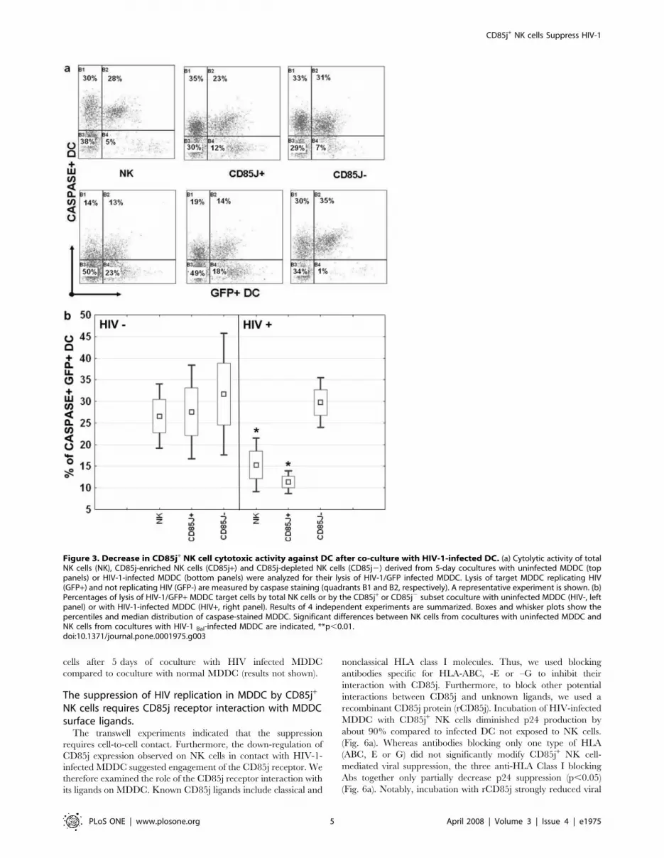
HIV-1 suppressive capacity of CD85j+ NK cell is inducedby infected MDDC despite decreased cytotoxic activity
To evaluate whether the inhibition of HIV-1 replication in infected
DC by CD85j+ NK cells might be related to NK cytolytic activity, we
first analyzed the effect of co-culturing NK cells with DC infected or
not infected by HIV-1 on NK cytolytic activity. The three NK cell
populations showed similar lytic capacities after coculture with
uninfected MDDC (Fig. 3a, top panels and Fig. 3b, left panel). In
contrast, total NK cells and the CD85j+ subset, but not by the CD85j2
subset, showed a significant reduction in their lytic activity after
coculture with HIV-1-infected MDDC in comparison with coculture
with uninfected MDDC (Fig 3a,b). Similar trends were observed for
the lysis of GFP2 and GFP+ MDDC (Fig 3a, B1 and B2 quadrants)
suggesting that HIV replication in MDDC does not modify MDDC
susceptibility to NK cell lysis. The decrease in the cytolytic capacity of
CD85j+ NK cells after coculture with HIV-1-infected MDDC
suggested that the inhibition of HIV-1 replication in MDDC by this
NK cell subset was not mediated by cytoxicity. However, we could not
exclude the possibility that CD85j+ NK cells might have ‘‘exhausted’’
their cytotoxic activity during coculture with infected MDDC and
then lost their suppressive activity. To specifically address this point,
we examined whether the anti-HIV-1 activity of CD85j+ NK cells was
maintained after coculture with HIV-1–infected MDDC, despite the
decrease in their cytolytic capacity. Total NK cells and CD85j2 NK
cells were studied in parallel. After 5 days of coculture with HIV-1Bal-
infected MDDC, each subset of NK cells was incubated with MDDC
previously infected with HIV-1/GFP and the GFP expression in
MDDC was monitored. HIV-1/GFP was used in these experiments
despite not expressing the Nef protein that affects the surface
expression of HLA class I molecules (ligands for CD85j and other NK
cell receptors), since the levels of HIV-1 suppression by CD85j NK
cells in DC infected with HIV-1/GFP or with the HIV-1Bal strain that
express Nef were equivalent (compare Fig 4a with Fig. 2b). The
CD85j+ NK cell subset was still able to suppress HIV-1 replication,
inducing levels of inhibition similar to those achieved when added to
HIV-1-infected DC immediately after infection (Fig. 4a,b). The
percentage of GFP+ MDDC in cocultures with CD85j+ NK cells was
significantly reduced throughout the study period in both cases, in
comparison with HIV-1/GFP-infected MDDC cultured without of
NK cells (Fig. 4a,b). As already observed in previous experiments,
CD85j2 NK cells only partially controlled HIV-1/GFP replication,
while total NK cells induced an intermediate level of control
(Fig. 4a,b). Interestingly, only a not stastistically significant decrease
CD85j+ NK cells Suppress HIV-1
PLoS ONE | www.plosone.org 2 April 2008 | Volume 3 | Issue 4 | e1975

of GFP+ MDDC was induced by both CD85j+ and CD85j2 NK cells
from cocultures with uninfected MDDC (Fig. 4c), despite both subsets
maintained cytolytic capacity (see Fig. 3b). These results confirmed
that the HIV-suppressive capacity of CD85j+ NK cells is induced by
HIV-1-infected MDDC, and that it cannot be explained by their
cytolytic capacity.
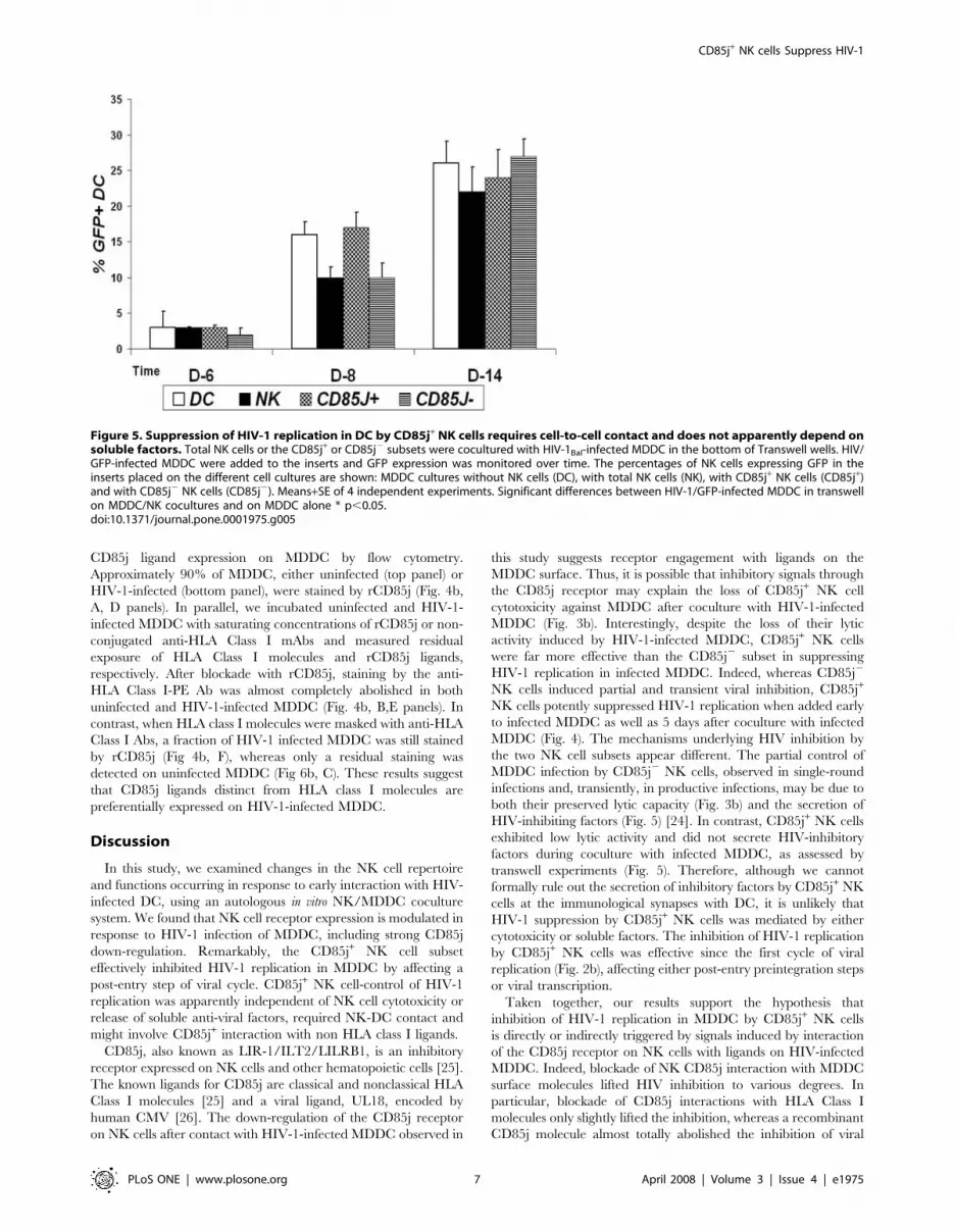
Control of HIV replication in DC by CD85j+ NK cellsrequires NK-DC contact and does not apparently dependon soluble factors
Activated NK cells secrete molecules that can control HIV
infection, such as b-chemokines and other inhibitory factors [24].
We then used transwell experiments to determine whether soluble
inhibitory factors, such as b-chemokines, secreted by NK cells in
coculture with HIV-1-infected MDDC, were involved in the
control of HIV infection, especially by the CD85j+ subset. Total
NK cells and NK cell subsets were incubated with HIV-1Bal-
infected DC or with uninfected DC (not shown), and HIV-1/GFP-
infected DC were then added, as target cells, to transwell inserts
that were placed in the NK-DC coculture-containing wells. A
transient decrease in the percentage of GFP+ MDDC on day 8 was
observed in the inserts placed on total NK cells or CD85j2 NK
cells cocultures with HIV-1-infected MDDC (Fig. 5). This weak
and transient inhibition of HIV-1/GFP replication may be due to
Figure 1. Changes in NK cell receptor expression after coculture with HIV-1-infected MDDC. (a) Dual-label flow cytometric analysis of NKcell proliferation (decrease in CFSE fluorescence) and NKR expression gated on CD16/CD56+CD32 cells. NK cells were stained after 5 days of coculturewith uninfected (top) or HIV-1-infected (bottom) MDDC. A representative experiment showing the expression of three NK cell receptors differentlymodulated by HIV-1-infected MDDC, CD85j (down-regulated), CD161 (up-regulated) and NKp30 (not significantly changed). (b) Overall modificationsin the percentage of NKR expression on NK cells after coculture with HIV-1-infected MDDC. Results are expressed as the percentage decrease(negative values) or increase (positive values) in NK cells expressing the different receptors after coculture with HIV-infected MDDC in comparison tococulture with uninfected MDDC. Means+SE of 9 independent experiments. * p,0.05, **p,0.01doi:10.1371/journal.pone.0001975.g001
CD85j+ NK cells Suppress HIV-1
PLoS ONE | www.plosone.org 3 April 2008 | Volume 3 | Issue 4 | e1975

the secretion of HIV-inhibiting molecules by NK cells upon
contact with infected MDDC. In contrast, CD85j+ NK cells
cocultures with HIV-1Bal-infected DC did not affect HIV-1/GFP
infection of DC in the inserts (Fig. 5). These results indicate that
that the suppression of HIV-1 replication by CD85j+ NK cells in
MDDC requires NK-MDDC contact but it is unlikely due to
soluble factors. Accordingly, intracellular b-chemokine staining
did not show differences in the percentages of stained CD85j+ NK
Figure 2. Suppression of HIV-1 replication in infected MDDC by the CD85j+ NK cell subset. (a) NKR expression on purified CD85j+ andCD85j2 NN cell subsets. Results are expressed as percentage of receptor expression in the given subset+SE. (b) MDDC were infected with HIV-1Bal andthen cultured alone (DC) or incubated with total NK cells (NK) or with the CD85j+ or CD85j2 NK cell subsets. HIV-1 replication was measured by p24production at the indicated time points. Mean+SE of p24 levels in 8 independent experiments using cells from different donors. p24 levels in NK/MDDC cocultures were compared to those in MDDC cultures * p,0.02, **p,0.001. (b) MDDC were one-round infected with HIV-1/BaL pseudotypesand then cultured alone (DC) or incubated with total NK cells (NK) or with the CD85j+ or CD85j2 NK cell subsets. Ninety-six hours later, cells werelysed and luciferase activities were measured. Results are expressed as means6SE of luciferase activity of 4 independent experiments. CD85j+ NK cellsinduced higher levels of inhibition than CD85j2 NK cells. * p,0.05doi:10.1371/journal.pone.0001975.g002
CD85j+ NK cells Suppress HIV-1
PLoS ONE | www.plosone.org 4 April 2008 | Volume 3 | Issue 4 | e1975
cells after 5 days of coculture with HIV infected MDDC
compared to coculture with normal MDDC (results not shown).
The suppression of HIV replication in MDDC by CD85j+
NK cells requires CD85j receptor interaction with MDDCsurface ligands.
The transwell experiments indicated that the suppression
requires cell-to-cell contact. Furthermore, the down-regulation of
CD85j expression observed on NK cells in contact with HIV-1-
infected MDDC suggested engagement of the CD85j receptor. We
therefore examined the role of the CD85j receptor interaction with
its ligands on MDDC. Known CD85j ligands include classical and
nonclassical HLA class I molecules. Thus, we used blocking
antibodies specific for HLA-ABC, -E or –G to inhibit their
interaction with CD85j. Furthermore, to block other potential
interactions between CD85j and unknown ligands, we used a
recombinant CD85j protein (rCD85j). Incubation of HIV-infected
MDDC with CD85j+ NK cells diminished p24 production by
about 90% compared to infected DC not exposed to NK cells.
(Fig. 6a). Whereas antibodies blocking only one type of HLA
(ABC, E or G) did not significantly modify CD85j+ NK cell-
mediated viral suppression, the three anti-HLA Class I blocking
Abs together only partially decrease p24 suppression (p,0.05)
(Fig. 6a). Notably, incubation with rCD85j strongly reduced viral
Figure 3. Decrease in CD85j+ NK cell cytotoxic activity against DC after co-culture with HIV-1-infected DC. (a) Cytolytic activity of totalNK cells (NK), CD85j-enriched NK cells (CD85j+) and CD85j-depleted NK cells (CD85j2) derived from 5-day cocultures with uninfected MDDC (toppanels) or HIV-1-infected MDDC (bottom panels) were analyzed for their lysis of HIV-1/GFP infected MDDC. Lysis of target MDDC replicating HIV(GFP+) and not replicating HIV (GFP-) are measured by caspase staining (quadrants B1 and B2, respectively). A representative experiment is shown. (b)Percentages of lysis of HIV-1/GFP+ MDDC target cells by total NK cells or by the CD85j+ or CD85j2 subset coculture with uninfected MDDC (HIV-, leftpanel) or with HIV-1-infected MDDC (HIV+, right panel). Results of 4 independent experiments are summarized. Boxes and whisker plots show thepercentiles and median distribution of caspase-stained MDDC. Significant differences between NK cells from cocultures with uninfected MDDC andNK cells from cocultures with HIV-1 Bal-infected MDDC are indicated, **p,0.01.doi:10.1371/journal.pone.0001975.g003
CD85j+ NK cells Suppress HIV-1
PLoS ONE | www.plosone.org 5 April 2008 | Volume 3 | Issue 4 | e1975

suppression (p,0.001). Viral suppression was further reduced by
simultaneous incubation of HIV-infected MDDC with both the
three anti-HLA class I antibodies and rCD85j (Fig. 6a). The
removal of HIV-1 inhibition by rCD85j was not attributable to
loss of NK cells by cytotoxicity, since NK cell number was
comparable in NK-MDDC cocultures with or without rCD85j
(data not shown). These results suggest that the control of HIV
infection by the CD85j+ NK subset requires CD85j interaction
with ligands expressed on infected MDDC, which may include,
but are not limited to HLA Class I molecules. We then measured
Figure 4. Maintenance of CD85j+ NK cell suppression of HIV-1 replication in DC despite the loss of cytotoxic activity after co-culturewith HIV-1-infected DC. (a) NK cells (NK) or the CD85j+ or CD85j2 NK cell subsets were added to HIV-1/GFP-infected MDDC 2 hours after infection.(b, c) NK cells or the CD85j+ or CD85j2 NK cell subsets derived from 5-day cocultures with HIV-1Bal-infected MDDC (b) or uninfected MDDC (c) wereadded to HIV-1/GFP-infected MDDC. Kinetics of HIV-1/GFP replication are expressed as percentages of MDDC cells expressing GFP (means+SE) at theindicated time points in eight independent experiments (a) or three independent experiments (b,c) Significant differences between HIV-1/GFP-infected MDDC cultures without NK cells (DC) and MDDC/NK cocultures on day 14 are indicated * p,0.05, **p,0.001.doi:10.1371/journal.pone.0001975.g004
CD85j+ NK cells Suppress HIV-1
PLoS ONE | www.plosone.org 6 April 2008 | Volume 3 | Issue 4 | e1975
CD85j ligand expression on MDDC by flow cytometry.
Approximately 90% of MDDC, either uninfected (top panel) or
HIV-1-infected (bottom panel), were stained by rCD85j (Fig. 4b,
A, D panels). In parallel, we incubated uninfected and HIV-1-
infected MDDC with saturating concentrations of rCD85j or non-
conjugated anti-HLA Class I mAbs and measured residual
exposure of HLA Class I molecules and rCD85j ligands,
respectively. After blockade with rCD85j, staining by the anti-
HLA Class I-PE Ab was almost completely abolished in both
uninfected and HIV-1-infected MDDC (Fig. 4b, B,E panels). In
contrast, when HLA class I molecules were masked with anti-HLA
Class I Abs, a fraction of HIV-1 infected MDDC was still stained
by rCD85j (Fig 4b, F), whereas only a residual staining was
detected on uninfected MDDC (Fig 6b, C). These results suggest
that CD85j ligands distinct from HLA class I molecules are
preferentially expressed on HIV-1-infected MDDC.
Discussion
In this study, we examined changes in the NK cell repertoire
and functions occurring in response to early interaction with HIV-
infected DC, using an autologous in vitro NK/MDDC coculture
system. We found that NK cell receptor expression is modulated in
response to HIV-1 infection of MDDC, including strong CD85j
down-regulation. Remarkably, the CD85j+ NK cell subset
effectively inhibited HIV-1 replication in MDDC by affecting a
post-entry step of viral cycle. CD85j+ NK cell-control of HIV-1
replication was apparently independent of NK cell cytotoxicity or
release of soluble anti-viral factors, required NK-DC contact and
might involve CD85j+ interaction with non HLA class I ligands.
CD85j, also known as LIR-1/ILT2/LILRB1, is an inhibitory
receptor expressed on NK cells and other hematopoietic cells [25].
The known ligands for CD85j are classical and nonclassical HLA
Class I molecules [25] and a viral ligand, UL18, encoded by
human CMV [26]. The down-regulation of the CD85j receptor
on NK cells after contact with HIV-1-infected MDDC observed in
this study suggests receptor engagement with ligands on the
MDDC surface. Thus, it is possible that inhibitory signals through
the CD85j receptor may explain the loss of CD85j+ NK cell
cytotoxicity against MDDC after coculture with HIV-1-infected
MDDC (Fig. 3b). Interestingly, despite the loss of their lytic
activity induced by HIV-1-infected MDDC, CD85j+ NK cells
were far more effective than the CD85j2 subset in suppressing
HIV-1 replication in infected MDDC. Indeed, whereas CD85j2
NK cells induced partial and transient viral inhibition, CD85j+
NK cells potently suppressed HIV-1 replication when added early
to infected MDDC as well as 5 days after coculture with infected
MDDC (Fig. 4). The mechanisms underlying HIV inhibition by
the two NK cell subsets appear different. The partial control of
MDDC infection by CD85j2 NK cells, observed in single-round
infections and, transiently, in productive infections, may be due to
both their preserved lytic capacity (Fig. 3b) and the secretion of
HIV-inhibiting factors (Fig. 5) [24]. In contrast, CD85j+ NK cells
exhibited low lytic activity and did not secrete HIV-inhibitory
factors during coculture with infected MDDC, as assessed by
transwell experiments (Fig. 5). Therefore, although we cannot
formally rule out the secretion of inhibitory factors by CD85j+ NK
cells at the immunological synapses with DC, it is unlikely that
HIV-1 suppression by CD85j+ NK cells was mediated by either
cytotoxicity or soluble factors. The inhibition of HIV-1 replication
by CD85j+ NK cells was effective since the first cycle of viral
replication (Fig. 2b), affecting either post-entry preintegration steps
or viral transcription.
Taken together, our results support the hypothesis that
inhibition of HIV-1 replication in MDDC by CD85j+ NK cells
is directly or indirectly triggered by signals induced by interaction
of the CD85j receptor on NK cells with ligands on HIV-infected
MDDC. Indeed, blockade of NK CD85j interaction with MDDC
surface molecules lifted HIV inhibition to various degrees. In
particular, blockade of CD85j interactions with HLA Class I
molecules only slightly lifted the inhibition, whereas a recombinant
CD85j molecule almost totally abolished the inhibition of viral
Figure 5. Suppression of HIV-1 replication in DC by CD85j+ NK cells requires cell-to-cell contact and does not apparently depend onsoluble factors. Total NK cells or the CD85j+ or CD85j2 subsets were cocultured with HIV-1Bal-infected MDDC in the bottom of Transwell wells. HIV/GFP-infected MDDC were added to the inserts and GFP expression was monitored over time. The percentages of NK cells expressing GFP in theinserts placed on the different cell cultures are shown: MDDC cultures without NK cells (DC), with total NK cells (NK), with CD85j+ NK cells (CD85j+)and with CD85j2 NK cells (CD85j2). Means+SE of 4 independent experiments. Significant differences between HIV-1/GFP-infected MDDC in transwellon MDDC/NK cocultures and on MDDC alone * p,0.05.doi:10.1371/journal.pone.0001975.g005
CD85j+ NK cells Suppress HIV-1
PLoS ONE | www.plosone.org 7 April 2008 | Volume 3 | Issue 4 | e1975

replication in MDDC. Inhibition of other NK receptors
recognizing HLA class I molecules might also contribute to the
partial removal of HIV-1 suppression by the anti-HLA Abs.
Conversely, removal of CD85j+ NK cell-mediated viral inhibition
by rCD85j points to the existence of a CD85j ligand other than
HLA Class I A, B, C, E and G molecules, and suggests that NK
cells use this ligand to inhibit HIV replication in MDDC. The
results of competition experiments with anti-HLA class I Abs and
rCD85j support this hypothesis. Indeed, a significant binding of
CD85j was detected on HIV-1 infected MDDC after masking
HLA class I molecules. It is tempting to speculate that the MDDC
non-HLA class I ligand that mediates NK cell inhibition of HIV
replication in HIV-1-infected MDDC might be modulated by
HIV-1 or related to HIV-1 proteins. Further work is needed to
Figure 6. Involvement of NK CD85j receptor interaction with CD85j/ligands, including non-HLA class I ligands, in the control of HIV-1 replication in MDDC. (a) MDDC were infected with HIVBal and incubated with CD85j+ NK cells after blocking CD85j receptor interactions with HLAclass I molecules on the MDDC by treatment with Mabs to HLA class I ABC (HLA-ABC), E (HLA-E) or G (HLA-G) or by incubating MDDC with arecombinant CD85j protein (rCD85j). Blocking reagents were also added during coculture. MDDC infection was evaluated by measuring p24 in thesupernatants 10 days later. The results are expressed as the percentage suppression of p24 (mean+SE) in comparison with HIVBal-infected MDDC notexposed to NK cells in 8 independent experiments. Significant differences in the levels of p24 suppression after incubation with the three anti-HLAclass I Abs, with rCD85j or with rCD8j more the three anti-HLA class I Abs in comparison to CD85j+ NK cells cultured with infected DC without Abs areindicated by asterisks: * p,0.05, **p,0.001 (b) Flow cytometric analysis of rCD85j binding to uninfected MDDC (top panels) or to HIVBal-infectedMDDC (bottom panels). MDDC staining with rCD85j as revealed by anti-hIgG-PE Abs (A and D). MDDC staining with a mix of anti-HLA Class I-PE mAbsafter incubation with rCD85j (B and E), or with rCD85j and anti-hIgG-PE Abs after incubation with a mix of anti-HLA Class I MAbs. A representativeexperiment of three is shown. Without rCD85j blockade, nearly all MDDC were stained by anti-HLA-PE (not shown). The concentrations of anti-HLAclass I Abs used in blocking experiments (10 mg/ml of each anti-HLA class I A,B,C, E and G) totally prevented the detection of HLA class I molecules onMDDC by using labelled anti-HLA class I Abs (not shown). Percentages of positive cells and MFI (italic) are shown. Range of positive rCD85j cells wasbetween 8.2 and 21.9.doi:10.1371/journal.pone.0001975.g006
CD85j+ NK cells Suppress HIV-1
PLoS ONE | www.plosone.org 8 April 2008 | Volume 3 | Issue 4 | e1975

identify the non-HLA class I ligand(s) recognized by the CD85j
receptor on the surface of HIV-infected MDDC.
An increased expression of CD85j on NK cells has been
reported in both viremic and long term nonprogressor HIV-1-
infected patients [14,27]. While the significance of this observation
is unclear, the role of the CD85j receptor in HIV-1 infection has
only been analyzed and discussed with respect to its inhibitory
activity on NK cytolytic activity (usually measured on a target cell
line) [14,27]. In this paper, we show evidence that CD85j+ NK
cells control HIV-1 replication in HIV-1-infected MDDC.
Recently, KIR3DS1+ NK cells have been shown to inhibit HIV-
1 replication in vitro in CD4 T cells [20]. It will be interesting to
assess whether CD85j+ NK cells can also control HIV-1
replication in CD4 T cells and macrophages.
Should the interaction between CD85j+ NK cells and HIV-1-
infected DC prove to contribute to host defences against HIV-1
infection, it is probable that it would intervene early, when NK cell
activity is increased [12] and the virus has not yet disseminated.
Indeed, the suppression of HIV-1 replication by CD85j+ NK cells
in the first viral cycle, before viral spread in DC cultures, suggests
that this mechanism may contribute to the early containment of
the infection. Unfortunately, data on CD85j expression by NK
during early HIV infection are lacking. Ex vivo studies of acutely
infected individuals and/or simian models should provide new
insights into the role of NK cells in early innate defences against
HIV infection.
Materials And Methods
Generation of MDDCPeripheral blood mononuclear cells (PBMC) were prepared
from blood of healthy donors by Ficoll-Paque density gradient
centrifugation. PBMC were obtained through the French blood
bank (Etablissement Francais du Sang) in the setting of EFS-
Institut Pasteur Convention. A written agreement was obtained for
each donor to use the cells for clinical research according to
French laws. Our study was approved by IRB, external
(Etablissement Francais du Sang Board) as required by French
law and internal (Biomedical Research Committee Board, Institut
Pasteur) as required by Pasteur Institute. CD14+ PBMC were
isolated with anti-CD14 MicroBeads, MS+/RS+ columns and a
MiniMACS separator. Monocyte-derived DC were generated
from CD14+ PBMC (106/ml) by seeding in 6-well plates in
DMEM medium plus 10% FCS; rhIL-4 and rhGM-CSF were
added at final concentrations of 500 and 1000 U/ml, respectively,
on days 0, 2 and 4. On day 5, floating immature DC (iDC) were
transferred to new plates at a concentration of 36105 cells/ml.
Phenotype of iDC was verified by CD80, CD83, HLA DR and
HLA Class I expression and the absence of CD14. Maturation
capacity of iDC was assessed upon LPS stimulation.
HIV-1 infection of MDDCiDC were infected either with 1021 m.o.i of the R5 HIV-1Bal
strain or of NL.4/nef-/Bal env/GFP HIV-1, containing the green
fluorescent protein (GFP) gene in the place of nef (a gift from P.
Ancuta [28]), referred to hereafter as HIV-1/GFP. For single
round infections, Bal Env pseudotyped NL-Luc-E-R+ HIV-1
particles were produced and titrated as described [29] and then
used at 400 ng p24/ml. After 96 h, cells were lysed with a
luciferase lysis buffer (Promega, Paris, France) and the luciferase
activity was measured with a Luminometer (Glomax, Turener
Biosystem, Promega, Paris, France). All infections were performed
by spinoculation (1 h at 800 g followed by 1 h incubation at 37uC).
Isolation of NK cellsNK cells were negatively selected by using the NK cell isolation
kit MicroBeads, MS+/RS+ columns and a MiniMACS separator.
The percentage of CD3-CD56+ NK cells in the isolated
population, evaluated by flow cytometry, was.95%. NK cells
were frozen in 90% fetal calf serum, 10% DMSO, until cocultured
with autologous MDDC. We have previously established that NK
cell activity was not affected by freezing [21,22], To purify CD85j
NK cell subsets, NK cells were incubated with an antiCD85j MAb
(Beckman Coulter, Paris, France) followed by anti-mouse IgG
microbeads. The proportion of NK cells expressing the CD85j
receptor before enrichment was 29616%. Purified preparations
contained more than 90% of CD85j+ cells.
NK/MDDC cocultureNK/MDDC were cocultured in DMEM+10% FCS in 24-well
plates at a ratio of 1:5. An NK-DC ratio of 1:5 that induced
optimal activation and proliferation of NK cells in preliminary
studies was chosen for experiments. After 5 days of culture with
HIV-infected or uninfected MDDC, NK cells were collected and
analysed. Cell viability during cocultures was measured by
propidium iodine (PI) staining: percentages of PI stained MDDC
at day 14 of coculture were 25%611 with total NK cells or
CD85j2 NK cells and 1366 with CD85J+ NK cells. PI staining of
NK cells was always below 5%. In Transwell experiments, NK
cells were cocultured with uninfected or HIV-1Bal-infected MDDC
in the bottom of the wells. HIV/GFP-infected MDDC were added
to the inserts and GFP expression was recorded over time.
Proliferation and phenotypic analysisNK cells (56104) were incubated with MDDC (2.56105) for
5 days in DMEM plus 10% FCS in 96-well round-bottom
microtiter plates. NK were loaded with 5 mM CFSE before
culture and proliferation was analyzed by measuring the decrease
in fluorescence intensity, simultaneously with NK receptor
expression. For the detection of NK cell receptors by flow
cytometry, MAbs from Beckman Coulter and RD Systems were
used (Paris, France).
Analysis of NK cell cytolytic activity against HIV-infectedMDDC
This study was based on immunostaining of NK cells (CD3-
CD8+/-CD16+CD56+) and detection of target MDDC cell lysis
by caspase flow cytometry (CyToxiLux kit, Oncoimmunin Inc,
USA). Specific lysis of HIV-infected MDDC populations was
evaluated by combining caspase detection with MDDC infection
by an HIV-1/GFP virus. Autologous MDDC were infected with
HIV1/GFP seven days before the test, then surface-stained
(following the manufacturer’s instructions) and placed in contact
with NK cells from 5-day MDDC cocultures for one hour in the
presence of a fluorogenic caspase substrate. After incubation, lysis
was analysed by flow cytometry gating on target cells (surface
staining and GFP expression). Measures of NK cytolytic activity by
caspase assay and the chromium release technique showed good
correlation (r = 0.89)
Blocking experiments. Blocking experiments were
performed with optimal concentrations of either anti-HLA class
I antibodies or recombinant CD85j protein. MDDC were
incubated with 10 mg/ml anti-HLA-class I mAb W6/32
(Serotec, Paris, France), or anti-HLA-class G mAb (Serotec,
Paris, France), or anti-HLA-class E Mab (Serotec, Paris, France) or
a mix of the three mAbs, or with a recombinant Fc-CD85j
(rCD85j) fusion protein (RD System, Paris, France) at 20 mg/ml
CD85j+ NK cells Suppress HIV-1
PLoS ONE | www.plosone.org 9 April 2008 | Volume 3 | Issue 4 | e1975

for 30 min at room temperature before adding NK cells.
Antibodies or rCD85j were added to the cocultures every two
days.
Statistical analyses. All experiments were done in
independent triplicates or quadruplicates. Data were analyzed
with nonparametric (Mann-Whitney test) and parametric tests
(Student’s two-sided paired t-test). The results are expressed as
means6SE.
Acknowledgments
We thank all the donors for their participation in this study. We thank Dr.
S. Kottilil for comments on the manuscript.
Author Contributions
Conceived and designed the experiments: FB DS GP. Performed the
experiments: DS VA CD TK GP. Analyzed the data: DS VA CD GP.
Wrote the paper: FB DS VA GP.
References
1. Bottino C, Moretta L, Moretta A (2006) NK cell activating receptors and tumorrecognition in humans. Curr Top Microbiol Immunol 298: 175–182.
2. Bryceson YT, March ME, Ljunggren HG, Long EO (2006) Activation,
coactivation, and costimulation of resting human natural killer cells. ImmunolRev 214: 73–91.
3. Moretta A, Marcenaro E, Sivori S, Della Chiesa M, Vitale M, et al. (2005) Earlyliaisons between cells of the innate immune system in inflamed peripheral tissues.
Trends Immunol 26: 668–675.
4. Boyton RJ, Altmann DM (2007) Natural killer cells, killer immunoglobulin-likereceptors and human leucocyte antigen class I in disease. Clin Exp Immunol
149: 1–8.5. Moretta L, Ferlazzo G, Bottino C, Vitale M, Pende D, et al. (2006) Effector and
regulatory events during natural killer-dendritic cell interactions. Immunol Rev214: 219–228.
6. Marcenaro E, Dondero A, Moretta A (2006) Multi-directional cross-regulation
of NK cell function during innate immune responses. Transpl Immunol 17:16–19.
7. Alter G, Altfeld M (2006) NK cell function in HIV-1 infection. Curr Mol Med 6:621–629.
8. Fortis C, Tasca S, Capiluppi B, Tambussi G (2002) Natural killer cell function in
HIV-1 infected patients. J Biol Regul Homeost Agents 16: 30–32.9. Mavilio D, Lombardo G, Kinter A, Fogli M, La Sala A, et al. (2006)
Characterization of the defective interaction between a subset of natural killercells and dendritic cells in HIV-1 infection. J Exp Med 203: 2339–2350.
10. Scott-Algara D, Vuillier F, Cayota A, Dighiero G (1992) Natural killer (NK) cellactivity during HIV infection: a decrease in NK activity is observed at the clonal
level and is not restored after in vitro long-term culture of NK cells. Clin Exp
Immunol 90: 181–187.11. Tasca S, Tambussi G, Nozza S, Capiluppi B, Zocchi MR, et al. (2003) Escape of
monocyte-derived dendritic cells of HIV-1 infected individuals from naturalkiller cell-mediated lysis. Aids 17: 2291–2298.
12. Alter G, Teigen N, Ahern R, Streeck H, Meier A, et al. (2007) Evolution of
innate and adaptive effector cell functions during acute HIV-1 infection. J InfectDis 195: 1452–1460.
13. Alter G, Teigen N, Davis BT, Addo MM, Suscovich TJ, et al. (2005) Sequentialderegulation of NK cell subset distribution and function starting in acute HIV-1
infection. Blood 106: 3366–3369.14. Mavilio D, Lombardo G, Benjamin J, Kim D, Follman D, et al. (2005)
Characterization of CD56-/CD16+ natural killer (NK) cells: a highly
dysfunctional NK subset expanded in HIV-infected viremic individuals. ProcNatl Acad Sci U S A 102: 2886–2891.
15. Kottilil S, Chun TW, Moir S, Liu S, McLaughlin M, et al. (2003) Innateimmunity in human immunodeficiency virus infection: effect of viremia on
natural killer cell function. J Infect Dis 187: 1038–1045.
16. Kottilil S, Shin K, Planta M, McLaughlin M, Hallahan CW, et al. (2004)Expression of chemokine and inhibitory receptors on natural killer cells: effect of
immune activation and HIV viremia. J Infect Dis 189: 1193–1198.
17. Mavilio D, Benjamin J, Daucher M, Lombardo G, Kottilil S, et al. (2003)Natural killer cells in HIV-1 infection: dichotomous effects of viremia on
inhibitory and activating receptors and their functional correlates. Proc NatlAcad Sci U S A 100: 15011–15016.
18. Fauci AS, Mavilio D, Kottilil S (2005) NK cells in HIV infection: paradigm for
protection or targets for ambush. Nat Rev Immunol 5: 835–843.19. Martin MP, Qi Y, Gao X, Yamada E, Martin JN, et al. (2007) Innate
partnership of HLA-B and KIR3DL1 subtypes against HIV-1. Nat Genet 39:733–740.
20. Alter G, Martin MP, Teigen N, Carr WH, Suscovich TJ, et al. (2007)Differential natural killer cell-mediated inhibition of HIV-1 replication based on
distinct KIR/HLA subtypes. J Exp Med 204: 3027–3036.
21. Scott-Algara D, Truong LX, Versmisse P, David A, Luong TT, et al. (2003)Cutting edge: increased NK cell activity in HIV-1-exposed but uninfected
Vietnamese intravascular drug users. J Immunol 171: 5663–5667.22. Ravet S, Scott-Algara D, Bonnet E, Tran HK, Tran T, et al. (2007) Distinctive
NK-cell receptor repertoires sustain high-level constitutive NK-cell activation in
HIV-exposed uninfected individuals. Blood 109: 4296–4305.23. Donaghy H, Wilkinson J, Cunningham AL (2006) HIV interactions with
dendritic cells: has our focus been too narrow? J Leukoc Biol 80: 1001–1012.24. Oliva A, Kinter AL, Vaccarezza M, Rubbert A, Catanzaro A, et al. (1998)
Natural killer cells from human immunodeficiency virus (HIV)-infectedindividuals are an important source of CC-chemokines and suppress HIV-1
entry and replication in vitro. J Clin Invest 102: 223–231.
25. Lopez-Botet M, Angulo A, Guma M (2004) Natural killer cell receptors formajor histocompatibility complex class I and related molecules in cytomegalo-
virus infection. Tissue Antigens 63: 195–203.26. Vales-Gomez M, Shiroishi M, Maenaka K, Reyburn HT (2005) Genetic
variability of the major histocompatibility complex class I homologue encoded
by human cytomegalovirus leads to differential binding to the inhibitory receptorILT2. J Virol 79: 2251–2260.
27. O’Connor GM, Holmes A, Mulcahy F, Gardiner CM (2007) Natural Killer cellsfrom long-term non-progressor HIV patients are characterized by altered
phenotype and function. Clin Immunol 124: 277–283.28. Ancuta P, Kunstman KJ, Autissier P, Zaman T, Stone D, et al. (2006) CD16+
monocytes exposed to HIV promote highly efficient viral replication upon
differentiation into macrophages and interaction with T cells. Virology 344:267–276.
29. Perez-Bercoff D, David A, Sudry H, Barre-Sinoussi F, Pancino G (2003)Fcgamma receptor-mediated suppression of human immunodeficiency virus
type 1 replication in primary human macrophages. J Virol 77: 4081–4094.
CD85j+ NK cells Suppress HIV-1
PLoS ONE | www.plosone.org 10 April 2008 | Volume 3 | Issue 4 | e1975
Copyright © 2022 FDOKUMEN