Function of c-mos proto-oncogene product in meiotic maturation in Xenopus oocytes
Upload
mkuniversityCategory
view
2download
0
Article
Meiotic recombination generates rich diversity in NKcell receptor genes, alleles, and haplotypesPaul J. Norman,1 Laurent Abi-Rached,1 Ketevan Gendzekhadze,1 John A. Hammond,1
Achim K. Moesta,1 Deepti Sharma,1 Thorsten Graef,1 Karina L. McQueen,1
Lisbeth A. Guethlein,1 Christine V.F. Carrington,2 Dasdayanee Chandanayingyong,3
Yih-Hsin Chang,4 Catalina Crespı,5 Guher Saruhan-Direskeneli,6 Kamran Hameed,7
Giorgi Kamkamidze,8 Kwadwo A. Koram,9 Zulay Layrisse,10 Nuria Matamoros,5
Joan Mila,5 Myoung Hee Park,11 Ramasamy M. Pitchappan,12 D. Dan Ramdath,2
Ming-Yuh Shiau,13 Henry A.F. Stephens,14 Siske Struik,15 Dolly Tyan,16
David H. Verity,17 Robert W. Vaughan,18 Ronald W. Davis,19 Patricia A. Fraser,20
Eleanor M. Riley,15 Mostafa Ronaghi,19 and Peter Parham1,21
1–20[A complete list of author affiliations appears at the end of the paper before the Acknowledgments section.]
Natural killer (NK) cells contribute to the essential functions of innate immunity and reproduction. Various genes encodeNK cell receptors that recognize the major histocompatibility complex (MHC) Class I molecules expressed by other cells.For primate NK cells, the killer-cell immunoglobulin-like receptors (KIR) are a variable and rapidly evolving family ofMHC Class I receptors. Studied here is KIR3DL1/S1, which encodes receptors for highly polymorphic human HLA-A and -Band comprises three ancient allelic lineages that have been preserved by balancing selection throughout human evolution.While the 3DS1 lineage of activating receptors has been conserved, the two 3DL1 lineages of inhibitory receptors werediversified through inter-lineage recombination with each other and with 3DS1. Prominent targets for recombination wereD0-domain polymorphisms, which modulate enhancer function, and dimorphism at position 283 in the D2 domain, whichinfluences inhibitory function. In African populations, unequal crossing over between the 3DL1 and 3DL2 genes produceda deleted KIR haplotype in which the telomeric ‘‘half’’ was reduced to a single fusion gene with functional propertiesdistinct from its 3DL1 and 3DL2 parents. Conversely, in Eurasian populations, duplication of the KIR3DL1/S1 locus byunequal crossing over has enabled individuals to carry and express alleles of all three KIR3DL1/S1 lineages. These resultsdemonstrate how meiotic recombination combines with an ancient, preserved diversity to create new KIR phenotypesupon which natural selection acts. A consequence of such recombination is to blur the distinction between alleles and lociin the rapidly evolving human KIR gene family.
[Supplemental material is available online at www.genome.org. The sequence data from this study have been submitted toGenBank (http://www.ncbi.nlm.nih.gov/Genbank/) under accession nos. EF582383, EU267269–71, FJ158650–60, andFJ459734.]
Among the most polymorphic and structurally diverse human loci
are genes related to immune function (Redon et al. 2006; Frazer
et al. 2007; Korbel et al. 2007). A principle example is the KIR locus,
which displays both polymorphic and structural diversity
throughout all human populations (Parham 2005; Bashirova et al.
2006). The protein products, the killer cell immunoglobulin-like
receptors (KIR), recognize determinants of conserved and poly-
morphic major histocompatibility complex (MHC) Class I mole-
cules (Boyington et al. 2001). Interaction of KIR on immune-
system cells with MHC Class I on other cell types allows the health
of tissues to be monitored and responded to when compromised
by infection or malignant transformation. In the human MHC,
the HLA complex, each of the highly polymorphic Class I gen-
es—HLA-A, HLA-B, and HLA-C—has some alleles that encode KIR
ligands. KIR are principally found on the surface of natural killer
(NK) cells, lymphocytes that function in the early, or innate, im-
mune response to virus infection (Lanier 2008), but they also
contribute to an early stage of reproduction, when they remodel
the maternal blood vessels that will supply the placenta and
nourish the fetus (Moffett and Loke 2006). KIR are also expressed
on some T-lymphocytes, cells that are central to the adaptive im-
mune response to infection (Snyder et al. 2004; Moretta et al.
2006).
In humans and other primates, the KIR are encoded by a di-
verse and rapidly evolving gene family that exhibits considerable
species specificity due to continual gene turnover (Parham 2005;
Bashirova et al. 2006). In contrast, in mice, the widely used animal
model in immunology, the KIR genes are few in number (two) and
do not encode NK cell receptors for MHC Class I, those functions
having been assumed by the independently evolved KLRA1 (also
known as Ly49) receptors (Kelley et al. 2005). This lability and
plasticity in genes encoding NK cell receptors likely reflects
the strengths of the different and sometimes conflicting selec-
tions imposed by the needs of immune defense and placental
21Corresponding author.E-mail [email protected]; fax (650) 723-7456.Article is online at http://www.genome.org/cgi/doi/10.1101/gr.085738.108.
19:757–769 � 2009 by Cold Spring Harbor Laboratory Press; ISSN 1088-9051/09; www.genome.org Genome Research 757www.genome.org

reproduction, but also by the functional and genetic complexity of
matching polymorphic ligands and receptors encoded by un-
linked genes (Parham 2005; Moffett and Loke 2006; Lanier 2008).
The KIR locus is part of the leukocyte receptor complex (LRC)
on human chromosome 19, which comprises several families of
cell-surface receptors expressed by cells of the immune system
(Wilson et al. 2000). The KIR genes are flanked on the centromeric
side by the leukocyte immunoglobulin-like receptor (LILR) gene
family and on the telomeric side by FCAR, the gene encoding the
receptor for immunoglobulin A that is expressed on phagocytic
cells. Human KIR haplotypes vary in gene content, having be-
tween seven and 15 genes (Uhrberg et al. 1997). Each KIR haplo-
type is divided into two parts by three conserved framework
regions. The centromeric part contains KIR2D genes encoding
HLA-C receptors, and the telomeric part contains KIR3D genes
encoding HLA-A and -B receptors (Bashirova et al. 2006). The latter
two genes, comprising KIR3DL1/S1 and KIR3DL2, are the subject
of this study. KIR3DL1/S1 recognizes sequence motifs at residues
77–83 of HLA-A and HLA-B allotypes that form the Bw4 antigen,
or epitope, defined by HLA serology (Cella et al. 1994; Gumperz
et al. 1995; Thananchai et al. 2007), and KIR3DL2 recognizes HLA-
A3 and –A11 (Dohring et al. 1996; Pende et al. 1996).
Like HLA-A and -B, KIR3DL1/S1 is a highly polymorphic
protein with more than 60 allotypes defined (Robinson et al.
2006). The natural variation affects receptor function by altering
the frequency of cellular expression, abundance at the cell surface
(Pando et al. 2003; Thomas et al. 2008), avidity and specificity
for ligand (O’Connor et al. 2007; Thananchai et al. 2007), and
the nature—either inhibitory or activating—of the intracellular
signals generated upon ligand engagement (Carr et al. 2007).
Furthermore, a person’s combination of HLA-A, HLA-B, and
KIR3DL1/S1 allotypes influences the development and function of
the NK cell repertoire (Foley et al. 2008; Yawata et al. 2008), and in
the population, such combinations are associated with disease
susceptibility and progression, notably for HIV infection (Martin
et al. 2007). The foundation for 3DL1/S1 variety is three ancient
lineages of alleles—3DS1 lineage encoding activating receptors
and 3DL1-005 and 015 lineages encoding inhibitory receptors—
maintained by balancing selection for >3 million years and pres-
ent in all modern human populations (Norman et al. 2007). Of the
three lineages, 3DS1 is essentially homogeneous, whereas both
3DL1 lineages have been extensively diversified by point mutation
and recombination. Because recombination with other KIR genes
and between KIR3DL1/S1 lineages has the potential to erode the
lineage distinctions, we examined the impact that meiotic re-
combination has had on the KIR3DL1/S1 locus and on human NK
cell functional diversity.
Results
Generation of KIR3DL1/S1 diversity by intergenicrecombination
In humans, the hominoid KIR lineage II is represented by two
genes: 3DL1/S1 encoding NK cell receptors for the Bw4 epitopes of
HLA-A and HLA-B; and 3DL2 encoding NK-cell receptors specific
for HLA-A*03 and HLA-A*11 (Rajalingam et al. 2004). Not fitting
with this picture is the 3DL1/2v cDNA, which encodes extracel-
lular domains like 3DL1 and intracellular domains like 3DL2
(Shilling et al. 2002). To distinguish if the 3DL1/2v cDNA arises
from transcription of a single gene or the splicing together of
transcripts from both 3DL1 and 3DL2, we analyzed genomic DNA
from three healthy donors having different 3DL1/2v variants and
one 3DL1/2v donor who lacked 3DL1/S1 because of deletion of
this locus from the other KIR haplotype (Norman et al. 2004). The
results unequivocally demonstrated that 3DL1/2v represents
a unique hybrid gene for which exons 1–5 and associated introns
are like 3DL1, and exons 6–9 and associated introns are like 3DL2
(Fig. 1A, upper haplotype). In all four donors, the 3DL1/2v gene
was shown to be flanked by 2DL4 on the upstream (centromeric)
side and by FCAR on the downstream (telomeric) side. This gene
organization is unusual, differing from the more common situa-
tion (Wilson et al. 2000) where 3DL1 is downstream from 2DL4,
3DL2 is upstream of FCAR, and 2DS4 lies between 3DL1 and 3DL2
(Fig. 1A, lower haplotype). These results raised the possibility that
3DL1/2v arose through a non-homologous recombination be-
tween 3DL1 and 3DL2 that deleted the 39 part of 3DL1, the entire
2DS4 gene, and the 59 part of 3DL2. If true, then individuals car-
rying 3DL1/2v should never be heterozygous for exons 1–5 of
3DL2 or exons 6–9 of 3DL1/S1. Population analysis was performed
to test this hypothesis.
KIR3DL1/2v is restricted to sub-Saharan Africans and related
populations such as African-Americans and Afro-Caribbeans, in
which it is present at frequencies of up to 6.5% (Norman et al.
2007). Panels of 65 3DL1/2v+ and 65 3DL1/2v� Africans were
genotyped for heterozygosity in the sequences encoding the ex-
tracellular and intracellular domains of 3DL1 and 3DL2, and also
for 2DL4 as a control. Whereas the 3DL1/2v� Africans exhibited
significant heterozygosity in both the 39 and 59 parts of the 3DL1
and 3DL2 genes, the 3DL1/2v+ Africans lacked heterozygosity in
the 39-exons of 3DL1/S1 and the 59-exons of 3DL2 (Fig. 1B). These
results, showing that 3DL1/2v is not on the same haplotypes as
3DL1 or 3DL2, are consistent with 3DL1/2v having been formed
by a recombination-mediated deletion that fused exons 1–5 of
3DL1 with exons 6–9 of 3DL2. As a consequence, 3DL1/2v has an
allelic relationship with both 3DL1 and 3DL2 (Supplemental Fig.
S1). Because 3DL1/2v is overall more structurally similar to 3DL1
than 3DL2, the three 3DL1/2v variants were assigned names in the
3DL1/S1 series: 3DL1*059, 3DL1*060, and 3DL1*061 (Robinson
et al. 2006). The encoded allotypes, which differ by one or two
amino acid substitutions, have extracellular domains that are part
of the 3DL1-005 lineage, as opposed to the 3DL1-015 lineage (Fig.
1C). That 3DL1*059 is geographically most widespread, being
present in all six African populations studied, compared to only
one and two populations for 3DL1*060 and 3DL1*061, respec-
tively (Fig. 1D), suggests that 3DL1*059 was the progenitor 3DL1/
2v from which 3DL1*060 and 3DL1*061 were independently
derived by single point substitutions. Further supporting this
evolutionary model, the 3DL1*059 sequence corresponds to a fu-
sion of sequences from common 3DL1 (e.g., *00501) and 3DL2
(e.g., *001) alleles, whereas 3DL1*060 and 3DL1*061 are each
distinguished by a unique and different substitution. From the
genomic sequences, the recombination that fused parts of the
3DL1 and 3DL2 genes to form 3DL1/2v can be located to a 30-bp
sequence within intron 5 that is located 356 bp upstream of exon
6, and 2780 bp downstream from exon 5 encoding the D2 domain
(Fig. 1E; Supplemental Fig. S2).
The genomic location of 3DL1/2v between 2DL4 and FCAR is
the same as that observed for Pt-KIR3DL1/2, the single chimpanzee
lineage II KIR gene (Rajalingam et al. 2004; Sambrook et al. 2005),
which has structural and functional properties in common with
both human 3DL1 and 3DL2 (Khakoo et al. 2000). Although these
similarities raised the possibility that 3DL1/2v and Pt-KIR3DL1/2
share a common origin that predated the human–chimpanzee
758 Genome Researchwww.genome.org
Norman et al.
separation, several lines of evidence point to 3DL1/2v having
evolved independently by recombination during human evolu-
tion, subsequent to the split from chimpanzees. First, the in-
clusion of exons 1–5 of 3DL1/2v in the 005 lineage of 3DL1/S1
alleles (Fig. 1C), and not in either the 015 or 3DS1 lineages, indi-
cates that 3DL1/2 was formed after lineage divergence, which
occurred subsequent to the human–
chimpanzee separation (Norman et al.
2007). Thus in phylogenetic analysis,
human 3DL1/2v was never an out-group
to 3DL1 when compared with Pt-
KIR3DL1/2 and other hominoid KIR lin-
eage II genes (Supplemental Fig. S3A).
Second, the 3DL1/2v Ig-domain is esti-
mated to have arisen ;0.74 million years
ago (Mya) (95% CI: 0.27–1.55 Mya)
(Supplemental Fig. S3B), after the three
allelic lineages of 3DL1/S1 split. As the Ig-
domain divergence preceded the geno-
mic deletion event, 3DL1/2v arose at
a time potentially close to the emergence
of modern humans (;200,000 yr ago)
(Relethford 2008). Lastly, our finding
that all three variants of 3DL1/2v
(3DL1*059, 3DL1*060, 3DL1*061) were
flanked by identical sequences and 2DL4
(*00801) and FCAR (*001) alleles, despite
genome-wide lack of LD in sub-Saharan
Africans (Campbell and Tishkoff 2008),
points to a recent origin for 3DL1/2v.
In conclusion, the results are all
consistent with 3DL1/2v having been
formed during a meiotic recombination
in a human ancestor that deleted DNA
from the telomeric region of a KIR hap-
lotype to fuse the 59 part of a 3DL1 allele
with the 39 part of a 3DL2 allele. Although
the size of the deletion is uncertain be-
cause of gene-content variability, the
most likely event would have involved
the A haplotype configuration shown in
Figure 1A with deletion of ;30 kb con-
taining all of 2DS4 and parts of 3DL1 and
3DS1. That the telomeric part of most
chimpanzee KIR haplotypes has similarly,
but independently, evolved to resemble
that of the KIR3DL1/2v-containing hap-
lotypes, points to this simplified structure
having conferred advantage in at least
two different circumstances.
Generation of KIR3DL1/S1 diversityby interlineage recombination
Domain-by-domain phylogenetic analy-
sis of 61 3DL1/S1 alleles shows that
a minimum of 12 (;20%) alleles arose
through intragenic meiotic recombina-
tion (Fig. 2). Most of these events (nine)
involved recombination between two of
the three different 3DL1/S1 lineages:
3DL1-005, 3DL1-015, and 3DS1. The two
regions of the molecule most affected by recombination are the D0
domain (five events) and position 283 (three events) near the
C-terminal end of the D2 domain (Fig. 2A). Both these regions
bear a strong signature of positive selection in human populations
(Norman et al. 2007) and have distinct functions that affect the
strength and specificity of the KIR3DL1/S1 interaction with MHC
Figure 1. The 3DL1/2v fusion gene is allelic to 3DL1/S1 and 3DL2. (A, top) Schematic of the four3DL1/2v haplotypes that were sequenced here (Supplemental Fig. S2). (Bottom) A KIR A haplotype;(dashed lines) the genomic segment absent from 3DL1/2v haplotypes; (yellow) 3DL1; (blue) 3DL2.Exons 1–5 encode the leader peptide and Ig domains (Ig), exon 6 the stalk, and exons 7–9 thetransmembrane and cytoplasmic domains (Tm/Cyt). (B, left) Representative genotypes from the groupof 65 subjects who carry 3DL1/2v (3DL1/2v+) and the group of 65 who do not (3DL1/2v�). (Right)Comparisons of heterozygosity observed in the 3DL1/2v+ and 3DL1/2v� groups from segments of 3DL1and 3DL2. All of the 3DL1/2v+ subjects are hemizygous for exons 6–9 of 3DL1/S1 and exons 1–5 of3DL2, which corresponds to the portion absent from the KIR A haplotype. This shows the 3DL1/2v+
subjects have 3DL1 and 3DL2 on one haplotype and 3DL1/2v on the other. (C) Shown are the nucle-otide differences in exons 1–5 that distinguish 3DL1/2v (3DL1*059, 3DL1*060, and 3DL1*061) from(white) 3DL1 and in exons 7–9 that distinguish 3DL1/2v from (gray) 3DL2. The many nucleotide dif-ferences (N = 102) that distinguish 3DL1 and 3DL2 are not shown. Shown are 3DL1*00501 and3DL1*01502, which represent the 005 and 015 lineages of inhibitory receptors; exons 1–5 of 3DL1/2vare related to 3DL1*00501. In exons 7–9, 3DL1*059 is identical to 3DL2*001; 3DL1*060 and 3DL1*061being distinguished by the SNPs boxed. Codons are numbered according to the mature protein, andamino acid changes are indicated by single letter code. (D) Shown are the frequencies of three 3DL1/2valleles (3DL1*059–61) in the five sub-Saharan African populations where they were detected. Thenumber of haplotypes examined from each population is shown in parentheses; error bars show the95% confidence interval of the allele frequency measurements. (E) Shown is a pairwise identity plotfrom alignment of genomic sequences. (Yellow line) Signifies identity of 3DL1/2v with 3DL1; (blue line)the identity of 3DL1/2v with 3DL2; (vertical bars) the SNP markers used in this analysis. (Shown below bythe vertical arrow) The crossover occurred in intron 5 during the interval from 386 to 356 bp upstreamfrom exon 6. A continuous sequence trace that spans the crossover is shown in Supplemental Figure S2.
NK diversity from meiotic recombination
Genome Research 759www.genome.org

Class I. The D1 and D2 domains form the ligand-binding site, and
the D0 domain is an enhancer that modulates the strength of the
ligand–receptor interaction (Khakoo et al. 2002).
Three 3DL1/S1 allotypes likely arose by meiotic gene conver-
sion. Genomic sequence comparison indicates that 3DL1*009 was
formed by conversion between 3DL1*001 and 3DS1*01301 that
involved a sequence of 1.4–1.5 kb containing exons 2 and 3 (Fig. 2C;
Supplemental Fig. S4). Consequently, 3DL1*009 combines the D0
domain from 3DS1*013 with the other domains from 3DL1*001.
In analogous fashion, 3DL1*054 combines the D1 domain from
3DS1*013 with the other domains from 3DL1*002 (Thomas et al.
2008). The third candidate for gene conversion involves the argi-
nine/histidine dimorphism at position 31 in the D0 domain.
Uniquely, this dimorphism has been introduced into all three 3DL1/
S1 lineages, through conversions involving a maximum of 70–110
bp of DNA (Fig. 2D). Residue 31 is part of a positively selected cluster
of surface residues implicated in the enhancing function of the D0
domain (Khakoo et al. 2002). In contrast to these three products of
interlineage gene conversion, 3DL1*042 combines the leader pep-
tide and D0 domain from 3DS1*013 with the other domains from
3DL1*005 and was likely the product of a simple crossover between
the two alleles (Fig. 2B). Similarly, 3DL1*001 and 3DL1*043 combine
a D0 domain derived from the 3DL1-005 lineage with the other
domains from the 015 lineage. Although the three lineages and their
distinctive differences have been maintained by balancing selection
over several million years, we also see that these features have
regularly been recombined to produce 3DL1/S1 allotypes with dis-
tinctive functions that presumably confer some selective advantage
in the short term.
KIR3DL1/2v is an inhibitory receptor with distinctivespecificity for Bw4 epitopes of HLA-A and -B
A characteristic of 3DL1/S1 gene expression is that each allele is
expressed only by a fraction of NK cells, and the fraction varies with
the allele (Gardiner et al. 2001; Chan et al. 2003; Trundley et al. 2007;
Thomas et al. 2008). We therefore assessed cell surface expression of
3DL1/2v. The KIR3DL1-specific antibody, DX9, bound to a subset
(17%) of peripheral blood NK cells from a donor heterozygous for
3DL1*059 and 3DL1*004. Because 3DL1*004 is not expressed at the
cell surface (Pando et al. 2003), this DX9 binding had to be due to the
expression of 3DL1*059 (Fig. 3A). Comparison of peripheral blood
NK cells from different donors, and expressing different 3DL1 allo-
types, showed that the level of DX9 binding to 3DL1*059 was in-
termediate between that observed for 3DL1*001 and 3DL1*005 (Fig.
3A).This bindinghierarchycorrelateswith the two dimorphisms that
distinguish the extracellular domains of the three allotypes. High-
binding 3DL1*001 has proline 182 and tryptophan 283, whereas
low-binding 3DL1*005 has serine 182 and leucine 283; 3DL1*059 is
a hybrid of the two that combines proline 182 with leucine 283.
We next assessed the capacity of 3DL1/2v to function as an
inhibitory receptor. To do this, we used an in vitro assay in which
the NKL cell line kills the HLA Class I–deficient 221 cell line. This
killing can be inhibited if the NKL cells are transduced with an
inhibitory KIR and the 221 target cells are transfected with
a complementary HLA Class I ligand (Gumperz et al. 1995).
Transduction of the NKL cell line with 3DL1*059 cDNA gave an
NK cell for which 3DL1*059 (3DL1/2v) is the only KIR expressed at
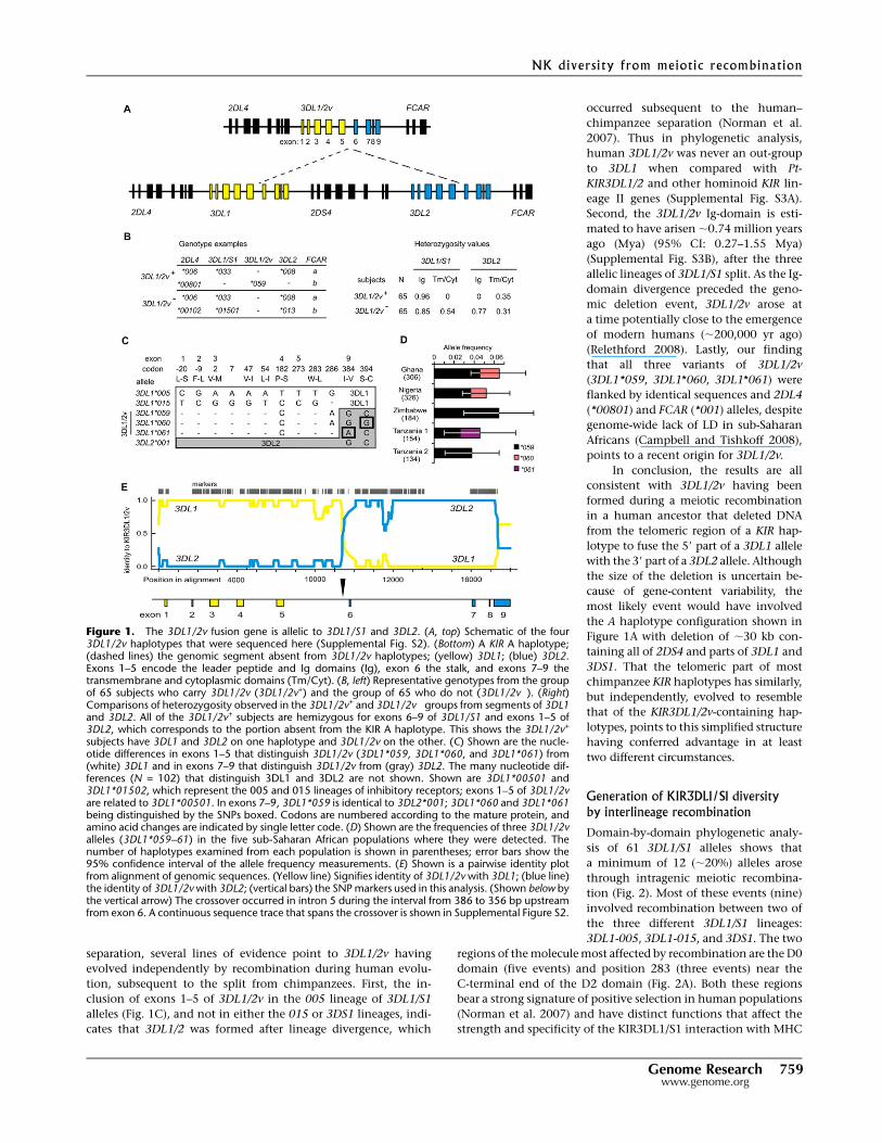
Figure 2. Motif and domain shuffling between three lineages diversifies3DL1/S1. (A) Shown to scale is the genomic organization of the 3DL1/S1locus. (Boxes) Exons; exons 2–5 encode the three Ig domains, D0, D1,and D2. (Vertical arrows) The genomic regions and the number of re-combination events detected from comparison of 3DL1/S1 alleles. (*)Recombination event that placed residue leucine 283 onto a differentbackground allotype. (B) Schematic of 12 recombinant alleles of 3DL1/S1that were identified using domain-by-domain phylogenetic analysis. Therecombinant allotypes are represented by segments colored according toallelic lineage: (red) 015, (blue) 005, (green) 3DS1. The allotypes shownat the top of the panel are encoded by the most common modern alleles:3DL1*01502, 3DL1*00501, and 3DS1*01301. For example, 3DL1*001 isidentical to 3DL1*00501 in exons 1–3 and 3DL1*01501 in exons 4–9.(Gray) Recombination from another locus (3DL2); (pink) within-lineagerecombinants. (White asterisk) Tryptophan–leucine substitution at resi-due 283; (black asterisk) recombination has replaced tryptophan at resi-due 283 with leucine; (z) threonine 118 that distinguishes 3DL1*043from 3DL1*001 and is shared with 3DL1*038. (C) The pairwise identityplot shows that 3DL1*009 formed by gene conversion. (Black line)Identity of 3DL1*009 with 3DL1*001; (green line) identity of 3DL1*009with 3DS1*01301. The recombination included exons 2 and 3, whichencode D0. (Across the top, vertical bars) SNP markers; (vertical arrows)the minimum and maximum limits of the gene conversion. Genomicsequences used for the RDP analysis include representatives of the threeallelic lineages of 3DL1/S1 (listed in Methods). The 3DL1*009 cDNA se-quence that was independently obtained (Middleton et al. 2007) corre-sponds precisely to the reading frame of the locus characterized here. (D)Shown for each of the 3DL1/S1 lineages is a pair of alleles that are di-morphic at (yellow) codon 31. Nucleotide differences in D0 are shown,and those that distinguish the three lineages are colored as in panel A.(Top) Amino acid substitutions with the ancestral residue first and theancestral nucleotide immediately below. (Right panel) A homology modelof D0; residue 31 (yellow) occurs in a patch of positively selected residues(orange) that were identified in Norman et al. (2007).
Norman et al.
760 Genome Researchwww.genome.org
the cell surface. The specificity and function of 3DL1*059 were
examined in cytotoxicity assays and compared to the 3DL1 and
3DL2 ‘‘parental’’ receptors. NKL cells expressing 3DL1*059,
3DL1*001, or 3DL1*01502 killed HLA Class I–deficient 221 cells as
effectively as untransfected NKL cells. When transfected 221 cells
expressing Bw4+ HLA-B*1513 were used as the target, reduced lysis
was observed for NKL cells expressing 3DL1*059, 3DL1*001, or
3DL1*015, compared to lysis by NKL cells that express no KIR or
KIR3DL2 that has no affinity for HLA-B*1513 (Fig. 3B, left). These
inhibitory effects were abrogated by the inclusion of either anti-
3DL1 (DX9) (Fig. 3B) or anti-HLA Class I (DX17) (data not shown)
monoclonal antibodies, showing that the inhibition observed for
the 3DL1-transduced NKL cells was dependent on the combina-
tion of 3DL1 on the NK cell and HLA-B*1513 on the target cell.
The inhibition mediated by the interaction of B*1513 with
3DL1*059 was comparable to that observed for 3DL1*001 and
greater than that seen for 3DL1*015. Thus, the Bw4 epitope of
HLA-B is demonstrated to serve as a ligand for 3DL1*059. Similar
experiments showed that interaction of 3DL1*059 with the Bw4+
HLA-A*3201 allotype also inhibited the lysis of transfected 221
cells, to an extent comparable to 3DL1*015, but less than
3DL1*001 (Fig. 3B, center). In contrast, the Bw4� HLA-A*1102
allotype did not engage 3DL1, but interacted with 3DL2*001 to
inhibit the lysis of 221 cells (Fig. 3B, right), in a manner consistent
with its HLA-A3, HLA-A11 specificity (Dohring et al. 1996; Pende
et al. 1996; Hansasuta et al. 2004).
In summary, these results demonstrate that 3DL1*059 is
a functional, inhibitory cell-surface receptor with a specificity for
Bw4+ HLA Class I that is characteristic of KIR3DL1. The unusual
3DL2-like signaling domain of 3DL1*059 (Fig. 4) generates in-
hibitory signals that are within the range of those obtained from
conventional 3DL1 signaling domains, but, as expected, does not
skew the specificity of 3DL1*059 toward the HLA-A*1102recognized
by KIR3DL2. Distinguishing 3DL1*059 from the other 3DL1 allo-
types examined here is the similar strength of the inhibition result-
ing from interactions with Bw4 epitopes on HLA-A and HLA-B
‘‘backgrounds’’ (Fig. 3B).
KIR3DL1/2v generates weaker inhibition than KIR3DL1
To examine further the effect of the 3DL2-like transmembrane
region and cytoplasmic tail of 3DL1*059, we made a mutant (m1)
in which they were replaced by the homologous domains of
3DL1*001. Reciprocally, a second mutant (m2) paired the extra-
cellular domains of 3DL1*001 with the transmembrane region and
cytoplasmic tail of 3DL1*059 (Fig. 3C, left). NKL transductants
expressing these two mutants and their natural allotypes were
compared in cytotoxicity assays against 221 target cells and 221
transfectants expressing A*3201 or B*1513. This analysis showed
that the combination of 3DL1*059 and B*1513 gave a weaker in-
hibition than the combination of m1 and B*1513 (Fig. 3C, center).
A similar trend was seen when the ligand was A*3201, but the
difference was much smaller (Fig. 3C, right). For the reciprocal
pair, 3DL1*001 gave a stronger inhibition than m2, a difference
that was greater with A*3201 as the ligand than with B*1513.
These results show that in all combinations, the trend is to weaker
inhibition when the transmembrane region and cytoplasmic do-
main are derived from 3DL2. Thus another potential selective
advantage of 3DL1/2v is its capacity to generate a weaker in-
hibitory signal than its progenitor 3DL1 allotype.
Because 3DL1*059 and m2 only differ at position 283, as do
3DL1*001 and m1, this mutational analysis also showed how the
dimorphism at position 283 affects 3DL1 function. Substitution of
tryptophan for leucine at position 283 increased the inhibitory
signal on binding to A*3201 but decreased the response to B*1513
(Fig. 3C). For 3DL1*059, the trend was similar—tryptophan 283
favored interaction with A*3201, leucine 283 its interaction with
B*1513—but the difference was smaller. Thus a potential advan-
tage of the recombinations that change residue 283 is that they
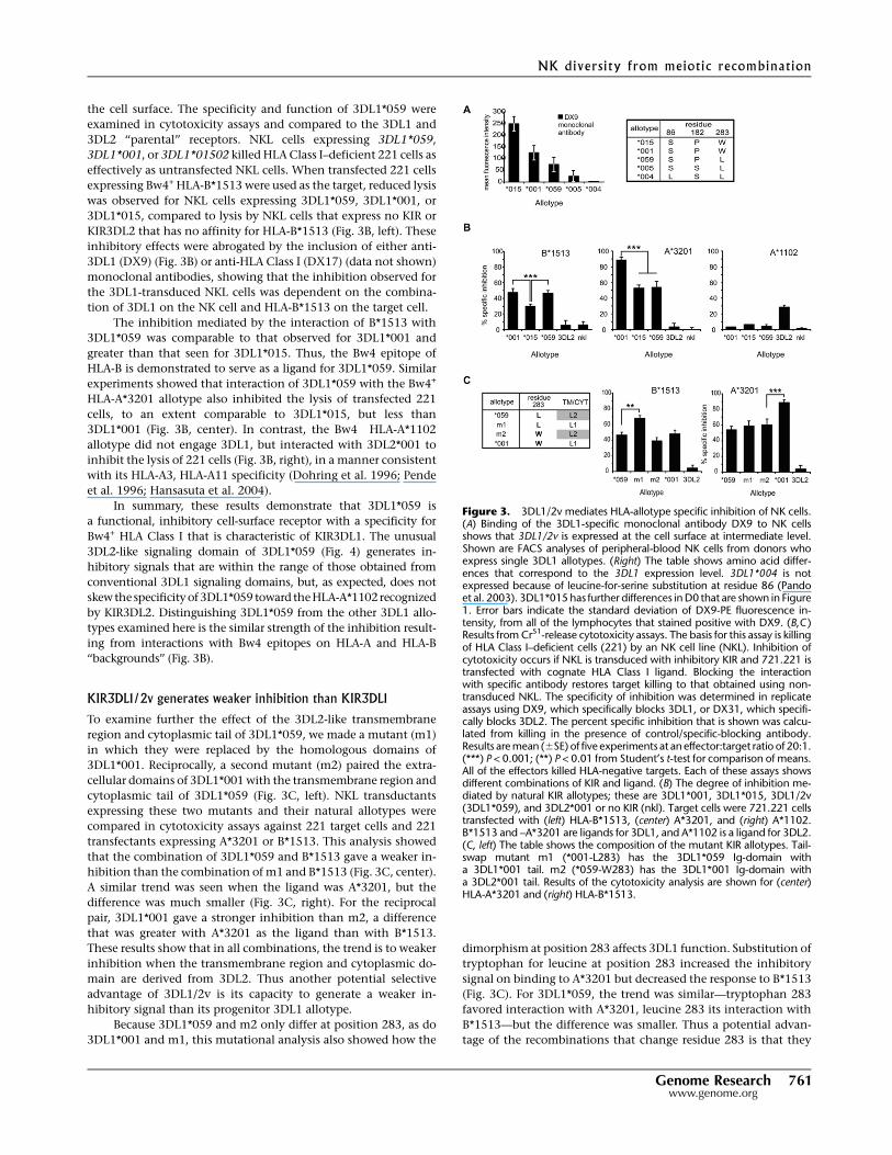
Figure 3. 3DL1/2v mediates HLA-allotype specific inhibition of NK cells.(A) Binding of the 3DL1-specific monoclonal antibody DX9 to NK cellsshows that 3DL1/2v is expressed at the cell surface at intermediate level.Shown are FACS analyses of peripheral-blood NK cells from donors whoexpress single 3DL1 allotypes. (Right) The table shows amino acid differ-ences that correspond to the 3DL1 expression level. 3DL1*004 is notexpressed because of leucine-for-serine substitution at residue 86 (Pandoet al. 2003). 3DL1*015 has further differences in D0 that are shown in Figure1. Error bars indicate the standard deviation of DX9-PE fluorescence in-tensity, from all of the lymphocytes that stained positive with DX9. (B,C )Results from Cr51-release cytotoxicity assays. The basis for this assay is killingof HLA Class I–deficient cells (221) by an NK cell line (NKL). Inhibition ofcytotoxicity occurs if NKL is transduced with inhibitory KIR and 721.221 istransfected with cognate HLA Class I ligand. Blocking the interactionwith specific antibody restores target killing to that obtained using non-transduced NKL. The specificity of inhibition was determined in replicateassays using DX9, which specifically blocks 3DL1, or DX31, which specifi-cally blocks 3DL2. The percent specific inhibition that is shown was calcu-lated from killing in the presence of control/specific-blocking antibody.Results are mean (6SE) of five experiments at an effector:target ratio of 20:1.(***) P < 0.001; (**) P < 0.01 from Student’s t-test for comparison of means.All of the effectors killed HLA-negative targets. Each of these assays showsdifferent combinations of KIR and ligand. (B) The degree of inhibition me-diated by natural KIR allotypes; these are 3DL1*001, 3DL1*015, 3DL1/2v(3DL1*059), and 3DL2*001 or no KIR (nkl). Target cells were 721.221 cellstransfected with (left) HLA-B*1513, (center) A*3201, and (right) A*1102.B*1513 and –A*3201 are ligands for 3DL1, and A*1102 is a ligand for 3DL2.(C, left) The table shows the composition of the mutant KIR allotypes. Tail-swap mutant m1 (*001-L283) has the 3DL1*059 Ig-domain witha 3DL1*001 tail. m2 (*059-W283) has the 3DL1*001 Ig-domain witha 3DL2*001 tail. Results of the cytotoxicity analysis are shown for (center)HLA-A*3201 and (right) HLA-B*1513.
NK diversity from meiotic recombination
Genome Research 761www.genome.org

can increase the compatibility of 3DL1/S1 with the particular Bw4-
bearing HLA-A and HLA-B allotypes present in a population.
Gene duplication by unequal crossing over allows individualsto carry all three 3DL1/S1 lineages
The three lineages of 3DL1/S1 alleles (3DL1-005, 3DL1-015, and
3DS1) have been maintained by balancing selection for more than
3 million years (Norman et al. 2007). However, most individuals
carry only one or two of the three lineages. Family studies iden-
tified a KIR haplotype in Europeans that contains both 3DS1 and
3DL1 and likely was formed by unequal crossing over, which du-
plicated both the KIR2DL4 and 3DLl/S1 genes (Martin et al. 2003;
Williams et al. 2003; Gomez-Lozano et al. 2005). The potential
benefit to such duplication haplotypes is that they permit some
individuals to carry all three 3DL1/S1 allelic lineages. To assess the
prevalence of the duplication haplotype, we used a newly developed
pyrosequencing method that detects 3DL1/S1 copy-number varia-
tion (Fig. 5) to study a panel of 2800ethnicallydiverse blood donors.
Eighty individuals (2.9%) were demonstrated to have a KIR hap-
lotype with two copies of 3DL1/S1 (Fig. 6A). By genotyping these
individuals for 2DL4, 3DL1/S1, and 3DL2 alleles, we defined
seven different duplication KIR haplotypes (Fig. 6B). Each dupli-
cation haplotype was differentially distributed; as a group, they
are most common in South and East Asians, with frequencies up
to 10%, less common in Caucasians, rare in Africans, and absent
from Amerindians. Among the 80 individuals carrying a duplica-
tion haplotype, 23 of them (28.8%) carried one allele from each of
the three KIR3DL1/S1 lineages.
All seven duplicated haplotypes have the same general
structure, containing two copies of 2DL4 and 3DL1/S1 (Supple-
mental Fig. S5). Common to the seven haplotypes are the
2DL4*005 and 3DS1*01301 alleles; these are associated with three
other 2DL4 alleles, four 3DL2 alleles, and seven different 3DL1
alleles embracing both 3DL1 lineages and their full ranges of
functional diversity. Each duplication haplotype contains a pair of
3DL1 and 3DL2 alleles that are common and in linkage disequi-
librium in the populations where the duplication haplotypes are
present (Norman et al. 2004; Middleton et al. 2007). This property,
combined with the constancy of
2DL4*005 and 3DS1*01301, supports an
evolutionary model in which all the du-
plication haplotypes originated with
a single event of unequal crossing over
(Fig. 6A), which was subsequently fol-
lowed by six independent events of ho-
mologous recombination that diversified
the 3DL1 and 3DL2 genes of the dupli-
cation haplotype. The restricted geo-
graphical distribution of the duplication
haplotypes, largely outside of Africa, and
the linkage disequilibrium of their 3DL1
and 3DL2 genes point to the emergence
of the duplication haplotypes during
the last 60,000 yr, and since humans
left Africa to populate Europe and Asia
(Campbell and Tishkoff 2008).
We identified a family in which
a son inherited a duplicated haplotype
containing 3DS1*01301 and 3DL1*002
from his mother. He inherited a second
copy of 3DS1*01301 from his father (Fig.
6C). Using the monoclonal antibodies DX9, specific for KIR3DL1,
and Z27 that binds much more strongly to 3DL1 than to 3DS1
(O’Connor et al. 2007; Trundley et al. 2007), we determined 3DL1/
S1 expression by the peripheral blood NK cells of members of this
family. The frequency of NK cells expressing 3DS1 in the son
(50.5%) was more than the sum (47.5%) of those cells in the
mother (24.2%) and father (23.3%) (Fig. 6C). This indicates that
both 3DS1*013 alleles inherited by the son are expressed by NK
cells. The son also expressed 3DL1*002 on 6.8% of his NK cells,
consistent with previous observation that this allele is usually
expressed at low frequency by NK cells (Chan et al. 2003). Thus,
each of the son’s three KIR3DL1/S1 alleles is expressed by NK cells.
A second son, who inherited two different KIR haplotypes from
the parents, had no 3DS1-expressing NK cells and exhibited a bi-
modal distribution of cells with high-expressing 3DL1*015
inherited from the mother and low-expressing 3DL1*007 inheri-
ted from the father (Fig. 6C). Although the mother’s NK cells
showed a high expression of 3DL1, we could not ascertain
whether both 3DL1*002 and 3DL1*1502 were expressed, because
they cannot be distinguished using available monoclonal anti-
bodies. Two unrelated donors carrying alleles of each 3DL1/S1
lineage—3DS1*01301 (3DS1 lineage), 3DL1*00501 (005 lineage)
and 3DL1*002 or 3DL*01502 (015 lineage)—were therefore iden-
tified. Because 3DL1*005 and 3DL1*015 are readily distinguished
by their low and high binding to Z27, respectively, we could
demonstrate that all three alleles were expressed by subsets of NK
cells in these individuals (Fig. 6D). In conclusion, individuals
carrying three 3DL1/S1 alleles representing the three allelic line-
ages express all three allotypes as NK cell receptors.
DiscussionNK cells provide an early defense against infection, particularly
viral infections (Lanier 2008), and at an early stage in re-
production, they remodel maternal blood vessels that provide the
fetus with nutrients via the placenta (Moffett and Loke 2006). In
performing these functions, NK cell receptors have become highly
diverse, rapidly evolving and largely species-specific (Kelley et al.
2005). Unlike the B-cell and T-cell receptors that diversify function
Figure 4. KIR3DL1 and KIR3DL2 differ at 68 amino acid residues spread throughout the polypeptide.Shown are the amino acid differences that distinguish 3DL1, 3DL2, and 3DL1/2v (3DL1*059). (Grayboxes) Differences that occur in the second ITIM motif of the cytoplasmic tail. Residue numbers arethose of the mature protein. The 3DL1*059 sequence that was independently obtained from cDNA(Shilling et al. 2002) is identical to the amino acid sequence encoded by the 3DL1/2v gene charac-terized here (Fig. 1).
Norman et al.
762 Genome Researchwww.genome.org

through somatic recombination and mutation, NK cell receptors
can only diversify through meiotic mutation and recombination.
And in general, immune system genes have become more di-
versified by meiotic recombination than other functional types of
human gene (Redon et al. 2006; Frazer et al. 2007) and display
greater variation as assessed by copy number and segmental in-
sertion/deletions (Dumas et al. 2007; Korbel et al. 2007).
The state of the killer cell immunoglobulin-like receptor (KIR)
locus in the leukocyte receptor complex (LRC) varies dramatically
between species. In some, like the seals, it is a single conserved and
nonpolymorphic gene, and in others, such as the dog, it appears to
have been deleted (Hammond et al. 2009). In contrast, in the
higher primates, the KIR locus has evolved to become a diverse
multigene family encoding MHC Class I receptors (Khakoo et al.
2000; Rajalingam et al. 2004; Sambrook et al. 2005; Guethlein et al.
2007). On the basis of current information, KIR diversity appears
greatest in the human species, where the combined effects of gene-
content diversity and allelic polymorphism are such that unrelated
individuals have distinct KIR genotypes (Shilling et al. 2002). The
extent of KIR genotype variability approaches that of HLA class I,
with the functionally important combination of KIR and HLA
genotypes being even more diverse (Parham 2005). Unequal
crossing over is the likely mechanism that expanded the primate
KIR locus from its origin as a single-copy gene, and it is clearly
implicated in the diversification of gene content in the modern
human KIR haplotypes (Khakoo et al. 2000; Wilson et al. 2000;
Martin et al. 2003). Further diversity is generated by homologous
recombination, in particular, by the reassortment of allelic and
gene-content motifs in the centromeric and telomeric parts of the
locus (Wilson et al. 2000; Norman et al. 2004; Parham 2005). Here
we have explored the role that meiotic recombination has played
in the diversification of an individual human KIR gene, KIR3DL1/
S1. This gene encodes highly polymorphic NK cell receptors that
recognize HLA-A and HLA-B, the most polymorphic MHC genes.
Balancing selection and coevolution with HLA are consis-
tently recognized features of the KIR (Hiby et al. 2004; Norman
et al. 2004; Gendzekhadze et al. 2006; Single et al. 2007; Yawata
et al. 2008). One of the strongest indicators for balancing selection
is the long-term preservation of alleles without fixation or loss
(Kimura and Ota 1969); another is the retention of alleles through
periods of restricted population size. The 3DL1/S1 locus comprises
three ancient lineages of alleles that have been maintained
by balancing selection over more than 3 million years and are
present in all modern human populations (Norman et al. 2007),
despite severe bottlenecks for some (Hey 2005). Contrasting with
the conservation of the 3DS1 lineage, the 3DL1-005 and 3DL1-015
Figure 5. Simultaneous detection of KIR3DL1/S1 polymorphism andcopy-number using pyrosequencing. (A, top) The diploid sequence ofa 17-bp fragment of exon 3 from an individual heterozygous for3DL1*01502 and 3DS1*01301. (Gray) SNP g336a. Underneath is a pyro-gram obtained from the same individual. To generate the pyrogram,nucleotides were added to a single-strand template in the sequenceshown, 1–11, and correspond to peaks of the same number. The peakheight is proportional to the quantity of nucleotides that were in-corporated, as shown by peak 1 (the sequence is gg), which is twice theheight of peak 6 (g). Each peak is a sum of the haplotypes present, sothat the monomorphic positions (relative peak height = 1) are used forcalibration. The combined peak height at the heterozygous positionshown (peaks 10 + 11) is equal to the peak from a single monomorphicposition. Shown are four different pyrograms; (top left of each diagram)the derived genotype; (brackets) the peak-height ratio compared with thesingle monomorphic peak. (A) At SNP g336a, there is one peak for g andone for a (peaks 10 and 11), and each peak is half the height of a singlepeak (peak 6). This individual has one copy of 3DL1*01502 and one copyof 3DS1*01301 (0.5g: 0.5a). (B) At SNP g336a, there is one peak for g(peak 10) that is the same height as a single monomorphic position (peak6) and no peak on addition of nucleotide a-11. This individual is homo-zygous, having two copies of 3DL1*01502 (1g: 0a). (C) There are twopeaks as for A, but g is twice the height of a (0.67g: 0.33a), and their sumis the same as peak 6. This individual has two copies of 3DL1*01502 andone 3DS1*01301. (D) This individual has one copy of 3DL1*01502 andtwo of 3DS1*01301 (0.33g: 0.67a).
NK diversity from meiotic recombination
Genome Research 763www.genome.org

lineages of inhibitory receptors are now
highly diverse. Of the approximately 60
alleles defined, 12 are recombinants,
most of which have been formed by re-
combination or conversion between
alleles from different lineages. Two main
targets for recombination have been
polymorphic motifs in the D0 domain,
the enhancer that modulates the
strength of the binding site formed by
the D1 and D2 domains (Khakoo et al.
2002) and a dimorphism at position 283
in D2 that we show alters the strength of
the inhibitory signal transduced upon
binding of 3DL1 to Bw4.
Onlythethreelineage-defining3DL1/
S1 alleles—3DS1*01301, 3DL1*00501, and
3DL1*01502—are common to all human
populations, the vast majority of the
others being specific to particular pop-
ulations, geographical regions, and eth-
nic groups (Norman et al. 2007). Since
humans originated in Africa and even-
tually went on to populate other con-
tinents (Goldstein and Chikhi 2002),
unequal crossing over has produced novel
recombinant alleles and haplotypes that
mark particular populations. For exam-
ple, the 3DL1*009 recombinant allotype,
in which the D0 domain of 3DL1*015 is
replaced with that of 3DS1*013, is re-
stricted to European and some South
Asian populations (Middleton et al.
2007; Norman et al. 2007). That we con-
sistently observe population stratifica-
tion of 3DL1/S1 alleles points to recent
or ongoing selection for variation. Up to
10% of sub-Saharan African populations
have a KIR haplotype that was formed
within 0.74 million years by an unequal
crossing over that caused the deletion of
>30 kb from the telomeric part of the KIR
locus and created a novel hybrid gene, by
fusing exons 1–5 from 3DL1*005 with
exons 7–9 of the 3DL2*001 gene. The
fusion protein encoded by this 3DL1/2v
allele (3DL1*059) combines the extra-
cellular, Ig-like domains of 3DL1*005
with the transmembrane and cytoplas-
mic domains of 3DL2*001. As expected,
3DL1*059 has specificity for Bw4, but it
has a more even response to HLA-A and
HLA-B allotypes having the Bw4 epitope,
and transduces a weaker inhibitory sig-
nal, than other 3DL1 allotypes. Since the
formation of 3DL1*059 by recombina-
tion, subsequent point substitutions in
the cytoplasmic tail produced 3DL1*060
and 3DL1*061 allotypes. Valine 384
that distinguishes 3DL1*061 is close to
an ITIM motif (Feng et al. 2005), and
the substitution of cysteine for serine at
Figure 6. Duplicating KIR loci diversifies the NK cell repertoire in quantity and quality. (A) Schematicof the donor haplotypes and duplication haplotype. (Across the top) The KIR loci; (shaded boxes) in-dicate presence of the locus. The donor KIR haplotypes B (black) and A (orange); the composite du-plication haplotype D is colored accordingly. (Dotted line) Non-allelic homologous recombination thatwas mediated by sequence similarity in the 59-regions of 3DP1 and 2DL5A (Martin et al. 2003; Gomez-Lozano et al. 2005). (B) The seven different 2DL4, 3DL1, and 3DL2 haplotypes deduced from analysis of80 individuals who have the duplication, and their haplotype frequencies in the populations where theywere detected. The number of haplotypes analyzed from each population is indicated in parentheses.Georgia is the country. The Piramalai Kaller and the Yadheva are two distinct populations from TamilNadu in Southern India. (C) Shown are FACS analyses of freshly isolated 3DL1/S1-expressing NK cellsfrom a European family with a duplicate haplotype. (At the top of each plot) The pyrosequencinggenotype. Son 1 inherited 3DS1*013 and 3DL1*002 (haplotype 3, panel B) from the mother and3DS1*013 from the father. Monoclonal antibody Z27 discriminates 3DL1/S1 allotypes, having high(3DL1*002 and 3DL1*015), low (3DL1*005 and 3DL1*007), and very low (3DS1*013) staining pat-terns. Showing that 3DL1/S1 expression depends on the number of copies of 3DS1 present, Son 1 hastwice the number of 3DS1+ NK cells as the mother or father. Son 2 has inherited the opposite pair ofhaplotypes to Son 1, and expresses two distinct populations of high (*015) and low (*007) 3DL1staining and no 3DS1. Further differences among allotypes are quantitative, with *015 being expressedby more cells than *002 (Chan et al. 2003), as observed here by comparing 3DL1 expression of themother and Son 1. (D) Three unrelated individuals chosen for their 3DL1/S1 genotype. (Left) An in-dividual heterozygous for 3DL1*015 and 3DL1*005 is shown as a negative control for the very low-staining 3DS1*013 peak. (Center and right) Two individuals who both express all three lineages of3DL1/S1 receptors; these FACS analyses are from NK cells that had been stored frozen. A comparison offresh and frozen NK cell staining is shown in Supplemental Figure S6.
Norman et al.
764 Genome Researchwww.genome.org

residue 394 in 3DL1*060 that eliminates a protein kinase C
phosphorylation site is predicted to increase inhibitory signals
(Alvarez-Arias and Campbell 2007). Thus, the variants of 3DL1/2v
have the potential for functional difference. That all hominoid
KIR haplotypes have a lineage II gene with an exon 9 orthologous
to that of 3DL2, despite much recombination, points to the im-
portance of the signaling domain it encodes (Rajalingam et al.
2001; Sambrook et al. 2005; Guethlein et al. 2007).
Gene fusion is a recognized means for generating genetic
novelty (Bailey and Eichler 2006), but to our knowledge, 3DL1/2v
is the first demonstration of the phenomenon occurring in
humans. Among the 600 gene fusions described for eukaryotes
(Kummerfeld and Teichmann 2005), all those identified as hu-
man-specific are associated with disease or abnormality. One
possible exception is the family of X chromosome gene fusions of
OPN1MW/LW that cause anomalous color detection in males
(Nathans et al. 1986), but may benefit female carriers (Deeb 2006).
As shown for other SNPs at these loci, heterozygous females have
enhanced color discrimination (Jameson et al. 2001).
Whereas the unequal crossing over that produced 3DL1/2v
reduced the size of a KIR haplotype, in Eurasia a different event of
unequal crossing over served to duplicate the 3DL1/S1 locus such
that the haplotype had one copy of 3DS1 and one copy of 3DL1.
Subsequent events of reciprocal crossing over have diversified
this duplicated haplotype so that at least seven different 3DL1
alleles, including most of the more common alleles of both the
005 and 015 lineages, are associated with 3DS1*013. Although we
do not know which 3DL1 allele was present on the original du-
plicated haplotype, that containing 3DL1*01502 appears the most
prevalent.
When first characterized from cDNA, 3DL1 and 3DS1 were
assigned to different loci based on the divergent sequence and
function of their transmembrane and cytoplasmic domains
(Robinson et al. 2006). Subsequent family and population studies
showed that 3DL1 and 3DS1 usually segregate as alleles (Middleton
et al. 2007), and sequence comparison indicated that 3DS1 was
formed from 3DL1 by recombination with a gene encoding an
activating KIR (Rajalingam et al. 2004). Consequently, 3DS1 and
3DL1 are generally considered to be alleles of a single locus:
3DL1/S1. The duplicated haplotypes are exceptional because they
have two genes of which one is always 3DS1 and the other is al-
ways 3DL1. So far no duplicated haplotype containing two copies
of 3DL1 and only one with two copies of 3DS1 (Gomez-Lozano
et al. 2005) have been identified, indicating that such re-
combination is rare or that such events are selected against. Thus
within the context of the duplicated haplotypes 3DS1 and 3DL1
are two different genes. This illustrates a more general complexity
of the KIR system, namely, imprecision or blurring in the dis-
tinction between loci and alleles. At the KIR, MHC, and other
immunological loci, an understanding of the synergy between
allelic polymorphism and copy-number variability that generates
diversity has required intensive, focused analysis (Bergstrom et al.
1999; Chung et al. 2002; Hollox et al. 2008). To determine the
extent to which the findings apply to other gene families will re-
quire analyses of similar commitment and intensity (Dumas et al.
2007; Perry et al. 2008).
Throughout human history, balancing selection has main-
tained the 3DS1, 3DL1-005, and 3DL1-015 lineages of alleles.
Today, the only three KIR3DL1/S1 alleles present in all the pop-
ulations studied are 3DS1*01301, 3DL1*00501, and 3DL1*01502.
This observation indicates that the three lineages have distinct
and complementary functions that have facilitated the survival
and propagation of human populations, a corollary being that
populations that lost a 3DL1/S1 lineage failed to compete and
survive over the long term. Despite the benefit of three allelic
lineages for populations, most modern humans carry alleles from
only one or two 3DL1/S1 lineages and suffer no obvious disad-
vantage in either survival or reproduction. In this context, the
potential advantage of the duplicated KIR haplotype is that it is
a mechanism by which individuals can carry all three 3DL1/S1
lineages and, as we have shown, express them as NK cell receptors.
In sub-Saharan African populations, the balance between the
three 3DL1/S1 lineages appears to have given way to selection for
the 3DL1-015 lineage and against the 3DS1 and 3DL1-005 lineages
(Norman et al. 2007). A further characteristic of sub-Saharan
Africans is that a significant proportion of the 3DL1-005 lineage
allotypes is 3DL1*059, 3DL1*060, and 3DL1*061, products of the
gene fusion between 3DL1*005 and 3DL2. Selection for these
alleles, might also reflect selection against conventional 3DL1*005
and for the modified functions imposed on its extracellular
domains by the distinctive transmembrane and cytoplasmic
domains of 3DL2.
KIR3DL1 and 3DL2 that recognize determinants of HLA-A and
-B are encoded by lineage II KIR genes, which are represented in
hominoid species by two or more genes. Exceptional is the chim-
panzee, which has one lineage II KIR gene (Pt-KIR3DL1/2) that
encodes a protein with structural and functional similarities to both
3DL1 and 3DL2. In chimpanzee KIR haplotypes, Pt-KIR3DL1/2 is
the only gene on the telomeric side of Pt-KIR2DL4, an arrangement
like that of the human haplotypes containing 3DL1/2v (3DL1*059,
3DL1*060, and 3DL1*061) in which 3DL1 and 3DL2 have been
reduced to a single fusion gene. We have shown that the similarities
of 3DL1/2v and Pt-KIR3DL1/2 are not due to common ancestry but
to convergence, further emphasizing the dynamic nature of the KIR
gene family and continuing cycles of gene expansion and con-
traction through meiotic recombination.
Methods
SubjectsThe subjects studied here are as described by Norman et al. (2007).Blood samples were collected and the research conducted withapproval from the appropriate Institutional Review Boards. Thetwo Tanzanian populations are from (1) a village that lives on thecoastal plain near the Indian Ocean and (2) a village at an altitudeof 1700 m in the West Usambara Mountains.
KIR nomenclature
KIR genes and alleles were named by the KIR NomenclatureCommittee (Marsh et al. 2003), formed from the WHO Nomen-clature Committee for factors of the HLA system and the HUGOGenome Nomenclature Committee. A curated database is avail-able at http://www.ebi.ac.uk/ipd/kir/ (Robinson et al. 2006). <D>
denotes the number of Ig-like Domains; <L> a Long, inhibitory,cytoplasmic tail; <S> a Short, activating, tail; and <P> a Pseudo-gene. For each locus, the alleles are named in the order of theirdiscovery, with the first three digits distinguishing alleles encod-ing different proteins. The second two digits are used to indicatesynonymous variation. As an example, KIR3DL1*01501 andKIR3DL1*01502 are synonymous variants of the 3DL1*015 allele,and both encode the 3DL1*015 allotype—an inhibitory receptorhaving three Ig-like domains. When the meaning is unambiguous,KIR or even KIR3DL1 can be dropped from allele names.
NK diversity from meiotic recombination
Genome Research 765www.genome.org

PCR/DNA sequencing
PCR was performed using a Perkin-Elmer 9600 thermal cycler witha 3-min denaturing step at 94°C, 10 cycles of (10 sec at 94°C; 60 secat 65°C) and 20 cycles of (10 sec at 94°C, 50 sec at 61°C, 30 sec at72°C). When the expected amplicons were >3.5 kb, long range-PCR (LR-PCR) was performed using TripleMaster/HiFi reagents(Eppendorf) and the oligonucleotide primers described in Sup-plemental Figure S2. LR-PCR conditions were 3 min at 94°C fol-lowed by 28 cycles of (20 sec at 94°C, 10 sec at 61°C, 4 min at 68°C)completed with 15-min extension at 68°C. Products were clonedusing the pCR 2.1-TOPO vector (Invitrogen) and sequenced usinginternal primers. Standard DNA sequencing was performed inforward and reverse directions using BigDye Terminator v3.1 andanalyzed using an ABI-377 sequencer (ABI). Three clones of thedesired allele were sequenced from each individual.
Characterizing the KIR3DL1/2v gene sequence
The KIR3DL1/2v gene sequence was characterized from one donorusing multiple PCR primer pairs (Supplemental Fig. S7) and fromfour donors using six overlapping PCR fragments that spanned thehaplotype from 2DL4 to FCAR (Supplemental Fig. S2). The fourdonors were selected to represent all three different 3DL1/2vsequences and also to have a distinct second KIR haplotype. Un-ambiguous sequence data were generated from one of the donors,who is hemizygous for 3DL1/2v so has no second 3DL1/S1 allele(Supplemental Fig. S2). For each heterozygous individual, suffi-cient SNPs were present to distinguish two haplotypes uponcloning, except for two of the reactions that were allele-specific(bands A and C) (Supplemental Fig. S2). Each amplicon was clonedusing pCR 2.1 and sequenced in triplicate for each allele present.All of the PCR primers were designed to be KIR gene- or allele-specific (Supplemental Fig. S2; Supplemental Table S1). Accessionnumbers for the newly generated 3DL1*059–61 genomic DNAsequences are EU267269–71 and FJ459734.
The 3DL1/S1*059 donor was an African-American from Cal-ifornia (genotype: 3DL1*031, 3DL1*059, 3DL2*001). The 3DL1/S1*060 donor was an African from Nigeria (genotype: 3DL1*035,3DL1*60, 3DL2*001). The 3DL1/S1*061 donor was an African-American from California (genotype: 3DL1*040, 3DL1*061,3DL2*010). The hemizygous donor was an African from Nigeriawho has a KIR haplotype that lacks 3DP1, 2DL4, and 3DL1/S1 butretains 3DL2 (Norman et al. 2004). This individual has one 3DL1/S1 allele and one 3DL2 allele (genotype: 3DL1*059, 3DL2*006).
DNA sequences and recombination analysis
Recombinant sequences were identified from a domain-by-domain phylogenetic analysis. Gene conversion was identifiedusing RDP (Recombination Detection Programs) version 2 (Martinet al. 2005) with a sliding window of 20 bp, with Bonferroni cor-rection and a threshold of P = 0.001. Coding sequences of 3DL1/S1used for alignments were obtained from the IPD database (http://www.ebi.ac.uk/ipd/kir/index.html) (Robinson et al. 2006) andthose described in Belle et al. (2008) and Thomas et al. (2008).cDNA from 3DS1*050 (EF582383) was fully characterized hereusing methods previously described (Shilling et al. 2002). Geno-mic DNA sequences that spanned exons 1–5 (encoding the ex-tracellular domains) and exons 7–9 (encoding the cytoplasmicdomain) were characterized by LR-PCR from the following alleles:3DL1*001, 3DL1*00501, 3DL1*009, 3DL1*01501, 3DL1*022,3DL1*029, 3DL1*030, 3DL1*031, 3DL1*035, 3DL1*036, and3DL1*040 (GenBank: FJ158650–60). 3DS1*01301 and 3DL2*007sequences were extracted from AL133414 (3DS1a) (Wilson et al.2000) and AY320039 (3DS1b). A second 3DL1*001 genomic
sequence, and 2DL4*00801 and 3DL2*001 were obtained fromAC011501 (Wilson et al. 2000). For the analysis indicated inSupplemental Figure 3B, a further 2 kb of sequence that surroundsthe crossover point in intron 5 was obtained from 3DL1*00501,3DL1*01501, and 3DL1*040; for 3DL1*002 the equivalent se-quence was obtained from CU151839 and 3DL2*010 fromCU464063 (Horton et al. 2006). pt2DL4 and pt3DL1/2 genomicDNA sequence was obtained from BX842589 (Sambrook et al.2005) and Popy2DL4 and Popy3DLa from EF014479 (Guethleinet al. 2007).
DNA sequence analysis
Nucleotide sequences were aligned using ClustalX (Thompsonet al. 1997) and BioEdit (www.mbio.ncsu.edu/BioEdit/bioedit.html). Neighbor-joining phylogenetic analyses were conductedusing the Tamura-Nei method of MEGA3.1 and 500 bootstrapreplicates (Kumar et al. 2004). PAML was used to reconstruct theancestral sequences at specified nodes, using the marginal re-construction method and the M8 model (Yang 2007).
Divergence time estimations
Divergence times were estimated using the MCMCTREE programfrom PAML 4 (Yang 2007), which uses Markov-chain Monte Carloanalysis for estimating mean posterior divergence times and their95% credibility interval (CI). The Markov chain was sampled 5000times every 10 cycles, and the burn-in stage was set to 5000 cycles.MODELTEST 3.7 (Posada and Crandall 1998) was used to gaugethe nucleotide-substitution model (HKY95) and the a-distributionof this substitution (0.83 and 1.03 for the sequences 59 and 39 fromthe 3DL1/2v crossover, respectively). Calibration points were setaccording to fossil data and the divergence times: CI = 23–33 Myafor macaque/hominoid, CI = 10–18 Mya (mean 12) orangutan/hominid, and CI = 6–8 Mya (mean 7) for chimpanzee/hominid(Benton and Donoghue 2007).
Genotyping by pyrosequencing
Using primers described in Supplemental Table 1, individual exonsfrom 2DL4 and 3DL2 were amplified and subjected to pyrose-quencing. Exon-specific PCR was performed using a Perkin-Elmer9600 thermal cycler with a 3-min denaturing step at 94°C, 10 cyclesof (10 sec at 94°C, 60 sec at 65°C), and 20 cycles of (10 sec at 94°C,50 sec at 61°C, 30 sec at 72°C). Identical conditions were used fora second specific reaction but with 35 cycles in the final step.Samples were purified using strepdavidin-coated Sephadex beads,and pyrosequencing reactions were performed using the Pyro-Goldreagents and a PSQ96A machine fitted with Capillary DispensingTips (Biotage Ltd.). 3DL1/S1 genotyping was performed as described(Norman et al. 2007). The genotyping procedures were performedin parallel with previously described family and control samples(Gardiner et al. 2001; Shilling et al. 2002; Artavanis-Tsakonas et al.2003; Norman et al. 2004, 2007). To further validate the techniques,a random selection of 25% donors was also re-sequenced froma freshly generated amplicon. According to the most recent se-quence alignments (http://www.ebi.ac.uk/ipd/kir/index.html), thecombination of techniques discriminates 11 2DL4 alleles, 52 3DL1/S1 alleles, 22 3DL2 alleles, three 3DL1/2v alleles, and copy-numbervariation for each locus.
Detection of KIR3DL1/2v by genotyping 3DL1/S1 and 3DL2
Because no SNP uniquely defines the 3DL1/2v sequences, thepyrosequencing protocol was designed to detect a unique
Norman et al.
766 Genome Researchwww.genome.org

combination of SNPs in exons 1–5 that distinguishes 3DL1/2vfrom 3DL1/S1 (Fig. 1). Three different 3DL1/2v alleles (3DL1*059,3DL1*060, 3DL1*061) were distinguished by one further SNP inexon 5 and two SNPs in exon 9. At the cDNA level, 3DL1*059(Shilling et al. 2002) and 3DL1*060 (Artavanis-Tsakonas et al.2003) have been described previously, and 3DL1*061 was dis-covered during the present study. Two other 3DL1/2v se-quences reported previously (AY366254-5) were not observed inthe present study. Heterozygosity was calculated using Arlequin(Schneider et al. 2000).
Detecting copy-number variation using pyrosequencing
Pyrosequencing uses enzyme-coupled reactions to generate lightfrom the pyrophosphate molecules released upon DNA elongation(Ronaghi et al. 1996). Nucleotides are added sequentially so thatthe reaction proceeds only when the correct base is added. Lightemission is plotted on a Pyrogram that has peak height pro-portional to the number of pyrophosphate molecules released.The technique is semiquantitative because the combined peakheights from a polymorphic position equal the height of the singlepeak at a monomorphic position (Fig. 5). With this method, wewere able to determine the presence of three 3DL1/S1 alleles inindividuals having a duplication of the 3DL1/S1 locus on onehaplotype even when two of the alleles were identical in sequence,because peak-height ratios were 2:1 in favor of the allele present intwo copies. All genotyping methods were designed so that eachgenotype gave a distinct pattern of several SNPs dispersed throughthe locus and did not rely on single marker SNPs. We were thusable to accurately assign three-copy genotypes by virtue of con-sistency in peak-height ratios throughout the locus.
To validate the copy-number detection method, two familiesshown previously by segregation analysis to have duplicated3DL1/S1 (Martin et al. 2003; Norman et al. 2004) were used asstandards. Furthermore, for eight of the 80 individuals in whomthe duplication was detected, PCR products that span exons 3–5 of2DL4 were generated, cloned, and sequenced (12 clones) to verifythe presence of three different 2DL4 alleles and thus of the du-plication. Some individuals may possess two copies of a 3DL1/S1-duplicated haplotype, and if the 3DL1/S1 pairs on the two hap-lotypes were identical, we would not have distinguished themfrom a normal heterozygote. However, the observed maximumfrequency of 10% for duplicated-3DL1/S1 haplotypes implies that,assuming Hardy-Weinberg equilibrium, only one or two individ-uals from the entire cohort had two identical duplicated-3DL1/S1haplotypes.
Haplotype composition and frequency were estimated in thefollowing way: Allele phase was assigned per population by link-age disequilibrium using Arlequin (Schneider et al. 2000). Geno-types found in only one or two individuals were first comparedwith other populations of similar origin, and then with the entiresample set, to derive the most parsimonious interpretation. Thisapproach was designed to estimate the minimum number of dif-ferent haplotypes that could be distinguished based on the com-position of 2DL4, 3DL1/S1, and 3DL2 alleles. After such iterativedeductions, the genotypes of only two individuals remained am-biguous. Fifty-one of the 80 individuals having a duplicationhaplotype were genotyped using locus-specific primers to de-termine their KIR locus genotype (Uhrberg et al. 1997).
Flow cytometry
Peripheral blood mononuclear cells (PBMC) were isolated on ficollgradients. Monoclonal antibodies used for flow cytometry wereCD3-PE-Cy5, CD56-FITC, 3DL1(DX9)-PE, and IgG1 isotype con-
trols (all BD Biosciences) and 3DL1(Z27)-PE (Beckman Coulter).CD3�CD56+ NK cells were monitored for expression of 3DL1/S1using DX9, which reacts with 3DL1, and Z27, which reacts withboth 3DL1 and 3DS1 (Supplemental Fig. S6; Gardiner et al. 2001;Carr et al. 2007; Trundley et al. 2007; Thomas et al. 2008). Thus,NK cells from individuals carrying 3DS1 and 3DL1 give two peaksof Z27 staining, but only one peak of DX9 staining. 3DL1*004 isnot expressed at the cell surface and binds neither antibody(Pando et al. 2003). Flow-cytometry data were analyzed usingFlowJo software.
Generation of NK effector and target cell lines
Effector cells were NKL cells transduced to express wild-type andmutant 3DL1 allotypes. Full-length KIR cDNA sequences wereamplified from PMBC and cloned into a pCR 2.1 vector (Invi-trogen). Site-directed mutagenesis was performed using theQuikChange mutagenesis kit (Stratagene) according to the man-ufacturer’s instructions. Full-length coding regions were thencloned into the pBMN retroviral vector and transferred by retro-virus to NKL cells. Recombinant retrovirus was generated bytransfection into Phi-NX cells, supernatants were used to infectNKL, and the cells were sorted for KIR expression level usinga FACSVantage cell sorter (BD Biosciences), as previously described(Moesta et al. 2008).
Target cells were the Class I–deficient B-cell line 221 andtransfectants of 221 expressing HLA allotypes: A*1102, A*3201, orB*1513. A*3201 cDNA was obtained from the WT47 cell line.A*1102 cDNA was generated by site-directed mutagenesis ofA*1101 cDNA, which had been obtained from the KT17 cellline. HLA-E can present peptides that are derived from the leaderpeptides of HLA Class I molecules and protect 221 cells from lysis,owing to interaction of HLA-E/peptide with the CD94/NKG2Ainhibitory receptor that is expressed by NK cells (Braud et al.1998). Recombinant PCR was used to replace the HLA-A leaderpeptide fragment with VMAPVTLLLLL; the R5V mutation(underlined) abrogates interaction of the HLA-E/peptide withCD94/NKG2A (Michaelsson et al. 2002). HLA Class I cDNA wastransfected into 221 cells using pBJneo as previously described(Gumperz et al. 1995).
NK cytotoxicity assays
Standard chromium-release assays were used to assess the degreeof inhibition conferred by specific 3DL1 allotypes. NKL cellsexpressing 3DL2*001 and 721.221 cells expressing HLA-A*1102were used as controls to investigate the specificity of 3DL1/2v-mediated inhibition. Two blocking agents were used in parallelexperiments as controls: DX9 (anti-3DL1) was used to block the3DL1/HLA interaction, and DX31 (anti-3DL2) was used to blockthe 3DL2/HLA-A11 interaction. Effector cells were mixed with51Cr loaded target cells for 4 h at 37°C at ratios ranging from 40:1to 10:1. Following incubation, supernatants were harvested and51Cr was quantified using a Wallac b-scintillation counter. Percentspecific lysis was calculated using the formula [(Specific Release �Spontaneous Release)/(Total Release � Spontaneous Release) 3
100]. Experiments were conducted in triplicate for each condition,and each experiment was repeated independently five times.Comparison of means at the 20:1 ratio was performed using theStudent’s t-test (8 df).
Complete list of author affiliations1Department of Structural Biology and Department of Microbiol-ogy and Immunology, Stanford University School of Medicine,
NK diversity from meiotic recombination
Genome Research 767www.genome.org

Stanford, California 94305, USA; 2Department of Preclinical Sci-ences, Faculty of Medical Sciences, University of West Indies, St.Augustine, Trinidad and Tobago; 3Department of TransfusionMedicine, Faculty of Medicine and Siriraj Hospital, Mahidol Uni-versity, Bangkok 10070, Thailand; 4School of Medical Laboratoryand Biotechnology, Chung Shan Medical University, Taichung402, Taiwan; 5Immunology Service, Hospital Universitari SonDureta, Palma 07014, Spain; 6Istanbul Medical Faculty, De-partment of Physiology, Istanbul University, Istanbul 34093, Tur-key; 7Department of Medicine, Aga Khan University Hospital,Karachi 74800, Pakistan; 8Department of Clinical Immunology,REA Centre, Tbilisi 0160, Georgia; 9Noguchi Memorial Institutefor Medical Research, University of Ghana, Legon LG581, Ghana;10Centre of Experimental Medicine ‘‘Miguel Layrisse’’ VenezuelanResearch Institute (IVIC), Caracas 21827, Venezuela; 11De-partment of Laboratory Medicine, Seoul National University Col-lege of Medicine, Seoul 110-744, Korea; 12Centre for AdvancedStudies in Functional Genomics, School of Biological Sciences,Madurai Kamaraj University Madurai 625-021, India; 13HungKuang University, Taichung 433, Taiwan; 14Centre for Nephrologyand The Anthony Nolan Trust, Royal Free and University CollegeMedical School, London NW3 2QG, United Kingdom; 15De-partment of Infectious and Tropical Diseases, London School ofHygiene and Tropical Medicine, London WC1E 7HT, UnitedKingdom; 16Department of Pathology, Stanford University Schoolof Medicine, Stanford, California 94305, USA; 17Moorfields EyeHospital, London EC1V 2PD, United Kingdom; 18Clinical Trans-plantation Laboratory, Guy’s and St. Thomas’ Foundation Trustand King’s College, London SE1 9RT, United Kingdom; 19StanfordGenome Technology Center, Stanford University School of Med-icine, Palo Alto, California 94304, USA; 20Immune Disease In-stitute, Boston, Massachusetts 02115, USA
AcknowledgmentsWe thank Lewis Lanier for supplying the DX17 and DX31 anti-bodies.
References
Alvarez-Arias, D.A. and Campbell, K.S. 2007. Protein kinase C regulatesexpression and function of inhibitory killer cell Ig-like receptors in NKcells. J. Immunol. 179: 5281–5290.
Artavanis-Tsakonas, K., Eleme, K., McQueen, K.L., Cheng, N.W., Parham, P.,Davis, D.M., and Riley, E.M. 2003. Activation of a subset of human NKcells upon contact with Plasmodium falciparum-infected erythrocytes.J. Immunol. 171: 5396–5405.
Bailey, J.A. and Eichler, E.E. 2006. Primate segmental duplications: Cruciblesof evolution, diversity and disease. Nat. Rev. Genet. 7: 552–564.
Bashirova, A.A., Martin, M.P., McVicar, D.W., and Carrington, M. 2006. Thekiller immunoglobulin-like receptor gene cluster: Tuning the genomefor defense. Annu. Rev. Genomics Hum. Genet. 7: 277–300.
Belle, I., Hou, L., Chen, M., Steiner, N.K., Ng, J., and Hurley, C.K. 2008.Investigation of killer cell immunoglobulin-like receptor gene diversityin KIR3DL1 and KIR3DS1 in a transplant population. Tissue Antigens 71:434–439.
Benton, M.J. and Donoghue, P.C. 2007. Paleontological evidence to datethe tree of life. Mol. Biol. Evol. 24: 26–53.
Bergstrom, T.F., Erlandsson, R., Engkvist, H., Josefsson, A., Erlich, H.A., andGyllensten, U. 1999. Phylogenetic history of hominoid DRB loci andalleles inferred from intron sequences. Immunol. Rev. 167: 351–365.
Boyington, J.C., Brooks, A.G., and Sun, P.D. 2001. Structure of killer cellimmunoglobulin-like receptors and their recognition of the class I MHCmolecules. Immunol. Rev. 181: 66–78.
Braud, V.M., Allan, D.S., O’Callaghan, C.A., Soderstrom, K., D’Andrea, A.,Ogg, G.S., Lazetic, S., Young, N.T., Bell, J.I., Phillips, J.H., et al. 1998.HLA-E binds to natural killer cell receptors CD94/NKG2A, B and C.Nature 391: 795–799.
Campbell, M.C. and Tishkoff, S.A. 2008. African genetic diversity:Implications for human demographic history, modern human origins,
and complex disease mapping. Annu. Rev. Genomics Hum. Genet. 9:403–433.
Carr, W.H., Rosen, D.B., Arase, H., Nixon, D.F., Michaelsson, J., and Lanier,L.L. 2007. Cutting Edge: KIR3DS1, a gene implicated in resistance toprogression to AIDS, encodes a DAP12-associated receptor expressed onNK cells that triggers NK cell activation. J. Immunol. 178: 647–651.
Cella, M., Longo, A., Ferrara, G.B., Strominger, J.L., and Colonna, M. 1994.NK3-specific natural killer cells are selectively inhibited by Bw4-positive HLA alleles with isoleucine 80. J. Exp. Med. 180: 1235–1242.
Chan, H.W., Kurago, Z.B., Stewart, C.A., Wilson, M.J., Martin, M.P., Mace,B.E., Carrington, M., Trowsdale, J., and Lutz, C.T. 2003. DNAmethylation maintains allele-specific KIR gene expression in humannatural killer cells. J. Exp. Med. 197: 245–255.
Chung, E.K., Yang, Y., Rennebohm, R.M., Lokki, M.L., Higgins, G.C., Jones,K.N., Zhou, B., Blanchong, C.A., and Yu, C.Y. 2002. Geneticsophistication of human complement components C4A and C4B andRP-C4-CYP21-TNX (RCCX) modules in the major histocompatibilitycomplex. Am. J. Hum. Genet. 71: 823–837.
Deeb, S.S. 2006. Genetics of variation in human color vision and the retinalcone mosaic. Curr. Opin. Genet. Dev. 16: 301–307.
Dohring, C., Scheidegger, D., Samaridis, J., Cella, M., and Colonna, M.1996. A human killer inhibitory receptor specific for HLA-A1,2. J.Immunol. 156: 3098–3101.
Dumas, L., Kim, Y.H., Karimpour-Fard, A., Cox, M., Hopkins, J., Pollack, J.R.,and Sikela, J.M. 2007. Gene copy number variation spanning 60 millionyears of human and primate evolution. Genome Res. 17: 1266–1277.
Feng, J., Garrity, D., Call, M.E., Moffett, H., and Wucherpfennig, K.W. 2005.Convergence on a distinctive assembly mechanism by unrelatedfamilies of activating immune receptors. Immunity 22: 427–438.
Foley, B.A., De Santis, D., Van Beelen, E., Lathbury, L.J., Christiansen, F.T.,and Witt, C.S. 2008. The reactivity of Bw4+ HLA-B and HLA-A alleleswith KIR3DL1: Implications for patient and donor suitability forhaploidentical stem cell transplantations. Blood 112: 435–443.
Frazer, K.A., Ballinger, D.G., Cox, D.R., Hinds, D.A., Stuve, L.L., Gibbs, R.A.,Belmont, J.W., Boudreau, A., Hardenbol, P., Leal, S.M., et al. 2007. Asecond generation human haplotype map of over 3.1 million SNPs.Nature 449: 851–861.
Gardiner, C.M., Guethlein, L.A., Shilling, H.G., Pando, M., Carr, W.H.,Rajalingam, R., Vilches, C., and Parham, P. 2001. Different NK cellsurface phenotypes defined by the DX9 antibody are due to KIR3DL1gene polymorphism. J. Immunol. 166: 2992–3001.
Gendzekhadze, K., Norman, P.J., Abi-Rached, L., Layrisse, Z., and Parham, P.2006. High KIR diversity in Amerindians is maintained using few gene-content haplotypes. Immunogenetics 58: 474–480.
Goldstein, D.B. and Chikhi, L. 2002. Human migrations and populationstructure: What we know and why it matters. Annu. Rev. Genomics Hum.Genet. 3: 129–152.
Gomez-Lozano, N., Estefania, E., Williams, F., Halfpenny, I., Middleton, D.,Solis, R., and Vilches, C. 2005. The silent KIR3DP1 gene (CD158c) istranscribed and might encode a secreted receptor in a minority ofhumans, in whom the KIR3DP1, KIR2DL4 and KIR3DL1/KIR3DS1genes are duplicated. Eur. J. Immunol. 35: 16–24.
Guethlein, L.A., Older Aguilar, A.M., Abi-Rached, L., and Parham, P. 2007.Evolution of killer cell Ig-like receptor (KIR) genes: Definition of anorangutan KIR haplotype reveals expansion of lineage III KIR associatedwith the emergence of MHC-C. J. Immunol. 179: 491–504.
Gumperz, J.E., Litwin, V., Phillips, J.H., Lanier, L.L., and Parham, P. 1995.The Bw4 public epitope of HLA-B molecules confers reactivity withnatural killer cell clones that express NKB1, a putative HLA receptor. J.Exp. Med. 181: 1133–1144.
Hammond, J.A., Guethlein, L.A., Abi-Rached, L., Moesta, A.K., and Parham,P. 2009. Evolution and survival of marine carnivores did not requirea diversity of KIR or Ly49 NK cell receptors. J. Immunol. 182: 3618–3627.
Hansasuta, P., Dong, T., Thananchai, H., Weekes, M., Willberg, C., Aldemir,H., Rowland-Jones, S., and Braud, V.M. 2004. Recognition of HLA-A3and HLA-A11 by KIR3DL2 is peptide-specific. Eur. J. Immunol. 34: 1673–1679.
Hey, J. 2005. On the number of New World founders: A population geneticportrait of the peopling of the Americas. PLoS Biol. 3: e193. doi:10.1371/journal.pbio.0030193.
Hiby, S.E., Walker, J.J., O’Shaughnessy, K.M., Redman, C.W., Carrington,M., Trowsdale, J., and Moffett, A. 2004. Combinations of maternal KIRand fetal HLA-C genes influence the risk of preeclampsia andreproductive success. J. Exp. Med. 200: 957–965.
Hollox, E.J., Barber, J.C., Brookes, A.J., and Armour, J.A. 2008. Defensins andthe dynamic genome: what we can learn from structural variation athuman chromosome band 8p23.1. Genome Res. 18: 1686–1697.
Horton, R., Coggill, P., Miretti, M.M., Sambrook, J.G., Traherne, J.A., Ward,R., Sims, S., Palmer, S., Sehra, H., Harrow, J., et al. 2006. The LRChaplotype project: A resource for killer immunoglobulin-like receptor-linked association studies. Tissue Antigens 68: 450–452.
Norman et al.
768 Genome Researchwww.genome.org

Jameson, K.A., Highnote, S.M., and Wasserman, L.M. 2001. Richer colorexperience in observers with multiple photopigment opsin genes.Psychon. Bull. Rev. 8: 244–261.
Kelley, J., Walter, L., and Trowsdale, J. 2005. Comparative genomics ofnatural killer cell receptor gene clusters. PLoS Genet. 1: 129–139.
Khakoo, S.I., Rajalingam, R., Shum, B.P., Weidenbach, K., Flodin, L., Muir,D.G., Canavez, F., Cooper, S.L., Valiante, N.M., Lanier, L.L., et al. 2000.Rapid evolution of NK cell receptor systems demonstrated bycomparison of chimpanzees and humans. Immunity 12: 687–698.
Khakoo, S.I., Geller, R., Shin, S., Jenkins, J.A., and Parham, P. 2002. The D0domain of KIR3D acts as a major histocompatibility complex class Ibinding enhancer. J. Exp. Med. 196: 911–921.
Kimura, M. and Ota, T. 1969. The average number of generations untilextinction of an individual mutant gene in a finite population. Genetics63: 701–709.
Korbel, J.O., Urban, A.E., Affourtit, J.P., Godwin, B., Grubert, F., Simons, J.F.,Kim, P.M., Palejev, D., Carriero, N.J., Du, L., et al. 2007. Paired-endmapping reveals extensive structural variation in the human genome.Science 318: 420–426.
Kumar, S., Tamura, K., and Nei, M. 2004. MEGA3: Integrated software forMolecular Evolutionary Genetics Analysis and sequence alignment.Brief. Bioinform. 5: 150–163.
Kummerfeld, S.K. and Teichmann, S.A. 2005. Relative rates of gene fusionand fission in multi-domain proteins. Trends Genet. 21: 25–30.
Lanier, L.L. 2008. Evolutionary struggles between NK cells and viruses. Nat.Rev. Immunol. 8: 259–268.
Marsh, S.G., Parham, P., Dupont, B., Geraghty, D.E., Trowsdale, J., Middleton,D., Vilches, C., Carrington, M., Witt, C., Guethlein, L.A., et al. 2003.Killer-cell immunoglobulin-like receptor (KIR) nomenclature report,2002. Immunogenetics 55: 220–226.
Martin, M.P., Bashirova, A., Traherne, J., Trowsdale, J., and Carrington, M.2003. Cutting edge: Expansion of the KIR locus by unequal crossingover. J. Immunol. 171: 2192–2195.
Martin, D.P., Williamson, C., and Posada, D. 2005. RDP2: Recombinationdetection and analysis from sequence alignments. Bioinformatics 21:260–262.
Martin, M.P., Qi, Y., Gao, X., Yamada, E., Martin, J.N., Pereyra, F., Colombo,S., Brown, E.E., Shupert, W.L., Phair, J., et al. 2007. Innate partnership ofHLA-B and KIR3DL1 subtypes against HIV-1. Nat. Genet. 39: 733–740.
Michaelsson, J., Teixeira de Matos, C., Achour, A., Lanier, L.L., Karre, K., andSoderstrom, K. 2002. A signal peptide derived from hsp60 binds HLA-Eand interferes with CD94/NKG2A recognition. J. Exp. Med. 196: 1403–1414.
Middleton, D., Meenagh, A., and Gourraud, P.A. 2007. KIR haplotypecontent at the allele level in 77 Northern Irish families. Immunogenetics59: 145–158.
Moesta, A.K., Norman, P.J., Yawata, M., Yawata, N., Gleimer, M., andParham, P. 2008. Synergistic polymorphism at two positions distal tothe ligand-binding site makes KIR2DL2 a stronger receptor for HLA-Cthan KIR2DL3. J. Immunol. 180: 3969–3979.
Moffett, A. and Loke, C. 2006. Immunology of placentation in eutherianmammals. Nat. Rev. Immunol. 6: 584–594.
Moretta, L., Bottino, C., Pende, D., Castriconi, R., Mingari, M.C., andMoretta, A. 2006. Surface NK receptors and their ligands on tumor cells.Semin. Immunol. 18: 151–158.
Nathans, J., Piantanida, T.P., Eddy, R.L., Shows, T.B., and Hogness, D.S.1986. Molecular genetics of inherited variation in human color vision.Science 232: 203–210.
Norman, P.J., Cook, M.A., Carey, B.S., Carrington, C.V., Verity, D.H.,Hameed, K., Ramdath, D.D., Chandanayingyong, D., Leppert, M.,Stephens, H.A., et al. 2004. SNP haplotypes and allele frequencies showevidence for disruptive and balancing selection in the human leukocytereceptor complex. Immunogenetics 56: 225–237.
Norman, P.J., Abi-Rached, L., Gendzekhadze, K., Korbel, D., Gleimer, M.,Rowley, D., Bruno, D., Carrington, C.V., Chandanayingyong, D.,Chang, Y.H., et al. 2007. Unusual selection on the KIR3DL1/S1 naturalkiller cell receptor in Africans. Nat. Genet. 39: 1092–1099.
O’Connor, G.M., Guinan, K.J., Cunningham, R.T., Middleton, D., Parham,P., and Gardiner, C.M. 2007. Functional polymorphism of the KIR3DL1/S1 receptor on human NK cells. J. Immunol. 178: 235–241.
Pando, M.J., Gardiner, C.M., Gleimer, M., McQueen, K.L., and Parham, P.2003. The protein made from a common allele of KIR3DL1 (3DL1*004)is poorly expressed at cell surfaces due to substitution at positions 86 inIg domain 0 and 182 in Ig domain 1. J. Immunol. 171: 6640–6649.
Parham, P. 2005. MHC class I molecules and KIRs in human history, healthand survival. Nat. Rev. Immunol. 5: 201–214.
Pende, D., Biassoni, R., Cantoni, C., Verdiani, S., Falco, M., di Donato, C.,Accame, L., Bottino, C., Moretta, A., and Moretta, L. 1996. The naturalkiller cell receptor specific for HLA-A allotypes: A novel member of thep58/p70 family of inhibitory receptors that is characterized by three
immunoglobulin-like domains and is expressed as a 140-kD disulphide-linked dimer. J. Exp. Med. 184: 505–518.
Perry, G.H., Ben-Dor, A., Tsalenko, A., Sampas, N., Rodriguez-Revenga, L.,Tran, C.W., Scheffer, A., Steinfeld, I., Tsang, P., Yamada, N.A., et al. 2008.The fine-scale and complex architecture of human copy-numbervariation. Am. J. Hum. Genet. 82: 685–695.
Posada, D. and Crandall, K.A. 1998. MODELTEST: Testing the model of DNAsubstitution. Bioinformatics 14: 817–818.
Rajalingam, R., Hong, M., Adams, E.J., Shum, B.P., Guethlein, L.A., andParham, P. 2001. Short KIR haplotypes in pygmy chimpanzee (Bonobo)resemble the conserved framework of diverse human KIR haplotypes. J.Exp. Med. 193: 135–146.
Rajalingam, R., Parham, P., and Abi-Rached, L. 2004. Domain shuffling hasbeen the main mechanism forming new hominoid killer cell Ig-likereceptors. J. Immunol. 172: 356–369.
Redon, R., Ishikawa, S., Fitch, K.R., Feuk, L., Perry, G.H., Andrews, T.D.,Fiegler, H., Shapero, M.H., Carson, A.R., Chen, W., et al. 2006. Globalvariation in copy number in the human genome. Nature 444: 444–454.
Relethford, J.H. 2008. Genetic evidence and the modern human originsdebate. Heredity 100: 555–563.
Robinson, J., Waller, M.J., Fail, S.C., and Marsh, S.G. 2006. The IMGT/HLAand IPD databases. Hum. Mutat. 27: 1192–1199.
Ronaghi, M., Karamohamed, S., Pettersson, B., Uhlen, M., and Nyren, P.1996. Real-time DNA sequencing using detection of pyrophosphaterelease. Anal. Biochem. 242: 84–89.
Sambrook, J.G., Bashirova, A., Palmer, S., Sims, S., Trowsdale, J., Abi-Rached,L., Parham, P., Carrington, M., and Beck, S. 2005. Single haplotypeanalysis demonstrates rapid evolution of the killer immunoglobulin-like receptor (KIR) loci in primates. Genome Res. 15: 25–35.
Schneider, S., Roessli, A., and Excoffier, L. 2000. Arlequin ver. 2.0: A softwarefor population genetics data analysis. Genetics and Biometry Laboratory,University of Geneva, Geneva, Switzerland.
Shilling, H.G., Guethlein, L.A., Cheng, N.W., Gardiner, C.M., Rodriguez, R.,Tyan, D., and Parham, P. 2002. Allelic polymorphism synergizes withvariable gene content to individualize human KIR genotype. J. Immunol.168: 2307–2315.
Single, R.M., Martin, M.P., Gao, X., Meyer, D., Yeager, M., Kidd, J.R., Kidd,K.K., and Carrington, M. 2007. Global diversity and evidence forcoevolution of KIR and HLA. Nat. Genet. 39: 1114–1119.
Snyder, M.R., Nakajima, T., Leibson, P.J., Weyand, C.M., and Goronzy, J.J.2004. Stimulatory killer Ig-like receptors modulate T cell activationthrough DAP12-dependent and DAP12-independent mechanisms. J.Immunol. 173: 3725–3731.
Thananchai, H., Gillespie, G., Martin, M.P., Bashirova, A., Yawata, N., Yawata,M., Easterbrook, P., McVicar, D.W., Maenaka, K., Parham, P., et al. 2007.Cutting Edge: Allele-specific and peptide-dependent interactionsbetween KIR3DL1 and HLA-A and HLA-B. J. Immunol. 178: 33–37.
Thomas, R., Yamada, E., Alter, G., Martin, M.P., Bashirova, A.A., Norman,P.J., Altfeld, M., Parham, P., Anderson, S.K., McVicar, D.W., et al. 2008.Novel KIR3DL1 alleles and their expression levels on NK cells:convergent evolution of KIR3DL1 phenotype variation? J. Immunol.180: 6743–6750.
Thompson, J.D., Gibson, T.J., Plewniak, F., Jeanmougin, F., and Higgins,D.G. 1997. The CLUSTAL_X windows interface: Flexible strategies formultiple sequence alignment aided by quality analysis tools. NucleicAcids Res. 25: 4876–4882.
Trundley, A., Frebel, H., Jones, D., Chang, C., and Trowsdale, J. 2007. Allelicexpression patterns of KIR3DS1 and 3DL1 using the Z27 and DX9antibodies. Eur. J. Immunol. 37: 780–787.
Uhrberg, M., Valiante, N.M., Shum, B.P., Shilling, H.G., Lienert-Weidenbach,K., Corliss, B., Tyan, D., Lanier, L.L., and Parham, P. 1997. Humandiversity in killer cell inhibitory receptor genes. Immunity 7: 753–763.
Williams, F., Maxwell, L.D., Halfpenny, I.A., Meenagh, A., Sleator, C.,Curran, M.D., and Middleton, D. 2003. Multiple copies of KIR 3DL/S1and KIR 2DL4 genes identified in a number of individuals. Hum.Immunol. 64: 729–732.
Wilson, M.J., Torkar, M., Haude, A., Milne, S., Jones, T., Sheer, D., Beck, S.,and Trowsdale, J. 2000. Plasticity in the organization and sequencesof human KIR/ILT gene families. Proc. Natl. Acad. Sci. 97: 4778–4783.
Yang, Z. 2007. PAML 4: Phylogenetic analysis by maximum likelihood. Mol.Biol. Evol. 24: 1586–1591.
Yawata, M., Yawata, N., Draghi, M., Partheniou, F., Little, A.M., and Parham,P. 2008. MHC class I-specific inhibitory receptors and their ligandsstructure diverse human NK cell repertoires towards a balance ofmissing-self response. Blood 112: 2369–2380.
Received August 29, 2008; accepted in revised form December 30, 2008.
NK diversity from meiotic recombination
Genome Research 769www.genome.org
Copyright © 2022 FDOKUMEN