The histone methylase Set2p and the histone deacetylase Rpd3p repress meiotic recombination at the...
19
The histone methylase Set2p and the histone deacetylase Rpd3p repress meiotic recombination at the HIS4 meiotic recombination hotspot in Saccharomyces cerevisiae Jason D. Merker a,b , Margaret Dominska c , Patricia W. Greenwell c , Erica Rinella a , David C Bouck a , Yoichiro Shibata d , Brian D. Strahl d , Piotr Mieczkowski c , and Thomas D. Petes c,* a Department of Biology, University of North Carolina, Chapel Hill, NC 27599-3280 c Department of Molecular Genetics and Microbiology, Duke University Medical Center, Durham, NC 27710-3054 d Department of Biochemistry and Biophysics University of North Carolina School of Medicine Chapel Hill, NC 27599 Abstract The rate of meiotic recombination in the yeast Saccharomyces cerevisiae varies widely in different regions of the genome with some genes having very high levels of recombination (hotspots). A variety of experiments done in yeast suggest that hotspots are a feature of chromatin structure rather than a feature of primary DNA sequence. We examined the effects of mutating a variety of enzymes that affect chromatin structure on the recombination activity of the well-characterized HIS4 hotspot including the Set2p and Dot1p histone methylases, the Hda1p and Rpd3p histone deacetylases, the Sin4p global transcription regulator, and a deletion of one of the two copies of the genes encoding histone H3–H4. Loss of Set2p or Rpd3p substantially elevated HIS4 hotspot activity, and loss of Hda1p had a smaller stimulatory effect; none of the other alterations had a significant effect. The increase of HIS4 hotspot activity in set2 and rpd3 strains is likely to be related to the recent finding that histone H3 methylation by Set2p directs deacetylation of histones by Rpd3p. 2. Introduction The rate of meiotic recombination varies considerably at different positions in the yeast genome (1,2). This variation reflects differences in the frequency of local meiosis-specific double- strand DNA breaks (DSBs), the recombination-initiating lesion (3,4) catalyzed by Spo11p and associated proteins (5). There are several lines of evidence that the frequency of DSBs is regulated by elements of chromatin structure rather than primary DNA sequence (2). First, recombination hotspots exhibit hypersensitivity to nucleases (6–10). Some loci (8,9) undergo meiosis-specific alterations in nuclease sensitivity prior to DSB formation, although no such changes are observed at other loci (6). Since all nuclease-hypersensitive regions are not meiotic *Corresponding author: Department of Molecular Genetics and Microbiology, Duke University, Medical Center, Durham, NC 27710-3054; phone: (919) 684-4986; FAX (919) 684-6033; email: [email protected]. b Current address: Department of Pathology, Stanford University Medical Center, Stanford, CA 94305-5324 Publisher's Disclaimer: This is a PDF file of an unedited manuscript that has been accepted for publication. As a service to our customers we are providing this early version of the manuscript. The manuscript will undergo copyediting, typesetting, and review of the resulting proof before it is published in its final citable form. Please note that during the production process errors may be discovered which could affect the content, and all legal disclaimers that apply to the journal pertain. Conflicts of interest None. NIH Public Access Author Manuscript DNA Repair (Amst). Author manuscript; available in PMC 2009 August 2. Published in final edited form as: DNA Repair (Amst). 2008 August 2; 7(8): 1298–1308. NIH-PA Author Manuscript NIH-PA Author Manuscript NIH-PA Author Manuscript
Transcript of The histone methylase Set2p and the histone deacetylase Rpd3p repress meiotic recombination at the...

The histone methylase Set2p and the histone deacetylase Rpd3prepress meiotic recombination at the HIS4 meiotic recombinationhotspot in Saccharomyces cerevisiae
Jason D. Merkera,b, Margaret Dominskac, Patricia W. Greenwellc, Erica Rinellaa, David CBoucka, Yoichiro Shibatad, Brian D. Strahld, Piotr Mieczkowskic, and Thomas D. Petesc,*
a Department of Biology, University of North Carolina, Chapel Hill, NC 27599-3280
c Department of Molecular Genetics and Microbiology, Duke University Medical Center, Durham, NC27710-3054
d Department of Biochemistry and Biophysics University of North Carolina School of Medicine Chapel Hill,NC 27599
AbstractThe rate of meiotic recombination in the yeast Saccharomyces cerevisiae varies widely in differentregions of the genome with some genes having very high levels of recombination (hotspots). A varietyof experiments done in yeast suggest that hotspots are a feature of chromatin structure rather than afeature of primary DNA sequence. We examined the effects of mutating a variety of enzymes thataffect chromatin structure on the recombination activity of the well-characterized HIS4 hotspotincluding the Set2p and Dot1p histone methylases, the Hda1p and Rpd3p histone deacetylases, theSin4p global transcription regulator, and a deletion of one of the two copies of the genes encodinghistone H3–H4. Loss of Set2p or Rpd3p substantially elevated HIS4 hotspot activity, and loss ofHda1p had a smaller stimulatory effect; none of the other alterations had a significant effect. Theincrease of HIS4 hotspot activity in set2 and rpd3 strains is likely to be related to the recent findingthat histone H3 methylation by Set2p directs deacetylation of histones by Rpd3p.
2. IntroductionThe rate of meiotic recombination varies considerably at different positions in the yeast genome(1,2). This variation reflects differences in the frequency of local meiosis-specific double-strand DNA breaks (DSBs), the recombination-initiating lesion (3,4) catalyzed by Spo11p andassociated proteins (5). There are several lines of evidence that the frequency of DSBs isregulated by elements of chromatin structure rather than primary DNA sequence (2). First,recombination hotspots exhibit hypersensitivity to nucleases (6–10). Some loci (8,9) undergomeiosis-specific alterations in nuclease sensitivity prior to DSB formation, although no suchchanges are observed at other loci (6). Since all nuclease-hypersensitive regions are not meiotic
*Corresponding author: Department of Molecular Genetics and Microbiology, Duke University, Medical Center, Durham, NC27710-3054; phone: (919) 684-4986; FAX (919) 684-6033; email: [email protected] address: Department of Pathology, Stanford University Medical Center, Stanford, CA 94305-5324Publisher's Disclaimer: This is a PDF file of an unedited manuscript that has been accepted for publication. As a service to our customerswe are providing this early version of the manuscript. The manuscript will undergo copyediting, typesetting, and review of the resultingproof before it is published in its final citable form. Please note that during the production process errors may be discovered which couldaffect the content, and all legal disclaimers that apply to the journal pertain.Conflicts of interestNone.
NIH Public AccessAuthor ManuscriptDNA Repair (Amst). Author manuscript; available in PMC 2009 August 2.
Published in final edited form as:DNA Repair (Amst). 2008 August 2; 7(8): 1298–1308.
NIH
-PA Author Manuscript
NIH
-PA Author Manuscript
NIH
-PA Author Manuscript

recombination hotspots (6,10), “open” chromatin appears necessary, but not sufficient, formeiotic recombination hotspot activity. Second, insertion of a nucleosome-excluding sequenceinto the yeast genome creates a recombination hotspot (11). Third, the activity of some hotspotsrequires the binding of transcription factors, but not high levels of transcription (2). This resulthas been interpreted as indicating that chromatin modifications associated with transcriptionfactor binding stimulate both transcription and recombination. Finally, some mutants that affectchromatin modifications reduce the frequency of meiosis-specific DSBs. In the yeast S.pombe, mutations in a histone acetylase or in a chromatin-remodeling activity reduce theactivity of the ade6-M26 recombination hotspot (12). In S. cerevisiae, loss of Set1p (a histonemethyltransferase) or Rad6p (a ubiquitin-conjugating enzyme) reduce DSB formation (13,14).
In our genetic background, the HIS4 gene is one of the strongest recombination hotspots in theS. cerevisiae genome (15) and is associated with a high-frequency DSB located upstream ofHIS4 (16,17). In cells sporulated at 25°, this DSB is responsible for initiating about half ofHIS4 conversion events (16). The binding of four transcription factors (Bas1p, Bas2p, Rap1p,and Gcn4p) is required for optimal levels of this DSB (16,18–20). One role of thesetranscription factors is presumably to recruit chromatin-modifying complexes to the HIS4promoter. The transcription factor Rap1p directly alters the positioning of nucleosomes in theHIS4 upstream region (21,22). Based on the evidence that HIS4 hotspot activity is likely relatedto a particular chromatin structure, in this study, we analyzed the effects of mutations affectingchromatin modifications on HIS4 hotspot activity.
Two broad classes of protein complexes maintain or alter local chromatin structure: proteinsthat mediate post-translational modification of histones and other chromatin-related proteins,and ATP-dependent nucleosome remodeling complexes (23). The amino-terminal ‘tails’ ofhistones are subject to various covalent modifications including acetylation, methylation,phosphorylation, ubiquitination, and ADP-ribosylation (24). These modifications affectchromatin structure by altering histone-DNA or histone-histone contacts (25), and/or bycollectively establishing a code that is recognized by downstream effector proteins andcomplexes (24). The chromatin-modifying activities are recruited to certain loci by sequence-specific transcription factors (26,27), or by association with RNA polymerase II (27,28).
Since there are many proteins involved in modifying chromatin, our analysis emphasizedmutants that were reported to affect the expression of HIS4 or to alter its chromatin structure.Mutations in RPD3, encoding an H4 histone deacetylase (26), were reported to reduce HIS4expression about three-fold in one study (29), although no significant change was observed inanother study (30). Deletion of the HDA1-encoded histone H3 and H2B deacetylase (31),resulted in a three-fold increase of HIS4 gene expression in one study (30), but had nosignificant effect on expression in another study (29). The deletion of SIN4, a transcriptionfactor that is part of the Mediator complex, reduced HIS4 transcription about 15-fold and alteredHIS4 chromatin structure (32). In addition, Wyrick et al. (33) found that depletion of histoneH4 decreased HIS4 expression two-fold.
In addition to examining these mutants previously demonstrated to affect HIS4 expression, weanalyzed the effects of several other related mutants. We examined mutants of SET2 (encodingan H3K36 histone methylase, 34), since it has been recently reported (35–37) that Set2p-dependent methylation recruits an Rpd3p-containing repressive complex (Rpd3C[S]). We alsoexamined the effects of eliminating Dot1p, an H3K79 histone methyltransferase (38,39).H3K79 methylation is at low levels at yeast telomeres (40), but occurs at high levels at actively-transcribed genes (41). As described below, we found that deletion of SET2 or RPD3 stronglystimulated HIS4 hotspot activity, whereas deletion of HDA1 had a more subtle stimulatoryeffect. None of the other mutants examined had any significant effect. We also showed that
Merker et al. Page 2
DNA Repair (Amst). Author manuscript; available in PMC 2009 August 2.
NIH
-PA Author Manuscript
NIH
-PA Author Manuscript
NIH
-PA Author Manuscript

strains that lacked either SET2 or RPD3 had elevated levels of H3K27 acetylation near theHIS4 recombination hotspot in meiosis, and elevated meiotic HIS4 gene expression.
2. Materials and methods2.1. Yeast strains
All strains used were derived by transformation from the haploid strains AS4 (∝trp1 arg4 tyr7ade6 ura3) and AS13 (a leu2 ura3 ade6 rme1) (42). Derivatives of AS4 and AS13 are listedin Table 1; diploids constructed by crossing AS4 and AS13 derivatives are described in Table2. Deletions were constructed by replacing the relevant gene with a kanMX4 cassette (43) orhphMX4 cassette (44), which contain a gene that confers geneticin or hygromycin resistance,respectively. The PCR synthesis of the cassettes and subsequent transformation wereperformed as described by Wach et al. (43), using the plasmid pFA6-kanMX4 (kanMX4cassette; 43) or pAG32 (hphMX4 cassette; 44) and the primer pairs listed in Table 3. One pairof set2::hphMX4 strains, ER4 and ER5, had complete deletions of SET2 and was constructedwith primers set2Δ-F and set2Δ-R. A second pair of strains, DCB4 and DCB6, had a substitutionthat deleted the last 100 amino acids of the Set2p (set2::kanMX4). In all the assays in our study,these two types of set2 mutations had the same phenotypes. All constructions, unless notedotherwise, were checked by PCR. The rad50S derivatives were constructed as described byAlani et al. (45).
2.2. Genetic analysisStandard materials and methods were used (46) except where noted. Diploids were sporulatedon plates at 25°C. Although the HIS4 hotspot is stronger at 18°C (16), not all of the mutantssporulated at this temperature. Following tetrad dissection on plates with rich growth medium(YPD), the spore colonies were replica-plated to various omission media; spore colonies onmedium lacking histidine were examined microscopically in order to detect small sectors.
2.3. Southern analysisCells were harvested from rad50S diploid strains just prior to being placed on a sporulationplate (0 hr) or after 24 hr at 25° C. Cells were washed with 0.5 ml 10 mM Tris (pH 8.0)-1 mMEDTA, and stored at −80° C. DNA isolation and Southern blot procedures were performed asdescribed by Nag and Petes (17). The HIS4 DSB was examined using a BglII digest, and anXhoI-BglII fragment of HIS4 derived from pDN42 as a hybridization probe (17). The ARG4DSB was examined using a BglII digest, and an EcoRV-BglII fragment derived from pAK1 asa hybridization probe (16). Hybridization was quantitated using a PhosphorImager (MolecularDynamics), and the percent of molecules with a DSB was calculated as described byKirkpatrick et al. (47). Since the percentage of molecules that receive a DSB is affected by thesporulation efficiency, the percentage of DSBs at HIS4 was normalized to the percentage ofDSBs at ARG4.
We also did Southern analysis of intact chromosomal DNA molecules. For these experiments,DNA molecules were separated using CHEF (contour-clamped homogeneous electric field)gel electrophoresis as described previously (48). DNA was isolated from cells immediatelyafter transfer to solid sporulation medium or after 24 hours in solid sporulation medium (roomtemperature, 23°–25° C). Following electrophoresis, DNA was transferred to a Nylonmembrane and hybridized to a probe derived from left end of chromosome III, within theCHA1 gene. The probe was generated by PCR amplification of yeast genomic DNA with theprimers ChrIII-15830 and ChrIII-16773 (Table 3).
Merker et al. Page 3
DNA Repair (Amst). Author manuscript; available in PMC 2009 August 2.
NIH
-PA Author Manuscript
NIH
-PA Author Manuscript
NIH
-PA Author Manuscript

2.4. Analysis of histone modifications using chromatin immunoprecipitation (ChIP)Cell extracts were prepared from the diploid strains used for ChIP analysis (FX1, JDM1093,JDM1095, and JDM1096) as described by Xiao et al. (49). Prior to extraction, the cells wereincubated on solid sporulation medium for six hours at room temperature (23°–25° C.). Cellsat this time point are beginning to form DSBs and binucleate cells appear at about 10 hours(17). Immunoprecipitation of formaldehyde-fixed chromatin was done with the followingantibodies: anti-H3K36me3 (AB9050, Abcam), anti-H3K27ac (07–360, Upstate), and anti-H3(AB1791, Abcam). Following the immunoprecipitation, we reversed the crosslinks bytreatment of the samples at 65° C and performed multiplex PCR to amplify sequences in theHIS4 and BIK1 region.
Four sets of primers were used (sequences in Table 3). A PCR reaction with primersBIK1codpre350F and BIK1codpre350R results in a 350 bp fragment (termed “A”) derivedfrom the 5′ end of BIK1. The 300 bp “B” fragment (located at the 3′ end of BIK1) and the 242bp “C” fragment (located in the intergenic region upstream of HIS4) flank the site of the DSBassociated with the HIS4 hotspot. The primers used to produce the “B” and “C” fragments areBIK1codpost300F and BIK1codpost300R, and HIS4pro242F and HIS4pro242R, respectively.The fourth primer pair, which generates the 180 bp “D” fragment are HIS4codpost180F andHIS4codpost180R. The “D” sequences are derived from the 3′ end of HIS4. We also utilizedprimers (IntergenicV-1 and IntergenicV-2) to amplify a control locus on chromosome V thatwas used in previous studies (49); this region has a low intrinsic level of histone methylation.The multiplex procedure involved using all five pairs of primers (four from the HIS4-BIK1region and the control pair) in one reaction. The products of the PCR reaction were analyzedon 12% polyacrylamide gels that were stained with ethidium bromide. The number of pixilsin each band was measured using the Kodak 100 Imaging System.
The levels of histone modifications for the four fragments near the HIS4 recombination hotspotwere calculated as a ratio (a/b) as described previously (49). The a value is also a ratio: (w/x)/(y/z), where w is the amount of the PCR fragment from the HIS4 region in the ChIP sample,x is the amount of control PCR fragment in the ChIP sample, y is the amount of PCR fragmentfrom the HIS4 region in the input DNA, and z is the amount of PCR fragment from the controllocus in the input DNA. The b values were calculated similarly using the data obtained withantibodies directed against H3 histone; the b values are used to control for histone occupancyat each site. At least three independent experiments were done for each strain. For eachexperiment, we calculated separately the (a/b) ratio and the ratios were averaged. In summary,we calculate the level of histone modifications in the HIS4 region relative to a locus onchromosome V and corrected for the level of histone occupancy at each position.
2.5. Analysis of meiotic expression of HIS4 and BIK1We examined the meiotic mRNA levels of HIS4 and BIK1 in cells harvested after six hoursincubation in sporulation medium at 23° C. RNA was isolated using the Yeast RNA Kit (OmegaBiotech) and these RNA samples were reverse-transcribed into DNA using the High CapacitycDNA Reverse Transcription Kit (Applied Biosystems) according to the instructions of themanufacturer. The real-time PCR was performed with the Power SYBR Green PCR MasterMix (Applied Biosystems) using the Applied Biosystems Step One Plus PCR machine. Aftera 10 minute 95°C “start-up” incubation, we used 40 cycles of PCR with the followingtemperatures and step times: 95° C, 15 seconds; 60° C, 1 minute.
Three sets of primers were used for this analysis. The pairs of primers used to monitor theHIS4 and BIK1 cDNAs were his4_185_U and his4_185_L, and BIK1-388-F and BIK1-531-R, respectively (Table 3). We measured the cDNA levels of the control RNA1 gene with primersRNA1_128_U and RNA1_128_L. The RNA1 gene was chosen as a control because its mRNA
Merker et al. Page 4
DNA Repair (Amst). Author manuscript; available in PMC 2009 August 2.
NIH
-PA Author Manuscript
NIH
-PA Author Manuscript
NIH
-PA Author Manuscript

level is not affected by sporulation (50) or by the rpd3 mutation (29,30). In our analysis, wefirst normalized the levels of BIK1 and HIS4 cDNAs to the level of RNA1 cDNA. Wesubsequently normalized the levels of BIK1 and HIS4 cDNAs to the level of HIS4 cDNA inthe wild-type strain.
2.6. Data analysisStatistical analysis was performed using Instat 1.12 (GraphPad Software) for the Macintosh.The Fisher’s exact test with a two-tailed P value or Chi-squared analysis (for comparisons thatinvolve more than two experimental measures) were used for all comparisons, and P<0.05 wasconsidered statistically significant. For the ChIP experiments, standard errors of the mean werecalculated using Excel.
3. ResultsWe used two different assays to monitor the effects of mutations affecting chromatin structureon the recombination activity of the HIS4 hotspot. First, we monitored the rate of aberrantsegregation of a heterozygous HIS4 marker in the wild-type strain and strains homozygous forvarious mutations. Second, for some strains, we examined the rate of meiosis-specific double-strand DNA breaks (DSBs) at the HIS4 and ARG4 loci. In our previous studies (16) and thoseof others (51), these two assays for recombination activity generally yield the same result.
3.1. Tetrad analysis of strains with mutations affecting chromatin structureWhen a yeast strain that is heterozygous for alleles A and a undergoes meiosis, most tetradssegregate 2A:2a spores (Mendelian segregation). There are two classes of aberrant segregationevents, gene conversion and postmeiotic segregation (PMS) events. In most models ofrecombination (52), both gene conversion and PMS events reflect heteroduplex formationinvolving the heterozygous site, followed by mismatch repair (gene conversion) or failure torepair (PMS events). Thus, the frequency of aberrant segregation tetrads is a measurement ofthe rate of local recombination events. Although the average rates of conversion for markersin most genomic regions are low (less than 5% of the tetrads), the rates of conversion formarkers at the HIS4 and ARG4 loci in our genetic background are very high. At the HIS4 locus,more than 50% of the tetrads have aberrant segregation when the cells are sporulated at 18°C.(16). When the cells are sporulated at 25° C., the rate of aberrant segregation is approximatelyhalved. In the experiments described below, we used the higher temperature of sporulationbecause the set2 strains failed to sporulate at 18° C.
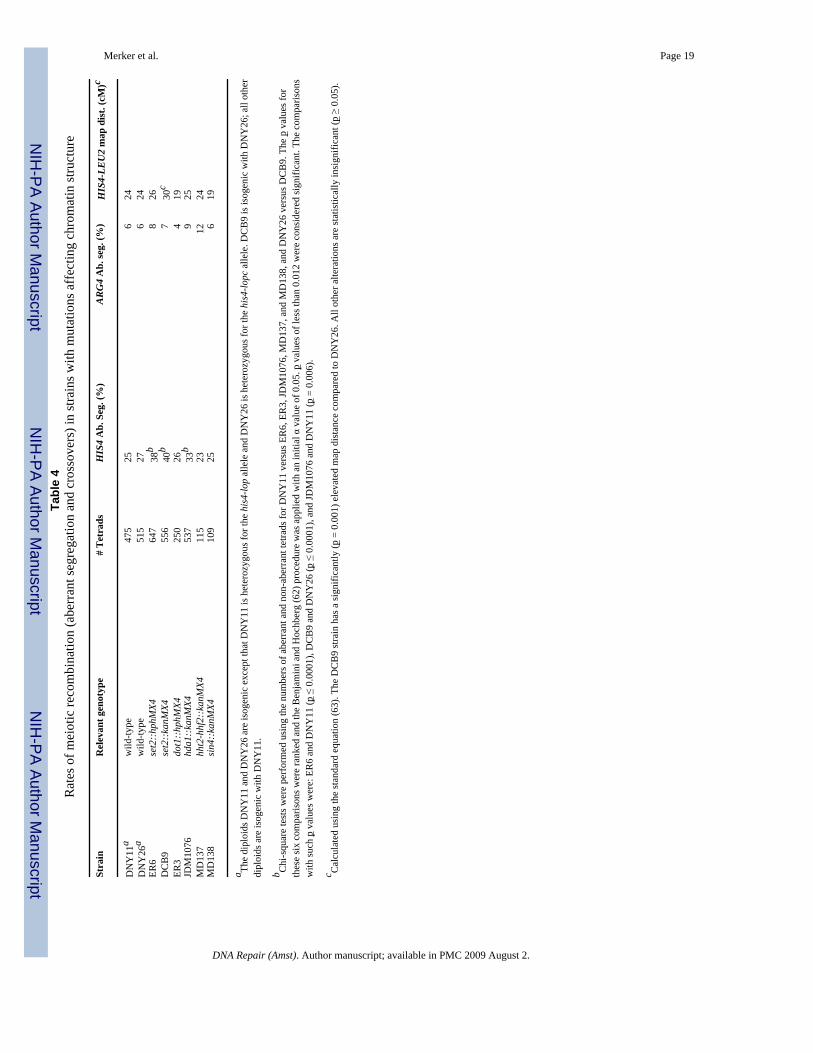
Our analysis is summarized in Table 4. The set2 mutation in two closely-related geneticbackgrounds resulted in a very significant (p < 0.0001) stimulation of the rate of aberrantsegregation for the HIS4, but not the ARG4, marker. Since the rate of aberrant segregation ismuch higher for HIS4 than ARG4 (which affects the power of the statistical test), we cannotrule out the possibility that both loci are affected by the set2 mutation to similar extents; thispossibility, however, is excluded by the physical analysis of DSBs described in Section 3.2.The wild-type strain DNY11 is isogenic (except for the set2 mutation) with ER6 and the wild-type strain DNY26 strain is isogenic with DCB9. We observed a significant increase in crossingover in the HIS4-LEU2 interval for the DNY26/DCB9 comparison, although the increase forthe DNY11/ER6 comparison was not significant. We also found that a mutation of the Hda1pdeacetylase significantly stimulated the rate of aberrant segregation of the HIS4 marker,although the magnitude of this stimulation was half of that observed for the set2 mutation. Lossof the Dot1p methylase, the Sin4p transcription factor, and loss of one copy of the H3-H4histone pair had no effect on the hotspot activity associated with the HIS4 gene.
Merker et al. Page 5
DNA Repair (Amst). Author manuscript; available in PMC 2009 August 2.
NIH
-PA Author Manuscript
NIH
-PA Author Manuscript
NIH
-PA Author Manuscript

As described in the Introduction, Set2p-dependent methylation of H3K36 is involved in therecruitment of the histone deacetylase Rpd3p. Thus, one explanation of the effects of theset2 mutation is that loss of the Set2p-dependent recruitment of Rpd3p results in hyper-acetylated chromatin that allows more efficient entry of the Spo11p recombination machineryand, therefore, more DSBs in the HIS4 region. This model predicts that loss of Rpd3p wouldalso result in a higher level of aberrant segregation. We could not check this prediction bytetrad analysis, since (as reported previously, 53) rpd3 mutants did not complete meiosis andfailed to produce viable spores. An adjustment of the sporulation conditions that allowedrpd3 mutants to complete sporulation in some genetic backgrounds (54) did not allowsporulation in our genetic background. To confirm our conclusions about the effects of theset2 mutation on recombination and to examine the effect of rpd3 mutants on HIS4recombination activity, we measured the rate of meiosis-specific DSBs at HIS4 in wild-type,set2, rpd3, and set2 rpd3 double mutant strains.
3.2. Measurement of meiosis-specific DSBs at the HIS4 and ARG4 loci in wild-type, set2,rpd3, and set2 rpd3 diploid strains
Since meiotic recombination is initiated by a DSB (3,4), the frequency of local DSBs measuredby Southern analysis is a direct measurement of recombination activity. We constructedderivatives of the wild-type, set2, rpd3, and set2 rpd3 diploids (Table 2) that were homozygousfor the rad50S mutation, which prevents subsequent processing of DSBs (45). These strainswere sporulated at 25° C., and DSBs were examined at both the HIS4 and ARG4 loci. TheSouthern analysis for one set of experiments in which we examined DSBs at the HIS4 locusis shown in Fig. 1. Since the efficiency of cells undergoing meiotic DSB formation can varyfrom one experiment to another, we normalized the levels of DSBs measured at the HIS4 locusto the levels observed at the ARG4 locus (as described in Materials and Methods). Normalizingthe ratio of HIS4 to ARG4 DSBs to 1 for the wild-type strains FX1 and FX3, we found thisratio averaged 3.3 for the set2 strains JDM1093 and DCB16, 2.7 for the rpd3 strain JDM1095,and 4.1 for the set2 rpd3 strain JDM1096. We also examined the % DSBs relative to theunbroken parental fragments for HIS4 and ARG4. These values are somewhat more variablethan the normalized HIS4/ARG4 ratio, since the fraction of cells in sporulation medium thatundergo DSB formation is somewhat variable from culture to culture. The % DSBs for HIS4and ARG4, respectively, in the various strains were: 4.5, 2.6 (FX1, wild-type); 3.0, 0.6 (FX3,wild-type), 12.9, 2.7 (JDM1093, set2); 8.3, 0.4 (DCB16, set2); 19.1, 3.9 (JDM1095, rpd3); 18,2.1 (JDM1096, set2 rpd3). These values represent the average of two independent experimentsfor each strain, except for FX3 and DCB16 which represent a single experiment. Although ouranalysis does not rule out the possibility that set2 and rpd3 mutations stimulate DSB formationat ARG4, the degree of stimulation is clearly stronger at HIS4.
These results confirm the conclusion, based on tetrad analysis, that the set2 mutation elevatesrecombination rates at the HIS4 hotspot. This elevation does not reflect the presence of novelDSBs near HIS4, but the strengthening of the same DSB site observed in the wild-type strain(Fig. 1A). The set2 and rpd3 mutations had similar effects on the frequency of HIS4-associatedDSBs, and the set2 rpd3 double mutant strain had an effect that was only slightly stronger thanthe single mutants. This result suggests that Set2p and Rpd3p may be acting in the samepathway to stimulate HIS4 hotspot activity. It should be noted that the increase in DSBformation observed in the mutant strains is substantially greater than the increase in geneconversion rates. In part, this difference is attributable to the observation that only half of theconversion events at HIS4 in cells sporulated at 25° are a consequence of the DSB site shownin Fig. 1A (16).
We also examined DSBs on chromosome III by Southern analysis of chromosomal DNAmolecules separated by CHEF (contour-clamped homogeneous electric field) gel
Merker et al. Page 6
DNA Repair (Amst). Author manuscript; available in PMC 2009 August 2.
NIH
-PA Author Manuscript
NIH
-PA Author Manuscript
NIH
-PA Author Manuscript

electrophoresis (Fig. 1B). As observed previously (55), DSBs on chromosome III occurred intwo clusters, one on the left arm (which includes the HIS4 DSB) and one on the right arm.From the analysis shown in Fig. 1B, it is clear that mutations in set2 and rpd3 substantiallyelevated the DSB frequency of the HIS4 hotspot relative to other DSBs on chromosome III. InFig. 1B, DSBs on the right arm of III appear to be repressed in strains with the set2 and rpd3mutations. It is possible that strengthening of the HIS4 DSB reduces the frequency of DSBson the same chromosome; we and others have previously observed local competition betweenDSB sites. Alternatively, it is possible that Rpd3p- and Set2p-mediated modifications repressmeiotic recombination at some loci and stimulate recombination at others.
3.3. Histone modifications near the HIS4 hotspot in wild-type, set2, rpd3, and set2 rpd3strains
Loss of the Rpd3p deacetylase results in increased acetylation of many sites in H3 and H4histones (56,57), whereas loss of the Set2p methylase directly affects only methylation ofH3K36 (34). The Set2p-mediated methylation, however, helps recruit a repressive Rpd3p-containing complex to chromatin, and elevated histone acetylation was observed for severalloci in set2 mutant strains (35,37). Consequently, we examined the histone acetylation levelsat H3K27 by chromatin immunoprecipitation (ChIP analysis) in the diploids FX1 (wild-type),JDM1093 (set2), JDM1095 (rpd3), and JDM1096 (set2 rpd3). For this analysis, chromatinwas isolated from cells sporulated for six hours, about the time of DSB formation (17). Weused four different PCR products (labeled A–D in Fig. 2A) to look at H3K27 acetylation levelsat four sites in the HIS4 region. The A fragment (located near the 5′ end of BIK1) and the Dfragment (located near the 3′ end of HIS4) are more than 1 kb from the site of the DSB. TheB and C fragments are within about 200 bp of the DSB site. ChIP experiments were done withantibodies directed against acetylation of H3K27, and unmodified histone H3 (as a histoneoccupancy control). In addition, we used an antibody directed against tri-methylation of H3K36in extracts derived from the wild-type, rpd3, and set2 strains.
The ChIP data were normalized in two ways: by comparison to a control DNA fragment (anintergenic region on chromosome V) and by comparison to the level of nucleosome occupancy(monitored using an antibody directed against histone H3); details of these normalizations aredescribed in Materials and Methods. We found that the regions near the DSB sites were hyper-acetylated at the H3K27 sites in the set2, rpd3, and set2 rpd3 strains (Fig. 2B). The acetylationwas elevated most strongly for the sites (B and C fragments) located near the site of the DSB.As expected, the region near the DSB site had lower nucleosome occupancy than the regionsrepresented by the A and D fragments (Fig. 2C). Finally, we found that the tri-methylation ofH3K36 was not substantially affected in rpd3 strains, but was, as expected, eliminated in theset2 strain (Fig. 2D). The interpretation of these results will be described below.
3.4. Meiotic expression of HIS4 and BIK1 is elevated in strains with set2 or rpd3 mutationsAlthough there is not a direct relationship between gene expression and the level of meioticrecombination (2), some mutations reduce both gene expression and meiotic recombination.For example, at HIS4, deletion of the Bas2p transcription factor binding site, which reducesthe level of HIS4 expression, also substantially reduces the HIS4 recombination hotspot activity(19). Consequently, we used real-time PCR to measure the level of BIK1 and HIS4 mRNA inwild-type, set2, rpd3, and set2 rpd3 strains incubated for six hours in sporulation medium. Asshown in Fig. 3, expression levels for both genes (BIK1 levels shown by white bars, HIS4 bystippled bars) were significantly elevated in the mutant strains relative to the wild-type strain.These results indicate that the chromatin alterations associated with the rpd3 and set2 mutationsat the HIS4 locus elevate both meiotic recombination and meiotic gene expression.
Merker et al. Page 7
DNA Repair (Amst). Author manuscript; available in PMC 2009 August 2.
NIH
-PA Author Manuscript
NIH
-PA Author Manuscript
NIH
-PA Author Manuscript

4. DiscussionThe main conclusions from our study are: 1) loss of the Set2 and Rpd3 proteins strongly elevatesHIS4 hotspot activity, whereas loss of the Hda1p has a weaker stimulating effect, and 2) lossof the Dot1 and Sin4 protein, or a reduction in the amount of histones H3 and H4 do not affectHIS4 hotspot activity. These results will be discussed further below.
Since mutations in SET2 and RPD3 stimulate meiosis-specific DSBs at the HIS4 hotspot toapproximately the same extent and since the double mutant strain has a level of DSBs that isnot substantially greater than the single mutants, we favor the possibility that Set2p and Rpd3pact in the same pathway to repress hotspot activity. Similarly, the BIK1 expression level iselevated to about the same extent in the double mutant compared to both single mutants,although HIS4 expression is somewhat greater in the double mutant than in either single mutant(Fig. 3). Based on the recent evidence that Set2p-mediated methylation of H3K36 is involvedin the recruitment of a repressive Rpd3 complex (35–37), the simplest explanation of theseobservations is that this complex negatively regulates both meiotic recombination andtranscription. This negative regulation is likely to be a consequence of a closed chromatinstructure caused by Rpd3p-mediated deacetylation. Our chromatin immunoprecipitationexperiments, although limited in scope, are also consistent with this possibility. We found thatacetylation of H3K27 was elevated in the set2, rpd3, and set2 rpd3 strains near the site of theHIS4 DSB in meiotic cells. These results suggest that the elevated recombination at HIS4reflects a more open chromatin structure resulting from increased acetylation of histones.
There are a number of caveats concerning this explanation. In our study, as in most studies ofthe effects of chromatin-modifying enzymes, it is difficult to distinguish direct from indirecteffects. For example, microarray analysis indicates rpd3Δ affects the expression of over 400genes (29) and one of these gene products could indirectly stimulate meiotic recombination.Although we cannot exclude indirect effects of the set2 and rpd3 mutations, a number ofarguments suggest that the effects at the HIS4 hotspot may be direct, in addition to the ChIPdata discussed above. First, Robyr et al. (58) found that the level of histone H4K12 acetylation(a modification regulated by Rpd3p; 57) in the region adjacent to the HIS4 DSB site is increasedapproximately three-fold in an rpd3Δ strain relative to wild type. Second, Kurdistani et al.(59) found that Rpd3p binds in the HIS4 intergenic region. Third, in our study, the set2 andrpd3 mutations differentially affect the rate of meiotic recombination at HIS4 and ARG4. Thefrequency of DSB formation in the rpd3 and set2 mutants is clearly more elevated at HIS4 thanat ARG4. In addition, the frequency of DSBs at HIS4 is elevated relative to many other DSBsites on chromosome III (Fig. 1B). Thus, our results are unlikely to be explained by theargument that the elevated level of HIS4 recombination reflects increased expression of Spo11pand other components of the meiotic recombination machinery in rpd3 and set2 strains.
Our observation that meiotic expression of HIS4 is somewhat elevated in rpd3 and set2 strainscontrasts with the observation that the level of HIS4 gene expression is reduced by the rpd3mutation (29), although no such reduction was observed in another study (30). Thesedifferences may reflect differences in the pattern of mitotic and meiotic expression and/or straindifferences. We also found that the hda1 mutation significantly elevated the activity of theHIS4 hotspot (Table 4), although this effect was smaller than that observed for the set2 andrpd3 mutations. Since the Hda1p histone deacetylase has somewhat different activities thanthe Rpd3p deacetylase (56), this result may argue that the general level of histone acetylationis more important in regulating HIS4 hotspot activity than a specific type of modification.Although we cannot rule out an indirect effect of hda1 on recombination, HIS4 gene expressionis elevated about three-fold by the hda1 mutation in one study (30), although no effect onexpression was found in another study (29).
Merker et al. Page 8
DNA Repair (Amst). Author manuscript; available in PMC 2009 August 2.
NIH
-PA Author Manuscript
NIH
-PA Author Manuscript
NIH
-PA Author Manuscript

As described in the Introduction, many mutations that reduce HIS4 transcription (deletion oftranscription factors or their binding sites upstream of HIS4) also reduce HIS4 meioticrecombination (11,18–20,47). White et al. (60) showed, however, that deletion of the TATAAsequence upstream of HIS4 significantly decreased HIS4 expression, but had no effect onmeiotic recombination. In this study, we found that the sin4 deletion, which reduces HIS4expression about 15-fold (32), had no effect on HIS4 hotspot activity. Similarly, a reductionin the level of histones H3 and H4 that stimulates HIS4 gene expression had no effect onHIS4 hotspot activity. These results support our earlier conclusion that, although some featuresof chromatin structure favor both gene expression and recombination, other features mayspecifically stimulate one process or the other.
Whatever the precise mechanism responsible for the elevated recombination in the set2 andrpd3 strains, our results demonstrate that the activity of one of the strongest hotspots in thegenome can be further elevated. In an analysis of the ade6-M26 hotspot in S. pombe, Yamadaet al. (12) reported that loss of the SpGcn5 histone acetylase or deletion of the Snf22 histone-remodeling proteins reduced the activity of the hotspot. Since the loss of a histone acetylasewould be expected to decrease acetylation at the ade6-M26 hotspot, the mechanism by whichloss of SpGcn5 reduces hotspot activity may be related to the mechanism by which loss ofSet2p, Rpd3p, and Hda1p stimulate hotspot activity.
Acknowledgements
We thank J. Lieb for discussions concerning chromatin structure. This research was supported by National Institutesof Health grants GM24110 (TDP) and GM68088 (BDS). B. D. Strahl is a Pew Scholar in the Biomedical Sciences.
References1. Lichten M, Goldman ASH. Meiotic recombination hotspots. Ann Rev Genet 1995;29:423–444.
[PubMed: 8825482]2. Petes TD. Meiotic recombination hot spots and cold spots. Nature 2001;2:360–369.3. Sun H, Treco D, Schultes NP, Szostak JW. Double strand breaks at an initiation site for meiotic gene
conversion. Nature 1989;338:87–90. [PubMed: 2645528]4. Szostak JW, Orr-Weaver TL, Rothstein R, Stahl FW. The double-strand-break repair model for
recombination. Cell 1983;33:25–35. [PubMed: 6380756]5. Keeney S, Giroux CN, Kleckner N. Meiosis-specific DNA double-strand breaks are catalyzed by
Spo11, a member of a widely conserved protein family. Cell 1997;88:375–384. [PubMed: 9039264]6. Fan QQ, Petes TD. Relationship between nuclease-hypersensitive sites and meiotic recombination hot
spot activity at the HIS4 locus of Saccharomyces cerevisiae. Mol Cell Biol 1996;16:2037–2043.[PubMed: 8628269]
7. Keeney S, Kleckner N. Communication between homologous chromosomes: genetic alterations at anuclease-hypersensitivity site can alter meiotic chromatin structure at that site both in cis and intrans. Genes Cells 1996;1:475–489. [PubMed: 9078379]
8. Mizuno K, Emura Y, Baur M, Kohli J, Ohta K, et al. The meiotic recombination hotspot created bythe single-base substitution ade6-M26 results in remodeling of chromatin structure in fission yeast.Genes Dev 1997;11:876–886. [PubMed: 9106659]
9. Ohta K, Shibata T, Nicolas A. Changes in chromatin structure at recombination initiation sites duringyeast meiosis. EMBO J 1994;13:5754–5763. [PubMed: 7988571]
10. Wu TC, Lichten M. Meiosis-induced double-strand breaks determined by yeast chromatin structure.Science 1994;263:515–518. [PubMed: 8290959]
11. Kirkpatrick DT, Wang YH, Dominska M, Griffith JD, Petes TD. Control of meiotic recombinationand gene expression in yeast by a simple repetitive DNA sequence that excludes nucleosomes. MolCell Biol 1999;19:7661–7671. [PubMed: 10523654]
Merker et al. Page 9
DNA Repair (Amst). Author manuscript; available in PMC 2009 August 2.
NIH
-PA Author Manuscript
NIH
-PA Author Manuscript
NIH
-PA Author Manuscript

12. Yamada T, Mizuno K, Hirota K, Kon N, Wahls WP, et al. Roles of histone acetylation and chromatinremodeling factor in a meiotic recombination hotspot. EMBO J 2004;23:1792–1803. [PubMed:14988732]
13. Sollier J, Lin W, Soustelle C, Suhre K, Nicolas A, et al. Set1 is required for meiotic S-phase onset,double-strand break formation and middle gene expression. EMBO J 2004;23:1957–1967. [PubMed:15071505]
14. Yamashita K, Shinohara M, Shinohara A. Rad6-Bre1-mediated histone H2B ubiquitylation modulatesthe formation of double-strand breaks during meiosis. Proc Natl Acad Sci USA 2004;101:11380–11385. [PubMed: 15280549]
15. Gerton J, Derisi J, Shroff R, Lichten M, Brown PO, et al. Global mapping of meiotic recombinationhotspots and coldspots in the yeast Saccharomyces cerevisiae. Proc Natl Acad Sci USA2000;97:11383–11390. [PubMed: 11027339]
16. Fan QQ, Xu F, Petes TD. Meiosis-specific double-strand DNA breaks at the HIS4 recombination hotspot in the yeast Saccharomyces cerevisiae: control in cis and trans. Mol Cell Biol 1995;15:1679–1688. [PubMed: 7862159]
17. Nag DK, Petes TD. Physical detection of heteroduplexes during meiotic recombination in the yeastSaccharomyces cerevisiae. Mol Cell Biol 1993;13:2324–2331. [PubMed: 8455614]
18. Abdullah MFF, Borts RH. Meiotic recombination frequencies are affected by nutritional states inSaccharomyces cerevisiae. Proc Natl Acad Sci USA 2001;98:14524–14529. [PubMed: 11724920]
19. White MA, Dominska M, Petes TD. Transcription factors are required for the meiotic recombinationhotspot at the HIS4 locus in Saccharomyces cerevisiae. Proc Natl Acad Sci USA 1993;90:6621–6625. [PubMed: 8341678]
20. White MA, Wierdl M, Detloff P, Petes TD. DNA-binding protein RAP1 stimulates meioticrecombination at the HIS4 locus in yeast. Proc Natl Acad Sci USA 1991;88:9755–9759. [PubMed:1946399]
21. Devlin C, Tice-Baldwin K, Shore D, Arndt K. RAP1 is required for BAS1/BAS2- and GCN4-dependent transcription for the yeast HIS4 gene. Mol Cell Biol 1991;11:3642–3651. [PubMed:1904543]
22. Yu L, Morse RH. Chromatin opening and transactivator potentiation by RAP1 in Saccharomycescerevisiae. Mol Cell Biol 1999;19:5279–5288. [PubMed: 10409719]
23. Workman JL, Kingston RE. Alteration of nucleosome structure as a mechanism of transcriptionalactivation. Annu Rev Biochem 1998;67:545–579. [PubMed: 9759497]
24. Strahl BD, Allis CD. The language of covalent histone modifications. Nature 2000;403:41–45.[PubMed: 10638745]
25. Hansen JC, Tse C, Wolffe AP. Structure and function of the core histone N-termini: more than meetsthe eye. Biochemistry 1998;37:17637–17641. [PubMed: 9922128]
26. Kadosh D, Struhl K. Targeted recruitment of the Sin3-Rpd3 histone deacetylase complex generatesa highly localized domain of repressed chromatin in vivo. Mol Cell Biol 1998;18:5121–5127.[PubMed: 9710596]
27. Vignali M, Hassan AH, Neely KE, Workman JL. ATP-dependent chromatin remodeling complexes.Mol Cell Biol 2000;20:1899–1910. [PubMed: 10688638]
28. Xiao T, Hall H, Kizer KO, Shibata Y, Hall MC, et al. Phosphorylation of RNA polymerase II CTDregulates H3 methylation in yeast. Genes Dev 2003;17:1–10. [PubMed: 12514094]
29. Bernstein BE, Tong JK, Schreiber SL. Genomewide studies of histone deacetylase function in yeast.Proc Natl Acad Sci USA 2000;97:13708–13713. [PubMed: 11095743]
30. Hughes TR, Marton MJ, Jones AR, Roberts CJ, Stoughton R, et al. Functional discovery via acompendium of expression profiles. Cell 2000;102:109–126. [PubMed: 10929718]
31. Wu J, Carmen AA, Kobayashi R, Suka N, Grunstein M. HDA2 and HDA3 are related proteins thatinteract with and are essential for the activity of the yeast histone deacetylase HDA1. Proc Natl AcadSci USA 2001;98:4391–4396. [PubMed: 11287668]
32. Jiang YW, Stillman DJ. Regulation of HIS4 expression by the Saccharomyces cerevisiae SIN4transcriptional regulator. Genetics 1995;140:103–114. [PubMed: 7635278]
Merker et al. Page 10
DNA Repair (Amst). Author manuscript; available in PMC 2009 August 2.
NIH
-PA Author Manuscript
NIH
-PA Author Manuscript
NIH
-PA Author Manuscript

33. Wyrick JJ, Holstege FCP, Jennings EG, Causton HC, Shore D, et al. Chromosomal landscape ofnucleosome-dependent gene expression and silencing in yeast. Nature 1999;402:418–421. [PubMed:10586882]
34. Strahl BD, Grant PA, Briggs SD, Sun ZW, Bone JR, et al. Set2 is a nucleosomal histone H3-selectivemethyltransferase that mediates transcriptional repression. Mol Cell Biol 2002;22:1298–1306.[PubMed: 11839797]
35. Carrozza MJ, Li B, Florens L, Suganua T, Swanson SK, et al. Histone H3 methylation by Set2 directsdeacetylation of coding regions by Rpd3S to suppress spurious transcription. Cell 2005;123:581–592. [PubMed: 16286007]
36. Joshi AA, Struhl K. Eaf3 chromodomain interaction with methylated H3-K36 links histonedeacetylation to Pol II elongation. Mol Cell 2005;20:971–978. [PubMed: 16364921]
37. Keogh MC, Kurdistani SK, Morris SA, Ahn SH, Podolny V, et al. Cotranscriptional Set2 methylationof histone H3 Lysine 36 recruits a repressive Rpd3 complex. Cell 2005;123:593–605. [PubMed:16286008]
38. Feng Q, Wang H, Ng HH, Erdjument H, Tempst P, et al. Methylation of H3-lysine 79 is mediated bya new family of HMTases without a SET domain. Curr Biol 2002;12:1052–1058. [PubMed:12123582]
39. Ng HH, Feng Q, Wang H, Erdjument-Bromage H, Tempst P, et al. Lysine methylation within theglobular domain of histone H3 by Dot1 is important for telomeric silencing and Sir proteinassociation. Genes Dev 2002;16:1518–1527. [PubMed: 12080090]
40. Ng HH, Ciccone DN, Morshead KB, Oettinger MA, Struhl K. Lysine-79 of histone H3 ishypomethylated at silenced loci in yeast and mammalian cells: a potential mechanism for position-effect variegation. Proc Natl Acad Sci USA 2003;100:1820–1825. [PubMed: 12574507]
41. Pokholok DK, Harbison CT, Levine S, Cole M, Hannett NM, et al. Genome-wide map of nucleosomeacetylation and methylation in yeast. Cell 2005;122:517–527. [PubMed: 16122420]
42. Stapleton A, Petes TD. The Tn3 beta-lactamase gene acts as a hotspot for meiotic recombination inyeast. Genetics 1991;127:39–51. [PubMed: 1849855]
43. Wach AA, Brachat A, Pohlmann R, Philippsen P. New heterologous modules for classical or PCR-based gene disruptions in Saccharomyces cerevisiae. Yeast 1994;10:1793–808. [PubMed: 7747518]
44. Goldstein AL, McCusker JH. Three new dominant drug resistance cassettes for gene disruption inSaccharomyces cerevisiae. Yeast 1999;15:1541–1553. [PubMed: 10514571]
45. Alani E, Padmore R, Kleckner N. Analysis of wild-type and rad50 mutants of yeast suggests anintimate relationship between meiotic chromosome synapsis and recombination. Cell 1990;61:419–436. [PubMed: 2185891]
46. Sherman, F.; Fink, GR.; Hicks, JB. Methods in Yeast Genetics. Cold Spring Harbor Laboratory; ColdSpring Harbor, NY: 1983.
47. Kirkpatrick DT, Fan QQ, Petes TD. Maximal stimulation of meiotic recombination by a yeasttranscription factor requires the transcription activation domain and a DNA binding domain. Genetics1999;152:101–115. [PubMed: 10224246]
48. Lobachev KS, Gordenin DA, Resnick MA. The Mre11 complex is required for repair of hairpin-capped double-strand breaks and prevention of chromosome rearrangements. Cell 2002;108:183–193. [PubMed: 11832209]
49. Xiao T, Shibata Y, Rao B, Laribee RN, O’Rourke R, et al. The RNA polymerase II kinase Ctk1regulates positioning of a 5′ histone methylation boundary along genes. Mol Cell Biol 2007;27:721–731. [PubMed: 17088384]
50. Chu S, DeRisi J, Eisen M, Mulholland J, Botstein D, Brown PO, et al. The transcriptional programof sporulation in budding yeast. Science 1998;282:699–705. [PubMed: 9784122]
51. Paques F, Haber JE. Multiple pathways of recombination induced by double-strand breaks inSaccharomyces cerevisiae. Microbiol Mol Biol Rev 1999;63:349–404. [PubMed: 10357855]
52. Petes, TD.; Malone, RE.; Symington, LS. Recombination in Yeast. In: Broach, J.; Jones, E.; Pringle,J., editors. The Molecular and Cellular Biology of the Yeast Saccharomyces: Genome Dynamics,Protein Synthesis, and Energetics. Cold Spring Harbor Laboratory Press; Cold Spring Harbor, NY:1991. p. 407-521.
Merker et al. Page 11
DNA Repair (Amst). Author manuscript; available in PMC 2009 August 2.
NIH
-PA Author Manuscript
NIH
-PA Author Manuscript
NIH
-PA Author Manuscript

53. Dora EG, Rudin N, Martell JR, Esposito MS, Ramirez RM. RPD3 (REC3) mutations affect mitoticrecombination in Saccharomyces cerevisiae. Curr Genet 1999;35:68–76. [PubMed: 10079324]
54. Burgess SM, Ajimura M, Kleckner N. GCN5-dependent histone H3 acetylation and RPD3-dependenthistone H4 deacetylation have distinct, opposing effects on IME2 transcription, during meiosis andduring vegetative growth in budding yeast. Proc Natl Acad Sci USA 1999;96:6835–6840. [PubMed:10359799]
55. Zenvirth D, Arbel T, Sherman A, Goldway M, Klein S, et al. Multiple sites for double-strand breaksin whole meiotic chromosomes of Saccharomyces cerevisiae. EMBO J 1992;11:3441–3447.[PubMed: 1324174]
56. Rundlett SE, Carmen AA, Kobayashi R, Bavykin S, Turner BM, et al. HDA1 and RPD3 are membersof distinct yeast histone deacetylase complexes that regulate silencing and transcription. Proc NatlAcad Sci USA 1996;93:14503–14508. [PubMed: 8962081]
57. Suka N, Suka Y, Carmen AA, Wu J, Grunstein M. New antibodies for sites of acetylation determinenovel histone site usage in heterochromatin and euchromatin. Mol Cell 2001;8:473–479. [PubMed:11545749]
58. Robyr D, Suka Y, Xenarios I, Kurdistani SK, Wang A, et al. Microarray deacetylation maps determinegenome-wide functions for yeast histone deacetylases. Cell 2002;109:437–446. [PubMed:12086601]
59. Kurdistani SK, Robyr D, Tavazoie S, Grunstein M. Genome-wide binding map of the histonedeacetylase Rpd3 in yeast. Nat Genet 2002;31:248–254. [PubMed: 12089521]
60. White MA, Detloff P, Strand M, Petes TD. A promoter deletion reduces the rate of mitotic, but notmeiotic, recombination at the HIS4 locus in yeast. Curr Genet 1992;21:109–116. [PubMed: 1568254]
61. Nag DK, White MA, Petes TD. Palindromic sequences in heteroduplex DNA inhibit mismatch repairin yeast. Nature 1989;340:318–320. [PubMed: 2546083]
62. Benjamini Y, Hochberg Y. Controlling the false discovery rate: a practical and powerful approach tomultiple testing. J R Stat Soc 1995;57:289–300.
63. Perkins DD. Biochemical mutants in the smut fungus Ustilago maydis. Genetics 1949;34:607–626.
Merker et al. Page 12
DNA Repair (Amst). Author manuscript; available in PMC 2009 August 2.
NIH
-PA Author Manuscript
NIH
-PA Author Manuscript
NIH
-PA Author Manuscript

Figure 1. Southern analysis of HIS4 meiosis-specific DSBs in chromatin structure mutantsDNA was isolated from cells just prior to (0 hr) and after a 24 hr (Fig. 1A) or 48 h (Fig. 1B)incubation period on solid sporulation medium. Samples were examined by standard gelelectrophoresis (Fig. 1A) or CHEF gel electrophoresis (Fig. 1B).A. The DNA was digested with BglII, and examined by Southern analysis using a XhoI-BglIIfragment of HIS4 as a hybridization probe (shown as a double-headed arrow in the lower partof the figure). The parental band (at roughly 3 kb) represents the unbroken BglII fragments,while the DSB band (at roughly 1.9 kb) represents fragments with a meiosis-specific DSB atHIS4. All strains are homozygous for the rad50S mutation. FX1 has no mutations in genesaffecting chromatin structure. JDM1093, JDM1095, and JDM1096 are isogenic derivatives ofFX1 homozygous for set2Δ, rpd3Δ, and set2Δ rpd3Δ mutations, respectively. In the schematic,horizontal arrows indicate the direction of transcription and the vertical arrow shows the DSBsite associated with the HIS4 hotspot.B. For CHEF gel electrophoresis, DNA was prepared and the resulting molecules separatedby electrophoresis using the methods described by Lobachev et al. (48). Followingelectrophoresis, the chromosomal DNA molecules were transferred to a Nylon membrane andhybridized to CHA1 sequences, a gene located near the left end of chromosome III. The bandof hybridization near the top of the gel represents chromosome III molecules without a DSB.The chromosomal DNA with a DSB at the HIS4 hotspot is shown with an arrow. ChromosomeIII is depicted in the lower part of the figure. The short vertical lines represent the positions ofmeiotic recombination hotspots determined by microarray analysis (15).
Merker et al. Page 13
DNA Repair (Amst). Author manuscript; available in PMC 2009 August 2.
NIH
-PA Author Manuscript
NIH
-PA Author Manuscript
NIH
-PA Author Manuscript

Figure 2. Chromatin modifications near the HIS4 hotspot examined by chromatinimmunoprecipitationCell extracts were prepared from the same diploid strains that were analyzed for DSBs: FX1(wild-type, white bars), JDM1093 (set2, striped bars), JDM1095 (rpd3, black bars), andJDM1096 (set2 rpd3, stippled bars). The levels of histone modifications for four differentfragments (A–D, Fig. 2A) were monitored by multiplex PCR. The arrow in Fig. 2A shows theapproximate location of the recombination-associated DSB. Antibodies against H3K27acetylation (panel B), histone H3 (panel C), or H3K36 tri-methylation (panel D) were used toprecipitate chromatin harvested from meiotic cells incubated for six hours in sporulationmedium. As described in the Materials and Methods, the levels of acetylation and methylationwere normalized to a control region on chromosome V. In addition, the levels of acetylationand methylation were normalized for histone occupancy, using the data derived from theimmunoprecipitations shown in Fig. 2C. Error bars represent the standard error of the mean.
Merker et al. Page 14
DNA Repair (Amst). Author manuscript; available in PMC 2009 August 2.
NIH
-PA Author Manuscript
NIH
-PA Author Manuscript
NIH
-PA Author Manuscript

Figure 3. Meiotic gene expression of HIS4 and BIK1 in wild-type, set2, rpd3, and set2 rpd3 strainsRNA was isolated from wild-type (FX1), set2 (JDM1093), rpd3 (JDM1095), and set2 rpd3strains that had been incubated for six hours in sporulation medium. Following reversetranscription of the RNA, we measured the levels of the resulting BIK1 (shown by the whitebars) and HIS4 (shown by the stippled bars) cDNAs by real-time PCR (details in Materials andMethods). Following normalization of the amounts to the level of RNA1 (a gene whoseexpression is unaffected by set2 or rpd3 mutations), the expression levels were normalized tothe level of HIS4 expression in the wild-type strain. Bars represent 95% confidence limits.
Merker et al. Page 15
DNA Repair (Amst). Author manuscript; available in PMC 2009 August 2.
NIH
-PA Author Manuscript
NIH
-PA Author Manuscript
NIH
-PA Author Manuscript

NIH
-PA Author Manuscript
NIH
-PA Author Manuscript
NIH
-PA Author Manuscript
Merker et al. Page 16Ta
ble
1G
enot
ypes
of h
aplo
id y
east
stra
ins
Stra
inR
elev
ant g
enot
ype;
ref
eren
ceN
ame
of ra
d50S
der
ivat
ive;
ref
eren
ce
AS4
-der
ived
hap
loid
sA
S4W
T; 4
3D
NY
107;
16
ER4
set2
::hp
hMX4
JDM
205
DC
B4
set2
::ka
nMX4
DC
B14
MD
136
rpd3
::ka
nMX4
JDM
210
ER2
dot1
::hp
hMX4
JDM
166
hda1
::ka
nMX4
MD
131
hht2
-hhf
2::k
anM
X4M
D13
3si
n4::
kanM
X4JD
M21
4se
t2::
hphM
X4 rp
d3::
kanM
X4 ra
d50S
JDM
214
AS1
3-de
rive
d ha
ploi
dsA
S13
WT;
42
DN
Y9
his4
-lop;
16
HF1
; 16
DN
Y25
his4
-lopc
; 16
HF4
; 16
ER5
his4
-lop
set2
::hp
hMX4
JDM
204
DC
B6
his4
-lopc
set2
::ka
nMX4
DC
B15
MD
135
his4
-lop
rpd3
::ka
nMX4
JDM
131
ER1
his4
-lop
dot1
::hp
hMX4
MD
141
his4
-lop
hda1
::ka
nMX4
MD
132
his4
-lop
hht2
-hhf
2::k
anM
X4M
D13
4hi
s4-lo
p si
n4::
kanM
X4JD
M21
3hi
s4-lo
p se
t2::
hphM
X4 rp
d3::
kanM
X4 ra
d50S
JDM
213
All
hapl
oid
stra
ins w
ere
deriv
ed fr
om A
S4 (α
arg
4-17
trp1
tyr7
-1 a
de6
ura3
) and
AS1
3 (a
leu2
ade
6 ur
a3 rm
e1) b
y tra
nsfo
rmat
ion
or b
y cr
osse
s with
isog
enic
stra
ins.
The
mut
ant h
is4-
lop
and
his4
-lo
pc a
re 2
6 bp
pal
indr
omic
inse
rtion
s tha
t res
ult i
n in
effic
ient
ly-r
epai
red
mis
mat
ches
(61)
. Onl
y th
ose
mar
kers
that
are
diff
eren
t fro
m th
e ha
ploi
d pr
ogen
itor s
train
s are
show
n. If
no
refe
renc
e is
giv
en,
the
stra
ins w
ere
cons
truct
ed in
this
stud
y.
DNA Repair (Amst). Author manuscript; available in PMC 2009 August 2.

NIH
-PA Author Manuscript
NIH
-PA Author Manuscript
NIH
-PA Author Manuscript
Merker et al. Page 17Ta
ble
2D
iplo
id y
east
stra
ins u
sed
in th
is st
udy
Stra
inR
elev
ant h
omoz
ygou
s mut
atio
nsC
ross
; ref
eren
cea
Stra
ins h
eter
ozyg
ous f
or h
is4-lo
p
DN
Y11
WT
AS4
X D
NY
9; 1
6FX
1ra
d50S
::U
RA3
DN
Y10
7 X
HF1
; 16
ER6
set2
::hp
hMX4
ER4
X E
R5
JDM
1093
set2
::hp
hMX4
rad5
0S::
URA
3JD
M20
5 X
JDM
204
JDM
1094
rpd3
::ka
nMX4
MD
136
X M
D13
5JD
M10
95rp
d3::
kanM
X4 ra
d50S
::U
RA3
JDM
210
X JD
M13
1ER
3do
t1::
hphM
X4ER
2 X
ER
1JD
M10
76hd
a1::
kanM
X4JD
M16
6 X
MD
141
MD
137
hht2
-hhf
2::k
anM
X4M
D13
1 X
MD
132
MD
138
sin4
::ka
nMX4
MD
133
X M
D13
4JD
M10
96se
t2::
hphM
X4 rp
d3::
kanM
X4 ra
d50S
::U
RA3
JDM
214
X JD
M21
3
Stra
ins h
eter
ozyg
ous f
or h
is4-lo
pc
DN
Y26
WT
AS4
X D
NY
25; 1
6FX
3ra
d50S
::U
RA3
DN
Y10
7 X
HF4
; 16
DC
B9
set2
::ka
nMX
DC
B4
X D
CB
6D
CB
16se
t2::
kanM
X ra
d50S
::U
RA3
DC
B14
X D
CB
15
a Gen
otyp
es o
f the
hap
loid
s use
d in
the
cons
truct
ions
are
giv
en in
Tab
le 1
. In
cros
ses,
AS4
- and
AS1
3-de
rived
stra
ins a
re sh
own
on th
e le
ft an
d th
e rig
ht o
f the
X, r
espe
ctiv
ely.
DNA Repair (Amst). Author manuscript; available in PMC 2009 August 2.

NIH
-PA Author Manuscript
NIH
-PA Author Manuscript
NIH
-PA Author Manuscript
Merker et al. Page 18Ta
ble
3Pr
imer
s use
d to
mak
e de
letio
ns w
ith k
anM
X4 o
r hph
MX4
cas
sette
sa and
for o
ther
type
s of a
naly
sis.
Prim
er n
ame
Sequ
ence
(5′-3′)
set2Δ-
FA
GT
CG
TG
CT
GT
CA
AA
CC
TT
TC
TC
CT
TT
CC
TG
GT
TG
TT
GT
TT
TA
CG
TG
AT
CC
GTA
CG
CTG
CA
GG
TCG
AC
set2Δ-
RC
TT
TG
GG
AC
AG
AA
AA
CG
TG
AA
AC
AA
GC
CC
CA
AA
TA
TG
CA
TG
TC
TG
GT
TA
AA
TCG
ATG
AA
TTC
GA
GC
TCG
Set2
del
UP
TC
AC
AT
TA
CC
TA
TC
AT
TA
CA
GT
GC
TA
CT
GA
TA
GC
CC
AC
TA
AT
GC
GG
AT
AT
CG
TAC
GC
TGC
AG
GTC
GA
CSe
t2 d
el D
NT
CT
CC
CA
GT
CC
CA
AA
GA
CT
AG
AG
CA
CA
AC
TG
GA
AT
AA
AT
TC
TT
TG
CA
TC
TA
TCG
ATG
AA
TTC
GA
GC
TCG
rpd3Δ-
FT
GC
GC
CA
TA
CA
AA
AC
AT
TC
GT
GG
CT
AC
AA
CT
CG
AT
AT
CC
GT
GC
AG
CG
TAC
GC
TGC
AG
GTC
GA
Crp
d3Δ-
RT
TG
TT
TC
AC
AT
TA
TT
TA
TA
TT
CG
TA
TA
TA
CT
TC
CA
AC
TC
TT
TT
TT
ATC
GA
TGA
ATT
CG
AG
CTC
dot1Δ-
FA
AG
GA
GG
TC
AC
CA
GT
AA
TT
GT
GC
GC
TT
TG
GT
TA
CA
TT
TT
GT
TG
TA
CA
GT
AC
GTA
CG
CTG
CA
GG
TCG
AC
dot1Δ-
RT
AT
TT
CT
AC
TT
AG
TT
AT
TC
AT
AC
TC
AT
CG
TT
AA
AA
GC
CG
TT
CA
AA
GT
GC
CA
TCG
ATG
AA
TTC
GA
GC
TCG
hda1Δ-
FG
AG
AA
AG
GG
AA
AG
TT
GA
GC
AC
TG
TA
AT
AC
GC
CG
AA
CA
GA
TT
AA
GC
CG
TAC
GC
TGC
AG
GTC
GA
Chd
a1Δ-
RG
GC
AT
GA
AG
GT
TG
CC
GA
AA
AA
AA
AT
TA
TT
AA
TG
GC
CA
GT
TT
TT
CC
ATC
GA
TGA
ATT
CG
AG
CTC
hht2
-hhf
2Δ-F
CC
CC
CA
GT
CT
AA
AT
GC
AT
AG
AA
AA
AA
AA
AA
AT
TC
CC
GC
TT
TA
TA
TC
GTA
CG
CTG
CA
GG
TCG
AC
hht2
-hhf
2Δ-R
bG
GC
AT
GA
AA
AT
AA
TT
TC
AA
CA
CC
GA
TT
GT
TT
AA
CC
AC
CG
AT
TG
TT
ATC
GA
TGA
ATT
CG
AG
CTC
Gsi
n4Δ-
FA
GA
AA
AG
AA
AC
TA
GC
AG
AC
CT
GA
CC
TT
CT
GT
TG
GT
AA
AT
AT
TA
GT
CG
TAC
GC
TGC
AG
GTC
GA
Csi
n4Δ-
RA
TG
TT
TA
AA
AC
AA
TT
CT
AT
AC
AA
AA
CT
AT
GC
TA
TA
GT
AC
TA
AT
AA
ATC
GA
TGA
ATT
CG
AG
CTC
GB
IK1c
odpr
e350
FG
GA
TAG
ATA
TCA
AA
GA
AA
GA
TAG
GA
TGTT
TCB
IK1c
odpr
e350
RG
GG
ATT
TAG
GA
TCA
TCC
ATG
GG
BIK
1cod
post
300F
GG
TGC
CA
AC
GA
AG
ATG
TCG
BIK
1cod
post
300R
GA
AA
ATT
GG
CA
AC
GA
TTC
CA
CH
IS4p
ro24
2FG
GC
AG
TCG
AA
CTG
AC
TCTA
ATA
GTG
HIS
4pro
242R
CTA
TTG
TATT
AC
TATT
AC
AC
AG
CG
CA
GH
IS4c
odpo
st18
0FC
GTG
TGG
ATA
TTG
TTC
GTA
AA
TGH
IS4c
odpo
st18
0RG
TAA
GC
AC
CC
AC
AA
ATA
CG
GA
CIn
terg
enic
V-1
GG
CTG
TCA
GA
ATA
TGG
GG
CC
GTA
GTA
Inte
rgen
icV
-2C
AC
CC
CG
AA
GC
TGC
TTTC
AC
AA
TAC
Chr
III-
1583
0C
TGG
AA
ATA
TGA
AA
TTG
TCA
GC
GA
CC
hrII
I-16
773
TGA
ATG
CC
TTC
AA
CC
AA
GTG
GC
TCC
TTC
his4
_185
_UC
GTT
GC
TGC
TATG
GC
TTA
CG
his4
_185
_LC
CA
CA
TCG
GC
ATC
TTC
ATC
GB
IK1-
388-
FC
AC
AG
CG
GTA
ATC
AA
CA
GTC
BIK
1-53
1-R
GC
TGG
TGG
TGTT
GTG
ATT
AG
RN
A1_
128_
UC
TGC
TGG
TTC
AG
ATG
AA
GTC
RN
A1_
128_
LTT
CC
ATA
GC
CG
GTA
AG
AA
GG
a Gen
e de
letio
ns w
ere
mad
e us
ing
PCR
frag
men
ts c
onta
inin
g a
sele
ctab
le m
arke
r fla
nked
by
sequ
ence
s fro
m th
e ge
ne to
be
dele
ted
(43,
44).
For m
ost o
f the
se c
onst
ruct
ions
, the
sequ
ence
s of t
heol
igon
ucle
otid
es m
atch
thos
e lo
cate
d im
med
iate
ly u
pstre
am a
nd d
owns
tream
of t
he g
ene
to b
e de
lete
d. T
he o
nly
exce
ptio
ns a
re th
e Se
t2 d
el U
P an
d Se
t2 d
el D
N o
ligon
ucle
otid
es. S
et2
DN
UP
is lo
cate
dw
ithin
SET
2, a
bout
300
bp
from
the
end
of th
e ge
ne, a
nd S
et2p
UP
is lo
cate
d ab
out 1
50 b
p do
wns
tream
of S
ET2.
Seq
uenc
es h
omol
ogou
s to
the
regi
on fl
anki
ng th
e ta
rget
gen
e ar
e sh
own
in b
old-
face
type
. Seq
uenc
es h
omol
ogou
s to
the
sele
ctab
le c
asse
tte u
sed
to m
ake
the
disr
uptio
n ar
e sh
own
in re
gula
r typ
e. T
he p
rimer
s lab
eled
“B
IK1”
, “H
IS4”
, and
“In
terg
enic
” w
ere
used
in c
hrom
atin
imm
unop
reci
pita
tion
expe
rimen
ts.
b The
prim
er h
ht2-
hhf2Δ-
R is
mis
sing
a si
ngle
bas
e re
lativ
e to
the
regi
on d
owns
tream
of H
HF2
, whi
ch c
reat
es a
sing
le b
ase
pair
dele
tion
whe
n th
e PC
R fr
agm
ent i
s ins
erte
d in
the
locu
s. Th
e co
rrec
tse
quen
ce is
GG
CA
TGA
AA
ATA
ATT
TCA
AA
CA
CC
GA
TTG
TTTA
AC
CA
CC
GA
TTG
TT, w
ith th
e de
lete
d ba
se u
nder
lined
.
DNA Repair (Amst). Author manuscript; available in PMC 2009 August 2.
NIH
-PA Author Manuscript
NIH
-PA Author Manuscript
NIH
-PA Author Manuscript
Merker et al. Page 19Ta
ble
4R
ates
of m
eiot
ic re
com
bina
tion
(abe
rran
t seg
rega
tion
and
cros
sove
rs) i
n st
rain
s with
mut
atio
ns a
ffec
ting
chro
mat
in st
ruct
ure
Stra
inR
elev
ant g
enot
ype
# T
etra
dsH
IS4
Ab.
Seg
. (%
)AR
G4
Ab.
seg.
(%)
HIS
4-LE
U2
map
dis
t. (c
M)c
DN
Y11
aw
ild-ty
pe47
525
624
DN
Y26
aw
ild-ty
pe51
527
624
ER6
set2
::hp
hMX4
647
38b
826
DC
B9
set2
::ka
nMX4
556
40b
730
cER
3do
t1::
hphM
X425
026
419
JDM
1076
hda1
::ka
nMX4
537
33b
925
MD
137
hht2
-hhf
2::k
anM
X411
523
1224
MD
138
sin4
::ka
nMX4
109
256
19
a The
dipl
oids
DN
Y11
and
DN
Y26
are
isog
enic
exc
ept t
hat D
NY
11 is
het
eroz
ygou
s for
the
his4
-lop
alle
le a
nd D
NY
26 is
het
eroz
ygou
s for
the
his4
-lopc
alle
le. D
CB
9 is
isog
enic
with
DN
Y26
; all
othe
rdi
ploi
ds a
re is
ogen
ic w
ith D
NY
11.
b Chi
-squ
are
test
s wer
e pe
rfor
med
usi
ng th
e nu
mbe
rs o
f abe
rran
t and
non
-abe
rran
t tet
rads
for D
NY
11 v
ersu
s ER
6, E
R3,
JDM
1076
, MD
137,
and
MD
138,
and
DN
Y26
ver
sus D
CB
9. T
he p
val
ues f
orth
ese
six
com
paris
ons w
ere
rank
ed a
nd th
e B
enja
min
i and
Hoc
hber
g (6
2) p
roce
dure
was
app
lied
with
an
initi
al α
val
ue o
f 0.0
5. p
val
ues o
f les
s tha
n 0.
012
wer
e co
nsid
ered
sign
ifica
nt. T
he c
ompa
rison
sw
ith su
ch p
val
ues w
ere:
ER
6 an
d D
NY
11 (p
≤ 0
.000
1), D
CB
9 an
d D
NY
26 (p
≤ 0
.000
1), a
nd JD
M10
76 a
nd D
NY
11 (p
= 0
.006
).
c Cal
cula
ted
usin
g th
e st
anda
rd e
quat
ion
(63)
. The
DC
B9
stra
in h
as a
sign
ifica
ntly
(p =
0.0
01) e
leva
ted
map
dis
tanc
e co
mpa
red
to D
NY
26. A
ll ot
her a
ltera
tions
are
stat
istic
ally
insi
gnifi
cant
(p ≥
0.0
5).
DNA Repair (Amst). Author manuscript; available in PMC 2009 August 2.