Chromosomal Protein HMGN1 Modulates Histone H3 Phosphorylation

Biochimica et Biophysica Acta 1819 (2012) 332–342
Contents lists available at ScienceDirect
Biochimica et Biophysica Acta
j ourna l homepage: www.e lsev ie r.com/ locate /bbagrm
Histone exchange and histone modifications during transcription and aging☆
Chandrima Das, Jessica K. Tyler ⁎Department of Biochemistry and Molecular Biology, University of Texas MD Anderson Cancer Center, 1515 Holcombe Boulevard, Houston, TX 77030, USA
☆ This article is part of a Special Issue entitled: Histoassembly.⁎ Corresponding author.
E-mail address: [email protected] (J.K. Tyler).
1874-9399/$ – see front matter © 2011 Elsevier B.V. Adoi:10.1016/j.bbagrm.2011.08.001
a b s t r a c t
a r t i c l e i n f oArticle history:Received 17 May 2011Received in revised form 29 July 2011Accepted 1 August 2011Available online 9 August 2011
Keywords:Histone chaperoneHistone modificationAgingLifespan extensionChromatinTranscription
The organization of the eukaryotic genome into chromatin enables DNA to fit inside the nucleus while alsoregulating the access of proteins to the DNA to facilitate genomic functions such as transcription, replicationand repair. The basic repeating unit of chromatin is the nucleosome, which includes 147 bp of DNA wrapped1.65 times around an octamer of core histone proteins comprising twomolecules each of H2A, H2B, H3 and H4[1]. Each nucleosome is a highly stable unit, being maintained by over 120 direct protein–DNA interactionsand several hundred water mediated ones [1]. Accordingly, there is considerable interest in understandinghow processive enzymes such as RNA polymerases manage to pass along the coding regions of our genes thatare tightly packaged into arrays of nucleosomes. Here we present the current mechanistic understanding ofthis process and the evidence for profound changes in chromatin dynamics during aging. This article is part ofa Special Issue entitled: Histone chaperones and Chromatin assembly.
ne chaperones and Chromatin
ll rights reserved.
© 2011 Elsevier B.V. All rights reserved.
1. Introduction
The vast majority of eukaryotic genes are transcribed by RNApolymerase II (Pol II). During transcription, the DNA template isextensively distorted as it passes through the narrow active centercleft of Pol II [2,3]. Given the intimate interaction between the DNAand Pol II during RNA synthesis, it is inconceivable that transcriptioncan occur in the absence of drastic disruptions to the nucleosome.Indeed, in vitro transcription experiments with nucleosomal tem-plates revealed that Pol II cannot traverse through nucleosomes [4]. Asa consequence, nature has evolved additional cellular activities inorder to disrupt the chromatin structure to facilitate Pol II passage.These cellular activities include ATP-dependent nucleosome remo-delers, which use the energy of ATP hydrolysis to break histone–DNAinteractions. ATP-dependent nucleosome remodelers allow the DNAto “inch-worm” around the histone octamer. However, this isextremely energy demanding. For example, movement of the histoneoctamer along the DNA by ATP-dependent nucleosome remodelersrequires the hydrolysis of 2–4 ATP molecules per base pair moved [5].It is now well appreciated that the action of ATP-dependentnucleosome remodelers is facilitated by a family of proteinscollectively termed histone chaperones, which appear to “collect”the histones after the histone–DNA interactions have been broken byATP-dependent nucleosome remodelers. By acting together, the ATP-dependent nucleosome remodelers and histone chaperones facilitate
the removal of histones from the DNA and their assembly onto theDNA. Here we review the large body of evidence for the dynamiceviction of histones from DNA in our cells and the subsequent returnof histones to the DNA, collectively termed “histone exchange”, thatoccurs independent of DNA replication. We will present the currentmechanistic understanding of how ATP-dependent chromatin remo-delers, histone chaperones and specific histone post-translationalmodifications function together to promote histone exchange duringtranscriptional elongation by eukaryotic RNA polymerase II. We willcover the distinct mechanisms that are used to either promote histoneexchange or polymerase passage through the nucleosome, dependingon the gene's rate of transcription. Highlighting the importance ofhistone exchange, we will discuss the functional outcome ofdisrupting this process experimentally and the growing evidence fora decline in these chromatin dynamics with increased organismal age.
2. The logistics of histone exchange
A full understanding of the mechanism of histone exchangerequires an appreciation for how the histones are organized withinthe nucleosome structure [1]. The central core of the nucleosome is aheterotetramer of H3/H4 that is maintained by dimerization of twoH3/H4 heterodimers via H3–H3 interactions. The H3/H4 heterote-tramer is flanked on either side by a dimer of H2A/H2B (Fig. 1).Although the intact histone octamer can be transferred from one DNAmolecule to another in vitro using anionic molecules that transientlyneutralize the highly basic histone proteins, this is not hownucleosomes are generally disassembled from the DNA or assembledonto the DNA in the cell. Consistent with the central location of theH3/H4 heterotetramer within the nucleosome, histones H3/H4 are

H2A/H2B heterodimer
H3/H4 heterotetramer
Key
Tetrasome
Hexasome
Nucleosome
DNA
Fig. 1. Stepwise chromatin assembly and disassembly. The black arrows indicateestablished reversible steps involved in chromatin assembly and disassembly, whereH3/H4 is deposited onto the DNA first and removed from the DNA last. The presence ofcellular factors that are not shown, such as histone chaperones, histone modifications,ATP-dependent nucleosome remodelers and polymerases, tip the equilibria in onedirection or the other. H2A/H2B can also be deposited onto the DNA before H3/H4,although histone chaperones such as NAP1 function to remove non-functionalH2A/H2B–DNA complexes in our cells as indicated by the blue arrows [103]. The redarrows indicate more speculative steps in chromatin assembly/disassembly. Forexample, it is formally possible that an H3/H4 tetramer may join the H2A/H2Bdimer–DNA complex to form a hexasome, although this has not been shown. Otherputative chromatin assembly and disassembly intermediates are shown where theinteraction(s) between the H2A/H2B dimer and DNA exist, while the weakerinteractions between the H2A/H2B dimer and H3/H4 tetramer have been released ornot yet established. Such intermediates have been observed recently in vitro [6].
333C. Das, J.K. Tyler / Biochimica et Biophysica Acta 1819 (2012) 332–342
deposited onto the DNA before H2A/H2B during nucleosomeformation in our cells (Fig. 1). Subsequently, the H2A/H2B dimersare incorporated on either side of the H3/H4 heterotetramer viaprotein–protein interactions between the C-terminal portion of eachH4 molecule and each H2B molecule. This nucleosome assemblyprocess also involves the wrapping of the DNA around the histoneoctamer, where the H3/H4 tetramer directs the wrapping of thecentral turn of the DNA and each H2A/H2B dimer directs thewrappingof the peripheral half-turns of the DNA as it enters and exits thenucleosome (Fig. 1).
Chromatin disassembly in our cells occurs via a stepwise processthat is the opposite of chromatin assembly (Fig. 1). The peripherallylocated histone H2A/H2B dimers are first removed from the
nucleosome, followed by removal of H3/H4 from the DNA. Chromatindisassembly of H2A/H2B appears to first involve release of all theinteractions between each H2A/H2B dimer and H3/H4 tetramer,followed by release of the H2A/H2B–DNA interactions, at least in vitro(Fig. 1) [6]. Histone exchange of H3/H4 presumably necessitateseviction of all four core histones from the DNA followed by completereassembly of the nucleosome, while histone exchange of H2A/H2Bwould only require the eviction of the peripheral H2A/H2B dimer(s)followed by the reassembly of H2A/H2B. This structural insight willhopefully provide the reader with a framework for a bettermechanistic understanding of the process of histone exchange.
3. Evidence for transcription-dependent incorporation ofnew histones
Much of the experimental evidence for histone exchange hasrelied on the fortuitous fact that at least some of the histones thatreturn to the DNA during histone exchange are not the same histonesthat left the DNA in the first place. This has enabled the developmentof approaches to follow transiently labeled new histones or epitope-tagged newly induced histones, in order to provide evidence forhistone exchange. In all the studies of histone exchange to date, it hasbeen critical to either use DNA replication inhibitors or cells in theappropriate cell cycle stage, to avoid the abundant chromatindisassembly and assembly that accompanies DNA replication(reviewed by Zhang et al. in this volume).
It is important for both veterans and newcomers to the field to bereminded of the ingenious studies of Vaughn Jackson and colleaguesin the 1980s. Several of the “high impact” mechanistic questions thathave been re-raised in the chromatin field in recent years wereanswered by the Jackson lab decades ago, using density labelingexperiments to follow the assembly of newly synthesized histonesinto octamers in human cells. These ground breaking studies includedthe unequivocal demonstration that seemingly all canonical parentalH3/H4 tetramers remain intact during DNA replication rather thanbeing split into H3/H4 dimers [7]. More relevant to the subject of thisreview, Jackson provided clear evidence for histone exchange of notonly H2A/H2B but also H3/H4 during transcription [8]. Specifically,upon inhibition of DNA replication the extent of incorporation of newH3/H4 into the genome was reduced approximately 10 fold, whileinhibition of both DNA polymerase and RNA polymerase passageresulted in a further 2 fold reduction in incorporation of new H3/H4onto the human genome (Fig. 2) [8]. These results demonstrated thatthe entire nucleosome is at least sometimes disassembled andreassembled during the process of RNA polymerase passage. Further-more, the ~5% of H3/H4 histone exchange that occurred independentof both transcription and DNA replication demonstrated thatpolymerase passage is not the sole driving force for histone exchange(Fig. 2). Rather, nucleosomes within the cell should be viewed asdynamic structures that are in a constant equilibrium between a stateof disassembly and reassembly. Polymerase passage presumablydirectly or indirectly increases the probability for tipping the histoneexchange equilibrium transiently towards histone eviction. Thequestion of whether H3/H4 tetramers remain intact during transcrip-tion has also been raised in the last few years, confirming Jackson'scareful pulse chase experiments of density labeled histones whichshowed transcription-specific splitting of H3/H4 tetramers intoH3/H4 dimers and even into occasional monomers (Fig. 2) [8]. Wespeculate that the mixing of old and new core histone monomersduring transcriptionmay promote the re-establishment of the originalpattern of post-translational histone modifications onto the newlyincorporated histones via trans-modification pathways followingPol II passage.
Exchange of H2A/H2B dimers during transcription occurs moreglobally and more rapidly than H3/H4 exchange [8], which is not toosurprising given their peripheral location andweaker interaction with

Only transcription
Only transcription
100% 5%
Cha
se
Pulse label
new histones Pulse
label
new h
iston
es
New H2A/H2B heterodimer
New H3/H4 heterodimer
New H3/H4 heterotetramer
Pul
se la
bel
new
his
tone
s
No replication, no transcription Replication + transcription
Old chromatin
10%
Key
Fig. 2. Summary of the results of Jackson's analysis of transcription dependent histone exchange [8]. Schematic representation of the form of new histones (H2A/H2B dimers, H3/H4heterotetramers, H3/H4 dimers or histone monomers — all shown in red by different shapes) that are assembled into human chromatin in the absence or presence of inhibitors oftranscription and/or DNA replication. The % numbers indicate the proportion of new histone incorporation, where 100% represents the extent of new histone incorporation whenneither replication nor transcription was inhibited. Histone dimer shapes with lines down the middle and of mixed colors indicate monomer exchange that was detected upon thechase, as discussed in the text.
334 C. Das, J.K. Tyler / Biochimica et Biophysica Acta 1819 (2012) 332–342
the DNA (Fig. 2). This observation has also been made in othersystems. For example, this was observed during chromatin immuno-precipitation (ChIP) analyses following the addition of exogenousepitope-tagged histone proteins into plasmodial slime molds whereall nuclei were naturally synchronized in G2 phase, away from theinterference of replication-dependent chromatin assembly [9]. Note-worthy, H3/H4 exchange was more apparent in highly transcribedgenes as compared to less transcribed genes. The addition of the Pol IIinhibitor α-amanitin greatly diminished the histone exchange,indicating that it was a consequence of transcriptional elongation.Related approaches have been used in budding yeast cells synchro-nized outside of S phase. These studies used galactose-induciblepromoters to produce new epitope-tagged histones in G1 phase of thecell cycle and directly compared their incorporation onto the genometo that of endogenous histones taggedwith a distinct epitope, referredto here as dual epitope histone labeling. While incorporation of newH2B occurred irrespective of whether genes were active or inactive,H3 was mainly incorporated into transcriptionally active genes [10].The extension of these studies using tiling arrays covering the yeastgenome revealed that replication-independent exchange of histoneH3 is a common feature of the coding regions of highly transcribedgenes as well as promoter regions [11].
Consistent with the idea that RNA polymerase passage stimulateshistone eviction, histone H3 replacement rates within coding regionscorrelate nicely with Pol II density [10,11]. High levels of new histoneincorporation outside of S phase occur at regions of high Pol IIoccupancy, while low levels of new histone incorporation occur atregions of low Pol II density. Dual epitope histone labeling approacheshave also been used in sequential ChIP analyses to reveal the existenceof dually labeled nucleosomes [12]. Dually labeled nucleosomespresumably represent the product of splitting the H3/H4 hetero-tetramer followed by mixing a new H3/H4 dimer with an old H3/H4
dimer. Consistent with the earlier studies by Jackson [8], these splittetramers were only detectable during transcription and only athighly transcribed genes [12]. This suggests that the mechanism ofchromatin assembly following Pol II passage is distinct from that usedfollowing DNA replication, where the canonical H3/H4 tetramers arenever split [13].
One potential caveat of all the yeast studies using newly inducedepitope-tagged histones is that these histones are strongly inducedfrom a promoter that is active throughout the cell cycle, whileendogenous histone H3 is expressed only during S-phase. As such, it ispossible that rates of histone exchange and the extent of H3/H4tetramer splitting are altered by having an excess of the epitope-tagged free histones present at times in the cell cycle during whichfree histones are not normally abundant. Notwithstanding, thesestudies demonstrate the relative potential for histone exchange atdifferent regions of the genome and they nicely complement otherapproaches using labeled endogenous histones or fluorescenceanalyses to examine the dynamics of relatively low levels of histonesthat are usually expressed from the endogenous promoters. One suchnewly developed system uses CRE recombinase-induced tag exchangeto switch epitope tags on the endogenous histone proteins, achievingdifferential labeling of new and old histones [14]. This approachwas used to show replication-independent histone exchange ofendogenous histones in yeast and should be valuable for accuratequantitation of histone exchange in vivo.
Fluorescence recovery after photobleaching (FRAP) studies inmammalian cells have provided key insights into the proportion ofthe cell's chromatin that is available for histone exchange as well asthe kinetics of histone exchange. Stable HeLa cell lines expressingH2B-GFP, H3-GFP or H4-GFP from a constitutive promoter were usedto follow histone exchange [15]. Because the GFP-tagged histoneswere expressed at a low level compared to the flood of endogenous
335C. Das, J.K. Tyler / Biochimica et Biophysica Acta 1819 (2012) 332–342
canonical histone proteins that are expressed and incorporated ontothe DNA exclusively during S phase, the genomic distribution of theGFP-tagged histone that is expressed throughout the cell cycle mostlikely reflects replication-independent histone exchange. H2B-GFPyielded a distribution similar to that of DNA suggesting thatreplication-independent histone H2A/H2B dimer exchange occursthroughout the genome [15]. By contrast, H3-GFP and H4-GFP residedmore in euchromatic regions consistentwith transcription-dependenthistone exchange. Upon photobleaching, the recovery of fluorescencerequires the exchange of the bleached GFP-tagged histone with a newGFP-tagged histone. By this approach, it was apparent that theexchange of H2B-GFP is faster than exchange of H3-GFP or H4-GFP.About 3% of the genomic H2B exchanged within minutes and thispopulation disappeared upon inhibition of transcription, consistentwith transcription-dependent H2A/H2B dimer exchange. Another 40%of the H2B and 15% of the H3 exchanged slowly, with a half-life of DNAoccupancy of around 130 min. Noteworthy, this length of time issignificantly shorter than the cell cycle and indicates that extensivehistone exchange occurs in mammalian cells independent of DNAreplication.
Studies of histone variants in Drosophila revealed that a histone H3variant, termed H3.3, is often used to replace the evicted histonesduring transcription [16]. The use of a heat-shock inducible promoterto drive expression of the GFP-tagged histones enabled the newhistones to be followed specifically. Transcription-dependent incor-poration of H3.3 into the genome is not merely due to the fact that,unlike canonical H3.1, it is expressed throughout the cell cycle.Drosophila H3.1 and H3.3 only differ by 4 amino acids (5 amino acidsdiffer between the human proteins) and three of these amino acidsdiffering between H3.1 and H3.3 specify the use of either thereplication-dependent histone chaperone CAF-1 or the replication-independent histone chaperone HIRA [16,17]. Phosphorylation onhistone H4 serine 47 (H4 S47p) also helps specify which histonechaperone binds to which histone variant, where H4 S47p promotesthe interaction of HIRA with H3.3/H4 but reduces the interaction ofCAF-1 with H3.1/H4 [18]. The existence of a histone variant inmetazoans that is preferentially incorporated during transcription-dependent histone exchange provides a powerful tool for the study ofglobal histone exchange independent of DNA replication [17,19].
The relative rates of histone exchange between open readingframes (ORFs) and promoters differ between yeast and metazoans. Inyeast, promoters have very high rates of histone exchange while mosttranscribed genes have low rates of histone exchange, with theexception of highly transcribed genes. However, in Drosophila, ORFshave the highest histone exchange rates and these are proportional totranscription rates, while promoters have little histone exchange. Theingenious approach used for the Drosophila histone exchange studies,termed CATCH-IT, relies on a brief metabolic labeling of nativehistones with a methionine analog which is then covalently attachedto biotin [20]. The chromatin is digested to mononucleosomes andcaptured on strepdavidin affinity beads, followed by DNA sequenceanalysis. This CATCH-IT approach should be very powerful formapping nucleosome exchange rates, based on the extent of newlysynthesized H3/H4 incorporation, over mammalian genomes, indifferent cell types and during stem cell differentiation.
All the studies described so far provided evidence for histoneexchange based on the observation of newly synthesized histonesbeing incorporated into the genome outside of DNA replication.Although these studies clearly showed the occurrence of histoneexchange and the proportion of the genome from which histoneexchange occurs, they did not reveal whether the DNA ever exists in ahistone-depleted form in between histone eviction and histonereplacement. Instead, the profound extent of histone eviction duringRNA polymerase passage was made apparent by ChIP analyses ofsteady state bulk histone occupancy where more than 50% histonedepletion occurs in highly transcribed yeast ORFs. Within the ORFs of
the galactose-inducible yeast genes for example, the density ofhistones inversely correlates with Pol II occupancy, and this histoneeviction is dependent on transcriptional elongation [21–23]. Usingcareful time course analyses of the first wave of Pol II passage upongene induction, it is clear that histone eviction temporally follows RNApolymerase entry into the ORF. Conversely, during the last wave oftranscription upon glucose induced repression, the histones return tothe DNAwithin 1 min of Pol II passage. This depletion of histones fromactively transcribed genes is not unique to galactose-inducible genes,as a global microarray study of histone occupancy in yeast identified apartial loss of histones H3 and H4 from the ORFs of the most highlytranscribed yeast genes [24]. The depletion of histones from highlytranscribed genes presumably reflects a state where the constantdynamic equilibrium of histone eviction and replacement is tippedtowards the disassembled state. Indeed histone occupancy per se overa particular region of the genome can be considered to reflect the stateof the equilibrium between histone eviction and histone replacement,and therefore indicates the rate of histone exchange at that particulargenomic region.
The ultimate evidence for the extent of the transient physicalremoval of histones from the DNA and their return to the DNAfollowing RNA polymerase passage was provided by the analysis ofyeast mutants lacking particular histones chaperones. As discussedbelow, the absence of the histone chaperones FACT or Spt6 cripplesthe return of histones to the DNA after RNA polymerase passage. Therequirement for reassembly of histones after RNA polymerase passageappears to be quite global because free histones, which are usuallybarely detectable, accumulate upon inactivation of FACT in atranscription-dependent manner [25]. Taken together, there isunquestionable evidence for the disassembly of nucleosomes duringPol II passage and their subsequent reassembly behind Pol II. Below,we will discuss whether this occurs during the transcription of allgenes and the mechanisms that determine whether the outcome ofPol II passage is the transient removal of all four core histones, justH2A/H2B, or no histones at all.
4. What is the mechanism of histone exchangeduring transcription?
It is clear from the evidence discussed above that H2A/H2B dimerexchange (removal fromDNA and replacementwith newH2A/H2B) ismore prevalent and faster than H3/H4 exchange during transcription.It is not clear whether this is due to preferential return of the originalH3/H4 proteins to the DNA rather than their being replaced by newH3/H4, or whether simultaneous breaking of all the DNA–H3/H4interactions (which is required for histone eviction) occurs lessfrequently than it does for the H2A/H2B–DNA interactions. Inagreement with the observation of more prevalent H2A/H2Bexchange during transcription in vivo, in vitro transcription systemshave been established in which only one H2A/H2B dimer is lost from amononucleosome during a single round of Pol II passage [26].Although these studies used a minimal transcription system lackingthe accessory factors present in the cell that modulate the chromatinstructure, it is noteworthy that the nucleosome was not translocatedalong the DNA by Pol II passage, as has been observed for RNApolymerase III passage [27]. Instead, the nucleosome remained in itsoriginal position on the DNA following Pol II passage suggesting thatthe mechanisms used for traversing the nucleosome by these twopolymerases are distinct.
How is a single H2A/H2B dimer removed from a nucleosome?Recent studies employing single molecule FRET analysis haveidentified a new intermediate of the nucleosome disassemblypathway [6]. This altered nucleosome has lost the protein–proteincontacts between the H2A/H2B dimer and the H3/H4 tetramer whilestill maintaining the H2A/H2B–DNA interactions (Fig. 1). Althoughthis analysis was not performed in the context of ongoing

336 C. Das, J.K. Tyler / Biochimica et Biophysica Acta 1819 (2012) 332–342
transcription or in the presence of chromatin modulating activitiessuch as histone chaperones and ATP-dependent nucleosome remo-delers, it indicates that the first step during spontaneous nucleosomedisassembly is the disruption of the H3/H4 tetramer–H2A/H2B dimerinteraction. This was followed by H2A/H2B dimer release from theDNA [6]. The removal of the H2A/H2B dimer from the nucleosome fitswell with the location of the major kinetic barrier to Pol II passage,which is immediately upstream of the high affinity H3/H4 tetramer–DNA interaction within the nucleosome [28].
4.1. The role of histone chaperones in histone exchange
In vitro, the release of the H2A/H2B dimer from a nucleosomeduring Pol II passage is facilitated by a histone chaperone termed FACT[29]. FACT (discussed in detail in the review by Tim Formosa in thisvolume) was originally identified biochemically by its ability topromote Pol II transcriptional elongation through a chromosomaltemplate in vitro. Mechanistically, it is intriguing to ask how can ahistone chaperone, which is not an enzyme, remove histones from thestable nucleosome structure? For FACT, part of this answer probablylies in its ability to bind to both H3/H4 and H2A/H2B at the same timeusing different interfaces [29]. FACT may utilize its interaction withH3/H4 to ratchet an H2A/H2B dimer off the nucleosome. In otherstudies, FACT has been observed to alter the overall structure of thenucleosome without the removal of an H2A/H2B dimer [30]. It istempting to speculate that this may reflect the intermediate state ofthe nucleosome prior to H2A/H2B removal where the H2A/H2Bdimer–H3/H4 tetramer interactions have been disrupted but all theDNA–histone contacts remain intact (Fig. 1).
In vivo, FACT appears to be more involved in reassemblingchromatin following Pol II passage. This is apparent from theobservation that inactivation of the FACT component Spt16 in yeastresults in loss of histones from the bodies of highly transcribed genes[22,31,32]. Yeast lacking FACT fail to efficiently restore histoneoccupancy on the GAL10 ORF upon transcriptional repression [22].Tellingly, the absence of FACT not only caused depletion of H2A/H2Bwithin ORFs, but also caused depletion of H3/H4. As such, FACT isimplicated in the return of not just H2A/H2B dimers to the DNA, butalso in the return of H3/H4 to the DNA (Fig. 3). Furthermore, it appearsthat FACT is more involved in returning the displaced H3/H4 histonesto the DNA rather than new H3/H4, because inactivation of Spt16
Highly transcribed gene
Moderately transcribed gene
FACT, Spt6, Asf1, HIRA, Swi/Snf and Chd1
facilitated histone eviction and replacement
Fig. 3. Different mechanisms for RNA polymerase II transcription through chromatintemplates, depending on the rate of transcription. The green shape represents Pol II.Nucleosomes are completely disassembled and reassembled during high rates oftranscription and this is facilitated by histone chaperones and ATP-dependentnucleosome remodelers. Other genes lose only H2A/H2B during transcription.Moderate and low levels of transcription are not necessarily accompanied by evictionof histones from the DNA, because the slow movement of Pol II along the DNA mayallow time for rebinding of the DNA to the histone octamer behind the polymerasebefore all the histone interactions ahead of the polymerase have been released.
favors the incorporation of new histones onto the genome [33]. Thiscould be explained if FACTwere also involved, directly or indirectly, inremoval of the H3/H4 from in front of Pol II.
Spt6 is an H3/H4 histone chaperone that has convincingly beenshown to promote chromatin reassembly following Pol II passage inyeast [31,32]. Global mapping of the genes that utilize Spt6 to returnH3/H4 to the DNA during transcriptional elongation identified mainlyhighly transcribed genes [31]. As discussed below, one of thefunctional outcomes of failure to reassemble chromatin followingtranscription is transcriptional initiation from accessible cryptic siteswithin ORFs in yeast. Cryptic internal initiation was first discovered inyeast lacking Spt6 or Spt16 [22,31,32]. However, cryptic internalinitiation has been used as an assay for factors that might be involvedin reassembly of chromatin after transcription, including three otherH3/H4 histone chaperones Asf1, HIR, and Rtt106 and the ATP-dependent nucleosome remodeling protein Chd1 [34]. In agreement,other studies have shown that lack of Asf1 or Chd1 leads to initiationfrom cryptic promoters within coding regions at many genes [35,36]and lack of fission yeast HIRA also results in cryptic initiation [37]. Assuch, many histone chaperones/chromatin remodelers appear to beinvolved, directly or indirectly, in the reassembly of chromatin behindRNA polymerase II (Fig. 3).
Asf1 is widely considered to be an upstream histone H3/H4chaperone that hands histones to downstream replication-indepen-dent chaperones such as HIR or replication-dependent histonechaperones such as CAF-1. Consistent with a role in replication-independent histone exchange, Asf1 associates with promoters andcoding regions of active genes that are sites of histone exchange andAsf1 has been shown to mediate the eviction of histone H3 (but notH2B) and its deposition during transcription elongation in yeast [35].As would be expected for a scenario where Asf1 hands histones to HIRfor chromatin assembly during transcription, yeast lacking either Asf1or HIR have similarly reduced bulk histone occupancy withintranscribed genes [23]. Unexpectedly, the source of the histonesthat are preferentially reassembled onto transcribed genes by thesetwo histones chaperones is quite distinct, where the asf1 mutantpreferentially incorporated new H3/H4 and the hir1 mutant prefer-entially incorporated old H3/H4 [23]. These results suggest that thenormal function of Asf1 is to promote the reassembly of the pre-existing H3/H4 onto DNA while the normal function of HIR is topromote the assembly of new H3/H4 following transcription. As such,the situation in the cell is more complicated than Asf1merely handinghistones to the HIR complex to deposit them following RNApolymerase passage.
4.1.1. Splitting of the H3/H4 tetramer during transcriptionThere is convincing evidence for splitting of the H3/H4 tetramer
during transcription, although it is not clear whether this occurs onthe DNA or off the DNA. Nearly 25 years ago, Vincent Allfrey notedthat mammalian nucleosomes enriched in actively transcribed re-gions have increased accessibility of the sulfhydryl groups of cysteine110 of H3 [38]. Within the nucleosome, cysteine 110 normallyinteracts with H113 of the other H3 molecule at the H3/H4 dimer–H3/H4 dimer interface (Fig. 4B). Although there is no evidence tosupport this idea, it is possible that the increased accessibility of thesulfhydryl of H3 C110 during transcription may be due to splitting ofthe H3/H4 tetramer during transcriptional elongation to reveal theH3–H3 interface. Noteworthy, this transcription-dependent accessi-bility of the H3–H3 interface was reversed upon transcriptionalrepression. Electron microscopy analysis of the nucleosomes that hadaccessible H3 C110 sulfhydryl groups revealed them to adopt anextended U-shape without any loss of histone proteins, consistentwith their representing an unfolded nucleosomal intermediate duringtranscription [39]. The potential for splitting of the H3/H4 tetramerwhilst still within the nucleosomal context is supported by theobservation of DNase I sensitivity at the dyad in yeast [40], although it
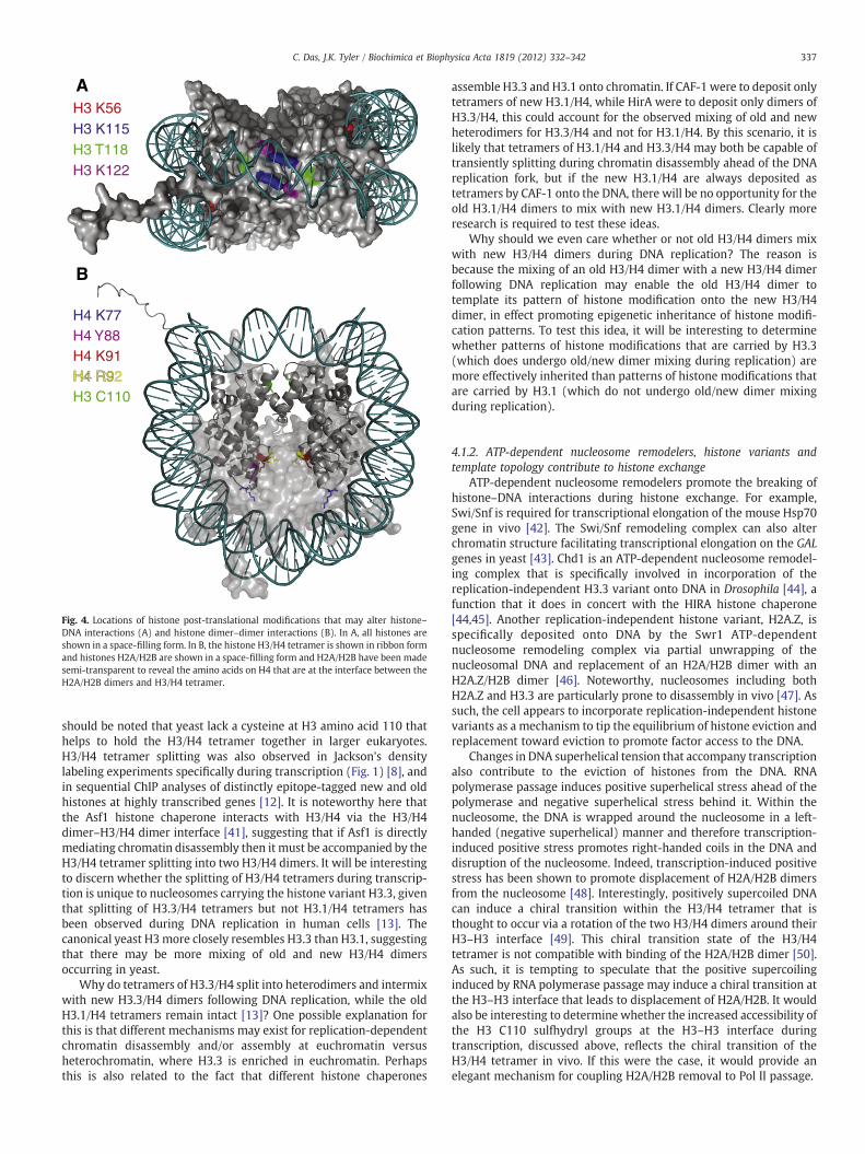
H3 K56H3 K115
H3 K122H3 T118
A
B
H4 K91
H4 K77H4 Y88
H4 R9H4 R92H3 C110
Fig. 4. Locations of histone post-translational modifications that may alter histone–DNA interactions (A) and histone dimer–dimer interactions (B). In A, all histones areshown in a space-filling form. In B, the histone H3/H4 tetramer is shown in ribbon formand histones H2A/H2B are shown in a space-filling form and H2A/H2B have been madesemi-transparent to reveal the amino acids on H4 that are at the interface between theH2A/H2B dimers and H3/H4 tetramer.
337C. Das, J.K. Tyler / Biochimica et Biophysica Acta 1819 (2012) 332–342
should be noted that yeast lack a cysteine at H3 amino acid 110 thathelps to hold the H3/H4 tetramer together in larger eukaryotes.H3/H4 tetramer splitting was also observed in Jackson's densitylabeling experiments specifically during transcription (Fig. 1) [8], andin sequential ChIP analyses of distinctly epitope-tagged new and oldhistones at highly transcribed genes [12]. It is noteworthy here thatthe Asf1 histone chaperone interacts with H3/H4 via the H3/H4dimer–H3/H4 dimer interface [41], suggesting that if Asf1 is directlymediating chromatin disassembly then it must be accompanied by theH3/H4 tetramer splitting into two H3/H4 dimers. It will be interestingto discern whether the splitting of H3/H4 tetramers during transcrip-tion is unique to nucleosomes carrying the histone variant H3.3, giventhat splitting of H3.3/H4 tetramers but not H3.1/H4 tetramers hasbeen observed during DNA replication in human cells [13]. Thecanonical yeast H3more closely resembles H3.3 than H3.1, suggestingthat there may be more mixing of old and new H3/H4 dimersoccurring in yeast.
Why do tetramers of H3.3/H4 split into heterodimers and intermixwith new H3.3/H4 dimers following DNA replication, while the oldH3.1/H4 tetramers remain intact [13]? One possible explanation forthis is that different mechanisms may exist for replication-dependentchromatin disassembly and/or assembly at euchromatin versusheterochromatin, where H3.3 is enriched in euchromatin. Perhapsthis is also related to the fact that different histone chaperones
assemble H3.3 and H3.1 onto chromatin. If CAF-1 were to deposit onlytetramers of new H3.1/H4, while HirA were to deposit only dimers ofH3.3/H4, this could account for the observed mixing of old and newheterodimers for H3.3/H4 and not for H3.1/H4. By this scenario, it islikely that tetramers of H3.1/H4 and H3.3/H4 may both be capable oftransiently splitting during chromatin disassembly ahead of the DNAreplication fork, but if the new H3.1/H4 are always deposited astetramers by CAF-1 onto the DNA, there will be no opportunity for theold H3.1/H4 dimers to mix with new H3.1/H4 dimers. Clearly moreresearch is required to test these ideas.
Why should we even care whether or not old H3/H4 dimers mixwith new H3/H4 dimers during DNA replication? The reason isbecause the mixing of an old H3/H4 dimer with a new H3/H4 dimerfollowing DNA replication may enable the old H3/H4 dimer totemplate its pattern of histone modification onto the new H3/H4dimer, in effect promoting epigenetic inheritance of histone modifi-cation patterns. To test this idea, it will be interesting to determinewhether patterns of histone modifications that are carried by H3.3(which does undergo old/new dimer mixing during replication) aremore effectively inherited than patterns of histone modifications thatare carried by H3.1 (which do not undergo old/new dimer mixingduring replication).
4.1.2. ATP-dependent nucleosome remodelers, histone variants andtemplate topology contribute to histone exchange
ATP-dependent nucleosome remodelers promote the breaking ofhistone–DNA interactions during histone exchange. For example,Swi/Snf is required for transcriptional elongation of the mouse Hsp70gene in vivo [42]. The Swi/Snf remodeling complex can also alterchromatin structure facilitating transcriptional elongation on the GALgenes in yeast [43]. Chd1 is an ATP-dependent nucleosome remodel-ing complex that is specifically involved in incorporation of thereplication-independent H3.3 variant onto DNA in Drosophila [44], afunction that it does in concert with the HIRA histone chaperone[44,45]. Another replication-independent histone variant, H2A.Z, isspecifically deposited onto DNA by the Swr1 ATP-dependentnucleosome remodeling complex via partial unwrapping of thenucleosomal DNA and replacement of an H2A/H2B dimer with anH2A.Z/H2B dimer [46]. Noteworthy, nucleosomes including bothH2A.Z and H3.3 are particularly prone to disassembly in vivo [47]. Assuch, the cell appears to incorporate replication-independent histonevariants as a mechanism to tip the equilibrium of histone eviction andreplacement toward eviction to promote factor access to the DNA.
Changes in DNA superhelical tension that accompany transcriptionalso contribute to the eviction of histones from the DNA. RNApolymerase passage induces positive superhelical stress ahead of thepolymerase and negative superhelical stress behind it. Within thenucleosome, the DNA is wrapped around the nucleosome in a left-handed (negative superhelical) manner and therefore transcription-induced positive stress promotes right-handed coils in the DNA anddisruption of the nucleosome. Indeed, transcription-induced positivestress has been shown to promote displacement of H2A/H2B dimersfrom the nucleosome [48]. Interestingly, positively supercoiled DNAcan induce a chiral transition within the H3/H4 tetramer that isthought to occur via a rotation of the two H3/H4 dimers around theirH3–H3 interface [49]. This chiral transition state of the H3/H4tetramer is not compatible with binding of the H2A/H2B dimer [50].As such, it is tempting to speculate that the positive supercoilinginduced by RNA polymerase passage may induce a chiral transition atthe H3–H3 interface that leads to displacement of H2A/H2B. It wouldalso be interesting to determine whether the increased accessibility ofthe H3 C110 sulfhydryl groups at the H3–H3 interface duringtranscription, discussed above, reflects the chiral transition of theH3/H4 tetramer in vivo. If this were the case, it would provide anelegant mechanism for coupling H2A/H2B removal to Pol II passage.

338 C. Das, J.K. Tyler / Biochimica et Biophysica Acta 1819 (2012) 332–342
4.1.3. Low level transcription is not associated with histone displacementfrom the DNA
Eviction of histones from the DNA may not be obligatorily coupledto the passage of RNA polymerases. The analysis of histone H3depletion from ORFs in the absence of the Spt6 chromatin reassemblyfactor demonstrates that transcription at low levels does not displacenucleosomes, while transcription at high levels does displace histones[31]. However, it is still possible that histone chaperones other thanSpt6 are responsible for reassembling chromatin after Pol II passage atgenes that are transcribed at a low rate. Regardless, transcriptionstudies on mononucleosome templates in vitro have revealed amechanism for nucleosome survival during Pol II passage [51]. OncePol II arrives at a strong DNA–histone interaction site, the DNA aheadof the polymerase unwinds from the histone octamer and a smallintranucleosomal DNA loop containing transcribing Pol II is formed(Fig. 3). This DNA loop formation leads to restoration of the DNA–histone interactions behind Pol II. This mechanism could explain whythe original H3/H4 histones are retained rather than being exchangedfor newH3/H4 at moderately transcribed genes, because in this modelall the H3/H4-DNA interactions are not simultaneously broken at anypoint in time. However, rate of transcription per se is not the onlydeterminant of the extent of histone H3/H4 exchange, as some genesshow rates of histone exchange that are higher or lower than wouldbe predicted from the Pol II transcription rate alone [52].
5. Histone modifications that regulate histone exchange
Histone proteins undergo a wide variety of post-translationalmodifications on specific amino acid residues during genomicprocesses. The vast majority of these modifications occur on theexposed N-terminal tails of the histones that extend out beyond thenucleosomal DNA. However, where examined the histone N-terminaltails are not essential for histone exchange. For example, the sites ofchromosomal incorporation of newly induced histones H2B and H3lacking their N-terminal tails in G1 phase arrested cells is unchangedfrom that of full length histones [10]. It is possible that the N-terminaltails of the histones act in a redundant manner during histoneexchange. Indeed, the N-terminal tails of histones slow down Pol IIpassage along chromatin templates in vitro [53]. In Drosophila, it hasbeen noted that N-terminally acetylated histones turn over morerapidly than unacetylated histones [54]. Whether the N-terminalhistone acetylation is causative or correlative for histone exchange isunknown.
Structurally, it makes more sense to consider the potentialinfluence of histone modifications that affect the histone–DNAinteractions on histone exchange, rather than those on the histoneN-terminal tails. The founding member of this class of histonemodification is acetylation of H3 lysine 56 (H3 K56Ac). Within thenucleosome structure, H3 K56 contacts the DNA via a water molecule(Fig. 4A). Addition of an acetyl group is predicted to break theinteractions between H3 K56 and the DNA as it enters and exits thenucleosome. Indeed, there is extensive evidence to indicate that H3K56Ac disrupts the nucleosome structure both in vivo and in vitro.Single molecule FRET experiments have revealed that H3 K56Acincreases the unwrapping of the ends of the nucleosomal DNA fromthe histone octamer by a factor of 7-fold [55]. By promoting this“breathing” of the nucleosomal DNA, H3 K56Ac can enhance thebinding of LexA to its nucleosome-buried DNA binding site by at least3-fold [56]. In yeast, failure to acetylate H3 K56 leads to increasedsuperhelical density and decreased nuclease accessibility, suggestingthat H3 K56Ac loosens the chromatin structure in vivo [57,58]. Duringhistone eviction from the yeast PHO5 promoter, the proportion ofhistone H3 carrying K56Ac drastically increases in a local manner,while a mutation to mimic K56 acetylation increases the rate ofhistone eviction from this promoter [59]. A similar situation is seenwithin ORFs where Asf1, presumably through its role in promoting H3
K56 acetylation, drives histone exchange during transcriptionalelongation [35]. Indeed, transcription coupled incorporation of H3K56Ac promotes transcription of heterochromatic regions [60].Genome-wide, histones carrying K56Ac mark regions of new histoneincorporation and high levels of replication-independent histoneexchange in yeast [61]. Furthermore, the rate of histone exchangeglobally is reduced in yeast unable to acetylate H3 K56Ac [62]. Takentogether all the evidence indicates that acetylation of H3 K56promotes histone eviction and subsequent histone exchange, atleast in yeast. It is noteworthy however, that yeast lacking the H3 K56histone acetyl transferase Rtt109, unlike asf1 mutants, do not displaycryptic internal initiationwhich is a consequence of failure to properlyreassemble chromatin after transcription [34]. As such, Asf1 wouldappear to have roles in chromatin assembly after RNA polymerasepassage that are distinct from its function in H3 K56 acetylation andhistone eviction.
We predict that H3 K56ac is the founding member of a large classof histone modifications that are used by the cell to regulate histoneeviction and histone replacement. To date the evidence for otherhistone modifications with this function is limited. However, thephysical locations within the nucleosome of several histone post-translational modifications are too coincidental to ignore. Forexample, there is a cluster of histone modifications that lie at theinterface between the nucleosomal DNA dyad and histone H3including acetylation and methylation of K115 and K122 andphosphorylation of T118 (Fig. 4A). Interestingly, these modificationsoften occur together, at least on peptides from bovine histones [63],suggesting that theymay act in a concertedmanner to disrupt the H3–DNA interactions. Peptide ligation has been used to generatenuclesomes carrying H3 K122Ac and H3 K115Ac [64]. Theseacetylation marks reduce the free energy for DNA binding to thehistone octamer and augment chromatin disassembly in vitro [64,65].Although the effects of H3 T118 phosphorylation on nucleosomestability has not yet been directly examined in vitro, it is noteworthythat T118I was uncovered as a SIN mutant (Swi/Snf independent)[66]. In vitro, the T118I mutation destabilizes nucleosomes [67,68]and H3 T118I promotes the ability of Pol II to pass the nucleosomebarrier [69]. Given the predicted drastic distortion of the nucleosomalDNA that phosphorylation of H3 T118 would cause, it will befascinating to determine when and where this modification and itsneighboring modifications are utilized by the cell.
Other histonemodificationsmay be used by the cell to regulate theinteraction between the H2A/H2B dimers and H3/H4 tetramer duringhistone exchange. One potential example of this type of modificationis acetylation of H4 on lysine 91. H4 K91Ac has been detected onnewly synthesized histones in yeast and its location at the interfacebetween H2A/H2B and the H3/H4 tetramer is suggestive of a role inregulating the interaction between the histone proteins [70]. Withinthe nucleosome, H4 K91 forms a salt bridge with E74 (E71 inmetazoans) of H2B and structurally it is likely that H4 K91 acetylationwould break this interaction (Fig. 4B). Indeed, H4 K91Ac destabilizesthe histone octamer [70]. Whether H4 K91Ac is used to help maintainthe H3/H4 tetramers free of H2A/H2B dimers during the process ofhistone exchange is not known. H4 K91 is also monoubiquitylated inresponse to DNA damage in mammals [71], which could potentiallydistort the nucleosome structure to facilitate repair.
Mapping other post-translational modifications that have beenuncovered by mass spectrometry analyses onto the nucleosomestructure reveals additional histonemodifications that could influencethe interaction between the H2A/H2B dimers and H3/H4 tetramer(Fig. 4B). For example, acetylation of H4 K77 was observed by massspectroscopy of bovine histones [63]. Within the nucleosomestructure, H4 K77 mediates Van der Waal's interactions with R92 ofH2B (in metazoans, R95 in yeast) and acetylation of K77 may disruptthis H4–H2B interaction. H4 R92 methylation has also been observedby mass spectroscopy of bovine histones [63]. Within the nucleosome

339C. Das, J.K. Tyler / Biochimica et Biophysica Acta 1819 (2012) 332–342
H4 R92 forms strong salt bridge interactions with E71 of H2B (inmetazoans, E74 in yeast). Like many of the other sites of modificationnoted above, mutation of H4 K77 and H4 R92 cause interestingphenotypic effects on transcriptional silencing in yeast [72]. Phos-phorylation of H4 Y88 [73] is likely to disrupt the aromatic ringstacking interactions between this residue and Y86 of H2B (in yeast,Y83 in metazoans) within the nucleosome. The stacking of Y88 of H4on Y86 of H2B has been proposed previously to form a spring likestructure that maintains tensile strength in the lower half of thenucleosome [74]. As such, one could imagine how phosphorylation ofH4 Y88 could potentially trigger release of the H2A/H2B dimer–H3/H4tetramer interaction. Yeast carrying substitution of Y88 to G are SINmutants [75], consistent with their having a loosened nucleosomestructure. In addition to overcoming the requirement for the ATP-dependent nucleosome remodeler Swi/Snf, Y88G also leads to defectsin transcription and proliferation [75]. It is important to appreciatethat the nucleosomal locations of these histone residues that mediateinteractions with DNA and with other histones (Fig. 4) makes themextremely inaccessible for enzymatic modification. As such, theirmodification must either occur on free histones or during extensivedisruption of the nucleosome structure.
Other histone modifications have been implicated indirectly inhistone eviction or histone return to the DNA. FACT-mediatedchromatin reassembly following Pol II passage is regulated by H2BK123 monoubiquitylation. In yeast, the absence of H2B ubiquitylationprevents the chromatin structure being properly restored in the wakeof elongating Pol II [76]. The function of FACT and H2B ubiquitylationare intertwined, where H2B ubiquitylation is required for the stableaccumulation of Spt16 at the GAL1 coding region, and Spt16 regulatesthe formation of ubiquitylated H2B both globally and at the GAL1gene. Related observations had been made earlier with an eleganthighly reconstituted transcription system in vitro [77]. Consistentwith its role in recruitment of FACT to promote chromatin reassembly,monoubiquitylated H2B stabilizes the nucleosome structure in yeast[78].
In mice sperm, acetylation of H4 K16 promotes global histoneeviction from the genome during their replacement with protamines[79]. However, it is not known whether this role of H4 K16Ac inhistone removal is direct or indirect. Given that H4 K16Ac breaksnucleosome–nucleosome interactions that are involved in chromatincondensation [80], it is possible that H4 K16Ac mediated decondensa-tion of chromosomes is required before histone removal duringspermatogenesis.
Although not a histone modification per se, proteolytic cleavage ofthe N-terminal tail of histone H3, which indirectly removes the N-terminal histone modifications, plays a role in histone eviction. N-terminal truncation of histone H3 after amino acid 21 precedes, and isrequired for, histone eviction from promoters during transcriptionalinduction [81]. The same study did not observe histone N-terminal H3cleavage within the IME1 ORF. However, this particular ORF does notdisplay significant H3 depletion during transcription and thereforemay not be the optimal locus to investigate a role for N-terminal tailclipping in promoting histone eviction during RNA polymerasepassage. For investigators employing N-terminal epitopes on H3 toexamine histone occupancy or histone dynamics, it is important forthem to consider the influence of N-terminal H3 clipping wheninterpreting their results.
6. Functional importance of histone exchange
Clearly the cell has placed a lot of emphasis on developing intricatemechanisms to mediate the eviction and replacement of histonesduring Pol II passage. This implies that improper histone exchange islikely to be detrimental to the cell. But what are those detrimentalconsequences? Failure to evict histones ahead of Pol II wouldpresumably hinder RNA polymerase passage through ORFs in cells,
as it does in vitro. Consistent with this idea, two of the histonechaperones that promote histone exchange within ORFs, FACT andSpt6 (Fig. 3), are both essential in yeast. Failure to restore the histonesonto the DNA in the wake of Pol II passage results in cryptictranscription internal initiation and the synthesis of their encodedproteins [34]. The consequence of making truncated proteins viacryptic internal initiation is presumably deleterious. Surprisingly,Drosophila lacking the transcription specific histone variant H3.3 areviable (although the males are infertile) [82], but this is largely due tocompensatory upregulation of the canonical histone H3.1 genes forreplication-independent chromatin assembly.
Histone exchange that occurs independent of transcription andDNA replication, for example at promoters and enhancers, provides anopportunity for transient access of factors to the genome. This is likelyto be essential for enabling effective transcriptional activation andrepression to occur. Without histone exchange, the pattern of histonemodifications and any information carried within this pattern wouldremain static long after the enzymes that established the histonemodifications had left that genomic region. One can see how thiswould be particularly problematic in post mitotic cells that lack theopportunity to refresh the histone proteins during DNA replication. Byconstantly refreshing the histones with new histones lacking the “old”histone modification pattern, histone exchange allows the chromatinstructure to be highly responsive to changes in the environment.
Histone exchange is likely to be especially important in pluripotentcells. Histone FRAP studies on pluripotent embryonic stem cellsrevealed that histone exchange is significantly faster and moreextensive than in differentiated cells [83]. This rapid fluorescencerecovery of the histones in stem cells was due to a population (18% forH2B and 25% for H3) of loosely bound (yet not free) histones thatwere exchanging within seconds. The size and speed of recovery ofthis dynamic histone pool in stem cells is much greater than indifferentiated cells. The mechanistic reason for the “hyperdynamic”chromatin state of stem cells is not presently known. Also, whetherthe hyperdynamic chromatin is the cause or consequence of theunique gene expression pattern and histone post-translationalmodification signature in pluripotent cells remains to be determined.
7. Changes in chromatin dynamics during aging
There is growing evidence that the chromatin structure andchromatin dynamics change during the aging process, and thefunctional consequences of these changes are likely to be profound.Although rates of histone exchange have not yet been directlyexamined in aged cells, there are hints that they will be significantlyaltered. For example, the replication-independent histone variantH3.3 accumulates on the genome of cells that are no longerreplicating, such as neurons [84,85]. The reason for this is probablydue to the rather trivial fact that the canonical H3.1 and H3.2 that areexpressed during S-phase are no long expressed in post mitotic cells.There is an interesting correlation between the fact that neurons havegenomes that are globally transcribed (approximately 80% of genesare expressed in adult mouse brain) [86] and have a genome that ismostly packaged with H3.3. As such, it is tempting to speculate thatglobal transcription of the neuronal genome is a consequence of theirgenome being packaged by the “looser” histone H3.3 variant that maypromote more frequent histone exchange and increased access of thetranscription machinery to the DNA. In fact, the H3.3 variant appearsto becomemore abundant during aging in general, not just in neurons.For example, H3.3 levels increase during postnatal development ofchicken and mice [87].
Cellular senescence is a state of irreversible growth arrest inculture. Whether or not a link exists between cellular senescence andin vivo aging is controversial and currently unproven. Notwithstand-ing, akin to the observations made during in vivo aging, H3.3 levelsincrease during the culture of human fibroblasts, which is a model for

340 C. Das, J.K. Tyler / Biochimica et Biophysica Acta 1819 (2012) 332–342
the entry into cell senescence [85]. Accordingly, the levels of thehistone H3.3 specific chaperone HIRA also increase during aging inbaboons [88]. Cellular senescence can be induced by multiple factorsin vitro including the BRAF oncogene. Interestingly, knockdown ofHIRA blocks BRAF-induced senescence [89], indicating that normallevels of HIRA may be important for entry into the senescent state.Perhaps related, HIRA is required for the formation of senescence-associated heterochromatin foci (SAHF) [90]. However, SAHF do notappear to accumulate during aging in vivo, at least not asmorphologically distinct structures that can be visualized by micros-copy in either mouse or baboon [91].
H2A.Z, the other histone variant that is incorporated into thegenome during replication-independent histone exchange is alsolinked to aging. Although H2A.Z levels do not appear to change duringthe aging process, H2A.Z knockdown in human fibroblasts causespremature entry into senescence [92]. Similarly, knockdown of p400,the human counterpart of the Swr1 H2A.Z histone exchanger, alsoinduces premature senescence in culture [93]. However, the physi-ological significance of these observations is unclear, as primaryhuman cells are highly prone to premature senescence when stressed.
While the levels of the histone variants that are incorporatedduring replication-independent histone exchange either rise duringaging or influence aging, the levels of the canonical histone proteinsdrastically drop during replicative aging (Fig. 5). Human fibroblaststhat have been through a high number of population doublings showa decrease in total histone protein levels compared to those that havebeen through a low number of population doublings [94]. This isaccompanied by a drastic decrease in the levels of the histonechaperones required for the incorporation of canonical histonesduring DNA replication, CAF-1 and Asf1 [94]. This profound loss of thecore histones with increasing numbers of cell divisions has also beenobserved in yeast, where it leads to expression of otherwise silencedgenes [95,96]. Meanwhile, total histone H3 protein levels do notchange between 10 day old and 40 day old Drosophila (a situation thatlargely represents post-mitotic aging) [97].
Studies have only just begun to examine the changes to histonemodifications during the aging process (reviewed by [98]). Theseinclude an increase in levels of H4 K16Ac with replicative age in bothyeast and with increasing numbers of population doublings of humanfibroblasts in culture [94,96]. Increased H4 K16Ac levels would reducethe nucleosome–nucleosome interactions, opening up the chromatinstructure in old cells. There is a decrease in the levels of H3 K56Acduring in vitro aging of fibroblasts [94]. From the function of H3 K56Acshown above, a drop in H3 K56Ac levels in old cells would bepredicted to reduce the rate of histone exchange. This drop in H3K56Ac levels in old cells is likely to be a consequence of the reducedlevels of Asf1 in old cells [94], which is required for H3 K56 acetylation[99]. However, given that at least half of the genome is nucleosome-free in old cells, this reduction in levels of H3 K56Ac may not impact
Tim
e
Young cell
Old cell
Loss of core histonesAsf1, CAF-1, HP1,
Gain of H3.3, HIRAH4 K16Ac
Fig. 5. Changes to the chromatin structure during aging. The schematic summarizesobservations made from mammalian systems, although the profound loss ofnucleosomes from the genome during aging has also been observed in yeast.
accessibility of the genome too greatly. In worms, excess H3 K4methylation, which correlates with active transcription, is detrimentalfor longevity, as deficiency in the ASH-2 methyltransferase complexextend lifespan [100]. Noteworthy, longevity in worms reflects post-mitotic aging, not mitotic/replicative aging that was the focus of theother studies in yeast and in cultured mammalian cells.
Heterochromatin is the repressed and less dynamic form ofchromatin, which is maintained by the binding of heterochromatinprotein 1 (HP1) [101]. Although mainly speculative, HP1 may act torepress transcription by stabilizing the nucleosome structure and indoing so HP1 may reduce the rate of histone exchange. As such, it isinteresting to note that cells from aged humans have less HP1 and lossof the specific modification that recruits HP1 to chromatin, H3 K9me3[102] (Fig. 5). However, other studies report that HP1 levels increaseduring replicative senescence of human fibroblasts [91] and thatlevels of HP1 and H3 K9me3 increase in 40 day old Drosophila ascompared to 10 day old flies [97]. Clearly the situation is confusingand much more research is required to fully understand how thechromatin structure changes during replicative aging and in vivoaging, and whether it is different in different organisms.
In general, the changes to the chromatin structure during aginginfer that cells that have divided more times may have a more openand dynamic chromatin state. Whether this is programmed or anunfortunate consequence of aging is not clear. However, supplyingadditional histones can extend replicative lifespan, at least in yeast,indicating that the decay of chromatin during aging is a cause of aging[95]. Functional analyses of the molecular consequences of the alteredchromatin structure during aging hold the potential to reveal keymediators of the aging process. In summary, while our understandingof the mechanisms of histone exchange has grown rapidly in recentyears, we still have a lot to learn about the full implications of histoneexchange on the activities of the genome.
Note added in proof
A recent careful in vitro analysis of the effect of H3 T118phosphorylation revealed that it increases nucleosome mobility by26 fold and increases DNA accessibility near the normally inaccessibledyad by 6 fold [104].
Acknowledgments
We are very grateful to Leisa McCord of the Department ofBiochemistry and Molecular Genetics UT MD Anderson Cancer Centerfor assistance with the figures. We would also like to acknowledgeSiddhartha Roy, for his assistance with analysis of the nucleosomestructure. JKT is supported by grants from NIH GM and NCI, and aCPRIT Rising Star award, CD is supported by a Susan G. Komen for theCure Fellowship.
References
[1] K. Luger, A.W. Mader, R.K. Richmond, D.F. Sargent, T.J. Richmond, Crystalstructure of the nucleosome core particle at 2.8 A resolution, Nature 389 (1997)251–260.
[2] X. Liu, D.A. Bushnell, D. Wang, G. Calero, R.D. Kornberg, Structure of an RNApolymerase II–TFIIB complex and the transcription initiation mechanism,Science 327 (2010) 206–209.
[3] A.C. Cheung, P. Cramer, Structural basis of RNA polymerase II backtracking, arrestand reactivation, Nature 471 (2011) 249–253.
[4] C.H. Chang, D.S. Luse, The H3/H4 tetramer blocks transcript elongation by RNApolymerase II in vitro, J. Biol. Chem. 272 (1997) 23427–23434.
[5] D.V. Fyodorov, J.T. Kadonaga, Dynamics of ATP-dependent chromatin assemblyby ACF, Nature 418 (2002) 897–900.
[6] V. Bohm, A.R. Hieb, A.J. Andrews, A. Gansen, A. Rocker, K. Toth, K. Luger, J.Langowski, Nucleosome accessibility governed by the dimer/tetramer interface,Nucleic Acids Res. 39 (2011) 3093–3102.
[7] V. Jackson, Deposition of newly synthesized histones: hybrid nucleosomes arenot tandemly arranged on daughter DNA strands, Biochemistry 27 (1988)2109–2120.

341C. Das, J.K. Tyler / Biochimica et Biophysica Acta 1819 (2012) 332–342
[8] V. Jackson, In vivo studies on the dynamics of histone–DNA interaction: evidencefor nucleosome dissolution during replication and transcription and a low levelof dissolution independent of both, Biochemistry 29 (1990) 719–731.
[9] C. Thiriet, J.J. Hayes, Replication-independent core histone dynamics attranscriptionally active loci in vivo, Genes Dev. 19 (2005) 677–682.
[10] A. Jamai, R.M. Imoberdorf, M. Strubin, Continuous histone H2B and transcrip-tion-dependent histone H3 exchange in yeast cells outside of replication, Mol.Cell 25 (2007) 345–355.
[11] M.F. Dion, T. Kaplan, M. Kim, S. Buratowski, N. Friedman, O.J. Rando, Dynamics ofreplication-independent histone turnover in budding yeast, Science 315 (2007)1405–1408.
[12] Y. Katan-Khaykovich, K. Struhl, Splitting of H3–H4 tetramers at transcriptionallyactive genes undergoing dynamic histone exchange, Proc. Natl. Acad. Sci. U. S. A.108 (2011) 1296–1301.
[13] M. Xu, C. Long, X. Chen, C. Huang, S. Chen, B. Zhu, Partitioning of histone H3–H4tetramers during DNA replication-dependent chromatin assembly, Science 328(2010) 94–98.
[14] K.F. Verzijlbergen, V. Menendez-Benito, T. van Welsem, S.J. van Deventer, D.L.Lindstrom, H. Ovaa, J. Neefjes, D.E. Gottschling, F. van Leeuwen, Recombination-induced tag exchange to track old and new proteins, Proc. Natl. Acad. Sci. U. S. A.107 (2010) 64–68.
[15] H. Kimura, P.R. Cook, Kinetics of core histones in living human cells: littleexchange of H3 and H4 and some rapid exchange of H2B, J. Cell Biol. 153 (2001)1341–1353.
[16] K. Ahmad, S. Henikoff, The histone variant H3.3 marks active chromatin byreplication-independent nucleosome assembly, Mol. Cell 9 (2002) 1191–1200.
[17] Y. Mito, J.G. Henikoff, S. Henikoff, Genome-scale profiling of histone H3.3replacement patterns, Nat. Genet. 37 (2005) 1090–1097.
[18] B. Kang, M. Pu, G. Hu, W. Wen, Z. Dong, K. Zhao, B. Stillman, Z. Zhang,Phosphorylation of H4 Ser 47 promotes HIRA-mediated nucleosome assembly,Genes Dev. 25 (2011) 1359–1364.
[19] S.L. Ooi, J.G. Henikoff, S. Henikoff, A native chromatin purification system forepigenomic profiling in Caenorhabditis elegans, Nucleic Acids Res. 38 (2010) e26.
[20] R.B. Deal, J.G. Henikoff, S. Henikoff, Genome-wide kinetics of nucleosome turnoverdetermined by metabolic labeling of histones, Science 328 (2010) 1161–1164.
[21] A. Kristjuhan, J.Q. Svejstrup, Evidence for distinct mechanisms facilitatingtranscript elongation through chromatin in vivo, EMBO J. 23 (2004) 4243–4252.
[22] M.A. Schwabish, K. Struhl, Evidence for eviction and rapid deposition of histonesupon transcriptional elongation by RNA polymerase II, Mol. Cell. Biol. 24 (2004)10111–10117.
[23] H.J. Kim, J.H. Seol, J.W. Han, H.D. Youn, E.J. Cho, Histone chaperones regulatehistone exchange during transcription, EMBO J. 26 (2007) 4467–4474.
[24] C.K. Lee, Y. Shibata, B. Rao, B.D. Strahl, J.D. Lieb, Evidence for nucleosome depletionat active regulatory regions genome-wide, Nat. Genet. 36 (2004) 900–905.
[25] M. Morillo-Huesca, D. Maya, M.C. Munoz-Centeno, R.K. Singh, V. Oreal, G.U.Reddy, D. Liang, V. Geli, A. Gunjan, S. Chavez, FACT prevents the accumulation offree histones evicted from transcribed chromatin and a subsequent cell cycledelay in G1, PLoS Genet. 6 (2010) e1000964.
[26] M.L. Kireeva, W. Walter, V. Tchernajenko, V. Bondarenko, M. Kashlev, V.M.Studitsky, Nucleosome remodeling induced by RNA polymerase II: loss of theH2A/H2B dimer during transcription, Mol. Cell 9 (2002) 541–552.
[27] V.M. Studitsky, G.A. Kassavetis, E.P. Geiduschek, G. Felsenfeld, Mechanism oftranscription through the nucleosome by eukaryotic RNA polymerase, Science278 (1997) 1960–1963.
[28] V.A. Bondarenko, L.M. Steele, A. Ujvari, D.A. Gaykalova, O.I. Kulaeva, Y.S.Polikanov, D.S. Luse, V.M. Studitsky, Nucleosomes can form a polar barrier totranscript elongation by RNA polymerase II, Mol. Cell 24 (2006) 469–479.
[29] R. Belotserkovskaya, S. Oh, V.A. Bondarenko, G. Orphanides, V.M. Studitsky, D.Reinberg, FACT facilitates transcription-dependent nucleosome alteration,Science 301 (2003) 1090–1093.
[30] H. Xin, S. Takahata, M. Blanksma, L. McCullough, D.J. Stillman, T. Formosa, yFACTinduces global accessibility of nucleosomal DNA without H2A–H2B displace-ment, Mol. Cell 35 (2009) 365–376.
[31] I. Ivanovska, P.E. Jacques, O.J. Rando, F. Robert, F. Winston, Control of chromatinstructure by spt6: different consequences in coding and regulatory regions, Mol.Cell. Biol. 31 (2011) 531–541.
[32] C.D. Kaplan, L. Laprade, F. Winston, Transcription elongation factors represstranscription initiation from cryptic sites, Science 301 (2003) 1096–1099.
[33] A. Jamai, A. Puglisi, M. Strubin, Histone chaperone spt16 promotes redepositionof the original h3–h4 histones evicted by elongating RNA polymerase, Mol. Cell35 (2009) 377–383.
[34] V. Cheung, G. Chua, N.N. Batada, C.R. Landry, S.W. Michnick, T.R. Hughes, F.Winston, Chromatin- and transcription-related factors repress transcriptionfrom within coding regions throughout the Saccharomyces cerevisiae genome,PLoS Biol. 6 (2008) e277.
[35] M.A. Schwabish, K. Struhl, Asf1 mediates histone eviction and deposition duringelongation by RNA polymerase II, Mol. Cell 22 (2006) 415–422.
[36] T.K. Quan, G.A. Hartzog, Histone H3K4 and K36 methylation, Chd1 and Rpd3Soppose the functions of Saccharomyces cerevisiae Spt4–Spt5 in transcription,Genetics 184 (2010) 321–334.
[37] H.E. Anderson, J. Wardle, S.V. Korkut, H.E. Murton, L. Lopez-Maury, J. Bahler, S.K.Whitehall, The fission yeast HIRA histone chaperone is required for promotersilencing and the suppression of cryptic antisense transcripts, Mol. Cell. Biol. 29(2009) 5158–5167.
[38] T.A. Chen, V.G. Allfrey, Rapid and reversible changes in nucleosome structureaccompany the activation, repression, and superinduction of murine fibroblast
protooncogenes c-fos and c-myc, Proc. Natl. Acad. Sci. U. S. A. 84 (1987)5252–5256.
[39] D.P. Bazett-Jones, E. Mendez, G.J. Czarnota, F.P. Ottensmeyer, V.G. Allfrey,Visualization and analysis of unfolded nucleosomes associated with transcribingchromatin, Nucleic Acids Res. 24 (1996) 321–329.
[40] M.S. Lee,W.T. Garrard, Transcription-inducednucleosome ‘splitting’: an underlyingstructure for DNase I sensitive chromatin, EMBO J. 10 (1991) 607–615.
[41] C.M. English, M.W. Adkins, J.J. Carson, M.E. Churchill, J.K. Tyler, Structural basisfor the histone chaperone activity of Asf1, Cell 127 (2006) 495–508.
[42] L.L. Corey, C.S. Weirich, I.J. Benjamin, R.E. Kingston, Localized recruitment of achromatin-remodeling activity by an activator in vivo drives transcriptionalelongation, Genes Dev. 17 (2003) 1392–1401.
[43] M.A. Schwabish, K. Struhl, The Swi/Snf complex is important for histone evictionduring transcriptional activation and RNA polymerase II elongation in vivo, Mol.Cell. Biol. 27 (2007) 6987–6995.
[44] A.Y. Konev, M. Tribus, S.Y. Park, V. Podhraski, C.Y. Lim, A.V. Emelyanov, E.Vershilova, V. Pirrotta, J.T. Kadonaga, A. Lusser, D.V. Fyodorov, CHD1 motorprotein is required for deposition of histone variant H3.3 into chromatin in vivo,Science 317 (2007) 1087–1090.
[45] A.D. Goldberg, L.A. Banaszynski, K.M. Noh, P.W. Lewis, S.J. Elsaesser, S. Stadler, S.Dewell, M. Law, X. Guo, X. Li, D. Wen, A. Chapgier, R.C. DeKelver, J.C. Miller, Y.L.Lee, E.A. Boydston, M.C. Holmes, P.D. Gregory, J.M. Greally, S. Rafii, C. Yang, P.J.Scambler, D. Garrick, R.J. Gibbons, D.R. Higgs, I.M. Cristea, F.D. Urnov, D. Zheng,C.D. Allis, Distinct factors control histone variant H3.3 localization at specificgenomic regions, Cell 140 (2010) 678–691.
[46] G. Mizuguchi, X. Shen, J. Landry, W.H. Wu, S. Sen, C. Wu, ATP-driven exchange ofhistone H2AZ variant catalyzed by SWR1 chromatin remodeling complex,Science 303 (2004) 343–348.
[47] C. Jin, C. Zang, G. Wei, K. Cui, W. Peng, K. Zhao, G. Felsenfeld, H3.3/H2A.Z doublevariant-containing nucleosomes mark ‘nucleosome-free regions’ of activepromoters and other regulatory regions, Nat. Genet. 41 (2009) 941–945.
[48] V. Levchenko, B. Jackson, V. Jackson, Histone release during transcription:displacement of the two H2A–H2B dimers in the nucleosome is dependent ondifferent levels of transcription-induced positive stress, Biochemistry 44 (2005)5357–5372.
[49] A. Hamiche, V. Carot, M. Alilat, F. De Lucia, M.F. O'Donohue, B. Revet, A. Prunell,Interaction of the histone (H3–H4)2 tetramer of the nucleosome with positivelysupercoiled DNA minicircles: potential flipping of the protein from a left- to aright-handed superhelical form, Proc. Natl. Acad. Sci. U. S. A. 93 (1996)7588–7593.
[50] S. Peterson, R. Danowit, A. Wunsch, V. Jackson, NAP1 catalyzes the formation ofeither positive or negative supercoils on DNA on basis of the dimer–tetramerequilibrium of histones H3/H4, Biochemistry 46 (2007) 8634–8646.
[51] O.I. Kulaeva, D.A. Gaykalova, N.A. Pestov, V.V. Golovastov, D.G. Vassylyev, I.Artsimovitch, V.M. Studitsky, Mechanism of chromatin remodeling and recoveryduring passage of RNA polymerase II, Nat. Struct. Mol. Biol. 16 (2009)1272–1278.
[52] I. Gat-Viks, M. Vingron, Evidence for gene-specific rather than transcription rate-dependent histone H3 exchange in yeast coding regions, PLoS Comput. Biol. 5(2009) e1000282.
[53] A. Ujvari, F.K. Hsieh, S.W. Luse, V.M. Studitsky, D.S. Luse, Histone N-terminal tailsinterfere with nucleosome traversal by RNA polymerase II, J. Biol. Chem. 283(2008) 32236–32243.
[54] P.V. Kharchenko, A.A. Alekseyenko, Y.B. Schwartz, A. Minoda, N.C. Riddle, J. Ernst,P.J. Sabo, E. Larschan, A.A. Gorchakov, T. Gu, D. Linder-Basso, A. Plachetka, G.Shanower, M.Y. Tolstorukov, L.J. Luquette, R. Xi, Y.L. Jung, R.W. Park, E.P. Bishop,T.K. Canfield, R. Sandstrom, R.E. Thurman, D.M. MacAlpine, J.A. Stamatoyanno-poulos, M. Kellis, S.C. Elgin, M.I. Kuroda, V. Pirrotta, G.H. Karpen, P.J. Park,Comprehensive analysis of the chromatin landscape in Drosophila melanogaster,Nature 471 (2011) 480–485.
[55] H. Neumann, S.M. Hancock, R. Buning, A. Routh, L. Chapman, J. Somers, T. Owen-Hughes, J. van Noort, D. Rhodes, J.W. Chin, A method for genetically installingsite-specific acetylation in recombinant histones defines the effects of H3 K56acetylation, Mol. Cell 36 (2009) 153–163.
[56] J.C. Shimko, J.A. North, A.N. Bruns, M.G. Poirier, J.J. Ottesen, Preparation of fullysynthetic histone h3 reveals that acetyl-lysine 56 facilitates protein bindingwithin nucleosomes, J. Mol. Biol. 408 (2011) 187–204.
[57] M.W. Adkins, J.K. Tyler, The histone chaperone Asf1p mediates global chromatindisassembly in vivo, J. Biol. Chem. 279 (2004) 52069–52074.
[58] R. Driscoll, A. Hudson, S.P. Jackson, Yeast Rtt109 promotes genome stability byacetylating histone H3 on lysine 56, Science 315 (2007) 649–652.
[59] S.K. Williams, D. Truong, J.K. Tyler, Acetylation in the globular core of histone H3on lysine-56 promotes chromatin disassembly during transcriptional activation,Proc. Natl. Acad. Sci. U. S. A. 105 (2008) 9000–9005.
[60] S. Varv, K. Kristjuhan, K. Peil, M. Looke, T. Mahlakoiv, K. Paapsi, A. Kristjuhan,Acetylation of H3 K56 is required for RNA polymerase II transcript elongationthrough heterochromatin in yeast, Mol. Cell. Biol. 30 (2010) 1467–1477.
[61] A. Rufiange, P.E. Jacques, W. Bhat, F. Robert, A. Nourani, Genome-wide replication-independent histone H3 exchange occurs predominantly at promoters andimplicates H3 K56 acetylation and Asf1, Mol. Cell 27 (2007) 393–405.
[62] T. Kaplan, C.L. Liu, J.A. Erkmann, J. Holik, M. Grunstein, P.D. Kaufman, N.Friedman, O.J. Rando, Cell cycle- and chaperone-mediated regulation of H3K56acincorporation in yeast, PLoS Genet. 4 (2008) e1000270.
[63] L. Zhang, E.E. Eugeni, M.R. Parthun, M.A. Freitas, Identification of novel histonepost-translational modifications by peptide mass fingerprinting, Chromosoma112 (2003) 77–86.

342 C. Das, J.K. Tyler / Biochimica et Biophysica Acta 1819 (2012) 332–342
[64] M.Manohar, A.M. Mooney, J.A. North, R.J. Nakkula, J.W. Picking, A. Edon, R. Fishel,M.G. Poirier, J.J. Ottesen, Acetylation of histone H3 at the nucleosome dyad altersDNA–histone binding, J. Biol. Chem. 284 (2009) 23312–23321.
[65] S. Javaid, M. Manohar, N. Punja, A. Mooney, J.J. Ottesen, M.G. Poirier, R. Fishel,Nucleosome remodeling by hMSH2–hMSH6, Mol. Cell 36 (2009) 1086–1094.
[66] W. Kruger, C.L. Peterson, A. Sil, C. Coburn, G. Arents, E.N. Moudrianakis, I.Herskowitz, Amino acid substitutions in the structured domains of histones H3and H4 partially relieve the requirement of the yeast SWI/SNF complex fortranscription, Genes Dev. 9 (1995) 2770–2779.
[67] H. Kurumizaka, A.P. Wolffe, Sin mutations of histone H3: influence onnucleosome core structure and function, Mol. Cell. Biol. 17 (1997) 6953–6969.
[68] U.M. Muthurajan, Y. Bao, L.J. Forsberg, R.S. Edayathumangalam, P.N. Dyer, C.L.White, K. Luger, Crystal structures of histone Sin mutant nucleosomes revealaltered protein–DNA interactions, EMBO J. 23 (2004) 260–271.
[69] F.K. Hsieh, M. Fisher, A. Ujvari, V.M. Studitsky, D.S. Luse, Histone Sin mutationspromote nucleosome traversal and histone displacement by RNA polymerase II,EMBO Rep. 11 (2010) 705–710.
[70] J. Ye, X. Ai, E.E. Eugeni, L. Zhang, L.R. Carpenter, M.A. Jelinek, M.A. Freitas, M.R.Parthun, Histone H4 lysine 91 acetylation a core domain modification associatedwith chromatin assembly, Mol. Cell 18 (2005) 123–130.
[71] Q. Yan, S. Dutt, R. Xu, K. Graves, P. Juszczynski, J.P. Manis, M.A. Shipp, BBAPmonoubiquitylates histone H4 at lysine 91 and selectively modulates the DNAdamage response, Mol. Cell 36 (2009) 110–120.
[72] E.M. Hyland, M.S. Cosgrove, H. Molina, D. Wang, A. Pandey, R.J. Cottee, J.D. Boeke,Insights into the role of histone H3 and histone H4 core modifiable residues inSaccharomyces cerevisiae, Mol. Cell. Biol. 25 (2005) 10060–10070.
[73] K. Rikova, A. Guo, Q. Zeng, A. Possemato, J. Yu, H. Haack, J. Nardone, K. Lee, C.Reeves, Y. Li, Y. Hu, Z. Tan, M. Stokes, L. Sullivan, J. Mitchell, R. Wetzel, J. Macneill,J.M. Ren, J. Yuan, C.E. Bakalarski, J. Villen, J.M. Kornhauser, B. Smith, D. Li, X. Zhou,S.P. Gygi, T.L. Gu, R.D. Polakiewicz, J. Rush, M.J. Comb, Global survey ofphosphotyrosine signaling identifies oncogenic kinases in lung cancer, Cell 131(2007) 1190–1203.
[74] J. Dai, E.M. Hyland, D.S. Yuan, H. Huang, J.S. Bader, J.D. Boeke, Probingnucleosome function: a highly versatile library of synthetic histone H3 and H4mutants, Cell 134 (2008) 1066–1078.
[75] M.S. Santisteban, G. Arents, E.N. Moudrianakis, M.M. Smith, Histone octamerfunction in vivo: mutations in the dimer–tetramer interfaces disrupt both geneactivation and repression, EMBO J. 16 (1997) 2493–2506.
[76] A.B. Fleming, C.F. Kao, C. Hillyer, M. Pikaart, M.A. Osley, H2B ubiquitylation playsa role in nucleosome dynamics during transcription elongation, Mol. Cell 31(2008) 57–66.
[77] R. Pavri, B. Zhu, G. Li, P. Trojer, S. Mandal, A. Shilatifard, D. Reinberg, Histone H2Bmonoubiquitination functions cooperatively with FACT to regulate elongation byRNA polymerase II, Cell 125 (2006) 703–717.
[78] M.B. Chandrasekharan, F. Huang, Z.W. Sun, Ubiquitination of histone H2Bregulates chromatin dynamics by enhancing nucleosome stability, Proc. Natl.Acad. Sci. U. S. A. 106 (2009) 16686–16691.
[79] L.Y. Lu, J. Wu, L. Ye, G.B. Gavrilina, T.L. Saunders, X. Yu, RNF8-dependent histonemodifications regulate nucleosome removal during spermatogenesis, Dev. Cell18 (2010) 371–384.
[80] M. Shogren-Knaak, H. Ishii, J.M. Sun, M.J. Pazin, J.R. Davie, C.L. Peterson, HistoneH4-K16 acetylation controls chromatin structure and protein interactions,Science 311 (2006) 844–847.
[81] H. Santos-Rosa, A. Kirmizis, C. Nelson, T. Bartke, N. Saksouk, J. Cote, T. Kouzarides,Histone H3 tail clipping regulates gene expression, Nat. Struct. Mol. Biol. 16(2009) 17–22.
[82] A. Sakai, B.E. Schwartz, S. Goldstein, K. Ahmad, Transcriptional and develop-mental functions of the H3.3 histone variant in Drosophila, Curr. Biol. 19 (2009)1816–1820.
[83] E. Meshorer, D. Yellajoshula, E. George, P.J. Scambler, D.T. Brown, T. Misteli,Hyperdynamic plasticity of chromatin proteins in pluripotent embryonic stemcells, Dev. Cell 10 (2006) 105–116.
[84] B. Pina, P. Suau, Changes in histones H2A and H3 variant composition indifferentiating and mature rat brain cortical neurons, Dev. Biol. 123 (1987)51–58.
[85] E.P. Rogakou, K.E. Sekeri-Pataryas, Histone variants of H2A and H3 families areregulated during in vitro aging in the same manner as during differentiation,Exp. Gerontol. 34 (1999) 741–754.
[86] E.S. Lein, M.J. Hawrylycz, N. Ao, M. Ayres, A. Bensinger, A. Bernard, A.F. Boe, M.S.Boguski, K.S. Brockway, E.J. Byrnes, L. Chen, T.M. Chen, M.C. Chin, J. Chong, B.E.Crook, A. Czaplinska, C.N. Dang, S. Datta, N.R. Dee, A.L. Desaki, T. Desta, E. Diep,T.A. Dolbeare, M.J. Donelan, H.W. Dong, J.G. Dougherty, B.J. Duncan, A.J. Ebbert, G.Eichele, L.K. Estin, C. Faber, B.A. Facer, R. Fields, S.R. Fischer, T.P. Fliss, C. Frensley,S.N. Gates, K.J. Glattfelder, K.R. Halverson, M.R. Hart, J.G. Hohmann, M.P. Howell,D.P. Jeung, R.A. Johnson, P.T. Karr, R. Kawal, J.M. Kidney, R.H. Knapik, C.L. Kuan,J.H. Lake, A.R. Laramee, K.D. Larsen, C. Lau, T.A. Lemon, A.J. Liang, Y. Liu, L.T.Luong, J. Michaels, J.J. Morgan, R.J. Morgan, M.T. Mortrud, N.F. Mosqueda, L.L. Ng,R. Ng, G.J. Orta, C.C. Overly, T.H. Pak, S.E. Parry, S.D. Pathak, O.C. Pearson, R.B.Puchalski, Z.L. Riley, H.R. Rockett, S.A. Rowland, J.J. Royall, M.J. Ruiz, N.R. Sarno, K.Schaffnit, N.V. Shapovalova, T. Sivisay, C.R. Slaughterbeck, S.C. Smith, K.A. Smith,B.I. Smith, A.J. Sodt, N.N. Stewart, K.R. Stumpf, S.M. Sunkin, M. Sutram, A. Tam,C.D. Teemer, C. Thaller, C.L. Thompson, L.R. Varnam, A. Visel, R.M. Whitlock, P.E.Wohnoutka, C.K. Wolkey, V.Y. Wong, M. Wood, M.B. Yaylaoglu, R.C. Young, B.L.Youngstrom, X.F. Yuan, B. Zhang, T.A. Zwingman, A.R. Jones, Genome-wide atlasof gene expression in the adult mouse brain, Nature 445 (2007) 168–176.
[87] M.K. Urban, A. Zweidler, Changes in nucleosomal core histone variants duringchicken development and maturation, Dev. Biol. 95 (1983) 421–428.
[88] J.C. Jeyapalan, M. Ferreira, J.M. Sedivy, U. Herbig, Accumulation of senescent cellsin mitotic tissue of aging primates, Mech. Ageing Dev. 128 (2007) 36–44.
[89] N. Wajapeyee, R.W. Serra, X. Zhu, M. Mahalingam, M.R. Green, Oncogenic BRAFinduces senescence and apoptosis through pathways mediated by the secretedprotein IGFBP7, Cell 132 (2008) 363–374.
[90] R. Zhang, M.V. Poustovoitov, X. Ye, H.A. Santos, W. Chen, S.M. Daganzo, J.P.Erzberger, I.G. Serebriiskii, A.A. Canutescu, R.L. Dunbrack, J.R. Pehrson, J.M.Berger, P.D. Kaufman, P.D. Adams, Formation of MacroH2A-containing senes-cence-associated heterochromatin foci and senescence driven by ASF1a andHIRA, Dev. Cell 8 (2005) 19–30.
[91] J.A. Kreiling, M. Tamamori-Adachi, A.N. Sexton, J.C. Jeyapalan, U. Munoz-Najar,A.L. Peterson, J. Manivannan, E.S. Rogers, N.A. Pchelintsev, P.D. Adams, J.M.Sedivy, Age-associated increase in heterochromatic marks in murine andprimate tissues, Aging Cell 10 (2011) 292–304.
[92] N. Gevry, H.M. Chan, L. Laflamme, D.M. Livingston, L. Gaudreau, p21 transcriptionis regulated by differential localization of histone H2A.Z, Genes Dev. 21 (2007)1869–1881.
[93] H.M. Chan, M. Narita, S.W. Lowe, D.M. Livingston, The p400 E1A-associatedprotein is a novel component of the p53→p21 senescence pathway, Genes Dev.19 (2005) 196–201.
[94] R.J. O'Sullivan, S. Kubicek, S.L. Schreiber, J. Karlseder, Reduced histonebiosynthesis and chromatin changes arising from a damage signal at telomeres,Nat. Struct. Mol. Biol. 17 (2010) 1218–1225.
[95] J. Feser, D. Truong, C. Das, J.J. Carson, J. Kieft, T. Harkness, J.K. Tyler, Elevatedhistone expression promotes life span extension, Mol. Cell 39 (2010) 724–735.
[96] W. Dang, K.K. Steffen, R. Perry, J.A. Dorsey, F.B. Johnson, A. Shilatifard, M.Kaeberlein, B.K. Kennedy, S.L. Berger, Histone H4 lysine 16 acetylation regulatescellular lifespan, Nature 459 (2009) 802–807.
[97] J.G. Wood, S. Hillenmeyer, C. Lawrence, C. Chang, S. Hosier, W. Lightfoot, E.Mukherjee, N. Jiang, C. Schorl, A.S. Brodsky, N. Neretti, S.L. Helfand, Chromatinremodeling in the aging genome of Drosophila, Aging Cell 9 (2010) 971–978.
[98] J. Feser, J. Tyler, Chromatin structure as a mediator of aging, FEBS Lett. (2010).[99] C. Das, M.S. Lucia, K.C. Hansen, J.K. Tyler, CBP/p300-mediated acetylation of
histone H3 on lysine 56, Nature 459 (2009) 113–117.[100] E.L. Greer, T.J. Maures, A.G. Hauswirth, E.M. Green, D.S. Leeman, G.S.Maro, S. Han,M.R.
Banko, O. Gozani, A. Brunet, Members of the H3K4 trimethylation complex regulatelifespan in a germline-dependent manner in C. elegans, Nature 466 (2010) 383–387.
[101] T. Cheutin, A.J. McNairn, T. Jenuwein, D.M. Gilbert, P.B. Singh, T. Misteli,Maintenance of stable heterochromatin domains by dynamic HP1 binding,Science 299 (2003) 721–725.
[102] P. Scaffidi, T. Misteli, Lamin A-dependent nuclear defects in human aging,Science 312 (2006) 1059–1063.
[103] A.J. Andrews, X. Chen, A. Zevin, L.A. Stargell, K. Luger, The histone chaperoneNap1 promotes nucleosome assembly by eliminating nonnucleosomal histoneDNA interactions, Mol. Cell 37 (2010) 834–842.
[104] J.A. North, S. Javaid, M.B. Ferdinand, N. Chatterjee, J.W. Picking, M. Shoffner, R.J.Nakkula, B. Bartholomew, J.J. Ottesen, R. Fishel, M.G. Poirier, Phosphorylation ofhistone H3(T118) alters nucleosome dynamics and remodeling, Nucleic AcidsRes. (2011) (May 16, Electronic publication ahead of print).
Copyright © 2022 FDOKUMEN