Female Meiotic Sex Chromosome Inactivation in Chicken
16
Female Meiotic Sex Chromosome Inactivation in Chicken Sam Schoenmakers 1,2 , Evelyne Wassenaar 1 , Jos W. Hoogerbrugge, Joop S. E. Laven 2 , J. Anton Grootegoed 1 , Willy M. Baarends 1 * 1 Department of Reproduction and Development, Erasmus MC, University Medical Center, Rotterdam, The Netherlands, 2 Department of Obstetrics and Gynaecology, Erasmus MC, University Medical Center, Rotterdam, The Netherlands Abstract During meiotic prophase in male mammals, the heterologous X and Y chromosomes remain largely unsynapsed, and meiotic sex chromosome inactivation (MSCI) leads to formation of the transcriptionally silenced XY body. In birds, the heterogametic sex is female, carrying Z and W chromosomes (ZW), whereas males have the homogametic ZZ constitution. During chicken oogenesis, the heterologous ZW pair reaches a state of complete heterologous synapsis, and this might enable maintenance of transcription of Z- and W chromosomal genes during meiotic prophase. Herein, we show that the ZW pair is transiently silenced, from early pachytene to early diplotene using immunocytochemistry and gene expression analyses. We propose that ZW inactivation is most likely achieved via spreading of heterochromatin from the W on the Z chromosome. Also, persistent meiotic DNA double-strand breaks (DSBs) may contribute to silencing of Z. Surprisingly, cH2AX, a marker of DSBs, and also the earliest histone modification that is associated with XY body formation in mammalian and marsupial spermatocytes, does not cover the ZW during the synapsed stage. However, when the ZW pair starts to desynapse, a second wave of cH2AX accumulates on the unsynapsed regions of Z, which also show a reappearance of the DSB repair protein RAD51. This indicates that repair of meiotic DSBs on the heterologous part of Z is postponed until late pachytene/diplotene, possibly to avoid recombination with regions on the heterologously synapsed W chromosome. Two days after entering diplotene, the Z looses cH2AX and shows reactivation. This is the first report of meiotic sex chromosome inactivation in a species with female heterogamety, providing evidence that this mechanism is not specific to spermatogenesis. It also indicates the presence of an evolutionary force that drives meiotic sex chromosome inactivation independent of the final achievement of synapsis. Citation: Schoenmakers S, Wassenaar E, Hoogerbrugge JW, Laven JSE, Grootegoed JA, et al. (2009) Female Meiotic Sex Chromosome Inactivation in Chicken. PLoS Genet 5(5): e1000466. doi:10.1371/journal.pgen.1000466 Editor: Jeannie T. Lee, Massachusetts General Hospital, Howard Hughes Medical Institute, United States of America Received August 27, 2008; Accepted April 3, 2009; Published May 22, 2009 Copyright: ß 2009 Schoenmakers et al. This is an open-access article distributed under the terms of the Creative Commons Attribution License, which permits unrestricted use, distribution, and reproduction in any medium, provided the original author and source are credited. Funding: This work was supported by the Netherlands Organisation for Scientific Research (NWO) through the Division for the Earth and Life Sciences (ALW) (VIDI 864.05.003). The funders had no role in study design, data collection and analysis, decision to publish, or preparation of the manuscript. Competing Interests: The authors have declared that no competing interests exist. * E-mail: [email protected] Introduction During meiotic prophase, homologous chromosomes pair and are held together by the synaptonemal complex (reviewed in [1]). In spermatocytes of male mammals, the heterologous X and Y chromosomes pair and synapse only in small pseudoautosomal regions (PARs). The presence of the largely unsynapsed X and Y chromosomal axes is associated with meiotic sex chromosome inactivation (MSCI) [2,3]. The two X chromosomes in meiotic prophase in oocytes show complete synapsis and are transcrip- tionally active. In birds, females are heterogametic, carrying Z and W chromosomes (ZW), whereas males have the homogametic ZZ constitution. The chicken Z chromosome is the larger of the two chromosomes (http://www.ensembl.org/Gallus_gallus/index.html). Similar to the mammalian X and Y sex chromosomes, the Z and W chromosomes share only a small pseudoautosomal region [4]. However, the behaviour of the ZW pair during female oogenesis in the chicken differs from that of the XY pair in mammalian spermatocytes, in that the ZW chromosomes appear to reach a stage of complete synapsis. Based on electron micrographs, Solari [5] analysed the pairing between Z and W throughout the pachytene stage and found that the chromosomal axes of the Z chromosome thickens and shortens (most likely by folding back on itself), and wraps itself around the W chromosome to achieve complete synapsis during the brief so-called equalized stage. Subsequently, the Z and W chromosomes desynapse but remain attached at their tips when the oocytes enter diplotene. The morphological changes of the Z and W axes have been explained by a mechanism called synaptic adjustment [5]. This mechanism describes the process of resolving an axial length difference between aligned chromosomes to achieve complete synapsis [6,7]. During mitotic prophase in female chicken cells, the small W chromosome appears to be heterochromatic [8] indicating that the W chromosome is mostly inactive in somatic cells. During early meiotic prophase in leptotene and zygotene oocytes, such a heteropycnotic area appears to be absent [9–11]. Subsequently, Z and W pair completely. Although the pairing is mainly heterologous, Jablonka and Lamb [12] have suggested that pairing, synapsis and subsequent retention of an active state is preferred above meiotic inactivation of Z and W, because of a requirement for Z- and/or W- linked genes for maintenance and growth of the large and long-living oocytes. However, Solari [10] describes the appearance of a more dense chromatin structure surrounding the ZW pair in late pachytene and early diplotene oocytes, and the appearance of a heteropycnotic body in some late pachytene and diplotene nuclei of chicken oocytes. This observation suggests that some form of Z and/ or W inactivation may occur during late meiotic prophase. PLoS Genetics | www.plosgenetics.org 1 May 2009 | Volume 5 | Issue 5 | e1000466
Transcript of Female Meiotic Sex Chromosome Inactivation in Chicken

Female Meiotic Sex Chromosome Inactivation in ChickenSam Schoenmakers1,2, Evelyne Wassenaar1, Jos W. Hoogerbrugge, Joop S. E. Laven2, J. Anton
Grootegoed1, Willy M. Baarends1*
1 Department of Reproduction and Development, Erasmus MC, University Medical Center, Rotterdam, The Netherlands, 2 Department of Obstetrics and Gynaecology,
Erasmus MC, University Medical Center, Rotterdam, The Netherlands
Abstract
During meiotic prophase in male mammals, the heterologous X and Y chromosomes remain largely unsynapsed, andmeiotic sex chromosome inactivation (MSCI) leads to formation of the transcriptionally silenced XY body. In birds, theheterogametic sex is female, carrying Z and W chromosomes (ZW), whereas males have the homogametic ZZ constitution.During chicken oogenesis, the heterologous ZW pair reaches a state of complete heterologous synapsis, and this mightenable maintenance of transcription of Z- and W chromosomal genes during meiotic prophase. Herein, we show that theZW pair is transiently silenced, from early pachytene to early diplotene using immunocytochemistry and gene expressionanalyses. We propose that ZW inactivation is most likely achieved via spreading of heterochromatin from the W on the Zchromosome. Also, persistent meiotic DNA double-strand breaks (DSBs) may contribute to silencing of Z. Surprisingly,cH2AX, a marker of DSBs, and also the earliest histone modification that is associated with XY body formation in mammalianand marsupial spermatocytes, does not cover the ZW during the synapsed stage. However, when the ZW pair starts todesynapse, a second wave of cH2AX accumulates on the unsynapsed regions of Z, which also show a reappearance of theDSB repair protein RAD51. This indicates that repair of meiotic DSBs on the heterologous part of Z is postponed until latepachytene/diplotene, possibly to avoid recombination with regions on the heterologously synapsed W chromosome. Twodays after entering diplotene, the Z looses cH2AX and shows reactivation. This is the first report of meiotic sex chromosomeinactivation in a species with female heterogamety, providing evidence that this mechanism is not specific tospermatogenesis. It also indicates the presence of an evolutionary force that drives meiotic sex chromosome inactivationindependent of the final achievement of synapsis.
Citation: Schoenmakers S, Wassenaar E, Hoogerbrugge JW, Laven JSE, Grootegoed JA, et al. (2009) Female Meiotic Sex Chromosome Inactivation in Chicken. PLoSGenet 5(5): e1000466. doi:10.1371/journal.pgen.1000466
Editor: Jeannie T. Lee, Massachusetts General Hospital, Howard Hughes Medical Institute, United States of America
Received August 27, 2008; Accepted April 3, 2009; Published May 22, 2009
Copyright: � 2009 Schoenmakers et al. This is an open-access article distributed under the terms of the Creative Commons Attribution License, which permitsunrestricted use, distribution, and reproduction in any medium, provided the original author and source are credited.
Funding: This work was supported by the Netherlands Organisation for Scientific Research (NWO) through the Division for the Earth and Life Sciences (ALW)(VIDI 864.05.003). The funders had no role in study design, data collection and analysis, decision to publish, or preparation of the manuscript.
Competing Interests: The authors have declared that no competing interests exist.
* E-mail: [email protected]
Introduction
During meiotic prophase, homologous chromosomes pair and
are held together by the synaptonemal complex (reviewed in [1]).
In spermatocytes of male mammals, the heterologous X and Y
chromosomes pair and synapse only in small pseudoautosomal
regions (PARs). The presence of the largely unsynapsed X and Y
chromosomal axes is associated with meiotic sex chromosome
inactivation (MSCI) [2,3]. The two X chromosomes in meiotic
prophase in oocytes show complete synapsis and are transcrip-
tionally active.
In birds, females are heterogametic, carrying Z and W
chromosomes (ZW), whereas males have the homogametic ZZ
constitution. The chicken Z chromosome is the larger of the two
chromosomes (http://www.ensembl.org/Gallus_gallus/index.html).
Similar to the mammalian X and Y sex chromosomes, the Z and W
chromosomes share only a small pseudoautosomal region [4].
However, the behaviour of the ZW pair during female oogenesis in
the chicken differs from that of the XY pair in mammalian
spermatocytes, in that the ZW chromosomes appear to reach a stage
of complete synapsis. Based on electron micrographs, Solari [5]
analysed the pairing between Z and W throughout the pachytene
stage and found that the chromosomal axes of the Z chromosome
thickens and shortens (most likely by folding back on itself), and
wraps itself around the W chromosome to achieve complete synapsis
during the brief so-called equalized stage. Subsequently, the Z and
W chromosomes desynapse but remain attached at their tips when
the oocytes enter diplotene. The morphological changes of the Z and
W axes have been explained by a mechanism called synaptic
adjustment [5]. This mechanism describes the process of resolving an
axial length difference between aligned chromosomes to achieve
complete synapsis [6,7].
During mitotic prophase in female chicken cells, the small W
chromosome appears to be heterochromatic [8] indicating that the
W chromosome is mostly inactive in somatic cells. During early
meiotic prophase in leptotene and zygotene oocytes, such a
heteropycnotic area appears to be absent [9–11]. Subsequently, Z
and W pair completely. Although the pairing is mainly heterologous,
Jablonka and Lamb [12] have suggested that pairing, synapsis and
subsequent retention of an active state is preferred above meiotic
inactivation of Z and W, because of a requirement for Z- and/or W-
linked genes for maintenance and growth of the large and long-living
oocytes. However, Solari [10] describes the appearance of a more
dense chromatin structure surrounding the ZW pair in late
pachytene and early diplotene oocytes, and the appearance of a
heteropycnotic body in some late pachytene and diplotene nuclei of
chicken oocytes. This observation suggests that some form of Z and/
or W inactivation may occur during late meiotic prophase.
PLoS Genetics | www.plosgenetics.org 1 May 2009 | Volume 5 | Issue 5 | e1000466
MSCI in mammals is thought to be a specialization of a more
general process that silences unsynapsed chromatin during meiotic
prophase, named MSUC (meiotic silencing of unsynapsed
chromatin) [13–15]. Similar, but mechanistically distinct mecha-
nisms (meiotic silencing by unpaired DNA; MSUD) are operative
in a variety of distant species such as Caenorhabditis elegans and
Neurospora crassa (reviewed in [16]).
In mammalian meiosis, chromosomal alignment and pairing is
preceded by induction of DNA double strand breaks (DSBs) by the
topoisomerase-like protein SPO11, and these DSBs are thought to
participate in homology recognition [17,18]. After formation of
DSBs, the homologous recombination repair protein RAD51
rapidly forms filaments on the 39 end single-strand DNA
overhangs of meiotic DSBs [19]. The presence of persistent
RAD51 foci on the unpaired X chromosome of mouse and man
indicates that DSBs in heterologous regions show delayed repair
[19–21]. This is most likely due to the fact that a non-sister
chromatid from a homologous chromosome is not available for
strand invasion and recombination repair. Ashley et al. [20]
reported a high concentration of RAD51 foci on the unsynapsed
axis of the Z chromosome in chicken oocytes during early
pachytene, which disappear as the oocytes progress through
pachytene. Unsynapsed sex chromatin, persistent DSBs, and
meiotic silencing are always associated in mice [13,15,22]. In
chicken oocytes, however, the ZW pair reaches a state of complete
synapsis, but possibly with persistent DSBs. In the present paper,
we have investigated whether meiotic DSBs in chicken oocytes
persist on the Z chromosome, analogous to persistence of X-
chromosomal meiotic DSBs in mouse spermatocytes, and whether
or not this would be associated with MSCI.
Materials and Methods
Isolation of Oocytes from Chicken OvariesOocytes were isolated from embryonic day 20 (E20), day 4 (P4)
and 7 (P7) post hatching female chickens. Ovaries were collected
and incubated for 30 min in 20 ml Dulbecco’s-PBS medium
containing 1 mg/ml collagenase, 1 mg/ml trypsin and 0,5 mg/ml
hyaluronidase (Worthington, Lakewood, USA) in a shaking
waterbath with an amplitude of 1 cm at 37uC (60 cpm/min). A
single cell suspension was obtained by repeated pipetting of the
suspension. After filtration through 70 mm gauze, the cell
suspension was centrifuged for 3 min at 800 g. 1 ml of cell
suspension in DMEMF12 was loaded on 9 ml of a 3-step gradient
of 1.012, 1.037 and 1.071 mg/ml Nycodenz (NycoprepTM
Universal, Axis Shield PoC AS, Oslo, Norway) and centrifuged
at 2400 g for 20 min at 20uC. The oocyte fraction was collected
from the 1,037 mg/ml layer, centrifuged for 3 min at 800 g and
the pellet was snap-frozen in liquid nitrogen and stored at 280uC.
Based on SYCP3 staining of spread nuclei preparations from the
purified fractions we calculated a purity of 70%, 40% and 50%
oocytes in fractions isolated from E20, P4 and P7, respectively.
Spreads and ImmunocytochemistryChicken (Gallus gallus domesticus) eggs were incubated at 37uC
and a humidity of 70–80% until hatching. Chickens were killed by
CO2 intoxication. The functional left ovary or left and right testes
were dissected and placed in Hanks’ solution. Spread nuclei
preparations of chicken oocytes and spermatocytes were prepared
using a modification of the drying-down technique described by
Peters et al. [23]. Briefly, ovaries and testes were minced in pieces
with forceps and cells were suspended in 500 ml of 100 mM
sucrose, containing EDTA-free complete protease inhibitor
cocktail (Roche Diagnostics, Almere, The Netherlands). Oocytes
and spermatocytes were dispersed on a glass slide dipped in 1%
paraformaldehyde fixative with 0.1% Triton X100. After two
hours in a humid chamber at room temperature, the slides were
allowed to dry for 30 minutes at room temperature, followed by a
single wash in 0.08% Photoflo (Kodak, Paris, France) and air-
dried. The slides were stored at 280uC.
For immunocytochemistry, frozen slides were defrosted at room
temperature and washed with PBS. The slides were blocked with
PBS containing 0.5% w/v BSA and 0.5% w/v milk powder, and
double stained with different combinations of the following
antibodies: rabbit polyclonal anti-SYCP3 (1:1000), rabbit poly-
clonal anti-SYCP1 (1:200) (gifts from C. Heyting, Wageningen),
mouse polyclonal anti-cH2AX (1:1000) (Upstate, Walthum, MA,
USA), rabbit polyclonal anti-cH2AX (1:1000) (Upstate), mouse
monoclonal IgM anti-H2AK119ub1 (1:1000) (Upstate), mouse
monoclonal anti-RNA polymerase II, (8WG16) directed against
the RNA polymerase II CTD repeat YSPTSPS (1:600) (Abcam,
Cambridge, United Kingdom), mouse monoclonal anti-H4K16ac
(1:200) (Upstate), mouse monoclonal anti-H3K27me3 (1:100)
(Abcam), rabbit polyclonal anti-H3K9me3 (1:500) (Upstate), and
rabbit anti-human RAD51 (1:500) [24]. For mouse monoclonal
primary antibodies, the secondary antibodies were fluorescein
isothiocyanate (FITC)-labeled goat anti-mouse IgG antibodies
(1:128) (Sigma, St Louis, USA) for anti-RNA polymerase II, anti-
cH2AX, and anti-H3K27me3, FITC-labeled goat anti-mouse
IgM (1:128) (Sigma) for anti-H2AK119ub1 and tetramethylrho-
damine isothiocyanate (TRITC)-labeled goat anti-mouse IgG
antibodies (1:128) (Sigma) for anti-cH2AX. The secondary
antibody for polyclonal rabbit primary antibodies was tetra-
methylrhodamine isothiocyanate (TRITC)-labeled goat anti-
rabbit IgG antibodies (1:200) (Sigma) for anti-SYCP3 and
fluorescein isothiocyanate (FITC)-labeled goat anti-rabbit IgG
antibodies (1:80) (Sigma) for anti-Rad51, anti-SYCP1, and anti-
cH2AX. Primary antibodies were diluted in 10% w/v BSA in PBS
and incubated overnight in a humid chamber. Thereafter, slides
were washed in PBS, blocked in 10% v/v normal goat serum
(Sigma) in blocking buffer (5% milk powder (w/v) in PBS,
Author Summary
Meiosis is a sequence of two specialized cell divisionsduring which haploid gametes are generated. Duringmeiotic prophase, homologous chromosomes pair andrecombine to allow proper separation of chromosomesduring the first meiotic division. The pairing mechanism ischallenged by the presence of the largely nonhomologoussex chromosomes in spermatocytes of male mammals,since X and Y pair only in the short regions of homology.The unpaired nonhomologous regions are recognized andtranscriptionally silenced, which leads to the formation ofthe so-called XY body. In mammalian females, which carrytwo homologous X chromosomes, no such structure isformed and the sex chromosomes are both active inoocytes. We asked whether meiotic silencing of sexchromosomes also occurs during gametogenesis inchickens. In this species, males carry two Z chromosomes,and females are ZW. We show that Z and W fully pair inoocytes, despite the overall lack of sequence homology.Surprisingly, the ZW pair is transcriptionally silencedduring meiotic prophase and remains inactive until thetwo chromosomes have largely separated. Reactivation ofZ at this stage may be necessary to allow expression ofgenes that are required for further oocyte development.These data show that meiotic sex chromosome silencingoccurs also in species with female heterogamety.
Female Meiotic Sex Chromosome Inactivation
PLoS Genetics | www.plosgenetics.org 2 May 2009 | Volume 5 | Issue 5 | e1000466

centrifuged at 13.200 rpm for 10 min), and incubated with
secondary antibodies in 10% v/v normal goat serum in blocking
buffer at room temperature for 2 hours. Next, the slides were
washed in PBS and embedded in Vectashield containing DAPI
(49,69-diamindino-2-phenylindole) (Vector Laboratories, Burlin-
game CA, USA). Double stainings of SYCP1 with SYCP3, of
RAD51 with SYCP3, and of SYCP3 with H3K9me3 (all rabbit
polyclonal antibodies) were obtained by sequential immunostain-
ings with the single antibodies. Images of SYCP1, RAD51 and
SYCP3 stainings respectively, were obtained prior to immuno-
staining with anti-SYCP3 or H3K9me3 of the same nuclei.
Real-Time RT-PCRFor real-time RT-PCR, RNA was prepared from embryonic
female liver, embryonic day (E20), post hatching day 4 (P4) and
day 7 (P7) ovaries and oocyte fractions by Trizol (Invitrogen,
Breda, The Netherlands), DNase-treated and reverse transcribed
using random hexamer primers and Superscript II reverse
transcriptase (Invitrogen). PCR was carried out with the Fast
SYBR green PCR mastermix (Applied Biosystems, Foster City,
USA) in the DNA engine Opticon 2 real-time PCR detection
system (Bio-Rad, Hercules, USA). For ACTB, SYCP3, SPO11, W
genes: NIBPL, SPIN, SMAD2, HINTW and Z genes: NIBPL, SPIN1,
SMAD2, HINT1, DMRT1, TXNL1, TXN, ILR7, PARP8, SLCA1A3
we used the following conditions: 3 minutes 95uC, then 10 sec-
onds 95uC, 30 seconds 58uC, 30 seconds 72uC for 40 cycles,
experiments were performed in triplicate. For data analysis, the
average threshold cycle (Ct) was converted to absolute amount of
transcript (E2Ct) (E = efficiency determined via a standard curve)
and presented as E Ct Actin -Ct gene of interest. To estimate the
expression of Z and W encoded genes in oocytes and to correct for
differences in purity, we used the following formulas:
Expo ~ P:Exoc z 1 � Pð ÞExr
Exov ~ F:Exoc z 1 � Fð ÞExr
Expo = measured expression level in the purified oocyte sample,
P = purity of the oocytes (0.7, 0.4 and 0.5 for E20, P4 and P7
respectively), Exoc = expression level in oocytes, Exr = expression
level in the rest of the ovarian cells, Exov = measured expression
level in the ovary, F = oocyte fraction in the ovary. We equalized
the Exr for SYCP3 to the expression measured in embryonic liver.
This allowed us to calculate the value of F in the different ovary
samples. The median value of F was found to be 0.06, and this
number was used to calculate Exoc. All –RT reactions were
negative. Forward and reverse primers (59 to 39): See Table 1.
Fluorescent In Situ Hybridisation (FISH)First, immunocytochemistry was performed as described above,
and images were made of selected nuclei. Probe mixture of
digoxigenin-labelled GGA (Gallus GAllus) W and biotin-labelled
GGA Z chromosome (heterochromatic part) painting probes
(Farmachrom, Kent, UK), salmon sperm DNA and hybridisation
buffer were mixed and denatured at 75uC. Slides were treated with
0.005% pepsine solution for 5 minutes at 37uC, washed in 26SSC
at room temperature for 5 minutes, rinsed in distilled water and
then air dried. Next, they were dehydrated, air-dried and
incubated for 1 hour at 75uC. Again, slides were dehydrated
and air dried. Subsequently, RNAse mix (100 mg/ml in PBS) was
placed on each slide, and slides were incubated in a humid
chamber at 37uC. After 1 hour, slides were again air dried. Slides
were denatured in 70% formamide with 30% 26SSC for
160 seconds at 75uC. This was followed by quenching the slides
in ice-cold 70% ethanol, then at room temperature in 80% ethanol
and finally in 100% ethanol. Probe mixture was placed on the
slide, covered with a coverslip and sealed. The slides were placed
in a pre-heated humid chamber and incubated overnight at 37uC.
After incubation, the slides with coverslip were placed in 26SSC
at room temperature for 5 minutes. After removal of the coverslip,
slides were then rinsed twice in 50% formamide and 50% 26SSC
for 10 minutes at 37uC, followed by rinsing in 26SSC with 0.1%
Triton X-100 at room temperature for 1 minute. Subsequently,
the slides where placed 1 hour in 46SSC with 0,05% Triton X-
100. Finally, the slides were placed in 46SCC, 0.05% Triton X-
100, 3% BSA for 25 minutes at room temperature. Slides were
incubated with anti-biotin-labelled Cy3 and anti-digoxigenin
Avidine Alexa Fluor 488-labelled antibodies (Invitrogen) in a dark
humid chamber for 35 minutes at room temperature. After
removing the coverslips, slides were washed 3 times for 3 minutes
in 46SSC with 0.05% Triton X-100, rinsed in distilled water and
air dried before a droplet of Vectashield mounting medium with
DAPI (49,69-diamidino-2-phenylindole) (Vector Laboratories) was
placed on the slide and covered with a coverslip.
Fluorescence Microscopy, Digital Image Preparation, andAnalysis
Analysis of the chicken oocyte nuclei was performed using a
Carl Zeiss Axioplan 2 imaging microscope (Jena, Germany) with a
plan-neofluar objective 1006/1.3 oil immersion. Images were
taken with a Coolsnap-pro digital camera (Photometrics, Water-
loo, Canada). The acquired digital images were processed with
Photoshop software (Adobe Systems).
Table 1. Primers used for real-time RT-PCR analyses.
Gene Forward primer (59–39) Reverse primer (59-39)
autosomal
SYCP3 AGAGCATGGAAGAGCTAGAG AGAGCATGGAAGAGCTAGAG
SPO11 AGAAGTGACTGCCCTGCAAC TGGCTACCAAACAGGAGCTT
W chromosomal genes
NIBPL AAAGTCCTGCGGGATATGTG ATGGGACTGGACACTGAAGC
SPIN TCAGCCACGAAGAAACATTG TGTCCCTTCCATTGTGTTA
SMAD2 ACCAGAAACACCACCTCCAG TTGGTTCAACTGCTGGTCAC
HINTW CTTCTTGGGCGTTTGATGAT GCGGTAGTCTGAAGGGACTG
Z chromosomal genes
NIBPL CAGGGTCTCATCCATCCTGT TCGCATAGAAGGCTCTGGAT
SPIN1 GTCTCTGCCAGCATGATGAA CACTCCCTTCTTTCCATCCA
SMAD2 GTCTCTGCCAGCATGATGAA GTCCCCAAATTTCAGAGCAA
HINT1 GTTTTGAATGAAGGGCCTGA CATGCAGCATCTCTTGTGGT
DMRT1 AGTGGCAGATGAAAGGGATG CGAGGCCAGTATCTGTGTGA
TXNL1 GCCCTGGAACTAACACCAGA TCCCCGTGATTAGACTGGAC
TXN AGAACGGAAAGAAGGTGCAG AGACATGCTCCGATGTCTCC
IL7R TTCCTACAGCAGCCTGACCT TGGTACACACAGCCAGGGTA
PARP8 CACCAGCCAAAGAATCCAAT CAGGATGGAATGCCAGTTTT
SLC1A3 TCTTGGATCGTCTCCGTACC CTTCAGCTCATGCCGTGATA
doi:10.1371/journal.pgen.1000466.t001
Female Meiotic Sex Chromosome Inactivation
PLoS Genetics | www.plosgenetics.org 3 May 2009 | Volume 5 | Issue 5 | e1000466
Results
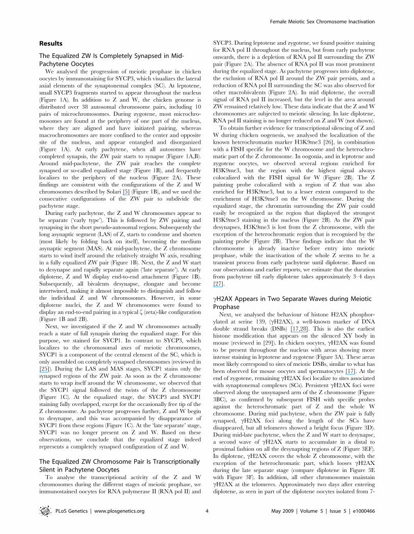
The Equalized ZW Is Completely Synapsed in Mid-Pachytene Oocytes
We analysed the progression of meiotic prophase in chicken
oocytes by immunostaining for SYCP3, which visualizes the lateral
axial elements of the synaptonemal complex (SC). At leptotene,
small SYCP3 fragments started to appear throughout the nucleus
(Figure 1A). In addition to Z and W, the chicken genome is
distributed over 38 autosomal chromosome pairs, including 10
pairs of microchromosomes. During zygotene, most microchro-
mosomes are found at the periphery of one part of the nucleus,
where they are aligned and have initiated pairing, whereas
macrochromosomes are more confined to the center and opposite
site of the nucleus, and appear entangled and disorganized
(Figure 1A). At early pachytene, when all autosomes have
completed synapsis, the ZW pair starts to synapse (Figure 1A,B).
Around mid-pachytene, the ZW pair reaches the complete
synapsed or so-called equalized stage (Figure 1B), and frequently
localizes to the periphery of the nucleus (Figure 2A). These
findings are consistent with the configurations of the Z and W
chromosomes described by Solari [5] (Figure 1B), and we used the
consecutive configurations of the ZW pair to subdivide the
pachytene stage.
During early pachytene, the Z and W chromosomes appear to
be separate (‘early type’). This is followed by ZW pairing and
synapsing in the short pseudo-autosomal regions. Subsequently the
long asynaptic segment (LAS) of Z, starts to condense and shorten
(most likely by folding back on itself), becoming the medium
asynaptic segment (MAS). At mid-pachytene, the Z chromosome
starts to wind itself around the relatively straight W axis, resulting
in a fully equalized ZW pair (Figure 1B). Next, the Z and W start
to desynapse and rapidly separate again (‘late separate’). At early
diplotene, Z and W display end-to-end attachment (Figure 1B).
Subsequently, all bivalents desynapse, elongate and become
intertwined, making it almost impossible to distinguish and follow
the individual Z and W chromosomes. However, in some
diplotene nuclei, the Z and W chromosomes were found to
display an end-to-end pairing in a typical f (zeta)-like configuration
(Figure 1B and 2B).
Next, we investigated if the Z and W chromosomes actually
reach a state of full synapsis during the equalized stage. For this
purpose, we stained for SYCP1. In contrast to SYCP3, which
localizes to the chromosomal axes of meiotic chromosomes,
SYCP1 is a component of the central element of the SC, which is
only assembled on completely synapsed chromosomes (reviewed in
[25]). During the LAS and MAS stages, SYCP1 stains only the
synapsed regions of the ZW pair. As soon as the Z chromosome
starts to wrap itself around the W chromosome, we observed that
the SYCP1 signal followed the twists of the Z chromosome
(Figure 1C). At the equalized stage, the SYCP3 and SYCP1
staining fully overlapped, except for the occasionally free tip of the
Z chromosome. As pachytene progresses further, Z and W begin
to desynapse, and this was accompanied by disappearance of
SYCP1 from these regions (Figure 1C). At the ‘late separate’ stage,
SYCP1 was no longer present on Z and W. Based on these
observations, we conclude that the equalized stage indeed
represents a completely synapsed configuration of Z and W.
The Equalized ZW Chromosome Pair Is TranscriptionallySilent in Pachytene Oocytes
To analyse the transcriptional activity of the Z and W
chromosomes during the different stages of meiotic prophase, we
immunostained oocytes for RNA polymerase II (RNA pol II) and
SYCP3. During leptotene and zygotene, we found positive staining
for RNA pol II throughout the nucleus, but from early pachytene
onwards, there is a depletion of RNA pol II surrounding the ZW
pair (Figure 2A). The absence of RNA pol II was most prominent
during the equalized stage. As pachytene progresses into diplotene,
the exclusion of RNA pol II around the ZW pair persists, and a
reduction of RNA pol II surrounding the SC was also observed for
other macrobivalents (Figure 2A). In mid diplotene, the overall
signal of RNA pol II increased, but the level in the area around
ZW remained relatively low. These data indicate that the Z and W
chromosomes are subjected to meiotic silencing. In late diplotene,
RNA pol II staining is no longer reduced on Z and W (not shown).
To obtain further evidence for transcriptional silencing of Z and
W during chicken oogenesis, we analysed the localization of the
known heterochromatin marker H3K9me3 [26], in combination
with a FISH specific for the W chromosome and the heterochro-
matic part of the Z chromosome. In oogonia, and in leptotene and
zygotene oocytes, we observed several regions enriched for
H3K9me3, but the region with the highest signal always
colocalized with the FISH signal for W (Figure 2B). The Z
painting probe colocalized with a region of Z that was also
enriched for H3K9me3, but to a lesser extent compared to the
enrichment of H3K9me3 on the W chromosome. During the
equalized stage, the chromatin surrounding the ZW pair could
easily be recognized as the region that displayed the strongest
H3K9me3 staining in the nucleus (Figure 2B). As the ZW pair
desynapses, H3K9me3 is lost from the Z chromosome, with the
exception of the heterochromatic region that is recognised by the
painting probe (Figure 2B). These findings indicate that the W
chromosome is already inactive before entry into meiotic
prophase, while the inactivation of the whole Z seems to be a
transient process from early pachytene until diplotene. Based on
our observations and earlier reports, we estimate that the duration
from pachytene till early diplotene takes approximately 3–4 days
[27].
cH2AX Appears in Two Separate Waves during MeioticProphase
Next, we analysed the behaviour of histone H2AX phosphor-
ylated at serine 139, (cH2AX), a well-known marker of DNA
double strand breaks (DSBs) [17,28]. This is also the earliest
histone modification that appears on the silenced XY body in
mouse (reviewed in [29]). In chicken oocytes, cH2AX was found
to be present throughout the nucleus with areas showing more
intense staining in leptotene and zygotene (Figure 3A). These areas
most likely correspond to sites of meiotic DSBs, similar to what has
been observed for mouse oocytes and spermatocytes [17]. At the
end of zygotene, remaining cH2AX foci localize to sites associated
with synaptonemal complexes (SCs). Persistent cH2AX foci were
observed along the unsynapsed arm of the Z chromosome (Figure
3BC), as confirmed by subsequent FISH with specific probes
against the heterochromatic part of Z and the whole W
chromosome. During mid pachytene, when the ZW pair is fully
synapsed, cH2AX foci along the length of the SCs have
disappeared, but all telomeres showed a bright focus (Figure 3D).
During mid-late pachytene, when the Z and W start to desynapse,
a second wave of cH2AX starts to accumulate in a distal to
proximal fashion on all the desynapting regions of Z (Figure 3EF).
In diplotene, cH2AX covers the whole Z chromosome, with the
exception of the heterochromatic part, which looses cH2AX
during the late separate stage (compare diplotene in Figure 3E
with Figure 3F). In addition, all other chromosomes maintain
cH2AX at the telomeres. Approximately two days after entering
diplotene, as seen in part of the diplotene oocytes isolated from 7-
Female Meiotic Sex Chromosome Inactivation
PLoS Genetics | www.plosgenetics.org 4 May 2009 | Volume 5 | Issue 5 | e1000466

Figure 1. Synaptonemal complex formation and ZW pairing during meiotic prophase in chicken oocyte nuclei. A) Overview of meioticprophase in chicken oocytes. The upper panel shows the different substages, based on the morphology of the lateral elements of the synaptonemalcomplexes immunostained for SYCP3 (red). The lower panel shows the corresponding DAPI stained nuclei. The ZW pair is encircled in the zygoteneand pachytene oocytes. Bar represents 10 mm. B) Overview of the different synaptic configurations of the ZW pair during zygotene, pachytene, anddiplotene, visualized by anti-SYPC3 (red). LAS = long asynaptic segment, MAS = medium asynaptic segment [38]; W indicates W chromosome, Zindicates Z chromosome. The panels on the right show schematic drawings of the morphological configurations of Z and W (Z chromosome in blue,W chromosome in green). Bar represents 5 mm. C) Progress of ZW synapsis during pachytene visualized by immunostaining for SYCP1 (green) andSYCP3 (red). Bar represents 10 mm. The higher magnifications show separate immunostainings for SYCP1 (green, upper), SYCP3 (red, middle) and themerge (bottom) for the ZW pair. Bar represents 5 mm.doi:10.1371/journal.pgen.1000466.g001
Female Meiotic Sex Chromosome Inactivation
PLoS Genetics | www.plosgenetics.org 5 May 2009 | Volume 5 | Issue 5 | e1000466

day-old chickens, cH2AX is lost from the Z chromosome
(Figure 3A).
Together with the RNA polymerase II and H3K9me3 staining
patterns, these data show that the second wave of cH2AX
accumulation starts after silencing of the ZW pair has been
established. Moreover, the second wave of cH2AX labelling
appears to be restricted to the Z chromosome. These findings
contrast with observations during mouse meiosis, where cH2AX
accumulation is essential for, and occurs concomitant with,
silencing of the sex chromosomes [30]. To obtain more insight
in the trigger for cH2AX accumulation on the Z chromosome
during late pachytene in chicken oocytes, we analysed the
immunolocalization of the DSB-repair protein RAD51.
DNA Double Strand Break Repair Associated ProteinsTransiently Disappear from the ZW Pair
We found RAD51 foci on the synapsed autosomes and the
unsynapsed axis of the Z chromosome during early pachytene.
However, we never observed RAD51 foci on the W chromosome
during pachytene (Figure 4A–F). During progression of pachytene,
RAD51 foci gradually disappeared from the autosomes. As
synapsis between Z and W progresses, RAD51 foci start to
disappear from the synapsed part, but remain present on the part
of Z that is still unsynapsed (Figure 4B–D). During the equalized
stage, only a diffuse RAD51 signal persists at the distal tip of Z
(Figure 4E). When the Z and W start to desynapse, we noticed a
reappearance of RAD51 foci along the desynapsing Z chromo-
somal axis (Figure 4F). When Z and W were almost completely
separated (‘late separate’ subtype), RAD51 foci covered the
complete Z chromosome (Figure 4G), whereas the W chromosome
remained devoid of these foci. Upon entering diplotene, all SC-
associated RAD51 foci gradually disappear (Figure 4A). The
observed temporal disappearance of RAD51 foci (and cH2AX)
during progression of synapsis between Z and W in pachytene
could indicate that the repair proteins are transiently lost, while
the breaks are not repaired. However, the lack of detectable
RAD51 foci could also be due to the tight winding and twisting of
Z around W, which may render the RAD51 proteins inaccessible
to the antibody. Still, the absence of cH2AX from the synapsed
Figure 2. Lack of RNA polymerase II and enrichment for H3K9me3 mark the ZW pair. A) Oocyte spread nuclei immunostained for RNApolymerase II (green) and SYCP3 (red). The RNA pol II signal is evenly spread in the zygotene nucleus. In pachytene nuclei (LAS and equalizedconfiguration of the ZW pair) and in diplotene, RNA pol II signal is reduced around the ZW pair. Bar represents 10 mm. B) Oocyte spread nucleiimmunostained for H3K9me3 (green) and SYCP3 (red) (upper panel), and DNA FISH with painting probes for the heterochromatic part of the Z(green) and W (red) chromosomes (lower panel). W and the heterochromatic part of Z are enriched for H3K9me3 already in early zygotene. Thehighest H3K9me3 signal is seen on the fully synapsed ZW pair in pachytene. In diplotene, as Z and W have desynapsed, H3K9me3 remains highlypositive on W and is lost from Z, with the exception of the constitutive heterochromatic part of the Z that is recognized by the painting probe whichis still positive for H3K9me3, although with much lower intensity than W. Bar represents 10 mm.doi:10.1371/journal.pgen.1000466.g002
Female Meiotic Sex Chromosome Inactivation
PLoS Genetics | www.plosgenetics.org 6 May 2009 | Volume 5 | Issue 5 | e1000466

Figure 3. Two separate waves of cH2AX on Z during meiotic prophase in chicken oocytes. A–F) A: Oocyte spread nuclei immunostainedfor cH2AX (green) and SYCP3 (red) B–F: Oocyte spread nuclei immunostained for cH2AX (green) and SYCP3 (red) (left panels), DNA FISH with paintingprobes for the heterochromatic part of the Z (green) and W (red) chromosomes (middle panels) and schematic drawings of the synapticconfigurations of Z (blue) and W (green) (right panels). At leptotene, cH2AX starts to appear, and in zygotene it is present throughout the nucleus (A).In pachytene, cH2AX marks all telomeres and is present as big foci on several chromosomes (B). cH2AX also coats the unsynapsed part of Z duringearly pachytene, but disappears from Z as synapsis between Z and W progresses (BC). At the equalized stage, around midpachytene, only thetelomeres are cH2AX positive (D). Upon unwinding or desynapsis of the Z and W, a second wave of cH2AX starts to coat the desynapsed part of Z (E).The heterochromatic part of Z has lost cH2AX from late pachytene onwards (F). In diplotene, only the telomeres and the desynapsed Z chromosomeare highly enriched for cH2AX (A). At later stages of diplotene, cH2AX disappears also from the telomeres and Z (A). Bar represents 10 mm.doi:10.1371/journal.pgen.1000466.g003
Female Meiotic Sex Chromosome Inactivation
PLoS Genetics | www.plosgenetics.org 7 May 2009 | Volume 5 | Issue 5 | e1000466

ZW indicates that the DSBs may also be temporarily undetectable
for the machinery that couples processing of these breaks to
cH2AX formation. Final repair of these breaks may therefore be
suppressed until Z and W desynapse.
H2A Ubiquitylation Marks the ZW Pair from EarlyPachytene until Diplotene
Next, we investigated the localization of several known other
mammalian and marsupial XY body markers in chicken oocyte
nuclei. First, we evaluated H2Ak119ub1, a histone modification
which is generally associated with gene silencing, in combination
with FISH for Z and W. It marks the inactive X chromosome in
female somatic cells [13,31,32], and the mammalian XY body
from mid-pachytene to early diplotene [33]. In chicken zygotene
oocytes, this histone modification marks the W chromosome
(Figure 5B) and from late zygotene onwards also accumulates on
centromeric chromatin. In early pachytene, H2Ak119ub1 starts to
spread from the centromeres on a few microbivalents. It also
begins to accumulate on the already synapsed part of the ZW pair,
and part of the distal unsynapsed arm of the Z chromosome
(Figure 5C). Around mid-pachytene, when the ZW pair is fully
synapsed, the H2Ak119ub1 signal increases and intensifies
specifically on the ZW pair (Figure 5D). When the Z and W
start to desynapse, H2Ak119ub1 remains present on the
desynapting Z chromosomal axis, but is eventually lost from the
W (Figure 5E). At the late separate stage and during early
diplotene, H2Ak119ub1 covers the Z chromosome (Figure 5FG),
with exception of the heterochromatic part recognized by the
FISH probe. A few days after entering diplotene, H2AK119ub1 is
lost from the Z chromosome, and appears in an evenly distributed
manner throughout the whole nucleus (Figure 5H).
Trimethylation of Lysine 27 of Histone H3 Is a ProminentMarker of the W Chromosome during Meiotic Prophase
H3K27me3, an early marker of X chromosome inactivation in the
female mouse embryo [34], is reduced on the XY body in mammals
Figure 4. Reappearance of RAD51 foci on the desynapsing Z chromosome. A–G) Oocyte spread nuclei immunostained for RAD51 (green)and SYCP3 (red). At leptotene and zygotene, RAD51 foci are dispersed throughout the nucleus (A). At early pachytene, some RAD51 foci are presenton almost all synaptonemal complexes, most prominently on the unsynapsed part of the Z chromosome (AB). With progression of pachytene, RAD51foci disappear from the autosomes, but remain present on the unsynapsed segment of the Z chromosome (CD). As synapsis between Z and Wproceeds (MAS to equalized), RAD51 foci disappear along the synapsed region of Z and W (CDE). When Z starts to unwrap itself, RAD51 foci reappearon the desynapsed part of Z (FG) and remain present until diplotene (A). Enlargements and the schematic drawings (Z in blue, W in green) of thesynaptic configurations of the ZW pair are shown. Bar represents 10 mm.doi:10.1371/journal.pgen.1000466.g004
Female Meiotic Sex Chromosome Inactivation
PLoS Genetics | www.plosgenetics.org 8 May 2009 | Volume 5 | Issue 5 | e1000466

[35] and marsupials [36]. In chicken leptotene oocyte nuclei, this
modification is virtually absent, whereupon the signal in zygotene
nuclei increases on W and some microbivalents (Figure 6A). During
pachytene, H3K27me3 is enriched on three microbivalents, but this
modification most prominently marks a subregion of the W
chromosome (Figure 6A). This enrichment of H3K27me3 on the
W chromosome was found to remain prominent only on the W
chromosome, even in late diplotene (Figure 6A).
Figure 5. Analysis of H2Ak119ub1 on the ZW pair during meiotic prophase. A–H) A+H: Immunostaining for H2Ak119ub1 (green) and SYCP3(red) on spread oocyte nuclei. B–G: Immunostaining for H2Ak119ub1 (green) and SYCP3 (red) on spread oocyte nuclei (left) and DNA FISH withpainting probes for the heterochromatic part of the Z (green) and W (red) chromosomes (right). In leptotene, some H2Ak119ub1 areas are visible (A).H2Ak119ub1 already marks the W chromosome during early zygotene (B) and accumulates at all centromeres at late zygotene (not shown). Inpachytene, H2Ak119ub1 still marks the centromeres, and H2Ak119ub1 starts to coat the Z and W chromosomes, covering both parts of the synapsedregions as well as the heterochromatic part of the unsynapsed Z (C). The ZW pair is completely covered by H2Ak119ub1 at its equalized stage (D).When the Z and W start to desynapse, H2Ak119ub1 only persists on the Z chromosome (E) (inset shows schematic drawing of Z (blue) and W (green)pair). At late pachytene, H2Ak119ub1 remains present on the Z, but is lost from its heterochromatic part (F). In diplotene oocytes from 7-day-oldchickens (G), H2Ak119ub1 briefly persists on Z, but eventually disappears and is distributed throughout the nucleus (H). Bar represents 10 mm.doi:10.1371/journal.pgen.1000466.g005
Female Meiotic Sex Chromosome Inactivation
PLoS Genetics | www.plosgenetics.org 9 May 2009 | Volume 5 | Issue 5 | e1000466

Acetylation of H4K16 on the ZW Pair Is Reduced fromMid Pachytene until Diplotene
Acetylation of H4K16 is associated with active transcription,
and in nuclei of female chicken somatic cells, a subregion of the Z
chromosome is specifically enriched for this histone modification
[37]. We performed double-immunostainings of oocytes for
H4K16ac and SYCP3, followed by a FISH for Z and W. During
zygotene, H4K16ac stained the nucleus more prominent then
during leptotene and pachytene, which could indicate a transient
global upregulation of transcription (not shown). Similar to what
was observed for RNA polymerase II, reduced H4K16ac staining
is observed on the completely synapsed ZW pair (Figure 6B). As
pachytene progresses, H4K16ac is also reduced around long
autosomal SCs. The low level of H4K16ac on ZW appears to
persist until mid diplotene. We also performed double-stainings for
cH2AX and H4K16ac and observed that when cH2AX
accumulates on the desynapting Z, H4K16ac is reduced, and this
persists up to diplotene (Figure 6C). Concomitantly, H4K16ac
signal increases on the rest of the genome. Together, these
observations show that the Z and W chromosome lose H4K16ac
around the midpachytene stage, indicating transcriptional silenc-
ing, in accordance with our other observations.
mRNAs of Z and W Genes Show TransientDownregulation in Oocytes during Early Postnatal OvaryDevelopment
If the Z and W chromosome are silenced during pachytene and
early diplotene, mRNAs for Z and W-encoded genes should be
decreased in these cells, compared to earlier and/or later stages of
oocyte development. To analyse this, we performed real-time RT-
Figure 6. Analysis of histone modifications on Z and / or W during meiotic prophase. A) Immunostaining of oocyte spread nucleipreparations for H3K27me3 (green) and SYCP3 (red) (upper panel) and DNA FISH with painting probes for the heterochromatic part of the Z (green)and W (red) chromosomes (lower panel). With progression of zygotene, part of the W chromosome becomes positive for H3K27me3. Duringpachytene, H3K27me3 is present on several microbivalents and coats a specific part of the W chromosome. This pattern persists up into diplotene.Bar represents 10 mm. B) Oocyte spread nuclei preparations immunostained for H4K16ac (green) and SYCP3 (red). From around midpachyteneonwards, H4K16ac is reduced around the ZW pair. In pachytene, also other chromosomes appear to have reduced levels of H4K16ac. Bar represents10 mm. C) Oocyte spread nuclei preparations immunostained for H4K16ac (green) and cH2AX (red). In diplotene, cH2AX and H4K16ac signals aremostly mutually exclusive. Bar represents 10 mm.doi:10.1371/journal.pgen.1000466.g006
Female Meiotic Sex Chromosome Inactivation
PLoS Genetics | www.plosgenetics.org 10 May 2009 | Volume 5 | Issue 5 | e1000466

PCR experiments using total RNA isolated from purified oocyte
fractions and total ovaries isolated on 3 different time points
(embryonic day 20, post hatching day 4 and day 7). Real time RT-
PCR was performed for 10 Z-encoded genes, 4 W-encoded genes
(Figure 7A) and 2 autosomal meiosis-specific genes, namely the
synaptonemal complex component SYCP3 and meiotic-DNA
double strand break-inducing enzyme SPO11. The expression
profiles of SYCP3 and SPO11 followed the expected pattern
(Figure 7B, Figure S1). The Z genes, HINT1, TXN, NIPBL and
SMAD2 all show a relative decrease in expression in oocytes of post
hatching day 4, followed by an increase in expression at day 7
(Figure 7B); Expression of the Z gene SLCA1A3 is measured in
oocytes for the first time at day 7. The W gene HINTW also shows
a decrease on post hatching day 4, and subsequently increased
expression at day 7. The other analysed Z- en W-encoded genes
showed no expression in oocytes at any timepoint, indicating that
they are inactive during meiotic prophase. Based upon earlier
descriptions of ovary development [5,10,38] and our own
observations, most oocytes are still in zygotene during embryonic
day 20, whereas the vast majority of the oocytes is in late
pachytene on post hatching day 4, and in late diplotene on day 7.
The observed decrease in mRNA levels of Z- and W-encoded
genes supports our immunocytochemical findings and confirms
that Z and W are transiently silenced during oocyte development.
The ZZ Chromosome Pair Behaves Similar to theAutosomal Chromosome Pairs during Male MeioticProphase
To establish that the ZW pair in oocytes behaves different from
the ZZ pair in spermatocytes, we also performed immunocyto-
chemical analyses on chicken spermatocytes isolated from adult
testes. Similar to what we observed in oocytes, , cH2AX was present
throughout the nucleus with areas showing more intense staining in
leptotene and zygotene spermatocytes (Figure S2A). At the end of
zygotene, remaining cH2AX foci localize to sites associated with
synaptonemal complexes, also resembling the pattern observed in
chicken oocytes. However, during pachytene, all chromosomes were
fully synapsed and cH2AX was present only on telomeres, and this
pattern persisted up to late diplotene (Figure S2A).
Next, we analyzed the presence of H3K9me3 in combination
with a FISH specific for the heterochromatic regions of the Z
chromosomes (Figure S2B). Several regions in leptotene and
zygotene spermatocytes were enriched for H3K9me3, but they
never colocalized with the FISH signal(s) of Z (Figure S2B). In
pachytene, the heterochromatic region of Z showed some
enrichment for H3K9me3, and this signal decreased again during
diplotene (Figure S2B).
H3K27me3 was present on a few microchromosome through-
out meiotic prophase, but not on Z (not shown).
Discussion
Meiotic Inactivation of the Synapsed ZW Pair in ChickenOocytes
Meiotic sex chromosome inactivation (MSCI) in male mammals
is thought to be triggered by the presence of unsynapsed axes of
the X and Y chromosome (reviewed in [29]). Recently, it was
discovered that in marsupial spermatocytes the unsynapsed X and
Y chromosomes are also inactivated in a manner similar to what
has been observed in mouse [36,39]. Herein, we show inactivation
of sex chromosomes during meiosis in the female Gallus gallus
domesticus, a species with female heterogamety and a ZW sex
chromosome system that evolved independent of XY. Female
oocytes undergo a much longer developmental process between
the initiation of meiotic prophase and ovulation, compared to the
time course that is involved during development of spermatocytes
to mature sperm. Therefore, it was suggested that meiotic
inactivation of Z (and W) would not occur because it would be
incompatible with the lengthy oocyte developmental process [12].
Herein, we have shown to the contrary that MSCI does occur, but
is transient in chicken oocytes; in diplotene, the Z chromosome
loses its specific ‘‘silencing’’ histone modifications (cH2AX and
H2Ak119ub1). In addition, the mRNA of several Z-encoded genes
is higher in oocytes isolated at posthatching day 7 compared to day
4. Reactivation of Z may allow Z-encoded genes to assist in further
oocyte development. HINTW is a W chromosomal multicopy gene
[40,41] that also shows increased expression in day 7 oocytes. It
localizes to the non-heterochromatic tip of W [42]. Based on its
female specificity and expression in differentiating ovaries of
early embryos, HINTW has been implicated in female sex
differentiation, but its exact function is unclear [42]. The W
chromosome is gene poor, and to date, only a few genes have
been described to be W-specific (ICBN Mapviewer, [41,43]). In
addition, the actual size of the pseudo-autosomal region between
Z and W has not been established. Based on the persistent
presence of H3K9me3 on W in diplotene oocytes, it could be
suggested that the W remains inactive throughout oocyte
development, perhaps with the exception of the non-hetero-
chromatic tip that contains the multicopy gene HINTW. This
nicely parallels the recent findings by Mueller et al [44], who
describe that X- linked multicopy genes that are subjected to
MSCI are specifically re-expressed in postmeiotic spermatids in
mouse, whereas the vast majority of single-copy genes remain
inactive.
In early mouse pachytene spermatocytes, the X and Y
chromosome show more extensive synapsis compared to the later
pachytene stages, when desynapsis progresses until the X and Y
show only an end to end association in some diplotene nuclei [45].
This resembles the dynamics of ZW association during chicken
oogenesis, with exception of the fact that complete synapsis is
never achieved in mouse, and always in chicken. Our data now
show that despite the complete (heterologous) synapsis, sex
chromosome inactivation is not prevented, and repair of meiotic
DSBs is delayed (see below).
No Compensation for Z Inactivation by RetrogeneExpression from Autosomal Sites
During mammalian MSCI, silencing of some essential X
chromosomal genes is compensated by expression from retroposed
copies on autosomal chromosomes. The expression of these copies
is male-specific and initiates concomitant with MSCI [46].
However, in the chicken genome, very few functional retroposed
genes appear to be present [47]. For the 15 identified functional
retroposed genes in chicken, no bias for genes from specific
chromosomes was detected. Due to the transient nature of the ZW
inactivation, Z-encoded mRNAs and proteins may be in large
enough supply to allow maintenance of function of essential Z-
linked genes during this short period. Genomic analyses and
analyses of EST databases have revealed that ovary-specific genes
are underrepresented on the chicken Z chromosome. In addition,
microarray analyses of gene expression in different chicken tissues
have shown that the average expression of Z-linked genes versus
autosomal genes is lowest in the embryonic ovary [48]. This
phenomenon could have different causes. In principle, so-called
sexually antagonistic genes (genes beneficial to one sex, detrimen-
tal to the other), are expected to accumulate on the sex
chromosomes. In species with male heterogamety, recessive male
beneficial genes would be expected to accumulate on the X. In
Female Meiotic Sex Chromosome Inactivation
PLoS Genetics | www.plosgenetics.org 11 May 2009 | Volume 5 | Issue 5 | e1000466

accordance with this notion, the mouse and human X chromo-
some are enriched for spermatogenesis-genes expressed prior to
meiotic prophase. Due to MSCI and PMSC (post meiotic sex
chromatin), the X is depleted for spermatogenesis-genes expressed
during later stages, with the exception of some single-copy and
multicopy genes, that show postmeiotic reactivation [44,49,50].
Since retroposition of Z genes to autosomes does not seem to occur
in chicken [47], it might be suggested that the evolutionary force to
drive oocyte-specific genes off the Z during evolution is relatively
weak, perhaps due to the transient nature of MSCI in chicken.
Still, the relative lack of ovary-specific genes, and the generally low
level of Z-encoded gene expression in embryonic ovary may
indicate that MSCI in chicken reduces the likelihood of oocyte-
specific genes that function during meiotic prophase to evolve on
the Z. However, the properties of the chicken Z chromosome can
also be explained by a dominant model of sexual antagonistic
genes, whereby dominant genes encoding proteins that are
beneficial to males would be downregulated in females to
minimize antagonism [51]. More detailed analyses of ovary-
specific genes is required, including separate analyses of genes
expressed in somatic and germ line cells of the ovary, to determine
whether MSCI in chicken affects gene content on Z.
Z Inactivation Precedes the Second Wave of cH2AXFormation
The inactivation of Z and W during chicken oogenesis shows
marked differences and similarities to MSCI in marsupial and
mouse (summarized in Figure 8). The timing of Z inactivation
(early pachytene) is similar to what has been observed in the other
vertebrates. However, the W chromosome appears to enter the
zygotene stage already in a (partially) inactivated configuration.
cH2AX appears as foci on the Z chromosome during zygotene,
and these foci appear to persist longer on the Z compared to the
autosomes, similar to what has been observed on the X
chromosome during zygotene in mouse [17,52]. However, during
the stage of complete synapsis, cH2AX is absent from the ZW
pair. Then, a second wave of cH2AX formation appears around
late pachytene on the desynapsed Z, and only after silencing has
been established. This is in marked contrast with the second wave
of cH2AX formation in mouse, which occurs on both X and Y,
and immediately as spermatocyes enter pachytene. The appear-
ance of cH2AX on the desynapting Z chromosome is accompa-
nied by a reappearance of RAD51. At this stage autosomal axes
also begin to desynapse, but do not show a reappearance of
RAD51 foci, and do not accumulate cH2AX. Thus, repair of
meiotic DSBs on the Z chromosome may be inhibited to avoid
recombination with the synapsed W chromosome, and postponed
until desynapsis. This provides a clear link between the second
wave of cH2AX formation and DSB-repair rather than with an
unsynapsed axis per se. At this late pachytene/early diplotene
stage, H2Ak119ub1 formation is also specifically enhanced on the
Z chromosome. This modification is known to be associated with
inactive chromatin, but has also been implicated in DSB-repair
[53]. Perhaps silencing is induced at sites containing persistent
DSBs to prevent aberrant (truncated) transcription through the
damaged region in somatic as well as germ-line cells. In somatic
cells, DSB repair can also be associated with the recruitment of
silencing factors [54]. We reported a link between the presence of
persistent DSBs and the frequency of meiotic silencing of
unsynaped chromatin (MSUC) in mouse [22].
Figure 7. Gene expression profile in oocytes in different stages of meiotic prophase. A) Schematic drawing of the location of the analyzedgenes on the Z and W chromosomes. The genes in gray did not show expression in oocytes at any of the analysed timepoints. The intermittent linesfrom the W chromosome indicate that the exact location of the genes is unknown [42]. B) Gene expression graphs as analyzed by real time RT PCR fortwo autosomal meiosis-specific genes; SPO11 and SYCP3, for 1 W chromosome gene (HINTW) and 5 Z chromosomal genes (HINT1, TNX, NIBPL, SMAD2,and SLCA1A3). Data were collected at 3 different time-points: embryonic day 20 (E20), 4 (P4) and 7 days post-hatching (P7), and expression in oocyteswas estimated as described in Materials and Methods. Finally, expression at E20 was set at 1, except for SLCA1A3, which was expressed in oocytes onlyon day 7 post hatching.doi:10.1371/journal.pgen.1000466.g007
Female Meiotic Sex Chromosome Inactivation
PLoS Genetics | www.plosgenetics.org 12 May 2009 | Volume 5 | Issue 5 | e1000466

Inactivation of Z during Pachytene Is Most LikelyAccomplished by Spreading of Heterochromatin from W
With the identification of meiotic sex chromosome inactivation
in a species that shows female heterogamety as well as complete
nonhomologous synapsis during pachytene, we provide indications
for the presence of an evolutionary force that drives meiotic sex
chromosome inactivation independent of the final achievement of
synapsis. The absence of homologous chromatin (as a template for
the repair of DNA double-strand breaks) could be instrumental in
initiating this silencing, since synapsis is only achieved after
silencing has been established. Based on the observations
described herein, we propose the following model for the
inactivation of the sex chromosome in the heterogametic female
oocyte during meiotic prophase (Figure 9): The W chromosome
enters meiosis in an inactive configuration, which includes
hypermethylation of H3K9 and ubiquitylation of H2AK119.
H3K27me3 is also present on the W chromosome from zygotene
onwards. H3K27me3 may recruit the polycomb protein complex
PRC1, which could then help to enhance ubiquitylation of
H2AK119, as has been observed during X inactivation in somatic
cells of female mammals [32]. In pachytene, H3K27me3 remains
enriched on a subregion of the synapsed ZW pair. Concomitantly,
the synapsis with Z allows spreading in trans of heterochromatic
marks such as H2Ak119ub1 and H3K9me3 from the inactive W
chromosome on the Z chromosome. Also, additional spreading in
cis of H3K9me3 and H2Ak119ub1 from the heterochromatic
region of Z may contribute to inactivation of Z, triggered by the
transient persistence of the meiotic DSB-associated cH2AX-signal.
The accumulated silencing histone modifications result in
inhibition of Z and W gene transcription, as visualized by reduced
RNA polymerase II staining around ZW, and reduced mRNA
expression of selected Z and W genes. During the compact
arrangement of the Z-chromosomal axis around the W axis, DSB-
repair is inhibited, and cH2AX and possibly also RAD51 are lost
from the DSB-repair sites. Subsequent desynapsis is accompanied
by reappearance of RAD51, a second wave of cH2AX formation
and enhanced H2Ak119ub1 formation on Z. The latter
modification may maintain silencing (despite the absence of
H3K9me3 on the desynapsed Z) until the breaks are repaired. The
W chromosome remains inactive due to the high levels of
H3K9me3 and H3K27me3.
Transcriptional inactivation of the ZW pair was first observed in
oocytes when Z and W show the LAS to MAS configuration, at
day 1 after hatching. Disappearance of cH2AX and H2Ak119ub1
from Z in diplotene was observed in oocytes isolated at the 7th day
after hatching. This indicates that the period of Z inactivation lasts
approximately 5.5–6 days.
Meiotic Sex Chromosome Inactivation and DosageCompensation
A wide variety of mechanisms exist that compensate for unequal
gene dosage in species with chromosomal sex determination.
Female marsupials show inactivation of the paternal X chromo-
some in somatic cells, to equalize the expression level of X-
encoded genes with that of males. The recent discovery of MSCI
and maintenance of X inactivation in postmeiotic cells of male
marsupials supports the hypothesis that inheritance of a ‘‘pre-
inactivated’’ X chromosome could contribute to the establishment
of paternal X-inactivation in female embryos [36]. Our findings
on transient ZW inactivation argue against the existence of such a
mechanism in birds. This is in accordance with data from the
literature that show that male birds do not show inactivation of
one of the two Z chromosomes [48,55,56]. In fact, dosage
compensation in birds appears to be far less complete than in
mammals, and it is not yet known whether dosage compensation,
if it occurs, is achieved via upregulation of Z-genes in females, or
downregulation in males. It cannot be excluded however, that the
transient inactivation of Z leads to epigenetic modifications that
persist and may influence gene expression in male (ZZ)
offspring.
Figure 8. Phylogenetic tree, and overview of marsupial and mammalian XY body and chicken ZW pair markers. Timescale for theavian (chicken) and mammalian (mouse, marsupials) vertebrates [59–63]. Overview of the localization of different markers on XY (mouse [29,35,64]and marsupial [36]) and ZW (chicken) during meiotic prophase.doi:10.1371/journal.pgen.1000466.g008
Female Meiotic Sex Chromosome Inactivation
PLoS Genetics | www.plosgenetics.org 13 May 2009 | Volume 5 | Issue 5 | e1000466

Meiotic Silencing in EvolutionDuring male meiosis in mice, a general mechanism named
meiotic silencing of unsynapsed chromatin (MSUC) silences all
unsynapsed chromosomes [13,15]. This mechanism could be
evolutionary related to meiotic silencing by unpaired DNA
(MSUD) which operates in Neurospora crassa [57]. However,
MSUD is a posttranscriptional silencing mechanism that acts at
the single gene level. It is not clear whether components of MSUD
are conserved and used in MSUC, which acts at a much larger
scale and is far less efficient. Meiotic silencing of sex chromosomes
(MSCI) in mammals is most likely a specialized form of MSUC.
The driving force behind MSUD and MSUC may be that it is
beneficial for the species to silence foreign DNA. Although sex
chromosomes are no foreign DNA, recognition as such may also
be beneficial, because it will help to suppress recombination
between the heterologous regions of the sex chromosomes. This
suppression of sex chromosome recombination could also be a
strong driving force to silence single or heterologous sex
chromosomes. Spreading of heterochromatin from W on Z in
female chicken oocytes to achieve meiotic sex chromosome
inactivation may be mechanism that evolved independent from
MSCI in mammals. In XO male grasshoppers, the single X
chromosome also enters spermatogenesis in an already inactive
configuration [58]. In chicken, the heterologous synapsis between
Z and W may be required to escape from a synapsis checkpoint,
and not to avoid meiotic silencing.
Supporting Information
Figure S1 Gene expression profile in ovaries in different stages
of meiotic prophase. Gene expression graphs with SEM as
analyzed by real time RT PCR using total ovary RNA for two
autosomal meiosis specific genes; SPO11 and SYCP3, for 1 W
chromosome gene (HINTW) and 5 Z chromosomal genes (HINT1,
TNX, NIBPL, SMAD2, and SLCA1A3). Data were normalized to
actin at 3 different time-points: embryonic day 20 (E20), 4 (P4) and
7 days post-hatching (P7). Expression at E20 was set at 1.
Found at: doi:10.1371/journal.pgen.1000466.s001 (0.22 MB TIF)
Figure S2 Analysis of histone modifications during meiotic
prophase of male chicken spermatocytes. A. Spermatocyte spread
nuclei immunostained for cH2AX (green) and SYCP3 (red). At
leptotene cH2AX starts to appear and in zygotene it is present
throughout the nucleus. In pachytene, cH2AX marks all
telomeres, then it gradually disappears from telomeres in
diplotene. Bar represents 10 micrometer. B. Spermatocyte spread
nuclei immunostained for H3K9me3 (green) and SYCP3 (red)
(upper panel) and DNA FISH with painting probes for the
heterochromatic part of the Z (light blue) and SYCP3 (red) (lower
panel). In leptotene and zygotene, H3K9m3 is present throughout
the nucleus with several regions of higher signal intensity, and the
Z chromosomal regions show the same H3K9me3 signal as the
majority of the nucleus. In mid pachytene, some microchromo-
somes and the heterochromatic part of Z have a slightly higher
Figure 9. Model for transient sex chromosome inactivation in the chicken oocyte. In zygotene, parts of the W chromosome are alreadyinactive and marked by increased H3K9me3 and H2Ak119ub1. In addition, part of the W chromosome acquires H3K27me3. The Z chromosome isenriched for H3K9me3 and H2Ak119ub1 only in the region that is known to be heterochromatic. During this phase, RAD51 foci are present and thefirst wave of cH2AX accumulation has occurred clearly on Z and most likely also to a lesser extent on W. Upon entrance of pachytene, Z starts tocondense and folds back on itself, while the W chromosome lengthens slightly, and RNA pol II and the first wave of cH2AX gradually disappear. Withprogression of pachytene, the ZW pair reaches full synapsis, H3K9me3 and H2AK119ub1 most likely spread in trans from W on Z while H4K16ac is lost.Also, silencing may spread on Z in cis, triggered by the presence of persistent meiotic DSBs. As the ZW starts to desynapse, RAD51 reappears on thedesynapting Z, and a second wave of cH2AX on the whole Z chromosome follows. H2AK119ub1 also spreads on the desynapting Z. During thisperiod, meiotic DNA double-strand breaks on Z are most likely repaired, and transcriptional silencing of Z is maintained by cH2AX and H2AK119ub1.Shortly after desynapting, both cH2AX and H2AK119ub1 are lost from the heterochromatic part of Z, while H3K9me3 is lost from Z, except from itsheterochromatic region. In diplotene, both sex chromosomes lengthen, and cH2AX and H2AK119ub1 are gradually lost, allowing re-activation of theZ chromosome, while W remains positive for H3K27me3 and H3K9me3, most likely indicating a maintenance of its inactive state.doi:10.1371/journal.pgen.1000466.g009
Female Meiotic Sex Chromosome Inactivation
PLoS Genetics | www.plosgenetics.org 14 May 2009 | Volume 5 | Issue 5 | e1000466

signal. In diplotene (2 nuclei are shown), H3K9me3 signal is found
in a patchy pattern on some macrochromosomes and minichro-
mosomes, and it is lost from the heterochromatic part of Z.
Found at: doi:10.1371/journal.pgen.1000466.s002 (5.84 MB TIF)
Acknowledgments
We want to thank Dr Martin Volker of the University of Kent for his
helpful advice on the FISH procedure and for providing us with the Z and
W painting probes for Gallus gallus domesticus.
Author Contributions
Conceived and designed the experiments: SS JAG WMB. Performed the
experiments: SS EW JWH WMB. Analyzed the data: SS EW WMB.
Contributed reagents/materials/analysis tools: SS JSEL WMB. Wrote the
paper: SS JSEL JAG WMB.
References
1. Zickler D (2006) From early homologue recognition to synaptonemal complex
formation. Chromosoma 115: 158–174.
2. Monesi V (1965) Differential rate of ribonucleic acid synthesis in the autosomes
and sex chromosomes during male meiosis in the mouse. Chromosoma 17:
11–21.
3. Turner JM, Mahadevaiah SK, Ellis PJ, Mitchell MJ, Burgoyne PS (2006)
Pachytene asynapsis drives meiotic sex chromosome inactivation and leads to
substantial postmeiotic repression in spermatids. Dev Cell 10: 521–529.
4. Solovei I, Gaginskaya E, Hutchison N, Macgregor H (1993) Avian sex
chromosomes in the lampbrush form: the ZW lampbrush bivalents from six
species of bird. Chromosome Res 1: 153–166.
5. Solari AJ (1992) Equalization of Z and W axes in chicken and quail oocytes.
Cytogenet Cell Genet 59: 52–56.
6. Moses MJ, Poorman PA (1981) Synaptosomal complex analysis of mouse
chromosomal rearrangements. II. Synaptic adjustment in a tandem duplication.
Chromosoma 81: 519–535.
7. Moses MJ, Poorman PA, Roderick TH, Davisson MT (1982) Synaptonemal
complex analysis of mouse chromosomal rearrangements. IV. Synapsis and
synaptic adjustment in two paracentric inversions. Chromosoma 84: 457–474.
8. Stefos K, Arrighi FE (1971) Heterochromatic nature of W chromosome in birds.
Exp Cell Res 68: 228–231.
9. Becak ML, Becak W (1981) Behaviour of the ZW sex bivalent in the snake
Bothrops jararaca. Chromosoma 83: 289–293.
10. Ohno S (1961) Sex chromosomes and microchromosomes of Gallus domesticus.
Chromosoma 11: 484–498.
11. Solari AJ (1977) Ultrastructure of the synaptic autosomes and the ZW bivalent in
chicken oocytes. Chromosoma 64: 155–165.
12. Jablonka E, Lamb MJ (1988) Meiotic pairing constraints and the activity of sex
chromosomes. J Theor Biol 133: 23–36.
13. Baarends WM, Wassenaar E, van der Laan R, Hoogerbrugge J, Sleddens-
Linkels E, et al. (2005) Silencing of unpaired chromatin and histone H2A
ubiquitination in mammalian meiosis. Mol Cell Biol 25: 1041–1053.
14. Schimenti J (2005) Synapsis or silence. Nat Genet 37: 11–13.
15. Turner JM, Mahadevaiah SK, Fernandez-Capetillo O, Nussenzweig A, Xu X,
et al. (2005) Silencing of unsynapsed meiotic chromosomes in the mouse. Nat
Genet 37: 41–47.
16. Kelly WG, Aramayo R (2007) Meiotic silencing and the epigenetics of sex.
Chromosome Res 15: 633–651.
17. Mahadevaiah SK, Turner JM, Baudat F, Rogakou EP, de Boer P, et al. (2001)
Recombinational DNA double-strand breaks in mice precede synapsis. Nat
Genet 27: 271–276.
18. Padmore R, Cao L, Kleckner N (1991) Temporal comparison of recombination
and synaptonemal complex formation during meiosis in S. cerevisiae. Cell 66:
1239–1256.
19. Moens PB, Chen DJ, Shen Z, Kolas N, Tarsounas M, et al. (1997) Rad51
immunocytology in rat and mouse spermatocytes and oocytes. Chromosoma
106: 207–215.
20. Ashley T, Plug AW, Xu J, Solari AJ, Reddy G, et al. (1995) Dynamic changes in
Rad51 distribution on chromatin during meiosis in male and female vertebrates.
Chromosoma 104: 19–28.
21. Moens PB, Kolas NK, Tarsounas M, Marcon E, Cohen PE, et al. (2002) The
time course and chromosomal localization of recombination-related proteins at
meiosis in the mouse are compatible with models that can resolve the early
DNA-DNA interactions without reciprocal recombination. J Cell Sci 115:
1611–1622.
22. Schoenmakers S, Wassenaar E, van Cappellen WA, Derijck AA, de Boer P, et al.
(2008) Increased frequency of asynapsis and associated meiotic silencing of
heterologous chromatin in the presence of irradiation-induced extra DNA
double strand breaks. Dev Biol 317: 270–281.
23. Peters AH, Plug AW, van Vugt MJ, de Boer P (1997) A drying-down technique
for the spreading of mammalian meiocytes from the male and female germline.
Chromosome Res 5: 66–68.
24. Essers J, Hendriks RW, Wesoly J, Beerens CE, Smit B, et al. (2002) Analysis of
mouse Rad54 expression and its implications for homologous recombination.
DNA Repair (Amst) 1: 779–793.
25. Costa Y, Cooke HJ (2007) Dissecting the mammalian synaptonemal complex
using targeted mutations. Chromosome Res 15: 579–589.
26. Nakayama J, Rice JC, Strahl BD, Allis CD, Grewal SI (2001) Role of histone H3
lysine 9 methylation in epigenetic control of heterochromatin assembly. Science
292: 110–113.
27. Hughes GC (1963) The Population of Germ Cells in the Developing Female
Chick. J Embryol Exp Morphol 11: 513–536.
28. Rogakou EP, Pilch DR, Orr AH, Ivanova VS, Bonner WM (1998) DNA double-
stranded breaks induce histone H2AX phosphorylation on serine 139. J Biol
Chem 273: 5858–5868.
29. Turner JM (2007) Meiotic sex chromosome inactivation. Development 134:
1823–1831.
30. Fernandez-Capetillo O, Mahadevaiah SK, Celeste A, Romanienko PJ,
Camerini-Otero RD, et al. (2003) H2AX is required for chromatin remodeling
and inactivation of sex chromosomes in male mouse meiosis. Dev Cell 4:
497–508.
31. de Napoles M, Mermoud JE, Wakao R, Tang YA, Endoh M, et al. (2004)
Polycomb group proteins Ring1A/B link ubiquitylation of histone H2A to
heritable gene silencing and X inactivation. Dev Cell 7: 663–676.
32. Fang J, Chen T, Chadwick B, Li E, Zhang Y (2004) Ring1b-mediated H2A
ubiquitination associates with inactive X chromosomes and is involved in
initiation of X inactivation. J Biol Chem 279: 52812–52815.
33. Baarends WM, Hoogerbrugge JW, Roest HP, Ooms M, Vreeburg J, et al. (1999)
Histone ubiquitination and chromatin remodeling in mouse spermatogenesis.
Dev Biol 207: 322–333.
34. Plath K, Fang J, Mlynarczyk-Evans SK, Cao R, Worringer KA, et al. (2003)
Role of histone H3 lysine 27 methylation in X inactivation. Science 300:
131–135.
35. Namekawa SH, Park PJ, Zhang LF, Shima JE, McCarrey JR, et al. (2006)
Postmeiotic sex chromatin in the male germline of mice. Curr Biol 16: 660–667.
36. Namekawa SH, VandeBerg JL, McCarrey JR, Lee JT (2007) Sex chromosome
silencing in the marsupial male germ line. Proc Natl Acad Sci U S A 104:
9730–9735.
37. Bisoni L, Batlle-Morera L, Bird AP, Suzuki M, McQueen HA (2005) Female-
specific hyperacetylation of histone H4 in the chicken Z chromosome.
Chromosome Res 13: 205–214.
38. Solari AJ (1977) Ultrastructure of the synaptic autosomes and the ZW bivalent in
chicken oocytes. Chromosoma 64: 155–165.
39. Franco MJ, Sciurano RB, Solari AJ (2007) Protein immunolocalization supports
the presence of identical mechanisms of XY body formation in eutherians and
marsupials. Chromosome Res 15: 815–824.
40. Mizuno S, Kunita R, Nakabayashi O, Kuroda Y, Arai N, et al. (2002) Z and W
chromosomes of chickens: studies on their gene functions in sex determination
and sex differentiation. Cytogenet Genome Res 99: 236–244.
41. O’Neill M, Binder M, Smith C, Andrews J, Reed K, et al. (2000) ASW: a gene
with conserved avian W-linkage and female specific expression in chick
embryonic gonad. Dev Genes Evol 210: 243–249.
42. Smith CA (2007) Sex determination in birds: HINTs from the W sex
chromosome? Sex Dev 1: 279–285.
43. Hori T, Asakawa S, Itoh Y, Shimizu N, Mizuno S (2000) Wpkci, encoding an
altered form of PKCI, is conserved widely on the avian W chromosome and
expressed in early female embryos: implication of its role in female sex
determination. Mol Biol Cell 11: 3645–3660.
44. Mueller JL, Mahadevaiah SK, Park PJ, Warburton PE, Page DC, et al. (2008)
The mouse X chromosome is enriched for multicopy testis genes showing
postmeiotic expression. Nat Genet 40: 794–799.
45. Tres LL (1977) Extensive pairing of the XY bivalent in mouse spermatocytes as
visualized by whole-mount electron microscopy. J Cell Sci 25: 1–15.
46. Wang PJ (2004) X chromosomes, retrogenes and their role in male reproduction.
Trends Endocrinol Metab 15: 79–83.
47. Consortium ICGS (2004) Sequence and comparative analysis of the chicken
genome provide unique perspectives on vertebrate evolution. Nature 432:
695–716.
48. Ellegren H, Hultin-Rosenberg L, Brunstrom B, Dencker L, Kultima K, et al.
(2007) Faced with inequality: chicken do not have a general dosage
compensation of sex-linked genes. BMC Biol 5: 40.
49. Khil PP, Oliver B, Camerini-Otero RD (2005) X for intersection: retrotrans-
position both on and off the X chromosome is more frequent. Trends Genet 21:
3–7.
Female Meiotic Sex Chromosome Inactivation
PLoS Genetics | www.plosgenetics.org 15 May 2009 | Volume 5 | Issue 5 | e1000466

50. Khil PP, Smirnova NA, Romanienko PJ, Camerini-Otero RD (2004) The mouse
X chromosome is enriched for sex-biased genes not subject to selection bymeiotic sex chromosome inactivation. Nat Genet 36: 642–646.
51. Mank JE, Ellegren H (2009) Sex-Linkage of Sexually Antagonistic Genes Is
Predicted by Female, but Not Male, Effects in Birds. Evolution;doi: 10.1111/j.1558-5646.2009.00618.x.
52. Hamer G, Roepers-Gajadien HL, van Duyn-Goedhart A, Gademan IS, Kal HB,et al. (2003) DNA double-strand breaks and gamma-H2AX signaling in the
testis. Biol Reprod 68: 628–634.
53. Mailand N, Bekker-Jensen S, Faustrup H, Melander F, Bartek J, et al. (2007)RNF8 ubiquitylates histones at DNA double-strand breaks and promotes
assembly of repair proteins. Cell 131: 887–900.54. O’Hagan HM, Mohammad HP, Baylin SB (2008) Double strand breaks can
initiate gene silencing and SIRT1-dependent onset of DNA methylation in anexogenous promoter CpG island. PLoS Genet 4: e1000155.
55. Itoh Y, Melamed E, Yang X, Kampf K, Wang S, et al. (2007) Dosage
compensation is less effective in birds than in mammals. J Biol 6: 2.56. Melamed E, Arnold AP (2007) Regional differences in dosage compensation on
the chicken Z chromosome. Genome Biol 8: R202.57. Bowring FJ, Yeadon PJ, Stainer RG, Catcheside DE (2006) Chromosome
pairing and meiotic recombination in Neurospora crassa spo11 mutants. Curr
Genet 50: 115–123.
58. Cabrero J, Teruel M, Carmona FD, Jimenez R, Camacho JP (2007) Histone H3
lysine 9 acetylation pattern suggests that X and B chromosomes are silenced
during entire male meiosis in a grasshopper. Cytogenet Genome Res 119:
135–142.
59. Bininda-Emonds OR, Cardillo M, Jones KE, MacPhee RD, Beck RM, et al.
(2007) The delayed rise of present-day mammals. Nature 446: 507–512.
60. Kumar S, Hedges SB (1998) A molecular timescale for vertebrate evolution.
Nature 392: 917–920.
61. Pereira SL, Baker AJ (2006) A mitogenomic timescale for birds detects variable
phylogenetic rates of molecular evolution and refutes the standard molecular
clock. Mol Biol Evol 23: 1731–1740.
62. Warren WC, Hillier LW, Marshall Graves JA, Birney E, Ponting CP, et al.
(2008) Genome analysis of the platypus reveals unique signatures of evolution.
Nature 453: 175–183.
63. Woodburne MO, Rich TH, Springer MS (2003) The evolution of tribospheny
and the antiquity of mammalian clades. Mol Phylogenet Evol 28: 360–385.
64. van der Heijden GW, Derijck AA, Posfai E, Giele M, Pelczar P, et al. (2007)
Chromosome-wide nucleosome replacement and H3.3 incorporation during
mammalian meiotic sex chromosome inactivation. Nat Genet 39: 251–258.
Female Meiotic Sex Chromosome Inactivation
PLoS Genetics | www.plosgenetics.org 16 May 2009 | Volume 5 | Issue 5 | e1000466