Direct translocation of histone molecules across cell membranes
10
Introduction In the eukaryotic chromatin, the histone octamer – which is composed of the four histone classes H2A, H2B, H3 and H4 – is surrounded by DNA molecules (Van-Holde, 1989), which are electrostatically bound to the histone (Luger et al., 1997). Being positively charged, histones share several properties with other basic macromolecules such as the polylysine (PLL) (Wagner, 1998), polyornithine (PLO) and polyethanolimine (PEI) (Boussif et al., 1995). Indeed, octamer-DNA complexes or complexes formed between DNA molecules and isolated histones (such as H2A) have been used as DNA carriers to transfect animal cells (Fritz et al., 1996; Balicki et al., 2000) in a similar way to the polyplexes (complexes between DNA and basic macromolecules) (Felgner et al., 1997). A peptide derived from H2A was shown recently to mediate the transfer of a plasmid encoding the β-galactosidase gene into COS-7 cells (Balicki et al., 2002). It was generally assumed that – similar to the polyplexes – histones are being taken into cells by clatherin-coated pits and by the endocytic pathway (Ryser and Hancock, 1965; Murphy et al., 1982). During the past few years, it became apparent that certain small molecular weight proteins are able to directly cross the cell plasma membrane without the involvement of the endocytic pathaway and thus are not susceptible to the intra- endosomal enzymes. The claim about direct penetration via a putative ‘inverted micelle pathway’ (Gariepy and Kawamura, 2001) was based on initial observations that cellular import occurred at 37°C and at 4°C, ruling out endocytosis as a possible transport mechanism. Several natural proteins or peptides such as the HIV-1 Tat or the ARM (arginine-rich motif) peptide derived from it (Vives et al., 1997) as well as mastoparan, the third alpha helix from the Antennapedia homeodomain of Drosophila (Higashijima et al., 1990), have been defined as cell penetrating proteins or peptides (CPPs) because of their ability to translocate cell plasma membrane independently of transporters or specific receptors. Very recently, however, this concept was re-evaluated, leading to the suggestion that the cellular accumulation of the HIV-1 Tat- ARM peptide – thus, of any positively charged peptides – is due to internalization via endocytosis and not to direct penetration (Richard et al., 2003). It was claimed that strong interaction of the CPPs with the cell membrane led to an overestimation of the observed cell-associated fluorescent signal (Vives et al., 2003). Furthermore, previous reports (Vives et al., 1997; Futaki, 2002) on Tat-ARM penetration were suggested to be an artefact due to the use of fixed cells – conditions that may cause a redistribution of the surface-bound Tat peptide, resulting in what appears by microscopic observations as cell penetration and nuclear import. In the present work we show that histones are able to directly transverse the cell plasma membrane. This was observed using different and independent experimental systems: fluorescent microscopy observations with fixed and unfixed HeLa cells and a quantitative assay system using unfixed Colo-205 cells from which surface-bound histones were removed by neutralization. Penetration of histones was also observed at 4°C, in the 4577 The present work shows that histones are able to directly cross cell plasma membranes and mediate penetration of macromolecules covalently attached to them. Adding a mixture containing the five nucleosomal histones, H1, H2A, H2B, H3 and H4, as well as each of the last four individual histones to intact HeLa and Colo-205 cultured cells resulted in cell penetration and nuclear import of these externally added histones. This was observed by fluorescent and confocal microscopy using fixed and unfixed cells, showing that penetration was not due to the fixation process. Accumulation was also estimated by a quantitative assay that did not require cell fixation and allowed neutralization of surface-bound histones. Translocation into the HeLa and Colo-205 cells occurred at 4°C, in ATP-depleted cells and in cells incubated with sucrose (0.5 M) – conditions that block the endocytic pathway. Furthermore, various endocytosis inhibitors such as colchicine, nocodazole, cytochalasin D, brefeldin A, chloroquine and nystatin did not have any effect on the penetration process. Thus, cellular uptake was mostly due to direct translocation of the histones through the cell plasma membrane and not to endocytosis. The histones were also able to mediate penetration of covalently attached bovine serum albumin (BSA) molecules, indicating their potential as carriers for the delivery of macromolecules into living mammalian cells. Key words: Histone, Direct penetration, CPP, Delivery, Endocytosis Summary Direct translocation of histone molecules across cell membranes Elana Hariton-Gazal 1 , Joseph Rosenbluh 2 , Adolf Graessmann 3 , Chaim Gilon 1 and Abraham Loyter 2, * 1 Department of Organic Chemistry, Institute of Chemistry and 2 Department of Biological Chemistry, The Alexander Silberman Institute of Life Sciences, The Hebrew University of Jerusalem, 91904 Jerusalem, Israel 3 Institut fuer Molekularbiologie und Biochemie, Free University of Berlin, Berlin 14195, Germany *Author for correspondence (e-mail: [email protected]) Accepted 8 July 2003 Journal of Cell Science 116, 4577-4586 © 2003 The Company of Biologists Ltd doi:10.1242/jcs.00757 Research Article
-
Upload
independent -
Category
Documents
-
view
1 -
download
0
Transcript of Direct translocation of histone molecules across cell membranes
IntroductionIn the eukaryotic chromatin, the histone octamer – which iscomposed of the four histone classes H2A, H2B, H3 and H4– is surrounded by DNA molecules (Van-Holde, 1989), whichare electrostatically bound to the histone (Luger et al., 1997).Being positively charged, histones share several propertieswith other basic macromolecules such as the polylysine (PLL)(Wagner, 1998), polyornithine (PLO) and polyethanolimine(PEI) (Boussif et al., 1995). Indeed, octamer-DNA complexesor complexes formed between DNA molecules and isolatedhistones (such as H2A) have been used as DNA carriers totransfect animal cells (Fritz et al., 1996; Balicki et al., 2000)in a similar way to the polyplexes (complexes between DNAand basic macromolecules) (Felgner et al., 1997). A peptidederived from H2A was shown recently to mediate the transferof a plasmid encoding the β-galactosidase gene into COS-7cells (Balicki et al., 2002). It was generally assumed that –similar to the polyplexes – histones are being taken into cellsby clatherin-coated pits and by the endocytic pathway (Ryserand Hancock, 1965; Murphy et al., 1982).
During the past few years, it became apparent that certainsmall molecular weight proteins are able to directly cross thecell plasma membrane without the involvement of theendocytic pathaway and thus are not susceptible to the intra-endosomal enzymes. The claim about direct penetration via aputative ‘inverted micelle pathway’ (Gariepy and Kawamura,2001) was based on initial observations that cellular importoccurred at 37°C and at 4°C, ruling out endocytosis as a
possible transport mechanism. Several natural proteins orpeptides such as the HIV-1 Tat or the ARM (arginine-richmotif) peptide derived from it (Vives et al., 1997) as well asmastoparan, the third alpha helix from the Antennapediahomeodomain of Drosophila (Higashijima et al., 1990), havebeen defined as cell penetrating proteins or peptides (CPPs)because of their ability to translocate cell plasma membraneindependently of transporters or specific receptors. Veryrecently, however, this concept was re-evaluated, leading to thesuggestion that the cellular accumulation of the HIV-1 Tat-ARM peptide – thus, of any positively charged peptides – isdue to internalization via endocytosis and not to directpenetration (Richard et al., 2003). It was claimed that stronginteraction of the CPPs with the cell membrane led to anoverestimation of the observed cell-associated fluorescentsignal (Vives et al., 2003). Furthermore, previous reports(Vives et al., 1997; Futaki, 2002) on Tat-ARM penetration weresuggested to be an artefact due to the use of fixed cells –conditions that may cause a redistribution of the surface-boundTat peptide, resulting in what appears by microscopicobservations as cell penetration and nuclear import.
In the present work we show that histones are able to directlytransverse the cell plasma membrane. This was observed usingdifferent and independent experimental systems: fluorescentmicroscopy observations with fixed and unfixed HeLa cells anda quantitative assay system using unfixed Colo-205 cells fromwhich surface-bound histones were removed by neutralization.Penetration of histones was also observed at 4°C, in the
4577
The present work shows that histones are able to directlycross cell plasma membranes and mediate penetration ofmacromolecules covalently attached to them. Adding amixture containing the five nucleosomal histones, H1, H2A,H2B, H3 and H4, as well as each of the last four individualhistones to intact HeLa and Colo-205 cultured cells resultedin cell penetration and nuclear import of these externallyadded histones. This was observed by fluorescent andconfocal microscopy using fixed and unfixed cells, showingthat penetration was not due to the fixation process.Accumulation was also estimated by a quantitative assaythat did not require cell fixation and allowed neutralizationof surface-bound histones. Translocation into the HeLa andColo-205 cells occurred at 4°C, in ATP-depleted cells and
in cells incubated with sucrose (0.5 M) – conditions thatblock the endocytic pathway. Furthermore, variousendocytosis inhibitors such as colchicine, nocodazole,cytochalasin D, brefeldin A, chloroquine and nystatin didnot have any effect on the penetration process. Thus,cellular uptake was mostly due to direct translocation ofthe histones through the cell plasma membrane and notto endocytosis. The histones were also able to mediatepenetration of covalently attached bovine serum albumin(BSA) molecules, indicating their potential as carriers forthe delivery of macromolecules into living mammaliancells.
Key words: Histone, Direct penetration, CPP, Delivery, Endocytosis
Summary
Direct translocation of histone molecules across cellmembranesElana Hariton-Gazal 1, Joseph Rosenbluh 2, Adolf Graessmann 3, Chaim Gilon 1 and Abraham Loyter 2,*1Department of Organic Chemistry, Institute of Chemistry and 2Department of Biological Chemistry, The Alexander Silberman Institute of LifeSciences, The Hebrew University of Jerusalem, 91904 Jerusalem, Israel3Institut fuer Molekularbiologie und Biochemie, Free University of Berlin, Berlin 14195, Germany*Author for correspondence (e-mail: [email protected])
Accepted 8 July 2003Journal of Cell Science 116, 4577-4586 © 2003 The Company of Biologists Ltddoi:10.1242/jcs.00757
Research Article
4578
absence of ATP and in the presence of various endocytosisinhibitors, suggesting that it proceeds in an endocytosis-independent pathway. Of the different histones, H2A and H4had the strongest ability to penetrate cells. We conclude thathistones can be considered to be cell-penetrating proteins.
Materials and MethodsCultured cellsHeLa cell monolayers were grown in Dulbecco’s Modified Eagle’sMedium (DMEM) supplemented with 10% fetal calf serum (FCS),0.3 g/l L-glutamine, 100 U/ml penicillin and 100 U/ml streptomycin(Beit Haemek, Israel). Colo-205 [human Colo-205 adenocarcinomacells (ATcc CCL 222)] were maintained in RPMI 1640 medium,supplemented with 10% FCS, 0.3 g/l L-glutamine, 100 U/mlpenicillin and 100 U/ml streptomycin. Cells were incubated at 37°Cin 5% CO2 atmosphere.
Human lymphocytes were obtained from fresh human blood by itsfractionation on a Ficol gradient (Jazwinski, 1990).
BuffersThe following buffers were used: transport buffer (TB): 20 mM HepespH 7.3, 110 mM potassium acetate, 5 mM sodium acetate, 0.5 mMEGTA, 2 mM DTT, 1 mg/ml leupeptin, 1 mg/ml pepstatin, 1 mg/mlaprotinin, 0.1 mM PMSF. Phosphate buffered saline (PBS): 140 mMsodium chloride buffered with 20 mM sodium phosphate, pH 7.3.
Expression and purification of importin alpha and betaThe vector pET28-hIMPα/β was kindly obtained from V. Citovsky(State University of New-York Stony Brook, NY) and was expressedin Escherichia coli strain BL21 (DE3). The histidine (His-Tag;Qiagen) tagged-importin alpha/beta fusion protein was expressed andpurified by standard protocols following growth at 37°C and inductionof the E. coli strains at 25°C.
Preparation and purification of recombinant histone proteinsExpression plasmids for the individual histones (H2A, H2B, H3 andH4) were kindly obtained from K. Luger (Department of MolecularBiology, Scripps Research Institute, La Jolla, CA) and T. J. Richmond(Institut fur Molekularbiologie und Biophysik, Zurich, Switzerland)and were expressed in E. coli strain BL21 (pLysS) and purified exactlyas described before (Luger et al., 1999). The purified recombinanthistones were solubilized in PBS to give 1 mg/ml.
Incubation of histone mixture and of pure recombinanthistones with cultured HeLa cells: microscopic observationsAn histone mixture containing all the five histones (Sigma H5505) aswell as the individual recombinant histones (H2A, H2B, H3 and H4)were labelled with lissamine rhodamine (Molecular Probes) orcovalently attached to fluorescently labelled bovine serum albumin(BSA) exactly as described previously for the labelling of otherprotein molecules (Hariton-Gazal et al., 2002) and solubilized in PBS.
For incubation with histones, HeLa cells (3×10⁴ per well) werecultured on 10 mm coverslips to subconfluent density. Following theremoval of the culture medium, the cells were washed three times withTB and then exposed to various concentrations of labelled-histonepreparations or histones-BSA conjugates at 37°C or at 4°C in finalvolume of 50 µl TB. At the end of the incubation period the cells werewashed three times with TB and in few experiments were observeddirectly by fluorescent microscopy without fixation. For most of theexperiments, the cells were fixed in 4% (v/v) formaldehyde dissolvedin TB. Fixed cells were examined by fluorescence microscope (Zeiss
Germany, a 40× objective; Apoplan) or by confocal microscopy usingan MRC 1024 confocal microscope (Bio-Rad). The microscope(Axiovert 135M; Zeiss Germany, a 63× objective; Apoplan; NA 1.4)was equipped with an argon ion laser for rhodamine excitation at 514nm (emission 580 nm).
Estimation of the amount of histones within the cytosol andnuclei of intact Colo-205 cellsFor quantitative estimation of intracellular histones, the histones wereconjugated to biotin maleimide or covalently attached to biotinylatedBSA (Sigma) as described previously (Hariton-Gazal et al., 2002),and for these studies a suspension of Colo-205 was used. The degreeof biotinylation was estimated using a spectrophotometric assay forbiotin; it was found to be in most of the experiments 4 molebiotin/mole of histone (Janolino, 1996), and the biotinylated BSAcontained 9 mole biotin/1mole BSA (Sigma).
Intact Colo-205 cells (15-20×105 cells) in TB were incubated witheither biotinylated histone mixture, with the four individualbiotinylated histones (H2A, H2B, H3 and H4) (0.1 mg/ml in TB) orwith biotinylated BSA-histone or with biotinylated BSA (Bb)conjugates (both at 1 mg/ml in TB) in a final volume of 60 µl for 1hour at 37°C or at 4°C. At the end of the incubation period, 200 µlTB were added and the extracellular histones were removed bycentrifugation of the cell suspension for 5 minutes at 2 g. Followingremoval of the supernatant and to neutralize any remaining cell-surface-bound biotinylated histones or conjugates, the pellets weresuspended in a volume of 100 µl avidin (Sigma) (1 mg/ml) in TB.After 30 minutes incubation at 37°C, unbound avidin was neutralizedby the addition of 100 µl of biocytin (2 mg/ml) in TB (Sigma).Following another 15 minutes of incubation, the samples werecentrifuged and the supernatant was removed. For estimation ofcytosol and nuclei biotinylated histone lysates of both compartmentswere obtained in two steps. First, the cells’ plasma membranes werepermeabilized – as followed by a phase microscope – with 30 µl ofdigitonin solution (0.08 mg/ml). Permeabilization was terminated bythe addition of 200 µl TB, usually within 30 seconds at 37°C. Thesamples were centrifuged and the supernatant, containing the cellcytosols, was removed and stored in the cold. Biotinylated moleculespresent in these samples were considered to be those accumulatedwithin the cell cytosol. Any exposed biotinylated histones remainedin the pellet (considered as extranuclear) were neutralized by theaddition 100 µl avidin (0.1 mg/ml) in TB. Following gentlesuspension, the pellet was incubated for 30 minutes at 4°C, and then100 µl of biocytin (0.2 mg/ml) in TB were added to neutralize anyremaining avidin and the samples were incubated for another 15minutes at 4°C. After centrifugation, as above, the supernatant wasremoved and the nuclei in the pellet were then lysed by the additionof 200 µl of lysis buffer (1% Triton X-100 in PBS). After vigorousmixing, lysis was completed by incubation overnight at 4°C.Biotinylated molecules present in these fractions were considered tobe those accumulated within the cell nuclei. For estimation of totalcellular accumulation of histones (cytosol + nuclei), complete cellslysates were obtained, after neutralization of surface-bound biotinmolecules as described above, by the addition of 200 µl lysis buffer.
For collecting the biotinylated histones, the various lysate fractionswere incubated with importin beta-coated plates (Fineberg et al.,2003). For coating, a solution of importin beta (4.3 mg/ml inNaHCO3/Na2CO3 buffer, pH 9.6) was added to 96 maxisorp plates(Nunc Inc.) and incubated overnight at 4°C. Importin beta was usedto attract and bind soluble biotinylated histones as it was shown toserve as a nuclear import receptor of histones (Johnson-Saliba et al.,2000).
When biotinylated BSA-histone conjugates were used, they wereattached to anti-BSA antibody-coated plates as described previouslyfor the attachment of nuclear localization signal (NLS)-BSAconjugates (Hariton-Gazal et al., 2002). Plates attached biotin
Journal of Cell Science 116 (22)
4579Histones directly penetrate cell membrane
molecules of the various samples were estimated following theaddition of avidin-HRP (horse radish peroxidase) exactly as describedpreviously (Melchior et al., 1993). The enzymatic activity of the HRPwas estimated by monitoring the optical density (O.D.) of productobtained at 490 nm. The results given are an average of triplicatedeterminations whose standard error is ±15%.
Estimation of the effect of the histone mixture on the overalltranscription processColo-205 cells, 10 ml (20×106 cells/ml RPMI) were incubated for 1
hour at 37°C in the absence (three samples of10 ml each) or in the presence of a 0.5 µMhistone mixture (three samples of 10 ml each)and then 100 µCi of 5-[3H]-uridine (28Ci/mmole; Amersham Biosciences), and thecells were cultured at 37°C for another 30minutes. At the end of incubation the cellswere centrifuged and after three wash cycleswith PBS, total RNA was extracted by TRIzol(Invitrogen) according to manufacturer
instructions. The RNA pellet obtained from each sample wasdissolved in 200 µl of double distilled water and the degree ofradioactivity was estimated on aliquots of 100 µl. The remaining 100µl, containing the total RNA, were used for isolation of mRNA usingDynabeads mRNA Purification (Dynal Biotech No. 610.06). Thedegree of radioactivity was estimated using aliquots of 20 µlcontaining purified mRNA.
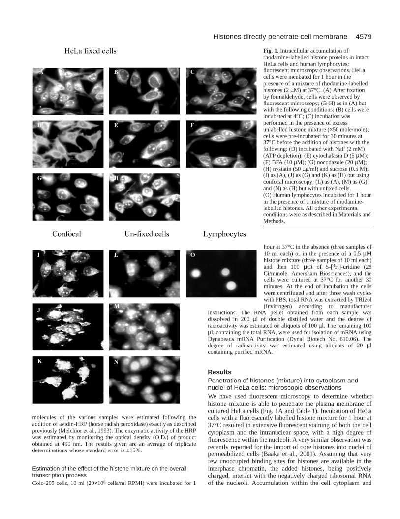
ResultsPenetration of histones (mixture) into cytoplasm andnuclei of HeLa cells: microscopic observationsWe have used fluorescent microscopy to determine whetherhistone mixture is able to penetrate the plasma membrane ofcultured HeLa cells (Fig. 1A and Table 1). Incubation of HeLacells with a fluorescently labelled histone mixture for 1 hour at37°C resulted in extensive fluorescent staining of both the cellcytoplasm and the intranuclear space, with a high degree offluorescence within the nucleoli. A very similar observation wasrecently reported for the import of core histones into nuclei ofpermeabilized cells (Baake et al., 2001). Assuming that veryfew unoccupied binding sites for histones are available in theinterphase chromatin, the added histones, being positivelycharged, interact with the negatively charged ribosomal RNAof the nucleoli. Accumulation within the cell cytoplasm and
Fig. 1. Intracellular accumulation ofrhodamine-labelled histone proteins in intactHeLa cells and human lymphocytes:fluorescent microscopy observations. HeLacells were incubated for 1 hour in thepresence of a mixture of rhodamine-labelledhistones (2 µM) at 37°C. (A) After fixationby formaldehyde, cells were observed byfluorescent microscopy; (B-H) as in (A) butwith the following conditions: (B) cells wereincubated at 4°C; (C) incubation wasperformed in the presence of excessunlabelled histone mixture (×50 mole/mole);cells were pre-incubated for 30 minutes at37°C before the addition of histones with thefollowing: (D) incubated with NaF (2 mM)(ATP depletion); (E) cytochalasin D (5 µM);(F) BFA (10 µM); (G) nocodazole (20 µM);(H) nystatin (50 µg/ml) and sucrose (0.5 M);(I) as (A), (J) as (G) and (K) as (H) but usingconfocal microscopy; (L) as (A), (M) as (G)and (N) as (H) but with unfixed cells.(O) Human lymphocytes incubated for 1 hourin the presence of a mixture of rhodamine-labelled histones. All other experimentalconditions were as described in Materials andMethods.
4580
nucleus was further confirmed by confocal microscopy (Fig.1I). Recent reports suggested that the fixation process can causeartifacts that can be misinterpreted as cellular penetration(Lundberg and Johansson, 2002). However, we also observed
the accumulation of externally added histones within the cellcytosol and nuclei in unfixed cells (Fig. 1L), ruling out thepossibility of an artefact caused by the fixation process.
Cellular uptake of histones occurs under conditions thatinhibit endocytosisTo obtain more insight into the machinery of histone penetrationinto cells, we have tested it under various experimental
Journal of Cell Science 116 (22)
Table 1. Effect of endocytic pathway inhibitors on the uptake of histone mixture, LDL and LY into intact HeLa cells: asummary of fluorescent microscopy observations
Histone mixture† Histone mixture‡ LDL† LY†
Experimental conditions Nuclei Cytosol Nuclei Cytosol Cytosol Cytosol
Control + + + + + +ATP-depleted cells* – + ND ND – –Colchicine (20 µM) + + + + – +Brefeldin A (10 µM) + + + + – –Nystatin (50 µg/ml) + + + + – –Cytochalasin D (5 µM) + + + + – +Chloroquine (50 µM) + + + + – –Nocodazole (20 µM) + + + + – +Sucrose (0.5 M) ± Nystatin (50 µg/ml) + + + + – –
Following 30 minutes incubation at 37°C with all the above inhibitors HeLa cells were incubated with histone mixture for 1 hour and with LDL and LY for 4hours.
*For ATP depletion, cells were incubated, before the addition of the histones with DNP (1 mM), NaF (2 mM) and iodoaceticacid (1 mM). At the end of theincubation period the cells were washed with TB and all subsequent steps of addition of fluorescently labelled molecules as well as fluorescent microscopy werecarried out as described in Materials and Methods.
†Cells were fixed with 4% (v/v) formaldehyde.‡Cells were observed without fixation. +, most of the nuclei/cytoplasm in the microscopic fields are highly fluorescent; –, no fluorescence in the nuclei/cytoplasm; ND, not determined.
0
20
40
60
80
100
120
no ATP
0
0.1
0.2
0.3
0.4
0 10 15 20 25 30 35
0
0.1
0.2
0.3
0.4
0.5
0.6
0 10 20 30 40 50
OD
(490
nm
)O
D (4
90 n
m)
A
C
B
% p
enet
ratio
n
time (min)
time (min)
5
prior neutdetergent
control37ºC
priorfixation depleted
cells
4ºCHistoneexcess
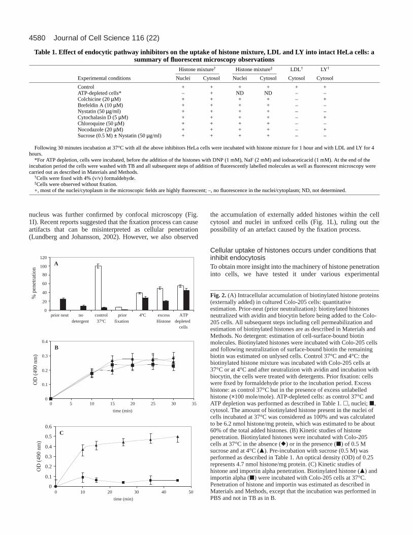
Fig. 2. (A) Intracellular accumulation of biotinylated histone proteins(externally added) in cultured Colo-205 cells: quantitativeestimation. Prior-neut (prior neutralization): biotinylated histonesneutralized with avidin and biocytin before being added to the Colo-205 cells. All subsequent steps including cell permeabilization andestimation of biotinylated histones are as described in Materials andMethods. No detergent: estimation of cell-surface-bound biotinmolecules. Biotinylated histones were incubated with Colo-205 cellsand following neutralization of surface-bound biotin the remainingbiotin was estimated on unlysed cells. Control 37°C and 4°C: thebiotinylated histone mixture was incubated with Colo-205 cells at37°C or at 4°C and after neutralizion with avidin and incubation withbiocytin, the cells were treated with detergents. Prior fixation: cellswere fixed by formaldehyde prior to the incubation period. Excesshistone: as control 37°C but in the presence of excess unlabelledhistone (×100 mole/mole). ATP-depleted cells: as control 37°C andATP depletion was performed as described in Table 1. h, nuclei; j,cytosol. The amount of biotinylated histone present in the nuclei ofcells incubated at 37°C was considered as 100% and was calculatedto be 6.2 nmol histone/mg protein, which was estimated to be about60% of the total added histones. (B) Kinetic studies of histonepenetration. Biotinylated histones were incubated with Colo-205cells at 37°C in the absence (r) or in the presence (j) of 0.5 Msucrose and at 4°C (m). Pre-incubation with sucrose (0.5 M) wasperformed as described in Table 1. An optical density (OD) of 0.25represents 4.7 nmol histone/mg protein. (C) Kinetic studies ofhistone and importin alpha penetration. Biotinylated histone (m) andimportin alpha (j) were incubated with Colo-205 cells at 37°C.Penetration of histone and importin was estimated as described inMaterials and Methods, except that the incubation was performed inPBS and not in TB as in B.
4581Histones directly penetrate cell membrane
conditions that inhibit the endocytic pathway. Incubation of theHeLa cells with histone mixture at 4°C resulted mainly in theappearance of the fluorescent molecules within the cellcytoplasm with very little staining of the cell nuclei (Fig. 1B),indicating inhibition only of nuclear import but not of cellularuptake. Addition of an excess (×50 mole/mole) of unlabelledhistones resulted in some inhibition of nuclear import of thehistones but not of their penetration, indicating a non-receptor-mediated process (compare Fig. 1A with 1C).
In a similar manner, the histone mixture was able topenetrate into ATP-depleted cells (Fig. 1D), showing thathistones uptake did not require ATP, whereas the nuclearimport was ATP-dependent, as expected. In these cells only thecytoplasm was fluorescent, whereas most of the nucleiremained dark. It was well established that nuclear import isan ATP- and GTP-dependent process (Gorlich et al., 2003). Wenext tested the effect of endocytosis inhibitors on the cellularuptake of histones. A battery of inhibitors, such as colchicine(Table 1) (Skrzypek et al., 1998), chloroquine (Table 1), as wellas cytochalasin D (Table 1 and Fig. 1E) (Elliott and O’Hare,1997), Brefeldin A (BFA) (Table 1 and Fig. 1F) andnocodazole (Bayer et al., 1998) (Table 1 and Fig. 1: G, fixedcells; J, confocal microscopy; M, unfixed cells), which are allknown to affect, directly or indirectly, internalization viaendocytosis or intracellular trafficking, did not cause anyinhibition of cellular penetration of histones. Even extremeconditions such as incubation of cells with a combination ofsucrose (0.5 M) and nystatin neither blocked cell penetrationnor nuclear import (Table 1 and Fig. 1: H-fixed cells, K-confocal microscopy, N-unfixed cells). Accumulation of thefluorescent molecules within the cell cytoplasm and nucleiappeared to be the same in the presence or absence of theseinhibitors. In addition to HeLa cells, the labelled histones wereable to accumulate also within human lymphocytes, as evidentfrom the nuclear staining of these cells (Fig. 1O). To confirmthat the endocytosis inhibitors were active, we tested theirability to inhibit the cellular uptake of both low densitylipoprotein (LDL) and lucifer yellow (LY), which are knownto be taken into cells via endocytosis and pinocytosis(Skrzypek et al., 1998; Catizone et al., 1996). As shown inTable 1, nystatin ± sucrose (0.5 M) and ATP depletion of cells(Schmid and Carter, 1990) by a combination of dinitrophenol(DNP), NaF and iodoacetic acid blocked the uptake of bothLDL and LY, whereas colchicine, cytochalasin D andnocodazole completely blocked the uptake of LDL, showingthat the inhibitors used were functional and active.
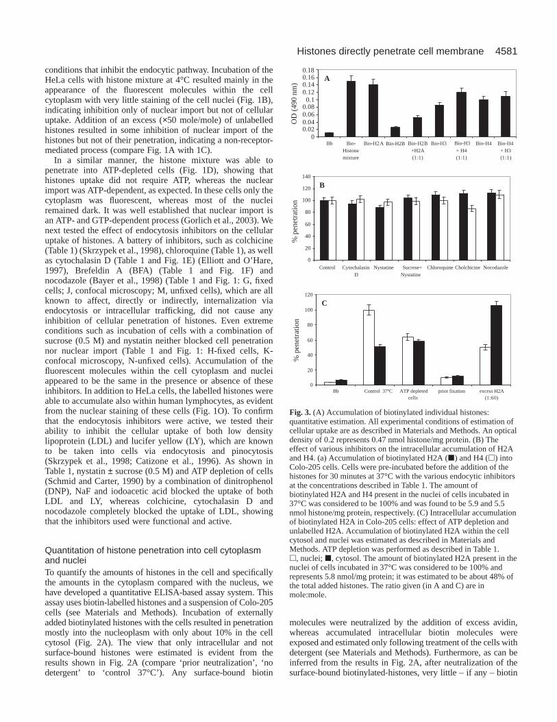
Quantitation of histone penetration into cell cytoplasmand nucleiTo quantify the amounts of histones in the cell and specificallythe amounts in the cytoplasm compared with the nucleus, wehave developed a quantitative ELISA-based assay system. Thisassay uses biotin-labelled histones and a suspension of Colo-205cells (see Materials and Methods). Incubation of externallyadded biotinylated histones with the cells resulted in penetrationmostly into the nucleoplasm with only about 10% in the cellcytosol (Fig. 2A). The view that only intracellular and notsurface-bound histones were estimated is evident from theresults shown in Fig. 2A (compare ‘prior neutralization’, ‘nodetergent’ to ‘control 37°C’). Any surface-bound biotin
molecules were neutralized by the addition of excess avidin,whereas accumulated intracellular biotin molecules wereexposed and estimated only following treatment of the cells withdetergent (see Materials and Methods). Furthermore, as can beinferred from the results in Fig. 2A, after neutralization of thesurface-bound biotinylated-histones, very little – if any – biotin
0
20
40
60
80
100
120
Bbcell s (1:60)
00.020.040.060.08
0.10.120.140.16
Bb Bio-Histonemixture
Bio-H2A Bio-H2B+H2A(1:1)
Bio-H3+ H4(1:1)
Bio-H4 Bio-H4+ H3(1:1)
0
20
40
60
80
100
120
140
Control CytochalasinD
Nystatine Sucrose+Nystatine
Chloroquine Cholchicine Nocodazole
OD
(49
0 nm
)%
pen
etra
tion
% p
enet
ratio
n
B
A
C
Bio-H3Bio-H2B
0.18
Control 37ºC ATP depleted prior fixation excess H2A
Fig. 3. (A) Accumulation of biotinylated individual histones:quantitative estimation. All experimental conditions of estimation ofcellular uptake are as described in Materials and Methods. An opticaldensity of 0.2 represents 0.47 nmol histone/mg protein. (B) Theeffect of various inhibitors on the intracellular accumulation of H2Aand H4. (a) Accumulation of biotinylated H2A (j) and H4 (h) intoColo-205 cells. Cells were pre-incubated before the addition of thehistones for 30 minutes at 37°C with the various endocytic inhibitorsat the concentrations described in Table 1. The amount ofbiotinylated H2A and H4 present in the nuclei of cells incubated in37°C was considered to be 100% and was found to be 5.9 and 5.5nmol histone/mg protein, respectively. (C) Intracellular accumulationof biotinylated H2A in Colo-205 cells: effect of ATP depletion andunlabelled H2A. Accumulation of biotinylated H2A within the cellcytosol and nuclei was estimated as described in Materials andMethods. ATP depletion was performed as described in Table 1.h, nuclei; j, cytosol. The amount of biotinylated H2A present in thenuclei of cells incubated in 37°C was considered to be 100% andrepresents 5.8 nmol/mg protein; it was estimated to be about 48% ofthe total added histones. The ratio given (in A and C) are inmole:mole.
4582
molecules were detected unless cells were lysed with Triton.Indeed, in all subsequent experiments the amount of biotindetected in neutralized unlysed cells never exceeded 10% of theamount of biotinylated molecules found intracellularly. Onlybiotin (–histones) molecules estimated after neutralization ofsurface-bound biotin and treatment of cells with Triton wereconsidered to be molecules present within the intracellular space.Very little accumulation of histones was observed informaldehyde-fixed cells (Fig. 2A), indicating that thepenetration process required a functional intact plasmamembrane. Our quantitative studies confirmed the microscopicobservations (Fig. 1B), showing accumulation within intact cellsfollowing incubation at 4°C (Fig. 2A). However, under theseconditions, translocation into the cell nuclei was inhibited, asexpected, and relatively higher amounts of histones were foundin the cell cytoplasm. The total amount of histones (nuclei +cytoplasm) accumulated at 4°C was 25-30% less than at 37°C(Fig. 2A). In the presence of ×100 molar excess of unlabelledhistones the relative amount of the histones in the cytosol wasincreased but a decrease of about 50% in the intranuclear histonecontent was observed, showing that the unlabelled molecules didnot compete with the penetration of the labelled ones. Similar tothe labelled histones, the externally added unlabelled histones
accumulated within the Colo-205 cellsand thus inhibited translocation oflabelled histones into the cells’ nuclei.It appears, however, that the externallyadded unlabelled histones caused verylittle inhibition of the penetrationprocess itself, which is consistent witha non-saturable membrane penetrationprocess, which is not an activetransport. Intracellular accumulation ofhistones was also obtained followingthe incubation of the histones withATP-depleted cells (Fig. 2A),providing further support to thenotion that penetration was energyindependent. In this case the relativeamount of the intracellular histones
was reduced in the nuclei and increased in the cytosol, indicatinginhibition of nuclear import. However, no reduction in the totalintracellular histone was detected.
Kinetics of histone accumulation in the cellWe have studied the kinetics of histone accumulation in the cellusing our quantitative ELISA-based system (Fig. 2B). Thesame amount of histones accumulated in the cells both at 37°Cand at 4°C, reaching saturation after 15 minutes of incubationat both temperatures. In the presence of sucrose (0.5 M), whichis known to completely inhibit the endocytic pathway, only areduction of 20% in the total amount of the intracellular histonewas observed, and the kinetics remained almost identical. Thecontrol protein importin alpha (Gorlich et al., 1995) did notpenetrate into Colo-205 cells (Fig. 2C), indicating that thepenetration is a specific property of the histones.
Penetration of the individual histones into cell cytoplasmand nucleiFollowing the above observations, we studied whether each ofthe four core histones was also able to penetrate intact cells,
Journal of Cell Science 116 (22)
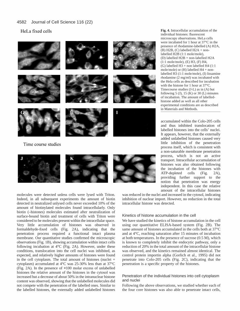
Fig. 4. Intracellular accumulation of theindividual histones: fluorescentmicroscopy observations. HeLa cellswere incubated for 1 hour at 37°C in thepresence of rhodamine-labelled (A) H2A,(B) H2B, (C) labelled H2A + non-labelled H2B (1:1 mole/mole),(D) labelled H2B + non-labelled H2A(1:1 mole/mole), (E) H3, (F) H4,(G) labelled H3 + non labelled H4 (1:1mole/mole) or (H) labelled H4 + non-labelled H3 (1:1 mole/mole), (I) lissaminerhodamine (2 mg/ml) was incubated withthe Hela cells as described for incubationwith the histone for 1 hour at 37°C.Timecourse studies (J-L) as in (A) butfollowing 5 (J), 15 (K) or 30 (L) minutesof incubation. The amount of labelled-histone added as well as all otherexperimental conditions are as describedin Materials and Methods.
4583Histones directly penetrate cell membrane
and to what extent. This was tested using the quantitativeassay system. All histones were able to penetrate into cells, inthe order: histone mixture > H2A > H4 > H3 > H2B. Theamount of the intracellular histone H2A was very close to thatof the histone mixture, whereas the accumulation of histoneH2B was significantly lower (Fig. 3A). This was confirmed byfluorescence microscopy: histone H2A readily accumulatedwithin the cell cytosol and nuclei (Fig. 4A), whereaspenetration of the H2B was low and mainly into the cytosol(Fig. 4B). The addition of labelled H2A to unlabelled H2B orof unlabelled H2A to labelled H2B increased theaccumulation of the labelled histone (Fig. 3A, and see alsoFig. 4C,D). The results were obtained when either unfixedcells were studied or by using the confocal microscope withfixed cells (not shown).
The extent of H3 accumulation was close to that of H4 butlower than that of the H2A or of the histone mixture (Fig. 3Aand Fig. 4E,F). Most of the intracellular H3 accumulatedwithin the cytoplasm, with very little – if any – in theintranuclear space (Fig. 4E). When a combination of the twoproteins (H3 and H4) was used, the extent of their penetrationwas always higher than of each individual histone (Fig. 3and compare Fig. 4E and 4F to 4G and 4H, respectively).Timecourse studies (Fig. 4J-L) confirmed our quantitative data(Fig. 2B), revealing that maximum accumulation of H2A wasreached within 15-30 minutes of incubation. But nointracellular fluorescent staining was observed, even after 60minutes incubation with lissamine rhodamine alone (Fig. 4I).Western blot analysis of the intracellular histones using HRP-strepavidin showed that after penetration, the histonemolecules remained intact (not shown). Also, similar to theobservations with the histone mixture, none of the endocyticpathway inhibitors had any effect on the penetration ability ofthe histones H2A and H4 (Fig. 3B).
The intracellular distribution of H2A was very similar to thatobserved with the histone mixture (compare Fig. 3C to Fig.2A). In control, untreated cells the large majority of theintracellular H2A accumulated within the cell nuclei (Fig. 3C),whereas in ATP-depleted cells it was equally distributedbetween the nucleus and the cytosol. However, the totalintracellular amount of the H2A in ATP-depleted cells wasvery close to that found in control untreated cells (Fig. 3C),indicating again that the penetration process is energyindependent. Similarly, very little change was observed inthe total amount of the intracellular H2A, although its
nuclear:cytosol ratio was altered when ×50 molar excess ofunlabelled H2A was added (Fig. 3C).
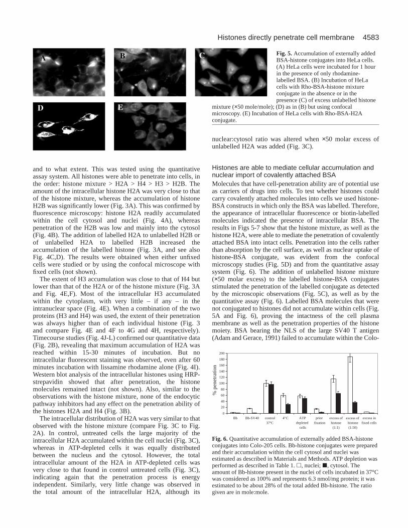
Histones are able to mediate cellular accumulation andnuclear import of covalently attached BSA Molecules that have cell-penetration ability are of potential useas carriers of drugs into cells. To test whether histones couldcarry covalently attached molecules into cells we used histone-BSA constructs in which only the BSA was labelled. Therefore,the appearance of intracellular fluorescence or biotin-labelledmolecules indicated the presence of intracellular BSA. Theresults in Figs 5-7 show that the histone mixture, as well as thehistone H2A, were able to mediate the penetration of covalentlyattached BSA into intact cells. Penetration into the cells ratherthan absorption by the cell surface, as well as nuclear uptake ofhistone-BSA conjugate, was evident from the confocalmicroscopy studies (Fig. 5D) and from the quantitative assaysystem (Fig. 6). The addition of unlabelled histone mixture(×50 molar excess) to the labelled histone-BSA conjugatesstimulated the penetration of the labelled conjugate as detectedby the microscopic observations (Fig. 5C), as well as by thequantitative assay (Fig. 6). Labelled BSA molecules that werenot conjugated to histones did not accumulate within cells (Fig.5A and Fig. 6), proving the intactness of the cell plasmamembrane as well as the penetration properties of the histonemoiety. BSA bearing the NLS of the large SV40 T antigen(Adam and Gerace, 1991) failed to accumulate within the Colo-
Fig. 5.Accumulation of externally addedBSA-histone conjugates into HeLa cells.(A) HeLa cells were incubated for 1 hourin the presence of only rhodamine-labelled BSA. (B) Incubation of HeLacells with Rho-BSA-histone mixtureconjugate in the absence or in thepresence (C) of excess unlabelled histone
mixture (×50 mole/mole); (D) as in (B) but using confocalmicroscopy. (E) Incubation of HeLa cells with Rho-BSA-H2Aconjugate.
0
20
40
60
80
100
120
140
160
180
200
Bb 4ºC ATP excess in
% p
enetr
atio
n
Bb-SV40 control37ºC depleted
cells
priorfixation
excess of excess ofhistone histone(1:1) (1:50)
fixed cells
Fig. 6.Quantitative accumulation of externally added BSA-histoneconjugates into Colo-205 cells. Bb-histone conjugates were preparedand their accumulation within the cell cytosol and nuclei wasestimated as described in Materials and Methods. ATP depletion wasperformed as described in Table 1. h, nuclei; j, cytosol. Theamount of Bb-histone present in the nuclei of cells incubated in 37°Cwas considered as 100% and represents 6.3 nmol/mg protein; it wasestimated to be about 28% of the total added Bb-histone. The ratiogiven are in mole:mole.
4584
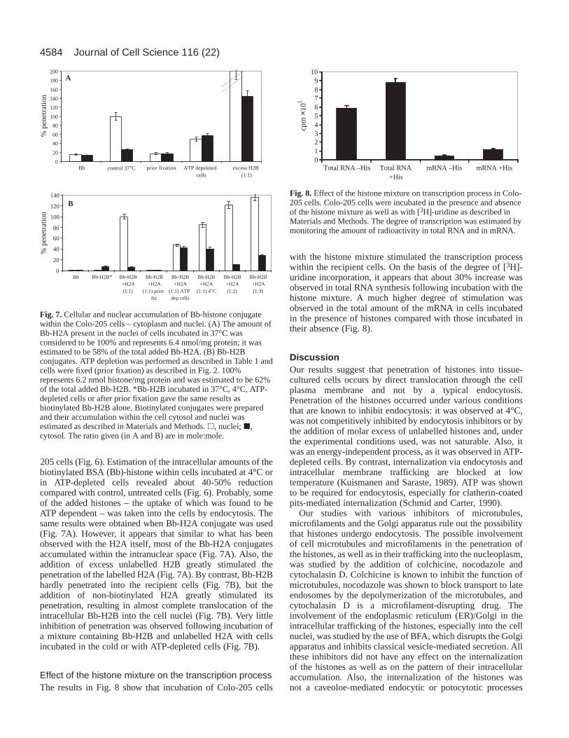
205 cells (Fig. 6). Estimation of the intracellular amounts of thebiotinylated BSA (Bb)-histone within cells incubated at 4°C orin ATP-depleted cells revealed about 40-50% reductioncompared with control, untreated cells (Fig. 6). Probably, someof the added histones – the uptake of which was found to beATP dependent – was taken into the cells by endocytosis. Thesame results were obtained when Bb-H2A conjugate was used(Fig. 7A). However, it appears that similar to what has beenobserved with the H2A itself, most of the Bb-H2A conjugatesaccumulated within the intranuclear space (Fig. 7A). Also, theaddition of excess unlabelled H2B greatly stimulated thepenetration of the labelled H2A (Fig. 7A). By contrast, Bb-H2Bhardly penetrated into the recipient cells (Fig. 7B), but theaddition of non-biotinylated H2A greatly stimulated itspenetration, resulting in almost complete translocation of theintracellular Bb-H2B into the cell nuclei (Fig. 7B). Very littleinhibition of penetration was observed following incubation ofa mixture containing Bb-H2B and unlabelled H2A with cellsincubated in the cold or with ATP-depleted cells (Fig. 7B).
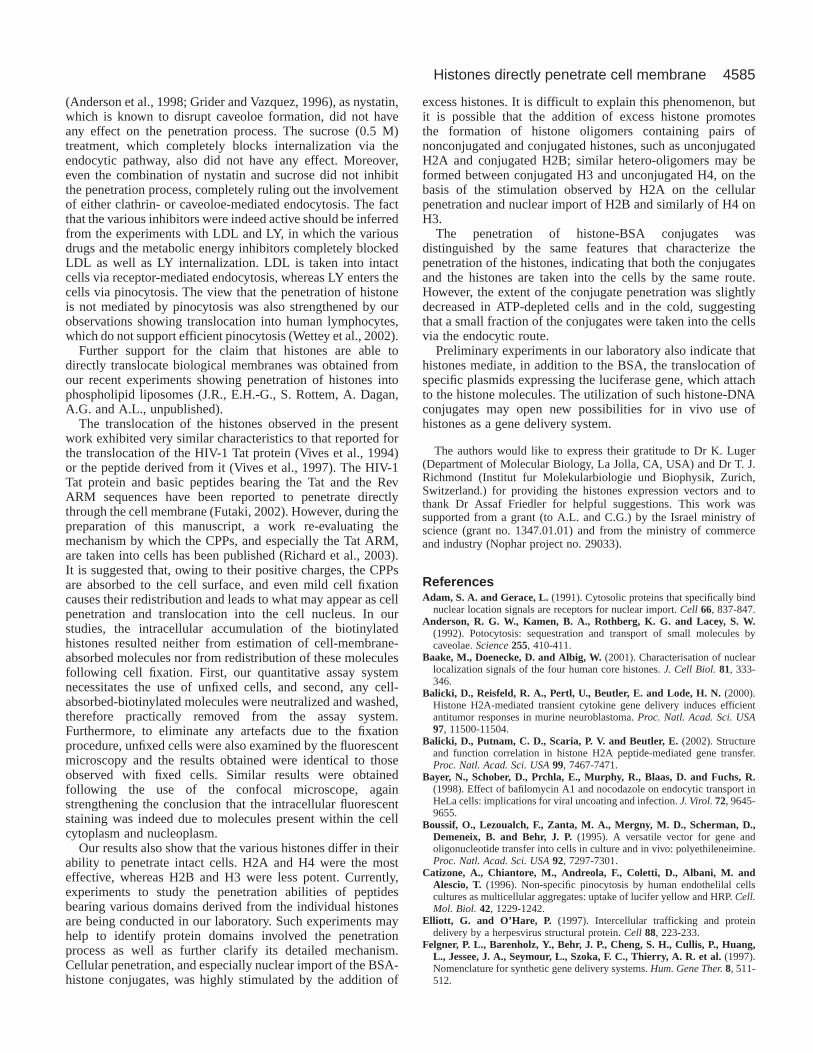
Effect of the histone mixture on the transcription processThe results in Fig. 8 show that incubation of Colo-205 cells
with the histone mixture stimulated the transcription processwithin the recipient cells. On the basis of the degree of [3H]-uridine incorporation, it appears that about 30% increase wasobserved in total RNA synthesis following incubation with thehistone mixture. A much higher degree of stimulation wasobserved in the total amount of the mRNA in cells incubatedin the presence of histones compared with those incubated intheir absence (Fig. 8).
DiscussionOur results suggest that penetration of histones into tissue-cultured cells occurs by direct translocation through the cellplasma membrane and not by a typical endocytosis.Penetration of the histones occurred under various conditionsthat are known to inhibit endocytosis: it was observed at 4°C,was not competitively inhibited by endocytosis inhibitors or bythe addition of molar excess of unlabelled histones and, underthe experimental conditions used, was not saturable. Also, itwas an energy-independent process, as it was observed in ATP-depleted cells. By contrast, internalization via endocytosis andintracellular membrane trafficking are blocked at lowtemperature (Kuismanen and Saraste, 1989). ATP was shownto be required for endocytosis, especially for clatherin-coatedpits-mediated internalization (Schmid and Carter, 1990).
Our studies with various inhibitors of microtubules,microfilaments and the Golgi apparatus rule out the possibilitythat histones undergo endocytosis. The possible involvementof cell microtubules and microfilaments in the penetration ofthe histones, as well as in their trafficking into the nucleoplasm,was studied by the addition of colchicine, nocodazole andcytochalasin D. Colchicine is known to inhibit the function ofmicrotubules, nocodazole was shown to block transport to lateendosomes by the depolymerization of the microtubules, andcytochalasin D is a microfilament-disrupting drug. Theinvolvement of the endoplasmic reticulum (ER)/Golgi in theintracellular trafficking of the histones, especially into the cellnuclei, was studied by the use of BFA, which disrupts the Golgiapparatus and inhibits classical vesicle-mediated secretion. Allthese inhibitors did not have any effect on the internalizationof the histones as well as on the pattern of their intracellularaccumulation. Also, the internalization of the histones wasnot a caveoloe-mediated endocytic or potocytotic processes
Journal of Cell Science 116 (22)
Fig. 7.Cellular and nuclear accumulation of Bb-histone conjugatewithin the Colo-205 cells – cytoplasm and nuclei. (A) The amount ofBb-H2A present in the nuclei of cells incubated in 37°C wasconsidered to be 100% and represents 6.4 nmol/mg protein; it wasestimated to be 58% of the total added Bb-H2A. (B) Bb-H2Bconjugates. ATP depletion was performed as described in Table 1 andcells were fixed (prior fixation) as described in Fig. 2. 100%represents 6.2 nmol histone/mg protein and was estimated to be 62%of the total added Bb-H2B. *Bb-H2B incubated in 37°C, 4°C, ATP-depleted cells or after prior fixation gave the same results asbiotinylated Bb-H2B alone. Biotinylated conjugates were preparedand their accumulation within the cell cytosol and nuclei wasestimated as described in Materials and Methods. h, nuclei; j,cytosol. The ratio given (in A and B) are in mole:mole.
Fig. 8.Effect of the histone mixture on transcription process in Colo-205 cells. Colo-205 cells were incubated in the presence and absenceof the histone mixture as well as with [3H]-uridine as described inMaterials and Methods. The degree of transcription was estimated bymonitoring the amount of radioactivity in total RNA and in mRNA.
% p
enet
ratio
n%
pen
etra
tion
0
20
40
60
80
100
120
140
Bb Bb-H2B* Bb-H2B+H2A
Bb-H2B+H2A
(1:1) priorfix
Bb-H2B+H2A
(1:1) ATPdep cells
Bb-H2B+H2A
Bb-H2B+H2A(1:2)
Bb-H2B+H2A(1:3)
0
20
40
60
80
100
120
140
160
180
200
Bb prior fixation ATP depeletedcells
excess H2B(1:1)
B
A
control 37°C
(1:1) (1:1) 4°C
0123456789
10
+HisTotal RNA –His Total RNA mRNA –His mRNA +His
cpm
×10
5
4585Histones directly penetrate cell membrane
(Anderson et al., 1998; Grider and Vazquez, 1996), as nystatin,which is known to disrupt caveoloe formation, did not haveany effect on the penetration process. The sucrose (0.5 M)treatment, which completely blocks internalization via theendocytic pathway, also did not have any effect. Moreover,even the combination of nystatin and sucrose did not inhibitthe penetration process, completely ruling out the involvementof either clathrin- or caveoloe-mediated endocytosis. The factthat the various inhibitors were indeed active should be inferredfrom the experiments with LDL and LY, in which the variousdrugs and the metabolic energy inhibitors completely blockedLDL as well as LY internalization. LDL is taken into intactcells via receptor-mediated endocytosis, whereas LY enters thecells via pinocytosis. The view that the penetration of histoneis not mediated by pinocytosis was also strengthened by ourobservations showing translocation into human lymphocytes,which do not support efficient pinocytosis (Wettey et al., 2002).
Further support for the claim that histones are able todirectly translocate biological membranes was obtained fromour recent experiments showing penetration of histones intophospholipid liposomes (J.R., E.H.-G., S. Rottem, A. Dagan,A.G. and A.L., unpublished).
The translocation of the histones observed in the presentwork exhibited very similar characteristics to that reported forthe translocation of the HIV-1 Tat protein (Vives et al., 1994)or the peptide derived from it (Vives et al., 1997). The HIV-1Tat protein and basic peptides bearing the Tat and the RevARM sequences have been reported to penetrate directlythrough the cell membrane (Futaki, 2002). However, during thepreparation of this manuscript, a work re-evaluating themechanism by which the CPPs, and especially the Tat ARM,are taken into cells has been published (Richard et al., 2003).It is suggested that, owing to their positive charges, the CPPsare absorbed to the cell surface, and even mild cell fixationcauses their redistribution and leads to what may appear as cellpenetration and translocation into the cell nucleus. In ourstudies, the intracellular accumulation of the biotinylatedhistones resulted neither from estimation of cell-membrane-absorbed molecules nor from redistribution of these moleculesfollowing cell fixation. First, our quantitative assay systemnecessitates the use of unfixed cells, and second, any cell-absorbed-biotinylated molecules were neutralized and washed,therefore practically removed from the assay system.Furthermore, to eliminate any artefacts due to the fixationprocedure, unfixed cells were also examined by the fluorescentmicroscopy and the results obtained were identical to thoseobserved with fixed cells. Similar results were obtainedfollowing the use of the confocal microscope, againstrengthening the conclusion that the intracellular fluorescentstaining was indeed due to molecules present within the cellcytoplasm and nucleoplasm.
Our results also show that the various histones differ in theirability to penetrate intact cells. H2A and H4 were the mosteffective, whereas H2B and H3 were less potent. Currently,experiments to study the penetration abilities of peptidesbearing various domains derived from the individual histonesare being conducted in our laboratory. Such experiments mayhelp to identify protein domains involved the penetrationprocess as well as further clarify its detailed mechanism.Cellular penetration, and especially nuclear import of the BSA-histone conjugates, was highly stimulated by the addition of
excess histones. It is difficult to explain this phenomenon, butit is possible that the addition of excess histone promotesthe formation of histone oligomers containing pairs ofnonconjugated and conjugated histones, such as unconjugatedH2A and conjugated H2B; similar hetero-oligomers may beformed between conjugated H3 and unconjugated H4, on thebasis of the stimulation observed by H2A on the cellularpenetration and nuclear import of H2B and similarly of H4 onH3.
The penetration of histone-BSA conjugates wasdistinguished by the same features that characterize thepenetration of the histones, indicating that both the conjugatesand the histones are taken into the cells by the same route.However, the extent of the conjugate penetration was slightlydecreased in ATP-depleted cells and in the cold, suggestingthat a small fraction of the conjugates were taken into the cellsvia the endocytic route.
Preliminary experiments in our laboratory also indicate thathistones mediate, in addition to the BSA, the translocation ofspecific plasmids expressing the luciferase gene, which attachto the histone molecules. The utilization of such histone-DNAconjugates may open new possibilities for in vivo use ofhistones as a gene delivery system.
The authors would like to express their gratitude to Dr K. Luger(Department of Molecular Biology, La Jolla, CA, USA) and Dr T. J.Richmond (Institut fur Molekularbiologie und Biophysik, Zurich,Switzerland.) for providing the histones expression vectors and tothank Dr Assaf Friedler for helpful suggestions. This work wassupported from a grant (to A.L. and C.G.) by the Israel ministry ofscience (grant no. 1347.01.01) and from the ministry of commerceand industry (Nophar project no. 29033).
ReferencesAdam, S. A. and Gerace, L. (1991). Cytosolic proteins that specifically bind
nuclear location signals are receptors for nuclear import. Cell 66, 837-847.Anderson, R. G. W., Kamen, B. A., Rothberg, K. G. and Lacey, S. W.
(1992). Potocytosis: sequestration and transport of small molecules bycaveolae. Science255, 410-411.
Baake, M., Doenecke, D. and Albig, W. (2001). Characterisation of nuclearlocalization signals of the four human core histones. J. Cell Biol.81, 333-346.
Balicki, D., Reisfeld, R. A., Pertl, U., Beutler, E. and Lode, H. N. (2000).Histone H2A-mediated transient cytokine gene delivery induces efficientantitumor responses in murine neuroblastoma. Proc. Natl. Acad. Sci. USA97, 11500-11504.
Balicki, D., Putnam, C. D., Scaria, P. V. and Beutler, E. (2002). Structureand function correlation in histone H2A peptide-mediated gene transfer.Proc. Natl. Acad. Sci. USA99, 7467-7471.
Bayer, N., Schober, D., Prchla, E., Murphy, R., Blaas, D. and Fuchs, R.(1998). Effect of bafilomycin A1 and nocodazole on endocytic transport inHeLa cells: implications for viral uncoating and infection. J. Virol.72, 9645-9655.
Boussif, O., Lezoualch, F., Zanta, M. A., Mergny, M. D., Scherman, D.,Demeneix, B. and Behr, J. P. (1995). A versatile vector for gene andoligonucleotide transfer into cells in culture and in vivo: polyethileneimine.Proc. Natl. Acad. Sci. USA92, 7297-7301.
Catizone, A., Chiantore, M., Andreola, F., Coletti, D., Albani, M. andAlescio, T. (1996). Non-specific pinocytosis by human endothelilal cellscultures as multicellular aggregates: uptake of lucifer yellow and HRP. Cell.Mol. Biol. 42, 1229-1242.
Elliott, G. and O’Hare, P. (1997). Intercellular trafficking and proteindelivery by a herpesvirus structural protein. Cell 88, 223-233.
Felgner, P. L., Barenholz, Y., Behr, J. P., Cheng, S. H., Cullis, P., Huang,L., Jessee, J. A., Seymour, L., Szoka, F. C., Thierry, A. R. et al. (1997).Nomenclature for synthetic gene delivery systems. Hum. Gene Ther.8, 511-512.

4586
Fineberg, K., Fineberg, T., Graessmann, A., Luedtke, N., Tor, Y., Lixin,R., Jans, D. A. and Loyter, A. (2003). Inhibition of Rev-arginine rich motif-mediated nuclear import by the HIV-1 Rev response element. Biochemistry42, 2625-2633.
Fritz, J., Herweijer, H., Zhang, G. and Wolff, J. (1996). Gene transfer intomammalian cells using histone condensed plasmid DNA. Hum. Gene Ther.7, 1395-1404.
Futaki, S. (2002). Arginine-rich peptides: potential for intracellular deliveryof macromolecules and the mystery of the translocation mechanisms. Int. J.Pharmacol.245, 1-7.
Gariepy, J. and Kawamura, K. (2001). Vectorial delivery ofmacromolecules into cells using peptide-based vehicles. TrendsBiotechnol.19, 21-28.
Gorlich, D., Vogel, E., Mills, A. D., Hartmann, E. and Laskey, R. A. (1995).Distinct functions for the two importin subunits in nuclear protein import.Nature377, 246-248.
Gorlich, D., Seewald, M. and Ribbeck, K. (2003). characterization of Randriven cargo transport and the RanGTPase system by kinetic measurementsand computer simulation. EMBO J.22, 1088-1100.
Grider, A. and Vazquez, F. (1996). Nystatin affects zinc uptake in humanfibroblasts. Biol. Trace Elem. Res.54, 97-104.
Hariton-Gazal, E., Friedler, D., Friedler, A., Zakai, N., Gilon, C. andLoyter, A. (2002). Inhibition of nuclear import by backbone cyclicpeptidomimetics derived from the HIV-1 MA NLS sequence. Biochim.Biophys. Acta1594, 234-242.
Higashijima, T., Burnier, J. and Ross, E. M. (1990). Regulation of Gi andGo by mastoparan, related amphiphilic peptides and hydrophobic amines.Mechanism and structural determinants of activity. J. Biol. Chem.265,14176-14186.
Janolino, V. G. (1996). A spectrometric assay for biotin-binding sites ofimmobilized avidin. Appl. Biochem. Biotechnol.56, 1-7.
Jazwinski, S. M. (1990). Preparation of extracts from yeast. Methods Enzymol.182, 154-174.
Johnson-Saliba, M., Siddon, N., Clarkson, M., Tremethick, D. and Jans,D. (2000). Distinct importin recognition properties of histones andchromatin assembly factors. FEBS Lett.467, 167-174.
Kuismanen, E. and Saraste, J. (1989). Low temperature induced transportblocks as tools to manipulate membrane traffic. Methods Cell Biol.32, 257-274.
Luger, K., Mader, A. W., Richmond, R. K., Sarget, D. F. and Richmond,T. J. (1997). Crystal structure of the nucleosome core particle at 2.8Aresolution. Nature389, 251-260.
Luger, K., Rechsteiner, T. and Richmond, T. (1999). Expression andpurification of recombinant histones and nucleosome reconstitution.Methods Mol. Biol.119, 1-16.
Lundberg, M. and Johansson, M. (2002). Positively charged DNA bindingproteins cause apparent cell membrane translocation. Biochem. Biophys.Res. Commun.291, 367-371.
Melchior, F., Paschal, B., Evance, J. and Gerace, L. (1993). Inhibition ofnuclear protein import by nonhydrolyzable analogues of GTP andidentification of the small GTPase Ran/TC4 as an essential transport factor.J. Cell Biol.123, 1649-1659.
Murphy, R. F., Jorgensen, E. D. and Cantor, C. R. (1982). Kinetics ofhistone endocytosis in chinese hamster ovary cells. J. Biol. Chem.257,1695-1701.
Richard, J. P., Melinkov, K., Vives, E., Ramos, C., Verbeure, B., Gait, M.J., Chernomordik, L. V. and Lebleu, B. (2003). Cell penetrating peptides:a re-evaluation of the mechanism of cellular uptake. J. Biol. Chem.278, 585-590.
Ryser, H. J. P. and Hancock, R. (1965). histone and basic polyamino acidsstimulate the uptake of albumin by tumor cells in culture. Science150, 501-503.
Schmid, S. and Carter, L. (1990). ATP is required for receptor mediatedendocytosis in intact cells. J. Cell Biol.111, 2307-2318.
Skrzypek, E., Cowan, C. and Straley, S. C. (1998). Targeting of the YersiniapestisYopM protein into HeLa cells and intracellular trafficking to thenucleus. Mol. Microbiol. 30, 1051-1065.
Van-Holde, K. (1989). Chromatin. New York: Springer.Vives, E., Charneau, P., VanRietschoten, J., Rochart, H. and Bahraoui, E.
(1994). Effects of the Tat basic domain on human immunodeficiency virustype 1 transactivation, using chemically synthesized Tat protein and Tatpeptides. J. Virol. 68, 3343-3353.
Vives, E., Brodin, P. and Lableu, B. (1997). A truncated HIV-1 Tat proteinbasic domain rapidly translocates through the plasma membrane andaccumulates in the cell nucleus. J. Biol. Chem.272, 16010-16017.
Vives, E., Richard, J. P., Rispal, C. and Lebleu, B. (2003). Tat PeptideInternalization: Seeking the Mechanism of Entry. Curr. Protein Pept. Sci.4,125-132.
Wagner, E. (1998). Polylysine conjugate based DNA delivery. In SelfAssembling Complexes for Gene Delivery(eds A. V. Kabanov, P. L. Felgnerand L. W. Seymour), pp. 309-322. Chichester: Wiley.
Wettey, F., Hawkins, S., Stewart, A., Luzio, J., Howard, J. and Jackson,A. (2002). Controlled elimination of clathrin heavy-chain expression inDT40 lymphocytes. Science297, 1521-1525.
Journal of Cell Science 116 (22)