Length-dependent translocation of polymers through nanochannels
FEMS Microbiology Immunology 105 (1992) 101-112 © 1992 Federation of European Microbiological Societies 0920-8534/92/$05.00 Published by Elsevier
101
FEMSIM 00236
Ion channel and membrane translocation of diphtheria toxin
Cesare Montecucco, Emanue le Papini, Giampietro Schiavo, Elisabetta Padovan and Ornella Rosset to
Centro CNR Biomembrane and Istituto di Patologia Generale, Universitd di Padova, Padova, Italy
Key words: Diphtheria toxin; Membrane translocation; Conformational change; Ion channel
1. SUMMARY
Diphtheria toxin is the best studied member of a family of bacterial protein toxins which act inside cells. To reach their cytoplasmic targets, these toxins, which include tetanus and bo- tulinum neurotoxins and anthrax toxin, have to cross the hydrophobic membrane barrier. All of them have been shown to form ion channels across planar lipid bilayer and, in the case of diphtheria toxin, also in the plasma membrane of cells. A relation between the ion channel and the process of membrane translocation has been sug- gested and two different models have been put forward to account for these phenomena. The two models are discussed on the basis of the available experimental evidence and in terms of the focal points of difference, amenable to fur- ther experimental investigations.
2. INTRODUCTION
Diphtheria toxin (DT) is secreted by toxigenic strains of Corynebacterium diphtheriae and it is
Correspondence to: C. Montecucco, Istituto di Patologia Gen- erale, Via Trieste, 75, 35121 Padova, Italy.
responsible for most clinical manifestations of diphtheria [1,2]. DT is the most studied member of a large family of bacterial protein toxins that act inside cells on cytoplasmic targets [3]. They share a similar mechanism of cell intoxication that can be conveniently divided into three steps: a) binding; b) membrane translocation; and c) target modification. This functional similarity is reflected in an overall similar structural organiza- tion. In fact these toxins are composed of two protomers generally held together by a single disulfide bond: protomer A has a catalytic activity while protomer B is responsible for cell binding and translocation into the cell [3]. The reduced toxin or the isolated protomers are non-toxic and toxicity can be recovered by reforming the disul- fide link. Most of these toxins display an ADP- ribosyltransferase activity, i.e. they transfer ADP-ribose from cytoplasmic NAD to a more or less specific cytoplasmic target [3,4]. This is the case of DT, exotoxin A from Pseudomonas aerug- inosa, cholera toxin, pertussis toxin and the C2 and C3 clostridial toxins. Shighella shigae and Shiga-like toxins are rRNA-adenine glycohydro- lases [5]. Tetanus and botulinum neurotoxins are zinc endopeptidases [6,7].
DT is secreted as an inactive single polypep- tide chain which is later activated by protease cleavage at an exposed loop [2], as schematized in

102
Fig. 1. The A chain (21164 Da) ADP-ribosylates specifically elongation factor 2 at a single residue, a modified histidine, termed diphtamide. This results in the blockage of protein synthesis and cell death by apoptosis [2,4]. The B chain (37194 Da) binds to a protein receptor via its 6-kDa carboxy-terminal part [8,9]. Cell sensitivity to DT is correlated to the amount of its receptor: the very sensitive Vero cell line displays tens of thou- sand receptors per cell [10]. The DT-receptor complex is collected into coated pits and endocy- tosed. Drugs which interfere with the acidifica- tion of the endosomal lumen by the ATPase proton pump also reduce or prevent cell intoxica- tion [11-14]. Moreover DT can be induced to enter into cells from the plasma membrane by a short incubation in an acid medium, thus by-pass- ing endocytosis [15-17]. These experiments have indicated that acidification is a prerequisite for the entry of DT into ceils. Moreover, the endo- some by-pass procedure is very important be- cause it allows one to test in vivo several parame- ters that may influence the phenomenon [18].
The discovery that a simple condition such as acid pH is sufficient to cause a rather remarkable phenomenon such as the translocation of a
~c**.
PROTEASE$
( 21 kDa )'NH2 A s1 ,s
( 37 kDa ) - C ~ H S
Fig. 1. Schematic structure of diphtheria toxin. The toxin is secreted as a single inactive polypeptide chain. Several pro- teases can cleave the chain at an exposed loop thus generating an active toxin composed of two subunits held together by a single interchain disulfide bond. Protomer B is responsible for cell binding and membrane translocation while protomer A is an ADP-ribosyltransferase specific for elongation factor two
of eukaryotes and arehaebacteria.
water-soluble protein across the hydrophobic membrane barrier has triggered a wealth of stud- ies aimed at a molecular understanding of such a process. Most of them were performed on model membrane systems employing a variety Of bio- physical and biochemical techniques. Attention was also paid to compare model and cellular experimental systems [19].
3. THE ION CHANNEL OF DIPHTHERIA TOXIN IN PLANAR LIPID BILAYERS
By applying DT to planar lipid bilayers at different pH values it was found that at low pH DT and the B fragment of the truncated mutant crm 45 (lacking the 16.5-kDa carboxy-terminus) form voltage-dependent ion channels [20-25] with the following main properties: 1) the DT channel has a low conductance, few picoSiemens (pS) at physiological ionic strength; 2) the DT-induced increase in conductance of the membrane occurs only at low pH and does not require a transmem- brane pH gradient, though the rate of increase of conductance is higher with a pH gradient. With no pH gradient across the planar lipid bilayer a DT-induced current is seen only in the presence of an applied voltage; voltage is not needed when a pH gradient is present; 3) the channel is cation selective at pH 5.5; 4) more than one toxin molecule, possibly two, participate in the forma- tion of a channel; 5) the conductance of planar lipid bilayers containing acidic phospholipids in the presence of DT at low pH is higher than those made only of zwitterionic lipids; and 6) the rate of conductance increase is strongly depen- dent on the existence of a positive potential (posi~ tive on the c/s side where DT is present).
Analysis of the change of permeability induced by DT on potassium-loaded liposomes led to es- sentially the same conclusions [26,27]. These stud- ies have been very important because, apart from introducing the use of model membrane systems, they provided a clear indication that DT under- goes a pH-driven structural change and that, as a consequence, at low pH it interacts deeply with the lipid bilayer.

4. T H E T U N N E L M O D E L FOR T H E MEM- BRANE TRANSLOCATION OF DIPHTHE- RIA TOXIN
Another important aspect of these biophysical studies was that they provided a first experimen- tal support for the tunnel mechanism of DT translocation across membranes proposed by Bo- quet et al. [28] on the basis of detergent binding experiments. Moreover, this model identified protons as a single agent capable of inducing the membrane interaction of DT. The main features of the tunnel model are schematically summa- rized in Fig. 2. Fragment B was suggested to undergo a conformational change from a neutral form to an acidic form, characterized by the presence of hydrophobic surfaces which mediate detergent and lipid binding. The acid form of fragment B is thus able to insert into lipid bilay- ers forming a transmembrane hydrophilic pore large enough to allow the passage of the unfolded A chain. The protein pore, probably made of two B polypeptide chains, protects subunit A from the interaction with lipids. In this model it is assumed that ions flow through the same channel used by the unfolded A chain to transverse the membrane and the formation of the channel is a prerequisite for A translocation to occur. On the basis of this model one may expect that a single
(9 ®
~ acid ic pH ,ons
Fig. 2. The tunnel model for the membrane translocation of diphtheria toxin. At acid pH fragment B becomes hydropho- bic and inserts in the lipid bilayer in an oligomeric form with a hydrophilic pore. Fragment A unfolds and crosses the mem- brane inside the tunnel protected from the contact with lipids. When free of the A chain, the protomer B tunnel is an ion
channel.
103
pore incorporated in a planar lipid bilayer is able to mediate the passage of more than one A chain. In the tunnel model the A and B subunits of DT are seen as two independent domains with re- spect to the conformational change triggered by pH: protomer B is suggested to become hy- drophobic and to insert into the lipid bilayer while protomer A is suggested to unfold before inserting into the channel.
5. T H E ION CH A N N EL OF D I P H T H E R I A TOXIN IN CELLS
Some aspects of the ion channel made by DT on planar lipid bilayers left the possibility open that the ion channel could be a peculiarity of this model system and, even if not so, that the chan- nel could be non-relevant to the translocation of D T across membranes. For example, as appears from Fig. 2, its pH dependence is shifted at least one pH unit toward lower pH values with respect to that of cell intoxication [18,29]. Another con- troversial aspect concerns the size of the channel. Though there is no direct relation between the size of a channel and its conductance, values of the order of few picoSiemens do not fit with the dimensions expected for a protein channel that has to accommodate a polypeptide chain with its lateral chains of different volume, charge and hydrophilicity. This consideration is the more valid in the light of the properties of the first protein-conducting channel characterized in pla- nar lipid bilayers [30]. Simon and Blobel [30] have shown that this channel of the rough endoplasmic reticulum is closed when plugged by the nascent polypeptide chain and is opened by puromycin, which induces the release of the peptidyl- puromycin complex. The channel shows a large conductance (220 picoSiemens) with no ion selec- tivity. The change in size or polarity of the ap- plied voltage does not influence either the con- ductance or the gating of this channel. These properties are those expected for a pore that has to accommodate amino acids with lateral chain differing in size, charge and polarity.
To obtain evidence for the formation of a DT channel on living cells, Vero cells in culture were

104
exposed to DT in an acid medium for various time periods. Sandvig and Olsnes measured the cellular uptake of various radioactive ions [31], Papini et al. measured the K ÷ and Na ÷ cellular content [32] and Alder et al. measured leakage of radioactive metabolites [33]. The following con- clusions were reached: a) at low pH DT forms an ion channel on the plasma membrane; b) its pH dependence is almost superimposable to that of cellular intoxication; c) the channel is specific for monovalent cations, does not leak out amino acids or phosphorylated metabolites and does not take up glucosamine; d) the membrane remains per- meable to monovalent cations as long as a trans- membrane pH gradient is maintained; on return- ing the extracellular medium to neutrality, the cell quickly recovers its normal K ÷ and Na ÷ content; and e) the channel itself does not con- tribute significantly to the death of the cell be- cause a DT mutant lacking ADP-ribosylating ac- tivity forms the channel but does not kill the cells.
Taken together, these studies on the perme- ability induced by DT on cells have also provided strong evidence for the formation of a DT ion channel on ceils. However, some differences among channels formed on planar membranes and on cell membranes are evident, as summa- rized in Table 1. In particular there is a different pH dependence and an apparent different size. These differences can be ascribed to the presence in the cellular system, but not in the model sys- tems, of the toxin receptor. Unfortunately no
single channel patch clamp studies of DT on cells have been reported so far and any further com- parison is not possible.
6. THE LOW pH-INDUCED INSERTION OF DIPHTHERIA TOXIN INTO THE LIPID BILAYER
The structural changes of DT-induced by pH in relation to model membranes have been stud- ied with a variety of techniques including lipo- some binding [34], membrane photolabelling with photoreactive lipids [35-37] and phospholipids [38,39], circular dichroism [36,40], tryptophan flu- orescence [41,42], liposome fusion and leakage [42-46], scanning calorimetry [47], polarized in- frared spectroscopy [48] and lipid monolayers [49]. Notwithstanding the differences among the ex- perimental approaches, these studies show a sub- stantial agreement on the main findings with a good correlation with results obtained with cellu- lar systems. The more relevant conclusions, de- rived from the above-quoted studies, can be sum- marized as follows:
(a) At neutral pH, DT interacts with the sur- face of negatively charged membranes. The possi- ble relevance of this membrane surface interac- tion of DT to cell surface binding has been dis- cussed elsewhere [39].
(b) A downshift of pH induces a true struc- tural change in DT from a neutral conformation
Table 1
Comparison of the properties of the channels made by diphtheria toxin planar lipid bilayers and in Vero cells with those of a protein-conducting channel of the endoplasmic reticulum
Property DT in planar DT in Vero cells Protein- lipid bilayers conducting channel
of ER
Conductance Few ps n.d. 220 pS Estimated few pS
Ion selectivity X + X + Non-selective Size of permeant ion > Glucosamine < Glucosamine > Gluconate pH Dependence < 5.0 5-6 n.d. Voltage gating + n.d. - Voltage dependence of conductance + n.d. -
n.d. = not determined.

to an acidic conformation. The two forms differ in their secondary structures and exposure of tryptophan residues. While the neutral form is water-soluble, the acidic form aggregates as a result of the exposure of previously hidden hy- drophobic regions. The hydrophobicity of acid DT makes it able to penetrate into the hydropho- bic core of lipid micelles or bilayers with a strong preference for negatively charged lipids.
(c) Both the A and B fragments of DT are involved in the low pH-induced interaction of the toxin with the fatty acid portion of phospholipids: such interaction extends all along the hydrocar- bon chains. In this process, a-helices of the inner part of B chain orientate parallel to the lipid acyl chains, while B-sheets of the carboxy terminal part of the B chain orient parallel to the mem- brane surface. While the low pH-induced lipid interaction was predicted by the tunnel model, the finding that A also interacts with the hy- drophobic lipid core of the membrane at low pH was unexpected.
(d) The low pH-induced lipid interaction of chain A is fully reversible, i.e. chain A appears to insert in the lipid bilayer at low pH and to re- escape when pH is brought back to neutrality.
(e) The neutral and acid structures of DT have different shapes with different apparent molecular areas; 2950 A 2 at pH 7.4 and 3450 A 2 at pH 5.0. The lipid-embedded part of DT at pH 5.0 has an apparent molecular area of 2100 A 2.
(f) The decrease of pH brings about the expo- sure of the interchain disulfide bond, which be- comes accessible to reduction by thioreductase [50].
(g) The transition between the neutral and acid forms of DT in the presence of negatively charged liposomes occurs between pH 5 and 6 in close agreement with the pH dependence of DT cell intoxication and ion channel formation, shown in Fig. 2, and at variance from that obtained in planar lipid membranes.
(h) Chain B is the first part of DT that inserts into the membrane and this B insertion is not the rate-limiting step of DT membrane translocation.
(i) The membrane translocation competent form of DT is likely to be a multimer-aggregated toxin.
105
7. A CLEFT MODEL FOR THE MEMBRANE TRANSLOCATION OF DIPHTHERIA TOXIN
Some well-documented experimental results such as the low pH lipid interaction of fragment A and the reduced size of the channel formed by DT in living cells, cannot be accommodated in the tunnel model of DT membrane translocation. Moreover, as discussed above and summarized in Table 1, the properties of the recently character- ized protein-conducting channel of the endoplas- mic reticulum are very different from those of DT [30].
At this stage one is left with two possibilities: (1) the ion channel formed by DT at acid pH is not directly involved in toxin membrane translo- cation. Such an epiphenomenon would easily ex- plain the above-mentioned experimental results; and (2) the DT ion channel is involved in the membrane translocation step, but there are dif- ferences among the fragment A-conducting and the ion-conducting forms of DT.
As the large majority of researchers in this field, we have adopted as working hypothesis the latter one. However, to account for all the experi- mental findings and theoretical considerations summarized above, we have put forward a 'cleft'
100
-.: -i o _ " ~ -
~ 8 '~
° -
• •
O 4 5
pH
Fig. 3. pH Dependence of diphtheria toxin block of Vero cells protein synthesis ( • ), ion channel formation in Vero cells (~)
and in planar lipid bilayers (e).
106
model for the m e m b r a n e t ranslocat ion of D T [19,51], as schemat ized in Fig. 3. We propose that toxin cell binding is the result o f the interactions of D T with bo th a prote in receptor and the headgroups of negatively charged phospholipids [39]. Experimental evidence for such a double interaction and its biological implications are dis- cussed elsewhere [39]. The D T protein receptor is a 14-20-kDa glycoprotein which binds D T via its polypept ide chain [7-9]. A n addit ional prote in may be involved in toxin binding [52]. The 6-kDa carboxy-terminal region of p ro tomer B is mainly responsible for the interact ion with the prote in receptor [9].
Recep to r -bound D T is internalized via coated pits and it is collected into endosomes. As a result of the acidification o f the endosomal lu- men, D T adopts an acidic structure, character- ized by the exposure of hydrophobic regions, in- cluding the interchain disulfide bond, previously
h idden in the prote in interior. Such hydrophobic surfaces can be shielded f rom the contact with water ei ther by involvement in p ro t e in -p ro t e in contacts or by insertion into the hydrophobic core of the lipid bilayer. Even though there are no data on the stoichiometry of the membrane- in- serting form of p ro tomer B, it is likely to be a dimer or a multimer, whose format ion is driven by hydrophobic segments exposed at acidic pH. A multimeric organizat ion of the D T channel is also suggested by the generally oligomeric structure of the known ion channels and by considerat ions on the number of f ragment B segments candidate for the insertion in the lipid bilayer.
The apparen t p K a of m e m b r a n e insertion of p ro tomer B is about 6. On this basis the protona- tion of histidine residues (15 out of the 16 such residues present in D T are clustered in chain B) is expected to play a major role in its conforma- tional transit ion together with prolines, which
(9 ® @
low ~
neutral neutral ~ pH pH )
tow
C Y T O P L A S M
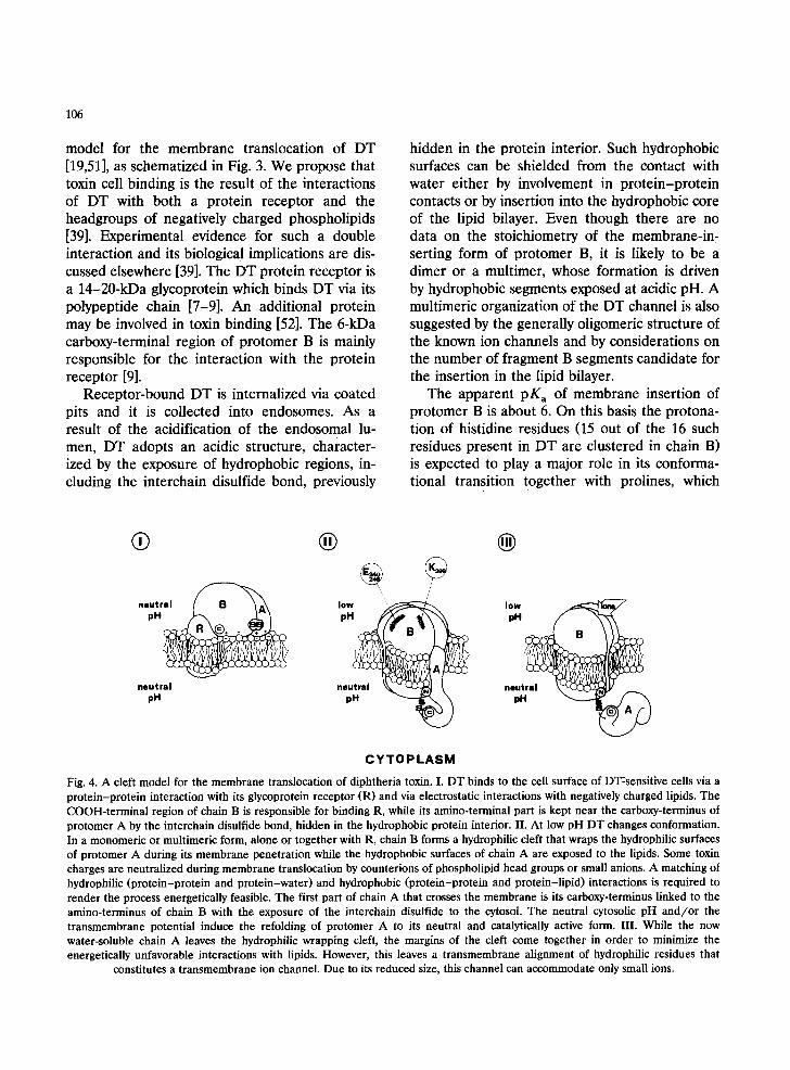
Fig. 4. A cleft model for the membrane translocation of diphtheria toxin. I. DT binds to the cell surface of DT:sensitive cells via a protein-protein interaction with its glycoprotein receptor (R) and via electrostatic interactions with negatively charged lipids. The COOH-terminal region of chain B is responsible for binding R, while its amino-terminal part is kept near the carboxy-terminus of protomer A by the interchain disulfide bond, hidden in the hydrophobic protein interior. II. At low pH DT changes conformation. In a monomeric or multimeric form, alone or together with R, chain B forms a hydrophilic cleft that wraps the hydrophilic surfaces of protomer A during its membrane penetration while the hydrophobic surfaces of chain A are exposed to the lipids. Some toxin charges are neutralized during membrane translocation by counterions of phospholipid head groups or small anions. A matching of hydrophilic (protein-protein and protein-water) and hydrophobic (protein-protein and protein-lipid) interactions is required to render the process energetically feasible. The first part of chain A that crosses the membrane is its carboxy-terminus linked to the amino-terminus of chain B with the exposure of the interchain disulfide to the cytosol. The neutral cytosolic pH and/or the transmembrane potential induce the refolding of protomer A to its neutral and catalytically active form. III. While the now water-soluble chain A leaves the hydrophilic wrapping cleft, the margins of the cleft come together in order to minimize the energetically unfavorable interactions with lipids. However, this leaves a transmembrane alignment of hydrophilic residues that
constitutes a transmembrane ion channel. Due to its reduced size, this channel can accommodate only small ions.

107
may undergo cis-trans isomerization as proposed by Brasseur et al. [53].
The central proposition of the cleft model is that, during the membrane penetration of DT, fragment B forms a hydrophilic cleft that drives the insertion of chain A with its hydrophobic segments exposed to lipids and their hydrophilic regions interacting with corresponding regions of the hydrophilic cleft of fragment B. Such a matching of hydrophobic and hydrophilic pro- tein-lipid and protein-protein interactions is of fundamental importance in making the transloca- tion process energetically feasible.
The cleft model can accommodate the possibil- ity that the DT receptor participates in the mem- brane translocation of protomer A [54] by assum- ing that together with protomer B it forms the hydrophilic cleft that nests chain A during its passage through the membrane; it is also possible that the efficiency of the chain-A-conducting channel made of protomer B plus DT receptor is higher than that formed by fragment B alone in model membranes, but that both structures are able to function as protein channels.
The disulfide-linked C-terminal part of A and N-terminal part of B are suggested to be the first portion of DT that inserts in the membrane to reach the cytoplasm, mainly driven by the hy- drophobicity of the disulfide bond. These two terminal segments are expected to play a crucial role in the process and thus not to tolerate insert- ions, deletions or substitutions altering their phys- ico-chemical properties. Indeed, it has recently been shown that DT mutants extended at the carboxy-terminus of chain are less or non-toxic [55].
The A chain crosses the membrane with its hydrophilic residues facing the protein channel and its hydrophobic lateral chains exposed to the hydrocarbon chains of lipids. The open structure of the B cleft can accommodate the different bulkinesses of different portions of the A chain. Moreover, such a transmembrane structure is ex- pected to be able to translocate across the mem- brane peptide segments added to the N-terminus of the A chain, but not to the C-terminus, as recently found by Stenmark et al. [55]. It should be emphasized that no unfolding of A is required
by the cleft model. Rather it is suggested that A undergoes a conformational change in concert with B. The low pH structure of A is amphipathic with mainly hydrophilic surfaces inside the pro- tomer B cleft and an hydrophobic side interacting with lipids.
The interchain disulfide bond, previously hid- den in the hydrophobic protein interior, becomes exposed on the other side of the membrane to the cytoplasm, where it is ready to be cleaved either by chemical or proteic reducing agents of yet unknown localization [50].
It is likely that the membrane penetrating form of DT has a net positive charge and that some Lys, Arg or His residues are not in the position to form salt bridges with corresponding negatively charged Asp and Glu residues. Due to the high energetic cost of driving charged groups across the hydrophobic core of the lipid bilayer (particu- larly the positive charges of lysines and arginines), membrane insertion and translocation of DT would be made less energetically unfavorable if these charges were neutralized and shielded by formation of ionic couples with negatively charged phospholipid head groups or small anions such as chloride, bromide or thiocyanate. This effect would help to explain the finding that anions are involved in the penetration of DT into ceils and that the more hydrophobic membrane permeant anions are the more efficient [56]. It is possible that the membrane-translocating form of DT is actually a transient anion-phospholipid-multi- meric toxin complex that assembles on the low pH lumen side of the endosomal membrane and lasts until it reaches the cytoplasmic face of the membrane, where the complex disassembles be- cause its ion couples are released by water solva- tion. Such a proposal emphasizes the role of non-bilayer lipidic configurations [57,58], though direct evidence for such structures in DT mem- brane translocation is lacking. However, this sug- gestion is in agreement with the well-documented ability of DT to induce fusion of lipid vesicles at low pH that is believed to occur via zones ot DT-induced lipid destabilization [44].
At the present stage it is hard to envisage how the polypeptide chain of protomer B folds across the membrane to form the hydrophilic cleft and
108
interacts with lipids at the same time. The only available information is that Lys 299 and Glu 248 and 249 are located on the face opposite to the cytosol [59]. Moreover, the loss of channel activity of DT on replacing I1e-364 with a Lys residue [60] suggests that I1e-364 is essential for the correct assembly of the channel.
The finding that chain A can escape from liposomes once pH is returned to neutrality sug- gests that, after reduction of the interchain disul- fide bond, fragment A can re-acquire its neutral water-soluble form on facing the neutral cytoplas- mic pH [38].
A comparison of the number of molecules of DT bound to a cell with the estimated number of A chains in the cytoplasm has indicated that the membrane-translocation step of DT is a ineffi- cient process [50]. However, in experiments aimed at testing the possible role of the endosomal proteases in the cellular intoxication of DT (Papini et al., in preparation), it was found that the translocation and release in the cytosol of the catalytic chain A subunit is a rather efficient process that occurs in competition with a proteol- ysis of DT stimulated by the acidification of the endosomal compartment. In other words, the A chains in the cytoplasm are those that have es- caped endosomal proteolysis by translocation in the cytosol. Endosomal proteolysis lowers the overall efficiency of intoxication and is responsi- ble for the previous underestimation of the effi- ciency of membrane translocation that is per se higher than previously thought. At the present stage, the proposal that endosomal proteolysis generates a B45-1ike fragment which is the form of B actually responsible for A translocation is still to be tested [28]. The fact that endosomal proteolysis is avoided in cells intoxicated with DT by exposure to a low pH extracellular medium [17,50] leaves open the possibility that a Bas-like fragment is generated by proteolysis in the endo- somal lumen and it mediates a more efficient translocation of A with respect to that occurring at the plasma membrane with the intact B frag- ment.
The cleft model suggests that, after chain A has reached the cytoplasm, the margins of the hydrophilic cleft embedded in the lipid bilayer,
formed by protomer B (or protomer B plus DT receptor), come together to reduce to a minimum the amount of hydrophilic protein surface ex- posed to the hydrocarbon chains of lipids. This leaves a transmembrane alignment of hydrophilic residues that constitute a peculiar fiat-shaped ion channel with more or less rigid protein walls and a very flexible lipid side. Such a channel is ex- pected to have a low conductance because of its reduced size and at the same time to be able to accommodate large charged objects provided that they have a hydrophobic fiat portion such as an aromatic ring. Such features would account for the finding that the DT channel on cells has a low permeability to choline, glucosamine, amino acids and phosphorylated intermediates [31-33].
In this simplified model the major driving force for the membrane insertion and translocation of DT is the transmembrane proton gradient be- cause it is the low pH that causes the conforma- tional transition on one side of the membrane and it is the neutral pH that induces the refolding of chain A on the other side of the membrane. However, a contribution of membrane potential is suggested by cellular studies [61,62]. Moreover, the voltage opening of DT channels at low pH with no transmembrane pH gradient [20-25] sug- gests that voltage is able to unplug the channel by inducing the release of the A chain. Also, the role of disulfide reduction in membrane translo- cation and in the refolding of protomer A in the cytoplasm remains to be investigated.
The tunnel model and the cleft model differ in several respects. The main differences amenable to experimental testing can be summarized as follows: 1) the low pH-driven conformational change of DT. In the tunnel model the A and B protomers are supposed to change conformation independently: B becomes more hydrophobic and inserts in the lipid bilayer forming a pore, while A is suggested to unfold in order to be able to enter in the pore. In the cleft model the confor- mational change induced by acid pH is seen as a concerted phenomenon involving B and A at the same time. There is no experimental evidence for an unfolding of A at low pH, rather it appears that the catalytic fragment of DT adopts an acidic conformation that can be crystallized and charac-

ter ized spectroscopically [48,63,64]; 2) lipid inter- action. In the tunnel model f ragment A crosses the m e m b r a n e inside an hydrophil ic pore with no contact with lipids. The model does not explain how the 65 hydrophobic residues of the A chain can get th rough an hydrophil ic pore, part icularly the 20 Trp, Tyr and Phe residues. In the cleft model the m e m b r a n e t ranslocat ion of f ragment A occurs at the lipid prote in boundary in a way compat ible with the different proper t ies of the lateral chains of the different amino acids; 3) proper t ies o f the D T ion channel. In the tunnel model the channel that mediates the transloca- t ion of A is also the one that carries ions across the membrane : the prote in conduct ing channel and the ion channel are the same entity with the same dimensions. The format ion of the tunnel is actually a prerequisi te for the m e m b r a n e translo- cat ion of f ragment A. In the cleft model the ion channel is re lated to the s tructure that has medi- ated the t ranslocat ion of A, but it is not the same entity, not having the same size and propert ies; the ion channel is a consequence of the mem- brane t ranslocat ion of A. This difference is such that the tunnel model would be dismissed by the finding of a muta t ion or of an agent that allows cell intoxication without channel formation.
8. M E M B R A N E T R A N S L O C A T I O N O F O T H E R B A C T E R I A L P R O T E I N T O X I N S
Much less is known about the mechanism of cell entry of o ther bacterial prote in toxins with cytosolic targets such as the exotoxin A f rom Pseudomonas aeruginosa or the clostridial neuro- toxins of te tanus and botulism, Diphther ia toxin has been the toxin pro to type in the study of all aspects o f cell intoxication. It is therefore likely that some aspects of its mechan i sm of act ion are relevant to those of o ther toxins. Several biophys- ical and biochemical studies indicate that these toxins also undergo an acid-driven structural t ransit ion with exposure o f hydrophobic surfaces and consequent interact ion with the hydrocarbon chains of lipids [65-74].
109
A C K N O W L E D G E M E N T S
We thank Drs. R. Bisson, G. Menes t r ina and D. Pie t robon for critical reading of the manuscr ipt and we acknowledge the suppor t of the ' C N R Target Project for Biotechnology and Bioinstru- mentat ion ' .
R E F E R E N C E S
[1] Pappenheimer, A.M. (1982) Diphtheria: studies on the biology of an infectious disease. Harvey Lect. 76, 45-73.
[2] Uchida, T. (1982) Diphtheria toxin: biological activity. In: Molecular Action of Toxins and Viruses (Cohen, P. and Van Heyningen, S., Eds.), 1-31, Elsevier, Amsterdam.
[3] Rappuoli, R. and Pizza, M.G. (1991) Structural and evo- lutionary aspects of ADP-ribosylating toxins. In: Source- book of Bacterial Protein Toxins (Alouf, J.E. and Freer, J.H., Eds.) Academic Press, London, 1-23.
[4] Collier, R.J. (1990) Diphtheria toxin: Structure and Func- tion of a Cytocidal Protein. In: ADP-ribosylating Toxins and G Proteins (Moss, J. and Vaugham, M., Eds.), 3-19, American Society for Microbiology, Washington D.C.
[5] Endo, Y., Tsurugi, K., Yutsudo, T., Takeda, Y., Oga- sawara, T. and Igarashi, K. (1988) Site of action of a Vero toxin from Escherichia coli 0157:H7 and of Shiga toxin on eukaryotic ribosomes. Eur. J. Biochem. 171, 45-50.
[6] Schiavo, G., Poulain, B., Rossetto, O., Benfenati, F., Tauc, L. and Montecucco, C. (1992) Tetanus toxin is a zinc protein and its inhibition of neurotransmitter release and protease activity depend on zinc. EMBO J. 11, (in press).
[7] Schiavo, G., Rossetto, O., Santucci, A., DasGupta, B.R. and Montecucco, C. (1992) Botulinum neurotoxins A, B and E are zinc proteins. J. Biol. Chem. 267, (in press).
[8] Mekada, E., Okada, Y. and Uchida, T. (1988) Identifica- tion of diphtheria toxin receptor and a nonproteinous diphtheria toxin-binding molecule in Vero cell mem- brane. J. Cell Biol. 107, 511-519.
[9] Naglich, J.G., Metherall, J.E., Russell, D.W. and Eidels, L. (1992) Expression cloning of a diphtheria toxin recep- tor: identity with a heparin-binding EGF-like growth factor precursor. Cell 69, 1051-1061.
[10] Middlebrook, J.L., Dorland, R.B. and Leppla, S.H. (1978) Association of diphtheria toxin with Vero cells. J. Biol. Chem. 253, 7325-7330.
[11] Moya, M., Dautry-Varsat, A., Gould, B., Louvard, D. and Boquet, P. (1985) Inhibition of coated pit formation in Hep2 cells blocks the cytotoxicity of diphtheria toxin but not that of ricin toxin. J. Cell. Biol. 101, 548-559.
[12] Morris, R.E., Gersten, A.S., Bonventre, P.F. and Salinger, C. (1985) Receptor-mediated entry of diphtheria toxin into Vero cells: electron microscopic evaluation. Infect. Immun. 50, 721-727.
110
[13] Marnell, M.H., Shia, S.P., Stookey, M. and Draper, R.K. (1984) Evidence for penetration of diphtheria toxin to the cytosol through a prelysosomal membrane. Infect. Im- mun. 44, 145-150.
[14] Umata, T., Moriyama, Y., Futai, M. and Mekada, E. (1990) The cytotoxic action of diphtheria toxin and its degradation in intact Vero ceils are inhibited by bafilomycin A1, a specific inhibitor of vacuolar-type H- ATPase. J. Biol. Chem. 265, 21940-21945.
[15] Sandvig, H. and Olsnes, S. (1980) Diphtheria toxin entry into cells is facilitated by low pH. J. Cell Biol. 87, 828-832.
[16] Draper, R.K. and Simon, M.I. (1980) The entry of diph- theria toxin into the mammalian cell cytoplasm: evidence for lysosomal involvement. J. Cell Biol., 87, 849-854.
[17] Sandvig, K. and Olsnes, S. (1981) Rapid entry of nicked diphtheria toxin into ceils at low pH. J. Biol. Chem. 256, 9068-9076.
[18] Sandvig, K., Tonnessen, T.I., Sand, O. and Olsnes, S. (1986) Requirement of a transmembrane pH gradient for the entry of diphtheria toxin into cells at low pH. J. Biol. Chem. 26t, 11639-11644.
[19] Montecucco, C., Papini, E. and Schiavo, G. (1991) Molecular models of toxin membrane translocation. In: Sourcebook of Bacterial Protein Toxins (Alouf, J.E. and Freer, J.H., Eds.), 45-56, Academic Press, London.
[20] Donovan, J.J., Simon, M.I., Draper, R.K. and Montal, M. (1981) Diphtheria toxin forms transmembrane channels in planar lipid bilayers. Proc. Natl. Acad. Sci. USA 78, 172-176.
[21] Kagan, B.L., Finkelstein, A. and Colombini, M. (1981) Diphtheria toxin fragment forms large pores in phospho- lipid bilayer membranes. Proc. Natl. Acad. Sci. USA 78, 4950-4954.
[22] Donovan, J.J., Simon, M. and Montal, M. (1982) Inser- tion of diphtheria toxin into and across membranes: role of phosphoinositide asymmetry. Nature 298, 669-672.
[23] Deleers, M., Beugnier, N., Falmagne, P., Cabiaux, V. and Ruysschaert, J.M. (1983) Localization in diphtheria toxin fragment B of a region that induces pore formation in planar lipid bilayers at low pH. FEBS Lett. 160, 82-86.
[24] Misler, S. (1983) Gating of ion channel made of diphthe- ria toxin fragment in phospholipid bilayer membranes. Proc. Natl. Acad. Sci. USA 80, 4320-4324.
[25] Hoch, D.H., Romero-Mira, M., Ehrlich, B.E., Finkel- stein, A., DasGupta, B.R. and Simpson, L.L. (1985) Channels formed by botulinum, tetanus, and diphtheria toxins in planar lipid bilayers: relevance to translocation of proteins across membranes. Proc. Natl. Acad. Sci. USA 82, 1692-1696.
[26] Boquet, P. and Duflot, E. (1982) Tetanus toxin fragment forms channels in lipid vesicles at low pH. Proc. Natl. Acad. Sci. USA 79, 7614-7618.
[27] Shiver, J.W. and Donovan, J.J. (1987) Interactions of diphtheria toxin with lipid vesicles: determinants of ion channel formation. Biochim. Biophys. Acta 903, 48-55.
[28] Boquet, P., Silverman, M.S., Pappenheimer, A.M. and Vernon, W.B. (1976) Binding of Triton X-100 to diphthe-
ria toxin, crossreacting material 45 and their fragments. Proc. Natl. Acad. Sci. USA 73, 4449-4453.
[29] Papini, E., Schiavo, G., Tomasi, M., Colombatti, M., Rappuoli, R. and Montecueco, C. (1987) Lipid interac- tion of diphtheria toxin and mutants with altered frag- ment B: hydrophobic photolabelling and cell intoxication. Eur. J. Biochem. 169, 637-644.
[30] Simon, S.M. and Blobel, G. (1991) A protein-conducting channel in the endoplasmic reticulum. Cell 65, 371-380.
[31] Sandvig, K. and Olsnes, S. (1988) Diphtheria toxin-in- duced channels in Vero cells selective for monovalent cations. J. Biol. Chem. 263, 12352-12359.
[32] Papini, E., Sandonh, D., Rappuoli, R. and Montecucco, C. (1988) On the membrane translocation of diphtheria toxin: at low pH the toxin induces ion channels on cells. EMBO J. 7, 3353-3359.
[33] Alder, G.M., Bashford, C.L. and Pasternak, C.A. (1990) Action of diphtheria toxin does not depend on the induc- tion of large, stable pores across biological membranes. J. Membr. Biol. 113, 67-74.
[34] Alving, C.E., Iglewski, B.H., Urban, K.A., Moss, J. Richards, R.L. and Sadoff, J.C. (1980) Binding of diph- theria toxin to phospholipids in liposomes. Proc. Natl. Acad. Sci. USA 77, 1986-1990.
[35] Zalman, L.S. and Wisnieski, B.J. (1984) Mechanism of insertion of diphtheria toxin: peptide entry and pore size determinations. Proc. Natl. Acad. Sci. USA 81, 3341- 3345.
[36] Hu, V.W. and Holmes, R.K. (1984) Evidence for direct insertion of fragment A and B of diphtheria toxin into model membranes. J. Biol. Chem. 259, 12226-12233.
[37] Dumont, M. and Richards, F.M. (1988) The pH-depen- dent conformational change of diphtheria toxin. J. Biol. Chem. 263, 2087-2097.
[38] Montecucco, C., Schiavo, G. and Tomasi, M. (1985) pH- dependence of the phospholipid interaction of diphthe- ria-toxin fragments. Biochem. J. 231, 123-128.
[39] Papini, E., Colonna, R., Schiavo, G., Cusinato, F., Tomasi, M., Rappuoli, R. and Montecucco, C. (1987a) Diphtheria toxin and its mutant c r m 197 differ in their interaction with lipids. FEBS Lett. 215, 73-78.
[40] Blewitt, M.G., Chung, L.A. and London, E. (1985) Effect of pH on the conformation of diphtheria toxin and its implications for membrane penetration. Biochemistry 24, 5458-5464.
[41] Chung, L.A. and London, E. (1988) Interaction of diph- theria toxin with model membranes. Biochemistry 27, 1245-1253.
[42] Cabiaux, V., Vandenbranden, M., Falmagne, P. and Ruysschaert, J.M. (1984) Diphtheria toxin induces fusion of small unilamellar vesicles at low pH. Biochim. Bio- phys. Acta 775, 31-36.
[43] Papini, E., Colonna, R., Cusinato, F., Montecucco, C., Tomasi, M. and Rappuoli, R. (1987) Lipid interaction of diphtheria toxin and mutants with altered fragment B: liposome aggregation and fusion. Eur. J. Biochem. 169, 629-635.
[44] Defrise-Quertain, F., Cabiaux, V., Vandenbranden, M.,

Wattiez, R., Falmagne, P. and Ruysschaert, J.M. (1989) pH-dependent bilayer destabilization and fusion of phos- pholipidie large unilamellar vesicles induced by diphthe- ria toxin and its fragments A and B. Biochemistry 28, 3406-3413.
[45] Jiang, G., Solow, R. and Hu, V.W. (1989) Characteriza- tion of diphtheria toxin-induced lesions of liposomal membranes. J. Biol. Chem. 264, 13424-13429.
[46] Jiang, G., Solow, R. and Hu, V.W. (1989) Fragment A of diphtheria toxin causes pH-dependent lesions in model membranes. J. Biol. Chem. 264, 17170-17173.
[47] Ramsay, G., Montgomery, D., Berger, D. and Freire, E. (1989) Energetics of diphtheria toxin membrane interac- tion and translocation: calorimetric characterization of the acid pH induced transition. Biochemistry 28, 529-533.
[48] Cabiaux, V., Brasseur, R., Wattiez, R., Falmagne, P., Ruysschaert, J.M. and Goormaghtigh (1989) Secondary structure of diphtheria toxin and its fragments interacting with acidic liposomes studied by polarized infrared spec- troscopy. J. Biol. Chem. 264, 4928-4938.
[49] Demel, R., Schiavo, G., de Kruijff, B. and Montecucco, C. (1991) Lipid interaction of diphtheria toxin and mu- tants: a study with phospholipid and protein monolayers. Eur, J. Biochem. 197, 481-486.
[50] Moskaug, J.O., Sandvig, K. and Olsnes, S. (1987) Cell- mediated reduction of the interfragment disulfide in nicked diphtheria toxin. J. Biol. Chem. 262, 10339-10345.
[51] Bisson, R. and Montecucco, C. (1987) Diphtheria toxin membrane translocation: an open question. Trends. Bio- chem. Sei. 12, 181-182.
[52] Mekada, E. (1991) The association of a 27 Kd membrane protein with the diphtheria toxin receptor. Zentr. Bakt. Mikrobiol. Hyg., in press.
[53] Brasseur, R., Cabiaux, V., Faimagne, P. and Ruysschaert, J.M. (1986) pH-dependent insertion of a diphtheria toxin B fragment peptide into the lipid membrane: a confor- mational analysis. Biochem. Biophys. Res. Commun. 136, 160-168.
[54] Stenmark, H., Olsnes, S. and Sandvig, IC (1988) Require- ment of specific receptors for efficient translocation of diphtheria toxin A fragment across the plasma mem- brane. J. Biol. Chem. 263, 13449-13455.
[55] Stenmark, H., Moskaug, J.O., Madshus, J.H., Sandvig, K. and Olsnes, S. (1991) Peptides fused to the amino-termi- nal end of diphtheria toxin are translocated into the cytosol. J. Cell Biol. 113, 1025-1032.
[56] Moskaug, J.O., Sandvig, K. and Olsnes, S. (1989) Role of anions in Iow-pH-induced translocation of diphtheria toxin. J. Biol. Chem. 264, 11367-11372.
[57] Nesmeyanova, M.A. (1982) On the possible participation of acid phospholipids in the translocation of secreted proteins through the bacterial cytoplasmic membrane. FEBS Lett. 142, 189-193.
[58] De Kruijff, B. and Cullis, P.R. (1980) Cytochrome c specifically induces non-bilayer structures in eardiolipin- containing model membranes. Biochim. Biophys. Acta 602, 477-490.
111
[59] Moskaug, J.O., Stenmark, H. and Olsnes, S. (1991) Inser- tion of diphtheria toxin B-fragment into the plasma membrane at low pH. J. Biol. Chem. 266, 2652-2659.
[60] Cabiaux, V., Mindell, J. and Collier, R.J. (1991) Structure and function of the DT-B fragment as detected via muta- genesis studies. Z~ntr. Bakter. Mikrobiol. Hyg., in press.
[61] Neville, D.M. and Hudson, T.H. (1986) Transmembrane transport of diphtheria toxin, related toxins and colicins. Annu. Rev. Biochem. 55, 195-224.
[62] Hudson, T.H., Scharff, J., Kimak, A.G. and Neville, D.M. (1988) Energy requirements for diphtheria toxin translo- cation are coupled to the maintenance of a plasma mem- brane potential and a proton gradient. J. Biol. Chem. 263, 4773-4781.
[63] Kantardjieff, K., Collier, R.J. and Eisenberg, D. (1989) X-ray grade crystals of the enzymatic fragment of diph- theria toxin. J. Biol. Chem. 264, 10402-10404.
[64] Jiang, J.X., Abrams, F.S. and London, E. (1991) Folding changes in membrane-inserted diphtheria toxin that may play important roles in its translocation. Biochemistry 30, 3857-3864.
[65] Boquet, P., Duflot, E. and Hauttecoeur, B. (1984) Low pH induces a hydrophobic domain in the tetanus toxin molecule. Eur. J. Biochem. 144, 339-344.
[66] Donovan, J.J. and Middlebrook, J.L. (1986) Ion-conduct- ing channels produced by botulinum toxin in planar lipid membranes. Biochemistry, 25, 2872-2876.
[67] Montecucco, C., Schiavo, G., Brunner, J., Dufiot, E., Boquet, P. and Roa, M. (1986) Tetanus toxin is labeled with photoactivatable phospholipids at low pH. Biochem- istry 25, 919-924.
[68] Farahbakhsh, Z.T., Baldwin, R.L. and Wisnieski, B.J. (1987) Effect of low pH on the conformation of Pseu- domonas exotoxin A. J. Biol. Chem. 262, 2256-2261.
[69] Shone, C.C., Hambleton, P. and Melling, J. (1987) A 50-kDa fragment from the NH2-terminus of the heavy subunit of Clostridium botulinum type A neurotoxin forms channels in lipid vesicles. Eur. J. Biochem. 167, 175-180.
[70] Gambale, F. and Montal, M. (1988) Characterization of the channel properties of tetanus toxin in planar lipid bilayers. Biophys. J., 53, 771-783.
[71] Montecucco, C., Schiavo, G. and DasGupta, B.R. (1989) Effect of pH on the interaction of botulinum neurotoxins A, B and E with liposomes. Biochem. J. 259, 47-53.
[72] Farahbaldash, Z.T. and Wisnieski, B.J. (1989) The acid- triggered pathway of Pseudomonas exotoxin A. Biochem- istry 28, 580-585.
[73] Menestrina, G., Forti, S. and Gambale, F. (1989) Interac- tion of tetanus toxin with lipid vesicles: effect of pH, surface charge and transmembrane potential on the ki- netics of channel formation. Biophys. J. 55, 393-405.
[74] Menestrina, G., Pederzolli, C., Forti, S. and Gambale, F. Lipid interaction of Pseudomonas aeruginosa exotoxin A. Acid-triggered permeabilization and aggregation of lipid vesicles. Biophys. J. 60, 1-13.
Copyright © 2022 FDOKUMEN