Human Immunodeficiency Virus-Related Microbial Translocation and Progression of Hepatitis C
19
Human Immunodeficiency Virus-related Microbial Translocation and Progression of Hepatitis C: HIV, HCV, Microbial Translocation and Liver Disease Ashwin Balagopal 1 , Frances H. Philp 1 , Jacquie Astemborski 1,2 , Timothy M. Block 3 , Anand Mehta 3 , Ronald Long 3 , Gregory D. Kirk 1,2 , Shruti H. Mehta 1,2 , Andrea L. Cox 1 , David L. Thomas 1,2 , and Stuart C. Ray 1 1 Division of Infectious Diseases, Viral Hepatitis Center, Department of Medicine, The Johns Hopkins Medical Institutions, Baltimore, Maryland, U.S.A 2 Department of Epidemiology, Johns Hopkins Bloomberg School of Public Health, Baltimore, Maryland, U.S.A 3 Hepatitis B Foundation and The Drexel Institute for Biotechnology and Virology Research of Drexel College of Medicine, Doylestown, Pennsylvania, U.S.A Abstract Background & Aims—HIV-1 infection has been associated with enhanced microbial translocation, and microbial translocation is a mechanism through which alcohol and some enteric conditions cause liver disease. We hypothesized that HIV promotes liver disease by enhancing microbial translocation. Methods—We studied human cohorts in which hepatitis C virus (HCV) and HIV outcomes were carefully characterized. Results—HIV-related CD4+ lymphocyte depletion was strongly associated with microbial translocation as indicated by elevated levels of circulating lipopolysaccharide (LPS), LPS binding protein, soluble CD14, fucose-binding lectin (AAL) reactive to IgG specific for the alpha galactose epitope, and suppressed levels of endotoxin-core antibodies (EndoCAb IgM) in HIV-infected subjects compared with the same persons before they had HIV infection and compared with HIV- uninfected subjects. The same measures of microbial translocation were strongly associated with HCV-related liver disease progression (cirrhosis), e.g. LPS, odds ratio 19.0 (p = 0.002), AAL, odds ratio 27.8 (p<0.0001); in addition, levels of LPS were elevated prior to recognition of cirrhosis. Conclusions—Microbial translocation may be a fundamental mechanism through which HIV accelerates progression of chronic liver disease. INTRODUCTION In Europe, the United States and Australia, liver disease has emerged as a leading cause of death among HIV-infected persons and most is due to chronic viral hepatitis. 1 Liver disease Correspondence: David L. Thomas, 1830 Building Room 455-ID, 600 North Wolfe Street, Baltimore, MD 21287, email: [email protected], fax: (410) 614-7564. The authors have no financial conflicts of interest to report. Publisher's Disclaimer: This is a PDF file of an unedited manuscript that has been accepted for publication. As a service to our customers we are providing this early version of the manuscript. The manuscript will undergo copyediting, typesetting, and review of the resulting proof before it is published in its final citable form. Please note that during the production process errors may be discovered which could affect the content, and all legal disclaimers that apply to the journal pertain. NIH Public Access Author Manuscript Gastroenterology. Author manuscript; available in PMC 2009 July 1. Published in final edited form as: Gastroenterology. 2008 July ; 135(1): 226–233. doi:10.1053/j.gastro.2008.03.022. NIH-PA Author Manuscript NIH-PA Author Manuscript NIH-PA Author Manuscript
Transcript of Human Immunodeficiency Virus-Related Microbial Translocation and Progression of Hepatitis C

Human Immunodeficiency Virus-related Microbial Translocationand Progression of Hepatitis C:HIV, HCV, Microbial Translocation and Liver Disease
Ashwin Balagopal1, Frances H. Philp1, Jacquie Astemborski1,2, Timothy M. Block3, AnandMehta3, Ronald Long3, Gregory D. Kirk1,2, Shruti H. Mehta1,2, Andrea L. Cox1, David L.Thomas1,2, and Stuart C. Ray1
1 Division of Infectious Diseases, Viral Hepatitis Center, Department of Medicine, The Johns HopkinsMedical Institutions, Baltimore, Maryland, U.S.A
2 Department of Epidemiology, Johns Hopkins Bloomberg School of Public Health, Baltimore, Maryland,U.S.A
3 Hepatitis B Foundation and The Drexel Institute for Biotechnology and Virology Research of Drexel Collegeof Medicine, Doylestown, Pennsylvania, U.S.A
AbstractBackground & Aims—HIV-1 infection has been associated with enhanced microbialtranslocation, and microbial translocation is a mechanism through which alcohol and some entericconditions cause liver disease. We hypothesized that HIV promotes liver disease by enhancingmicrobial translocation.
Methods—We studied human cohorts in which hepatitis C virus (HCV) and HIV outcomes werecarefully characterized.
Results—HIV-related CD4+ lymphocyte depletion was strongly associated with microbialtranslocation as indicated by elevated levels of circulating lipopolysaccharide (LPS), LPS bindingprotein, soluble CD14, fucose-binding lectin (AAL) reactive to IgG specific for the alpha galactoseepitope, and suppressed levels of endotoxin-core antibodies (EndoCAb IgM) in HIV-infectedsubjects compared with the same persons before they had HIV infection and compared with HIV-uninfected subjects. The same measures of microbial translocation were strongly associated withHCV-related liver disease progression (cirrhosis), e.g. LPS, odds ratio 19.0 (p = 0.002), AAL, oddsratio 27.8 (p<0.0001); in addition, levels of LPS were elevated prior to recognition of cirrhosis.
Conclusions—Microbial translocation may be a fundamental mechanism through which HIVaccelerates progression of chronic liver disease.
INTRODUCTIONIn Europe, the United States and Australia, liver disease has emerged as a leading cause ofdeath among HIV-infected persons and most is due to chronic viral hepatitis.1 Liver disease
Correspondence: David L. Thomas, 1830 Building Room 455-ID, 600 North Wolfe Street, Baltimore, MD 21287, email:[email protected], fax: (410) 614-7564.The authors have no financial conflicts of interest to report.Publisher's Disclaimer: This is a PDF file of an unedited manuscript that has been accepted for publication. As a service to our customerswe are providing this early version of the manuscript. The manuscript will undergo copyediting, typesetting, and review of the resultingproof before it is published in its final citable form. Please note that during the production process errors may be discovered which couldaffect the content, and all legal disclaimers that apply to the journal pertain.
NIH Public AccessAuthor ManuscriptGastroenterology. Author manuscript; available in PMC 2009 July 1.
Published in final edited form as:Gastroenterology. 2008 July ; 135(1): 226–233. doi:10.1053/j.gastro.2008.03.022.
NIH
-PA Author Manuscript
NIH
-PA Author Manuscript
NIH
-PA Author Manuscript

burden is increased because HIV-infected persons are at increased risk of chronic hepatitis Bvirus (HBV) and hepatitis C virus (HCV) infections and HIV-related immunosuppressionaccelerates liver disease progression.2, 3 However, the mechanisms through which HIVinfection increases the risk of liver disease are unknown.
HIV infection causes CD4+ lymphocyte depletion that occurs in gastrointestinal tissues withinthe first months of infection.4–9 HIV-related depletion of mucosal CD4+ lymphocytes hasbeen linked with disruption of gut epithelial integrity and increased mucosal translocation ofbacteria and bacterial products including lipopolysaccharide (LPS)10, the inflammatorycomponent of the Gram-negative bacteria cell wall. Recently, the magnitude of microbialtranslocation, as reflected by blood levels of LPS and host components required for its bindingand recognition by macrophages, was strongly correlated with HIV-related immune activation,eventual CD4+ lymphocyte depletion in peripheral blood, and clinical expression of disease.10 Additionally, naturally occurring LPS-binding immunoglobulin (EndoCAb IgM) was foundto have an inverse relationship with LPS.
Hepatic tissues, and in particular, liver macrophages (Kupffer cells) are directly affected bymicrobial translocation. Free LPS binds to Kupffer cells via interactions with circulating LPSbinding protein (LBP) and CD14. The membrane-bound LPS inflammatory complex signalsvia Toll-like receptor 4 (TLR4) and the transcription factor NFκB, which upregulatesproinflammatory and profibrogenic cytokines such as tumor necrosis factor (TNF)α, IL-1, IL-6,and IL-12.11, 12
Alcohol-induced liver disease has been linked to microbial translocation.13, 14 In severalanimal studies, alcohol use has been associated with increased enteric microbial burden andtranslocation, resulting in increased markers of microbial translocation, including LPS.14,15 In animal models, both sensitization and tolerance of Kupffer cells has been described andalcohol related liver disease can be reduced by suppression of microbial burden with antibioticsor inhibition of effector cytokines such as TNFα.15 Recently, it was shown that TLR4activation by LPS upregulates chemokine secretion and sensitizes stellate cells to transforminggrowth factor β and the activating effects of Kupffer cells.16 Microbial translocation has alsobeen implicated in liver disease associated with other enteric processes, such as graft versushost disease and celiac sprue.17–20
We hypothesized that, like alcohol, HIV might accelerate liver disease through microbialtranslocation caused by CD4+ lymphocyte depletion and immune activation, especially in acontext like chronic HCV infection in which there is chronic hepatic inflammation. To test thehypothesis, we studied cohorts of human subjects before and after HIV and HCV infections,as HIV-related CD4+ lymphocyte depletion occurred, and according to carefully-defined liverdisease outcomes.
METHODSStudy Population
For this investigation, subjects were chosen from three distinct ongoing cohorts in which liverdisease, HIV infection, and HCV infection were carefully evaluated and serum specimens werearchived at −80 °C. All subjects provided informed consent for testing through a protocolapproved by the Committees on Human Research of the Johns Hopkins School of Medicineor Bloomberg School of Public Health.
Prevalent liver disease group—We identified the first 100 consecutive samples withpaired liver biopsies for testing, then excluded those with unclear liver disease status. Caseshad either clinically-manifest end stage liver disease (defined as ascites, esophageal varices,
Balagopal et al. Page 2
Gastroenterology. Author manuscript; available in PMC 2009 July 1.
NIH
-PA Author Manuscript
NIH
-PA Author Manuscript
NIH
-PA Author Manuscript

or hepatic encephalopathy) or liver-biopsy-proven cirrhosis (metavir 3–4) and controls had atleast two liver biopsies 3–5 years apart with no more than minimal fibrosis (metavir 0–1). Ofthe seventy-one controls, all had at least two liver staging determinations during the studyinterval. For all but two subjects this included two biopsies, while two subjects had a biopsyand a fibroscan. All results at two time points were consistent with a Metavir 0/1 stage at themost recent determination, except for five subjects who had a Metavir 2 stage on the secondbiopsy. These subjects derived from a community based study of HCV progression amonginjection drug users (The AIDS Link to Intravenous Experience [ALIVE] Cohort), aspreviously described.21–23 Alcohol use in the six months prior to outcome ascertainment wasmeasured as a combined variable representing no use, less than one drink per day, and greaterthan or equal to one drink per day.
HIV seroconversion cohort—Within the ALIVE cohort, subjects were screened for HIVinfection every six months since 1988.24 By March of 2002, 309 persons had acquired HIV-1infection. Twenty-nine of these subjects with chronic hepatitis C were studied because therewere already detailed studies of HIV RNA, CD4+ lymphocyte trajectories and HCV humoralimmune responses and serum samples were available before HIV infection (2.8 to 26.4 monthsbefore first antibody positive visit) and at two intervals after HIV infection (“first post”: 7.7–88.8 months after first positive and “second post” 16.6–141.1 months after first positive).25
HCV seroconversion cohort—HCV-uninfected injection drug users are followed monthlyin the Risk Evaluation Assessment of Community Health (REACH) Cohort, as previouslydescribed.26 From 1997 to 2002, a total of 179 subjects were enrolled, and 62 (34.6%) hadHCV seroconversion. From these 34 were studied because serum was available at defined timeintervals before and after HCV seroconversion.
Microbial translocation and immune activation assaysTo inactivate plasma proteins, plasma and serum samples were diluted to 20% in endotoxin-free water and heated to 80°C for 12 minutes. LPS levels were measured using a LimulusAmebocyte Lysate assay (LONZA, Walkersville, MD, USA) according to the manufacturer’sprotocol. To avoid sample interference with substrate absorbance, p-nitroaniline wasderivatized via the addition of diazo-coupling reagents. 27 Samples were run in duplicate andbackground, if present, was subtracted. Commercially available ELISA kits were used tomeasure plasma and serum levels of LBP, EndoCAb IgM (Cell Sciences, Canton, MA, USA)and soluble CD14 (sCD14, R&D Systems, Minneapolis, MN, USA). Lectin Fluorophore-Linked Immunosorbent Assay (FLISA): the amount of an altered IgG, itself specific for aheterophilic (alpha-galactose) epitope, has previously been strongly associated with stage ofliver disease.28 The altered (agalactosylated) antibody has an exposed fucose residue, whichallows it to be detected by a fucose-binding lectin derived from Aleuria aurantia (AAL). AnAAL-based FLISA was used to quantify the amount of this altered immunoglobulin.
Statistical analysisThe distributions of microbial translocation marker tests were examined and log10 transformedto normalize. Values of LPS <10 pg/mL were below the linear range and were all assigned avalue of 5 pg/mL before transformation. Linear regression was used to model microbialtranslocation markers. Wilcoxon-rank sum tests were used to compare microbial translocationmarkers between HIV-stratified groups. Logistic regression was used to examine therelationship between the binary liver outcome and the covariates. A Generalized EstimatorEquation (G.E.E.) was used to compare longitudinally-obtained HIV seroconversion data andmarkers of microbial translocation. In all models CD4+ lymphocyte count was included toassess the association of depletion with change in marker level, adjusting for time and thatperson’s pre-HIV baseline. Shown is a model of the final microbial translocation marker,
Balagopal et al. Page 3
Gastroenterology. Author manuscript; available in PMC 2009 July 1.
NIH
-PA Author Manuscript
NIH
-PA Author Manuscript
NIH
-PA Author Manuscript

adjusting for time and pre-seroconversion value. Also computed (and showing similar findings)were models of the change of marker values divided by time and the difference in the lastcompared to the first, adjusted for time. Covariates with a p-value <0.05 in univariate analysiswere entered into a multivariate model in a stepwise manner. A p-value of <0.05 was consideredsignificant.
RESULTSPrevalent liver disease group and microbial translocation markers
A total of 88 HCV-infected persons had either cirrhosis or end stage liver disease (17 cases)or clear evidence of minimal liver disease (71 controls). The mean age of subjects was 43.4years at time of testing, 79.6 % were male, 96.6 % were African American, mean HCV RNAlevel was 12,928,360 IU/mL (7.1 log10) and 31.8 % were HIV-infected (Table 1). Theassociations of microbial translocation markers with factors like age, gender, and alcohol usethat might affect these markers and/or liver disease were examined by linear regression. Olderage was associated with LPS (p=0.038), sCD14 (p=0.013), and AAL (p<0.0001). Similarly,female gender was associated with higher LPS (p=0.007), sCD14 (p=0.003), and AAL(p=0.007). Statistically significant associations were not detected between microbialtranslocation markers and race, or alcohol use (data not shown). Persons who acknowledgedactive injection drug use in the six month period before study had higher levels of EndoCAbIgM (p=0.0005).
HIV infection is associated with microbial translocationCompared to 60 HIV-uninfected subjects, HIV-infected persons (n=28) had higher levels ofLPS (p=0.011) and LBP (p=0.024), and lower EndoCAb IgM (p=0.018) when analyzed usinglinear regression. There was also a trend towards increased levels of sCD14 in HIV-infectedpersons (p=0.067). No difference was detected in AAL in HIV-infected versus uninfectedpersons overall. To evaluate the role of CD4+ lymphocyte depletion, data were furtherstratified. HIV-infected persons with CD4+ lymphocyte counts <350/mm3 had significantdifferences in LPS (p=0.028), sCD14 (p=0.038), and EndoCAb IgM (p=0.031) compared topersons without HIV (Figure 1).
To assess potential interactions between HIV, liver disease stage and microbial translocation,bivariate models of LPS were constructed. The odds of having elevated LPS were highest inthe group with both HIV and cirrhosis. Individually, it appeared that each (HIV or liver disease)was associated with elevated LPS levels, but the association was much stronger for liver diseaseand that for HIV without liver disease was of marginal statistical significance (Table 2).
HIV seroconversion and microbial translocationTo assess the temporal association between microbial translocation and HIV infection,microbial translocation markers were compared in the same persons in serum collected beforeand at two points after HIV infection. After adjusting for the duration of follow-up and the pre-HIV infection value, subjects with CD4+ lymphocyte counts <200/mm3 had higher LPS (0.88log increase, p=0.03) and higher sCD14 (0.1 log increase, p=0.0006) compared to personswhose CD4+ lymphocyte counts remain higher.
HCV infection itself does not cause increased microbial translocationTo assess if HCV infection itself causes microbial translocation, we evaluated markers in 34persons with acute HCV infection (REACH cohort). When compared to pre-HCV infectionlevels, statistically significant increases were only detected in sCD14 in samples collected a
Balagopal et al. Page 4
Gastroenterology. Author manuscript; available in PMC 2009 July 1.
NIH
-PA Author Manuscript
NIH
-PA Author Manuscript
NIH
-PA Author Manuscript

median (range) 3.7 (0.3 – 17.2) months before seroconversion and then a median of 19 (12.9– 32.6) months after seroconversion (Figure 2).
Liver disease progression, HIV, and microbial translocationIn the 88 subjects of the prevalent disease cohort, we examined the association between HIV-infection and liver disease progression. Specifically, we were interested in the effect of CD4+lymphocyte depletion. Compared to persons with preserved CD4+ lymphocyte counts, liverdisease progression was detected 7.0-fold more often (95% CI 1.36 – 36.31, p=0.02) in HIV-infected persons with CD4+ lymphocyte count <350/mm3.
When compared to subjects with undetectable or moderately elevated LPS, liver diseaseprogression was detected 19.0-fold more often (95% CI 2.98 – 120.79, p=0.0018) in those whohad levels in the upper quartile (>42 pg/mL). Similar results were found for sCD14 and AAL(Table 3). Consistent with this finding AAL, a marker of liver disease in HCV-infected persons,was positively correlated with LPS (r=0.24, p=0.03) and sCD14 (r=0.41, p<0.0001) (data notshown). An inverse association was noted with EndoCAb IgM, consistent with the earlierassociation with HIV. The associations of HIV and microbial translocation with liver diseaseprogression were maintained in a multivariate logistic regression model adjusting for age, CD4+ lymphocyte count, and alcohol exposure measured in the six months prior to outcomeascertainment.
Temporal association between microbial translocation and liver diseaseA look back study was performed on 53 persons in the prevalent disease cohort who had atleast 2 specimens collected at least 8 years before disease ascertainment. A total of 188 serumspecimens collected a median (range) 4.4 (0.1 – 17.1) years before liver disease progressionwere examined. Compared to baseline (first sample tested), a statistically significant differencewas seen in LPS levels up to 1 year before liver disease though not earlier (p=0.001, data notshown).
DISCUSSIONIn this study, we confirmed that HIV-related CD4+ lymphocyte depletion was associated withmicrobial translocation and established a link between HIV-related microbial translocation andthe severity of liver disease. These data suggest that microbial translocation may be a novelmechanism by which HIV accelerates liver disease.
The link between HIV and microbial translocation was already made by Brenchley et al.10 Weconfirm their cross sectional analyses and extend these observations by finding that microbialtranslocation is chiefly evident after CD4+ lymphocyte depletion and in individuals withcirrhosis. By studying persons before and after HIV seroconversion, we were able to accountfor differences between subjects in the ‘baseline’ levels of these markers and to examine theassociation of HIV infection before CD4+ lymphocyte depletion occurs. These data supportthe proposed role of microbial translocation in the pathogenesis of HIV-related disease.
Two subjects with CD4+ lymphocyte depletion were noted to have extremely high LPS levelsand an additional two subjects had very high sCD14 levels. However, we do not believe thatthese outlier data disproportionately contribute to the results because non-parametric tests wereused that assessed rank order rather than absolute value. In addition, we repeated the analysesby Winsorizing 29 and associations between LPS and HIV CD4+ lymphocyte depletionremained (p=0.011 → p=0.013).
In this investigation, the risk of cirrhosis was 7.0 fold higher in persons with CD4+ lymphocytedepletion, a finding that is consistent with (or stronger than) what has been reported by other
Balagopal et al. Page 5
Gastroenterology. Author manuscript; available in PMC 2009 July 1.
NIH
-PA Author Manuscript
NIH
-PA Author Manuscript
NIH
-PA Author Manuscript

investigators. 2, 30, 31 Liver disease was also strongly associated with microbial translocation.While not proven, these findings are consistent with the hypothesis that microbial translocationcontributes to both immune activation and progression of liver disease (Figure 3).
Microbial translocation has previously been associated with liver disease. For example,elevated LPS levels were associated with HCV-related liver disease by Caradonna et al., andLPS levels diminished in those who achieved interferon-related virus suppression.32Additionally, in animal models alcohol has been linked to LPS and cirrhosis.15, 33–35Interestingly, like HIV infection, alcohol use is strongly associated in epidemiologic studieswith HCV-related liver disease progression.36–40 In fact, for each (alcohol or HIV-infection)there is a more than additive association with HCV-related liver disease, but the mechanismis not known.2, 36–39
Microbial translocation is central to the pathogenesis of alcohol-related liver disease.13, 15,33–35 Recent work in murine models suggests a model in which LPS activation of TLR4increases stellate cell susceptibility to inflammatory stimuli.16 While not evaluatedexperimentally, this work provides a context in which chronic LPS stimulation might havemore than an additive effect on the liver disease caused by a second process like chronic viralhepatitis. Interestingly, the effect of alcohol on liver disease progression can be abrogated bygut sterilization with antimicrobials.15 Recent literature in an acute liver injury model hasfurther suggested that replacement of LPS producing bacteria with so-called probiotic floradiminishes acute liver injury.41 We are not aware of studies extending this work to chronicviral hepatitis. We did not find a confounding effect of alcohol in our study, though alcoholuse remains a difficult variable to quantify in experimental human studies.
Of the markers studied, we found the strongest associations among liver disease, HIV-relatedCD4+ lymphocyte depletion, and the amount of AAL-reactive IgG specific for the alphagalactose epitope, which is a recently described biomarker closely correlated with liver fibrosisand cirrhosis.28 The heterophillic alpha-galactose epitope is most notably a major antigen onGram-negative micro-organisms. Thus, although the natural source for the alpha-galactoseantigen stimulating production of the AAL-reactive IgG is not currently known, given itscorrelation with the other markers of microbial translocation it is tempting to speculate that itis derived from translocated components of intestinal microorganisms.
We, and others, have observed that the progression of HCV-related liver disease appears to bediminished in injection drug users when compared to persons who acquire infection bytransfusion or other routes, even after adjusting for age.22, 42 We have also previously reportedthat liver enzyme values are lower when HCV-infected persons have been injecting in thepreceding months.43 Interestingly, we found that active injection drug use was associated withhigher levels of protective EndoCAb IgM, which bind LPS. Higher binding antibodiesproduced in response to chronic LPS stimulation could provide a mechanism for diminishedliver disease progression among active injection drug users.
In the absence of an experimental model, we cannot exclude the possibility that microbialtranslocation is a result of liver disease progression and not a cause. Kupffer cells bindLPS44 and other gut-derived microbial products and shunting of portal blood past the liver isa well known consequence of cirrhosis and could explain the elevated levels of plasma LPS.45 Indeed, HIV remained associated with liver disease when LPS levels were included in amultivariable model. Admittedly, we were only adjusting for a single LPS determination andconsequently underestimating microbial translocation. However, it is most plausible thatmicrobial translocation is both a cause and an effect of liver disease progression and systemicimmune activation. As with alcohol-related liver disease, it is possible that abundant microbial
Balagopal et al. Page 6
Gastroenterology. Author manuscript; available in PMC 2009 July 1.
NIH
-PA Author Manuscript
NIH
-PA Author Manuscript
NIH
-PA Author Manuscript
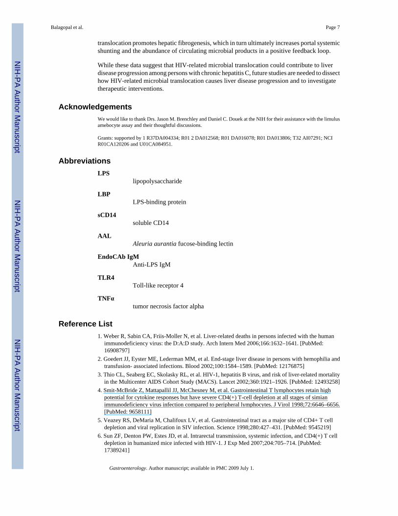
translocation promotes hepatic fibrogenesis, which in turn ultimately increases portal systemicshunting and the abundance of circulating microbial products in a positive feedback loop.
While these data suggest that HIV-related microbial translocation could contribute to liverdisease progression among persons with chronic hepatitis C, future studies are needed to dissecthow HIV-related microbial translocation causes liver disease progression and to investigatetherapeutic interventions.
AcknowledgementsWe would like to thank Drs. Jason M. Brenchley and Daniel C. Douek at the NIH for their assistance with the limulusamebocyte assay and their thoughtful discussions.
Grants: supported by 1 R37DA004334; R01 2 DA012568; R01 DA016078; R01 DA013806; T32 AI07291; NCIR01CA120206 and U01CA084951.
AbbreviationsLPS
lipopolysaccharide
LBP LPS-binding protein
sCD14 soluble CD14
AAL Aleuria aurantia fucose-binding lectin
EndoCAb IgM Anti-LPS IgM
TLR4 Toll-like receptor 4
TNFα tumor necrosis factor alpha
Reference List1. Weber R, Sabin CA, Friis-Moller N, et al. Liver-related deaths in persons infected with the human
immunodeficiency virus: the D:A:D study. Arch Intern Med 2006;166:1632–1641. [PubMed:16908797]
2. Goedert JJ, Eyster ME, Lederman MM, et al. End-stage liver disease in persons with hemophilia andtransfusion- associated infections. Blood 2002;100:1584–1589. [PubMed: 12176875]
3. Thio CL, Seaberg EC, Skolasky RL, et al. HIV-1, hepatitis B virus, and risk of liver-related mortalityin the Multicenter AIDS Cohort Study (MACS). Lancet 2002;360:1921–1926. [PubMed: 12493258]
4. Smit-McBride Z, Mattapallil JJ, McChesney M, et al. Gastrointestinal T lymphocytes retain highpotential for cytokine responses but have severe CD4(+) T-cell depletion at all stages of simianimmunodeficiency virus infection compared to peripheral lymphocytes. J Virol 1998;72:6646–6656.[PubMed: 9658111]
5. Veazey RS, DeMaria M, Chalifoux LV, et al. Gastrointestinal tract as a major site of CD4+ T celldepletion and viral replication in SIV infection. Science 1998;280:427–431. [PubMed: 9545219]
6. Sun ZF, Denton PW, Estes JD, et al. Intrarectal transmission, systemic infection, and CD4(+) T celldepletion in humanized mice infected with HIV-1. J Exp Med 2007;204:705–714. [PubMed:17389241]
Balagopal et al. Page 7
Gastroenterology. Author manuscript; available in PMC 2009 July 1.
NIH
-PA Author Manuscript
NIH
-PA Author Manuscript
NIH
-PA Author Manuscript
7. Mehandru S, Poles MA, Tenner-Racz K, et al. Primary HIV-1 infection is associated with preferentialdepletion of CD4(+) T lymphocytes from effector sites in the gastrointestinal tract. J Exp Med2004;200:761–770. [PubMed: 15365095]
8. Guadalupe M, Reay E, Sankaran S, et al. Severe CD4(+) T-cell depletion in gut lymphoid tissue duringprimary human immunodeficiency virus type 1 infection and substantial delay in restoration followinghighly active antiretroviral therapy. J Virol 2003;77:11708–11717. [PubMed: 14557656]
9. Kewenig S, Schneider T, Hohloch K, et al. Rapid mucosal CD4(+) T-cell depletion and enteropathyin simian immunodeficiency virus-infected rhesus macaques. Gastroenterology 1999;116:1115–1123.[PubMed: 10220503]
10. Brenchley JM, Price DA, Schacker TW, et al. Microbial translocation is a cause of systemic immuneactivation in chronic HIV infection. Nature Medicine 2006;12:1365–1371.
11. Jerala R. Structural biology of the LPS recognition. International Journal of Medical Microbiology2007;297:353–363. [PubMed: 17481951]
12. Creagh EM, O’Neill LAJ. TLRs, NLRs and RLRs: a trinity of pathogen sensors that cooperate ininnate immunity. Trends in Immunology 2006;27:352–357. [PubMed: 16807108]
13. Thurman RG. Mechanisms of Hepatic Toxicity II. Alcoholic liver injury involves activation ofKupffer cells by endotoxin. American Journal of Physiology-Gastrointestinal and Liver Physiology1998;38:G605–G611.
14. Paik YH, Schwabe RF, Bataller R, et al. Toll-like receptor 4 mediates inflammatory signaling bybacterial lipopolysaccharide in human hepatic stellate cells. Hepatology 2003;37:1043–1055.[PubMed: 12717385]
15. Enomoto N, Yamashina S, Kono H, et al. Development of a new, simple rat model of early alcohol-induced liver injury based on sensitization of Kupffer cells. Hepatology 1999;29:1680–1689.[PubMed: 10347108]
16. Seki E, De Minicis S, Osterreicher CH, et al. TLR4 enhances TGF-beta signaling and hepatic fibrosis.Nature Medicine 2007;13:1324–1332.
17. Rubio-Tapia A, Murray JA. The liver in celiac disease. Hepatology 2007;46:1650–1658. [PubMed:17969053]
18. Hill GR, Crawford JM, Cooke KR, et al. Total body irradiation and acute graft-versus-host disease:the role of gastrointestinal damage and inflammatory cytokines. Blood 1997;90:3204–3213.[PubMed: 9376604]
19. Gerbitz A, Schultz M, Wilke A, et al. Probiotic effects on experimental graft-versus-host disease: letthem eat yogurt. Blood 2004;103:4365–4367. [PubMed: 14962899]
20. Feld JJ, Meddings J, Heathcote EJ. Abnormal intestinal permeability in primary biliary cirrhosis. DigDis Sci 2006;51:1607–1613. [PubMed: 16937077]
21. Rai R, Wilson LE, Astemborski J, et al. Severity and correlates of liver disease in hepatitis C virus-infected injection drug users. Hepatology 2002;35:1247–1255. [PubMed: 11981775]
22. Wilson LE, Torbenson M, Astemborski J, et al. Progression of liver fibrosis among injection drugusers with chronic hepatitis C. Hepatology 2006;43:788–795. [PubMed: 16557548]
23. Mehta SH, Lucas GM, Mirel LB, et al. Limited effectiveness of antiviral treatment for hepatitis C inan urban HIV clinic. AIDS 2006;20:2361–2369. [PubMed: 17117023]
24. Vlahov D, Anthony JC, Muñoz A, et al. The ALIVE Study: a longitudinal study of HIV-1 infectionin intravenous drug users: description of methods. J Drug Issues 1991;21:759–776.
25. Netski DM, Mosbruger T, Astemborski J, et al. CD4(+) T cell-dependent reduction in hepatitis Cvirus-specific humoral immune responses after HIV infection. J Infect Dis 2007;195:857–863.[PubMed: 17299716]
26. Netski DM, Mosbruger T, Depla E, et al. Humoral immune response in acute hepatitis C virusinfection. Clin Infect Dis 2005;41:667–675. [PubMed: 16080089]
27. Gan EV, Haberman HF, Menon IA. Simple and sensitive test for the determination of phenoliccompounds in urine and its application to melanoma. J Invest Dermatol 1975;64:139–144. [PubMed:1117172]
28. Mehta AS, Long RE, Communale MA, et al. Increased levels of galactose-deficient anti-gal IgG inthe sera of HCV infected individuals with fibrosis and cirrhosis. 2007
Balagopal et al. Page 8
Gastroenterology. Author manuscript; available in PMC 2009 July 1.
NIH
-PA Author Manuscript
NIH
-PA Author Manuscript
NIH
-PA Author Manuscript

29. Shorack GR. Linear rank statistics, finite sampling, permutation tests and Winsorizing. The Annalsof Statistics 1996;24:1371–1385.
30. Rockstroh JK, Spengler U, Sudhop T, et al. Immunosuppression may lead to progression of hepatitisC virus- associated liver disease in hemophiliacs coinfected with HIV. Am J Gastroenterol1996;91:2563–2568. [PubMed: 8946987]
31. Di Martino V, Rufat P, Boyer N, et al. The influence of human immunodeficiency virus coinfectionon chronic hepatitis C in injection drug users: A long-term retrospective cohort study. Hepatology2001;34:1193–1199. [PubMed: 11732009]
32. Caradonna L, Mastronardi ML, Magrone T, et al. Biological and clinical significance of endotoxemiain the course of hepatitis C virus infection. Current Pharmaceutical Design 2002;8:995–1005.[PubMed: 11945146]
33. Bhagwandeen BS, Apte M, Manwarring L, et al. Endotoxin Induced Hepatic-Necrosis in Rats on AnAlcohol Diet. J Pathol 1987;152:47–53. [PubMed: 3305847]
34. Adachi Y, Bradford BU, Gao WS, et al. Inactivation of Kupffer Cells Prevents Early Alcohol-InducedLiver-Injury. Hepatology 1994;20:453–460. [PubMed: 8045507]
35. Hansen J, Cherwitz DL, Allen JI. The Role of Tumor-Necrosis-Factor-Alpha in Acute Endotoxin-Induced Hepatotoxicity in Ethanol-Fed Rats. Hepatology 1994;20:461–474. [PubMed: 8045508]
36. Ostapowicz G, Watson KJR, Locarnini SA, et al. Role of alcohol in the progression of liver diseasecaused by hepatitis C virus infection. Hepatology 1998;27:1730–1735. [PubMed: 9620350]
37. Frieden TR, Ozick L, McCord C, et al. Chronic liver disease in central Harlem: the role of alcoholand viral hepatitis. Hepatology 1999;29:883–888. [PubMed: 10051493]
38. Corrao G, Aricò S. Independent and combined action of hepatitis C virus infection and alcoholconsumption on the risk of symptomatic liver cirrhosis. Hepatology 1998;27:914–919. [PubMed:9537428]
39. Coelho-Little ME, Jeffers LJ, Bernstein DE, et al. Hepatitis c virus in alcoholic patients with andwithout clinically apparent liver disease. Alcoholism (NY) 1995;19:1173–1176.
40. Peters MG, Terrault NA. Alcohol use and hepatitis C. Hepatology 2002;36:S220–S225. [PubMed:12407597]
41. Ewaschuk J, Endersby R, Thiel D, et al. Problotic bacteria prevent hepatic damage and maintaincolonic barrier function in a mouse model of sepsis. Hepatology 2007;46:841–850. [PubMed:17659579]
42. Gordon SC, Elloway RS, Long JC, et al. The pathology of hepatitis C as a function of mode oftransmission: blood transfusion vs. intravenous drug use Hepatology 1993;18:1338–1343.
43. Inglesby TV, Rai R, Astemborski J, et al. A prospective, community-based evaluation of liverenzymes in individuals with hepatitis C after drug Use. Hepatology 1999;29:590–596. [PubMed:9918940]
44. Praaningvandalen DP, Brouwer A, Knook DL. Clearance Capacity of Rat-Liver Kupffer, Endothelial,and Parenchymal-Cells. Gastroenterology 1981;81:1036–1044. [PubMed: 7286581]
45. Lumsden AB, Henderson JM, Kutner MH. Endotoxin Levels Measured by A Chromogenic Assay inPortal, Hepatic and Peripheral Venous-Blood in Patients with Cirrhosis. Hepatology 1988;8:232–236. [PubMed: 3281884]
Balagopal et al. Page 9
Gastroenterology. Author manuscript; available in PMC 2009 July 1.
NIH
-PA Author Manuscript
NIH
-PA Author Manuscript
NIH
-PA Author Manuscript

Balagopal et al. Page 10
Gastroenterology. Author manuscript; available in PMC 2009 July 1.
NIH
-PA Author Manuscript
NIH
-PA Author Manuscript
NIH
-PA Author Manuscript

Balagopal et al. Page 11
Gastroenterology. Author manuscript; available in PMC 2009 July 1.
NIH
-PA Author Manuscript
NIH
-PA Author Manuscript
NIH
-PA Author Manuscript

Balagopal et al. Page 12
Gastroenterology. Author manuscript; available in PMC 2009 July 1.
NIH
-PA Author Manuscript
NIH
-PA Author Manuscript
NIH
-PA Author Manuscript

Balagopal et al. Page 13
Gastroenterology. Author manuscript; available in PMC 2009 July 1.
NIH
-PA Author Manuscript
NIH
-PA Author Manuscript
NIH
-PA Author Manuscript
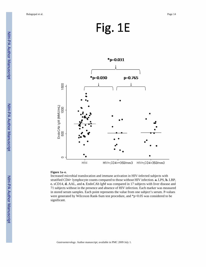
Figure 1a–e.Increased microbial translocation and immune activation in HIV-infected subjects withstratified CD4+ lymphocyte counts compared to those without HIV infection. a. LPS, b. LBP,c. sCD14, d. AAL, and e. EndoCAb IgM was compared in 17 subjects with liver disease and71 subjects without in the presence and absence of HIV infection. Each marker was measuredin stored serum samples. Each point represents the value from one subject’s serum. P-valueswere generated by Wilcoxon Rank-Sum test procedure, and *p<0.05 was considered to besignificant.
Balagopal et al. Page 14
Gastroenterology. Author manuscript; available in PMC 2009 July 1.
NIH
-PA Author Manuscript
NIH
-PA Author Manuscript
NIH
-PA Author Manuscript

Figure 2.HCV seroconversion is not significantly associated with microbial translocation. Thirty-fourpersons had levels of microbial translocation markers drawn before and after acute HCVseroconversion. LPS levels were not different after seroconversion, as shown. Similar resultswere found for other microbial translocation markers.
Balagopal et al. Page 15
Gastroenterology. Author manuscript; available in PMC 2009 July 1.
NIH
-PA Author Manuscript
NIH
-PA Author Manuscript
NIH
-PA Author Manuscript
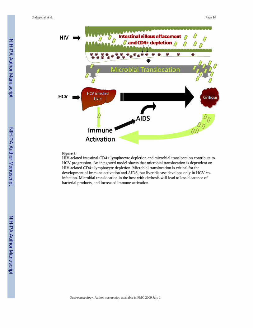
Figure 3.HIV-related intestinal CD4+ lymphocyte depletion and microbial translocation contribute toHCV progression. An integrated model shows that microbial translocation is dependent onHIV-related CD4+ lymphocyte depletion. Microbial translocation is critical for thedevelopment of immune activation and AIDS, but liver disease develops only in HCV co-infection. Microbial translocation in the host with cirrhosis will lead to less clearance ofbacterial products, and increased immune activation.
Balagopal et al. Page 16
Gastroenterology. Author manuscript; available in PMC 2009 July 1.
NIH
-PA Author Manuscript
NIH
-PA Author Manuscript
NIH
-PA Author Manuscript

NIH
-PA Author Manuscript
NIH
-PA Author Manuscript
NIH
-PA Author Manuscript
Balagopal et al. Page 17
Table 1Demographic characteristics of study subjectsa.
Prevalent Liver Disease Cohort HIV seroconverter Cohort HCV seroconverter Cohort
Cirrhosis Minimal disease
Total number 17 71 29 34
Mean age (years±SD) 50.5 ± 6.2 41.7 ± 6.0 35.8 ± 6.4 24.4 ± 3.0
Gender, male (%) 10 (58.8) 60 (84.5) 19 (65.5) 21 (61.7)
Race, black (%) 16 (94.1) 69 (97.2) 28 (96.6) 5 (14.7)
HIV (%) 8 (47.1) 20 (28.2) 29 (100) 0 (0)b
Proportion injectingillicit drugs (in prior 6mo)
7 (41.2) 39 (54.9) 26 (89.7) 100%
Alcohol use
None 11 (64.7) 26 (36.6) 2 (7.1) not available
>1 drink/day 5 (29.4) 38 (53.5) 16 (57.1)
7 days/week 1 (5.9) 7 (9.9) 35.7
aCirrhosis was determined by liver biopsy and/or clinical events including esophageal varices, encephalopathy, or ascites.
bTwo subjects declined HIV testing and therefore their HIV status was unknown.
Gastroenterology. Author manuscript; available in PMC 2009 July 1.

NIH
-PA Author Manuscript
NIH
-PA Author Manuscript
NIH
-PA Author Manuscript
Balagopal et al. Page 18
Table 2Independent associations of HIV and liver disease with LPS levels in 88 subjects.a
HIV Cirrhosis OR 95% CI p-value
− − 1.0 n/a n/a
+ − 1.21 0.97 – 1.51 0.086
− + 1.61 1.19 – 2.18 0.0024
+ + 2.07 1.51 – 2.84 <0.0001
aCirrhosis was determined by liver biopsy and/or clinical events including esophageal varices, encephalopathy, or ascites.
Gastroenterology. Author manuscript; available in PMC 2009 July 1.

NIH
-PA Author Manuscript
NIH
-PA Author Manuscript
NIH
-PA Author Manuscript
Balagopal et al. Page 19
Table 3Association of microbial translocation markers and cirrhosis in 88 subjectsa
OR 95% CI p-value
Univariate
LPS ≥ 42 pg/mLb 19.0 2.98 – 120.79 0.0018
LBP (highest quartile) 4.39 0.99 – 19.36 0.05
sCD14 (highest quartile)b 8.65 1.98 – 37.72 0.0041
AAL ≥ 5-fold above controlb 27.77 5.64 – 136.71 <0.0001
EndoCAb IgM (highest quartile) 0.10 0.01 – 0.86 0.036
CD4+ lymphocyte <350/mm3b 7.02 1.36 – 36.31 0.02
Multivariatec
LPS ≥ 42 pg/mL 18.71 2.62 – 133.78 0.0035
CD4+ lymphocyte <350/mm3 6.29 0.97 – 40.69 0.054
aLPS-lipopolysaccharide; LBP – LPS binding protein; sCD14 – soluble CD14; EndoCAb IgM antibody to LPS core; AAL- Aleuria aurantia lectin.
Cirrhosis was determined by liver biopsy and/or clinical events including esophageal varices, encephalopathy, or ascites.
bAdjusted for age because of an association detected in univariate analysis
cShown is a model of cirrhosis, with values adjusted for CD4+ lymphocyte depletion. Similar results were obtained for models of cirrhosis and the other
markers (sCD14, LBP, EndoCAb IgM, and AAL) but are not shown. Inclusion of more than one marker in the same model of cirrhosis did not improvethe fit significantly.
Gastroenterology. Author manuscript; available in PMC 2009 July 1.