Leptomycin B is an inhibitor of nuclear export: inhibition of nucleo-cytoplasmic translocation of...
9
Research Pa,,er 139 Leptomycin B is an inhibitor of nuclear export: inhibition of nucleo-cytoplasmic translocation of the human immunodeficiency virus type 1 (HIV-l) Rev protein and Rev-dependent mRNA Barbara Wolff’, Jean-Jacques Sangher and Ying Wang2 Background: The human immunodeficiency virus type 1 (HIV-I) regulatory protein Rev is required for unspliced and incompletely spliced viral mRNAs to appear in the cytoplasm and thus for viral replication. Translocation of Rev from the nucleus to the cytoplasm is essential if Rev is to function. We wanted to identify inhibitors of this transport process because they would be potential antiviral agents. Results: The Streptomyces metabolite, leptomycin 8, and other antibiotics of the leptomycinlkazusamycin family were identified as inhibitors of the nucleo-cytoplasmic translocation of Rev at nanomolar concentrations. Rev- dependent export of mRNA into the cytoplasm is also blocked by leptomycin B, which inhibits Rev-dependent, but not Rev-independent gene expression in a short-term transfection assay. In primary human monocytes, leptomycin B suppresses HIV-l replication. Conclusions: Leptomycin B is the first low molecular weight inhibitor of nuclear export to be identified. Although it cannot be used therapeutically, it should serve as a valuable tool for dissecting nuclear evporl pathways Introduction The acquired immunodeficiency syndrome (AIDS) is a life-threatening disease caused by HIV-l [l]. The HIV-1 genome encodes several regulatory proteins, in particular Tat and Rev, that are essential for virus replication [2;3j. Rev is necessary for the appearance in the cytoplasm of unspliced and singly spliced viral mRNAs that encode the structural proteins Gag, Pal and Env [4,5]. Rev is a nuclear protein [6] and it interacts with a highly structured RNA, the Rev response element (RRE), in the viral LED gene [7-101. Since Rev is a viral protein lacking a cellular counterparr, scvc~al different strategies have been employed to inhibit HIV-1 replication by inhibiting the function of the Rev protein. One strategy has heen to use anrisense phospho- rothioace oligonucleotides against Rev to suppress HlV in chronically infected cells [ll]. Using gene therapy approaches, tmnc-dominant mutants of HIV-1 Rev [12], dominant-negative mufants of the ccllnlar Rev cofactor eIF-5.4 [13], intracellular expression of an anti-Rev single chain antibody construct [14], and RR&decoys [15] have all been shown to inhibit lIIV-1 infection in cells. In a more classical approach using low molecular weight substances, neomycin was found IO block Rev-RRE binding and to antagonize Rev function [16] and an %alkylpyrimidinone was found to antagonize cellular Rev response [17]. Recently, we and others have demonstrated that the export of Rev from the nucleus to the cytoplasm is crucially Addresses: ‘Sandoz Research Institute. Brunner Strasse 59. Al 230 Vienna, Austria and ?4ovartis Preclinicai Research, Basis, Switzerland. Correspondence: Barbara Wolff E-mail: wolff_b@al .wienvl sandozcom Keywords: HIV-I Rev, leptomycin 8. nuclear export Received: 17 October 1996 Revisions requested: 7 November 1996 Revisions received: 28 January 1997 Accepted: 29 January ,997 Electronic identifier: 1074-5521-004-00139 Chemistry&Biology February 1997, 4:1X-147 0 Current Biology Lfd ISSN 1074-5521 important for its function and the export depends on the Rev activation domain [l&-20], which serves as a nuclear export signal [21,22]. Rev translocates from the nucleus to the cytoplasm in HeLa and COS cells transfected with Rev under conditions where rRNA synthesis is inhibited by, for example, actinomycin D (AD). It is clear that the appearance of Rev in the cytoplasm reflects export and nor de now synthesis of Rev protein in the cytoplasm, because the reaction takes place in the presence of the protein synthesis inhibitor, cycloheximide 11%201. Furthermore, dominant-negative mutants of Rev with mutations in the activation domain (nuclear export signal) are unable to leave the ndcleus upon treatment with AD [lS-201. In a screening assay for low molecular weight inhibitors of Rev nuclear export, we have now found four antibiotics of the leptomycinlkazusamycin family that completely block this process at nanomolar concentrations. 1,eptomycin B (LMB) also inhibits Rev fnnction and HIV-l replication. Results LME inhibits cytoplasmic localization of Rev, but not protein transport into the nucleus Nucleo-cytoplasmic translocarion of Rev induced by AD in HeLa cells transfected with Rev (HeLa-Rev cells) was used as a screen for inhibitors of Rev function. A strepto- mycete extract was identified with very potent inhibitory activity, and four compounds, leptomycins A and B and kazusamycins A and B (Fig. l), were isolated as the activ~~components.
-
Upload
independent -
Category
Documents
-
view
0 -
download
0
Transcript of Leptomycin B is an inhibitor of nuclear export: inhibition of nucleo-cytoplasmic translocation of...
Research Pa,,er 139
Leptomycin B is an inhibitor of nuclear export: inhibition of nucleo-cytoplasmic translocation of the human immunodeficiency virus type 1 (HIV-l) Rev protein and Rev-dependent mRNA Barbara Wolff’, Jean-Jacques Sangher and Ying Wang2
Background: The human immunodeficiency virus type 1 (HIV-I) regulatory protein
Rev is required for unspliced and incompletely spliced viral mRNAs to appear in the cytoplasm and thus for viral replication. Translocation of Rev from the nucleus
to the cytoplasm is essential if Rev is to function. We wanted to identify inhibitors
of this transport process because they would be potential antiviral agents.
Results: The Streptomyces metabolite, leptomycin 8, and other antibiotics of
the leptomycinlkazusamycin family were identified as inhibitors of the
nucleo-cytoplasmic translocation of Rev at nanomolar concentrations. Rev- dependent export of mRNA into the cytoplasm is also blocked by leptomycin B,
which inhibits Rev-dependent, but not Rev-independent gene expression in a
short-term transfection assay. In primary human monocytes, leptomycin B suppresses HIV-l replication.
Conclusions: Leptomycin B is the first low molecular weight inhibitor of nuclear
export to be identified. Although it cannot be used therapeutically, it should serve
as a valuable tool for dissecting nuclear evporl pathways
Introduction The acquired immunodeficiency syndrome (AIDS) is a life-threatening disease caused by HIV-l [l]. The HIV-1 genome encodes several regulatory proteins, in particular Tat and Rev, that are essential for virus replication [2;3j.
Rev is necessary for the appearance in the cytoplasm of unspliced and singly spliced viral mRNAs that encode the structural proteins Gag, Pal and Env [4,5]. Rev is a nuclear protein [6] and it interacts with a highly structured RNA, the Rev response element (RRE), in the viral LED gene [7-101.
Since Rev is a viral protein lacking a cellular counterparr,
scvc~al different strategies have been employed to inhibit HIV-1 replication by inhibiting the function of the Rev protein. One strategy has heen to use anrisense phospho- rothioace oligonucleotides against Rev to suppress HlV in chronically infected cells [ll]. Using gene therapy approaches, tmnc-dominant mutants of HIV-1 Rev [12], dominant-negative mufants of the ccllnlar Rev cofactor eIF-5.4 [13], intracellular expression of an anti-Rev single chain antibody construct [14], and RR&decoys [15] have all been shown to inhibit lIIV-1 infection in cells. In a more classical approach using low molecular weight substances, neomycin was found IO block Rev-RRE binding and to antagonize Rev function [16] and an %alkylpyrimidinone was found to antagonize cellular Rev response [17].
Recently, we and others have demonstrated that the export
of Rev from the nucleus to the cytoplasm is crucially
Addresses: ‘Sandoz Research Institute. Brunner Strasse 59. Al 230 Vienna, Austria and ?4ovartis Preclinicai Research, Basis, Switzerland.
Correspondence: Barbara Wolff E-mail: wolff_b@al .wienvl sandozcom
Keywords: HIV-I Rev, leptomycin 8. nuclear export
Received: 17 October 1996 Revisions requested: 7 November 1996 Revisions received: 28 January 1997 Accepted: 29 January ,997
Electronic identifier: 1074-5521-004-00139
Chemistry&Biology February 1997, 4:1X-147
0 Current Biology Lfd ISSN 1074-5521
important for its function and the export depends on the Rev activation domain [l&-20], which serves as a nuclear export signal [21,22]. Rev translocates from the nucleus to the cytoplasm in HeLa and COS cells transfected with Rev under conditions where rRNA synthesis is inhibited by, for example, actinomycin D (AD). It is clear that the appearance of Rev in the cytoplasm reflects export and nor de now synthesis of Rev protein in the cytoplasm, because the reaction takes place in the presence of the protein synthesis inhibitor, cycloheximide 11%201. Furthermore, dominant-negative mutants of Rev with mutations in the activation domain (nuclear export signal) are unable to leave the ndcleus upon treatment with AD [lS-201.
In a screening assay for low molecular weight inhibitors of Rev nuclear export, we have now found four antibiotics of
the leptomycinlkazusamycin family that completely block this process at nanomolar concentrations. 1,eptomycin B (LMB) also inhibits Rev fnnction and HIV-l replication.
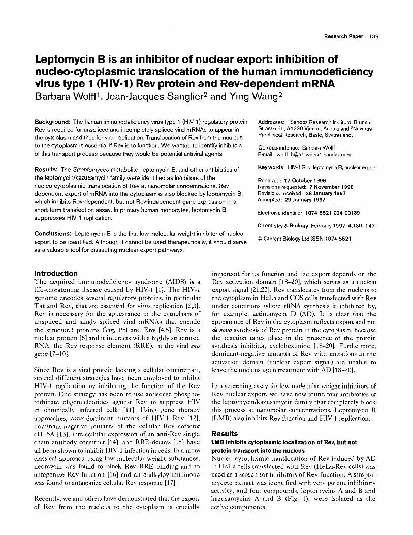
Results LME inhibits cytoplasmic localization of Rev, but not protein transport into the nucleus Nucleo-cytoplasmic translocarion of Rev induced by AD in HeLa cells transfected with Rev (HeLa-Rev cells) was used as a screen for inhibitors of Rev function. A strepto- mycete extract was identified with very potent inhibitory activity, and four compounds, leptomycins A and B and kazusamycins A and B (Fig. l), were isolated as the activ~~components.
Leptomycin A j % CH,
kptomycin S W CH,CH,
Kazusamycin A CH,OH CH,CH,
Karusamycin S CH,OH W
Structures of leptomycin and kazusamycin antibiotics,
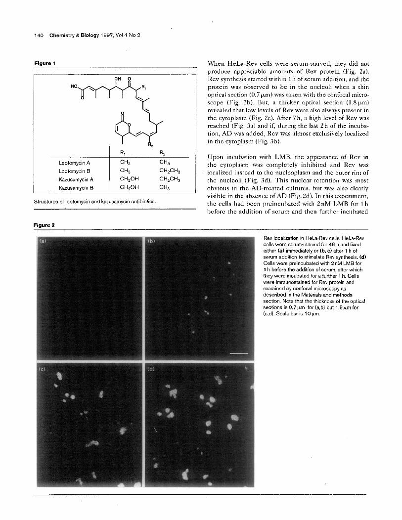
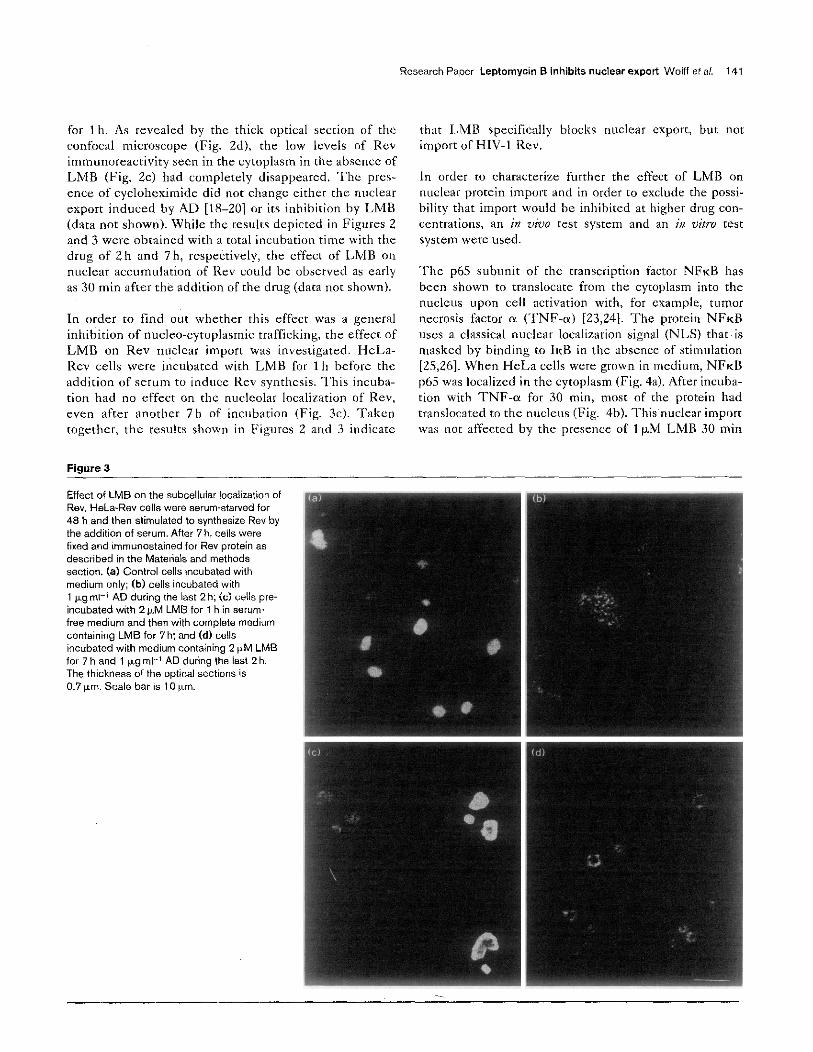
When HeLa-Rev cells were serum-starved, they did nor produce appreciahlc amounts of Rev protein (Fig. Za). Rev synthesis starred within 1 h of xwm addition, and the protein was observed to he in the nucleoli when a thin optical section (0.7 km) was taken with the confocal micro- scope (Fig. Zb). But, a thicker optical section (1.8b.m) revealed that low lcvcls of Rev were also always present in the cytoplasm (Fig. Zc). After 7 h, a high level of Rev was reached (Fig. 3a) and if, during the last 2 h of the incuha- tion, AD was added, Rev was almost exclusively localized in the cytoplasm (Fig. 3h).
Upon incubation with LMB, the appearance of Rev in the cytoplasm was completely inhibited and Rev was localized instead to the nucleoplasm and the outer rim of the nucleoli~ (Fig. 3d). This nuclear retention was most obvious in the AD-treated cultures, but was also clearly visible in the ahscncc of AD (Fig. Zd). In this experiment, the cells had heen preincubated with 2nM LMB for 1 h hcfore the addition of serum and then further incubated
Figure 2
Rev localization in H&a-Rev ceils. H&-Rev cells were serum-starved for 48 h and fixed either (8) immediately or (b, c) after 1 h of serum addition to stimulate Rev synthesis. (d) Cells were preincubated with 2 nM LMS for 1 h before the addition oi serum, after which they were incubated for a further 1 h. Cells were immunastained ior Rev protein and examined by confocal microscopy as described in the Materials and methods section. Note that the thickness of the optical sections is 0.7 pm for (a,b) but 1.8 &rn ior (c,d). Scale bar is 10 pm.
for lh. As revealed by the thick optical section of the confocal microscope (Fig. Zd), the low levels of Rev immunoreactiviry seen in the cytoplasm in the absence of LMB (Fig. Zc) had completely disapprxed. ‘The prcs- encc of cycloheximide did not change either the nuclear export induced by AD [tS-201 or its inhibition by LMB (data not shown). While the results depicted in Figures 2 and 3 WSK obtained with a total incubation time with the drug of 2 h and 7 h, respectively, the effect of LMB on nuclear accumulation of Rev could be observed as early as 30 min after the addition of the drug (data nor shown).
In order to find out whether this effect was a general inhibition of nucleo-cytoplasmic trafficking, the effect of LMB on Rev nuclear import was investigated. HcLa- Rev cells were incubated with LMB for 1 h before the addition of serum to induce Rev synthesis. This incuba- tion had no effect on the nucleolar localization of Rev, even after another 7h of incubation (Fig. 3~). Taken together, the results shown in Figures 2 and 3 indicate
Figure 3
Effect of LMB on the subcellular localization of Rev. HeLa.Re” cells were serum-starved for 48 h and then stimulated to synthesize Rev by the addition of serum. After 7 h. ceik were fixed and immunostained for Rev protein as described in the Materials and methods section. (a) Control cells incubated with medium only; (b) cells incubated with 1 kg ml-’ AD during the last 2 h; (c) ells pre- incubated with 2 &M LMB for 1 h in serum free medium and then with complete medium containing LMB for 7 h: and Cd) cells incubated with medium containing 2 CM LMB for 7 h and 1 p.g ml-’ AD during the last 2 h. The thickness of the optical sections IS 0.7 wm. Scale bar is 10 p.m.
Research Paper Leptomycin B inhibits nuclear export Wolff eta,. 141
that LMB specifically blocks nuclear exporr, but nor import of HIV-1 Rev.
In order m characterize further the effect of LMB on nuclear protein import and in order to exclude the possi- bility that import would be inhibited at higher drug con- centrations, an in vWo test system and an ix oirrv text system were used.
The p65 subunit of the transcription factor NFKB has been shown to translocare from the cytoplasm into the nucleus upon cell activation with, for example, tumor ~CCIOS~S factor a (TNF-a) [23,241. The protein NFKB uses a classical nuclear localization signal (NLS) that is masked by binding to IKB in the absence of stimulation [25,26]. When HeLa cells were grown in medium, NFKB 1~65 was localized in the cytoplasm (Fig. 4a). After incuba- tion with TNF-a for 30 min, most of the protein had translocated to the nucleus (Fig. 4b). This nuclear import was not affected by the presence of 1 &M LMB 30 min
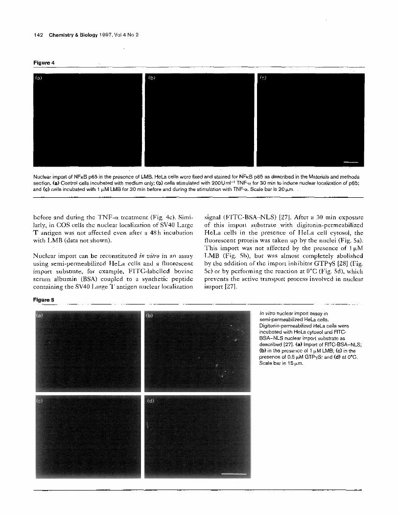
Figure 4
Nuclear import of NFKB p65 in the presence of LMB. HeLa cells were fixed and stained for NFKEI p65 as described in the Materials and methods section. (a) Control cells incubated with medium only; (b) cells stimulated with 200U ml-’ TNF-a for 30 min to induce nuclear localization of ~65; and W cells incubated with 1 +M LMB for 30 min before and during the stimulation with TNF-a. Scale bar is 20 km.
before and during the TNF-a ~~eafmcnc (Fig. 4~). Simi- larly, in COS cells the nuclear localization of SV40 Large T antigen was not affected even after a 48 h inabation with LMB (data not shown).
Nuclear import can be reconstituted in z&o in an assay using semi-permeabilized HeLa cells and a fluorescent import substrate, for example, FITC-labelled bovine serum albumin (BSA) coupled fo a synthetic peptide conmining the SV40 1,arg.e T antigen nuclear localization
Fioure 5
signal (I’ITC-BSA-NLS) [27]. After a 30 min exposure of this import substrate with digitonin-pcrmeahilized HeLa cells in the presence of I-l&a cell cyrosol, the fluorescent protein was taken up by the nuclei (Fig. Sa). This import was not affected by the presence of 1pM
,LMB (Fig. Sb), but was almost completely abolished by the addition of the import inhibitor GTPyS [28] (Fig. 5c) or by performing the reaction at 0°C (Fig. Sd), which prevents the active transport process involved in nuclear import [271. In vitro nuclear import assay in
semi-permeabilized HeLa ceils. Digitonin-permeabilized HeLa ceils were incubated with HeLa cytosal and FITC- BSA-NLS nuclear import substrate as described f271. (a) Import oi FITC-BSA-NLS; (b) in the presence of 1 $4 LMB; (c) in the presence of 0.5 ,dv, GTP-,S: and (d) at 0°C. Scale bar is 15 em.
Research Paper Leptomycin B inhibits nuclear export Wolff ef ai. 143
Taken together, the in viva and in vitro import results
show that LMB has no effect on protein import, even at concentrations more than lOOO-fold higher than chose required for the inhibition of Rev export (Table 1).
An inhibitory effect on Rev export was found not just for LMB but also for- lepromycin A and kazusamycins A and B. The concentrations required for a 50% inhibition of transport are given in Table 1, togethcr with the values for the antiproliferative effect after 7h, 24h, 48h and 72h. While that: appeared to be a rherapcutic window after short periods of incubation (7h and 24h), the concentra- tions required for inhibition of Rev export and 50% inhibi- tion of cell growth aftcr 72h were almost identical. Thcreforc, for most subsequent experiments, incubation times shorter than 72 h had to be chosen in order to evalu- ate a Rev-sprcific effect. Since LMB was the most abun- dant and the most potent of the four S~~ptomyces metabolitcs, it was used for all additional experiments.
LMB inhibits the appearance of RRE-containing mRNA in
the cytoplasm
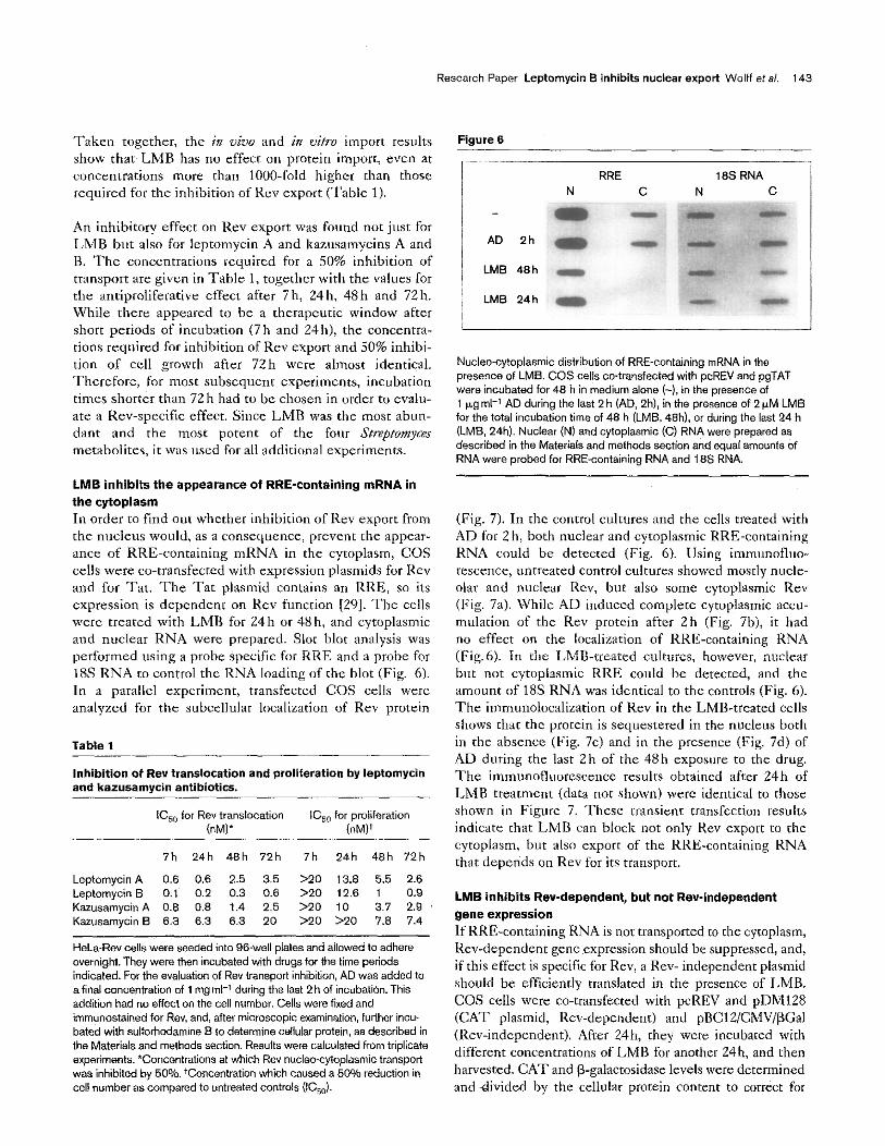
In order to find out whether inhibition of Rev export from the nucleus would, as a consequence, prevent the appear- ance of RRE-containing mRNA in the cytoplasm, COS cells were co-rransfecced with expression plasmids for Rev and for Tat. The Tat plasmid contains an RRE, so its expression is dcpendcnt on Rev function [29]. ‘l-he cells were treated with LMR for 2411 or 48 h, and cyroplasmic and nuclear RNA were prepared. Slot blot analysis was performed using a probe specific for RRE and a probe for 18s RNA to control the RNA loading of the blot (Fig. 6). In a parallel experiment, transfected COS cells were analyzed for the subcellular localization of Rev protein
Table 1
Inhibition of Rev translocation and proliferation by leptomycin and kazusamycin antibiotics.
7h 24h 48h 72h 7h 24h 48h 72h
Leptomycin A 0.6 0.6 2.6 8.6 >*o 13.8 5.5 2.6 Leptamycin B 0.1 0.2 0.3 0.6 >20 12.6 1 0.9 Kazusamycin A 0.8 0.8 1.4 2.5 >20 10 3.7 2.9 Karusamycin B 8.3 8.3 6.8 20 >20 920 7.8 7.4
HeLa-Rev cells were seeded into 96well plates and allowed to adhere overnight. They were then incubated with drugs for the time periods indicated. For the evaluation of Rev transport inhibition. AD was added to a final concentration of 1 mg ml-’ during the last 2 h of incubatirin. This addition had no effect on the cell number. Cells were fixed and immunastained for Rev, and, after microscopic examination, futiher incu. b&d with sulforhodamine 8 to determine cellular protein, as described in the Materials and methods section. Results were calculated ham triplicate experiments. *Concentrations at which Rev nucleo-cytoplasmic transpoti wan inhibited by 50%. ‘Concentration which caused a 50% reduction in cell number as compared to untreated controls (IC,,).
Figure 6
I RRE 16s RNA N C N C
AD 2h :
LMB 48h [;ssl, j, .,.
LMB 24h : q@j
Nucleo-cytaplasmic distribution of RRE-containing mRNA in the presence of LMB. COS cells cwtransfected with pcREV and pgTAT were incubated for 48 h in medium alone (-). in the presence of 1 p,g ml-’ AD during the last 2 h (AD, Zh), in the presence of 2pM LMB for the total incubation time of 48 h (LMB. 48h). or during the last 24 h (LMB, 24h). Nuclear (N) and cytoplasmic (Cl RNA were prepared as described in the Materials and methods section and equal amounts of RNA were probed far RRE-containing RNA and 18s RNA.
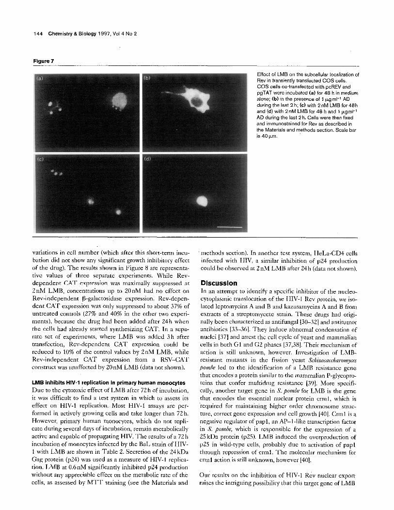
(Fig. 7). In the control cultures and the cells treated with AD for 2 h, both nuclear and cytoplasmic RRE-containing RNA could be detected (Fig. 6). Using immunofluo- rescence, untreated control cultures showed mostly nucle- alar and nuclear Rev, but also some cytoplasmic Rev (Fig. 7a). While AD induced complete cytoplasmic accu- mulation of the Rev protein after Zh (Fig. 7b), it had no effect on the localization of RR&containing RNA (Fig.6). in the LMB-treated altuxs, however, nuclear but nor cytoplasmic RRE could be detected, and the amount of 18s RNA was identical to the controls (Fig. 6). The immunolocalization of Rev in the LMB-treated czlls shows that the protein is sequestered in the nucleus both in the absence (Fig. 7~) and in the presence (Fig. 7d) of AD duririg rhe last 2 h of the 48 h exposure to the drug. The immuaofluorcscencc results obtained after 24h of LMB treatment (data nof shown) wcrc identical to those shown in Figure 7. These transient transfection results indicate that LMB can block not only Kev export to the cytoplasm, but also exporr of the RRE-containing RNA that depends on Rev for its transport.
LMB inhibits Rev-dependent, but not Rev-independent gene expression
If RRE-containing RNA is not transported to rhc cytoplasm, Kev-dependent gcnc expression should be suppressed, and, if this effect is specific for Rev, a Rev- independent plasmid should be efticienrly translated in the presence of LMB. COS cells were co-transfected with pcREV and pDMt28 (CAT plasmid, Rev-dependent) and pBClZ/CMV/PGal (Rev-independent). After 24h, they were incubated with differcnr concentrations of LMB for another 24h, and then harvested. CAT and P-galacrosidase levels wc~c determined and -divided by the cellular protein content to correct for
144 Chemistry & Biology 1997, Vol 4 No 2 . 1
Effect of LMB on the subcell& localization of Rev in transiently transfected COS cells. COS ceils co-transfected with pcREV and pgTAT were incubated (a) for 48 h in medium alone; (b) in the presence af 1 ,~.grnl-l AD during the last 2 h; (c)with 2nM LMB for 48h and (d) with 2 nM LMB for 48 h and 1 Fg ml-1 AD during the last 2 h. Cells were then fixed and immunostained for Rev a~ described in the Materials and methods sectian. Scale bar
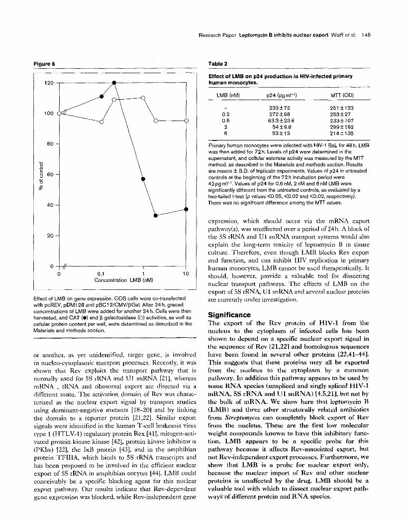
variations in cell number (which after this short-term incu- bation did not show any significant growth inhibitory effect of the drug). The results shown in Figure 8 are represenra- tivc values of three scparatc experiments. While Rev- dzpcndcnr CAST expression was maximally suppressed at 2nM LMB, concenrrations up to 20nM had no effect on Rev-independent P-galactosidase expression. Rev-depen- dent CAT expression was only suppressed to about 37% of untreated controls (27% and 40% in the other two experi- mats), because the drug had been added after 24h when the cells had already started synthesizing CAT. In a sepa- rate set of experiments, where LMB was added 3h after transfcction, Rev-dependent CAT expression could be reduced to 10% of the control values by 2nM LMB, while Rev-independent CA’T expression from a RSV-CAT construct was unaffected by 20nM LMB (data not shown).
LMB inhibits HIV-1 replication in primary human monocytes Due fo the cytotoxic effect of LMB after 72h of incubation, it was difficult to find a test system in which to assess its effect on HIV-l replication.~Most HIV-1 assays arc per- formed in actively growing cells and take longer than 72 h. However, primary human monocytes, which do not repli- cate during several days of incubation, remain metabolically active and capable of propagating HIV. ‘The rcsulrs of a 72 h incubation of monocytes infected by the BaL strain of HIV- 1 with LMB are shown in Table 2. Secretion of the 24kDa Gag protein (~24) was used as a measure of HIV-l replica- tion. LMR at O.6nM significantly inhibited p24 production without any appreciable effect on the metabolic raw of the cells, as assessed by MTT staining (see the Materials and
methods sccrion). In another test system, H&a-CD4 cells infected with HIV, a similar inhibition of p24 production could be observed at 2nM LMB aftcr 24h (data not shown).
Discussion In an attempt to identify a specific inhibitor of the nucleo- cycoplasmic translocation of the HIV-l Rev protein, wc iso- lated leptomycins A and B and kazusamycins A and B from exfracm of a streptwnycetc strain. These drugs had origi- nally been characterized as antifungal 130-321 and antirumor antibiotics [33-361. They induct: abnormal condensation of nuclei [37] and arrest the cell cycle of yeast and mammalian cells in both Gl and G2 phases [37,38]. Their mcclranism of action is still unknown, however. Investigation of LMB- resistant mutants in the fission yeast Sc&~a&~rom~~5 pum/x led to the idcntificarion of a LMB resistance gene that cncodes a protein similar to the mammalian P-glycopro- teins that confer multidrug resistance 1391. More spccifi- ally, another target gene in S. pombe for LMB is the gene that encodes the essential nuclear protein crml, which is required for maintaining higher order chromusome stwc- rue, correct gem expression and cell growth [40]. Crml is a negative regulator of papl, an AP-l-like transcription factor in S. pumbc, which is responsible for the expression of a 2SkDa prorcin (~25). LMB induced the overproducrion of ~25 in wild-type cells, probably due to activation of pap1 through repression of crml. The molecular mechanism for crml action is still unknown, howevcr [4O].
Our results on the inhibition of HIV-I Rev nuclear export raises the intriguing possibility that this target gene of LMB
Research Paper Leptomycin B inhibits nuclear export Wolff et al 145
Figure 8
120
100
80
$
g 60 ‘s B
40
20
0
T
rf 0 ;L- -
0.1 1 Concentration LMB (nM)
. 10
Effect of LMS on gene expressibn. COS cells were co-transfected with pcRE”, pDMl28 and pBC1 Z/CMV/@Gal. After 24h. graded cmxentrations of LMS were added for another 24 h. Cells were then harvested. and CAT (0) and p galactosidase (0) activities, as well as cellular protein content per well, were determined as described in the Materials and methods section.
or another, as yet unidentified, target gene, is involved in nucleo-cyroplasmic transport processes. Recently, it was shown that Rev exploits the rransport pathway that is normally used for SS rRNA and CJl snRNA [21j, whereas mRNA , tRNA and ribosomal export are directed via a different mutt. The activation domain of Rev was charac- terized as the nuclear export signal by transport studies using dominant-negative mutants [18-201 and by linking the domain to a reporter protein [21,22]. Similar export signals were identified in the human T-cell leukemia virus type 1 (HTLV-1) regulatory protein Rex [41], mitogen-acti- vated protein kinase kinase 1421, protein kinase inhibitor a (PKIrr) [Z’Z], the I& protein [43], and in the amphibian protein TFIIIA, which binds to SS rRNA transcripts and has been proposed to be involved in the efficient nuclear export of 5s [RNA in amphibian oocytes [441. LMB could conceivably be a specific blocking agent for this nuclear
cxport pathway. Our results indicate that Rev-dependent gene expression was blocked, while Rev-independent gene
Table 2
IWed of LMB on ~24 production in HIV-infected primary human monocytes.
LMB (nM) 1124 bgml-‘) MTT (00)
233+75 251*123 0.2 2723~98 253+27 0.6 m.3+23.5 233+107
2 54*9x 299?192 6 x3*,3 214+135
Primary human manocytes were infected with HIV.1 BaL for 48 h. LMB was then added for 72 h. Levels of ~24 were determined in the supernatant, and cellular e~terase activity was measured by the Mm method, as described in the Materials and methods section. Results are means f SD. of triplicate experiments. Values of p24 in untreated contr& at the beginning of the 72 h incubation period were 42 pg ml-‘. Values of ~24 for 0.6 nM. 2 nM and 6 nM LMB were significantly different from the untreated controls. as evaluated by a two-tailed t-test (p values <0.05. -CO.02 and <0.02. respectively). There was no significant difference among the MTT values.
expression, which should occur via the mRNA cxport
pathway(s), was unaffected over a period of 24h. A block of
the SS rKNA and Ul snRNA transport systems would also explain the long-term toxicity of leptomycin B in tissue
culture. Therefore, even though LMB blocks Rev export and function, and can inhibit HIV, replication in primary human monocyres, LMB cannot be used therapeutically. It should, however, provide a valuable tool for dissecting
nuclear transport pathways. The effects of LMB on the export of SS rRNA, Ul snRNA and several nuclear proteins
are currently under investigation.
Significance The export of the Rev protein of HIV-1 from the nucleus to the cytoplasm of infected cells has been shown to depend on n specific nuclear export signal in the sequence of Rev [21,221 and homologous sequences have been ‘found in several other proteins [22,41-441. This suggests that these proteins may al1 be exported from the nucleus to the cytoplasm by a common pathway. In addition this pathway appears to be used by some RNA species (unspliced and singly spliced HIV-1 mRNA, SS rRNA and Ul snRNA) [4,5,211. but not by the bulk of mRNA. We show here that leptomycin B (LMB) and three other structurally related antibiotics from Str@tonzyces can completely block export of Rev from the nucleus. These are the first low molecuInr weight compounds known to have this inhibitory func- tion. LMB appears to be a specific probe for this pathway because it affects Rev-associated export, but not Rev-independent export processes. Furthermore, we show that LMB is a probe for nuclear export only, because the nuclear import of Rev and other nuclear proteins is unaffected by the drug. LMB should be a valuable tool with which to dissect nuclear export path- waysof different protein and RNA species.
146 Chemistry&Biology 1997, Vol 4 No 2
Materials and methods Isolation and purification of leptomycin and kazusamycin antibiotics Leptomycins A and 8 and kazusamycins A and B were isolated from the Streptomyces sp. strain A87-18203, which was deposited with the Deutsche Sammlung van Mikroorganismen und Zellkulturen. Mascherod& Weg 1 b, D.3300 Sraunschweig. Germany (reference number DSM 7517). Ethyl acetate extracts of Streptomyces A87. 18203 were fractionated by gel filtration an Sephadex LH-20 (Pharma- cia). Four compounds were isolated by preparative HPLC as will be described elsewhere (Y. Wang et al., unpublished observations). The structures and the stereochemistry were established by spectroscopic and chemical methods.
Rev nucleo-cytoplasmic translocatkx assay HeLa-Rev cells are HeLa cells stably transfected with an expression plasmid far HIV-I Rev [291 and selected by co-transfection with the gene for dihydrololate reductase. The original cells were a generous gift from B.R. Cullen and were grown in Iscove’s modified Dulbecca’s medium (IMDM) supplemented with 10% heat-inactivated fetal calf serum and 1 OOnM amethapterin. These cells were subcloned to achieve a high level of Rev expression in all cells. and clone 403E3 was used for all localization experiments.
Cells were seeded into W-well plates (Nunc) and synchronized by serum starvation lor 48 h. Rev synthesis was induced by the addition of serum. At this time, test compounds or streptomycete broth extracts were added in complete medium lacking amethopterin and incubated with the cells for a total of 7 h. During the last 2 h. actinomycin D (AD; Sigma) was added at 1 pgmi-’ in order to induce complete cytoplae mic localization of Rev in the medium controls [I 81. The cells were then fixed and immunastained for Rev protein using a mouse monoclonal antibody against Rev and a dichlorotriarinyl amino fluorescein (DTAF)- lab&d goat-anti-mouse antibody as described elsewhere I1 81. Inhibitors of Rev translocation into the cytoplasm were identified by microscopic examination of individual wells using a 32x objective an a Zeiss IM IO inverted microscope equipped with epifluorescence optics. For the images shown in Figures 2 and 3, cells were grown on 8-well Lab-Tek chamber slides (Nunc). immunostained for Rev and examined in a BioRad MRC 600 confocal laser scanning microscope as described [I 81. Results in Figures ?a, 2b and 3 are represented as 0.7 &rn thick optical sections, while Figure 2c and 2d show optical sec- tions of 1.8 vrn thickness.
In order to assess the influence of LMB on Rev translocation from the cytoplasm into the nucleus. HeLa-Rev cells were serum-starved for 48 h. and the drug was added 1 h before the re-addition of serum.
COS cells in 6-well plates (Nunc) were transfected and treated with AD and LMB as described below (RNA transport assay) and immunos- tained for Rev. They were examined in an Olympus IX-70 inverted microscope equipped with a 100 x (1.35 N.A.) objective and epifluarew cence optics and photographed with a PM-30 camera (Olympus) using TMav 400 film (Eastman Kodak).
NFKB p6.5 nuclear import assay HeLa cells (ATCC, Rackville. MD, USA) were grown in IMDM supple- mented with 10% heat%activated fetal calf serum. They were seeded into B-well LabTek chamber slides and allowed lo adhere overnight. They were then stimulated by the addition of 200 U ml-’ TN& (Boehringer Mannheim. Germany) for 30 min. LMB (1 +M) was added for SO min before and during the stimulation. The cells were fixed and permeabilized with 4% paraformaldehyde and 0.5% Triton- X1 00, respectively. The cells were immunostained using a polyclonal rabbit antibody directed against NFKB ~65 (Santa Cruz Biatechnol- ogy) and a tetramethylrhodamine-labelled goat-anti-rabbit antibody (Accurate Scientific) and examined by confocal laser scanning microscopy as described [I 81.
The in vftra nuclear impoit assay was conducted as described far FAGS analvsis L271. The suswnsion of nuclei was transierred to a alass slide and examined in the Olympus microscope as described above.
RNA transport assay COS cells were seeded into T150 flasks (Nunc) and transfected with expression plasmids pcREV [291 and pgTAT [291 at 1 sgml-’ and 1.5 +gml-‘, respectively, using DEAE-dextran and chloraquine [45]. LMS was added at 2 nM for 24 h or 48 h; in one sample, AD was added 2 h prior to the end of the experiment. Total incubation time after transfection was 48 h. Nuclear and cytoplasmic RNAs were prepared according to 1461. and slot blot analysis was performed using probes specific for RRE WCCT GTA CCG TCA GCG TCA XG ACG CTG CGC-3’) and 185 RNA WGCA CCA GAC TTG CCC TCC AAT GGA TCC TCG-3’).
Reporter gene assays CCS cells were co-transfected with pcREV (1 kgml-I), a R&depen- dent chloramphenical acetyl transferase (CAT) indicator gene (pDM128; 1.5 pgml-‘) [471 and a Rev-independent gene for the expression of p-galactosidase (pBC12ICMVIPGal; 1 pg ml-‘) [481 in 6-well plates (Costar). 24 h after transfection, different concentrations of LMB were added to the ceils, and incubation was continued for another 24 h. The cells were prepared for the simultaneous determina- tion of CAT and P-galactasidase activities as described [491. Cellular protein was determined from an aliquot of the suspended cells, digested in 0.5 M N&H. using the BioRad protein assay.
HIV-1 ~2’4 Gag protein production in primary human monocytes Five-day adherent monocyte cultures were prepared from the blood oi healthy, HIV-negative donors [501. The cells were maintained in 48.well plates (Costar). They were infected with HIV-I BaL strain for 48 h, and LME was then added at different concentrations. Three days after sub- stance addition. p24 Gag protein was measured in the supernatant using a commercially available ELISA kit (Coulter). The cell number per well was determined using the MTT method [51].
Sulforhodamine B assay In order to determine the effect of the drugs on cell growth, HeLa-Rev cells were seeded into g&well plates at 5000 cells per well and allowed to adhere overnight. They were then incubated with test compounds for 7 h, 24 h, 48 h or 72 h. fixed with 3.7% formaldehydeW/o sucrose in PBS and stained with sulforhodamine B (Sigma) as described WI.
Acknowledgements We wouid iike to thank D. Bevec for performin the slot blot analysis. H. Jaksche for conducting the in vitro n”clear import assay, E. Pursch for carrying out the experiment with HIV-1 infected monocytes, and D. Bevec and A.P. Winiski ior critically reading the manuscript.
References 1.
2.
3.
4.
5.
6.
Papovic. M., Samgadharan M.G.. Read. E. & Gallo. R.C. (1984). Detection, is&Am. and continuous production of cytopathic reboviruses WTLV-III) from patients with AIDS and ore-AIDS. Science 224,497~500. "aml"s. H. (1968 I). Regulation of HIV and HTLV gene expression. Gs~~s Dw. 2. tm-ioez. Cullen, S.R. &Greene, W.C. (1989). Regulatory pathways governing HIV-I replication. Cen 58.423-426. Arys, S., Guo, C., Joseph% SF. & Wang-Staal, F. (1985). Trans- activator gene of human T-lymphotmpic vies type III (HTLV-III). ^ --7.69-73.
i., BurghoR, R., Sia, R., Sadroski. J., Haseltine. W. & Rosen, C. (1988). The art gene product of human immunodeficiency virus is required for replication. 1. Irirol. 62, 655-658. Bbhnlein, E.. Berger. J. & Hauber, J. (1981). Functional mapping of the human immunodeficiency virus type 1 Rev RNA binding domain: new insights into the domain sfructwe of Rev and Rex. 1 Wooi. 65, 7061-7055.
7.
8.
9.
10.
11.
12.
13.
14.
15.
16.
17.
18.
19.
20.
21.
22.
23.
24.
25.
26.
27.
28.
Arrigo. S.J. & Chen, I.S.Y. (1991). Rev is necessary ‘or transiation but not cytopkmic acc”m”,atio” a‘ HIV-I “if. vpr, and envivpu 2 RN!% Genes DW 5,808-819. Felber. B.. Had~opoulou-Cladaras, M.. Cladaras. c.. Copeland. T. & Paulakis, G.N. (1989). Rev protein of human immunodeficiency virus type 1 affects the stability and transpofi of the viral mRNA. Proc. Natl *cad. Sci. US.4 86.1495-1499. Hammarskjdid. M.L., Heumer, J.. Hammarskjijld, 8.. Sangwan, I., Albert, L. & Rskosh. D. (1989). Regulation of human immuno- deficiency virus env expression by the rev gene product. 1. Virirol 63. 1959-1966. Malim. M.H.. Hauber. 1.. Le. S.-Y., Maizel. J.V. & C&n, B.R. (1989). The HIV-1 rev trans-activator acts through a structured target Sequence lo activate nuclear export of unspliced viral mRN.4. Nahire
Duan. L., Bagasra, 0.. La”9hlin, MA.. Cakes. J.W. & Pomerantz R.J. (19941. Patent inhibition of human immunodeficiency virus type 1 replication by an intracellular anti.Rev single-chain antibody. Proc. Nat! Acad. sci us* 91.5075-5079. Lee. T.C., Sullenger, B.A., Gallardo. H.F., “ngers, GE & Gilboa, E. (1992). Overexpression of RRE-derived sequences inhibits HIV-1 replication in CEM 4s. New Bml 4, 66-74. Zapp. M.L.. Stern, S. &Green. MR. (1993). Small moiecule~ that selectively block RNA binding of HIV-l Rev protein inhibit Rev function and viral pioductian. Cell 74. 969-978. Ciccarelli, R.B.. et al.. & Hughes. J.V. (1994). Inhibition of the cellular Rev response and HIV-1 replication by 8-aikyl-2-(4-py~idyl)pyrido[2,3- dlpyrimidin-5(8M-anes. Antiviral Chem. C?iemoffxr. 5, 169-I 75. Woiti, B., Cohen. G.. Hauber, J.. Meshcheryakova, 0. & Rabeck. C. (1995). Nucleocytopiasmic transport of the Rev protein of human immunadeficiency virus type 1 is dependent on the activation domain of the protein. Exp. Cd, Res. 21,. 31-41. Meyer, B.E. & Malim M.H. (1994). The HIV-l Rev transectiuator shuttles between nuclew and cytoplasm. Genes Dev. 8.1538-1548. Szilvay, A.M., Brokstad. K.A.. Kopperud, R., Haukenes, G. & Kailand. K.-H. (1995). Nuclear export of the human immunodeficiency virus type 1 nucleacytaplasmic shuttle protein Rev is mediated by its activation domain and is blocked by trans dominant negative mutants. ,. vim,. 69. X315-3323. Fischer. U.. Huber, J., Boelens, W.C.. Mattaj, I.W. & Liihrmann, R. (1995). The HIV.1 Rev activation domain is a nuclear export signal that accesses an export pathway used by specific cellular RNAs. Cd 82,475-483. Wm. W., M&k& J.L.. Tsien. R.Y. &Taylor. S.S. (1995). Identification of a signal for rapid export of proteins from the nucleus Cell 82. 463473. Baeuerle, PA & Baltimare. D. (1989). Activation of DNA binding activity in an apparently cytoplasmic precursor of the NFKB transcription factor. Cell 53, 21 I-21 7. Arenzana-Seisdsdos. F., et a/.. &Hay. R.T. I1 995). Inducible nuclear expression of newly synthesized IcBa negatively regulates DNA-binding and transcriptional activities of N&B. MO/. Cell Bioi 15.2689-2696. &be,, U.. Henkeel, T., dos Santos Silva. M. & Baeuerla, P.A. (1993). Nuclear uptake control of NFKB by MAD-3 an IxB protein present in the nucieus. EMBCI J. 12.201-211. Beg, A.A.. ef a,.. &Baldwin. A.S. (1992). IKB interacts with the nuclear localization sequences of the subunits of NFnB: a mechanism ior cfloplasmic retention. Genes Dev. 6. 1899-1913. Paschal. B.M. & Getace, L. (1995). Identification of NTF2. a cytosolic factor far nuclear imps* that interacts with nuclear pore complex protein pm ,. Cd Bid 129, 925-937. M&him F., Paschal, B., Evans. J. & Gerace. L. (1993). inhibition of mclear protein impoti by nonhydroiyrable analagues of GTP and identification of the small GTPase RanlTCd as an essential transport factor. 1. Cell Bid 123, 1649-1659.
29.
30.
31.
32.
33.
34.
35.
36.
37.
38.
39.
40.
41.
42.
43.
44.
45.
46.
47.
48.
49.
50.
51.
52.
36,646-65-o. ,~
Hamamato, T.. Uozumi, T. & Beppu, T. (1985). Leptomycins A and B. new antifungal antibiotics 111. Mode of action of leptomycin 8 on schizosaccharomyces pombe. 1 Antibiot (Tokyo) 58, 1573-I 580. Komiyama. K., et al., & “merawa, I. (1985). Structural study of a new antitumor antibiotic, karusamycin. J. Ant;biot, (Tok~ Komiyama, K., Okada, K., Hirokawa, Y.. Masuda, K..Tomisaka, S. & Umezawa. I. (1985). Antitumor activity of anew antibiotic. kazusamycin. I Antibiot. (Tokyo1 38, 224-220. Yoshida, E.. et al.. & Umezawa. I. (19.87). Antitumor effect of karusamycin 8 on experimental tumors. J. Anfibiot. (Tokyo) 40. 1596-1604. Roberts. B .J.. Hameiehle, K.L., Sebalt, ,.S. &Leopold, W.R. (1986). In vim and fn vitro anticancer activity of the structurally nwei and highly potent antibiotic Cl-940 and its hydroxy analog (PD 114,721). Cancer Chemothe,. mamaco,. 16.95-101. Takamiya, K., Yoshida. E., Takahashi, T., Okura, A. & Okanishi, M. (1988). The effect of kazusamycin Bon the cell cycle and morphology of cultured L1210 cells. 1 Anfibiot. (Tokyo) 41, 1854-I 861. Yoshida. M.. Nishikawa, M.. Nishi, K.. Abe, K.. Horinouchi, S. & Beppu, T. (1990). Effects of ieptomycin B on the ceil cycle of fibroblasts and fission yeast ceils. Exp. Cell es. 187. 150-156. Nishi. K.. ef al, & Beppu. T. (1992). A leptomycin B resistance gene of Schizasaccharomyces pombe encodes a protein similar to the mammalian P glycoproteins. Mel Mkrobioi. 6. 761-769. Nishi, K.. Yoshida, M.. Fujiwara, D.. Nishikawe. M.. Horinouchi. S. & Beppu. T. (1994). Leptomycin B targets a regulatory cascade of crml a fission yeast nuclear protein, involved in control of higher order chromosome structure and wne expression. 1. Sir,,, Chem. 259.