Nucleo-cytoplasmic shuttling dynamics of the transcriptional regulators XYR1 and CRE1 under...
17
Nucleo-cytoplasmic shuttling dynamics of the transcriptional regulators XYR1 and CRE1 under conditions of cellulase and xylanase gene expression in Trichoderma reesei Alexander Lichius, 1 * Verena Seidl-Seiboth, 1 Bernhard Seiboth 1 and Christian P. Kubicek 1,2 1 Research Division Biotechnology and Microbiology, Institute of Chemical Engineering, Vienna University of Technology, Vienna, Austria. 2 Austrian Center of Industrial Biotechnology, Graz, Austria. Summary Trichoderma reesei is a model for investigating the regulation of (hemi-)cellulase gene expression. Cellu- lases are formed adaptively, and the transcriptional activator XYR1 and the carbon catabolite repressor CRE1 are main regulators of their expression. We quantified the nucleo-cytoplasmic shuttling dynamics of GFP-fusion proteins of both transcription factors under cellulase and xylanase inducing conditions, and correlated their nuclear presence/absence with transcriptional changes. We also compared their sub- cellular localization in conidial germlings and mature hyphae. We show that cellulase gene expression requires de novo biosynthesis of XYR1 and its simul- taneous nuclear import, whereas carbon catabolite repression is regulated through preformed CRE1 imported from the cytoplasmic pool. Termination of induction immediately stopped cellulase gene tran- scription and was accompanied by rapid nuclear deg- radation of XYR1. In contrast, nuclear CRE1 rapidly decreased upon glucose depletion, and became recy- cled into the cytoplasm. In mature hyphae, nuclei con- taining activated XYR1 were concentrated in the colony center, indicating that this is the main region of XYR1 synthesis and cellulase transcription. CRE1 was found to be evenly distributed throughout the entire mycelium. Taken together, our data revealed novel aspects of the dynamic shuttling and spatial bias of the major regulator of (hemi-)cellulase gene expression, XYR1, in T. reesei. Introduction In nature fungi contribute as essential decomposers of complex organic molecules to the biological carbon cycle. These organic polymers, produced through carbon dioxide fixation by plants, mainly comprise plant cell wall polysac- charides including cellulose, hemicelluloses, pectins and the polymer lignin. The major plant cell wall component is the β-(1,4)-linked glucose polymer cellulose, that alone exhibits an annual production of 7.2 × 10 10 tons, and its microbial degradation is therefore a key transformation step in the biological carbon cycle. The ascomycete Tricho- derma reesei (the anamorph of the pantropical saprotroph Hypocrea jecorina) has become a model system for the regulation of formation of plant cell wall polysaccharide hydrolases such as cellulases and hemicellulases (Seiboth et al., 2012; Kubicek, 2013). Notably, its hyper- producing strains are applied for the industrial production of the respective (hemi)cellulase enzymes for their appli- cations in the pulp and paper, food and textile industry, as well as for the conversion of plant biomass materials into second-generation biofuels or platform chemicals (Chundawat et al., 2011; Viikari et al., 2012). Most T. reesei cellulases are not formed during growth on monosaccharides and their full expression requires the presence of an inducer. Cellulase gene expression is regulated by the action of at least four positive transcrip- tional activators, including the xylanase regulator 1 (XYR1), the activators of cellulase 2 (ACE2) and 3 (ACE3), and a tetramer of heme-activated proteins (HAP2/3/5 complex), as well as two repressors, including ACE1 and the carbon catabolite repressor CRE1 (Kubicek et al., 2009; Seiboth et al., 2012; Häkkinen et al., 2014). XYR1 is a Zn 2Cys6-type regulator protein that binds to a 5′-GGCW4-3′ DNA motif and is the key transcriptional activator of cellulase and xylanase gene expression in T. reesei. Cellulases are coordinately upregulated through XYR1 activity upon induction by cellulose, lactose, or sophorose (Foreman et al., 2003; Kubicek et al., 2009), whereas expression of xylanases is differentially regu- lated. Expression of xyn1 and xyn2, for instance, is induced by D-xylose (Herold et al., 2013), whereas expression of xyn2 is also upregulated in the presence of Accepted 7 October, 2014. *For correspondence. E-mail [email protected]; Tel. (+43)(1)58801 166552; Fax (+43)(1)58801 17299. Molecular Microbiology (2014) ■ doi:10.1111/mmi.12824 © 2014 The Authors. Molecular Microbiology published by John Wiley & Sons Ltd. This is an open access article under the terms of the Creative Commons Attribution License, which permits use, distribution and reproduction in any medium, provided the original work is properly cited.
Transcript of Nucleo-cytoplasmic shuttling dynamics of the transcriptional regulators XYR1 and CRE1 under...

Nucleo-cytoplasmic shuttling dynamics of the transcriptionalregulators XYR1 and CRE1 under conditions of cellulase andxylanase gene expression in Trichoderma reesei
Alexander Lichius,1* Verena Seidl-Seiboth,1
Bernhard Seiboth1 and Christian P. Kubicek1,2
1Research Division Biotechnology and Microbiology,Institute of Chemical Engineering, Vienna University ofTechnology, Vienna, Austria.2Austrian Center of Industrial Biotechnology, Graz,Austria.
Summary
Trichoderma reesei is a model for investigating theregulation of (hemi-)cellulase gene expression. Cellu-lases are formed adaptively, and the transcriptionalactivator XYR1 and the carbon catabolite repressorCRE1 are main regulators of their expression. Wequantified the nucleo-cytoplasmic shuttling dynamicsof GFP-fusion proteins of both transcription factorsunder cellulase and xylanase inducing conditions,and correlated their nuclear presence/absence withtranscriptional changes. We also compared their sub-cellular localization in conidial germlings and maturehyphae. We show that cellulase gene expressionrequires de novo biosynthesis of XYR1 and its simul-taneous nuclear import, whereas carbon cataboliterepression is regulated through preformed CRE1imported from the cytoplasmic pool. Termination ofinduction immediately stopped cellulase gene tran-scription and was accompanied by rapid nuclear deg-radation of XYR1. In contrast, nuclear CRE1 rapidlydecreased upon glucose depletion, and became recy-cled into the cytoplasm. In mature hyphae, nuclei con-taining activated XYR1 were concentrated in thecolony center, indicating that this is the main regionof XYR1 synthesis and cellulase transcription. CRE1was found to be evenly distributed throughout theentire mycelium. Taken together, our data revealednovel aspects of the dynamic shuttling and spatialbias of the major regulator of (hemi-)cellulase geneexpression, XYR1, in T. reesei.
Introduction
In nature fungi contribute as essential decomposers ofcomplex organic molecules to the biological carbon cycle.These organic polymers, produced through carbon dioxidefixation by plants, mainly comprise plant cell wall polysac-charides including cellulose, hemicelluloses, pectins andthe polymer lignin. The major plant cell wall component isthe β-(1,4)-linked glucose polymer cellulose, that aloneexhibits an annual production of 7.2 × 10¹⁰ tons, and itsmicrobial degradation is therefore a key transformationstep in the biological carbon cycle. The ascomycete Tricho-derma reesei (the anamorph of the pantropical saprotrophHypocrea jecorina) has become a model system for theregulation of formation of plant cell wall polysaccharidehydrolases such as cellulases and hemicellulases(Seiboth et al., 2012; Kubicek, 2013). Notably, its hyper-producing strains are applied for the industrial productionof the respective (hemi)cellulase enzymes for their appli-cations in the pulp and paper, food and textile industry, aswell as for the conversion of plant biomass materials intosecond-generation biofuels or platform chemicals(Chundawat et al., 2011; Viikari et al., 2012).
Most T. reesei cellulases are not formed during growthon monosaccharides and their full expression requires thepresence of an inducer. Cellulase gene expression isregulated by the action of at least four positive transcrip-tional activators, including the xylanase regulator 1(XYR1), the activators of cellulase 2 (ACE2) and 3 (ACE3),and a tetramer of heme-activated proteins (HAP2/3/5complex), as well as two repressors, including ACE1 andthe carbon catabolite repressor CRE1 (Kubicek et al.,2009; Seiboth et al., 2012; Häkkinen et al., 2014).
XYR1 is a Zn2Cys6-type regulator protein that binds to a5′-GGCW4-3′ DNA motif and is the key transcriptionalactivator of cellulase and xylanase gene expression in T.reesei. Cellulases are coordinately upregulated throughXYR1 activity upon induction by cellulose, lactose, orsophorose (Foreman et al., 2003; Kubicek et al., 2009),whereas expression of xylanases is differentially regu-lated. Expression of xyn1 and xyn2, for instance, isinduced by D-xylose (Herold et al., 2013), whereasexpression of xyn2 is also upregulated in the presence of
Accepted 7 October, 2014. *For correspondence. [email protected]; Tel. (+43)(1)58801 166552; Fax(+43)(1)58801 17299.
Molecular Microbiology (2014) ■ doi:10.1111/mmi.12824
© 2014 The Authors. Molecular Microbiology published by John Wiley & Sons Ltd.This is an open access article under the terms of the Creative Commons Attribution License, which permits use, distribution andreproduction in any medium, provided the original work is properly cited.

the cellulase inducing carbohydrates cellulose andsophorose (Zeilinger et al., 1996). Furthermore, xyn1 andxyn2 respond to carbon catabolite repression in differentways (Mach et al., 1996). Deletion of xyr1 completelyeliminates the induction of both cellulases and xylanasesby all known inducers (i.e. cellulose, lactose, sophorose,xylan, xylose and arabinose) and in addition XYR1 alsoregulates lactose, L-arabinose and D-xylose metabolismmainly at the level of the D-xylose reductase (Strickeret al., 2006; Seiboth et al., 2007; Akel et al., 2009).Similar regulatory dynamics of XYR1 or its XlnR orthologsoccur in other species, including Aspergillus spp., Neuro-spora crassa, and Fusarium spp., although species-specific adaptations have been found (van Peij et al.,1998; de Vries et al., 1999; Hasper et al., 2000; Maruiet al., 2002; Calero-Nieto et al., 2007; Stricker et al.,2008; Seiboth et al., 2012; Sun et al., 2012).
Xyr1 transcription under non-inducing conditions occursat a low basal level. It appears not to be further inducedduring growth on xylan (Mach-Aigner et al., 2008), but tobe clearly induced by lactose and cellulose (Portnoy et al.,2011b; Bischof et al., 2013). So far, no information isavailable on how XYR1 reaches the nucleus and becomesactive for cellulase transcription. Nevertheless, the major-ity of Zn2Cys6 regulators are permanently present in thenucleus (MacPherson et al., 2006), and only a minority –like Aspergillus AflR, NirA and AmyR – have so far beendemonstrated to be shuttled between the cytoplasm andthe nucleus (Shimizu et al., 2003; Berger et al., 2006;Makita et al., 2009).
Carbon catabolite repression by CRE1 (or its CreAorthologs in other filamentous fungal species), a memberof the C2H2 class of DNA-binding proteins that recognizesa 5′SYGGRG-3′ consensus sequence (Felenbok et al.,2001), is the major negative regulator of cellulase andhemicellulase gene expression because it inhibits bothbasal and the inducible expression, and moreover alsoprevents expression of the positive regulator gene xyr1(Mach-Aigner et al., 2008; Seiboth et al., 2012). Carboncatabolite repression by CRE1/CreA appears to occur indifferent ways in different fungi: in T. reesei and Aspergil-lus nidulans, the cre1/creA genes are auto-regulated andtranscribed at lower levels in repressing conditions (Arstet al., 1990; Strauss et al., 1995; Ilmen et al., 1996; Shroffet al., 1996), whereas glucose-dependent transcriptionalexpression patterns of cre1 are positively correlated withthe extracellular glucose concentration in Acremoniumchrysogenum (Jekosch and Kück, 2000). Further, nuclearlocalization of CRE1 in Sclerotium sclerotiorum is depend-ent on the presence of glucose (Vautard-Mey et al., 1999),whereas nuclear localization of CreA is independent ofglucose in A. nidulans (Roy et al., 2008).
Up to now, the shuttling of positive and negative tran-scription factors during cellulase and hemicellulase gene
expression is completely obscure. The objective of thisarticle therefore was to set up a system that allows thequantification of subcellular localization of XYR1 andCRE1 in T. reesei under cellulase and xylanase inducingand non-inducing conditions, and thus to provide the firsttemporal resolution of transcription factor shuttling duringthe induction of cellulase gene expression in T. reesei.The study reveals some interesting differences betweenthe subcellular localization of XYR1 and CRE1 in ger-mlings growing in submerged cultures and the functionallystratified, mature colony as it develops on agar platesunder laboratory conditions. Our data show that cellulasegene expression depends on de novo biosynthesis andimmediate nuclear import of XYR1, and furthermoresuggest that XYR1-mediated cellulase expression occursmainly in the central area of the colony. This provides thefirst evidence that the mycelial organization of the fungalcolony might be another important element that regulatesthe high-level production of cellulolytic enzymes.
Results
XYR1 requires N-terminal and CRE1 requiresC-terminal GFP labeling
In order to test potential influences of GFP labeling on thefunction of the two transcription factors, we first com-pared the basic subcellular localization patterns of N- andC-terminally labeled XYR1 and CRE1 fusion proteins,expressed under native conditions from their endogenousloci (Fig. S1). Although all four versions, i.e. XYR1–GFP,GFP–XYR1, CRE1–GFP and GFP–CRE1, becamerecruited into nuclei under inducing or repressing condi-tions, respectively, we also noted significant differencesin their individual expression level and efficiency ofnuclear recruitment, judged by localized fluorescenceintensity measurements: attachment of GFP to theC-terminus of XYR1 resulted in approximately eightfoldreduced expression and nuclear recruitment compared tothe GFP–XYR1 version, whereas opposite effects, i.e.about fivefold reduced expression and nuclear recruit-ment of the N-terminal GFP version, were observed forCRE1 (Fig. S1).
To verify that the fusion proteins remained functional,Δxyr1 and Δcre1 gene deletion strains of T. reesei weretransformed with each of the four fusion constructs:thereby expression of GFP–XYR1 completely rescued thegrowth defect of Δxyr1 on 1% (w/v) xylose medium,whereas XYR1–GFP did so only partially (Fig. S2). Like-wise, the severe growth defect of Δcre1 was fully rescuedby expression of CRE1–GFP, whereas strains comple-mented with GFP–CRE1 retained significantly reducedcolony extension rates (Fig. S2). These data strongly sug-gested that XYR1 requires N-terminal and CRE1 requires
2 A. Lichius, V. Seidl-Seiboth, B. Seiboth and C. P. Kubicek ■
© 2014 The Authors. Molecular Microbiology published by John Wiley & Sons Ltd., Molecular Microbiology

C-terminal GFP labeling in order to preserve protein func-tion. Consequently, only T. reesei transformants express-ing GFP–XYR1 and CRE1–GFP, respectively, were usedfor all subsequent experiments.
The subcellular localization dynamics of GFP–XYR1and CRE1–GFP reporter proteins reflect the metabolicimpact of the utilization of simple and complexcarbon sources
Initial tests showed that CRE1 appeared to be equallylocalized to nuclei under cellulase inducing (lactose) andrepressing (glucose) conditions, whereas XYR1 showed asignificant increase of nuclear import only under cellulaseinducing conditions (Fig. 1). The distinct shuttlingresponses between both fluorescently labeled transcrip-tion factors, and the apparently specific, i.e. induction-dependent, response of GFP–XYR1 strongly suggestedthat both fusion proteins can be used as diagnostic tool tomonitor cellulase gene expression in T. reesei.
This notion was further substantiated by comparing thesubcellular localization dynamics of GFP–XYR1 andCRE1–GFP after replacement onto various other cellu-lase inducing and non-inducing carbon sources (Fig. S3).The most potent carbon source triggering nuclear importof XYR1 was sophorose, followed by lactose and cellu-lose, which both required extended incubation times tolead to a noticeable increase in nuclear fluorescencecompared to the pre-culture. For CRE1, the most potentcarbon sources triggering nuclear import were found to beglucose, high concentrations (65 mM) of xylose, and glyc-erol. Together these data demonstrate that on simple,easily metabolized carbon sources, nuclear accumulationof both transcription factors reached high levels within2–4 h after carbon source replacement, whereas on moreslowly metabolized, complex carbon sources more than18 h were required to reach similar levels.
In order to confirm that GFP–XYR1 and CRE1–GFPwere expressed as full-length proteins and hence respon-sible for the observed changes in subcellular fluorescence,
CRE1-GFP glucoseD
CRE1-GFP lactoseC
GFP-XYR1 glucoseB
GFP-XYR1 lactoseAFig. 1. Nuclear import of XYR1 wassignificantly increased under cellulaseinducing conditions.A and B. (A) GFP–XYR1 became stronglyrecruited into nuclei in the presence oflactose, whereas it was only weaklyexpressed under repressing/non-inducingconditions on glucose (B).C and D. CRE1 did not show this specificresponse and appeared equally recruited tonuclei under cellulase inducing and repressingconditions. 3D surface plots (right row) of theinverted fluorescence images (middle row)illustrate the relative differences of localizedfluorescence signal intensities. Thecorresponding transmission light images areshown in the left row. The displayed strainswere incubated for 48 h at 28°C on MA agarplates supplemented with lactose or glucoserespectively.Scale bars, 5 μm.
XYR1 and CRE1 shuttling in T. reesei 3
© 2014 The Authors. Molecular Microbiology published by John Wiley & Sons Ltd., Molecular Microbiology

SDS-PAGE and Western blot analyses were performed(Fig. 2). Full-length CRE1–GFP (71.07 kDa) was readilydetected from crude protein extracts of TRAL003.1 after18 h growth on glucose medium. Several degradationproducts, most prominently banding at around 60 and justbelow 40 kDa, were also detected in minor amounts. Full-length GFP–XYR1 (128.41 kDa) was also readily detectedin protein extracts of TRAL002.1 induced by sophorose for1, 2 and 3 h. However, in this case a higher concentrationof apparent degradation products of GFP–XYR1, mostnoticeably banding at around 25–30 kDa, were alreadyseen at the earliest time point and increased throughout
the time period, suggesting a higher proteolytic suscepti-bility of the GFP–XYR1 construct. The very prominentdegradation product of about 25 kDa after 3 h is most likelythe mere GFP (Fig. 2B and C), indicating that GFP–XYR1degradation leaves GFP intact.
In order to rule out that free GFP, resulting from hypo-thetical GFP–XYR1 or CRE1–GFP degradation in thecytoplasm, enters the nucleus and causes the observedincrease in nuclear fluorescence, we used a control strainthat expresses GFP under the constitutive Ptef1 promoter.Nuclei in that strain stained negatively against the brightlyfluorescent cytoplasm, strongly suggesting that GFP doesnot readily cross the nuclear envelope (Fig. S4). Hence,the observed changes in nuclear fluorescence must bedue to shuttling of the full-length GFP-transcription factorfusion proteins.
XYR1 generally shuttles slower than CRE1 and it is notmaintained as cytoplasmic pool
We next investigated the time needed for XYR1 andCRE1 to shuttle between the cytoplasm and the nucleus.To this end, we monitored subcellular dynamics of bothtranscription factors in conidial germlings in real-timeupon replacement onto a new carbon source (Fig. 3).Nuclear recruitment of XYR1 and nuclear loss of CRE1,respectively, were triggered by addition of sophorose to
A
170130100
70
55
40
35
25
10
kDa170130100 70
55
40
35
25
10kDa
150100 80
605040
20
30
150100 80
605040
20
30
B
C
M
GFP (18h gluc)
CR
E1-G
FP (18h gluc)
GFP
-XY
R1 (1h soph)
GFP
-XY
R1 (2h soph)
GFP
-XY
R1 (3h soph)
Mwild type (18h gluc)
µg0.35 20 20 20 20 20Fig. 2. Protein turn-over kinetics of both fusion reporters differ,leading to significant nuclear accumulation of free GFP fromGFP–XYR1 degradation.A. Coomassie-stained SDS-PAGE gel. Except for the GFP positivecontrol, of which only 0.35 μg total protein extract of QM6a Ptef1::gfpwere loaded, 20 μg of all other protein extract samples were loadedper lane. Molecular weight marker on both sides is PageRuler10–170 kDa ladder.B. Western blot of the sister SDS-PAGE gel shown in (A),incubated with monoclonal α-GFP(B-2) antibody, ECL 2 reagentand imaged on a Typhoon FLA700 chemifluorescence imager.Molecular weight marker on the left (lane 1) is PageRuler10–170 kDa ladder, on the right (lane 8) is SuperSignal Enhanced20 – 150 kDa ladder. Cytosolic GFP (26.98 kDa) as positive controlis exclusively detected in lane 2. Full-length CRE1–GFP(71.07 kDa) bands in lane 3 just above the 70 kDa marker, andsome of its degradation products with decreasing abundancetowards low-molecular-weight species below. Lanes 4–6 showfull-length GFP–XYR1 (128.41 kDa) with decreasing abundanceafter 1–3 h of sophorose induction. Interestingly, with time,abundance of its higher-molecular-weight degradation products(40–80 kDa) decreases, while amounts of its low-molecular-weightdegradation products (25 – 30 kDa) significantly increase. Thenegative control in lane 7 (protein extract from an 18 h glucoseculture of QM9414 Δtku70) reveals minor non-specific backgrounddetection in the higher molecular weight range > 40 kDa.C. The same membrane as shown in (B) developed by x-ray filmexposure, shows identical results with less non-specific backgroundsignals, however, inferior spatial resolution. Intense nuclearfluorescence of GFP–XYR1 and CRE1–GFP, respectively, wasconfirmed by live-cell imaging in all samples prior to biomassharvest and protein extraction.
4 A. Lichius, V. Seidl-Seiboth, B. Seiboth and C. P. Kubicek ■
© 2014 The Authors. Molecular Microbiology published by John Wiley & Sons Ltd., Molecular Microbiology

germlings pre-cultured for 22 h in glucose, whereasnuclear loss of XYR1 and nuclear recruitment of CRE1,respectively, were initiated by addition of glucose to ger-mlings pre-cultured for 44 h on cellulose.
Nuclear recruitment of XYR1 was detectable within5 min after addition of sophorose, however, requiredabout 30 min more until a significantly high increase innuclear signal (n/c-ratio tripled) could be observed(Fig. 3A). Interestingly, cytoplasmic fluorescence intensityremained constant over the whole induction period, whilethe nuclear signal increased steadily. The fact that thecytoplasmic signal of XYR1 remained constantly lowduring that period indicates that any newly produced
XYR1 became immediately imported into nuclei. On theother hand, nuclear loss of XYR1, triggered by transfer toglucose-containing medium, occurred very slowly andabout 65% of nuclear fluorescence was still visible after60 min (Fig. 3B). The cytoplasmic pool of XYR1 did notincrease during this period, suggesting either that signifi-cant export of GFP–XYR1 from the nucleus did not occur,or that GFP, as stable end-product of GFP–XYR1 degra-dation, remained inside the nucleus. In any case, thesedata indicate that XYR1 is only produced when needed,i.e. in response to a cellulase inducing signal, and that asignificant cytoplasmic pool of the transcription factor isnot maintained.
CRE1
22h gluc 10min soph 20min soph 40min soph 60min sophD
10min gluc 20min gluc 40min gluc 60min gluc
XYR1
44h cellB
11.7 1.8 2.2
3.5
5.1
0
2
4
6
8
0
500
1000
1500
n/c-
ratio
fluor
esce
nce
inte
nsity
Sophorose-induced nuclear recruitment of XYR1
nuclei cytoplasm n/c-ratio
13.5 12.710.9 9.8 10.0
0
4
8
12
16
0
1000
2000
3000
4000
n/c-
ratio
fluor
esce
nce
inte
nsity
Glucose-induced nuclear loss of XYR1
nuclei cytoplasm n/c-ratio
6.5
3.42.5 2.2 2.3
0
2
4
6
8
0
500
1000
1500
n/c-
ratio
fluor
esce
nce
inte
nsity
Sophorose-induced nuclear loss of CRE1
nuclei cytoplasm n/c-ratio
11.2 1.7 2.2 2.8
4.5
0
2
4
6
8
0
500
1000
1500
n/c-
ratio
fluor
esce
nce
inte
nsity
Glucose-induced nuclear recruitment of CRE1
nuclei cytoplasm n/c-ratio
44h cell 5min gluc 10min gluc 15min glucC 25min gluc
CRE1
22h gluc 5min soph 15min soph 25min soph 35min sophA
XYR1
Fig. 3. XYR1 and CRE1 showed distinct nuclear shuttling dynamics. Inverted fluorescence images show the degree of nuclear transcriptionfactor recruitment at representative time points, with the corresponding quantitative analysis below.A. Time-course of sophorose-induced nuclear import of GFP–XYR1. Quantitative image analysis showing immediate but weak nuclearrecruitment which started to significantly increases after 30 min upon induction, probably correlated with the maturation time of newlysynthetized GFP.B. Time-course of glucose-induced nuclear loss of GFP–XYR1. Quantitative image analysis showing only slow decrease of nuclear XYR1signal within the first hour upon carbon source replacement.C. Time-course of glucose-induced nuclear import of CRE1–GFP. Quantitative image analysis showing rapid nuclear import of CRE1 from thecytoplasmic pool.D. Time-course of sophorose-induced nuclear loss of CRE1–GFP. Quantitative image analysis showing rapid nuclear export of CRE1 uponcellulase induction with sophorose. Please refer to the text for more detailed explanation.Scale bars, 5 μm.
XYR1 and CRE1 shuttling in T. reesei 5
© 2014 The Authors. Molecular Microbiology published by John Wiley & Sons Ltd., Molecular Microbiology

For CRE1, the nucleo-cytoplasmic shuttling dynamicswere strikingly different. Already after 5 min upon replace-ment onto glucose, clear nuclear recruitment could beobserved, and it took only another 15 min to triple then/c-ratio (Fig. 3C). However, comparing the fluorescenceintensity in the nucleus and in the cytoplasm after 25 minrevealed that there was only a small increase (1.6-foldincrease) in total CRE1, and the 4.5-fold n/c-ratio increasewas due to a reduction of the cytoplasmic concentration.This indicates that nuclear import of CRE1 occurred almostinstantly (within less than 5 min) and exclusively from apre-formed cytoplasmic pool.An equally fast response wasobserved for sophorose-induced loss of nuclear CRE1. Inthis scenario, the nuclear signal of CRE1 dropped veryrapidly (by a factor of 1.9 within 10 min), coinciding with aninitial increase in the cytoplasmic signal, which subse-quently, however, stabilized at a medium level (Fig. 3D).
XYR1 nuclear recruitment dynamics correlate with ratesof cellulase and xylanase gene expression
Since the nuclear presence of a transcription factor doesnot necessarily imply that it is also transcriptionally active,we tested whether carbon source-induced nuclear importof XYR1 would correlate with cellulase and xylanase geneexpression. As shown in Fig. 4, using sophorose as aninducer, the nuclear presence of XYR1 correlated withupregulation of the major cellulase gene (cel6a/cbh1), aswell as its own (xyr1) upregulation, whereas the expressionof the major xylanase gene (xyn2) was only marginallyincreased. When low concentrations (1 mM) of xylosewere used for induction, the comparably moderate andslower increase in nuclear XYR1 signal correlated withcbh1 transcription and significantly higher xyn2 expressioncompared to sophorose. Neither cellulase nor xylanasegene expression were detected at high concentrations(65 mM) of xylose or on glucose. Interestingly, undercarbon starvation (= incubation without any carbonsource), moderate cbh1 gene expression was detected,which coincided with weak xyr1 expression and onlyslightly elevated nuclear recruitment of XYR1. Cre1 tran-script levels did not show any significant regulation underthese conditions.
In order to rule out that mechanical stress duringbiomass transfer for carbon source replacement disturbedthe cells and introduced artificial responses, we performedcontrol experiments in which the inductive sophorosepulse was directly administered into the glucose pre-culture medium. No significant changes in GFP–XYR1import dynamics nor subsequent marker gene expressioncould be observed (Fig. S5). Initiation of GFP–XYR1 denovo production, its nuclear import and the subsequentexpression of cellulase and hemicellulase genes occurredequally well in the presence of sophorose and residual
amounts of glucose as they did in the presence ofsophorose alone. This confirmed that the applied carbonsource replacement procedure did not introduce mechani-cal stress into the assay. It also showed that residualamounts of glucose did not interfere with sophorose-mediated cellulase induction.
XYR1-mediated gene regulation depends on its ownde novo protein biosynthesis
The slightly delayed nuclear import of XYR1 in comparisonto CRE1, and its own rapid upregulation under cellulaseinducing conditions suggested that de novo protein biosyn-thesis of XYR1 is required for its gene regulatory function.To test this hypothesis we compared transcription factorshuttling dynamics and marker gene expression in thepresence and absence of the protein biosynthesis inhibitorcycloheximide (CHX) (Schneider-Poetsch et al., 2010).The results clearly show that in the presence of 50 μMCHX, biosynthesis and intense nuclear accumulation ofXYR1 were completely blocked (Fig. 5A), and conse-quently XYR1-dependent genes (cbh1 and xyn2) were notupregulated (Fig. 5E). Xyr1 gene expression, however,remained largely unaffected by CHX treatment. In thepresence of CHX, a slightly enhanced nuclear loss ofXYR1 was observed (Fig. 5C), whereas no significanteffects of the inhibitor on the shuttling dynamics and tran-scriptional activity of CRE1 were found (Fig. 5B, D and F).The latter confirming that nuclear import and transcrip-tional activity of CRE1 did not depend on its own de novobiosynthesis, at least not during the initial hour followingcarbon source replacement.
Import of XYR1 is elevated in nuclei of the central areasof the mycelium
The nature of the available carbon source has a strongimpact on colony development, particularly the rate ofcolony extension, hyphal density and onset and degree ofconidiation (Fig. S2). In general, cellulase non-inducing,carbon catabolite repressing conditions [glucose or highconcentrations (65 mM) of xylose] promote faster growthand an increased rate of hyphal branching while delayingconidiation, whereas cellulase inducing conditions(sophorose or cellulose) result in slower and sparsergrowing hyphae, and an early onset of conidiation. Lactoseresults in an intermediate phenotype with respect to thesemorphological features.
In order to assess how XYR1 and CRE1 nuclear recruit-ment are correlated with colony development, we analyzedthe subcellular localization dynamics of both transcriptionfactors in three distinct zones of the functionally stratifiedmycelium: the leading edge of the colony periphery, thesubperiphery and the central area (Fig. 6).
6 A. Lichius, V. Seidl-Seiboth, B. Seiboth and C. P. Kubicek ■
© 2014 The Authors. Molecular Microbiology published by John Wiley & Sons Ltd., Molecular Microbiology

XYR1 showed a steep distribution gradient across thecolony profile on cellulose and lactose, with its strongestnuclear localization occurring in the central colony area. Itsnuclear concentration in leading hyphae at the colonyperiphery was comparable to that in the cytoplasm. Similardata were also obtained with sophorose induction,although under these conditions also the cytoplasmic poolof XYR1 was increased in the central area, resulting in alower n/c-ratio than in the subperiphery, although overallnuclear accumulation was considerably higher (Fig. S6).
This may indicate that the biosynthesis of XYR1 occurspredominantly in the central area, followed by the subpe-riphery. CRE1, on the other hand, was found to be stronglyrecruited to the nuclei in all regions of the colony, and thispattern was similar with any of the tested carbon sources.
Discussion
The XYR1 cellulase regulator is a Zn2Cys6 binuclear zinccluster protein (Stricker et al., 2006). It is commonly
0.1
1
10
100
1000
rela
tive
expr
essi
on
Fold-change transcription after C-source replacement(normalized to tef1 expression after 18h gluc)
cbh1 xyn2 xyr1 cre1
2.7 3.4 3.9 4.67.4
0
4
8
12
16
0
500
1000
1500
n/c-
ratio
fluor
esce
nce
inte
nsity
Xylose-induced nuclear recruitment of XYR1low xylose [1mM]
nuclei cytoplasm n/c-ratio
3.4 3.44.8
10.815.1
0
4
8
12
16
0
500
1000
1500
n/c-
ratio
fluor
esce
nce
inte
nsity
Sophorose-induced nuclear recruitment of XYR1
nuclei cytoplasm n/c-ratio
BA
(A) (B) (C) (D) (D)
2.4 2.9 3.3 3.1 3.8
0
4
8
12
16
0
500
1000
1500
n/c-
ratio
fluor
esce
nce
inte
nsity
Xylose-induced supression of XYR1high xylose [65mM]
nuclei cytoplasm n/c-ratio
C
2.4 2.75.1
0
4
8
12
16
0
500
1000
1500
n/c-
ratio
fluor
esce
nce
inte
nsity
Starvation-induced nuclear recruitment of XYR1
nuclei cytoplasm n/c-ratio
D
E
Fig. 4. XYR1 nuclear import dynamics correlated with gene expression.A. Sophorose-induced nuclear import of XYR1 represents cellulase inducing conditions.B. Low-xylose (1 mM) induced nuclear import of XYR1 represents cellulase and xylanase inducing conditions.C. High-xylose (65 mM) induced XYR1 suppression represents carbon-catabolite repressing conditions.D. Starvation-induced nuclear import of XYR1 represents carbon-catabolite de-repressing conditions.E. Gene expression analysis from biomass samples collected at the end of each imaging experiment quantified in (A–D) showed that uponsophorose addition cellulase cbh1 and xyr1 itself were strongly upregulated, low concentration (1 mM) of xylose strongly induce cbh1 andxyn2, but not xyr1, whereas in the presence of 65 mM xylose none of these three marker genes was significantly upregulated. This is differentunder de-repressing conditions, i.e. in the absence of any experimentally added carbon source, where nuclear import of XYR1 evidentlyinduced moderate upregulation of cbh1, xyn2 and of itself.
XYR1 and CRE1 shuttling in T. reesei 7
© 2014 The Authors. Molecular Microbiology published by John Wiley & Sons Ltd., Molecular Microbiology

believed that most Zn2Cys6 zinc cluster proteins are local-ized within the nucleus on a constitutive and permanentbasis (MacPherson et al., 2006). However, there is now anincreasing number of these zinc cluster proteins frommulticellular fungi known, which do not share this behavior(Shimizu et al., 2003; Berger et al., 2006; Makita et al.,2009; Dinamarco et al., 2012). Here we showed that theXYR1 transcription factor of T. reesei also joins this list.Interestingly, the majority of zinc cluster proteins perma-nently localized to nuclei, such as Lys14, Oaf1, War1, Put3,and Leu3, have been described in yeast and regulate
amino acid or nucleotide biosynthesis (Axelrod et al.,1991; Kirkpatrick and Schimmel, 1995; El Alami et al.,2000; Kohlhaw, 2003; Kren et al., 2003; Braun et al.,2005). Of these, Oaf1, War1, Put3, and Leu3 are constitu-tively bound to their target promoters, and their transcrip-tional activity is controlled by direct interaction with theinducing metabolites (Sellick and Reece, 2003; 2005;Harbison et al., 2004). Evidently, this is a very efficient wayto quickly induce transcription, especially in small cells inwhich the diffusion distance necessary for the metabolite toreach the binding protein is short. This mechanism,
0.001
0.01
0.1
1
10
100
1000
10000
100000
rela
tive
expr
essi
on
Fold-change transcription 1h after sophorose addition(normalized to tef1 expression after 22h glucose)
cbh1 xyn2 xyr1 cre1
(A) GFP-XYR1
+ CHX
(B) CRE1-GFP
- CHX + CHX - CHX
0.001
0.01
0.1
1
10
100
1000
10000
100000
rela
tive
expr
essi
onFold-change transcription 1h after glucose addition
(normalized to tef1 expression after 44h cellulose)
cbh1 xyn2 xyr1 cre1
1
2.63.5
4.2 4.1
11.8
3.8
5.05.4
0
2
4
6
8
0
500
1000
1500
n/c-
ratio
fluor
esce
nce
inte
nsity
Glucose-induced nuclear recruitment of CRE1in the presence or absence of cycloheximide
nuclei cytoplasm n/c-ratio
14.9
8.7 9.57.8
9.2
14.913.3
15.3
12.514.3
0
4
8
12
16
0
1000
2000
3000
4000
n/c-
ratio
fluor
esce
nce
inte
nsity
Glucose - induced nuclear loss of XYR1in the presence or absence of cycloheximide
nuclei cytoplasm n/c-ratio
1.62.4 2.4 2.2 2.3 1.6
3.5
5.8
10.7
14.5
0
4
8
12
16
0
500
1000
1500
n/c-
ratio
fluor
esce
nce
inte
nsity
Sophorose-induced nuclear recruitment of XYR1 in the presene or absence of cycloheximide
nuclei cytoplasm n/c-ratio
6.9
4.43.7 3.5
2.9
6.9
3.42.6
2.1 2.4
0
2
4
6
8
0
500
1000
1500
n/c-
ratio
fluor
esce
nce
inte
nsity
Sophorose-induced nuclear loss of CRE1in the presence or absence of cycloheximide
nuclei cytoplasm n/c-ratio
A
C
E F
+ CHX + CHX
+ CHX
B
D
+ CHX
(C) GFP-XYR1
+ CHX - CHX + CHX - CHX
(D) CRE1-GFP
- CHX - CHX
- CHX- CHX
Fig. 5. Complete inhibition of XYR1 nuclear import and cellulase gene expression by cycloheximide treatment. (A)–(D) show the shuttlingdynamics of fluorescently labeled XYR1 and CRE1 upon cellulase induction and repression, in the presence and absence of 50 μMcycloheximide (CHX) respectively. (E) and (F) show the corresponding transcriptional analyses.A. Addition of CHX completely blocked de novo protein biosynthesis of XYR1 and consequently its nuclear import.B. Nuclear loss of CRE1 remained unaffected under the same conditions.C. Nuclear loss of XYR1, on the other hand, was slightly enhanced in the presence of CHX in comparison to the untreated control.D. Glucose-induced nuclear import of CRE1 showed no significant differences between CHX-treated sample and untreated control.E. Inhibition of de novo XYR1 protein biosynthesis and nuclear import was directly correlated to the inhibition of transcription ofXYR1-dependent genes (cbh1 and xyn2). Transcription of xyr1 itself, however, responded normally to the induction signal and becameupregulated to the same extend as in the untreated controls.F. Apart from a weak upregulation of cre1 in the presence of CHX, all other tested marker genes became downregulated as expected undercellulase non-inducing conditions.
8 A. Lichius, V. Seidl-Seiboth, B. Seiboth and C. P. Kubicek ■
© 2014 The Authors. Molecular Microbiology published by John Wiley & Sons Ltd., Molecular Microbiology

however, could be a significant shortcoming for filamen-tous fungi in which many processes depend on long-distance transport and sophisticated spatial regulation;especially in a multinucleate environment with non-synchronized nuclei. In addition, the localization of otheryeast Zn2Cys6 regulators that are not involved in nitrogenmetabolism – such as Gal4 (Wightman et al., 2008) – alsodepend on induction. We therefore believe that the gener-
alization about nuclear localization of zinc cluster proteinsmay not be valid and/or only be true for such that areinvolved in processes critical for cell survival.
The way in which XYR1 receives the inductive signal isnot yet clear. Nevertheless, here we have shown thatupon cellulase induction, de novo protein biosynthesis ofXYR1 is essential for it to enter the nucleus in sufficientamounts in order to upregulate cellulase and xylanase
8.6 8.4
1.0
7.76.7
2.6 2.8 2.60.9 1.1
2.51.1
0
4
8
12
16
0
1000
2000
3000
4000
n/c-
ratio
fluor
esce
nce
inte
nsity
XYR1 localization in three functional zones of the mature colony
nuclei cytoplasm n/c ratio
8.4
6.0
3.3
6.8 6.4
2.8
6.5 6.0
3.1
4.2
6.14.7
0
4
8
12
16
0
1000
2000
3000
4000
n/c-
ratio
fluor
esce
nce
inte
nsity
CRE1 localization in three functional zones of the mature colony
nuclei cytoplasm n/c ratio
cellulose lactose glucose xylose
C
D
cellulose lactose glucose xylose
B
leading edge
subperiphery
central area
**
*
*
*
1-2mm
2-6mm
>6mmA
GFP-XYR1 48h lacto
se
Fig. 6. Transcription factor recruitment in thefunctionally stratified colony.A. Forty-eight-hour lactose plate culture of aGFP–XYR1 expressing strain. The whitedotted rectangle indicates the sampling areawhich includes all three main functional zonesof the colony (central area, subperiphery andleading edge) assessed by live-cell imagingmicroscopy.B. Bright-field images show the typical hyphalphenotypes: dense hyphal ‘network’ withconidiation (conidia indicated with asterisks)and a high degree of vacuolation (vacuolesindicated with arrowheads) in the central area,branched hyphae with beginning vacuolationbut no conidiation in the subperiphery, andlargely non-vacuolated leading hyphae withclear apical nuclear-exclusion zone (indicatedwith black lines). Representative images ofthe GFP–XYR1 fluorescence signal fromthese three areas show the evident decreasein nuclear XYR1 signal from colony center tocolony edge. Scale bars, 5 μm.C and D. Quantification of nuclear recruitmentof XYR1 and CRE1, respectively, in thesethree zones on inducing (cellulose andlactose) and non-inducing/repressing (glucoseand xylose) carbon sources showed thatnuclear import of XYR1 peaked in the centralcolony area exclusively under cellulaseinducing conditions. CRE1, on the other hand,was equally recruited to nuclei in all regionsof the colony with no significant differencesbetween the tested carbon sources. Allcultures have been incubated for 48 h at 28°Con MA medium supplemented with 1% w/v ofthe respective carbon source.
XYR1 and CRE1 shuttling in T. reesei 9
© 2014 The Authors. Molecular Microbiology published by John Wiley & Sons Ltd., Molecular Microbiology

gene transcription, as well as its own expression. The factthat transcriptional activation of xyr1 occurred also uponinhibition of de novo protein biosynthesis would argueagainst an auto-regulatory mechanism of xyr1 transcrip-tion. However, pre-formed XYR1 present before the addi-tion of cycloheximide might be sufficient to specificallytrigger xyr1 gene expression under these conditions. Butbecause subsequent translation of de novo xyr1 tran-scripts is blocked by the drug, XYR1 protein is missing toactivate cellulase and xylanase gene expression.
The inducibility of XYR1 by cellulose or lactose hasbeen demonstrated earlier (Portnoy et al., 2011b; Bischofet al., 2013), and is in line with the presented data. Onxylose (1 or 65 mM) expression of xyr1 was higher com-pared to glucose but still lower compared to the no carbonsource condition. These findings raise the question as tohow XYR1 receives its signal for activation of cellulaseand xylanase gene expression. The correlation of nuclearrecruitment of XYR1 and cellulase formation suggeststhat if activation would occur in the nucleus it must be veryrapid. Alternatively, the same signal regulates its importand activation. However, the indicated auto-regulatory denovo biosynthesis mechanism of XYR1 following a cellu-lase inducing signal renders it also possible that no acti-vation of XYR1 is in fact required, because it is onlysynthesized when needed. In Aspergillus niger, XlnR hasbeen reported to be cytoplasmic in its inactive state andnuclear import of XlnR accompanies induction by 10 mMxylose (Hasper et al., 2004). Although this very likelyreflects the general trend of xylose induced nuclear importof XlnR, which is consistent with XYR1 behavior undersimilar conditions (1 mM xylose) in T. reesei, overexpres-sion of the GFP–XlnR construct under the constitutivepkiA promotor, however, might have distorted theamounts of the transcription factor found in the nuclei andcytoplasm under these conditions. Comparative data onXlnR subcellular localization dynamics under cellulaseinducing or non-inducing conditions would indeed be veryhelpful at this point. A recent study identified large differ-ences in the set of genes that are controlled by XlnR/XYR1 in five different filamentous fungal species,suggesting a fine-tuned (hemi-)cellulolytic regulatorysystem dependent on the occupied biotope (Klaubaufet al., 2014).
In contrast to XYR1, the cellular pool of CRE1 proved tobe very stable and the addition of glucose or high xylosecaused a very rapid shift from cytoplasmic to nuclearlocalization. This finding is consistent with an efficientcarbon sensing mechanism that aids to almost instantlyadjust T. reesei metabolism to changes in the nutrientstatus, in this case to the most favorable condition ofavailable glucose. This behavior is essentially similar tothat of the Saccharomyces cerevisiae carbon cataboliterepressor Mig1 (De Vit et al., 1997).
The fast nuclear accumulation of both transcriptionfactors was remarkable: increases in nuclear fluorescenceof XYR1 and CRE1 could be measured within 5 min aftercarbon source replacement. In this context, it has to betaken into account that the maturation time of GFP delaysdetection of a newly synthesized fusion protein by about30 min (Heim et al., 1995; Heim and Tsien, 1996), and thusthese fast kinetics reflect the immediate import of pre-formed protein. It is conceivable that nuclear import ofnewly synthesized transcription factor occurs at the samerate, but due to the experimental limitation of GFP matura-tion is picked up with a delay. Possibly, with a moresensitive microscopy set up providing higher temporalresolution, and by using faster maturing and/or fasterdecaying GFP variants, such as TurboGFP (Evdokimovet al., 2006) or labile GFPs (Deichsel et al., 1999; Zhaoet al., 1999), it could be demonstrated that the cells actu-ally respond and reach higher nuclear transcription factorconcentrations much quicker.
Another interesting aspect from this work was that thetermination of transcriptional activity of CRE1 seemed tobe accompanied by its nuclear export. At this time, wemust emphasize that the mechanism involved has notbeen fully resolved yet, and we thus avoid the term‘export’. Nevertheless, our data show that the decay ofnuclear fluorescence upon carbon source replacementwas very fast with CRE1 (50% reduction in about 10 min).As mentioned earlier, the observed transient increase inthe cytoplasmic signal of CRE1 is probably causedthrough exported CRE1 that is used to replenish the cyto-plasmic pool, at least to a certain extent. Cytoplasmicpools of CRE1 were generally significantly larger becauseCRE1 was not as rapidly degraded as XYR1, and further-more, CRE1 shuttling and function was not affected byCHX treatment. Together these data suggest that, in con-trast to XYR1, CRE1 is being recycled.
It is thus possible that CRE1 initially has to be exportedfrom the nucleus to initiate its degradation, while thisproteolysis later on also continues in the nucleus or actsimmediately on the protein transported into the cytoplasm.Again, this is similar to S. cerevisiae Mig1 whose nuclearexport signal is phosphorylated by Snf1 upon glucoseremoval, causing it to be recognized by the nuclear expor-tin Msn5 and carried out of the nucleus into the cytoplasm(DeVit and Johnston, 1999).
In contrast to CRE1–GFP, the fluorescence intensity ofGFP–XYR1 in nuclei decreased only very slowly aftertermination of induction, and its cytoplasmic poolremained essentially unchanged. One explanation for thisfinding would be an inactivation of XYR1 in the nucleus by,for example, proteolysis. In fact, such nuclear degradationhas been demonstrated for the model Zn2Cys6-type tran-scription factor Gal1 of S. cerevisiae, which is alsodegraded while still being bound to DNA (Muratani et al.,
10 A. Lichius, V. Seidl-Seiboth, B. Seiboth and C. P. Kubicek ■
© 2014 The Authors. Molecular Microbiology published by John Wiley & Sons Ltd., Molecular Microbiology
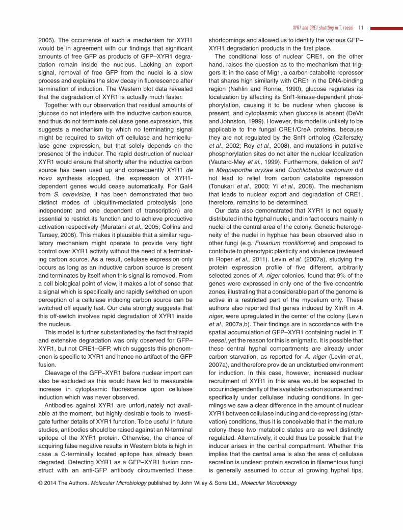
2005). The occurrence of such a mechanism for XYR1would be in agreement with our findings that significantamounts of free GFP as products of GFP–XYR1 degra-dation remain inside the nucleus. Lacking an exportsignal, removal of free GFP from the nuclei is a slowprocess and explains the slow decay in fluorescence aftertermination of induction. The Western blot data revealedthat the degradation of XYR1 is actually much faster.
Together with our observation that residual amounts ofglucose do not interfere with the inductive carbon source,and thus do not terminate cellulase gene expression, thissuggests a mechanism by which no terminating signalmight be required to switch off cellulase and hemicellu-lase gene expression, but that solely depends on thepresence of the inducer. The rapid destruction of nuclearXYR1 would ensure that shortly after the inductive carbonsource has been used up and consequently XYR1 denovo synthesis stopped, the expression of XYR1-dependent genes would cease automatically. For Gal4from S. cerevisiae, it has been demonstrated that twodistinct modes of ubiquitin-mediated proteolysis (oneindependent and one dependent of transcription) areessential to restrict its function and to achieve productiveactivation respectively (Muratani et al., 2005; Collins andTansey, 2006). This makes it plausible that a similar regu-latory mechanism might operate to provide very tightcontrol over XYR1 activity without the need of a terminat-ing carbon source. As a result, cellulase expression onlyoccurs as long as an inductive carbon source is presentand terminates by itself when this signal is removed. Froma cell biological point of view, it makes a lot of sense thata signal which is specifically and rapidly switched on uponperception of a cellulase inducing carbon source can beswitched off equally fast. Our data strongly suggests thatthis off-switch involves rapid degradation of XYR1 insidethe nucleus.
This model is further substantiated by the fact that rapidand extensive degradation was only observed for GFP–XYR1, but not CRE1–GFP, which suggests this phenom-enon is specific to XYR1 and hence no artifact of the GFPfusion.
Cleavage of the GFP–XYR1 before nuclear import canalso be excluded as this would have led to measurableincrease in cytoplasmic fluorescence upon cellulaseinduction which was never observed.
Antibodies against XYR1 are unfortunately not avail-able at the moment, but highly desirable tools to investi-gate further details of XYR1 function. To be useful in futurestudies, antibodies should be raised against an N-terminalepitope of the XYR1 protein. Otherwise, the chance ofacquiring false negative results in Western blots is high incase a C-terminally located epitope has already beendegraded. Detecting XYR1 as a GFP–XYR1 fusion con-struct with an anti-GFP antibody circumvented these
shortcomings and allowed us to identify the various GFP–XYR1 degradation products in the first place.
The conditional loss of nuclear CRE1, on the otherhand, raises the question as to the mechanism that trig-gers it: in the case of Mig1, a carbon catabolite repressorthat shares high similarity with CRE1 in the DNA-bindingregion (Nehlin and Ronne, 1990), glucose regulates itslocalization by affecting its Snf1-kinase-dependent phos-phorylation, causing it to be nuclear when glucose ispresent, and cytoplasmic when glucose is absent (DeVitand Johnston, 1999). However, this model is unlikely to beapplicable to the fungal CRE1/CreA proteins, becausethey are not regulated by the Snf1 ortholog (Cziferszkyet al., 2002; Roy et al., 2008), and mutations in putativephosphorylation sites do not alter the nuclear localization(Vautard-Mey et al., 1999). Furthermore, deletion of snf1in Magnaporthe oryzae and Cochliobolus carbonum didnot lead to relief from carbon catabolite repression(Tonukari et al., 2000; Yi et al., 2008). The mechanismthat leads to nuclear export and degradation of CRE1,therefore, remains to be determined.
Our data also demonstrated that XYR1 is not equallydistributed in the hyphal nuclei, and in fact occurs mainly innuclei of the central area of the colony. Genetic heteroge-neity of the nuclei in hyphae has been observed also inother fungi (e.g. Fusarium moniliforme) and proposed tocontribute to phenotypic plasticity and virulence (reviewedin Roper et al., 2011). Levin et al. (2007a), studying theprotein expression profile of five different, arbitrarilyselected zones of A. niger colonies, found that 9% of thegenes were expressed in only one of the five concentriczones, illustrating that a considerable part of the genome isactive in a restricted part of the mycelium only. Theseauthors also reported that genes induced by XlnR in A.niger, were upregulated in the center of the colony (Levinet al., 2007a,b). Their findings are in accordance with thespatial accumulation of GFP–XYR1 containing nuclei in T.reesei, yet the reason for this is enigmatic. It is possible thatthese central hyphal compartments are already undercarbon starvation, as reported for A. niger (Levin et al.,2007a), and therefore provide an undisturbed environmentfor induction. In this case, however, increased nuclearrecruitment of XYR1 in this area would be expected tooccur independently of the available carbon source and notspecifically under cellulase inducing conditions. In ger-mlings we saw a clear difference in the amount of nuclearXYR1 between cellulase inducing and de-repressing (star-vation) conditions, thus it is conceivable that in the maturecolony these two metabolic states are as well distinctlyregulated. Alternatively, it could thus be possible that theinducer arises in the central compartment. Whether thisimplies that the central area is also the area of cellulasesecretion is unclear: protein secretion in filamentous fungiis generally assumed to occur at growing hyphal tips,
XYR1 and CRE1 shuttling in T. reesei 11
© 2014 The Authors. Molecular Microbiology published by John Wiley & Sons Ltd., Molecular Microbiology

because only the growing cell wall would be porousenough to allow the secreted proteins to pass through.However, it has been shown in A. niger that α-amylase issecreted both at the hyphal tips and at the septa byexocytosis (Hayakawa et al., 2011; Read, 2011). Nostudies have so far been performed with T. reesei, but weassume in analogy that cellulases are also secreted atthese two places, hence implying long-distance transportof their mRNA or the precursor proteins. What has beendemonstrated recently, is a significant increase of theconidiation-associated transcriptome in T. reesei, includingthe cellulase and xylanase marker genes cel6a/cbh1 andxyn2 (Metz et al., 2011). As mentioned above, onset anddegree of conidiation greatly depend on the availablecarbon source, and are usually restricted to the centralcolony area. This obviously provides a functional linkbetween conidiogenesis and nuclear import of XYR1 in thesame region. However, in the present study, we haverestricted the analysis of transcription factor shuttlingdynamics to vegetative hyphae of the mycelium, excludingaerial hyphae and conidiophores/conidia. In contrast, Metzet al. (2011) investigated transcriptional changes in amixture of aerial hyphae, conidiophores and conidia. Thisleaves the interesting question as to how cellulase regula-tion and associated XYR1 and CRE1 transcriptionfactor shuttling might differ between these distinct mor-phological stages, and certainly warrants further, moredetailed investigation.
Taken together, the present data have revealed adynamic shuttling and a spatial bias of the major regulatorof cellulase and hemicellulase gene expression of T.reesei, XYR1, during the process of induction of expres-sion of the respective hydrolase genes. This clearly offersnew cellular areas for manipulation of T. reesei strains forcellulase production.
Experimental procedures
Strains and growth conditions
Trichoderma reesei strain QM9414 (ATCC 26921), an earlycellulase overproducing mutant derived from the wild-typeisolate QM6a (ATCC 26921) and the knock-out strains Δxyr1(Stricker et al., 2006), and Δcre1 (Portnoy et al., 2011a) wereused throughout this work. T. reesei transformants expressinggreen fluorescent protein (GFP)-labeled fusion proteins werecreated in QM9414 or QM9414 Δtku70 (C. Ivanova et al.,unpubl. data) parental strain backgrounds, in the latter theΔtku70 gene was replaced in strain QM9414 by the pyrithi-amine marker (Kubodera et al., 2002) essentially as described(Guangtao et al., 2009), and ultimately comprised strainsTRAL001 (xyr1–gfp), TRAL002 (gfp–xyr1), TRAL003 (cre1–gfp) and TRAL004 (gfp–cre1). QM9414 and QM9414 Δtku70respectively, served as reference strains in all experiments.Strain propagation, transformant selection and purificationwere performed on potato dextrose agar (PDA, Difco). For
experimentation, strains were grown in Mandels–Andreotti(MA) medium (Mandels and Andreotti, 1978), using glucose,xylose, glycerol, lactose, sophorose or cellulose as solecarbon sources at final concentrations as indicated below. Allstrains are maintained as glycerol stocks at −80°C. All strainsused and generated in this study are listed in Table S1.
Escherichia coli strains JM109 (cat.no. P9751, Promega,Madison, Wisconsin), One Shot®Top10 (cat.no. C4040-10,Life Technologies-Invitrogen, Austria) or Stellar® (cat.no.636763,Takara Bio Europe/Clontech, Saint-Germain-en-Laye, France) were used for plasmid construction and ampli-fication using standard molecular cloning techniques(Sambrook and Russell, 2001).
Generation of gene replacement cassettes
To express fluorescently labeled transcription factors XYR1and CRE1 from their native loci, we choose a simultaneousknock-out/knock-in strategy. For this, gene replacements cas-settes were constructed, that exchanged the complete xyr1 orcre1 open reading frame with full-length copies of each gfp-fusion gene plus a hygromycin resistance marker; the latterexpressing the E. coli hygromycin B phosphotransferase gene(hph) under control of the promoter and terminator region of T.reesei glyceraldehyde 3-phosphate dehydrogenase (gpd1)gene (Hartl et al., 2007). Homologous recombination wasfacilitated via the native ∼ 1 kb 5′ and 3′ non-coding regions(Fig. S1). Assembly of the xyr1 and cre1 gene replacementcassettes was performed using InFusion® recombinationalPCR cloning (cat.no. 639649, Takara Bio Europe/Clontech,Saint-Germain-en-Laye, France) with oligonucleotides sum-marized in Table S3. Briefly, pLH1-hph (Hartl et al., 2007) wasfirst double-digested with XhoI/SalI and the linearized 5 kbvector backbone was purified by gel extraction. Subsequently,the 1 kb non-coding 3′-flanks of xyr1 and cre1 were PCR-amplified from genomic T. reesei DNA and inserted intopLH1-hph to generate pLH1-hph-X3f and pLH1-hph-C3frespectively. Next, pLH1-hph-X3f and pLH1-hph-C3f werereopened by BamHI restriction digestion, gel-purified, and theORFs of xyr1 (4.8 kb) and cre1 (3.1 kb), each comprising 1 kbof the promoter region, the gene coding sequence, and 850bpof the terminator region, were amplified from T. reesei genomicDNA and integrated into the linearized vector backbone toresult in pXYR1 and pCRE1. Finally, pXYR1 and pCRE1 wereconverted by full-length PCR amplification into linear DNAfragments of 11 and 9.1 kb length, respectively, and the GFP-coding sequences (750 bp), beforehand amplified from pAL1-MAK1 (Lichius et al., 2012), were joined in to generatepGFP–XYR1, pXYR1–GFP, pGFP–CRE1 and pCRE1–GFP.All generated plasmids were verified by control restrictiondigestion and sequencing. Correct plasmids were amplifiedthrough E. coli, and subsequently used as templates for theamplification of the 9.5 and 7.6 kb sized xyr1 and cre1 replace-ment cassettes, respectively, which were transformed intoT. reesei.
Transformation of T. reesei
Gene replacements cassettes were amplified from therespective plasmids by PCR and transformed into T. reesei
12 A. Lichius, V. Seidl-Seiboth, B. Seiboth and C. P. Kubicek ■
© 2014 The Authors. Molecular Microbiology published by John Wiley & Sons Ltd., Molecular Microbiology

QM 9414 Δtku70 as linear DNA fragments using electropo-ration as described previously (Schuster et al., 2012). Trans-formants were selected on PDA hygromycin medium (100 μgml−1 final concentration) Homokaryons were obtained byrepeated rounds of vegetative spore propagation on selectivemedium and individual isolates were genotypically verified toconfirm targeted integration of the replacement cassette byPCR as described in detail elsewhere (Lichius et al., 2012),using genotyping primers listed in Table S3.
Real-time monitoring of transcription factor shuttling
In order to monitor rapid changes in the subcellular locali-zation of fluorescently labeled XYR1 and CRE1 upon cel-lulase induction or repression, respectively, carbon sourcereplacement experiments were performed using submergedgermling cultures. For this, conidia from 1-week-old carbonsource-free MA plate cultures were harvested in sterilewater and cell concentration was determined using aThoma cell counting chamber. A total of 1 × 10⁸ cells wereused to inoculate 100 ml MA pre-culture medium (liquid MAmedium with peptone to aid germination) in 500 ml shakeflasks, supplemented with either 1% w/v cellulose in its car-boxymethyl form (cellulase inducing condition) or 1% w/vglucose (cellulase repressing condition) as sole carbonsource, and incubated at 28°C and 200 r.p.m. overnight inthe dark. The next morning biomass from 20 ml pre-culturealiquots were washed twice with sterile tap water and trans-ferred into 20 ml carbon source-free MA replacementmedium (liquid MA medium without peptone) in 100 mlshake flasks. Initially, germlings were starved for up to 1.5 hunder identical incubation conditions to deplete cell internalcarbon storage. This, however, was not generally requiredand thus omitted during later experiments. At time pointt = 0 min a new carbon source for either cellulase and xyla-nase induction [0.062% w/v sophorose (1.4 mM), 0.015%w/v xylose (1 mM), 1% w/v lactose (25 mM) or 1% w/v cel-lulose] or for cellulase repression [1% w/v glucose (50 mM)or 1% w/v xylose (65 mM)] was added and incubation con-tinued. Germling samples for microscopical analysis weredrawn from overnight pre-cultures as well as before andafter carbon source replacement at desired time points.Carbon source-free MA cultures were used as controls forde-repressing conditions.
Transcription factor recruitment colony profiling
In order to quantify localized nuclear recruitment of fluores-cently labeled XYR1 and CRE1 in the three main regions ofthe functionally stratified fungal colony (periphery/leadingedge, subperiphery and central area) MA agar plates supple-mented with the respective carbon source for cellulase-inducing and -repressing conditions were centrally inoculatedwith T. reesei strains expressing either GFP–XYR1 or CRE1–GFP. Depending on the rate of colony development on thevarious carbon sources (growth on complex carbon sourcesgenerally required longer incubation times than growth onmonosaccharides), incubation was performed for 48–72 h at28°C and 12 h/12 h light/dark cycles until all three functionalzones have been established and conidiation commenced.
Agar block samples carrying mycelial sectors representativefor all three main colony regions were cut out and preparedfor live-cell imaging analysis.
Quantitative live-cell imaging
Expression and subcellular localization of GFP-labeled tran-scription factors was quantified using scanning confocalmicroscopy and image analysis. Fungal cells were preparedeither as germling samples from submerged flask cultures,simply by sandwiching 20 μl of cell suspension between twoglass coverslips, or as mycelial samples prepared fromgrowing plate cultures using the ‘inverted agar block’ method(Hickey et al., 2005).
Live-cell imaging was performed using a Nikon C1 con-focal laser scanning unit mounted on a Nikon EclipseTE2000-E inverted microscope base (Nikon GmbH, Vienna,Austria). GFP-labeled proteins were excited with the488 nm laser line of an argon ion laser, and emitted fluo-rescence light separated by a Nikon MHX40500b/C100332filter cube was detected with a photomultiplier tube withinthe range of 500–530 nm. A Nikon Plan Apo VC 60×/1.2water immersion objective lens was used, and laser inten-sity and laser dwell time during image acquisition were keptto a minimum to reduce photobleaching and phototoxiceffects while providing a sufficient signal-to-noise ratio forquantitative image analysis. Bright-field images were cap-tured simultaneously with a Nikon C1-TD transmitted lightdetector mounted behind the condenser turret. Images wererecorded with a maximum resolution of 1024 × 1024 pixelsand saved as TIFF. Apart from display range adjustmentsand cropping using the ImageJ software platform (http://rsb.info.nih.gov/ij/), images were not subjected to furthermanipulation. Fluorescence intensity measurements andinteractive 3D surface plots were performed with the corre-sponding plugins of the MacBiophotonics ImageJ workpackage available at (https://www.macbiophotonics.ca/software.htm), and statistically evaluated using MicrosoftExcel.
For each data bar, up to 12 images covering 160 × 160 μmfield of view of mycelial sample from the respective colonyareas or of 25 μl submerged germling cultures, were ran-domly recorded, and in those images mean fluorescenceintensities of 120 nuclei and 120 surrounding cytoplasmicareas were measured and averaged.
Trichoderma reesei is a multinucleate organism, and itsnuclei are not mitotically synchronized. Thus, to account forthe inherently high degree of variation in gene regulationbetween hyphae and within the nuclear population of onehypha, we calculated the nucleo-cytoplasmic fluorescenceratio (n/c-ratio) for each tested condition (Fig. S1). This valuerepresents the ratio between the average nuclear fluores-cence intensity within the population of nuclei and the averagecytoplasmic fluorescence intensity between these nuclei, andshould compensate for local differences in transcription factorexpression and subcellular localization within the imagedhyphal population. Taking the detection limit of the microscopeinto account, an n/c-ratio of ≥ 1.3 indicates significant nuclearaccumulation of fluorescently labeled transcription factorabove residual autofluorescence background. Note: ‘errorbars’ in the respective graphs represent the considerable
XYR1 and CRE1 shuttling in T. reesei 13
© 2014 The Authors. Molecular Microbiology published by John Wiley & Sons Ltd., Molecular Microbiology

biological variation of transcription factor recruitment to indi-vidual nuclei within the population rather than a statistical error(see example in Fig. S1C).
Total RNA extraction from fungal biomass
In order to correlate nuclear presence or absence of bothtranscription factors with changes in gene expression, fungalbiomass for subsequent RNA extraction and RT-qPCR analy-sis was collected immediately after the respective live-cellimaging shuttling experiments. For this, T. reesei germlingswere harvested from induced or repressed 20 ml submergedcultures, and preserved by immediate shock-freezing in liquidnitrogen, followed by storage at −80°C. Total RNA extractionwas performed according to Chirgwin et al. (1979), and RNAquality and quantity were determined using a NanoDrop spec-trophotometer (Thermo Scientific, Vienna, Austria).
Gene expression analysis by reverse transcriptasequantitative PCR (RT-qPCR)
DNase I-treated (cat.no. EN0521, Fermentas) RNA (3 μg)was reverse-transcribed with the RevertAid First StrandcDNA Kit (cat.no. K1632, Thermo Scientific) according to themanufacturer’s protocol with a 1:1 combination of the pro-vided oligo-dT and random hexamer primers. All RT-qPCRexperiments were performed on an Eppendorf realplex2 Mas-tercycler (Eppendorf, Hamburg, Germany). Each sample wasprepared as 25 μl reaction using the iQ SYBR Green Super-mix (cat.no. 170-8882, Bio-Rad) with a final primer concen-tration of 100 nM forward and reverse primer each. All assayswere carried out as triplicates in a 96-well plate formatcovered with optical tape, including no-template controls.Measurements with the housekeeping gene transcriptionelongation factor 1α (tef1) were performed for reference cal-culation and data normalization. Determination of the PCRefficiency was performed using triplicate reactions from adilution series of cDNA (1; 0.1; 0.01; 0.001). Primers, ampli-fication efficiency and R2 values are given in Table S2. Ampli-fication efficiency of each sample mRNA was then calculatedfrom the given slopes in the iQ5 Optical System Softwarev2.0 and relative fold-changes in gene expression werecalculated using the Relative Expression Software Tool(REST) (http://www.gene-quantification.de/rest.html; Pfafflet al., 2002). All samples were analyzed in two independentexperiments with three replicates in each run.
Protein extraction, SDS-PAGE and Westernblot analysis
Overnight liquid cultures (100 ml MA medium, 1% w/vglucose) were prepared as described above for shuttlingexperiments. Samples for positive and negative controls, andfor CRE1–GFP were directly drawn from these pre-culturesafter 18 h of incubation. Biosynthesis of GFP–XYR1 wasinduced by transferring fungal biomass from 50 ml pre-culture into 50 ml fresh MA medium containing 1.4 mMsophorose. These induced cultures were harvested after 1, 2and 3 h of additional incubation. For each sample, fungal
biomass from 50 ml liquid pre- or induced culture was har-vested on a glass microfiber filter (Whatmann, cat.no. 1822-047) using vacuum-driven filtration, and, after washing twicewith sterile tap water, immediately shock frozen in liquid nitro-gen. Subsequently, the biomass was ground to a fine powderin liquid nitrogen, and approximately 100 mg of it were addedto a 2 ml Eppendorf tube already containing 1 ml of proteinextraction buffer (10 ml PBS containing 5 mM EDTA and5 mM PMSF plus one cOmplete ULTRA protease inhibitorcocktail tablet (Roche, cat.no. 05 892 791 001), pH 7.4), and1 g of small (0.25 mm diameter) and four large (3 mm diam-eter) glass beads to aid cell destruction. The mix was sub-jected to three 1 min rounds of homogenization at 30 Hz with1 min cooling intervals at −20°C. Cell debris and aqueousphase were separated by centrifugation with 17 000 g for5 min and at 4°C. The clear supernatant containing all solubleproteins was transferred into fresh, pre-cooled Eppendorftubes and stored at −20°C until further use. Total proteinconcentration was determined against BSA using Bradfordreagent (Bio-Rad, cat.no. 500-0006) according to manufac-tures recommendations. Typically, protein yields between 1and 3 mg ml−1 were achieved.
Except for the GFP control (cytosolic GFP expressedunder control of the constitutive Ptef1 promoter in T. reeseiQM6a) of which only 0.35 μg per lane were loaded, 20 μgof the crude total protein extract of all other samples wereseparated by SDS-PAGE as outlined in detail elsewhere(Sambrook and Russell, 2001). Generally, two identical 12%SDS-PAGE gels were prepared, one for colloidal Coomas-sie staining, and the second for semi-dry electro blotting ofthe separated proteins onto ImmobileFL PVDF membrane(Millipore, cat.no. IPFL00010). Subsequent blocking wasachieved by incubation in PBS-T (PBS, 0.3% Tween 20)supplemented with 2% w/v milk powder (Roth, cat.no.T145.1) for 1 h at room temperature. For the specific labe-ling of GFP and GFP-fusion proteins, respectively, themonoclonal mouse anti-GFP–HRP antibody α-GFP(B-2)(Santa Cruz, cat.no. sc-9996) was used, diluted 1:1000 inPBS-T containing 0.5% w/v milk powder, and incubated onthe membrane for 2 h at room temperature, followed by fourwashing steps with PBS-T.
Detection of the labeled proteins was performed with thePierce ECL2 kit (Thermo Scientific, cat.no. 80197) accordingto manufactures recommendations. Chemifluorescent signalswere recorded on a Typhoon FLA700 imager (GE Healthcare),and chemiluminescent signals were visualized by x-ray film(Amersham Hyperfilm ECL, GE Healthcare, cat.no. 28-9068-35) exposure. For protein band size estimation, two molecularweight markers were used: PageRuler Pre-stained ProteinLadder 10–170 kDa (Thermo Scientific, cat.no. 26616), andSuperSignal Enhanced Protein Ladder 20–150 kDa (ThermoScientific, cat.no. 84786), with only the latter one being appli-cable for ECL detection on x-ray film.
Acknowledgements
We are thankful to Dr. Christa Ivanova and Dr. Rita Linke forproviding strains prior to publication, and gratefully appreci-ate the help of Sara Ghassemi during some stages of thiswork. We especially thank Dr. Lisa Kappel and JonasRamoni, as well as Dr. Eddy Sanchez León-Hing (CICESE,
14 A. Lichius, V. Seidl-Seiboth, B. Seiboth and C. P. Kubicek ■
© 2014 The Authors. Molecular Microbiology published by John Wiley & Sons Ltd., Molecular Microbiology

Mexico) and Dr. Michael Freitag (University of Oregon, USA)for helpful advice on protein biochemistry methods. This workwas supported by grants from the Austrian Science Founda-tion (FWF P-23202) to CPK and (FWF P-24219) to BS.
Conflict of interest
The authors declare no conflict of interest.
References
Akel, E., Metz, B., Seiboth, B., and Kubicek, C.P. (2009)Molecular regulation of arabinan and L-arabinose metabo-lism in Hypocrea jecorina (Trichoderma reesei). EukaryotCell 8: 1837–1844.
Arst, H.N., Jr, Tollervey, D., Dowzer, C.E., and Kelly, J.M.(1990) An inversion truncating the creA gene of Aspergillusnidulans results in carbon catabolite derepression. MolMicrobiol 4: 851–854.
Axelrod, J.D., Majors, J., and Brandriss, M.C. (1991) Proline-independent binding of PUT3 transcriptional activatorprotein detected by footprinting in vivo. Mol Cell Biol 11:564–567.
Berger, H., Pachlinger, R., Morozov, I., Goller, S., Narendja,F., Caddick, M., and Strauss, J. (2006) The GATA factorAreA regulates localization and in vivo binding site occu-pancy of the nitrate activator NirA. Mol Microbiol 59: 433–446.
Bischof, R., Fourtis, L., Gamauf, C., Seiboth, B., and Kubicek,C.P. (2013) Comparative analysis of the Trichodermareesei transcriptome during growth on the cellulase induc-ing substrates wheat straw and lactose. Biotechnol Biofu-els 6: 127.
Braun, B.R., van Het Hoog, M., d’Enfert, C., Martchenko, M.,Dungan, J., Kuo, A., et al. (2005) A human-curated anno-tation of the Candida albicans genome. PLoS Genet 1:36–57.
Calero-Nieto, F., Di Pietro, A., Roncero, M.I., and Hera, C.(2007) Role of the transcriptional activator xlnR ofFusarium oxysporum in regulation of xylanase genes andvirulence. Mol Plant Microbe Interact 20: 977–985.
Chirgwin, J.M., Przybyla, A.E., MacDonald, R.J., and Rutter,W.J. (1979) Isolation of biologically active ribonucleic acidfrom sources enriched in ribonuclease. Biochemistry 18:5294–5299.
Chundawat, S.P., Beckham, G.T., Himmel, M.E., and Dale,B.E. (2011) Deconstruction of lignocellulosic biomass tofuels and chemicals. Ann Rev Chem Biomol Eng 2: 121–145.
Collins, G.A., and Tansey, W.P. (2006) The proteasome: autility tool for transcription? Curr Opin Genet Dev 16: 197–202.
Cziferszky, A., Mach, R.L., and Kubicek, C.P. (2002) Phos-phorylation positively regulates DNA binding of the carboncatabolite repressor Cre1 of Hypocrea jecorina (Tricho-derma reesei). J Biolog Chem 277: 14688–14694.
De Vit, M.J., Waddle, J.A., and Johnston, M. (1997) Regu-lated nuclear translocation of the Mig1 glucose repressor.Mol Biol Cell 8: 1603–1618.
Deichsel, H., Friedel, S., Detterbeck, A., Coyne, C., Hamker,U., and MacWilliams, H.K. (1999) Green fluorescent pro-
teins with short half-lives as reporters in Dictyostelium dis-coideum. Dev Genes Evol 209: 63–68.
DeVit, M.J., and Johnston, M. (1999) The nuclear exportinMsn5 is required for nuclear export of the Mig1 glucoserepressor of Saccharomyces cerevisiae. Curr Biol 9: 1231–1241.
Dinamarco, T.M., Almeida, R.S., de Castro, P.A., Brown,N.A., dos Reis, T.F., Ramalho, L.N., et al. (2012) Molecularcharacterization of the putative transcription factor SebAinvolved in virulence in Aspergillus fumigatus. EukaryotCell 11: 518–531.
El Alami, M., Feller, A., Pierard, A., and Dubois, E. (2000)Characterisation of a tripartite nuclear localisationsequence in the regulatory protein Lys14 of Saccharomy-ces cerevisiae. Curr Genet 38: 78–86.
Evdokimov, A.G., Pokross, M.E., Egorov, N.S., Zaraisky,A.G., Yampolsky, I.V., Merzlyak, E.M., et al. (2006) Struc-tural basis for the fast maturation of Arthropoda greenfluorescent protein. EMBO Rep 7: 1006–1012.
Felenbok, B., Flipphi, M., and Nikolaev, I. (2001) Ethanolcatabolism in Aspergillus nidulans: a model system forstudying gene regulation. Prog Nucleic Acid Res Mol Biol69: 149–204.
Foreman, P.K., Brown, D., Dankmeyer, L., Dean, R., Diener,S., Dunn-Coleman, N.S., et al. (2003) Transcriptional regu-lation of biomass-degrading enzymes in the filamentousfungus Trichoderma reesei. J Biol Chem 278: 31988–31997.
Guangtao, Z., Hartl, L., Schuster, A., Polak, S., Schmoll, M.,Wang, T., et al. (2009) Gene targeting in a nonhomologousend joining deficient Hypocrea jecorina. J Biotechnol 139:146–151.
Harbison, C.T., Gordon, D.B., Lee, T.I., Rinaldi, N.J.,Macisaac, K.D., Danford, T.W., et al. (2004) Transcriptionalregulatory code of a eukaryotic genome. Nature 431:99–104.
Hartl, L., Kubicek, C.P., and Seiboth, B. (2007) Induction ofthe gal pathway and cellulase genes involves no transcrip-tional inducer function of the galactokinase in Hypocreajecorina. J Biol Chem 282: 18654–18659.
Hasper, A.A., Visser, J., and de Graaff, L.H. (2000) TheAspergillus niger transcriptional activator XlnR, which isinvolved in the degradation of the polysaccharides xylanand cellulose, also regulates D-xylose reductase geneexpression. Mol Microbiol 36: 193–200.
Hasper, A.A., Trindade, L.M., van der Veen, D., van Ooyen,A.J., and de Graaff, L.H. (2004) Functional analysis of thetranscriptional activator XlnR from Aspergillus niger. Micro-biology 150: 1367–1375.
Hayakawa, Y., Ishikawa, E., Shoji, J.Y., Nakano, H., andKitamoto, K. (2011) Septum-directed secretion in the fila-mentous fungus Aspergillus oryzae. Mol Microbiol 81:40–55.
Häkkinen, M., Valkonen, M.J., Westerholm-Parvinen, A., Aro,N., Arvas, M., Vitikainen, M., et al. (2014) Screening ofcandidate regulators for cellulase and hemicellulase pro-duction in Trichoderma reesei and identification of a factoressential for cellulase production. Biotechnol Biofuels 7:14.
Heim, R., and Tsien, R.Y. (1996) Engineering green fluores-cent protein for improved brightness, longer wavelengths
XYR1 and CRE1 shuttling in T. reesei 15
© 2014 The Authors. Molecular Microbiology published by John Wiley & Sons Ltd., Molecular Microbiology
and fluorescence resonance energy transfer. Curr Biol 6:178–182.
Heim, R., Cubitt, A.B., and Tsien, R.Y. (1995) Improved greenfluorescence. Nature 373: 663–664.
Herold, S., Bischof, R., Metz, B., Seiboth, B., and Kubicek,C.P. (2013) Xylanase gene transcription in Trichodermareesei is triggered by different inducers representing differ-ent hemicellulosic pentose polymers. Eukaryot Cell 12:390–398.
Hickey, P.C., Swift, S.R., Roca, M.G., and Read, N.D. (2005)Live-cell Imaging of filamentous fungi using vital fluores-cent dyes and confocal microscopy. In Methods inMicrobiology. Savidge, T., and Charalabos, P. (eds). Phila-delphia, USA: Elsevier B. V., pp. 63–87.
Ilmen, M., Thrane, C., and Penttila, M. (1996) The glucoserepressor gene cre1 of Trichoderma: isolation and expres-sion of a full-length and a truncated mutant form. Mol GenGenet 251: 451–460.
Jekosch, K., and Kück, U. (2000) Loss of glucose repressionin an Acremonium chrysogenum beta-lactam producerstrain and its restoration by multiple copies of the cre1gene. Appl Microbiol Biotechnol 54: 556–563.
Kirkpatrick, C.R., and Schimmel, P. (1995) Detection ofleucine-independent DNA site occupancy of the yeastLeu3p transcriptional activator in vivo. Mol Cell Biol 15:4021–4030.
Klaubauf, S., Narang, H.M., Post, H., Zhou, M., Brunner, K.,Mach-Aigner, A.R., et al. (2014) Similar is not the same:differences in the function of the (hemi-)cellulolytic regula-tor XlnR (Xlr1/Xyr1) in filamentous fungi. Fungal Genet Bioldoi:10.1016/j.fgb.2014.07.007 [Epub ahead of print].
Kohlhaw, G.B. (2003) Leucine biosynthesis in fungi: enteringmetabolism through the back door. Microbiol Mol Biol Rev67: 1–15, table of contents.
Kren, A., Mamnun, Y.M., Bauer, B.E., Schuller, C., Wolfger,H., Hatzixanthis, K., et al. (2003) War1p, a novel transcrip-tion factor controlling weak acid stress response in yeast.Mol Cell Biol 23: 1775–1785.
Kubicek, C.P. (2013) Systems biological approaches towardsunderstanding cellulase production by Trichoderma reesei.J Biotechnol 163: 133–142.
Kubicek, C.P., Mikus, M., Schuster, A., Schmoll, M., andSeiboth, B. (2009) Metabolic engineering strategies for theimprovement of cellulase production by Hypocrea jecorina.Biotechnol Biofuels 2: 19.
Kubodera, T., Yamashita, N., and Nishimura, A. (2002) Trans-formation of Aspergillus sp. and Trichoderma reesei usingthe pyrithiamine resistance gene (ptrA) of Aspergillusoryzae. Biosci Biotechnol Biochem 66: 404–406.
Levin, A.M., de Vries, R.P., Conesa, A., de Bekker, C., Talon,M., Menke, H.H., et al. (2007a) Spatial differentiation in thevegetative mycelium of Aspergillus niger. Eukaryot Cell 6:2311–2322.
Levin, A.M., de Vries, R.P., and Wosten, H.A. (2007b) Locali-zation of protein secretion in fungal colonies using a novelculturing technique; the ring-plate system. J MicrobiolMethods 69: 399–401.
Lichius, A., Lord, K.M., Jeffree, C.E., Oborny, R.,Boonyarungsrit, P., and Read, N.D. (2012) Importance ofMAP kinases during protoperithecial morphogenesis inNeurospora crassa. PLoS ONE 7: 1–21.
Mach, R.L., Strauss, J., Zeilinger, S., Schindler, M., andKubicek, C.P. (1996) Carbon catabolite repression of xyla-nase I (xyn1) gene expression in Trichoderma reesei. MolMicrobiol 21: 1273–1281.
Mach-Aigner, A.R., Pucher, M.E., Steiger, M.G., Bauer, G.E.,Preis, S.J., and Mach, R.L. (2008) Transcriptional regula-tion of xyr1, encoding the main regulator of the xylanolyticand cellulolytic enzyme system in Hypocrea jecorina. ApplEnviron Microbiol 74: 6554–6562.
MacPherson, S., Larochelle, M., and Turcotte, B. (2006) Afungal family of transcriptional regulators: the zinc clusterproteins. Microbiol Mol Biol Rev 70: 583–604.
Makita, T., Katsuyama, Y., Tani, S., Suzuki, H., Kato, N., Todd,R.B., et al. (2009) Inducer-dependent nuclear localizationof a Zn(II)(2)Cys(6) transcriptional activator, AmyR, inAspergillus nidulans. Biosci Biotechnol Biochem 73: 391–399.
Mandels, M.M., and Andreotti, R.E. (1978) The cellulose tocellulase fermentation. Proc Biochem 13: 6–13.
Marui, J., Tanaka, A., Mimura, S., de Graaff, L.H., Visser, J.,Kitamoto, N., et al. (2002) A transcriptional activator,AoXlnR, controls the expression of genes encoding xylano-lytic enzymes in Aspergillus oryzae. Fungal Genet Biol 35:157–169.
Metz, B., Seidl-Seiboth, V., Haarmann, T., Kopchinskiy, A.,Lorenz, P., Seiboth, B., and Kubicek, C.P. (2011) Expres-sion of biomass-degrading enzymes is a major eventduring conidium development in Trichoderma reesei.Eukaryot Cell 10: 1527–1535.
Muratani, M., Kung, C., Shokat, K.M., and Tansey, W.P.(2005) The F box protein Dsg1/Mdm30 is a transcriptionalcoactivator that stimulates Gal4 turnover and cotranscrip-tional mRNA processing. Cell 120: 887–899.
Nehlin, J.O., and Ronne, H. (1990) Yeast MIG1 repressor isrelated to the mammalian early growth response andWilms’ tumour finger proteins. EMBO J 9: 2891–2898.
van Peij, N.N., Gielkens, M.M., de Vries, R.P., Visser, J., andde Graaff, L.H. (1998) The transcriptional activator XlnRregulates both xylanolytic and endoglucanase geneexpression in Aspergillus niger. Appl Environ Microbiol 64:3615–3619.
Pfaffl, M.W., Horgan, G.W., and Dempfle, L. (2002) Relativeexpression software tool (REST) for group-wise compari-son and statistical analysis of relative expression results inreal-time PCR. Nucleic Acids Res 30: e36.
Portnoy, T., Margeot, A., Linke, R., Atanasova, L., Fekete, E.,Sandor, E., et al. (2011a) The CRE1 carbon cataboliterepressor of the fungus Trichoderma reesei: a masterregulator of carbon assimilation. BMC Genomics 12:269.
Portnoy, T., Margeot, A., Seidl-Seiboth, V., Le Crom, S.,Ben Chaabane, F., Linke, R., et al. (2011b) Differentialregulation of the cellulase transcription factors XYR1,ACE2, and ACE1 in Trichoderma reesei strains producinghigh and low levels of cellulase. Eukaryot Cell 10: 262–271.
Read, N.D. (2011) Exocytosis and growth do not occur only athyphal tips. Mol Microbiol 81: 4–7.
Roper, M., Ellison, C., Taylor, J.W., and Glass, N.L. (2011)Nuclear and genome dynamics in multinucleate ascomy-cete fungi. Curr Biol 21: R786–R793.
16 A. Lichius, V. Seidl-Seiboth, B. Seiboth and C. P. Kubicek ■
© 2014 The Authors. Molecular Microbiology published by John Wiley & Sons Ltd., Molecular Microbiology

Roy, P., Lockington, R.A., and Kelly, J.M. (2008) CreA-mediated repression in Aspergillus nidulans does notrequire transcriptional auto-regulation, regulated intracellu-lar localisation or degradation of CreA. Fungal Genet Biol45: 657–670.
Sambrook, J., and Russell, D.W. (2001) Molecular Cloning: ALaboratory Manual. Painview, NY: Cold Spring Harbor Lab.Press.
Schneider-Poetsch, T., Ju, J., Eyler, D.E., Dang, Y., Bhat, S.,Merrick, W.C., et al. (2010) Inhibition of eukaryotic transla-tion elongation by cycloheximide and lactimidomycin. NatChem Biol 6: 209–217.
Schuster, A., Bruno, K.S., Collett, J.R., Baker, S.E., Seiboth,B., Kubicek, C.P., and Schmoll, M. (2012) A versatile toolkitfor high throughput functional genomics with Trichodermareesei. Biotechnol Biofuels 5: 1.
Seiboth, B., Gamauf, C., Pail, M., Hartl, L., and Kubicek, C.P.(2007) The D-xylose reductase of Hypocrea jecorinais the major aldose reductase in pentose and D-galactose catabolism and necessary for beta-galactosidase and cellulase induction by lactose. MolMicrobiol 66: 890–900.
Seiboth, B., Herold, S., and Kubicek, C.P. (2012) Metabolicengineering of inducer formation for cellulase and hemicel-lulase gene expression in Trichoderma reesei. SubcellBiochem 64: 367–390.
Sellick, C.A., and Reece, R.J. (2003) Modulation of transcrip-tion factor function by an amino acid: activation of Put3p byproline. EMBO J 22: 5147–5153.
Sellick, C.A., and Reece, R.J. (2005) Eukaryotic transcriptionfactors as direct nutrient sensors. Trends Biochem Sci 30:405–412.
Shimizu, K., Hicks, J.K., Huang, T.P., and Keller, N.P. (2003)Pka, Ras and RGS protein interactions regulate activity ofAflR, a Zn(II)2Cys6 transcription factor in Aspergillus nidu-lans. Genetics 165: 1095–1104.
Shroff, R.A., Lockington, R.A., and Kelly, J.M. (1996) Analy-sis of mutations in the creA gene involved in carbon cat-abolite repression in Aspergillus nidulans. Can J Microbiol42: 950–959.
Strauss, J., Mach, R.L., Zeilinger, S., Hartler, G., Stoffler, G.,Wolschek, M., and Kubicek, C.P. (1995) Cre1, the carboncatabolite repressor protein from Trichoderma reesei.FEBS Lett 376: 103–107.
Stricker, A.R., Grosstessner-Hain, K., Wurleitner, E., andMach, R.L. (2006) Xyr1 (xylanase regulator 1) regu-lates both the hydrolytic enzyme system and D-xylose
metabolism in Hypocrea jecorina. Eukaryot Cell 5:2128–2137.
Stricker, A.R., Mach, R.L., and de Graaff, L.H. (2008) Regu-lation of transcription of cellulases- and hemicellulases-encoding genes in Aspergillus niger and Hypocrea jecorina(Trichoderma reesei). Appl Microbiol Biotechnol 78: 211–220.
Sun, J., Tian, C., Diamond, S., and Glass, N.L. (2012) Deci-phering transcriptional regulatory mechanisms associatedwith hemicellulose degradation in Neurospora crassa.Eukaryot Cell 11: 482–493.
Tonukari, N.J., Scott-Craig, J.S., and Walton, J.D. (2000) TheCochliobolus carbonum SNF1 gene is required for cellwall-degrading enzyme expression and virulence onmaize. Plant Cell 12: 237–248.
Vautard-Mey, G., Cotton, P., and Fevre, M. (1999) Expressionand compartmentation of the glucose repressor CRE1 fromthe phytopathogenic fungus Sclerotinia sclerotiorum. Eur JBiochem 266: 252–259.
Viikari, L., Vehmaanperä, J., and Koivula, A. (2012) Lignocel-lulosic ethanol: from science to industry. Biomass Bioen-ergy 46: 13–24.
de Vries, R.P., Visser, J., and de Graaff, L.H. (1999) CreAmodulates the XlnR-induced expression on xylose ofAspergillus niger genes involved in xylan degradation. ResMicrobiol 150: 281–285.
Wightman, R., Bell, R., and Reece, R.J. (2008) Localizationand interaction of the proteins constituting the GAL geneticswitch in Saccharomyces cerevisiae. Eukaryot Cell 7:2061–2068.
Yi, M., Park, J.H., Ahn, J.H., and Lee, Y.H. (2008) MoSNF1regulates sporulation and pathogenicity in the rice blastfungus Magnaporthe oryzae. Fungal Genet Biol 45: 1172–1181.
Zeilinger, S., Mach, R.L., Schindler, M., Herzog, P., andKubicek, C.P. (1996) Different inducibility of expression ofthe two xylanase genes xyn1 and xyn2 in Trichodermareesei. J Biol Chem 271: 25624–25629.
Zhao, X., Duong, T., Huang, C.C., Kain, S.R., and Li, X.(1999) Comparison of enhanced green fluorescent proteinand its destabilized form as transcription reporters.Methods Enzymol 302: 32–38.
Supporting information
Additional supporting information may be found in the onlineversion of this article at the publisher’s web-site.
XYR1 and CRE1 shuttling in T. reesei 17
© 2014 The Authors. Molecular Microbiology published by John Wiley & Sons Ltd., Molecular Microbiology