A Phosphorylated Cytoplasmic Autoantigen, GW182, Associates with a Unique Population of Human mRNAs...
14
Molecular Biology of the Cell Vol. 13, 1338 –1351, April 2002 A Phosphorylated Cytoplasmic Autoantigen, GW182, Associates with a Unique Population of Human mRNAs within Novel Cytoplasmic Speckles Theophany Eystathioy,* Edward K. L. Chan, †‡ Scott A. Tenenbaum, § Jack D. Keene, § Kevin Griffith,* and Marvin J. Fritzler* *Departments of Medicine and Biochemistry and Molecular Biology, University of Calgary, Calgary, Alberta AB T2N 4N1, Canada; † W.M. Keck Autoimmune Disease Center, Department of Molecular and Experimental Medicine, The Scripps Research Institute, La Jolla, California 92037; § Department of Microbiology, Duke University Medical Center, Durham, North Carolina 27710 Submitted November 9, 2001; Revised December 17, 2001; Accepted January 8, 2002 Monitoring Editor: Joseph Gall A novel human cellular structure has been identified that contains a unique autoimmune antigen and multiple messenger RNAs. This complex was discovered using an autoimmune serum from a patient with motor and sensory neuropathy and contains a protein of 182 kDa. The gene and cDNA encoding the protein indicated an open reading frame with glycine-tryptophan (GW) repeats and a single RNA recognition motif. Both the patient’s serum and a rabbit serum raised against the recombinant GW protein costained discrete cytoplasmic speckles designated as GW bodies (GWBs) that do not overlap with the Golgi complex, endosomes, lysosomes, or peroxi- somes. The mRNAs associated with GW182 represent a clustered set of transcripts that are presumed to reside within the GW complexes. We propose that the GW ribonucleoprotein complex is involved in the posttranscriptional regulation of gene expression by sequestering a specific subset of gene transcripts involved in cell growth and homeostasis. INTRODUCTION Historically human autoantibodies have been used to dis- cover, identify, and understand the function of novel cellu- lar constituents and macromolecules (Tan, 1991; von Muhlen and Tan, 1995). For example, small nuclear ribo- nucleoproteins (snRNPs) and the spliceosome were initially elucidated through the use of human autoantibodies (Lerner and Steitz, 1981; Tan, 1991). In addition, a number of unique nucleolar (Reimer et al., 1987), Golgi complex (Chan and Fritzler, 1998), and endosome (Waite et al., 1998; Selak et al., 1999) proteins have been identified using human auto- antibodies. Several known autoantigens such as SS-A/Ro, SS-B/La, Sm, Hu, and Nova bind distinct RNAs and, in concert with other proteins, form macromolecular complexes that are key components of gene expression at multiple levels including transcription, translation, transport, and stability (Keene, 1999; Brennan and Steitz, 2001; Keene, 2001; Musunuru and Darnell, 2001). Proteins associated with mRNA are of inter- est because it is thought that messenger ribonucleoproteins may be involved in stabilizing, degrading, or in regulating translation of the mRNAs upon exit from the nucleus (Bren- nan and Steitz, 2001; Keene, 2001). One of the approaches to identification of proteins involved in mRNA processing, referred to as ribonomics, involves biochemically or immu- nologically recovering protein/RNA complexes, followed by microarray analysis to determine their structural and functional relationships (Tenenbaum et al., 2000). Using this approach, some of the mRNA subsets and profiles have been deciphered for proteins such as eIF-4E, PABP, and ELAV/Hu (Tenenbaum et al., 2000) as well as Fragile X Mental Retardation RNA-binding protein (FMRP; Brown et al., 2001). It has been proposed that the expression of struc- turally related mRNAs is coordinated by multitargeted RNA binding proteins in order to regulate complex processes (Keene, 2001). It is not known whether expression of certain clustered mRNAs might be coordinated and if multiple mRNAs reside together within a physical particle (Keene, 2001). Article published online ahead of print. Mol. Biol. Cell 10.1091/ mbc.01–11– 0544. Article and publication date are at www.molbiol- cell.org/cgi/doi/10.1091/mbc.01–11– 0544. Corresponding author. E-mail address: [email protected]. ‡ Alternate corresponding author. E-mail address: [email protected] Abbreviations used: EEA1, early endosome antigen 1; IIF, indi- rect immunofluorescence; IP, immunoprecipitation; NLS, nu- clear localization signal; ORF, open reading frame; RRM, RNA recognition motif; snRNP, small nuclear ribonucleoprotein; TnT, in vitro transcription and translation. 1338 © 2002 by The American Society for Cell Biology
Transcript of A Phosphorylated Cytoplasmic Autoantigen, GW182, Associates with a Unique Population of Human mRNAs...

Molecular Biology of the CellVol. 13, 1338–1351, April 2002
A Phosphorylated Cytoplasmic Autoantigen, GW182,Associates with a Unique Population of HumanmRNAs within Novel Cytoplasmic SpecklesTheophany Eystathioy,* Edward K. L. Chan,†‡ Scott A. Tenenbaum,§Jack D. Keene,§ Kevin Griffith,* and Marvin J. Fritzler*�
*Departments of Medicine and Biochemistry and Molecular Biology, University of Calgary, Calgary,Alberta AB T2N 4N1, Canada; †W.M. Keck Autoimmune Disease Center, Department of Molecularand Experimental Medicine, The Scripps Research Institute, La Jolla, California 92037; §Department ofMicrobiology, Duke University Medical Center, Durham, North Carolina 27710
Submitted November 9, 2001; Revised December 17, 2001; Accepted January 8, 2002Monitoring Editor: Joseph Gall
A novel human cellular structure has been identified that contains a unique autoimmune antigenand multiple messenger RNAs. This complex was discovered using an autoimmune serum froma patient with motor and sensory neuropathy and contains a protein of 182 kDa. The gene andcDNA encoding the protein indicated an open reading frame with glycine-tryptophan (GW)repeats and a single RNA recognition motif. Both the patient’s serum and a rabbit serum raisedagainst the recombinant GW protein costained discrete cytoplasmic speckles designated as GWbodies (GWBs) that do not overlap with the Golgi complex, endosomes, lysosomes, or peroxi-somes. The mRNAs associated with GW182 represent a clustered set of transcripts that arepresumed to reside within the GW complexes. We propose that the GW ribonucleoproteincomplex is involved in the posttranscriptional regulation of gene expression by sequestering aspecific subset of gene transcripts involved in cell growth and homeostasis.
INTRODUCTION
Historically human autoantibodies have been used to dis-cover, identify, and understand the function of novel cellu-lar constituents and macromolecules (Tan, 1991; vonMuhlen and Tan, 1995). For example, small nuclear ribo-nucleoproteins (snRNPs) and the spliceosome were initiallyelucidated through the use of human autoantibodies (Lernerand Steitz, 1981; Tan, 1991). In addition, a number of uniquenucleolar (Reimer et al., 1987), Golgi complex (Chan andFritzler, 1998), and endosome (Waite et al., 1998; Selak etal., 1999) proteins have been identified using human auto-antibodies.
Several known autoantigens such as SS-A/Ro, SS-B/La,Sm, Hu, and Nova bind distinct RNAs and, in concert with
other proteins, form macromolecular complexes that are keycomponents of gene expression at multiple levels includingtranscription, translation, transport, and stability (Keene,1999; Brennan and Steitz, 2001; Keene, 2001; Musunuru andDarnell, 2001). Proteins associated with mRNA are of inter-est because it is thought that messenger ribonucleoproteinsmay be involved in stabilizing, degrading, or in regulatingtranslation of the mRNAs upon exit from the nucleus (Bren-nan and Steitz, 2001; Keene, 2001). One of the approaches toidentification of proteins involved in mRNA processing,referred to as ribonomics, involves biochemically or immu-nologically recovering protein/RNA complexes, followedby microarray analysis to determine their structural andfunctional relationships (Tenenbaum et al., 2000). Using thisapproach, some of the mRNA subsets and profiles have beendeciphered for proteins such as eIF-4E, PABP, andELAV/Hu (Tenenbaum et al., 2000) as well as Fragile XMental Retardation RNA-binding protein (FMRP; Brown etal., 2001). It has been proposed that the expression of struc-turally related mRNAs is coordinated by multitargeted RNAbinding proteins in order to regulate complex processes(Keene, 2001). It is not known whether expression of certainclustered mRNAs might be coordinated and if multiplemRNAs reside together within a physical particle (Keene,2001).
Article published online ahead of print. Mol. Biol. Cell 10.1091/mbc.01–11–0544. Article and publication date are at www.molbiol-cell.org/cgi/doi/10.1091/mbc.01–11–0544.
� Corresponding author. E-mail address: [email protected].‡ Alternate corresponding author. E-mail address: [email protected]
Abbreviations used: EEA1, early endosome antigen 1; IIF, indi-rect immunofluorescence; IP, immunoprecipitation; NLS, nu-clear localization signal; ORF, open reading frame; RRM, RNArecognition motif; snRNP, small nuclear ribonucleoprotein; TnT,in vitro transcription and translation.
1338 © 2002 by The American Society for Cell Biology

In this study, we used sera from a patient with a motorand sensory polyneuropathy to identify a novel 182-kDaprotein that we designate as GW182. This protein localizesto a novel cytoplasmic domain that appears distinct fromendosomes, lysosomes, peroxisomes, or the Golgi complex.We discuss the possible role of these autoimmune mRNPcomplexes in posttranscriptional gene expression.
MATERIALS AND METHODS
Patient Serum and AntibodiesAll human sera used in this study were obtained from serum banksat the Advanced Diagnostics Laboratory, University of Calgary andthe W.M. Keck Autoimmune Disease Center, The Scripps ResearchInstitute. The index human serum was obtained from a 49-year-oldCaucasian woman with a mixed motor and sensory polyneurop-athy. The selection of this serum was based on its unique reactivityto an apparently novel cytoplasmic domain.
cDNA Cloning and AnalysisAn HeLa uni-ZAP XR cDNA expression library (catalogue no.937216; Stratagene, La Jolla, CA) was screened with the index hu-man serum using techniques described in detail elsewhere (Fritzleret al., 1993, 1995). A single positive clone identified as 5.1 wassubcloned into pBS SK� plasmid, and the complete nucleotidesequence was determined in both strands using BigDye terminatorsequencing and a semiautomated sequencer (Model 377; AppliedBiosystems Inc., Foster City, CA). To search for similar knownsequences, the nucleotide and deduced amino acid sequences of 5.1were analyzed using BLAST search (Altschul et al., 1990) and Ex-PASy Proteomics tools on the World Wide Web Internet server(http://www.expasy.org/tools). An EST clone KIAA1460 (Gen-Bank accession no. AB040893) with 99% identity to clone 5.1 wasobtained from the cDNA bank section at the Kazusa DNA ResearchInstitute (Chiba, Japan).
Confirmation of the 5� Sequence of GW182 andConstruction of Full-Length GW182PCR amplification was conducted on HeLa marathon ready cDNA(CLONTECH, Palo Alto, CA) and isolated HeLa genomic DNAusing primer 1: 5�-TTTCTCGAGGATCCGCCATGGATGCT-GATTCTGCCTCCAGTTCT-3�, and primer 2: 5�-TTATCTATACAT-GCCCCTGAGTT-3� (also see Figure 2). The annealing temperaturefor PCR amplification was 55°C. The PCR products obtained werepurified and sequenced using the aforementioned primers. Thefull-length cDNA for GW182 was constructed by subcloning the 5�sequence derived from the above PCR into the KIAA1460 cDNAthat encodes the 3� sequence.
Recombinant Protein Production and Generation ofRabbit AntibodyThe cDNA insert from clone 5.1 was subcloned into the expressionvector pET28 (Novagen, Madison, WI) and transformed to Esche-richia coli JM109(DE3) for recombinant protein production. The N-terminal 6� histidine fusion recombinant protein from 1-liter cul-ture was purified using Ni2� affinity chromatography (QIAGEN,Valencia, CA) by following the manufacturer’s instructions. TwoNew Zealand White female rabbits were immunized by subcutane-ous injections of 0.5 mg of the purified protein in an equal volumeof Freund’s complete adjuvant (Difco Laboratories, Detroit, MI;Fritzler et al., 1995). After 4 weeks, the animals were boosted with0.5 mg of the same recombinant protein in Freund’s incompleteadjuvant (Difco). Preimmune rabbit sera before immunization andsera obtained after the booster injections were analyzed by immu-
noprecipitation and immunofluorescence. The postimmune rabbitserum was referred to a rabbit–anti-GW182p, where p is the anti-body raised to the partial length protein of GW182.
Cell Labeling and ImmunoprecipitationHeLa cell (ATCC, Rockville, MD) extracts were prepared after in-cubation of cells overnight in the presence of either [32P]orthophos-phate (ICN Biomedicals, Costa Mesa, CA) or [35S]methionine (Trans35S label; ICN) as previously described (Chan et al., 1986; Bestagnoet al., 1987; Chan and Tan, 1987). The lysis buffer (10 mM Tris-HCl,pH 7.5, 150 mM NaCl, 1.5 mM MgCl2, 1.0% Nonidet P-40) wassupplemented with Complete protease inhibitor cocktail (Boehr-inger Mannheim, Indianapolis, IN). Immunoprecipitation (IP) reac-tions were prepared by combining 100 �l 10% protein A-Sepharosebeads (catalogue no. P-3391; Sigma, St. Louis, MO), 10 �l humanserum, 500 �l NET2 buffer (50 mM Tris-HCl, pH 7.4, 150 mM NaCl,5 mM EDTA, 0.5% Nonidet P-40, 0.5% deoxycholic acid, 0.1% SDS,0.02% sodium azide), and 50–100 �l of labeled cell extract. After 1 hof incubation at 4–8°C, the Sepharose beads were washed five timesin NET2, and the proteins were eluted in 10 �l of sample buffer(Laemmli, 1970). The proteins were analyzed by 10 or 12.5% SDS-PAGE (Laemmli, 1970) as described. RNA-protein complexes wereimmunoprecipitated, and the purified RNA was analyzed by auto-radiography after removal of proteins with phenol/chloroform andprecipitation with isoamyl alcohol. The proteins were analyzed on a10% gel SDS-PAGE, and RNA was analyzed in 7 M urea gelscontaining 10% ployacrylamide.
Immunoprecipitation of mRNP ComplexesHeLa S3 cells ( catalogue no. CCL-2.2; ATCC) were grown in sus-pension to a confluence of approximately 1 � 106/ml. After centrif-ugation, the cells were washed with cold PBS and resuspended inapproximately two pellet volumes of polysome lysis buffer contain-ing 100 mM KCl, 5 mM MgCl2, 10 mM Hepes, pH 7.0, and 0.5%Nonidet P-40. One millimolar DTT, 100 units/ml RNase OUT(GIBCO/BRL, Rockville, MD), 0.2% vanadyl ribonuclease com-plexes (GIBCO/BRL), 0.2 mM PMSF, 1 mg/ml pepstatin A, 5mg/ml bestatin, and 20 mg/ml leupeptin were added at the time ofuse. The resuspended cells were incubated on ice for 5 min and thenfrozen and stored at �100°C. At the time of use the cell lysate wasthawed and centrifuged at 16,000 � g in a microfuge for 10 min at4°C. The supernatant from the cell lysate typically contained ap-proximately 30 mg/ml total protein.
IP reactions were performed essentially as described by Tenen-baum et al. (2000). Protein A-Sepharose beads were swollen 1:5vol/vol in NT2 buffer containing 50 mM Tris-HCl, pH 7.4, 150 mMNaCl, 1 mM MgCl2, 0.5% Nonidet P-40 supplemented with 5% BSA.A 300-�l aliquot of the 1:5 vol/vol preswollen protein A bead slurrywas used per IP reaction and incubated overnight at 4°C with excessimmunoprecipitating anti-GW182 antibodies (typically 10–20 �l de-pending on the titer of the reagent). The antibody-coated beadswere washed with ice-cold NT2 buffer and resuspended in 900 �l ofNT2 buffer supplemented with 100 units/ml RNase OUT, 0.2%vanadyl ribonuclease complex, 1 mM DTT, and 20 mM EDTA. Thebeads were briefly mixed, and then 100 �l of the cell lysate contain-ing mRNPs was added and immediately centrifuged. A 100-�laliquot was removed and used to represent total cellular mRNA.The IP reactions were tumbled at room temperature for 2 h and thenwashed six times with ice-cold NT2 buffer. Washed beads wereresuspended in 100 �l NT2 buffer supplemented with 0.1% SDS and30 �g proteinase-K and incubated for 30 min in a 55°C water bath.The immunoprecipitated mRNA was isolated by phenol-chloro-form-iso-amyl alcohol extraction and ethanol precipitation.
Probing of cDNA ArrayscDNA array analysis was performed by using Atlas Human 1.2Arrays (CLONTECH) that contain 1200 cDNA segments spotted on
Cytoplasmic mRNA Binding Protein GW182
Vol. 13, April 2002 1339

a nylon membrane. Probing of cDNA arrays was performed asdescribed in the CLONTECH Atlas cDNA Expression Arrays UserManual (PT3140-1). Briefly, total RNA or GW-associated RNA wasisolated and used to produce [�-32P]dATP reverse-transcribed ra-diolabeled probes using a pooled set of primers complementary tothe genes represented on the cDNA array. The radiolabeled probeswere purified by passage over CHROMA SPIN-200 columns(CLONTECH). Radiolabeled probes were hybridized to the cDNAarray membranes according to the manufacturer’s directions andthen washed and visualized using a phosphorimaging screen (Mo-lecular Dynamics, Sunnyvale, CA).
Analysis of cDNA ArraysPhosphorimages were scanned by using the Molecular DynamicsSTORM 860 System at 100-�m resolution and stored as .gel files.Images were analyzed by using ATLASIMAGE 2.1 software(CLONTECH). A default external background setting was used inconjunction with a background-based signal threshold to determinegene signal significance. The signal for a gene was consideredsignificantly above background if the adjusted intensity (total signalminus background) was more than twofold the background signal.Comparisons of multiple cDNA array images were performed byusing an average of all of the gene signals on a specific array (globalnormalization) to normalize the signal intensity between arrays(Tenenbaum et al., 2000). cDNA array images and overlays wereprepared by using Adobe Photoshop 5.0.2 (Adobe Systems, SanJose, CA).
In Vitro Transcription/Translation andImmunoprecipitationClone 5.1 and the full-length GW182 construct were used as tem-plates for in vitro transcription and translation (TnT; Promega,Madison, WI) in the presence of [35S]methionine (Fritzler et al., 1995;Griffith et al., 1997). TnT reactions were conducted at 30°C for 1.5–2h. To confirm the presence of translation products, 2- to 5-�l sam-ples were fractionated by SDS-PAGE and analyzed by autoradiog-raphy. The in vitro translated products were then used as thesubstrate in IP reactions, as described above.
Indirect ImmunofluorescenceIndirect Immunofluorescence (IIF) analyses used commercially pre-pared HEp-2 cells (ImmunoConcepts, Sacramento, CA) with fluo-rescein-conjugated goat anti-human antibodies as previously de-scribed (Fritzler et al., 1993). For colocalization studies primaryantibodies to the following proteins were used: golgin-97 (murinemonoclonal cdf4; Griffith et al., 1997), p58 (murine monoclonal, a giftfrom Dr. Tom Hobman, University of Alberta), TGN38 (murinemonoclonal; Affinity Bioreagents, Golden, CO), clathrin heavy chain(murine monoclonal; Transduction Laboratories, Lexington, KY),rab9 (murine monoclonal, Affinity Bioreagents, Golden, CO),PMP70 (murine monoclonal; Zymed, San Francisco, CA), LAMP1(murine monoclonal; Drs. August and Hildreth, Pharmacology andMolecular Sciences, John Hopkins University School of Medicine,Baltimore, MD), EEA1 (rabbit polyclonal antibody; Selak et al.,1999), cullin-1 (rabbit polyclonal antibody; NeoMarkers, Fremont,CA), caveolin (rabbit polyclonal antibody, Transduction Laborato-ries), and p19skp1 (rabbit polyclonal antibody; NeoMarkers). IIFused a Leica confocal microscope fitted with the appropriate filtersets for rhodamine, FITC, and DAPI. Images were taken at 1-�mincrements.
Immunogold Electron MicroscopyHeLa cells were grown in log phase, removed from the flask bygentle scraping, and fixed for 1.5 h in ice-cold 4% paraformaldehydeand 0.025% glutaraldehyde in 0.1 M phosphate buffer, pH 7.4. After
fixation, free aldehydes were quenched in 50 mM glycine in 0.1 Mphosphate buffer. The cells were then resuspended in warm 7.5%gelatin in PBS in microfuge tubes for 10 min and then pelleted at10,000 rpm for 1 min. The tubes were placed on ice, and thesolidified gelatin pellets were removed, transferred to an ice-cold 50mM glycine solution, cut into small cubes, and then transferred to2.3 M sucrose in 0.1 M phosphate buffer at 4°C. Each block of tissuewas placed on a cryo pin-head, excess sucrose solution was re-moved, and the pin was immersed and stored in liquid nitrogen.Thin sections (�100 nm) were cut using glass knives and picked upin wire loops dipped in a mixture of methylcellulose and sucrose.The sections were placed directly onto carbon-coated 200-meshnickel grids. Each grid was processed on individual droplets of thefollowing solutions at room temperature: 50 mM glycine, 10% fetalcalf serum (FCS) in PBS, primary antibody diluted 1/400 in 10%FCS, washed in 0.2% FCS, incubated in protein A tagged with 10 nmgold (Dr. J. Slot, University of Utrecht), washed in PBS, fixed in 1%glutaraldehyde in PBS, washed in double-distilled H2O, and con-trasted in uranyl oxalate (pH 7). Individual grids were then pickedup in ice-cold uranyl acetate/methyl cellulose (pH 4) mixture usingcopper wire loops that were then blotted with filter paper to removeexcess solution. The grids were then allowed to dry overnightbefore examination on a Philips CM-100 transmission electron mi-croscope (Mahwah, NJ). Controls included normal incubation withprotein A-gold alone, normal human serum, and rabbit antibodiesto the Golgi protein, giantin (Lindstedt and Hauri, 1993).
Transfection of HEp-2 Cells with GFP-taggedGW182 cDNAThe XhoI/SacII cDNA fragment derived from EST clone KIAA1460was subcloned into the corresponding sites of the phrGFP-N1 ex-pression vector (Stratagene, La Jolla, CA) and the in-frame fusion ofGFP to GW182 fragment was verified by direct nucleotide sequenc-ing. HEp-2 cells were grown in 8-well chamber slides to 50% con-fluency and transfected with 2 �g GFP-fusion in 100 �l of serum-free media and 6 �l of FuGENE 6 (Roche, Laval, PQ, Canada). Afterincubation at room temperature for 45 min, this mix was added toHEp-2 cells and incubated for 3 h at 37°C. The media was thenreplaced with serum containing media and cultured for 41 h at 37°C.The cells were fixed with ice-cold acetone-methanol (3:1 vol/vol),and immunofluorescence was performed as described above. As acontrol, cells were transfected with the phrGFP-N1 vector alone.
Northern Blot HybridizationNorthern blot analysis was performed using a Multiple HumanTissue blot (catalogue no. 7760–1; CLONTECH). A 1.4-kb ClaI/EcoRI fragment of clone 5.1 was gel-purified and labeled with[�-32P]dATP using a random 9-mer kit (catalogue no. 300385; Strat-agene). �-Actin cDNA (CLONTECH) was used as a control. Theprobes were purified on size exclusion columns (Stratagene), andthe hybridization reactions were carried out according to the man-ufacturer’s instructions.
The GenBank accession number for GW182 is AY035864.
RESULTS
GW182 Is Localized to Cytoplasmic SpecklesOne of the original interests in the prototype serum was itsdistinctive staining of HEp-2 cytoplasmic bodies. We soughtthe identity of these structures by using appropriate markersand reagents for cytoplasmic organelles and vesicles in co-localization reactions with the index human serum (Figure1). The staining obtained with the index human serum didnot colocalize with the Golgi compartments, as determinedby studies that used antibodies to golgin-97 (Figure 1A;
T. Eystathioy et al.
Molecular Biology of the Cell1340

Griffith et al., 1997) and the marker of the ER/Golgi inter-mediate compartment p58 (Lahtinen et al., 1996; Figure 1B)or TGN38, a marker for the trans-Golgi network (TGN38;our unpublished results; Luzio et al., 1990). Brefeldin, afungal toxin known to rapidly dissociate the Golgi complex
(Orci et al., 1991) did not affect the morphology, size, ornumber of these structures (our unpublished results). Thestaining did not colocalize with clathrin-coated vesicles (Fig-ure 1C), with the early endosomal protein EEA1 (Figure 1D;Mu et al., 1995; Selak et al., 1999; Lawe et al., 2000), with the
Figure 1. GW bodies are distinctfrom known cytoplasmic com-partments in HEp-2 cell. The firstcolumn represents staining withthe index human serum at 1/600dilution using as a secondary goatanti-human immunoglobulinconjugated to rhodamine. Thesecond column is composed ofseveral different cytoplasmicmarkers as follows. (A) Mousemonoclonal antigolgin-97 anti-body; (B) mouse monoclonal anti-p58 antibody; (C) mouse mono-clonal anticlathrin antibody; (D)rabbit anti-EEA1 antibody; (E)mouse monoclonal anti-rab9 anti-body; (F) rabbit anti-PMP70 anti-body; (G) mouse monoclonal anti-LAMP1 antibody. All thesemarker antibodies were used at1/100 dilution. The third columnshows corresponding DAPIstained cell nuclei. The fourth col-umn represents images mergedfrom the three columns to the left.The bar lines in the first columnrepresents 10 �m.
Cytoplasmic mRNA Binding Protein GW182
Vol. 13, April 2002 1341
late endosomal protein rab9 (Lombardi et al., 1993; Figure1E), with the peroxisome membrane protein PMP70 (Figure1F; Kamijo et al., 1990), with LAMP1 (Figure 1G), a lysosomemarker (Fukuda et al., 1988), or with caveolin (our unpub-lished results; Conrad et al., 1995; Song et al., 1995). Last,antibodies to ubiquitin ligases, cullin-1 (Kipreos et al., 1996;Lisztwan et al., 1998) and p19Skp1 (Zhang et al., 1995; Bai etal., 1996; Connelly and Hieter, 1996), members of the SCFfamily of ubiquitin ligases (for Skp1, Cdc53/Cullin, F-boxreceptor; Feldman et al., 1997; Skowyra et al., 1997), did notcolocalize with these structures either (our unpublished re-sults). To ensure that the method of fixation had not influ-enced the staining pattern, we compared methanol/acetone-fixed cells with cells fixed in 4% paraformaldehyde/TritonX-100 and found no differences as determined by IIF stain-ing of HEp-2 cells (our unpublished results).
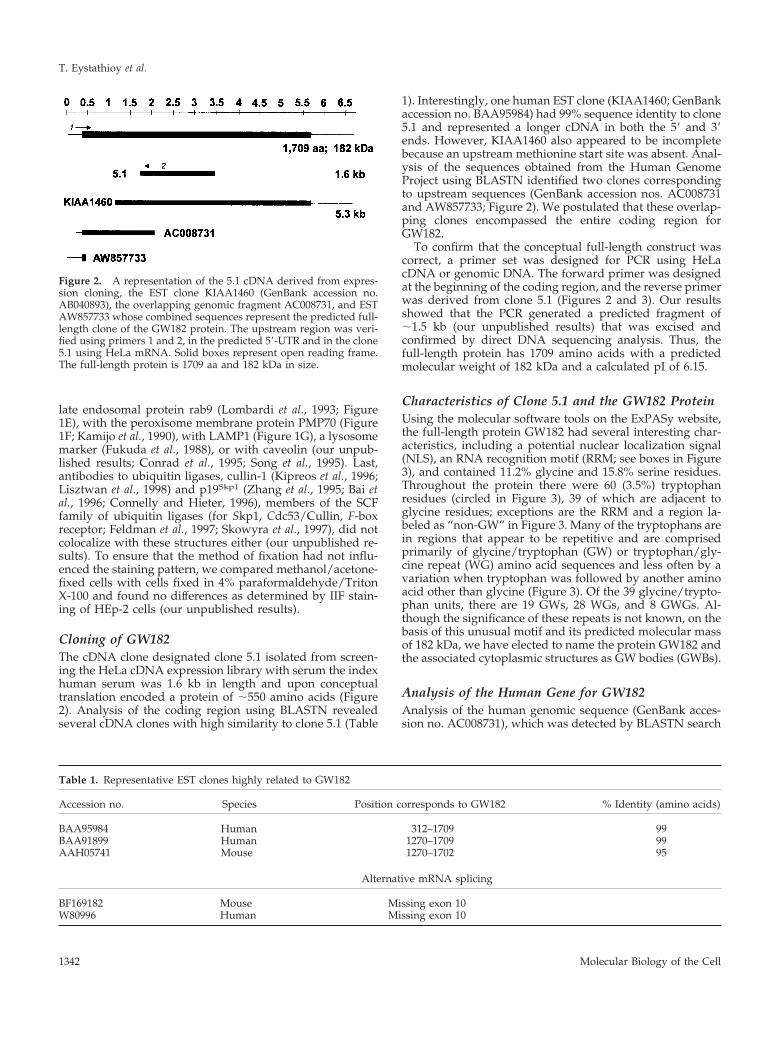
Cloning of GW182The cDNA clone designated clone 5.1 isolated from screen-ing the HeLa cDNA expression library with serum the indexhuman serum was 1.6 kb in length and upon conceptualtranslation encoded a protein of �550 amino acids (Figure2). Analysis of the coding region using BLASTN revealedseveral cDNA clones with high similarity to clone 5.1 (Table
1). Interestingly, one human EST clone (KIAA1460; GenBankaccession no. BAA95984) had 99% sequence identity to clone5.1 and represented a longer cDNA in both the 5� and 3�ends. However, KIAA1460 also appeared to be incompletebecause an upstream methionine start site was absent. Anal-ysis of the sequences obtained from the Human GenomeProject using BLASTN identified two clones correspondingto upstream sequences (GenBank accession nos. AC008731and AW857733; Figure 2). We postulated that these overlap-ping clones encompassed the entire coding region forGW182.
To confirm that the conceptual full-length construct wascorrect, a primer set was designed for PCR using HeLacDNA or genomic DNA. The forward primer was designedat the beginning of the coding region, and the reverse primerwas derived from clone 5.1 (Figures 2 and 3). Our resultsshowed that the PCR generated a predicted fragment of�1.5 kb (our unpublished results) that was excised andconfirmed by direct DNA sequencing analysis. Thus, thefull-length protein has 1709 amino acids with a predictedmolecular weight of 182 kDa and a calculated pI of 6.15.
Characteristics of Clone 5.1 and the GW182 ProteinUsing the molecular software tools on the ExPASy website,the full-length protein GW182 had several interesting char-acteristics, including a potential nuclear localization signal(NLS), an RNA recognition motif (RRM; see boxes in Figure3), and contained 11.2% glycine and 15.8% serine residues.Throughout the protein there were 60 (3.5%) tryptophanresidues (circled in Figure 3), 39 of which are adjacent toglycine residues; exceptions are the RRM and a region la-beled as “non-GW” in Figure 3. Many of the tryptophans arein regions that appear to be repetitive and are comprisedprimarily of glycine/tryptophan (GW) or tryptophan/gly-cine repeat (WG) amino acid sequences and less often by avariation when tryptophan was followed by another aminoacid other than glycine (Figure 3). Of the 39 glycine/trypto-phan units, there are 19 GWs, 28 WGs, and 8 GWGs. Al-though the significance of these repeats is not known, on thebasis of this unusual motif and its predicted molecular massof 182 kDa, we have elected to name the protein GW182 andthe associated cytoplasmic structures as GW bodies (GWBs).
Analysis of the Human Gene for GW182Analysis of the human genomic sequence (GenBank acces-sion no. AC008731), which was detected by BLASTN search
Figure 2. A representation of the 5.1 cDNA derived from expres-sion cloning, the EST clone KIAA1460 (GenBank accession no.AB040893), the overlapping genomic fragment AC008731, and ESTAW857733 whose combined sequences represent the predicted full-length clone of the GW182 protein. The upstream region was veri-fied using primers 1 and 2, in the predicted 5�-UTR and in the clone5.1 using HeLa mRNA. Solid boxes represent open reading frame.The full-length protein is 1709 aa and 182 kDa in size.
Table 1. Representative EST clones highly related to GW182
Accession no. Species Position corresponds to GW182 % Identity (amino acids)
BAA95984 Human 312–1709 99BAA91899 Human 1270–1709 99AAH05741 Mouse 1270–1702 95
Alternative mRNA splicing
BF169182 Mouse Missing exon 10W80996 Human Missing exon 10
T. Eystathioy et al.
Molecular Biology of the Cell1342

Figure 3. Human GW182 cDNA and deduced amino acid sequence. The sequence has been submitted to the GenBank under accession no.AY035864. Potential translation start sites (double underlined) and upstream in-frame stop codon (TGA, underlined) are indicated. TheGW182 protein consists of 1709 amino acids with a putative nuclear localization signal (NLS; aa 918–937) and RNA recognition motif (RRM;aa 1528–1600) outlined in boxes, respectively. The GW182 protein is rich in tryptophan residues (W, circled), which are often adjacent toglycine (G). Two regions with repeats of GW or WG in tandem are highlighted in bold and double underlined. The region lacking GW is alsobracketed. In addition the primers used to amplify the upstream region of the GW182 are shown. Exon–intron junctions and the exactpositions of the cDNA clones are indicated.
Cytoplasmic mRNA Binding Protein GW182
Vol. 13, April 2002 1343

using the cDNA sequence of GW182, revealed the completegene structure (Figure 4). The gene has 22 exons, is �46 kblong, and resides on chromosome 16p12. Analysis of humanand mouse EST clones from BLASTN search revealed atleast two clones that are missing exon 10 at the exact sitespredicted for the exon–intron junctions (Table 1). This sug-gests that alternative products derived from mRNA splicingof GW182 gene exist. The exon–intron junctions of the hu-man GW182 gene are summarized in Figure 4.
GW182 ProteinThe predicted molecular weight of GW182 was verified byIP using extract of [35S]methionine-labeled HeLa cells andthe index human serum (Figure 5A). The index humanserum immunoprecipitated two predominant proteins of�180 and 50 kDa. The 180-kDa protein is also specificallyprecipitated by other human autoimmune sera with a sim-ilar staining pattern on HEp-2 cells (our unpublished re-sults). Whether the 50-kDa protein represents a degradationproduct or alternative mRNA splicing–derived product ofGW182 is unclear and awaits further investigation.
GW182 Is PhosphorylatedThroughout the GW182 protein there were multiple pre-dicted serine-threonine and tyrosine phosphorylation sites.To determine whether the GW protein was phosphorylated,HeLa cells were labeled with [32P]orthophosphate, and thecell extracts were immunoprecipitated using the index hu-man serum (Figure 5B). A 180-kDa phosphorylated proteinwas immunoprecipitated with the index serum (lane 2), andnormal human serum did not immunoprecipitate any phos-phoproteins (lane 1). In contrast to the results obtained from
[35S]methionine-labeled cells (Figure 5A), no protein prod-uct of 50 kDa was observed.
GFP-tagged Expression of GW182To determine whether GW182 is able to localize to GWBs, agreen fluorescent protein (GFP) fusion construct pGFP-GWaa313–1709 was prepared and transfected into HEp-2cells as described in MATERIALS AND METHODS. TheGFP vector showed both nuclear and cytoplasmic staining intransfected cells alone (our unpublished results). In contrast,the GFP-tagged partial protein of GW182 shows distinctcytoplasmic domains (Figure 6A) that are recognized andstained with the index human serum, as shown in Figure 6D.It is noted that different GWBs incorporated varyingamounts of GFP-fusion protein relatively to endogenousGW182 detected by the index human serum (Figure 6).
Antibodies to Recombinant Protein Derived fromClone 5.1 Recognize GW BodiesRabbit antibodies to the recombinant protein encoded byclone 5.1 (rabbit anti-GW182p) were raised to verify that theGW182 resided in the cytoplasmic bodies observed with theindex human serum (see below). To ensure that the rabbitantibodies did indeed recognize the protein product en-coded by clone 5.1, in vitro transcription and translation(TNT) was used in IP with the index human serum and withrabbit antibodies to the recombinant protein (Figure 7A).The recombinant protein was immunoprecipitated by boththe index human serum (lane 3) and the rabbit antiserum(lane 5) but not by the rabbit preimmune serum (lane 4) or
Figure 4. The human GW182 gene is located on chromosome16p12 and has 22 exons.
Figure 5. Immunoprecipitation analysis detected a cellular phos-phoprotein of �180 kDa specifically recognized by the human indexserum. (A) Immunoprecipitation of an extract from [35S]methionine-labeled HeLa cells using normal human serum (lane 2) and thehuman index serum (lane 3) resolved by SDS-PAGE on a 12.5% gel.Lane 1 contains the 14C-labeled molecular weight standard. B. Im-munoprecipitation of an extract of [32P]phosphate-labeled HeLacells using normal human serum (lane 1) and the index humanserum (lanes 2) on 10% gel. The specific signals at �180 kDa areindicated by arrow. The nonspecific bands at 220 and 45 kDadetected in both lanes 1 and 2 of A are most likely nonmusclemyosin and actin, respectively.
T. Eystathioy et al.
Molecular Biology of the Cell1344

the NHS (lane 6). The same IP specificity was observed withthe in vitro–derived full-length GW182 protein, which mi-grated at �180 kDa (Figure 7B).
The most striking feature of IIF using the index humanserum was cytoplasmic bodies numbering from 0 in mitoticcells to 30 in some interphase cells (Figure 8B). In addition,finer cytoplasmic speckling was observed as well. IIF studieswith the index human serum did not show any nuclearstaining of HEp-2 cells. On average, �20–30 of the cytoplas-mic bodies are clearly seen as larger bodies but highermagnification revealed polymorphic GWBs varying in sizefrom �0.2 to 1 �m. The cytoplasmic staining observed withthe immune rabbit serum was similar to that observed withthe human serum, and in colocalization studies, the rabbitand the index human serum reacted with the same cytoplas-mic domains (Figure 8).
The observation that GWBs are distinct from endosomes,lysosomes, mitochondria, and vesicular elements of theGolgi complex is supported by immunogold electron mi-croscopy (IEM; Figure 9). The structures postimmunola-beled with protein A-gold and anti-GW182 are granular,electron dense bodies that are devoid of membranous struc-
tures (Figure 9, A–C). This is distinct from multivesicularbodies (Figure 9A), mitochondria (Figure 9, A and D), andthe Golgi complex (Figure 9D). No immunogold stainingwas seen with normal or preimmune serum or with immu-nogold in the absence of primary antibodies (our unpub-lished results). By comparison, antibodies to giantin boundto the Golgi membrane stack and vesicles in the lateralaspect of the membrane stack (Figure 9D). In addition, inagreement with size estimated by confocal microscopy, theimmunogold-labeled GWBs vary in size from 100 to 200 nm(Figure 9, A–C).
GW182 Binds to mRNABecause GW182 contained an RRM, we questioned whetherthis protein could bind small RNA species that are targets ofother autoimmune diseases. The prototype serum that im-munoprecipitated GW182 did not immunoprecipitate tRNA,snRNA, or hYRNA (Figure 10). By contrast, human anti–Jo-1(antihistidyl tRNA synthetase) precipitated tRNA, humananti-Sm serum precipitated U1, U2, U4, U5, and U6, humananti-U1 serum precipitated U1 RNA, human anti–SS-A/Bprecipitated hYRNA, and human anti–SS-A/Ro antibodiesprecipitated hY2/4 RNA (Figure 10).
Although anti-GW182 did not immunoprecipitate tRNA,snRNA, or hYRNA, HeLa cell mRNA was recovered in IPpellets and was used to probe a 1200-probe set humancDNA array on which many candidate mRNA targets wereidentified (Figure 11). Table 2 identifies the genes thatshowed high levels (fold enrichment � 2) of reactivity withthe mRNA immunoprecipitated by the prototype humananti-GW182. They included glycoprotein hormone alphasubunit precursor, 60S ribosomal protein L6 (RPL6), metal-loproteinase inhibitor 1 precursor (TIMP1), activated RNApolymerase II transcriptional coactivator p15, cAMP-depen-dent transcription factor ATF-4, serum- and glucocorticoid-regulated serine/threonine protein kinase (SGK), PTP-CAAX1 nuclear tyrosine phosphatase, and neuroleukin(NLK). Sequence analysis of these genes and their relatedmRNAs did not disclose any obvious common sequencemotif in the translated or untranslated regions that mightaccount for binding to GW182. The specificity of these reac-tions was confirmed and validated as illustrated before us-ing normal human serum and sera from patients with sys-temic lupus erythematosus and paraneoplastic syndromesthat react with proteins associated with known mRNAs(Tenenbaum et al., 2000; Brown et al., 2001).
Northern Blot AnalysisNorthern blot analysis using a human multiple tissue blotprobed with the 1.4 kb labeled fragment derived from cloneKIAA1460, showed highest expression in heart (Figure 12).In addition, expression of the GW182 gene was observed inall tissues suggesting that this protein is ubiquitously ex-pressed. The actin control shows that mRNA loading is quitecomparable among the lanes. IIF studies of a number oftissues and cell lines (HeLa, 3T3, MOLT-4, human chondro-cyte and osteoblast, chicken fibroblast) suggest that GWBsare variably but widely expressed in species as well astissues and cell types (our unpublished results).
Figure 6. Transfection of HEp-2 cells with a construct of GFP fusedto a GW182 cDNA fragment. The transfected GFP-tagged proteinproduced a staining pattern in transfected cells (A) highly similar tothe GWBs recognized by the index human serum (B). When theimages in panels A and B were merged with the DAPI stained nucleiof C, colocalization of the GFP product was observed in the cyto-plasmic GWBs (D). Three populations of GWBs can be distin-guished by the difference in green to red signal. Some GWBs haveapparently equal intensity of green and red signals (arrow), whereasothers have more green (arrowhead) or more red signal (doublearrowhead). Bar, 10 �m. Original magnification, �600.
Cytoplasmic mRNA Binding Protein GW182
Vol. 13, April 2002 1345

DISCUSSION
We have identified a novel protein that we have namedGW182 because of its multiple glycine-tryptophan repeatsfound throughout the protein and the predicted and ob-served molecular mass of 182 kDa. By IIF, antibodies tonative and recombinant GW182 appear to identify a cyto-plasmic domain that we have tentatively named GWBs. Thecontention that GW182 resides in a distinct cytoplasmicdomain is supported by the observation that antibodies toGW182 did not colocalize with markers of the Golgi com-plex, endosomes, lysosomes, or peroxisomes. Although
there may appear to be a few overlaps with of some of thesecytoplasmic markers, the overall IIF patterns observed withthese markers and the IIF staining by the prototype humanserum or rabbit anti-GW182p are clearly very different. An-other possibility is that the GWBs might represent aggre-somes or sites of protein degradation. However, our exper-iments using proteosome inhibitors or a target of the SCFubiquitin ligase–mediated pathway for protein degradationsuggests that GWBs are not aggresomes (Garcia-Mata et al.,1999; Johnston et al., 2000). Last, the observation that GWBsare not colocalized to sites marked by anticlathrin suggests
Figure 7. Analysis of in vitro–translated GW182 pro-teins specifically recognized by the index serum andrabbit antibodies raised to recombinant protein. (A) Im-munoprecipitation of translation products derived fromclone 5.1. Lane 1 shows the 14C-labeled molecular weightstandard. Lane 2 shows the proteins produced from invitro transcription and translation with the largestpolypeptide migrating at �66 kDa. Immunoprecipita-tion of the in vitro–translated protein product was per-formed using the index human serum (lane 3), rabbitpreimmune serum (lane 4), postimmune rabbit serum(lane 5), and normal human serum (lane 6). (B) Immu-noprecipitation of translation product derived from thefull-length construct (lane 1) with the major protein mi-grating at �180 kDa. The latter was immunoprecipitatedby the index human serum (lane 2) and rabbit antiserum(lane 5) but not by a normal human serum control (lane2) and the rabbit preimmune serum (lane 4).
Figure 8. Immunofluorescenceanalysis of HeLa cell GWBs showscostaining of rabbit anti-GW182 an-tibody and the index human se-rum. (A) HeLa cell staining withrabbit anti-GW182 antibody at1/100 dilution. (B) Staining withthe index human serum at 1/600dilution. (C) Nuclei counterstainedwith DAPI. (D) Merged image withGWBs (arrowheads) recognized byboth antibodies. Note that the rab-bit anti-GW182 serum also showsunrelated staining of centrioles (ar-rows) that is also evident in preim-mune rabbit serum (our unpub-lished results). Bar, 10 �m. Originalmagnification, �600.
T. Eystathioy et al.
Molecular Biology of the Cell1346

that these bodies are probably not involved in clathrin-dependent vesicle transport (Hirst and Robinson, 1998).These conclusions are supported by IEM data showing thatanti-GW182 identifies a granular, electron-dense body in thecytoplasm of HeLa cells that is the same size as that identi-fied by IIF and colocalization studies.
The data showing that the GW182 protein binds to severaldifferent mRNA species is quite intriguing. The lack of co-localization of GWBs with various known cytoplasmic or-ganelles may not be surprising considering the interaction ofGW182 with mRNA. Perhaps the GW182 protein is involvedin stabilizing and/or regulating translation and/or storingmRNAs. Several mRNAs as members of mRNP complexeshave been identified using antibodies to RNA proteins suchas ELAV/Hu, elf-4E, and poly(A) binding proteins (Tenen-baum et al., 2000; Keene, 2001). ELAV/Hu proteins are in-volved in stabilizing and/or regulating translation of earlyresponse gene transcripts expressed primarily in neurons(Keene, 1999; Brennan and Steitz, 2001). Analysis of thetranscripts revealed some limited sequence similarity, sug-gesting that a common theme exists among these transcriptsthat allows recognition by ELAV/Hu proteins. Interestingly,ELAV/Hu proteins along with a fraction of polyadenylatedmRNA have been observed in discrete clusters within thecytoplasm of medulloblastoma cells (Antic and Keene, 1998).It is possible that transcripts may be clustered in vivo withsimilar fates and/or functions. For example, upon retinoicacid treatment of P19 cells to induce neuronal differentia-tion, the population of mRNAs identified in the ELAV/Huprotein complex changed; additional mRNAs with AU-richelements known to be upregulated in neurons were found(Tenenbaum et al., 2000).
At the present time, we have not identified common func-tional or structural features among the mRNAs that areimmunoprecipitated by the anti-GW182 antibodies. Some ofthe proteins are known to be key components of the cellcycle, whereas others have no immediately apparent rela-
Figure 9. Immunogold electron microscopy (IEM) localization ofGW182 in HeLa cells. Frozen sections of fixed and gelatin-embed-ded HeLa cells were incubated with the index human serum orrabbit antigiantin diluted 1/400 and then postimmunolabeled withprotein A-gold. Examples of the structures labeled by anti-GW182are shown in A–C and by antigiantin in D. Antibodies to GW182labeled an electron dense cytoplasmic body that is not bound by amembrane and is distinct from mitochondria (m) and multivesicularbodies (mvb). (B) High-power view of the electron dense body in A.(C) A high-power view of another representative immunogold la-beled cytoplasmic electron dense body. Antibodies to giantin labelthe lateral aspects of the Golgi membrane stack (g). Scale bars areshown in the lower right.
Figure 10. Immunoprecipitation of RNA from 32P-labeled HeLacell extracts. IP reactions were performed using 100 �l of the cellextract. Normal human serum (lane 1) and the prototype humanserum (lane 2) did not precipitate small RNAs. However, prototypeanti-tRNA synthetase (lane 3), anti-Sm serum (lane 4), anti-U1RNPserum (lane 5), anti–SS-A/Ro and anti–SS-B/La serum (lane 6), andanti–SS-A/Ro serum (lane 7) all precipitated expected RNA mole-cules.
Cytoplasmic mRNA Binding Protein GW182
Vol. 13, April 2002 1347

tionship with one other. New approaches to elucidateunique structural features of mRNA are being developedand may shed light on the role of GW182 and related pro-teins. We are pursuing in situ hybridization using the mR-NAs identified in this study and appropriate controls todetermine if these mRNAs are located in GWBs.
In general the various functions of the cell are highlycoordinated and regulated, and among these functions, gene
transcription and translation are also highly regulated. Themovement of mRNA transcripts into the cytoplasm andsubsequent coordinated translation after appropriate endog-enous or exogenous signals would necessarily involvehighly sophisticated levels of control (Keene, 1999; Tenen-baum et al., 2000; Keene, 2001). For example, some mRNAsmay be required for rapid protein production and may bestored or protected from degradation. Because the cytoplas-
Figure 11. Identification of mRNAs associated with GW182-mRNP complexes using cDNA microarray analysis. As described in MATE-RIALS AND METHODS, RNA was extracted from immunoprecipitated GW182-mRNPs or total cell lysate and used to make reverse-transcribed radiolabeled probes for Atlas Human 1.2 Arrays containing 1188 singly spotted cDNA segments (CLONTECH). Phosphorimageswere scanned and stored as gel files and then analyzed using ATLASIMAGE 1.01 software (CLONTECH). A default external backgroundsetting was used in conjunction with a background-based signal threshold to determine gene signal significance. Comparison of cDNA arrayimages was performed using an average of all of the gene signals on the array (global normalization) to normalize the signal intensitybetween arrays. Messenger RNAs associated with GW182–mRNP complexes were considered significant if they were twofold or greaterenriched over total cellular RNA. (A) Total cellular RNA; (B) mRNAs associated with GW182–mRNP complexes; (C) computer-generatedoverlay comparison of representative arrays A and B. Red bars represent species of mRNAs that were enriched twofold or greater ascompared with total cellular RNA (genes listed in Table 2). Green bars indicate mRNAs that were present on one or both of the arrays butwere not enriched at least twofold.
Table 2. mRNA targets enriched in the GW182 mRNP
Foldenrichment Gene name GenBank
17.50 Glycoprotein hormone alpha subunit precursor V0051816.00 60S ribosomal protein L6 (RPL6); TAX-responsive enhancer element binding
protein 107 (TAXREB107); neoplasm-related protein C140X69391
12.00 Metalloproteinase inhibitor 1 precursor (TIMP1); erythroid potentiatingactivity (EPA); fibroblast collagenase inhibitor
X03124
8.00 Activated RNA polymerase II transcriptional coactivator p15; PC4 U129798.00 cAMP-dependent transcription factor ATF-4; DNA-binding protein
TAXREB67; cAMP-response element binding protein (CREB2)D90209
5.00 Serum- and glucocorticoid-regulated serine/threonine protein kinase (SGK) AJ0005125.00 PTPCAAX1 nuclear tyrosine phosphatase (PRL-1) U482965.00 Neuroleukin (NLK); glucose-6-phosphate isomerase (GPI); phosphoglucose
isomerase (PGI); phosphohexose isomerase (PHI)K03515
4.00 Proliferating cyclic nuclear antigen (PCNA); cyclin M157963.00 Prothymosin alpha (ProT-alpha; PTMA) M267083.00 40S ribosomal protein S19 (RPS19) M817573.00 Ubiquitin-conjugating enzyme E2 H10; ubiquitin-protein ligase; ubiquitin
carrier proteinU73379
3.00 Serine/threonine-protein kinase PCTAIRE 1 (PCTK1) X663633.00 ALG-2 calcium-binding protein AF0356063.00 Mitochondrial matrix protein P1 precursor; p60 lymphocyte protein;
chaperonin homolog; HUCHA60; heat shock protein 60 (HSP-60); HSPD1M34664
2.75 Alpha1 catenin (CTNNA1); cadherin-associated protein; alpha E-catenin D138662.50 Transcription factor AP-2 (TFAP2; AP2TF) M367112.33 Acyl-CoA-binding protein (ACBP); diazepam binding inhibitor (DBI);
endozepine (EP)M14200
T. Eystathioy et al.
Molecular Biology of the Cell1348

mic domains observed by IIF using the index human seraand rabbit anti-GW182 antibody failed to colocalize withmarkers of several cytoplasmic organelles and because ofour observation that specific mRNAs are bound by GW182,it seems reasonable to conclude that GW182 is involved inmRNA expression and that this occurs in a defined cytoplas-mic domain that we identify as GWBs. It might also be thatGW182 is involved in degradation of mRNA but GWBs donot colocalize with the SCF ubiquitin ligase-mediated path-way for protein degradation.
Along these lines of discussion, the GWBs described inthis study may be related to mRNA-associated particlesdescribed in other systems, particularly neuronal cells(Triedge et al., 1991; Miyashiro et al., 1994; Knowles et al.,1996; Martone et al., 1996; Gazzaley et al., 1997; Racca et al.,1997; Bassell et al., 1998; Steward et al., 1998). In oligoden-drocytes, injected fluorescent-labeled myelin basic proteinmRNA localized to granules that had a radius of 0.6–0.8�m, contained elongation factors, rRNA and other mRNAs.It was concluded that these granules may represent su-pramolecular complexes of a translational unit (Barbarese etal., 1995; Ainger et al., 1997). These observations are consis-tent with the size of HeLa and HEp-2 cell granules observedin our study and suggest that GW182 marks a subset of thesecytoplasmic bodies that contain a distinct subset of mRNAs.RNA granules containing mRNAs have also been describedin fibroblasts (Ross et al., 1997) and mast cells (Dvorak andMorgan, 2000; Dvorak and Morgan, 2001). In other organ-isms, similar observations have been made for the 3� UTR ofASH1 mRNA in budding yeast (Hazelrigg, 1998) and the bcdand PROSPERO mRNA in Drosophila oocytes (Wang andHazelrigg, 1994; Oleynikov and Singer, 1998; Bassell andOleynikov, 1999). In studies of fibroblasts, the �-actinmRNA is localized at the leading edge of the cell, and the3�UTR binds a protein called zipcode-binding protein 1(ZBP-1; Ross et al., 1997). GW182 does not have significantsequence similarity to ZBP-1, but they both have a RRM andNLS in common. Although the putative NLS motif suggeststhat ZBP-1 and GW182 proteins may be able to enter thenucleus, both proteins are predominantly found in the cy-toplasm. In the case of GW182, the evidence showed thatcytoplasmic staining was observed using both the indexhuman serum and the rabbit antibodies to the GW182 re-combinant protein. In addition, the partial protein of GW182tagged with GFP was localized to the cytoplasm, specifically
to GWBs, and no expression was detected in the nucleus.Last, no differences were observed in the IIF staining patternwith formaldehyde or other fixation was used (our unpub-lished data compared methanol/acetone fixation with para-formaldehyde/Triton X-100 fixation). Interestingly, HuB,HuC, and HuD isoforms are mainly cytoplasmic in neuronswith a small amount of protein observed with the nucleus aswell (Gao and Keene, 1996). Therefore, we believe that thepotential NLS motif may either be nonfunctional or isblocked in cells grown under our conditions and requires astimulus.
It is likely that GW182 protein is one member of a familyof proteins residing in GWBs. As observed with many or-ganelles and vesicles in the cell, it is simplistic to think thatone protein comprises these complex structures. It is possi-ble that more than one alternatively spliced protein productfrom the GW gene may reside in GWBs. First, the existenceof two EST clones missing exon 10 (accession nos. BF169182and W80996, from mouse and human, respectively) sup-ports this possibility (Table 1). Second, IP of extracts fromradiolabeled HeLa cells by the index human serum revealsthe presence of two proteins of 180 and 50 kDa. This couldeither be due to cross-reactivity or the association of thesetwo proteins in a complex. We are currently pursuing iso-lating other proteins that are associated with GWBs.
Considering the sequence characteristics of GW182, it isnot surprising that we were able to show that GW182 islikely a phosphoprotein. GWBs are heterogeneous in sizeand vary in number in individual cells. The variation in sizeand number may be related to the physiological state of thecell and the stage of the cell cycle. Detailed studies arecurrently underway to confirm this preliminary observation.
The significance of the GW repeats in GW182 is unclear.Although ESTs that contain this motif are in the database, todate no other mammalian proteins with this motif have beenreported. A clue to the function of GW repeats may comefrom studies of bacteria such as Listeria monocytogenes, Staph-ylococcus caprae, and Erysipelothrix rhusiopathiae, where it hasbeen suggested that proteins bearing these repeats play animportant role in anchoring bacterial proteins to the surfaceof the cell (Makino et al., 1998) and anchoring bacteria totarget cells (Braun et al., 1997; Milohanic et al., 2001). Themode of binding is not clearly understood, but is thought tooccur via interaction with lipoteichoic acid or with specificcell surface proteins. For example, the protein internalin B(InIB) produced by L. monocytogenes, is a key protein inpromoting adherence and entry of the bacteria to host cellsduring infection. InIB has a C-terminal cell wall–anchoringdomain containing 80-amino-acid GW repeats (Braun et al.,1997). Deletion of this domain impaired adherence to hostcells, whereas addition of GW repeats improved the bindingto the cell surface. Similarly, the autolysin Ami in L. mono-cytogenes contains a N-terminal catalytic domain and a C-terminal domain that is homologous to the GW domain inInIB but contains 8 GW modules arranged in tandem (Braunet al., 1997; Milohanic et al., 2001). Similar six to eight tandemrepeat motifs have been described in the S. caprae atlC geneproduct (Alligent et al., 2001) and the surface protectiveantigen (SpaA) of E. rhusiopathiae (Makino et al., 1998). Ofinterest, the 6 GW repeats in S. caprae occur in the fibronec-tin-binding domain of the atlC protein. This may have rele-vance to the GW182 protein in mammalian cells that might
Figure 12. Northern blot analysis of GW182 mRNA. The order ofpolyA� RNA from human tissues was as follows (lanes 1–8): heart,whole brain, placenta, lung, liver, skeletal muscle, kidney, andpancreas. The membrane was probed with a ClaI/EcoRI fragmentfrom the clone KIAA1460 (top panel) and reprobed using actin�-actin cDNA (bottom panel).
Cytoplasmic mRNA Binding Protein GW182
Vol. 13, April 2002 1349

bind to cytoskeletal elements to promote stabilization ormovement of certain RNA species to their physiologicaltarget. The role of GW182 in binding or adherence of RNAor other proteins to cytoskeletal components or membranemoieties requires further study.
Summary
We have identified a novel protein GW182 that containsmultiple glycine/tryptophan repeats and a classical RNA-binding domain at the C terminus. GW182 appears to reactwith a subset of mRNAs and localizes to cytoplasmic do-mains that do not belong to any of the known conventionalorganelles in the cytoplasm. Hence, we tentatively identifiedthe cytoplasmic domain marked by antibodies directedagainst GW182 as GW bodies or GWBs.
ACKNOWLEDGMENTS
The authors acknowledge the assistance of Joan Miller, Carol L.Peebles, and Dr. Doug Zochodne (University of Calgary) for arrang-ing the collection of the serum samples. Malcolm R. Wood providedvaluable assistance with immunogold electron microscopy in TheScripps Research Institute Core Microscopy Unit. This work wassupported by in part by the Canadian Institutes for Health Researchgrant MOP-38034 and the National Institutes of Health (NIH) grantsCA56956 and AI39645, and CA79907 and AI46451. This work wasalso supported in part by the Sam and Rose Stein Charitable Trustand NIH grant M01RR00833 to the General Clinical Research Centerof the Scripps Research Institute. M.J.F. hold the Arthritis SocietyChair at the University of Calgary. This is publication 14253-MEMfrom the Scripps Research Institute.
REFERENCES
Ainger, K., Avossa, D., Diana, A.S., Barry, C., Barbarese, E., andCarson, J.H. (1997). Transport and localization elements in myelinbasic protein mRNA. J. Cell Biol. 138, 1077–1087.
Alligent, J., Aubert, S., Dyke, K.G.H., and Solh, N.E. (2001). Staphylo-coocus caprae strains carry determinants known to be involved inpathogneicity: a gene encoding an autolysin-binding fibronectin andthe ica operon involved in biofilm formation. Infect. Immun. 69, 712–718.
Altschul, S.F., Gish, W., Miller, W., Myers, E.W., and Lipman, D.J.(1990). Basic local alignment search tool. J. Mol. Biol. 215, 403–410.
Antic, D., and Keene, J.D. (1998). Messenger ribonucleoprotein com-plexes containing human ELAV proteins: interactions with cytoskele-ton and translational apparatus. J. Cell Sci. 111, 183–197.
Bai, C., Sen, P., Hofmann, K., Ma, L., Goebl, M., Harper, J.W., andElledge, S.J. (1996). SKP1 connects cell cycle regulators to the ubiquitinproteolysis machinery through a novel motif, the F-box. Cell 86, 263–274.
Barbarese, E., Koppel, D.E., Deutscher, M.P., Smith, C.L., Ainger, K.,Morgan, F., and Carson, J.H. (1995). Protein translation components arecolocalized in granules in oligodendrocytes. J. Cell Sci. 108, 2781–2790.
Bassell, G.J., Zhang, H.L., Byrd, A.L., Femino, A.M., Singer, R.H.,Taneja, K.L., Liftshitz, L.M., Herman, I.M., and Kosik, K.S. (1998).Sorting of beta actin mRNA and protein to neurites and growth conesin culture. J. Neurosci. 18, 251–265.
Bassell, G., and Oleynikov, Y. (1999). The travels of mRNAs through allcells large and small. FASEB J. 13, 447–454.
Bestagno, M., Cerino, A., Riva, S., and Ricotti, G.C.B.A. (1987). Im-provements of Western blotting to detect monoclonal antibodies. Bio-chem. Biophys. Res. Comm. 146, 1509–1514.
Braun, L., Dramsi, S., Dehoux, P., Bierne, H., Lindahl, G., and Cossart,P. (1997). InIB: an invasion protein of Listeria monocytogenes with anovel type of surface association. Mol. Microbiol. 25, 285–294.
Brennan, C.M., and Steitz, J.A. (2001). HuR and mRNA stability. CellMol. Life Sci. 58, 266–277.
Brown, V., Jin, P., Ceman, S., Darnell, J.D., O’Donnell, W.T., Tenen-baum, S.T., Jin, X., Feng, Y., Wilkinson, K.D., Keene, J.D., Darnell, R.B.,Warren, S.T. (2001). Microarray identification of Fragile X-Mental Re-tardation protein (FMRP)-associated brain mRNAs and altered mRNAtranslational profiles in fragile X syndrome. Cell 107, 477–487.
Chan, E.K.L., Francoeur, A.-M., and Tan, E.M. (1986). Epitopes, struc-tural domains, and asymmetry of amino acid residues in SS-B/Lanuclear protein. J. Immunol. 136, 3744–3749.
Chan, E.K.L., and Fritzler, M.J. (1998). Golgins: coiled-coil-rich proteinsassociated with the Golgi complex. Electronic J. Biotechnol. 1, http://ejb.ucv.cl/content/vol1/issue2/full/1.
Chan, E.K.L., and Tan, E.M. (1987). Human autoantibody-reactiveepitopes of SS-B/La are highly conserved in comparison with epitopesrecognized by murine monoclonal antibodies. J. Exp. Med. 166, 1627–1640.
Connelly, C., and Hieter, P. (1996). Budding yeast SKP1 encodes anevolutionarily conserved kinetochore protein required for cell cycleprogression. Cell 86, 275–285.
Conrad, P.A., Smart, E.J., Ying, Y.S., Anderson, R.G.W., and Bloom,G.S. (1995). Caveolin cycles between plasma membrane caveolae andthe Golgi complex by microtubule-dependent and microtubule-inde-pendent steps. J. Cell Biol. 131, 1421–1433.
Dvorak, A.M., and Morgan, E.S. (2000). Ultrastructural immunogoldcytochemistry with autoimmune human sera and an antibody to uri-dine implicate human mast cell granules in RNA biology. Histochem.J. 32, 685–696.
Dvorak, A.M., and Morgan, E.S. (2001). Ribosomes and secretory gran-ules in human mast cells: close associations demonstrated by stainingwith a chelating agent. Immunol. Rev. 179, 94–101.
Feldman, R.M., Correll, C.C., Kaplan, K. .B., and Deshaies, R.J. (1997).A complex of Cdc4p, Skp1p, and Cdc53p/cullin catalyzes ubiquitina-tion of the phosphoylated CDK inhibitor Sic1p. Cell 91, 221–230.
Fritzler, M.J., Hamel, J.C., Ochs, R.L., and Chan, E.K.L. (1993). Molec-ular characterization of two human autoantigens: unique cDNAs en-coding 95- and 160-kD proteins of a putative family in the Golgicomplex. J. Exp. Med. 178, 49–62.
Fritzler, M.J., Lung, C.-C., Hamel, J.C., Griffith, K., and Chan, E.K.L.(1995). Molecular characterization of golgin-245: a novel Golgi complexprotein containing a granin signature. J. Biol. Chem. 270, 31262–31268.
Fukuda, M., Vitalla, J., Matteson, J., and Carlsson, S.R. (1988). Cloningof cDNAs encoding human lysosomal membrane glycoproteins, h-lamp-1 and h-lamp-2. Comparison of their deduced amino acid se-quences. J. Biol. Chem. 263, 18920–18928.
Gao, F.B., and Keene, J.D. (1996). Hel-N1/Hel-N2 proteins are boundto poly(A)� mRNA in granular RNP structures and are implicated inneuronal differentiation. J. Cell Sci. 109, 579–589.
Garcia-Mata, R., Bebok, Z., Sorscher, E.J., and Sztul, E.S. (1999). Char-acterization and dynamics of aggresome formation by a cytososlicGFP-chimera. J. Cell Biol. 146, 1239–1254.
Gazzaley, A., Benson, D., Huntley, G., and Morrison, J. (1997). Differ-ential subcellular regulation of NMDAR1 protein and mRNA in den-drites of dentate. Neuron 14, 433–445.
T. Eystathioy et al.
Molecular Biology of the Cell1350
Griffith, K., Chan, E.K.L., Hamel, J.C., Miyach, K., and Fritz, M.J.(1997). Molecular characterization of a novel 97 kDa Golgi complexautoantigen recognized by autoimmune antibodies from patientswith Sjogren’s syndrome. Arthritis Rheum. 40, 1693–1702.
Hazelrigg, T. (1998). The destinies and destinations of RNAs. Cell 95,451–460.
Hirst, J., and Robinson, M.S. (1998). Clathrin and adaptors. Biochim.Biophys. Acta 1404, 173–193.
Johnston, J.A., Ward, C.L., and Kopito, R.R. (2000). Aggresomes. Acellular response to misfolded proteins. J. Cell Biol. 143, 1883–1898.
Kamijo, K., Taketani, S., Yokota, S., Osumi, T., and Hashimoto, T.(1990). The 70-kDa peroxisomal membrane protein is a member of theMdr (P-glycoprotein)-related ATP-binding protein superfamily. J. Biol.Chem. 265, 4534–4540.
Keene, J.D. (1999). Why is Hu where? Shuttling of early-response-genemessenger RNA subsets. Proc. Natl. Acad. Sci. USA 96, 14085–14090.
Keene, J.D. (2001). Ribonucleoprotein infrastructure regulating the flowof genetic information between the genome and proteome. Proc. Natl.Acad. Sci. USA 98, 7018–7024.
Kipreos, E.T., Lander, L.E., Wing, J.P., He, W.W., and Hedgecock, E.M.(1996). cul-1 is required for cell cycle exit in C. elegans and identifies anovel gene family. Cell 85, 829–839.
Knowles, R.B., Sabry, J.H., Martone, M.E., Deerinck, T.J., Ellisman,M.H., Basell, G.J., and Kosik, K.S. (1996). Translocation of RNA gran-ules in living neurons. J. Neurosci. 16, 7812–7820.
Kopito, R.R. (2000). Aggresomes, inclusion bodies and protein aggre-gation. Trends Cell Biol. 10, 524–530.
Laemmli, U.K. (1970). Cleavage of structural proteins during the as-sembly of the head of bacteriophage T4. Nature 227, 680.
Lahtinen, U., Hellman, U., Wernstedt, C., Saraste, J., and Pettersson,R.F. (1996). Molecular cloning and expression of a 58-kDa cis-Golgi andintermediate compartment protein. J. Biol. Chem. 271, 4031–4037.
Lawe, D.C., Patki, V., Heller-Harrison, R., Lambright, D., Corvera, S.(2000). The FYVE domain of early endosome antigen 1 is required forboth phosphatidylinositol 3-phosphate and Rab5 binding—critical roleof this dual interaction for endosomal localization. J. Biol. Chem. 275,3699–3705.
Lerner, M.R., and Steitz, J.A. (1981). Snurps and scyrps. Cell 25, 298–300.
Lindstedt, A.D., and Hauri, H.P. (1993). Giantin, a novel conservedGolgi membrane protein containing a cytoplasmic domain of at least350 kDa. Mol. Biol. Cell 4, 679–693.
Lisztwan, J., Marti, A., Sutterluty, H., Gstaiger, M., Wirbelauer, C.,Krek, W. (1998). Association of human CUL-1 and ubiquitin-conjugat-ing enzyme CDC34 with the F-box protein p45(SKP2): evidence forevolutionarily conservation in the subunit composition of the CDC34-SCF pathway. EMBO J. 17, 368–383.
Lombardi, D., Soldati, T., Riederer, M.A., Goda, Y., Zerial, M., andPfeffer, S.R. (1993). Rab9 functions in transport between late endosomesand the trans Golgi network. EMBO J. 12, 677–682.
Luzio, J.P., Brake, B., Banting, G., Howell, K.E., Braghetta, P., andStanley, K.K. (1990). Identification, sequencing and expression of anintegral membrane protein of the trans Golgi network (TGN38). Bio-chem. J. 270, 97–102.
Makino, S., Yamamoto, K., Murakami, S., Shirahata, T., Uemura, K.,Sawada, T., Wakamoto, H., and Morita, T. (1998). Properties of repeatdomain found in a novel protective antigen, SaA, of Erysipelothrixrhusiopathiae. Microbiol. Pathogen. 25, 101–109.
Martone, M.E., Pollock, J.A., Jones, Y.Z., and Ellisman, M.H. (1996).Ultrastructural localization of dendrite messenger RNA in adult rathippocampus. J. Neurosci. 16, 7437–7446.
Milohanic, E., Jonquieres, R., Cossart, P., Berche, P., and Gaillard, J-L.(2001). The autolysin Ami contributes to the adhesion of Listeria mono-cytogenes to eukaryotic cells via its cell wall anchor. Mol. Microbiol. 39,1212–1224.
Miyashiro, K., Dichter, M., and Eberwine, J. (1994). On the nature anddifferential distribution of mRNAs in hippocampal neurites: implica-tions for neuronal functioning. Proc. Natl. Acad. Sci. USA 91, 10800–10804.
Mu, F.T., Callaghan, J.M., Steele-Mortimer, H.S., Parton, R.G., Camp-bell, P.L., McCluskey, J., Yeo, J.P., Tock, E.P.C., and Toh, B.H. (1995).EEA1, an early endosomal protein. J. Biol. Chem. 270, 13503–13511.
Musunuru, K., and Darnell, R.B. (2001). Paraneoplastic neurologicdisease antigens: RNA-binding proteins and signaling proteins in neu-ronal degeneration. Annu. Rev. Neurosci. 24, 239–262.
Oleynikov, Y., and Singer, R.H. (1998). RNA localization: differentzipcodes, same postman? Trends Cell Biol. 8, 381–383.
Orci, L., Tagaya, M., Amherdt, A., Perrelett, A., Donaldson, J.G., Lip-pincott-Schwartz, J., Klausner, R.D., and Rothman, J.E. (1991). BrefeldinA, a drug that blocks secretion, prevents the assembly of non-clathrin-coated buds on Golgi cisternae. Cell 64, 1183–1195.
Racca, C., Gardiol, A., and Triller, A. (1997). Dendritic and postsynapticlocalization of glycine receptor RNA. J. Neurosci. 17, 1691–1700.
Reimer, G., Raska, I., Tan, E.M., and Scheer, U. (1987). Human auto-antibodies: probes for nucleolus structure and function. Virch. Arch. BCell Pathol. 54, 131–143.
Ross, A.F., Oleynikov, Y., Kislauskis, E.H., Taneja, K.L., and Singer,R.L. (1997). Characterization of a �-actin mRNA zipcode-binding pro-tein. Mol. Cell Biol. 17, 2158–2165.
Selak, S., Scheonroth, L., Senecal, J-L., and Fritzler, M.J. (1999). Earlyendosome antigen 1: an autoantigen associated with neurological dis-eases. J. Invest. Med. 47, 311–318.
Skowyra, D., Craig, K.L., Tyers, M., Elledge, S.J., and Harper, J.W.(1997). F-box proteins are receptors that recruit phosphorylated sub-strates to the SCF ubiquitin-ligase complex. Cell 91, 209–219.
Song, K.S., Tang, Z., Li, S., and Lisanti, M.P. (1995). Mutational analysisof the properties of caveolin-1. A novel role for the C-terminal domainin mediating homo-typic caveolin-caveolin. J. Biol. Chem. 272, 4398–4403.
Steward, O., Wallaca, C.S., Lyford, G.L., and Worley, P.F. (1998). Syn-aptic activation causes the mRNA for Arc to localize selectively nearactivated postsynaptic sites on dendrites. Neuron 21, 1–20.
Tan, E.M. (1991). Autoantibodies in pathology and cell biology. Cell 67,841–842.
Tenenbaum, S.A., Carson, C.C., Lager, P.J., and Keene, J.D. (2000).Identifying mRNA subsets in messenger ribonucleoprotein complexesby using cDNA arrays. Proc. Natl. Acad. Sci. USA 97, 14085–14090.
Triedge, H., Fremeau, J., Weinstock, P.H., and Arancio, O. (1991).Dendritic location of neural BC1 RNA. Proc. Natl. Acad. Sci. USA 88,2093–2097.
von Muhlen, C.A., and Tan, E.M. (1995). Autoantibodies in the diag-nosis of systemic rheumatic disease. Semin. Arth. Rheum. 24, 323–358.
Waite, R.L., Sentry, J.W., Stenmark, H., and Toh, B.H. (1998). Autoan-tibodies to a novel early endosome antigen 1. Clin. Immunol. Immu-nopath. 86, 81–87.
Wang, S., and Hazelrigg, T. (1994). Implications for bcd mRNA local-ization from spatial distribution of exu protein in Drosophila oogenesis.Nature 369, 400–403.
Zhang, H., Kobayashi, R., Galaktionov, K., and Beach, D. (1995).p19Skp1 and p45Skp2 are essential elements of the cyclin A-CDK2 Sphase kinase. Cell 82, 915–922.
Cytoplasmic mRNA Binding Protein GW182
Vol. 13, April 2002 1351