Immunodetection of retinoblastoma-related protein and its phosphorylated form in interphase and...
14
Journal of Experimental Botany, Vol. 62, No. 6, pp. 2155–2168, 2011 doi:10.1093/jxb/erq413 Advance Access publication 31 December, 2010 This paper is available online free of all access charges (see http://jxb.oxfordjournals.org/open_access.html for further details) RESEARCH PAPER Immunodetection of retinoblastoma-related protein and its phosphorylated form in interphase and mitotic alfalfa cells Edit A ´ braha ´m 1, *, Pa ´ l Miskolczi 1,2, *, Ferhan Ayaydin 1 , Ping Yu 1 , Edit Kotoga ´ ny 1 , La ´ szlo ´ Bako ´ 1,2 , Krisztina O ¨ tvo ¨s 1 , Ga ´ bor V. Horva ´ th 1 and De ´ nes Dudits 1,† 1 Institute of Plant Biology, Biological Research Center, Hungarian Academy of Sciences, Temesva ´ ri krt. 62, H-6726 Szeged, Hungary 2 Department of Plant Physiology, Umea ˚ Plant Science Center, Umea ˚ University, SE-901 87 Umea ˚ , Sweden * These authors contributed equally to this work. y To whom correspondence should be addressed. E-mail: dudits @brc.hu Received 22 September 2010; Revised 19 November 2010; Accepted 23 November 2010 Abstract Plant retinoblastoma-related (RBR) proteins are primarily considered as key regulators of G 1 /S phase transition, with functional roles in a variety of cellular events during plant growth and organ development. Polyclonal antibody against the C-terminal region of the Arabidopsis RBR1 protein also specifically recognizes the alfalfa 115 kDa MsRBR protein, as shown by the antigen competition assay. The MsRBR protein was detected in all cell cycle phases, with a moderate increase in samples representing G 2 /M cells. Antibody against the human phospho-pRb peptide (Ser807/811) cross-reacted with the same 115 kDa MsRBR protein and with the in vitro phosphorylated MsRBR protein C-terminal fragment. Phospho-MsRBR protein was low in G 1 cells. Its amount increased upon entry into the S phase and remained high during the G 2 /M phases. Roscovitine treatment abolished the activity of alfalfa MsCDKA1;1 and MsCDKB2;1, and the phospho-MsRBR protein level was significantly decreased in the treated cells. Colchicine block increased the detected levels of both forms of MsRBR protein. Reduced levels of the MsRBR protein in cells at stationary phase or grown in hormone-free medium can be a sign of the division-dependent presence of plant RBR proteins. Immunolocalization of the phospho-MsRBR protein indicated spots of variable number and size in the labelled interphase nuclei and high signal intensity of nuclear granules in prophase. Structures similar to phospho-MsRBR proteins cannot be recognized in later mitotic phases. Based on the presented western blot and immunolocalization data, the possible involvement of RBR proteins in G 2 /M phase regulation in plant cells is discussed. Key words: Auxin, cell cycle, cell synchronization, colchicine, cyclin-dependent kinases, phosphorylation, prophase, retinoblastoma-related protein, roscovitine, immunolocalization. Introduction In the last decades, substantial progress has been made in the discovery of genes and protein complexes regulating cell division in different organs of higher plants, as discussed in recent reviews (Berckmans and De Veylder, 2009; Nieuwland et al., 2009; Sabelli and Larkins, 2009; Boruc et al., 2010). From these studies one can conclude that several compo- nents and mechanisms are functional in plant cell cycle control that also play a role in other eukaryotic cells. In addition to this phylogenetic conservation, plant-specific regulatory elements are responsible for perception or media- tion of developmental and environmental signals to modulate cell division activities during completion of the plant life cycle. One surprising example is the family of plant retinoblastoma-related (RBR) proteins sharing homology with the human tumour suppressor retinoblastoma (pRb) protein and operating in gametophyte and sporophyte development (Johnston et al., 2010) or in the maintenance of meristem integrity and function (Borghi et al., 2010). Maize cDNA clones encoding structural and functional counterparts of the pRb protein from higher eukaryotes ª 2010 The Author(s). This is an Open Access article distributed under the terms of the Creative Commons Attribution Non-Commercial License (http://creativecommons.org/licenses/by- nc/2.5), which permits unrestricted non-commercial use, distribution, and reproduction in any medium, provided the original work is properly cited.
Transcript of Immunodetection of retinoblastoma-related protein and its phosphorylated form in interphase and...
Journal of Experimental Botany, Vol. 62, No. 6, pp. 2155–2168, 2011doi:10.1093/jxb/erq413 Advance Access publication 31 December, 2010This paper is available online free of all access charges (see http://jxb.oxfordjournals.org/open_access.html for further details)
RESEARCH PAPER
Immunodetection of retinoblastoma-related protein and itsphosphorylated form in interphase and mitotic alfalfa cells
Edit Abraham1,*, Pal Miskolczi1,2,*, Ferhan Ayaydin1, Ping Yu1, Edit Kotogany1, Laszlo Bako1,2, Krisztina Otvos1,
Gabor V. Horvath1 and Denes Dudits1,†
1 Institute of Plant Biology, Biological Research Center, Hungarian Academy of Sciences, Temesvari krt. 62, H-6726 Szeged, Hungary2 Department of Plant Physiology, Umea Plant Science Center, Umea University, SE-901 87 Umea, Sweden
* These authors contributed equally to this work.y To whom correspondence should be addressed. E-mail: dudits @brc.hu
Received 22 September 2010; Revised 19 November 2010; Accepted 23 November 2010
Abstract
Plant retinoblastoma-related (RBR) proteins are primarily considered as key regulators of G1/S phase transition, withfunctional roles in a variety of cellular events during plant growth and organ development. Polyclonal antibody
against the C-terminal region of the Arabidopsis RBR1 protein also specifically recognizes the alfalfa 115 kDa
MsRBR protein, as shown by the antigen competition assay. The MsRBR protein was detected in all cell cycle
phases, with a moderate increase in samples representing G2/M cells. Antibody against the human phospho-pRb
peptide (Ser807/811) cross-reacted with the same 115 kDa MsRBR protein and with the in vitro phosphorylated
MsRBR protein C-terminal fragment. Phospho-MsRBR protein was low in G1 cells. Its amount increased upon entry
into the S phase and remained high during the G2/M phases. Roscovitine treatment abolished the activity of alfalfa
MsCDKA1;1 and MsCDKB2;1, and the phospho-MsRBR protein level was significantly decreased in the treated cells.Colchicine block increased the detected levels of both forms of MsRBR protein. Reduced levels of the MsRBR
protein in cells at stationary phase or grown in hormone-free medium can be a sign of the division-dependent
presence of plant RBR proteins. Immunolocalization of the phospho-MsRBR protein indicated spots of variable
number and size in the labelled interphase nuclei and high signal intensity of nuclear granules in prophase.
Structures similar to phospho-MsRBR proteins cannot be recognized in later mitotic phases. Based on the
presented western blot and immunolocalization data, the possible involvement of RBR proteins in G2/M phase
regulation in plant cells is discussed.
Key words: Auxin, cell cycle, cell synchronization, colchicine, cyclin-dependent kinases, phosphorylation, prophase,
retinoblastoma-related protein, roscovitine, immunolocalization.
Introduction
In the last decades, substantial progress has been made in the
discovery of genes and protein complexes regulating cell
division in different organs of higher plants, as discussed in
recent reviews (Berckmans and De Veylder, 2009; Nieuwland
et al., 2009; Sabelli and Larkins, 2009; Boruc et al., 2010).From these studies one can conclude that several compo-
nents and mechanisms are functional in plant cell cycle
control that also play a role in other eukaryotic cells. In
addition to this phylogenetic conservation, plant-specific
regulatory elements are responsible for perception or media-
tion of developmental and environmental signals to modulate
cell division activities during completion of the plant life
cycle. One surprising example is the family of plant
retinoblastoma-related (RBR) proteins sharing homology
with the human tumour suppressor retinoblastoma (pRb)protein and operating in gametophyte and sporophyte
development (Johnston et al., 2010) or in the maintenance of
meristem integrity and function (Borghi et al., 2010).
Maize cDNA clones encoding structural and functional
counterparts of the pRb protein from higher eukaryotes
ª 2010 The Author(s).
This is an Open Access article distributed under the terms of the Creative Commons Attribution Non-Commercial License (http://creativecommons.org/licenses/by-nc/2.5), which permits unrestricted non-commercial use, distribution, and reproduction in any medium, provided the original work is properly cited.

were first identified from endosperm and seedling libraries
(Grafi et al., 1996; Xie et al., 1996; Ach et al., 1997). The list
of cloned plant RBR genes is continuously expanding, and
the phylogenetic tree reveals significant differences between
dicot and monocot plant species as far as the number of
RBR genes is concerned. While dicot plants only have
a single gene, monocot cereal species carry at least two
distinct genes with characteristic expression patterns (Lendvaiet al., 2007; Miskolczi et al., 2007). Microarray analysis
showed constitutive expression of the Arabidopsis RBR1
gene during cell cycle progression in synchronized cells
(de Almeida et al., 2009).
Studies on the mammalian cell division cycle have revealed
that pRb proteins act as ‘pocket domain’ proteins regulating
G1 to S phase transition through phosphorylation-dependent
interaction with the E2F family transcription factors, repres-sing or activating genes required for cell cycle progression.
The Rb–E2F complexes are involved in several basic cellular
events such as oncological transformation, apoptosis, or cell
differentiation (reviewed by Korenjak and Brehm, 2005;
Poznic, 2009).
Similarly, in plants, the RBR protein functions are
controlled by phosphorylation and protein–protein interac-
tions (reviewed by Gutierrez, 1998; Durfee et al., 2000).Like human pRB proteins, plant RBR proteins are com-
posed of an N-terminal region, A and B domains in the
pocket region, and a C-terminal domain. These proteins
have several potential cyclin-dependent kinase (CDK)
phosphorylation sites (Durfee et al., 2000; Boniotti and
Gutierrez, 2001; Miskolczi et al., 2007). Divergent functions
of CDK–cyclin complexes in plant cell cycle control are
determined by specific binding between various members ofthe kinase and cyclin families (Dudits et al., 2007; Nieuwland
et al., 2007). More than 150 CDK proteins from 41 species
can be categorized into eight classes: CDKA–CDKG and
the CDK-like kinases (CDKLs) by using the predicted
cyclin-binding motifs as an essential criterion. Characteristic
members of the CDKA class share the canonical PSTAIRE
motif, and their interactions with D-type cyclins are
required for the formation of active kinase complexes thatcan phosphorylate RBR proteins, as was shown for
tobacco, Arabidopsis, and wheat (Nakagami et al., 1999;
Boniotti and Gutierrez, 2001). These plant cyclins may have
the LxCxE motif that mediates the binding of a variety of
proteins to RBR proteins (Soni et al., 1995; Huntley et al.,
1998). The NtRBR1 protein of tobacco was phosphorylated
by the cyclin D3;3–CDKA complex and this kinase activity
was detected in extracts from G1 and S phase cells ofsynchronized tobacco BY-2 culture (Nakagami et al., 2002).
The recombinant C-terminal domain [glutathione S-trans-
ferase (GST)–ZmRBR-C] of the maize RBR protein could
serve as a substrate for p13SUC1-bound kinase complex
from synchronized wheat cells. This kinase activity
remained high for several hours after release from the
hydroxyurea (HU) block (Boniotti and Gutierrez, 2001).
Kawamura et al. (2006) generated antibodies against theC-terminal region of the NtRBR1 protein and different
phosphoserine peptides containing sequences from
NtRBR1. The NtRBR1 protein was phosphorylated by
both CDKA and CDKB immunoprecipitated from actively
growing cells. Antibodies recognizing specific phosphoser-
ines cross-reacted differentially with the NtRBR1 protein in
various phases of the cell cycle. The recently described
PsRBR1 protein from pea was found to be able to form
a complex with D-type cyclin (Pissa; cyclin D3;1) containing
the canonical pRb-binding LxCxE motif in the N-terminalregion (Shimizu-Sato et al., 2008). The authors detected the
phosphorylated forms of the PsRBR1 protein by immuno-
precipitation after in vivo labelling with [32P]inorganic
phosphate.
Since cellular structures undergo dynamic changes during
progression through the consecutive phases of the cell
division cycle, therefore the localization of regulatory
proteins is a key determinant in functionality. Boruc et al.
(2010) presented the spatiotemporal occurrence of 60
Arabidopsis cell cycle proteins fused to green fluorescent
protein in Arabidopsis and tobacco cells. In this study the
AtRBR1 protein was shown to be localized in the nucleus
of interphase cells. So far there have been no reports on the
localization of phospho-RBR proteins in plant cells.
According to the dominant view, RBR proteins are
responsible for a major G1 checkpoint, blocking S phaseentry and cell growth. In this work, the molecular tools for
monitoring both the MsRBR and the phospho-MsRBR
proteins in cultured Medicago cells are extended. Limited
fluctuation in the MsRBR protein level during the whole
cell cycle including G2/M phases is shown. Western blot
analysis revealed a lower level of phospho-MsRBR protein
in G1 cells as compared with S or G2/M cells. Localization
of phospho-MsRBR protein in spots of interphase nucleiand in nuclear granules in prophase cells is a novel finding
in plant RBR research. Taking together the presented
immunodetection data, a functional role for plant RBR
proteins in mitotic events is postulated. Plant hormones can
directly influence plant cell division activity. Reduced
amounts of MsRBR and phospho-MsRBR proteins in cells
during the stationary phase of growth, or the lack of
MsRBR protein accumulation in non-dividing cells culturedin hormone-free medium for a prolonged time suggest a link
between the presence of RBR proteins and plant cell
division activity.
Materials and methods
Plant cell cultures, cell synchronization, and hormone starvationexperimentsMedicago sativa ssp. varia genotype A2 cell suspension culture
was maintained by weekly subculturing in Murashige and Skoog(MS) medium (Murashige and Skoog, 1962) supplemented with2 mg l�1 2,4-dichlorophenoxyacetic acid (2,4-D) and 0.2 mg l�1
kinetin according to Bogre et al. (1988).Synchronization of the cell cycle was started by a 1:4 dilution of
a 7-day-old alfalfa suspension culture. After 3 d cells were treatedwith 10 mM HU (Sigma, St Louis, MO, USA) for 36 h. The cellswere then washed three times with pre-conditioned MS medium(taken from an A2 suspension culture of the same age aftersubculture) and cultured further for synchronous growth in the
2156 | Abraham et al.

original volume (Magyar et al., 1993). Second inhibitors, 100 lMroscovitine or 0.05% colchicine, were used to stop cells in thedesired cell cycle phases by adding them to the medium, 2 h afterwashing the HU from the cells. Samples were collected at theindicated time points for protein extraction, cytology, and flowcytometric analysis.The hormone starvation and re-addition experiment was started
by extensive washing of a 7-day-old Medicago A2 cell culture withhormone-free MS medium. Subsequently the cell culture wasdivided and grown in either hormone-free MS medium or in thepresence of 1 mg l�1 2,4-D with 0.2 mg l�1 kinetin for 5 d.Afterwards hormones were re-added to the medium lackinggrowth hormones (1 mg l�1 2,4-D with 0.2 mg l�1 kinetin). Theparallel cell culture was grown in the presence of 1 mg or 2 mg of2,4-D and samples were taken at the indicated time points.
Protein extraction and immunoblotting
Protein extracts from cells were prepared according to Magyaret al. (1997) in buffer containing 25 mM TRIS-HCl pH 7.6,15 mM MgCl2, 15 mM EGTA, 75 mM NaCl, 60 mM b-glycer-ophosphate, 1 mM dithiothreitol (DTT), 0.1% NP-40, 0.1 mMNa3VO4, 1 mM NaF, 1 mM phenylmethylsulphonyl fluoride(PMSF), protease inhibitors (Complete, Roche, Mannheim,Germany), and PhosSTOP phosphatase inhibitor cocktail (Roche,Mannheim, Germany). A 100 lg aliquot or the indicated amountof cleared protein extracts were subjected to SDS–PAGE with theappropriate concentration of acrylamide from 6% to 12% andtransferred to polyvinyldifluoride (PVDF; Millipore). The amountof RBR protein was determined by immunoblot analysis asdescribed previously by Horvath et al. (2006). To test the cross-reaction of anti-AtRBR1 antibody with the MsRBR protein, 5 ngof purified recombinant His-tagged C-terminal fragment of theMsRBR protein was used in immunoblot assays. In order to testspecific cross-reaction of anti-AtRBR1 antibody with theMsRBR1 protein, an antigen competition assay was performed.The antibody was pre-incubated with the purified (His)6-taggedC-terminal part of the MsRBR1 protein at 4 �C for 3 h prior touse in the immunoblotting assay. The immunoblotting experimentwas run in duplicate; the first time with antibody pre-incubatedwith recombinant protein and the second time with a controlantibody (not pre-incubated). All other parameters of the immu-noblotting experiment remained constant throughout the experi-ment. Polyclonal antibody produced against a phosphopeptidecorresponding to residues around Ser807/811 of human Rb protein[Phospho-Rb (Ser807/811) Antibody #9308, Cell Signaling Tech-nology] was used according to the manufacturer’s instructions tomonitor the phospho-MsRBR protein level. To demonstrate cross-reaction of the antibody with phospho-MsRb protein in vitro,phosphorylation was carried out. The purified recombinant His-tagged C-terminal fragment of the MsRBR protein was phosphor-ylated by the CDK complex bound to p13SUC1 (Sigma). Sampleswere taken at the indicated time points. The reaction was monitoredusing an immunoblot (with anti-phospho-pRb antibody) and bydetecting incorporated [32P]inorganic phosphate (using Phosphor-Imager SI, Molecular Dynamics). Anti-MedsaCDKA1;1 or anti-MedsaCDKB2;1 antibodies were used according to Magyar et al.(1997).
Immunoprecipitation and kinase activity assays
For the immunoprecipitation procedures, equal amounts of pro-tein extracts in homogenization buffer (see above) were incubatedwith 2–4 lg of antibodies (anti-MsCDKA1;1 and anti-MsCDKB2;1). The kinase reaction was initiated by the additionof 30 ll of reaction mixture (50 mM TRIS-HCl, pH 7.6, 15 mMMgCl2, 5 mM EGTA, 1 mM DTT, 10 lM ATP) containing 2.5 lgof histone H1 or GST–MsRBR substrate and 0.25 MBq of[32P]ATP. Detailed descriptions of these methods have been givenpreviously (Magyar et al., 1993).
In vitro phosphatase treatment
Samples from M. sativa ssp. varia genotype A2 suspension culturewere harvested both 4 d (exponential growing phase) and 7 d(stationary phase) after subculturing. Protein extracts were pre-pared in buffer containing 20 mM HEPES pH 7.6 and EDTA-freecomplete protease inhibitor cocktail (Roche, Mannheim, Ger-many). A 50 lg aliquot of total protein was incubated in calfintestinal alkaline phosphatase (CIAP) buffer at 37 �C for 30 minin the presence or absence of 5 U of enzyme. The reactions werestopped by boiling in SDS loading buffer. Samples were separatedon a 6% polyacrylamide gel, then blotted onto a PVDF membrane.
Flow cytometry
For flow cytometric analysis, 1 ml of cell culture was filtered withmiracloth, then the cells were chopped with a sharp razor blade inGalbraith’s buffer (45 mM MgCl2, 20 mM MOPS, 30 mM sodiumcitrate, 0.1% Triton X-100, pH 7.0) in 6 cm plastic Petri dishes onice (Galbraith et al., 1983). Nuclei (in 1 ml of buffer) were filteredinto 1.5 ml microfuge tubes through 20 lm sieves. Nuclei werestained with propidium iodide (4 lg ml�1).Nuclei (103103) were used for flow cytometric determination of
the relative DNA content with a FACSCalibur flow cytometerfrom Becton Dickinson (Franklin Lakes, NJ, USA). Cell cyclephase analysis was carried out by the ModFit� software.
Immunolocalization and confocal laser scanning microscopy
A 2-day-old suspension culture of Medicago was fixed for 15 minin 4% (w/v) formaldehyde solution in phosphate-buffered saline(PBS) with 0.1% Triton X-100. Fixed cells were washed twice for5 min with PBS and once for 5 min with 0.5% MES (2-N-morpholinoethanesulphonic acid) at pH 5.8. Cell walls werepartially digested for 35 min with chromatographically purifiedlyophilized enzymes from Worthington Biochemical Corporation(Lakewood, NJ, USA). The enzyme mixture was 1% cellulase and0.5% pectinase in 0.5% MES, pH 5.8. After washing with PBS(335 min), cells were settled on poly-L-lysine-coated coverslips.Excess solution was removed and cells were permeabilized for20 min with 0.5% Triton X-100 in PBS to allow antibodypenetration, followed by 235 min washes with PBS and 1320 minwith PBS+ [PBS with 5% (v/v) fish gelatin]. Cells were incubatedfor 1.5 h at 37 �C (or overnight at 4 �C) with primary antibodies inPBS+. Chicken anti-retinoblastoma antibody and rabbit anti-phospho-Rb antibodies were used at a 1:200 and 1:100 dilution,respectively. Following 335 min washes with PBS+, cells wereincubated for 1 h at 37 �C with anti-chicken fluorescein isothiocya-nate (FITC)-conjugated and anti-rabbit AlexaFluor 488-conjugatedantibodies (Invitrogen) diluted 1:300 in PBS+. Cells were thenwashed 335 min with PBS containing 100 ng ml�1 DNA stainingdye DAPI (4’,6-diamidino-2-phenylindole; Invitrogen) and mountedwith Fluoromount-G anti-fade mounting solution (Southern Bio-tech). Confocal laser scanning microscopy was performed using anOlympus Fluoview FV1000 confocal laser scanning microscope(Olympus Life Science Europa GmbH, Hamburg, Germany). Themicroscope configuration is as follows: objective lenses, UPLSAPO320 (dry, NA:0.75), UPLFLN 340 (oil, NA:1.3), and UPLSAPO360 (oil, NA:1.35); sampling speed, 4 ls pixel�1; line averaging, 23;scanning mode, sequential unidirectional; excitation, 405 nm(DAPI) and 488 nm (FITC and AlexaFluor 488); laser trans-missivity, <1% and 5% were used for DAPI and Alexa Fluor 488,respectively; main dichroic beamsplitter, DM405/488; intermediatedichroic beamsplitter, SDM 490; DAPI signal was detected between425 nm and 475 nm, FITC and Alexa Fluor 488 were detectedbetween 500 nm and 600 nm, and pseudocoloured red and green,respectively. Differential interference contrast (DIC) images werecaptured with a 488 nm laser line. Composite images were preparedusing the ‘import image sequence’ and ‘make montage’ functions of
Retinoblastoma-related protein in G2/M alfalfa cells | 2157

the ImageJ software (National Institutes of Health, USA, version1.41).
Results
Immunodetection of MsRBR and phospho-MsRBRproteins in extracts from cultured alfalfa cells
At present a polyclonal antibody is available that was
produced against the recombinant AtRBR1 protein
C-terminal fragment consisting of 236 amino acids from
Arabidopsis (Horvath et al., 2006). The cross-reaction of
this anti-AtRBR1 antibody with the His-tagged C-termi-
nal fragment of the MsRBR protein produced in vitro was
tested. This antibody recognized even a low amount
(5 ng) of recombinant protein (data not shown), anda 115 kDa protein was detected as a major band in
western blots of total protein extract (Fig. 1A, lane 1).
The 115 kDa protein band corresponds to the predicted
molecular mass (114.2 kDa) of the alfalfa RBR protein.
The specificity of this cross-reaction was tested by antigen
competition assay. Pre-incubation of this antibody with
the purified (His)6-tagged C-terminal part of the MsRBR
protein removed the cross-reacting antibody and resulted
in the disappearance of the 115 kDa band (Fig. 1A,
lane2). This finding supports the use of this antibody in
the alfalfa experimental system. Since the amount of
phosphorylated MsRBR protein has functional signifi-
cance, the phosphoprotein was monitored by immuno-
blotting with antibody produced against thephosphorylated pRb peptide containing Ser807/811 (Cell
Signaling Technology). In the alfalfa MsRBR protein
Ser931 corresponds to the SPL motif. In order to test the
specificity of this peptide antibody, the recombinant
(His)6-tagged C-terminal fragment of the MsRBR protein
was phosphorylated using CDK complex bound to GST–
p13SUC1 beads. (Fig. 1B, middle panel). The phosphory-
lation reaction was also monitored by detection of 32Pincorporation (Fig. 1B, lower panel). The two parameters
are in good agreement with each other. Using whole-cell
extracts, both antibodies produced against either
AtRBR1 protein or phospho-pRb peptide recognized the
same 115 kDa size protein (Fig. 1C, D). Here the
sensitivity of phospho-MsRBR protein to CIAP
Fig. 1. Specificity test of antibodies used for detection of the Medicago retinoblastoma-related protein (MsRBR) and its phosphorylated
form (phospho-MsRBR) in cells at stationary or exponential growing phase. (A) Antigen competition assay for anti-AtRBR1 antibody: 1,
western blot of total protein extract with anti-AtRBR1 antibody detected the MsRBR1 protein (115 kDa); 2, immunoblot of total protein
extract with anti-AtRBR1 antibody pre-incubated with the purified (His)6-tagged C-terminal part of the MsRBR1 protein failed to detect
the MsRBR protein (115 kDa). (B) Functionality test of anti-human pRb phosphopeptide antibody by western blot of the in vitro
phosphorylated recombinant C-terminal fragment of MsRBR protein after incubation with p13SUC1-bound kinase complex. Upper panel,
Ponceau S-stained filter used for immunoblot assay; middle panel, immunoblot with antibody produced against the phosphopeptide
corresponding to residues around Ser807/811 of human pRb; lower panel, detection of incorporated [32P]inorganic phosphate by
Phosphor Imager SI (Molecular Dynamics). (C) Alfalfa cells at exponential phase have an increased amount of the MsRBR protein in
comparison with cells at stationary phase. Lanes 1–3, protein extracts from 7-day-old cultures (stationary phase); lanes 4–6, protein
extracts from 4-day-old cultures (exponential phase); 1, 4, control cultures; 2, 5, protein extracts treated with phosphatase buffer; 3, 6,
protein extracts with calf intestinal alkaline phosphatase (CIAP). (D) Western blots with the anti-human pRb phosphopeptide antibody
detected reduced amounts of phospho-MsRBR protein after phosphatase treatment and showed significantly higher amounts of
phospho-MsRBR protein in cells at exponential growing phase. Lanes 1–3, protein extracts from 7-day-old cultures (stationary phase);
lanes 4–6, protein extracts from 4-day-old cultures (exponential phase); 1, 4, control cultures; 2, 5, protein extracts treated with
phosphatase buffer; 3, 6, protein extracts with CIAP.
2158 | Abraham et al.
treatment is also demonstrated. Dephosphorylation re-
duced the amount of detected protein cross-reacting with
the anti-phospho-pRb antibody (Fig. 1D, lanes 3 and 6).
The results of these western blots provide additional
information on the accumulation of MsRBR and phos-
pho-MsRBR proteins in cells at different phases of the
growing period of the A2 suspension culture. The frequency
of dividing cells harvested at 4 d after subculturing is high,because the suspension culture is in the exponential phase.
As shown in Fig. 1C, D, lanes 4–6 these cells contain higher
amounts of both forms of MsRBR proteins than do cells
harvested at 7 d after subculturing representing the station-
ary phase of growth (Fig. 1C, D, lanes 1–3). In particular,
the elevated levels of phospho-MsRBR protein in cells in
a state of active division are indicated by this experiment.
Careful tests using the above antibody set have opened upa way to monitor different forms of MsRBR protein in
synchronized or hormone-treated alfalfa cells.
Medicago CDKA- and CDKB-type complexesphosphorylate the recombinant MsRBR protein
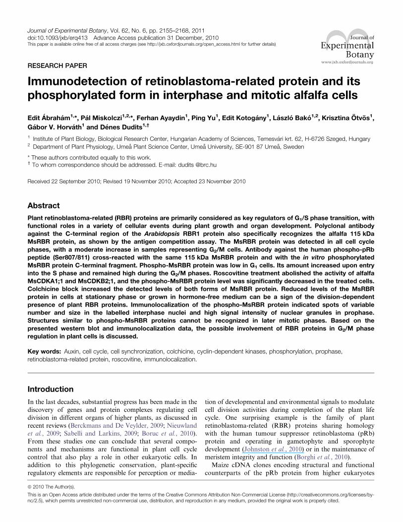
Based on the number of CDK phosphorylation sites and cell
cycle phase-dependent activities of different plant CDKs, the
phosphorylation of the MsRBR protein C-terminal fragmentwas tested by immunoprecipitated MedsaCDKA1;1/1;2
and MedsaCDKB2;1 complexes in vitro (Fig. 2). Both the
A- and B-type kinases phosphorylated the histone H1 and
the C-terminal fragment of the MsRBR protein. Previously
it had been demonstrated that the phosphorylation function
of MedsaCDKA1;1 is dependent on the presence of a cyclin
partner (MedsaCYCD3,1) (Dudits et al., 2007). This cyclin
variant has the characteristic LxCxE pRb-binding motif.On the other hand the mitosis-specific MedsaCDKB2;1 also
interacted with the MedtrCYCD1;1 cyclin, having a similar
pRb-binding motif, in yeast two-hybrid assays (Meszaros
et al., 2000).
Depletion of growth hormones causes a significantreduction of the level of RBR protein in cultured alfalfacells
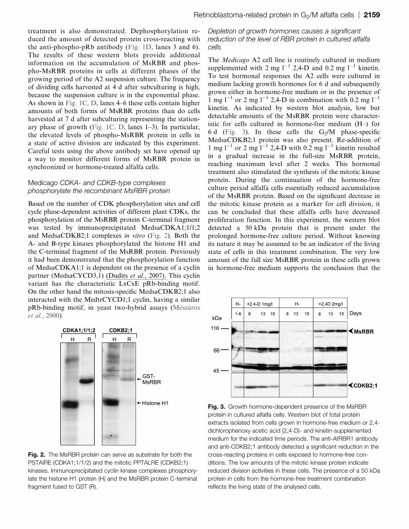
The Medicago A2 cell line is routinely cultured in medium
supplemented with 2 mg l�1 2,4-D and 0.2 mg l�1 kinetin.
To test hormonal responses the A2 cells were cultured in
medium lacking growth hormones for 6 d and subsequentlygrown either in hormone-free medium or in the presence of
1 mg l�1 or 2 mg l�1 2,4-D in combination with 0.2 mg l�1
kinetin. As indicated by western blot analysis, low but
detectable amounts of the MsRBR protein were character-
istic for cells cultured in hormone-free medium (H–) for
6 d (Fig. 3). In these cells the G2/M phase-specific
MedsaCDKB2;1 protein was also present. Re-addition of
1 mg l�1 or 2 mg l�1 2,4-D with 0.2 mg l�1 kinetin resultedin a gradual increase in the full-size MsRBR protein,
reaching maximum level after 2 weeks. This hormonal
treatment also stimulated the synthesis of the mitotic kinase
protein. During the continuation of the hormone-free
culture period alfalfa cells essentially reduced accumulation
of the MsRBR protein. Based on the significant decrease in
the mitotic kinase protein as a marker for cell division, it
can be concluded that these alfalfa cells have decreasedproliferation function. In this experiment, the western blot
detected a 50 kDa protein that is present under the
prolonged hormone-free culture period. Without knowing
its nature it may be assumed to be an indicator of the living
state of cells in this treatment combination. The very low
amount of the full size MsRBR protein in these cells grown
in hormone-free medium supports the conclusion that the
Fig. 2. The MsRBR protein can serve as substrate for both the
PSTAIRE (CDKA1;1/1/2) and the mitotic PPTALRE (CDKB2;1)
kinases. Immunoprecipitated cyclin kinase complexes phosphory-
late the histone H1 protein (H) and the MsRBR protein C-terminal
fragment fused to GST (R).
Fig. 3. Growth hormone-dependent presence of the MsRBR
protein in cultured alfalfa cells. Western blot of total protein
extracts isolated from cells grown in hormone-free medium or 2,4-
dichlorophenoxy acetic acid (2,4-D)- and kinetin-supplemented
medium for the indicated time periods. The anti-AtRBR1 antibody
and anti-CDKB2;1 antibody detected a significant reduction in the
cross-reacting proteins in cells exposed to hormone-free con-
ditions. The low amounts of the mitotic kinase protein indicate
reduced division activities in these cells. The presence of a 50 kDa
protein in cells from the hormone-free treatment combination
reflects the living state of the analysed cells.
Retinoblastoma-related protein in G2/M alfalfa cells | 2159

presence of the RBR protein is linked to division activity in
in vitro cultured alfalfa cells.
Cell cycle phase-dependent variation in amounts ofMsRBR and phospho-MsRBR proteins
Actively dividing Medicago cells grown in suspension culture
can be synchronized with HU treatment. As shown by
representative profiles and frequency data from flow cytom-
etry in Fig. 4A the control culture represents G1 cells (82%).
Alfalfa cells were subjected to 36 h HU treatment and, after
removing the inhibitor, cells progress through the S phasewith the highest frequency at 2–4 h (53% and 45%). The
maximum number of G2/M cells could be detected in samples
from cultures collected after 10–16 h (56, 61, 59, and 50%).
The frequency of G1 cells increased after 16 h, indicating the
initiation of a new cycle. A western blot of total protein
extracts detected the 115 kDa full size MsRBR protein in all
samples (Fig. 4B, lane 1). The amounts of cross-reacting
MsRBR protein showed limited variation during cell cycleprogression. Interestingly the G2/M cells had slightly elevated
levels of this protein. After 16 h the blot showed a gradual
decrease of the MsRBR protein level, which is in good
agreement with the increasing frequency of G1 cells. Western
blotting of the same protein samples with anti-phospho-pRb
antibody indicated a low protein phosphorylation level in the
control culture with 82% G1 cells (Fig. 4B, lane 3). The
protein level was elevated in samples with S phase cells (53%and 45%). The protein cross-reacting with antibodies against
the phospho-pRb peptide was found to be high in protein
extracts isolated from cell populations representing a signifi-
cant frequency (56, 61, and 59%) of G2/M phase cells.
Subsequently the phospho-MsRBR protein level was reduced
in samples containing more G1 cells. In this experiment both
antibodies cross-reacted with the full size MsRBR protein of
115 kDa. Both forms of the MsRBR protein are present inall cell cycle phases, and the phosphorylated MsRBR protein
exhibits higher variation. The differences between samples
from G1 and G2/M cells are significant.
Roscovitine reduced whereas colchicine increased bothforms of MsRBR protein
In order to dissect the G2/M phases in the analysis of levels
of the MsRBR protein and its phosphorylated form, double
synchronization based on HU treatment followed by
roscovitine or colchicine inhibition of cell cycle progression
was applied. Roscovitine is a specific inhibitor of CDKs and
it can block cell cycle progression in either late G1 phase orlate G2 phase, as was shown in BY-2 tobacco cells
(Planchais et al., 1997). Colchicine is a well-known inhibitor
of mitotic chromosome segregation by interacting with
tubulin (Bhattacharyya et al., 2008). In the present study
alfalfa cells were exposed to HU (10 mM) for 36 h. After
the release of the HU block the cells were grown in culture
Fig. 4. Variation in the amounts of the MsRBR and the phospho-
MsRBR proteins in alfalfa cells during cell cycle progression after
synchronization with 10 mM hydroxyurea (HU) treatment. (A)
Frequency data of G1, S, and G2 cells at various time points after
removal of the inhibitor (HU) with characteristic DNA histograms
from flow cytometry. (B) Western blot of protein extracts by anti-
AtRBR1 antibody (1) and anti-human phospho-pRb peptide
antibody (3). Ponceau S-stained filter with 50 lg protein samples(2). C, control, non-synchronized culture; BW, before washing out
HU; AW, after washing out HU.
2160 | Abraham et al.
medium for 2 h and subsequently treated with either roscovi-
tine (100 lM) or colchicine (0.05%). The reference HU-treated
cells were further grown in culture medium. First the cell cycle
parameters were identified by flow cytometry after these
treatment combinations (Fig. 5A). The maximum number of
S phase cells (43, 44, and 56%) was found in samples from
the time points of washing out HU and 2 h after the release
of the HU block. Cultures from 10, 16, and 22 h containan increased percentage of cells in G2/M phase with
increased frequency (54, 69, and 42%). G1 cells form the
majority in samples from 28 h. After roscovitine treatment
G1 phase cells could be detected at 54% and later at 48%
frequency. In the colchicine-treated cultures G2/M cells
represented the majority (61% and 67%) of all cell types.
Figure 5B (upper panel) presents the variation in levels of
MsRBR protein as detected by western blot of proteinextracts from cells synchronized by the above treatments.
Using the control non-treated G1 cells as reference, in-
creased MsRBR levels could be observed in cells grown in
the presence of HU (before washing). After release from the
HU block (after washing) a clear reduction was recognized
in samples with a significant number of cells in mid to late S
phase. The minimum amount was found in the sample from
2 h with 56% S phase cells. Compared with these timepoints, increased amounts of MsRBR protein were detected
in samples from 10–16 h and 22 h, with a high representa-
tion of G2/M cells. Later the cross-reacting protein levels
decreased when the frequency of G1 cells increased in the
cultures. These data show similar trends in cell cycle phase-
dependent changes as in the previous experiment (Fig. 4B,
lane 1). As far as MsRBR protein amounts are concerned,
the subsequent treatments of the synchronized cells resultedin different responses. The inhibitor roscovitine lowered the
MsRBR protein levels, whereas the colchicine block en-
hanced the accumulation of this protein in cells enriched for
G2/M phases. Using the same protein samples, the phospho-
MsRBR levels were monitored in cell populations with
various frequencies of cells in different division phases. As
shown previously, G1 cells have relatively low amounts of the
cross-reacting protein (Fig. 5B, lower panel). The roscovitinetreatment significantly reduced the amount of the phosphor-
ylated form of MsRBR protein. In contrast, the presence of
colchicine caused accumulation of phospho-MsRBR protein.
The lower molecular weight bands may represent unspecific
cross-reactions since they show different patterns when
compared with the full-size MsRBR protein.
To characterize kinase markers in cultures exposed to the
double synchronization treatment, an additional experimentwas carried out. As shown in Fig. 6A, the distribution of cells
in various phases of the cell cycle was similar to that in the
previous experiment (see Fig. 5A). Cell samples enriched in
different cell cycle stages were used for analysis of CDK
activities and quantification of these kinase proteins (Fig. 6B).
The MedsaCDKA1;1 protein was constantly present in cells
Fig. 5. Roscovitine reduces and colchicine increases the amounts
of MsRBR and phospho-MsRBR proteins in alfalfa cells exposed
to double synchronization. (A) Frequency data of G1, S, and G2
cells at various time points after removal of the inhibitor [hydroxy-
urea (HU) at 10 mM] and treatment with roscovitine or colchicine
for the indicated time periods. Characteristic DNA histograms from
flow cytometry are also shown. (B) Western blot of protein extracts
with anti-AtRBR1 antibody (upper) and anti-human pRb phospho-
peptide antibody (low). C, control, non-synchronized culture; BW,
before washing out HU; AW, after washing out HU.
Retinoblastoma-related protein in G2/M alfalfa cells | 2161

released from HU inhibition as well as in those treated with
either roscovitine or colchicine. Western blot of the same
protein samples failed to detect the mitotic MedsaCDKB2;1
in the roscovitine-treated cells. In the presence of this
inhibitor, the histone H1 phosphorylation function of both
CDKs was blocked. After HU treatment these CDKspresented different activity profiles, with a maximum histone
H1 phosphorylation at 10 h and 16 h in the case of CDKA
and 16 h and 22 h for CDKB. In the colchicine-treated cells
the activity of the mitotic MedsaCDKB2;1 was increased to
a maximum in samples from 22 h.
These sets of experiments confirmed differences in cell
cycle-dependent changes in the amounts of MsRBR and
phospho-MsRBR proteins. Roscovitine treatment of cellscaused only limited reduction in MsRBR protein levels,
whereas the phosphorylated form was significantly lowered
possibly because of the lack of CDK activities. In contrast,
the colchicine-treated cells found in G2 phase at high
frequency (>70%) showed elevated levels of both protein
forms. Accumulation of MsRBR proteins after DNA replica-
tion may be linked to the block of mitotic progression.
Immunolocalization of the phospho-MsRBR protein incultured alfalfa cells
In the present study it was not possible to obtain clear
immunolocalization signals by using antibody produced in
hens against the C-terminus of the AtRBR1 protein in
immunofluorescence microscopy of cultured alfalfa cells.
Non-uniform and inconsistent cytoplasmic and cell wall
staining of cells may be due to enhanced recognition of
certain non-specific epitopes detected by this antibody
during the immunolocalization procedure based on theformaldehyde fixation. During sample preparation, nuclei
were released from some cells. These nuclei showed
homogenous staining with the anti-AtRBR protein anti-
body. In contrast, the anti-phospho-pRb peptide antibody
recognized nuclear spots of varying sizes, indicating a con-
centrated presence of the phosphorylated form in these
structures (Fig. 7). These spots were randomly scattered in
the nuclei. Similar structures were detected in nuclei ofintact alfalfa cells by using this antibody. Figures 8 and 9
show the localization of the phospho-MsRBR protein in
interphase and mitotic cells from asynchronous suspension
cultures. The series of immunofluorescence images shown in
Fig. 8 present cells with interphase nuclei together with cells
at various mitotic phases. The nuclei of interphase cells
were stained intensively with anti-phospho-pRb peptide
antibodies and these signals were concentrated into spots asobserved with isolated nuclei (Fig. 8B–L). Analysis of
mitotic cells revealed labelled granules in prophase cells
(Fig. 8B). These structures detected in prophase cells cannot
be recognized in later mitotic stages such as prometaphase,
metaphase, anaphase, and telophase (Fig. 8D–L). Images in
Fig. 6. Changes in amounts of CDKA1;1 and CDKB2;1 proteins and their activities in alfalfa cells after double synchronization based on
10 mM hydroxyurea (HU) treatment combined with roscovitine (100 lM) or colchicine (0.05%) treatment. (A) Frequency data of G1, S,
and G2 cells at various time points after removal of the inhibitor (HU) subsequently grown in normal culture medium and treated with
either roscovitine or colchicine. (B) Top, western blot of protein extracts with anti-CDKA1;1 antibody; middle, western blot of protein
extracts with anti-CDKB2;1 antibody; bottom, histone H1 phosphorylation activities of CDKA1;1 and CDKB2;1 in the synchronized cells.
AW, after washing out HU.
2162 | Abraham et al.

Fig. 9 provide a more detailed insight into both interphase
nuclei with immunoreactive structures (Fig. 9B) and pro-
phase cells with positive nuclear granules (Fig. 9E). In this
cell the condensation of chromosomes is well advanced andthe merged pictures indicate an interchromosomal position
of nuclear granules. The presented immunolocalization data
strengthen the previous western blot results indicating the
presence of phospho-MsRBR proteins not only in in-
terphase cells before and during S phase but also in G2/M
phases of the cell cycle. An unexpected finding is that this
form of MsRBR protein is localized in spots or granules
possibly representing protein complexes with unknowncomposition and function. Additional studies are needed to
characterize these structures.
Discussion
The retinoblastoma pathway has emerged as an important
regulatory component in the cell division cycle of higherplants just like it is in other eukaryotes. Studies on plant
RBR proteins discovered a wide variety of additional roles
such as cell fate determination and influencing chromatin
dynamics or genomic imprinting (de Jager et al., 2005; Park
et al., 2005; Sablowski, 2007; Costa and Gutierrez-Marcos,
2008; Paz Sanchez et al., 2008; Chen et al., 2009; Sabelli and
Larkins, 2009). For a deeper understanding of the molecu-
lar basis of these divergent functions of plant RBR proteinsit is essential to know the cell cycle phase-dependent
changes in levels and post-translational modifications of
these proteins. In these studies, immunodetection techni-
ques were used to monitor the MsRBR and the phospho-
MsRBR proteins in samples from synchronized alfalfa cells.
Antibodies against the C-terminal fragment of AtRBR1
protein specifically recognized the 115 kDa MsRBR protein
as shown by the antigen competition assay (Fig. 1A). In
total protein extracts from cultured alfalfa cells this poly-
clonal antibody detected smaller cross-reacting bands,
indicating a wider spectrum of unspecific interactions for
the antibody tested. The higher signal intensity in lane 2
(Fig. 1A) might originate from the concentration ofantibody during the pre-incubation procedure. The present
observation that antibodies generated either against the
C-terminal fragment of the Arabidopsis AtRBR1 protein or
the phosphopeptide from human pRb protein cross-reacted
with the same 115 kDa alfalfa protein with the predicted
molecular size provides a sufficient basis to use these
antibodies in alfalfa cell systems (Fig. 1C, D). According to
the sequence data, the C-terminal regions of the Arabidopsisand alfalfa RBR proteins share a significant degree of
amino acid sequence identity (74%). The homologous
regions cover a substantial portion of these fragments
including the NVYVSPLRGS motif that may correspond
to the NIYISPLK-S motif of the human pRb protein which
has a conserved 807/811serine (S) phosphorylation site. In
attempts to monitor the phosphorylated form of the
MsRBR protein by using anti-phosphopeptide antibody, ithas to be emphasized that this methodology detects a single
phosphorylation site whereas post-translational modifica-
tions can occur at additional residues with different
functional consequences. Therefore, the methodology used
here is not sufficient for determining the ratio between the
MsRBR and the phospho-MsRBR proteins. The function-
ality and specificity of this anti-phosphopeptide antibody
was demonstrated by detection of the in vitro phosphory-lated form of the Medicago RBR protein C-terminal
fragment by western blot (Fig. 1B). The pRb proteins from
mammalian cells show phosphorylation-dependent migra-
tion patterns, and the hyperphosphorylated pRb protein is
usually detected as a more slowly migrating band (Farkas
et al., 2002). As a result of the protein separation protocol
employed (electrophoresis in denaturing polyacrylamide
gels) the MsRBR protein band was diffuse and a clear shiftbetween the two protein forms could not be recognized.
Similarly, no band shift was reported in the case of the
NtRBR1 and the phospho-NtRBR1 proteins (Kawamura
et al., 2006). After inorganic phosphate incorporation into
pea axillary buds the anti-PsRBR antibody immunoprecipi-
tated higher and lower molecular mass forms of the
phosphorylated PsRBR1 that were sensitive to k-phospha-tase treatment (Shimizu-Sato et al., 2008). In the presentstudy the intensity of the band corresponding to the
phospho-MsRBR (115 kDa) protein was reduced by phos-
phatase treatment, as an indication of correct recognition of
the phosphorylated form by the anti-phosphopeptide anti-
body used (Fig. 1D, lanes 3 and 6).
It was demonstrated previously that Arabidopsis, tobacco,
wheat, and maize RBR proteins can serve as substrates
for CDK–cyclin complexes (Boniotti and Gutierrez, 2001).The in vitro phosphorylation reactions revealed that
PSTAIRE-type CDKs and D-type cyclins are involved in
Fig. 7. Differences in immunofluorescence labelling of MsRBR
and phospho-MsRBR proteins in isolated nuclei released from
alfalfa cells indicate different nuclear organization for the two forms
of MsRBR protein. Upper panel: diffused, uniform staining of
nucleoplasm (green) after immunolocalization with anti-AtRBR1
antibody. Lower panel: granular labelling of the nucleus (green)
after immunolocalization with anti-human pRb phosphopeptide
antibody. DNA stained with DAPI and pseudocoloured red.
Retinoblastoma-related protein in G2/M alfalfa cells | 2163

this post-translational modification. Nakagami et al. (2002)
reported tobacco NtRBR1 phosphorylation by NictaCycD3;3-
associated kinase during a very short period in the middle ofthe G1 phase whereas p13SUC1-bound kinases were active in
phosphorylation of NtRBR1 during late G1 phase to late S
phase. These authors also showed that different types of
tobacco CDKs (CDKA and CDKB) can phosphorylate
the 110 kDa NtRBR protein in vitro (Kawamura et al.,
2006). The present study supports the potential role of
non-PSTAIRE kinases; namely the mitotic Cdc2F–
MedsaCDKB2;1 complex is also capable of phosphorylat-ing the C-terminal region of the alfalfa MsRBR protein as
is shown in the case of the MedsaCDKA1;1 PSTAIRE
kinase (Fig.2). The activity of MedsaCDKB2;1 is linked to
cells in G2 phase and mitosis (see Magyar et al., 1997 and
Fig. 6B). The MedsaCDKA1;1 protein is detectable during
the whole cell cycle, with increased activities in cells
accumulated in G1–S and early G2 phase after HU block
(Fig. 6B). The histone H1 phosphorylation function of bothCDKs was sensitive to roscovitine treatment. It is an
interesting finding that the MedsaCDKB2;1 protein was
undetectable in treated cells. That is a selective effect of this
inhibitor in comparison with the response of Med-
saCDKA1;1. The lack of B-type kinase protein accumula-tion can originate from blocking an unknown component
required for either expression of the MedsaCDKB2;1 gene
or synthesis and stability of this kinase protein. One potential
candidate can be the A-type CDK since this kinase is active
earlier in G1–S phases and its function is known to be
inhibited by roscovitine. Detection of this unexpected effect
of roscovitine may initiate further experiments.
Analysis of the MsRBR protein in cells from cultures atdifferent growing phases revealed an increase in both forms
of this protein during exponential growth (Fig. 1C, D, lanes
4–6). Differences between cells in the stationary and
exponential phase of the culture period were more pro-
nounced in the case of the phospho-MsRBR protein.
Linking the presence of the MsRBR proteins to the
proliferative state was also supported by the hormone
starvation experiment (Fig. 3). In this experiment quantifi-cation of the mitotic kinase (CDKB2;1) protein was used as
a marker for division activity that was induced already
Fig. 8. Organization of the phospho-MsRBR protein into granular structures is restricted to mitotic prophase. According to the
distribution of the phospho-MsRBR protein these characteristic granules cannot be detected in later mitotic stages. Immunodetection
of phospho-MsRBR protein (green panels) is shown at various phases of the cell division cycle in formaldehyde-fixed suspension-
cultured cells of alfalfa. Arrowheads indicate various mitotic phases (A, B, prophase; C, D, prometaphase; E, F, metaphase; G, H,
anaphase; I, J, telophase) or early G1 cells (K, L). Note the significantly reduced labelling intensities at metaphase, anaphase, and
telophase stages of mitosis. This figure also presents interphase nuclei (B, D, F, H, J, and L) with concentration of labelling signals in
nuclear spots. Localization of phospho-MsRBR protein is shown in the green panel. DNA is stained with DAPI and pseudocoloured red
(red panels). Bar¼10 lm
2164 | Abraham et al.

during the first 2 d by re-addition of hormones after
a hormone-free growing period of 6 d. The amounts of the
MsRBR protein(115 kDa) increased gradually and reached
a maximum after 15 d of growth in a complete culture
medium. Based on the observed changes, close correlation
between these two proteins is not expected since the
MsRBR protein slightly changes between cell cycle phases,whereas the mitotic kinase CDKB;2,1 protein is exclusively
found in G2/M phase cells (Figs 4B, 6B). When alfalfa cells
were cultured in hormone-free medium for a further 8–15 d,
both the MsRBR protein and the mitotic kinase CDKB;2,1
proteins were reduced to the detection threshold in protein
extracts; therefore, it was concluded that the prolonged
withdrawal of synthetic auxin and kinetin stopped the
division of A2 alfalfa cells and the non-dividing state ofthese cells was reflected by the very low level of the MsRBR
protein. The cell division-dependent presence of plant RBR
proteins could also be seen in the Arabidopsis experimental
system. Only the non-phosphorylated AtRBR1 protein was
detected in stationary phase MM2d Arabidopsis cells.
Transfer into fresh medium resulted in the detection of
increasing amounts of AtRBR1 protein and after 8 h its
phosphorylated form was recognized by western blots(Hirano et al., 2008). The role of RBRs in dividing plant
cells can also be deduced from experiments by these authors
where MM2d cells were grown in low sucrose medium.
AtRBR1 was depleted to undetectable levels within 12 h
after transfer of cells into the medium lacking sucrose. In
interpreting these findings as an indication for a link
between the dividing state of cells and the RBR protein it
should be emphasized that the present and cited studies
were carried out with plant cells cultured in vitro. The
functional characteristics of plant RBRs can be essentially
different in vivo. Wyrzykowska et al. (2006) showed growth
inhibition and initiation of differentiation by transient
overexpression of the AtRBR1 gene. In tobacco plants the
induced RBR protein synthesis caused a complete and rapid
cessation of cell division. These observations highlight anessential divergence of RBR protein functions in plant
organs such as shoot apical meristem and in hormone-
stimulated, fast cycling cultured cells.
Despite the above limitation, cultured plant cells at
present provide an optimal experimental system for efficient
synchronization of the division cycle to analyse phase-
specific events. In the present study the A2 cell line of
M. sativa that is well adapted to in vitro conditions was usedand its short cycle time allowed the enrichment of cell
populations in various phases by using specific inhibitors
such as HU, roscovitine, or colchicine. Western blotting of
total protein extracts from alfalfa cells with either antibody
against the C-terminal fragment of AtRBR1 protein or anti-
human phospho-pRb peptide antibody detected the
expected size of RBR protein (115 kDa) and additional
cross-reacting bands of various sizes and quantities (Figs 1,4B, 5B). These signals are considered to be the results of
non-specific interactions or degradation; therefore, the
present analysis is restricted to the full-size MsRBR protein
and its phosphorylated form. Western blot analysis of
protein samples detected the MsRBR protein during the
whole division cycle with limited changes. The observed
increase in MsRBR protein levels in samples with a higher
frequency of G2/M phase cells was shown more clearly by
Fig. 9 Concentration of the phospho-MsRBR protein (green panel) into nuclear spots in interphase cells (A–C) and granules in prophase
cells (D–I) after formaldehyde fixation. Prophase cells with advanced chromosome condensation have a considerable number of
phospho-MsRBR protein-positive granules with an interchromosomal position. DNA is stained with DAPI (bar¼10 lm) and
pseudocoloured red (red panel).
Retinoblastoma-related protein in G2/M alfalfa cells | 2165

the colchicine-blocked cells, although a cell cycle phase-
independent action of this inhibitor on the RBR protein
accumulation cannot be excluded. During HU+roscovitine
treatment the MsRBR protein level was slightly lower in
comparison with the HU-treated cells. The sequentially
applied HU and colchicine combination resulted in a signifi-
cantly elevated MsRBR protein level compared with the single
HU treatment (Fig. 5B). Cell cycle phase dependence couldbe more clearly recognized in alterations of phospho-MsRBR
protein levels (Figs 4B, 5B). The reduced level of the
phosphorylated form in G1 cells can be safely concluded
from the presented data. Comparing amounts of cross-
reacting MsRBR and phospho-MsRBR proteins in Fig. 4B
and Fig. 5B reveals that G1 cells primarily have the
hypophosphorylated form. Hirano et al. (2008) demon-
strated by pull-down assay that the non-phosphorylatedAtRBR1 protein could bind to E2Fa protein. These
findings are in agreement with the general model of G1/S
phase transition control where the hypophosphorylated Rb
proteins inhibit E2F functions in the activation of S phase-
specific genes (Poznic, 2009). The release of this inhibition is
expected to occur when the RBR proteins are phosphory-
lated by CDK complexes. The role of CDKs in phosphor-
ylation of RBR protein was shown by different studies onplant systems. The present work demonstrates that in
alfalfa cells the MedsaCDKA1;1 and the mitosis-specific
MedsaCDKB1,2 can be responsible for this modification of
the MsRBR protein (Fig. 2).
Based on the widely accepted model emphasizing the
regulatory role of the Rb proteins in G1/S phase transition,
experimental works on plant RBR proteins have essentially
focused on this checkpoint. Functional roles for plant RBRproteins in the G2/M phases have not been proposed so far.
The present western blot studies detected significant
amounts of both MsRBR and phospho-MsRBR proteins
in cells representing G2/M phases after chemical synchroni-
zation (Figs 4B, 5B). The presence of these proteins after S
phase was more clearly demonstrated by western blot of
proteins from the colchicine-treated cells. Occurrence of the
phosphorylated form coincided with CDK activities in-cluding the PSTAIRE CDKA1;1 and the mitosis-specific
CDKB2;1. In accordance with western blot data the
immunolocalization studies also revealed the presence of
phospho-MsRBR protein as granular structures in inter-
phase nuclei and prophase cells (Figs 7–9). These figures
present several interphase cells with nuclei having intense
labelling concentrated into spots of various sizes. According
to the results from labelling with anti-AtRBR1 antibody,a homogenous distribution of signals could be seen in the
released nuclei. This observation is in agreement with the
localization data presented by Boruc et al. (2010). Although
the functional significance of these differences in the
organization of MsRBR and phospho-MsRBR proteins
has yet to be elucidated, one possible hypothesis is that
only the phosphorylated form participates in protein com-
plexes with the capability to form the detected nuclearstructures. This question clearly is worthy of further
investigation by using additional approaches. As shown by
the present immunolocalization experiments, the phospho-
MsRBR protein is detectable as granules in prophase cells
with an advanced stage of chromosome condensation.
Based on this observation it will be interesting to test
a possible involvement of plant RBR proteins in building
up chromosome structures. In Drosophila the Rb family
proteins can have a function in the mitotic cell. The Rbf1
protein is required to target the condensin II subunitdCAPD3 to chromosomes and thus promotes chromosome
condensation and faithful transmission (Longworth et al.,
2008). These authors showed that the overexpression of the
Drosophila Rbf1 gene resulted in hypercondensation of
mitotic chromosomes. Additional mitotic functions of pRb
proteins have been widely demonstrated in mammalian cell
systems. Loss of pRb protein functions may induce cell
cycle deregulation, leading to a malignant phenotype(Giacinti and Giordano, 2006). The pRb protein has been
shown to regulate the expression of the mitotic checkpoint
protein Mad2, which promotes chromosome instability
when misregulated in mammalian cells (Hernando et al.,
2004; Sotillo et al., 2007). Zheng et al. (2000) demonstrated
that the pRb protein can interact with hsHec1p carrying the
LxCxE motif during G2/M phases. This protein is required
for proper chromosome segregation. The serine/threonineAurora kinases have been shown to play a critical role in
mitosis. Nair et al. (2009) demonstrated that phosphoryla-
tion of the pRb protein at Ser780 by Aurora B influences
the polyploid cell formation caused by inhibition of this
kinase. As shown by Karantza et al. (1993), cells over-
expressing the pRb proteins accumulated uniformly in the
G2 phase, whereas cells expressing endogenous levels of the
pRb protein were found throughout the whole cell cycle.These results suggest that the pRb protein may interact with
some protein of the cell cycle-regulatory machinery during
the G2 phase. The present immunodetection study based on
both western blot analysis and immunolocalization with
immunofluorescence microscopy provides substantial sup-
port for considering plant RBR proteins to be present in
G2/M cells as regulators of the cell cycle during post-DNA
synthetic events.
Acknowledgements
This work was supported by a grant from OTKA (The
Hungarian Scientific Research Grant), grant number: NK-
69227. EA was supported by the Janos Bolyai ResearchFellowship of the Hungarian Academy of Sciences. The
authors thank Matyas Cserhati and Erzsebet Fejes for
critical reading, and Klara Godo for valuable assistance in
the preparation of this manuscript.
References
Ach RA, Durfee T, Miller AB, Taranto P, Hanley-Bowdoin L,
Zambrski P, Gruissem W. 1997. RRB1 and RRB2 encode maize
retinoblastoma-related proteins that interact with a plant D-type cyclin
2166 | Abraham et al.

and geminivirus replication protein. Molecular and Cellular Biology 17,
5077–5086.
Berckmans B, De Veylder L. 2009. Transcriptional control of the cell
cycle. Current Opinion in Plant Biology 12, 599–605.
Bhattacharyya B, Panda D, Gupta S, Banerjee M. 2008. Anti-
mitotic activity of colchicine and the structural basis for its interaction
with tubulin. Medicinal Research Reviews 28, 155–183.
Blom N, Sicheritz-Ponten T, Gupta R, Gammeltoft S, Brunak S.
2004. Prediction of post-translational glycosylation and
phosphorylation of proteins from the amino acid sequence.
Proteomics 4, 1633–1649.
Bogre L, Olah Z, Dudits D. 1988. Ca2-dependent protein kinase
from alfalfa (Medicago varia): partial purification and
autophosphorylation. Plant Science 58, 135–144.
Boniotti MB, Gutierrez C. 2001. A cell-cycle-regulated kinase
activity phosphorylates plant retinoblastoma protein and contains, in
Arabidopsis, a CDKA/cyclin D complex. The Plant Journal 28,
341–350.
Borghi L, Gutzat R, Futterer J, Laizet Y, Hennig L, Gruissem W.
2010. Arabidopsis RETINOBLASTOMA-RELATED is required for stem
cell maintenance, cell differentiation, and lateral organ production. The
Plant Cell 22, 1792–1811.
Boruc J, Mylle E, Duda M, De Clercq R, Rombauts S, Geelen D,
Hilson P, Inze D, Van Damme D, Russinova E. 2010. Systematic
localization of the Arabidopsis core cell cycle proteins reveals novel cell
division complexes. Plant Physiology 152, 553–565.
Chen Z, Hafidh S, Poh SH, Twell D, Berger F. 2009. Proliferation
and cell fate establishment during Arabidopsis male gametogenesis
depends on the retinoblastoma protein. Proceedings of the National
Academy of Sciences, USA 106, 7257–7262.
Costa LM, Gutierrez-Marcos JF. 2008. Retinoblastoma makes its
mark on imprinting in plants. PLoS Biology 6, 1631–1633.
de Almeida J, Engler J, De Veylder L, et al. 2009. Systematic
analysis of cell-cycle gene expression during Arabidopsis
development. The Plant Journal 59, 645–660.
de Jager SM, Maughan S, Dewitte W, Scofield S, Murray JA.
2005. The developmental context of cell-cycle control in plants.
Seminars in Cell and Developmental Biology 16, 385–396.
Dudits D, Cserhati M, Miskolczi P, Horvath VG. 2007. The
growing family of plant cyclin-dependent kinases with multiple
functions in cellular and developmental regulation. In: Inze D, ed. Cell
cycle control and plant development. Oxfore: Blackwell Publishing,
1–30.
Durfee T, Feiler HS, Gruissem W. 2000. Retinoblastoma-related
proteins in plants: homologues or orthologues of their metazoan
counterparts? Plant Molecular Biology 43, 635–642.
Farkas T, Hansen K, Holm K, Lukas J, Bartek J. 2002. Distinct
phosphorylation events regulate p130- and p107-mediated repression
of E2F-4. Journal of Biological Chemistry 277, 26741–26752.
Galbraith DW, Harkins KR, Maddox JM, Ayres NM, Sharma DP,
Firoozabady E. 1993. Rapid flow cytometric analysis of the cell cycle
in intact plant tissues. Science 220, 1049–1051.
Giacinti C, Giordano A. 2006. RB and cell cycle progression;
Oncogene 25, 5220–5227.
Grafi G, Burnett RJ, Helentjaris T, Larkins BA, DeCaprio JA,
Sellers WR, Kaelin WG Jr. 1996. A maize cDNA encoding a member
of the retinoblastoma protein family: involvement in endoreduplication.
Proceedings of the National Academy of Sciences, USA 93,
8962–8967.
Gutierrez C. 1998. The retinoblastoma pathway in plant cell cycle and
development. Current Opinion in Plant Biology 1, 492–497.
Hall TA. 1999. BioEdit: a user-friendly biological sequence alignment
editor and analysis program for Windows 95/98/NT. Nucleic Acids
Symposium Series 41, 95–98.
Hernando E, Nahle Z, Juan G, et al. 2004. Rb inactivation promotes
genomic instability by uncoupling cell cycle progression from mitotic
control. Nature 430, 797–802.
Hirano H, Harashima H, Shinmyo A, Sekine M. 2008. Arabidopsis
retinoblastoma-related protein1 is involved in G1 phase cell cycle arrest
caused by sucrose starvation. Plant Molecular Biology 66, 259–275.
Horvath BM, Magyar Z, Zhang Y, Hamburger AW, Bako L,
Visser RG, Bachem CW, Bogre L. 2006. EBP1 regulates organ size
through cell growth and proliferation in plants. EMBO Journal 25,
4909–4920.
Huntley R, Healy S, Freeman D, et al. 1998. The maize
retinoblastoma protein homologue ZmRb-1 is regulated during leaf
development and displays conserved interactions with G1/S regulators
and plant cyclin D (CycD) proteins. Plant Molecular Biology 37, 155–169.
Johnston AJ, Kirioukhova O, Barrell PJ, Rutten T, Moore JM,
Baskar R, Grossniklaus U, Gruissem W. 2010. Dosage-sensitive
function of RETINOBLASTOMA RELATED and convergent epigenetic
control are required during the Arabidopsis life cycle. PLoS Genetics 6,
e1000988.
Karantza V, Maroo A, Fay D, Sedivy JM. 1993. Overproduction of
Rb protein after the G1/S boundary causes G2 arrest. Molecular Cell
Biology 13, 6640–6652.
Kawamura K, Kato K, Shinmyo A, Sekine M. 2006. Tobacco
retinoblastoma-related protein is phosphorylated by different types of
cyclin-dependent kinases during the cell cycle. Plant Biotechnology
23, 467–473.
Korenjak M, Brehm A. 2005. E2F–Rb complexes regulating
transcription of genes important for differentiation and development.
Current Opinion in Genetics and Development 15, 520–527.
Lendvai A, Pettko-Szandtner A, Csordas-Toth E, Miskolczi P,
Horvath GV, Gyorgyey J, Dudits D. 2007. Dicot and monocot
plants differ in retinoblastoma-related protein subfamilies. Journal of
Experimental Botany 58, 1663–75.
Longworth MS, Herr A, Ji JY, Dyson NJ. 2008. RBF1 promotes
chromatin condensation through a conserved interaction with the
Condensin II protein dCAP-D3. Genes and Development 22,
1011–1024.
Magyar Z, Bako L, Bogre L, Dedeoglu D, Kapros T, Dudits D.
1993. Active cdc2 genes and cell cycle phase-specific cdc2-related
kinase complexes in hormone-stimulated alfalfa cells. The Plant
Journal 4, 151–161.
Magyar Z, Meszaros T, Miskolczi P, et al. 1997. Cell cycle phase
specificity of putative cyclin-dependent kinase variants in synchronized
alfalfa cells. The Plant Cell 9, 223–235.
Retinoblastoma-related protein in G2/M alfalfa cells | 2167

Meszaros T, Miskolczi P, Ayaydin F, Pettko-Szandtner A,
Peres A, Magyar Z, Horvath GV, Bako L, Feher A, Dudits D. 2000.
Multiple cyclin-dependent kinase complexes and phosphatases control
G2/M progression in alfalfa cells. Plant Molecular Biology 43, 595–605.
Miskolczi P, Lendvai A, Horvath GV, Pettko-Szandtner A,
Dudits D. 2007. Conserved functions of retinoblastoma proteins: from
purple retina to green plant cells. Plant Science 172, 671–683.
Murashige T, Skoog F. 1962. A revised medium for rapid growth and
bioassays with tobacco tissue culture. Plant Physiology 15, 473–497.
Nair JS, Ho AL, Tse AN, Coward J, Cheema H, Ambrosini G,
Keen N, Schwartz GK. 2009. Aurora B kinase regulates the postmitotic
endoreduplication checkpoint via phosphorylation of the retinoblastoma
protein at serine 780. Molecular Biology of the Cell 20, 2218–2228.
Nakagami H, Kawamura K, Sugisaka K, Sekine M, Shinmyo A.
2002. Phosphorylation of retinoblastoma-related protein by the cyclin
D/cyclin-dependent kinase complex is activated at the G1/S-phase
transition in tobacco. The Plant Cell 14, 1847–1857.
Nakagami H, Sekine M, Murakami H, Shinmyo A. 1999. Tobacco
retinoblastoma-related protein phosphorylated by a distinct cyclin-
dependent kinase complex with Cdc2/cyclin D in vitro. The Plant
Journal 18, 243–252.
Nieuwland J, Menges M, Murray JAH. 2007. The plant cyclins. In:
Inze D, ed. Cell cycle control and plant development. Oxford:
Blackwell Publishing, 31–62.
Nieuwland J, Scofield S, Murray JA. 2009. Control of division and
differentiation of plant stem cells and their derivatives. Seminars in Cell
and Developmental Biology 20, 1134–1142.
Park JA, Ahn JW, Kim YK, Kim J, Kim SJ, Kim JK, Kim WT,
Pai HS. 2005. Retinoblastoma protein regulates cell proliferation,
differentiation, and endoreduplication in plants. The Plant Journal 42,
153–163.
Paz Sanchez M, Caro E, Desvoyes B, Ramirez-Parra E,
Gutierrez C. 2008. Chromatin dynamics during the plant cell cycle.
Seminars in Cell and Developmental Biology 19, 537–546.
Planchais S, Glab N, Trehin C, Perennes C, Bureau JM, Meijer L,
Bergounioux C. 1997. Roscovitine, a novel cyclin-dependent kinase
inhibitor, characterizes restriction point and G2/M transition in tobacco
BY-2 cell suspension. The Plant Journal 12, 191–202.
Poznic M. 2009. Retinoblastoma protein: a central processing unit.
Journal of Biosciences 34, 305–312.
Sabelli PA, Larkins BA. 2009. The contribution of cell cycle
regulation to endosperm development. Sexual Plant Reproduction 22,
207–219.
Sablowski R. 2007. The dynamic plant stem cell niches. Current
Opinion in Plant Biology 10, 639–644.
Shimizu-Sato S, Ike Y, Mori H. 2008. PsRBR1 encodes a pea
retinoblastoma-related protein that is phosphorylated in axillary buds
during dormancy-to-growth transition. Plant Molecular Biology 66,
125–135.
Soni R, Carmichael JP, Shah ZH, Murray JAH. 1995. A family of
cyclin D homologs from plants differentially controlled by growth
regulators and containing the conserved retinoblastoma protein
interaction motif. The Plant Cell 7, 85–103.
Sotillo R, Hernando E, Diaz-Rodriguez E, Teruya-Feldstein J,
Cordon-Cardo C, Lowe SW, Benezra R. 2007. Mad2
overexpression promotes aneuploidy and tumorigenesis in mice.
Cancer Cell 11, 9–23.
Wyrzykowska J, Schorderet M, Pien S, Gruissem W,
Fleming AJ. 2006. Induction of differentiation in the shoot apical
meristem by transient overexpression of a retinoblastoma-related
protein. Plant Physiology 141, 1338–1348.
Xie Q, Sanz-Burgos AP, Hannon GJ, Gutierrez C. 1996. Plant
cells contain a novel member of the retinoblastoma family of growth
regulatory proteins. EMBO Journal 15, 4900–4908.
Zheng L, ChenY Riley DJ, Chen PL, Lee WH. 2000.
Retinoblastoma protein enhances the fidelity of chromosome
segregation mediated by hsHec1p. Molecular and Cellular Biology 20,
3529–3537.
2168 | Abraham et al.