Similarities of Retinoblastoma Function between Plants and ...
28
International Journal of Molecular Sciences Review Beyond What Your Retina Can See: Similarities of Retinoblastoma Function between Plants and Animals, from Developmental Processes to Epigenetic Regulation Estephania Zluhan-Martínez 1,2 , Vadim Pérez-Koldenkova 3 , Martha Verónica Ponce-Castañeda 4 , María de la Paz Sánchez 1 , Berenice García-Ponce 1 , Sergio Miguel-Hernández 5 , Elena R. Álvarez-Buylla 1, * and Adriana Garay-Arroyo 1, * 1 Laboratorio de Genética Molecular, Epigenética, Desarrollo y Evolución de Plantas, Instituto de Ecología, Universidad Nacional Autónoma de Mexico, 3er Circuito Ext. Junto a J. Botánico, Ciudad Universitaria, UNAM 04510, Mexico; [email protected] (E.Z.-M.); [email protected] (M.d.l.P.S.); [email protected] (B.G.-P.) 2 Posgrado en Ciencias Biomédicas, Universidad Nacional Autónoma de México, Av. Universidad 3000, Coyoacán 04510, Mexico 3 Laboratorio Nacional de Microscopía Avanzada, Centro Médico Nacional Siglo XXI, Instituto Mexicano del Seguro Social, Av. Cuauhtémoc, 330. Col. Doctores, Alc. Cuauhtémoc 06720, Mexico; [email protected] 4 Unidad de InvestigaciónMédica en Enfermedades Infecciosas, Centro Médico Nacional SXXI, Instituto Mexicano del Seguro Social, Mexico City 06720, Mexico; [email protected] 5 Laboratorio de Citopatología Ambiental, Departamento de Morfología, Escuela Nacional de Ciencias Biológicas, Instituto Politécnico Nacional, Campus Zacatenco, Calle Wilfrido Massieu Esquina Cda, Manuel Stampa 07738, Mexico; [email protected] * Correspondence: [email protected] (E.R.Á.-B.); [email protected] (A.G.-A.) Received: 28 May 2020; Accepted: 7 July 2020; Published: 12 July 2020 Abstract: The Retinoblastoma protein (pRb) is a key cell cycle regulator conserved in a wide variety of organisms. Experimental analysis of pRb’s functions in animals and plants has revealed that this protein participates in cell proliferation and differentiation processes. In addition, pRb in animals and its orthologs in plants (RBR), are part of highly conserved protein complexes which suggest the possibility that analogies exist not only between functions carried out by pRb orthologs themselves, but also in the structure and roles of the protein networks where these proteins are involved. Here, we present examples of pRb/RBR participation in cell cycle control, cell differentiation, and in the regulation of epigenetic changes and chromatin remodeling machinery, highlighting the similarities that exist between the composition of such networks in plants and animals. Keywords: retinoblastoma protein; cell proliferation; cell differentiation; plants; animals; cell cycle; stem cells; epigenetics; DNA damage; morphogenetic regulatory networks 1. Introduction Eukaryotic organisms evolved from a common unicellular ancestor, from which they diverged 1500 million years ago [1–3]. Testimonies of this ancient kinship are some processes and genetic components that have been preserved throughout evolution, along with many others which subsequently emerged exclusively in plants or animals [4–6]. Interestingly, it has been shown that plants and animals evolved multicellularity independently, but unlike animals, plants have an extensive post-embryonic growth and development, that is highly influenced by the environment [7,8]. However, in both multicellular Int. J. Mol. Sci. 2020, 21, 4925; doi:10.3390/ijms21144925 www.mdpi.com/journal/ijms
-
Upload
khangminh22 -
Category
Documents
-
view
2 -
download
0
Transcript of Similarities of Retinoblastoma Function between Plants and ...

International Journal of
Molecular Sciences
Review
Beyond What Your Retina Can See: Similarities ofRetinoblastoma Function between Plants andAnimals, from Developmental Processes toEpigenetic Regulation
Estephania Zluhan-Martínez 1,2, Vadim Pérez-Koldenkova 3 , Martha Verónica Ponce-Castañeda 4,María de la Paz Sánchez 1, Berenice García-Ponce 1 , Sergio Miguel-Hernández 5,Elena R. Álvarez-Buylla 1,* and Adriana Garay-Arroyo 1,*
1 Laboratorio de Genética Molecular, Epigenética, Desarrollo y Evolución de Plantas, Instituto de Ecología,Universidad Nacional Autónoma de Mexico, 3er Circuito Ext. Junto a J. Botánico, Ciudad Universitaria,UNAM 04510, Mexico; [email protected] (E.Z.-M.); [email protected] (M.d.l.P.S.);[email protected] (B.G.-P.)
2 Posgrado en Ciencias Biomédicas, Universidad Nacional Autónoma de México, Av. Universidad 3000,Coyoacán 04510, Mexico
3 Laboratorio Nacional de Microscopía Avanzada, Centro Médico Nacional Siglo XXI, Instituto Mexicano delSeguro Social, Av. Cuauhtémoc, 330. Col. Doctores, Alc. Cuauhtémoc 06720, Mexico;[email protected]
4 Unidad de Investigación Médica en Enfermedades Infecciosas, Centro Médico Nacional SXXI,Instituto Mexicano del Seguro Social, Mexico City 06720, Mexico; [email protected]
5 Laboratorio de Citopatología Ambiental, Departamento de Morfología, Escuela Nacional de CienciasBiológicas, Instituto Politécnico Nacional, Campus Zacatenco, Calle Wilfrido Massieu Esquina Cda,Manuel Stampa 07738, Mexico; [email protected]
* Correspondence: [email protected] (E.R.Á.-B.); [email protected] (A.G.-A.)
Received: 28 May 2020; Accepted: 7 July 2020; Published: 12 July 2020�����������������
Abstract: The Retinoblastoma protein (pRb) is a key cell cycle regulator conserved in a wide varietyof organisms. Experimental analysis of pRb’s functions in animals and plants has revealed that thisprotein participates in cell proliferation and differentiation processes. In addition, pRb in animalsand its orthologs in plants (RBR), are part of highly conserved protein complexes which suggest thepossibility that analogies exist not only between functions carried out by pRb orthologs themselves,but also in the structure and roles of the protein networks where these proteins are involved. Here,we present examples of pRb/RBR participation in cell cycle control, cell differentiation, and in theregulation of epigenetic changes and chromatin remodeling machinery, highlighting the similaritiesthat exist between the composition of such networks in plants and animals.
Keywords: retinoblastoma protein; cell proliferation; cell differentiation; plants; animals; cell cycle;stem cells; epigenetics; DNA damage; morphogenetic regulatory networks
1. Introduction
Eukaryotic organisms evolved from a common unicellular ancestor, from which they diverged 1500million years ago [1–3]. Testimonies of this ancient kinship are some processes and genetic componentsthat have been preserved throughout evolution, along with many others which subsequently emergedexclusively in plants or animals [4–6]. Interestingly, it has been shown that plants and animals evolvedmulticellularity independently, but unlike animals, plants have an extensive post-embryonic growthand development, that is highly influenced by the environment [7,8]. However, in both multicellular
Int. J. Mol. Sci. 2020, 21, 4925; doi:10.3390/ijms21144925 www.mdpi.com/journal/ijms

Int. J. Mol. Sci. 2020, 21, 4925 2 of 28
organisms, morphogenesis depends on a delicate balance between cell division and differentiationrates. In animals, when this balance is lost, it can cause tumors and cancer, not seen during most ofplant development. Nonetheless, Arabidopsis thaliana (hereafter Arabidopsis), the most studied modelplant, has been shown to be an important model system to understand basic regulatory mechanismsinvolved in human diseases [9–11]. For example, Arabidopsis has homologous genes for 70% ofthose involved in human cancer. Interestingly, a higher percentage than that found in the genome ofDrosophila melanogaster or of Saccharomyces cerevisiae [9,10]. Hence, Arabidopsis has already been usedas a screening tool to evaluate the action and efficacy of some drugs to treat human cancer and otherdiseases [11–13].
Retinoblastoma (RB1) is a highly conserved gene, that encodes the so-called tumor suppressorprotein, that regulates different developmental processes of phylogenetically distant organisms.The name of this gene comes from the retinoblastoma illness, a rare intraocular malignant tumor thathas its onset during early childhood. As determined by karyotyping, this disease is associated withlosses in chromosome 13, which contains the RB1 gene [14–16]. RB1 was identified by positional cloningand after subsequent molecular analysis, it became known as the first tumor suppressor gene, givingrobust evidence for the genetic predisposition of cancer development in some cases [17,18]. After itsdiscovery, alterations in this gene were described in other malignant tumors such as osteosarcomas,cervical cancer, prostate carcinoma, small cell lung cancer, and some forms of leukemia [19,20]. RB1 isan essential gene whose best studied function is the regulation of the cell cycle transition from G1 toS phase through formation of a protein complex with transcription factors of the E2F-family; that areregulated by the Retinoblastoma protein (pRb) multiple phosphorylation states. In many cancer types,an altered regulation of pRb, like permanent hyperphosphorylation that promotes pRb dissociationfrom the complex with E2F, leads to an unregulated cell proliferation [21,22]. Moreover, alteredregulation of the pRb pathway is considered one of the most common traits in different types ofcancer [23,24], and several studies have proposed targeting pRb regulation pathway as alternativetreatments [25–27]. In fact, cyclin dependent kinases (CDKs), the kinases that phosphorylate pRb arecommonly deregulated in many malignant tumors. From the therapeutic standpoint, pRb cannotbe a target of drugs, however, CDKs are therapeutic targets, and several generations of non-specificcell cycle CDKs inhibitors have been under clinical evaluation as cancer treatments with mixedresults. More recently specific cell cycle CDK4/6 and transcriptional CDKs inhibitors may becomealternative therapeutic strategies under current clinical evaluation [28–30]. In summary, a morethorough understanding of pRb’s developmental functions could help find new efficient treatmentsfor different cancer types.
In this review, we will focus on how the protein encoded by the RB1 gene, and its plant orthologRETINOBLASTOMA-RELATED (RBR), participates in important developmental processes such ascell cycle control, cell differentiation, as well as in the homeostasis of stem cell/pluripotency, that arecellular processes shared by plants and animals. First, we analyze the structure of the RB/RBR proteins,secondly, we describe their interaction with proteins that participate in different developmentalprocesses and regulatory networks. Finally, we analyze their participation in the epigenetic andchromatin structure regulation, which is important for different developmental decisions. Interestingly,many of the protein interactions involving RETINOBLASTOMA, as well as the overall structures ofthe regulatory networks in which this protein participates are similar between plants and animals.This suggests that once such regulatory networks were assembled during evolution, the key role ofthis protein as an integrator of internal developmental cues remained functionally constrained amongeukaryotic organisms’ evolution.
2. Structure of the Retinoblastoma Protein
In humans, Retinoblastoma susceptibility gene is a member of a small gene family that includesRB1 (p105/pRb), RBl1 (p107/pRBL1), and RBl2 (p130/pRBL2), whose protein structure are very similar,and that share some overlapping functions [31–33]. From the three family members, RB1 has been the
Int. J. Mol. Sci. 2020, 21, 4925 3 of 28
most studied gene since it participates in tumor onset and progression, while RBl1 and RBl2 rarelydisplay mutations in human retinal cancer [34,35].
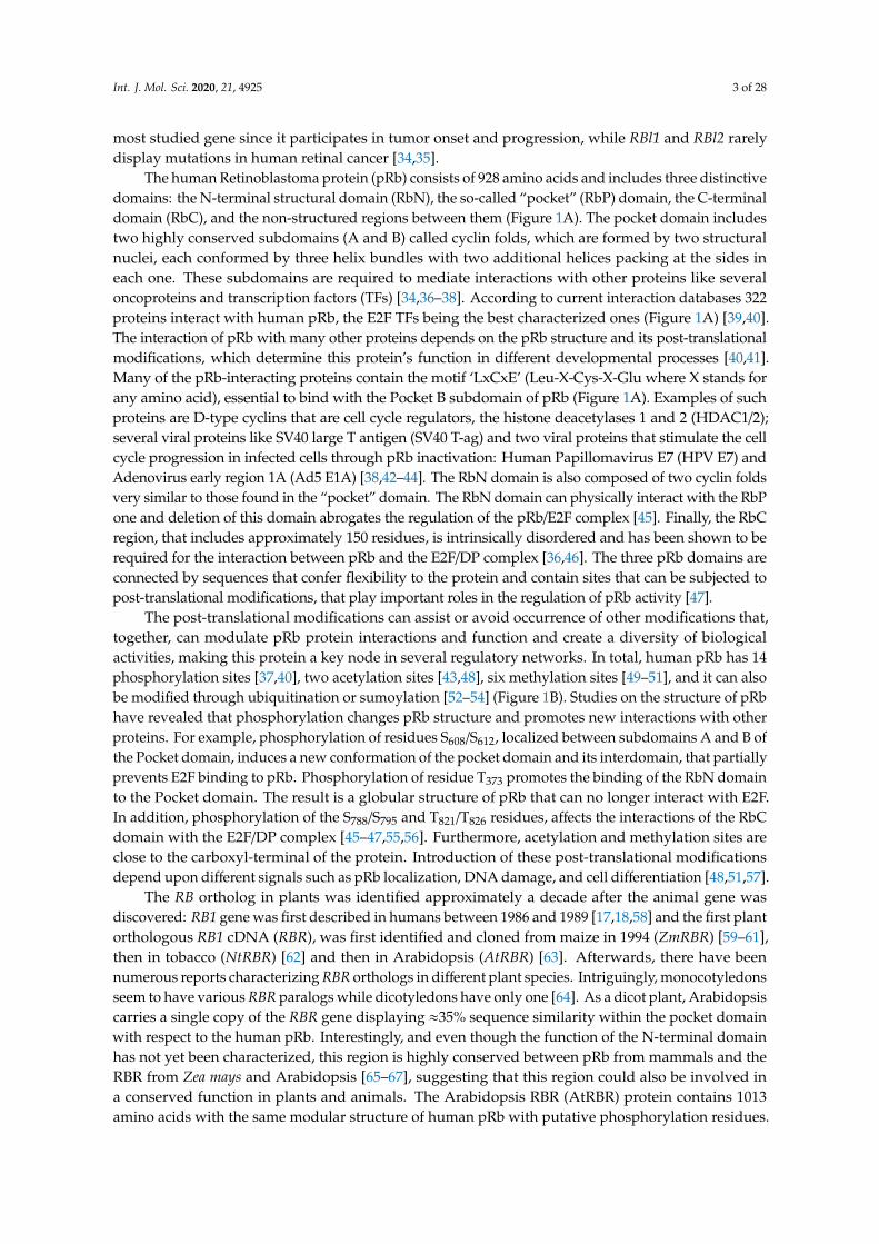
The human Retinoblastoma protein (pRb) consists of 928 amino acids and includes three distinctivedomains: the N-terminal structural domain (RbN), the so-called “pocket” (RbP) domain, the C-terminaldomain (RbC), and the non-structured regions between them (Figure 1A). The pocket domain includestwo highly conserved subdomains (A and B) called cyclin folds, which are formed by two structuralnuclei, each conformed by three helix bundles with two additional helices packing at the sides ineach one. These subdomains are required to mediate interactions with other proteins like severaloncoproteins and transcription factors (TFs) [34,36–38]. According to current interaction databases 322proteins interact with human pRb, the E2F TFs being the best characterized ones (Figure 1A) [39,40].The interaction of pRb with many other proteins depends on the pRb structure and its post-translationalmodifications, which determine this protein’s function in different developmental processes [40,41].Many of the pRb-interacting proteins contain the motif ‘LxCxE’ (Leu-X-Cys-X-Glu where X stands forany amino acid), essential to bind with the Pocket B subdomain of pRb (Figure 1A). Examples of suchproteins are D-type cyclins that are cell cycle regulators, the histone deacetylases 1 and 2 (HDAC1/2);several viral proteins like SV40 large T antigen (SV40 T-ag) and two viral proteins that stimulate the cellcycle progression in infected cells through pRb inactivation: Human Papillomavirus E7 (HPV E7) andAdenovirus early region 1A (Ad5 E1A) [38,42–44]. The RbN domain is also composed of two cyclin foldsvery similar to those found in the “pocket” domain. The RbN domain can physically interact with the RbPone and deletion of this domain abrogates the regulation of the pRb/E2F complex [45]. Finally, the RbCregion, that includes approximately 150 residues, is intrinsically disordered and has been shown to berequired for the interaction between pRb and the E2F/DP complex [36,46]. The three pRb domains areconnected by sequences that confer flexibility to the protein and contain sites that can be subjected topost-translational modifications, that play important roles in the regulation of pRb activity [47].
The post-translational modifications can assist or avoid occurrence of other modifications that,together, can modulate pRb protein interactions and function and create a diversity of biologicalactivities, making this protein a key node in several regulatory networks. In total, human pRb has 14phosphorylation sites [37,40], two acetylation sites [43,48], six methylation sites [49–51], and it can alsobe modified through ubiquitination or sumoylation [52–54] (Figure 1B). Studies on the structure of pRbhave revealed that phosphorylation changes pRb structure and promotes new interactions with otherproteins. For example, phosphorylation of residues S608/S612, localized between subdomains A and B ofthe Pocket domain, induces a new conformation of the pocket domain and its interdomain, that partiallyprevents E2F binding to pRb. Phosphorylation of residue T373 promotes the binding of the RbN domainto the Pocket domain. The result is a globular structure of pRb that can no longer interact with E2F.In addition, phosphorylation of the S788/S795 and T821/T826 residues, affects the interactions of the RbCdomain with the E2F/DP complex [45–47,55,56]. Furthermore, acetylation and methylation sites areclose to the carboxyl-terminal of the protein. Introduction of these post-translational modificationsdepend upon different signals such as pRb localization, DNA damage, and cell differentiation [48,51,57].
The RB ortholog in plants was identified approximately a decade after the animal gene wasdiscovered: RB1 gene was first described in humans between 1986 and 1989 [17,18,58] and the first plantorthologous RB1 cDNA (RBR), was first identified and cloned from maize in 1994 (ZmRBR) [59–61],then in tobacco (NtRBR) [62] and then in Arabidopsis (AtRBR) [63]. Afterwards, there have beennumerous reports characterizing RBR orthologs in different plant species. Intriguingly, monocotyledonsseem to have various RBR paralogs while dicotyledons have only one [64]. As a dicot plant, Arabidopsiscarries a single copy of the RBR gene displaying ≈35% sequence similarity within the pocket domainwith respect to the human pRb. Interestingly, and even though the function of the N-terminal domainhas not yet been characterized, this region is highly conserved between pRb from mammals and theRBR from Zea mays and Arabidopsis [65–67], suggesting that this region could also be involved ina conserved function in plants and animals. The Arabidopsis RBR (AtRBR) protein contains 1013amino acids with the same modular structure of human pRb with putative phosphorylation residues.

Int. J. Mol. Sci. 2020, 21, 4925 4 of 28
The role of four of them, located in the protein’s inter-domains, have been experimentally tested(Figure 1B) [68–70]. Interestingly, we observe that the S685 phosphorylation site in AtRBR is conservedin the same interdomain, between RbP A and RbP B subdomains, found in pRb (S608/S612), suggestinga conserved function in mediating the interaction with E2F TFs in animals and plants (Figure 1B).Additionally, human antibodies for human phospho-pRb protein in S807/811 can bind to RBR of Medicagosativa and Arabidopsis [71,72]. Even though the specific regulatory functions of these sites are stillunknown, it has been shown in plants that cyclin-dependent kinases also phosphorylate RBR toregulate cell cycle through its inactivation and release of E2F, similar to what it has been described inhumans [73,74].
Int. J. Mol. Sci. 2020, 21, x FOR PEER REVIEW 3 of 34
The human Retinoblastoma protein (pRb) consists of 928 amino acids and includes three distinctive domains: the N-terminal structural domain (RbN), the so-called “pocket” (RbP) domain, the C-terminal domain (RbC), and the non-structured regions between them (Figure 1A). The pocket domain includes two highly conserved subdomains (A and B) called cyclin folds, which are formed by two structural nuclei, each conformed by three helix bundles with two additional helices packing at the sides in each one. These subdomains are required to mediate interactions with other proteins like several oncoproteins and transcription factors (TFs) [34,36–38]. According to current interaction databases 322 proteins interact with human pRb, the E2F TFs being the best characterized ones (Figure 1A) [39,40]. The interaction of pRb with many other proteins depends on the pRb structure and its post-translational modifications, which determine this protein’s function in different developmental processes [40,41]. Many of the pRb-interacting proteins contain the motif ‘LxCxE’ (Leu-X-Cys-X-Glu where X stands for any amino acid), essential to bind with the Pocket B subdomain of pRb (Figure 1A). Examples of such proteins are D-type cyclins that are cell cycle regulators, the histone deacetylases 1 and 2 (HDAC1/2); several viral proteins like SV40 large T antigen (SV40 T-ag) and two viral proteins that stimulate the cell cycle progression in infected cells through pRb inactivation: Human Papillomavirus E7 (HPV E7) and Adenovirus early region 1A (Ad5 E1A) [38,42–44]. The RbN domain is also composed of two cyclin folds very similar to those found in the “pocket” domain. The RbN domain can physically interact with the RbP one and deletion of this domain abrogates the regulation of the pRb/E2F complex [45]. Finally, the RbC region, that includes approximately 150 residues, is intrinsically disordered and has been shown to be required for the interaction between pRb and the E2F/DP complex [36,46]. The three pRb domains are connected by sequences that confer flexibility to the protein and contain sites that can be subjected to post-translational modifications, that play important roles in the regulation of pRb activity [47].
Figure 1. Retinoblastoma protein structure. (A) Representation of the human Rb protein structure with the domains RbN (blue), Pocket (RbP), with the RbP A (purple) and B (grey) subdomains interacting with an E2F TF (red), the RbC domain (orange) and the inter-domains (black lines) are also shown. The “P” inside a circle represents three examples of phosphorylation sites that change
Figure 1. Retinoblastoma protein structure. (A) Representation of the human Rb protein structure withthe domains RbN (blue), Pocket (RbP), with the RbP A (purple) and B (grey) subdomains interactingwith an E2F TF (red), the RbC domain (orange) and the inter-domains (black lines) are also shown.The “P” inside a circle represents three examples of phosphorylation sites that change the structure ofthe protein. The position of the LxCxE cleft that allows Retinoblastoma protein (pRb) to interact withdifferent proteins is also shown. (B) Comparison of the domains of human Rb protein (blue foreground)and Arabidopsis RBR protein (green foreground) and their reported post-translational modifications.Phosphorylation sites (P) are shown in black, methylation sites (M) in red, acetylation sites (A) inorange, and sumoylation sites (S) in pink.
Like in humans, in plants there are viral proteins (e.g., RepA from geminivirus) that also have theability to interact with the pocket domain of RBR [61,75,76]. This suggests that the protein–proteininteraction between pRb/RBR with specific viral proteins is a viral mechanism that controls cellcycle progression both in plants and animals. Such viral–eukaryotic cell interaction could have beenestablished before plant and animal divergence, or it has evolved independently.
pRb is also found in unicellular organisms such as the algae Chlamydomonas reinhardtii,the choanoflagellate Monosiga brevicollis, and the amoeba Dictyostelium discoideum, suggesting that itwas already present in the common eukaryotic ancestor before the separation of animal and plant
Int. J. Mol. Sci. 2020, 21, 4925 5 of 28
lineages, before they diverged. In addition, at least one copy of RB has been identified in species fromeach of the eukaryotic supergroups [68,77].
3. Cell Cycle Control through Retinoblastoma
The cell cycle is a well-studied process essential for the growth and reproduction of all eukaryoticorganisms. It assures the faithful duplication of the genetic material and its distribution between twodaughter cells [78,79]. The cell cycle has two major phases: Interphase and Mitosis phase. Additionally,in the interphase other three stages can be distinguished: G1, S, and G2. Many of the components thatregulate these phases and the transition between them, are conserved among different organisms [80,81].The availability of growth factors, nutrients, and intrinsic developmental signals determine whether acell remains in a quiescent state, when the cell does not divide (G0), or transits from phase G1 to S,during which the genetic material is duplicated to later divide. In humans, this transition is under thecontrol of the pRb/E2F pathway, that regulates the transcription of genes encoding proteins involvedin DNA synthesis [22].
The G1/S transition is one of the main regulation checkpoints of the cell cycle, being the“commitment point”, also known as the “restriction point” in animals, the one that determines thecell’s commitment to engage proliferation in a way independent from environmental signals [82,83].Therefore, at this point the cell integrates environmental and intrinsic signals to prepare its nucleus tostart cell division. A deregulation of this transition in humans can lead to the generation of tumorsand cancer [22,84,85]. pRb hypophosphorylated acts as a negative regulator of cell cycle progressionthrough its interaction with the E2F proteins. The heterodimer keeps the chromatin in a closedconformation in the regulatory regions of E2F-regulated genes [22,86]. The E2F family includesthe transcription factors E2F1, E2F2, and E2F3a, which typically promote transcription and E2F3b,E2F4-E2F8 that are associated with transcriptional repression. However, it has been reported that,depending on the developmental stage, some E2Fs, like E2F1-4, can function both as activators oras repressors of transcription [87–89]. E2F1-6 members can heterodimerize with the DimerizationPartner (DP) proteins, although this interaction is not always required for transcriptional activation [90].E2F7 and E2F8 are independent of DP and these TFs are also different to other E2Fs because theypossess two DNA-binding domains [87,91,92]. The hypophosphorylated pRb form binds and inhibitsactivating E2F1-3, whereas E2F4 and E2F5 bind to pRBL1 (p107) and pRBL2 (p130) at the promoters oftarget genes, to repress transcription [22,93]. In addition, E2F1-3 carry a nuclear localization signal,whereas E2F4 and E2F5 lack it, and apparently rely on pRBL1 and pRBL2 for their nuclear translocation.On the other hand, E2F6-8 lacks the sequences required to bind with pRbs [94–96]. At the sametime, the expression of different E2Fs is itself subjected to spatiotemporal regulation during cell cycleprogression and different stages of development [97]. Moreover, when the pRb/E2F interaction isdisrupted by loss or reduction of pRb, a high rate of cell proliferation is observed, and this generallytriggers cancer [98].
pRb phosphorylation by cyclin/CDKs (cyclin-dependent kinases) complexes changes the pRbprotein structure and its interactions with other proteins, inducing the release of E2F. For instance,this occurs in human cells when cyclin type D or E (CYCD/E) associate with cyclin kinases 4 or 2(CDK4/2), respectively (Figure 2A) [22,99,100]. When pRb phosphorylation is altered, for example byoverexpression of CYCDs, or by a miss regulation of CDKs, or by a disruption of the LxCxE-bindingfunction the cell cycle is altered [101–103].
In humans, pRBL1 and pRBL2 participate in the repression of genes when cells are in theG0 quiescent state, through a complex called DREAM (DP-Rb-E2F-MuvB), that coordinates therepression of genes during quiescence and also the periodic gene expression during the G1/S and G2/Mtransitions [104,105]. DREAM is a multimeric protein complex that in humans is composed of DP(DP1-DP3), pRBL1 or pRBL2, E2F proteins (E2F4 and E2F5), and the subcomplex MuvB (Multi vulvalclass B). The MuvB subcomplex acts as a repressor when it is part of the DREAM complex, and iscomposed of LIN (LIN9, LIN37, LIN52, LIN54) and RBBP4 proteins. When pRb is phosphorylated,

Int. J. Mol. Sci. 2020, 21, 4925 6 of 28
the DREAM complex is disassembled and the MuvB subcomplex can associate with TFs such as B-Myb(Myb type) and Fox-M1 (Forkhead box protein M 1), to promote the regulation of gene expressionand the transition from quiescence to proliferation (Figure 2A) [104,106,107]. This complex is alsoconserved in phylogenetically distant organisms, but some of its components can vary among species,as in Arabidopsis compared to mammals (see below).
Int. J. Mol. Sci. 2020, 21, x FOR PEER REVIEW 6 of 34
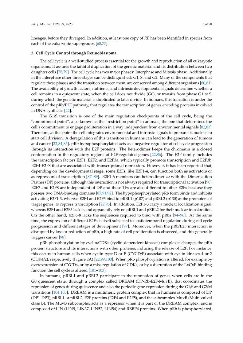
Figure 2. Interaction of the Retinoblastoma protein involved in cell cycle regulation of humans and plants. (A) pRb- and pRBL1/2-containing protein complexes from humans, formed at different stages of the cell cycle (G0/G1; G1/S; G2/M). (B) Arabidopsis protein complexes formed at different stages of the cell cycle (G0/G1; G1/S; G2/M), including AtRBR as a component. (C) Maize protein complexes formed at different stages of the cell cycle (G0/G1; G2/M), involving the ZmRBR proteins (RBR1 and RBR3) as components. Similar components in humans, Arabidopsis, and maize are displayed using the same colors: Rb proteins (blue), E2F transcription factors (TFs) (green), DP (grey), cyclins (CYC), and cyclin-dependent kinases (CDK) (orange), Muv complex proteins (red and pink), Myb TFs (purple), FOXM1 TF (pink).
At about the same time that the pRb ortholog in plants was discovered, homologs of other components of the animal’s cell cycle regulatory machinery were identified and characterized in corn, as well as in Arabidopsis and Medicago sativa (alfalfa) [61,108–111]. In these plants, it was shown that the phosphorylation of RBR by the CDKA/CYCD protein complex regulates cell cycle progression, as it occurs in humans (Figure 2B) [73,74].
In Arabidopsis, the null mutant plants of AtRBR are gametophytic lethal because of supernumerary nuclei alterations in the divisions at late stages of female gametogenesis; whereas the male gametophyte (pollen) contains multiple sperm cells [112,113]. Therefore, in order to study the function of RBR function of Arabidopsis and maize, either its transcript accumulation has been reduced by RNAi or conditional repression has been employed. In addition, protein competence for RBR binding, has also been used to address the role of RBR in cell proliferation [114–117].
In monocots, like rice, wheat, barley, and sugarcane, at least two different RBR types are present, RBR1 and RBR3 [64], although maize carries four ZmRBR genes: ZmRBR1, ZmRBR3, and two paralogs, ZmRBR2 and ZmRBR4 [118,119]. ZmRBR1 is constitutively expressed, and its protein interacts with E2F and with a HDAC (ZmRpd3I) that lacks the characteristic LxCxE motif. Besides, the two paralogs, ZmRBR1/2, negatively regulate cell cycle as it occurs in humans and Arabidopsis (Figure 2C) [119–122]. In addition, ZmRBR3/4 transcripts accumulate in mitotic tissues from the endosperm. Recently, high levels of ZmRBR3/4 were found in tumor-like formations (induced by the fungus Ustilago maydis) from maize leaves [118,123]. Interestingly, the protein complex ZmRBR3/4/E2F has a unique role not observed in other plants or animals: high levels of ZmRBR3/4 in complex with E2F promotes the expression of genes involved in DNA replication and cell cycle
Figure 2. Interaction of the Retinoblastoma protein involved in cell cycle regulation of humans andplants. (A) pRb- and pRBL1/2-containing protein complexes from humans, formed at different stagesof the cell cycle (G0/G1; G1/S; G2/M). (B) Arabidopsis protein complexes formed at different stages ofthe cell cycle (G0/G1; G1/S; G2/M), including AtRBR as a component. (C) Maize protein complexesformed at different stages of the cell cycle (G0/G1; G2/M), involving the ZmRBR proteins (RBR1 andRBR3) as components. Similar components in humans, Arabidopsis, and maize are displayed usingthe same colors: Rb proteins (blue), E2F transcription factors (TFs) (green), DP (grey), cyclins (CYC),and cyclin-dependent kinases (CDK) (orange), Muv complex proteins (red and pink), Myb TFs (purple),FOXM1 TF (pink).
At about the same time that the pRb ortholog in plants was discovered, homologs of othercomponents of the animal’s cell cycle regulatory machinery were identified and characterized in corn,as well as in Arabidopsis and Medicago sativa (alfalfa) [61,108–111]. In these plants, it was shown thatthe phosphorylation of RBR by the CDKA/CYCD protein complex regulates cell cycle progression, as itoccurs in humans (Figure 2B) [73,74].
In Arabidopsis, the null mutant plants of AtRBR are gametophytic lethal because of supernumerarynuclei alterations in the divisions at late stages of female gametogenesis; whereas the male gametophyte(pollen) contains multiple sperm cells [112,113]. Therefore, in order to study the function of RBRfunction of Arabidopsis and maize, either its transcript accumulation has been reduced by RNAi orconditional repression has been employed. In addition, protein competence for RBR binding, has alsobeen used to address the role of RBR in cell proliferation [114–117].
In monocots, like rice, wheat, barley, and sugarcane, at least two different RBR types are present,RBR1 and RBR3 [64], although maize carries four ZmRBR genes: ZmRBR1, ZmRBR3, and two paralogs,ZmRBR2 and ZmRBR4 [118,119]. ZmRBR1 is constitutively expressed, and its protein interacts withE2F and with a HDAC (ZmRpd3I) that lacks the characteristic LxCxE motif. Besides, the two paralogs,

Int. J. Mol. Sci. 2020, 21, 4925 7 of 28
ZmRBR1/2, negatively regulate cell cycle as it occurs in humans and Arabidopsis (Figure 2C) [119–122].In addition, ZmRBR3/4 transcripts accumulate in mitotic tissues from the endosperm. Recently, highlevels of ZmRBR3/4 were found in tumor-like formations (induced by the fungus Ustilago maydis)from maize leaves [118,123]. Interestingly, the protein complex ZmRBR3/4/E2F has a unique rolenot observed in other plants or animals: high levels of ZmRBR3/4 in complex with E2F promotesthe expression of genes involved in DNA replication and cell cycle progression; contrary to whathas been reported for overexpression of pRb and AtRBR (Figure 2C) [118,119,123,124]. Besides,ZmRBR3/4 is negatively regulated by the ZmRBR1-E2F complex; as in lines expressing RepA, whichinhibits ZmRBR1, ZmRBR3 is upregulated, suggesting a compensatory mechanism to ensure cell cycleprogression [118,125]. This mechanism has also been observed in undifferentiated germline cells inwhich the absence of pRb is compensated by pRBL1 (p107) to maintain the cell’s quiescent state [126].
The conservation in plants and animals of the “restrictions point” and of key cell cycle regulatorslike Cyclin-CDKs and RB/E2F, suggests that the common ancestor of these two groups of organismsalready had these components. Interestingly, there is a different number of genes encoding for eachcell cycle protein suggesting the emergence of different and novel lineage-dependent proteins betweenplants and animals [78]. For example, it is known that Arabidopsis has 10 genes that code for cyclin-D(CYCD) classified into seven subtypes [127,128], while in humans there are only three [129]. In contrast,nine E2F are found in humans (three activators and six repressors), and only six in Arabidopsis.Of these, AtE2Fa functions as activator, AtE2Fb functions either as an activator or repressor, dependingon the plant developmental stages; E2Fc is a repressor; and the other three members of this family(E2Fd/e/f) are atypical since they have a duplicated DNA-binding domain, do not heterodimerizewith DP and lack the trans-activating and Rb pocket-binding domains, and thus resemble E2F7/8 inanimals [130–133]. Finally, some components of the cell cycle regulatory machinery are plant-specificsuch as type B cyclin dependent kinases (CDKB1/2) [134,135], while others are animal-specific, such ascyclin E in humans [136,137].
The DREAM complex also appears to be conserved in plants [138,139]. In addition to the presenceof AtRBR, E2Fs/DPs, a MuvB-like complex has also been found that contains ALY2/3 (orthologsof LIN9) and TCX5 that is part of the TSO1-like family members (orthologs of LIN54) [140,141].Furthermore, MYB3R, a transcription factor of the Myb family, has a protein structure resembling theDNA binding domain of B-Myb, which in humans forms part of the MuvB complex. These plant’sMYB3Rs also participate in the regulation of the G2/M transition as follows: MYB3R3 associateswith E2Fc and AtRBR to repress G2/M genes, while MYB3R4 associates with E2Fb and AtRBR toactivate G2/M genes (Figure 2B) [142–144]. CDKA;1 and cyclins could as well be involved in theplant DREAM complex since MYB3R3 and MYB3R4 interact with CDKA;1 and, in tobacco, CDKA;1regulates MYB3R phosphorylation [139,144,145]. Moreover, E2Fc can physically interact with CDKA;1,CYCD2;1, and CYCD2;2 in vitro (Figure 2B) [146]. Interestingly, the function of the activation complexMYB3R4/AtRBR1/E2Fb is similar to the function of ZmRBR3 in maize that positively regulates transcriptaccumulation of genes involved in DNA replication and cell cycle progression in the transition G2/M(Figure 2B,C) [124]. However, in contrast to what has been reported in animals, RBR presence in theDREAM complex is able to control cell cycle in different stages of cell cycle in plants (Figure 2B,C).
4. The Roles of Retinoblastoma in Cell Differentiation
The formation of any organ relies on two different but interlinked cellular processes: cellproliferation and cell differentiation. Proliferation produces all the cells that later will acquirefates, specialized functions, and morphologies through differential gene expression during celldifferentiation [147,148]. pRb has been widely studied in proliferation and, recently, its participation inmany different animal differentiation processes in the eye, brain, peripheral nervous system, muscle,liver, placenta, lung, cerebellum, pituitary gland, and heart has been elucidated (Figure 3A) [149–154].
The participation of pRb in these processes has been studied in vivo in mutant mice and/or cellcultures derived from cancer cells, characterized by alterations in their RB1 expression levels [155–157].

Int. J. Mol. Sci. 2020, 21, 4925 8 of 28
Mice with RB1 ablation are embryonic lethal, and those with low levels of the three pRb (RB1,RBl1, and RBl2) not only die in utero but also present defects in erythroid, neuronal, and musculardifferentiation [158–160]. Additionally, in mice with conditionally-regulated levels of the three pRb,cells overproliferated inducing retinal cancer (Retinoblastoma) and also display defects in laminarorganization of the retina and the crystalline [161–163]. E2Fs target genes are also important fordifferentiation of the adipose tissue, bone, nervous system, and muscles [164–167]. Interestingly,expression of pRb in mutants with altered E2F function has shown that pRb can also independentlyregulate tissue-specific genes in mammals, pointing to its broad roles in development [168,169].
Similar to pRb in animals, RBR participates in differentiation processes in the rootand shoot meristems, the vascular system, leaves, stomas, and trichomes tissues in plants(Figure 3A) [116,117,170,171]. In Arabidopsis, AtRBR functions as a negative regulator of primary rootdevelopment; its downregulation leads to longer roots with larger meristems whereas its overexpressionresults in shorter roots with smaller meristems [172]. In this organ, AtRBR forms a protein complexwith the TF ARABIDOPSIS RESPONSE REGULATOR12 (ARR12) that activates the transcriptionof the AUXIN RESPONSE FACTOR19 (ARF19) TF [172]. These TFs participate in two differenthormone signaling pathways: ARR12 is part of the cytokinin signaling pathway involved in rootdifferentiation, while ARF19 is part of the auxin signaling pathway which is important for rootcell proliferation [172,173]. ARF19 is activated by RBR mostly in between the meristematic and theelongation zones in roots and it is suggested to promote differentiation [172].
The role of RBR in differentiation at the shoot apical meristem (SAM) has also been analyzed.In this case, overexpression of AtRBR accelerates differentiation, increasing the expression of genesinvolved in metabolic pathways that are not present in the SAM and decreasing the expression ofgenes that are only expressed in the SAM [114,170]. AtRBR RNA interference (RNAi) in leaf primordiadelays differentiation and, consequently, increments two to four-fold the cell number in both theadaxial and abaxial sides of the leaves [114,170]. In addition, also in leaves, AtRBR participates in theendoreplication and differentiation of trichomes, in which the AtRBR transcription is regulated by theTFs GLABRA1 (GL1) and GLABRA3 (GL3) [174,175]. In the vascular system, AtRBR associates withXYLEM NAC DOMAIN1 (XND1), a negative regulator of differentiation, thus controlling processesrelated to the differentiation of tracheary elements [176,177].
It is difficult to compare the roles of pRb and RBR during differentiation due to lack of informationand because the components involved in cell differentiation are more species-specific and less conservedthan those involved in cell cycle regulation. However, taking as examples skeletal muscle differentiationin humans and stomatal guard cells differentiation in Arabidopsis, conserved factors that coordinatecell differentiation can be identified, namely Rb, MADS-box TFs, and TFs of the bHLH family, as wellas the cyclins/CDKs protein complexes (Figure 3D) [178,179].
At post-embryonic stages, myocyte differentiation in mammals can be triggered in response tomuscle damage or a specific growth-inducing stimulus. This last provokes massive proliferationof myoblasts through the hierarchical activation of several MRF (Myogenic Regulatory Factors):Myogenic Factor 5 (Myf5), myoblast determination protein 1 (MyoD), Myogenin (MyoG), and MyogenicRegulatory Factor 4B (MRF4), which are TFs that belong to the bHLH family. These MRF are requiredtogether with pRb for the muscle regeneration process [180–182]. In resting state conditions, muscle stemcells or satellite cells express only the box protein Pax7 [183]. When the muscle is injured, satellite cellsget activated and become myoblasts that massively proliferate to generate the myogenic progenitors,which express Myf5 and/or MyoD TFs [184]. Later, when myoblasts start differentiation into myocytes,Myf5 and Pax7 are repressed and MRF4 and myogenin are expressed promoting cell cycle exit. Finally,when the cells fuse to become myofibrils, myogenin and MRF4 are expressed as part of the final steps ofdifferentiation (Figure 3B) [180,182]. In this process, the protein complex of MyoD with pRb, is thoughtto initiate differentiation, as it promotes cell cycle arrest [185–188]. Despite immunoprecipitationexperiments that proved that pRb and MyoD can interact [189], later experiments using nuclearmagnetic resonance allowed to determine that there is no direct protein–protein interaction between

Int. J. Mol. Sci. 2020, 21, 4925 9 of 28
MyoD and pRb [190]. Still, MyoD does activate RB1 expression through its association with thecAMP response element-binding (CREB) TF and the coactivators p300 and P/CAF (Figure 3) [191,192].Myocyte Enhancer Factor 2 (MEF2), that belongs to the MADS-box family of TFs, is also a componentnecessary to carry out the final stages of muscle differentiation [149,193,194].
Int. J. Mol. Sci. 2020, 21, x FOR PEER REVIEW 9 of 34
regulated by the joint action of MyoD and pRb through the regulation of the upstream intermediary gene Fra-1 (antigen related to FOS 1) (Figure 3B) [196,197]. Antagonistically, cyclin D1 inhibits the activity of MyoD: overexpression of cyclin D1 promotes nuclear accumulation of CDK4, that binds MyoD, preventing its interaction with DNA, and inhibiting the CDK4-dependent phosphorylation of pRb (Figure 3B) [198–200]. Additionally, when HDAC1 interacts with pRb, the MyoD protein can bind its target DNA regulatory sites [201,202]. In summary, there are two different protein complexes formed during different stages of muscle development: HDAC1/MyoD during proliferation and HDAC1/hypophosphorylated pRb during differentiation (Figure 3B) [201,202].
Figure 3. Retinoblastoma proteins are involved in cell differentiation in mammals and plants. (A) Mammals’ and plants’ organs whose differentiation depends on pRb/RBR. (B) Sequential steps of
Figure 3. Retinoblastoma proteins are involved in cell differentiation in mammals and plants.(A) Mammals’ and plants’ organs whose differentiation depends on pRb/RBR. (B) Sequential stepsof skeletal muscle differentiation in mammals. Shown are pRb interactions and bHLHs-familyproteins (Mef2, MyoD, Myogenin, MRF4), involved in muscle quiescence maintenance, proliferation,and differentiation. (C) Sequential steps of guard cells differentiation in Arabidopsis. Shown areRBR interactions and bHLHs-family proteins (SPCH, MUTE, FAMA, SCRM, SCRM2) involved inquiescence maintenance, proliferation, and differentiation of guard cells. (D) Correlations betweencomponents involved in muscle and guard cell development in mammals and Arabidopsis, respectively.Differentiation in both lineages involves proteins of the pRb, bHLH TF, cyclins and CDKs, MADS-boxTF families.

Int. J. Mol. Sci. 2020, 21, 4925 10 of 28
Induction of muscle biogenesis also requires the regulation by cyclins-CDKs in association withpRb [195]. The stable repression of cyclin D1, required for cell cycle arrest during differentiation,is regulated by the joint action of MyoD and pRb through the regulation of the upstream intermediarygene Fra-1 (antigen related to FOS 1) (Figure 3B) [196,197]. Antagonistically, cyclin D1 inhibits theactivity of MyoD: overexpression of cyclin D1 promotes nuclear accumulation of CDK4, that bindsMyoD, preventing its interaction with DNA, and inhibiting the CDK4-dependent phosphorylationof pRb (Figure 3B) [198–200]. Additionally, when HDAC1 interacts with pRb, the MyoD protein canbind its target DNA regulatory sites [201,202]. In summary, there are two different protein complexesformed during different stages of muscle development: HDAC1/MyoD during proliferation andHDAC1/hypophosphorylated pRb during differentiation (Figure 3B) [201,202].
In plants, some epidermal cells undergo differentiation producing the two mature guard cellsthat form the stomata pores, structures that are conserved among land plants and allow them toregulate gas exchange and water loss [203,204]. Stomatal development is hierarchically regulated bythe sequential activation of several TFs of the bHLH family: SPCH (SPEECHLESS), MUTE, and FAMA.These three bHLHs form heterodimers with either the bHLHs SCREAM (SCRM, also called ICE1) orSCRM2 [205–207], that belong to the same family of TFs that participate in muscle development inmammals (MRFs) (Figure 3D). These plant TFs orchestrate cell division events of protodermal cells,which give origin to guard cells (stomatogenesis). SPCH triggers the maturation of a protodermalcell into a meristemoid mother cell (MMC) and is also involved in the asymmetric cell division of theMMC, that results in one meristemoid cell and one larger sister cell (SLGC). The meristemoid cellexits stemness and engages in differentiation to become a guard meristemoid cell (GMC). MUTE mustbe expressed at this stage, to direct further differentiation of a GMC, and to ensure that this cellundergoes a single symmetric division. In addition MUTE regulates the expression of FAMA, whichcontrols the final stages of differentiation, promoting guard cell (GC) identity acquisition and theirreversible termination of the meristematic activity of the cells (Figure 3C) [208–210]. AtRBR playsimportant roles in the regulatory network of stomata development, and its downregulation at theGMC or GC stages, induces extra divisions in differentiated GCs and the formation of aberrantstomata-in-stomatal nested structures [114,211]. In fact, AtRBR hyperphosphorylation inhibits stomatalinitiation affecting the asymmetric division of protodermal cells that produces MMCs, this seems to becontrolled by CDKA;1, that negatively regulates AtRBR, and regulates positively SPCH TF throughphosphorylation (Figure 3C) [212–214]. It has also been hypothesized that AtRBR hyperphosphorylationby CDKB1;1-CYCD7;1 inhibits the AtRBR/FAMA repression complex leading to the induction ofcell-cycle regulators of the GMC symmetric division event [215–217]. At the same time, MUTE directlyupregulates FAMA and FLP; and FAMA represses cell-cycle control genes such as CDKB1;1, ensuring asingle symmetric division to form GCs (Figure 3C) [208,218]. Mutation in the FAMA LxCxE sequenceprevents the formation of the AtRBR/FAMA complex, making cells unable to maintain the long-termcommitment to differentiate into GC, and arresting this process at the GMC stage [211,215]. A similarmechanism is present in mammals, in which downregulation of MyoD allows cells to dedifferentiate;an ability that determines the muscular capacity to regenerate [219]. Finally, it is also possible thatFAMA functions at early steps of guard cell differentiation since the AtRBR/FAMA heterodimer bindsto SPCH and FAMA promoters, and this complex negatively regulates the accumulation of the SPCHtranscript, which is normally expressed at early stages of guard cells development (Figure 3C) [211,220].
As it can be appreciated, the differentiation processes of skeletal muscle in mammals and guardcells in Arabidopsis are both regulated by a similar set of conserved elements: pRb/RBR, bHLHs, cyclinsand CDKs (Figure 3D). Moreover, in both organisms, members of the MADS-box family participate inthese processes: MEF2 is involved in early stages of muscle differentiation and in cell proliferation ofother tissues (Figure 3B) [221–223]. Similarly, a plant MADS-box gene, AGAMOUS-LIKE16 (AGL16),is expressed and participates in GC development (Figure 3D) [224,225]. AGL16 mediates the stomatadevelopment process at the MMC cell lineage level and represses FAMA as well as other genes involvedin the development and differentiation of guard cells (Figure 3C) [225,226].

Int. J. Mol. Sci. 2020, 21, 4925 11 of 28
5. Retinoblastoma Function in Stem Cells Homeostasis
Accumulated evidence indicates that pRb loss of function in mammals results in altered progenitorcells (or stem cells) quiescence. Quiescent cells are usually in the G0 phase of the cell cycle or in aprolonged G1 phase; and have a very low proliferative activity. Through asymmetric division stem cellscan give origin to a new quiescent stem cell and a new daughter cell that can proliferate several timesto eventually produce one or more differentiated cell types [227–229]. pRb participates in pluripotencymaintenance, inhibition of differentiation, and in self-renewal of stem cells [230–232]. Mutants withlow expression levels of Rb display an increased cell division of both embryonic and post-embryonicstem cells from retina, mesenchymal, and early osteoblasts progenitors, as well as of post-embryonicstem cells of the liver, muscle, and nervous system [233–239]. Interestingly, in these pRb mutantssomatic cells acquire stem cells features, as it occurs in some human cancers [240,241]. In humanpluripotent stem cells (hPSC), the pRb/E2F pathway enhances differentiation towards all germ layersin response to a DMSO stimulus [153]. pRb together with E2F1 can bind and suppress the transcriptaccumulation of pluripotency promoting factors, such as SEX DETERMINING REGION Y-BOX 2(SOX2) and OCTAMER-BINDING TRANSCRIPTION FACTOR 4 (OCT4). In addition, pRb can alter theaccumulation of transcripts by directing chromatin modifiers to promoters of specific TFs, as it happensin the case of KRUPPEL-LIKE FACTOR 4 (KLF4), the homeobox NANOG and TRANSCRIPTIONFACTOR 3 (TCF3), that are part of the induced stem cell pluripotency regulatory network; and also inthe case of ENHANCER OF ZESTE HOMOLOG 2 (EZH2), which is a methyltransferase that participatesin establishing stem cells establishment [242,243].
The plant ortholog, RBR, is also implicated in pluripotency maintenance. Lowering transcriptlevels of AtRBR induces disorganization of the root stem cell niche (SCN) in Arabidopsis, as well as inthe shoot meristem that harbors flower and leaf stem cells [114–116]. The root SCN consists of a centralorganizer, the quiescent center (QC), which is surrounded by four type of stem cells called initialcells, that in Arabidopsis are columella initials (CI), cortex/endodermis initials (CEI), epidermis/lateralroot cap initials (ELRCI), and stele initials (SI) [244]. Under normal growth conditions, proliferationrate at the QC is very low compared to adjacent zones, although the division rate increases at theQC in older seedlings [245,246]. AtRBR silencing increases the proliferation of both QC and CI cells,resulting in an overgrowth of undifferentiated cell layers. Conversely, overexpression of AtRBR causespremature differentiation of CIs [115,116,247]. Interestingly, absence of AtRBR favors the duplicationof differentiated columella cells that normally do not duplicate in wild-type plants [247].
Within CEI cells and their progeny, the TF SCARECROW (SCR), a member of the GRAS TFsfamily, interacts, through its LxCxE motif, with AtRBR forming a ternary complex with SHORTROOT(SHR), which is another member of the GRAS family. This AtRBR/SCR/SHR complex inhibits thetranscription of target genes of the SHR/SCR heterodimer. One of these target genes is CYCLIN D6;1(CYCD6;1), which controls the cell cycle progression of the CEI cells progeny. Moreover, CYCD6;1together with the kinase CDKB1;1, in turn, regulates the phosphorylation of AtRBR, liberating theSCR/SHR complex, favoring in this way CEI cells’ asymmetric division [115,248,249]. AtRBR, SCR,and CYCD6;1 are degraded by the proteasome before mitosis, which is consistent with a model wherethe degradation of these proteins allows CEI cells to restart the asymmetric divisions [248]. Moreover,the same AtRBR/SCR interaction is necessary to establish the QC cells and is regulated by the interactionof AtRBR with the ETHYLENE RESPONSE FACTOR115 (ERF115) TF, that belongs to the AP2/ERFfamily. This interaction also occurs through the LxCxE domain of ERF115, that competes with SCRfor AtRBR, reducing in this way the levels of the AtRBR-SCR heterodimer [250]. Importantly, most ofthe AtRBR functions related to differentiation and cell cycle arrest in the SCN are cell-autonomous,highlighting the crucial role of AtRBR activity level in the QC, in the columella stem cells, and in theirimmediate progeny in the acquisition of niche-specific features [247].
Interestingly, it has also been observed that the TOPOISOMERASE 1α (TOP1α) together withAtRBR, is also involved in the control of stem cell maintenance during root development. TOP1α is anenzyme present in plants and animals that creates breaks in double DNA strands to relax supercoiled

Int. J. Mol. Sci. 2020, 21, 4925 12 of 28
structures. In humans, it has been used as a target to stop the proliferation of breast cancer stem cellsin cell cultures [251,252]. In Arabidopsis roots it has been shown that TOP1α is downregulated byAtRBR to maintain the undifferentiated state of cells and the number of CI cells in the SCN. In addition,TOP1α is epistatic over AtRBR and its overexpression results in an increased number of CI cells, as ithappens in AtRBR loss of function mutants [253]. Besides, mutations that disrupt the activity of TOP1α,induce cell death in the initial cells of the stele (SI) which can be partially reversed by the activation ofERF115 expression, since TOP1α negatively regulates the expression of ERF115 [253], and as it wasmentioned above, ERF115 also interacts with AtRBR to establish the QC [250]. Although it is not yetknown whether TOP1α participates with AtRBR in the shoot meristem or not, TOP1α also regulatesthe establishment of stem cells through indirect transcriptional repression of WUS (WUSCHEL) [254].
In the shoot meristem, silencing of AtRBR also produces disordering of the stem cell divisionswithin the SAM, resulting in a significant reduction in the typical height-to-width ratio of the SAM; andalso in alterations in stem cell maintenance and differentiation [114]. Overexpression of AtRBR triggerscells toward a more differentiated state; but the molecular mechanism has not yet been described toexplain this phenotype [170]. In addition, AtRBR regulates proliferation and differentiation of MMC ofguard cells in leaves as it has been described above.
It can be noted from the examples presented, that pRb/RBR of animals and plants, respectively,participate in the maintenance of stem cells by controlling the cell cycle and cell differentiation, as wellas regulating specific genes that give identity to these cells.
6. Function of Retinoblastoma in Epigenetic Modifications, Chromatin Regulation,and DNA Damage
Epigenetic modifier proteins participate in the regulation of gene expression throughout development.This allows cells with the same genetic background to exhibit different phenotypes [255,256]. Epigeneticmodifications alter DNA accessibility and chromatin structure by mechanisms such as DNA methylationand histones modifications by acetylation, methylation, ubiquitination, phosphorylation, sumoylation,etc. [257,258]. In mammals and plants, many epigenetic modifier proteins interact with proteincomplexes that include pRb/RBR, allowing them to regulate different developmental processes.
Human pRb has been reported to interact with over 300 proteins and many such protein interactionsare epigenetic modifier proteins or interact with the latter [40,256]. pRb levels decrease leads to anincomplete chromosome condensation and segregation during mitosis, as it has been observed incancer cells; some alterations of the chromatin structure are also induced by changes in histonemethylation and acetylation [259–261]. pRb can interact with chromatin remodeling factors, such ashistone deacetylases, DNA methyltransferases, histone methyltransferases, and with complexes likeSWI/SNF; and like Polycomb group (PcG), the latter is a chromatin-modifying complex that maintainsrepressed gene expression states and is subdivided into two main complexes: Polycomb repressivecomplex 1 (PRC1) and PRC2 [259,262–265].
The interaction of pRb-epigenetic with modifiers complexes are also important to maintainheterochromatin in intergenic zones as well as in centromeres and telomeres (Figure 4A) [260,261].The interaction of pRb with ENHANCER OF ZESTE HOMOLOG 2 (EZH2), a histone methyltransferaseof the PRC2 complex, allows the deposition of the trimethylation of lysine 27 of histone H3 (H3K27me3),a repressive mark, on pRb target genes (Figure 4A). In turn, pRb-E2F negatively regulates EZH2transcript accumulation and proliferation; conversely high expression of EZH2 is observed incancer stem cells as has a critical function in regulating stem cell expansion and maintenance(Figure 4A) [242,266]. Interestingly, it has been observed that pRb can also recruit EZH2 protein intosequences within introns and intergenic regions, specifically in repeated sequences, transposons, longinterspersed nuclear elements (LINEs), short interspersed nuclear elements (SINEs), and long terminalretroviruses (LTR). The loss of the pRb-EZH2 complex provokes loss of the H3K27me3 mark at theseelements, leading to dispersion or loss of heterochromatin and probably disorganized proliferation asobserved in cancer cells (Figure 4A) [267].

Int. J. Mol. Sci. 2020, 21, 4925 13 of 28
Int. J. Mol. Sci. 2020, 21, x FOR PEER REVIEW 14 of 34
Figure 4. pRb and RBR are involved in modifications and DNA repair mechanisms both in mammals and plants. (A) Mammalian pRb participates in developmental processes and chromatin localization together with EZH2, a component part of the PRC2 complex. (B) Arabidopsis RBR participates in development and chromatin localization together with the PRC2 complex. (C) Mammalian pRb is part of the machinery involved in DNA repair. (D) Arabidopsis RBR is part of the machinery involved in DNA repair. Proteins and complexes conserved between mammals and Arabidopsis are marked with the same colors.
In plants, there are also numerous reports of RBR interactions with epigenetic modifiers, which are important in the regulation of different developmental processes [277]. In Arabidopsis, like in
Figure 4. pRb and RBR are involved in modifications and DNA repair mechanisms both in mammalsand plants. (A) Mammalian pRb participates in developmental processes and chromatin localizationtogether with EZH2, a component part of the PRC2 complex. (B) Arabidopsis RBR participates indevelopment and chromatin localization together with the PRC2 complex. (C) Mammalian pRb is partof the machinery involved in DNA repair. (D) Arabidopsis RBR is part of the machinery involved inDNA repair. Proteins and complexes conserved between mammals and Arabidopsis are marked withthe same colors.
DNA integrity in mammals is altered when pRb is absent and, in some cases, this can be provokedby the overexpression of E2F regulated genes that are able to introduce double-strand DNA breaks,or by stress conditions that generate aneuploidies [37]. In the case of a double strand break, pRbis necessary to form the complex of the heterodimer E2F1-pRb with TopBP1 (DNA topoisomerase

Int. J. Mol. Sci. 2020, 21, 4925 14 of 28
2-binding protein 1) [268]. TopBP1 is a protein that interacts with Topoisomerase 2β (Top2β) and withother proteins that participate in DNA replication and in the maintenance of the DNA integrity andgenome stability [261,269]. In addition, the protein complex E2F1-pRb-TopBP1 interacts with BRG1(also known as ATP-dependent chromatin remodeler SMARCA4) which is a member of the SWI/SNFcomplex that is necessary to reduce nucleosome density at injury sites, allowing DNA end resectionand reparation by homologous recombination (HR) (Figure 4C) [268,270]. Another novel aspect of thepRb-BRG1 interaction is its influence in mediating cell cycle arrest, by the regulation of different genesalso involved in human cancer cells (Figure 4C) [271–273]. pRb interacts with the tumor suppressorBRCA1 (Breast cancer 1), which is also involved in DNA repair via Homologous Recombination.The BRCA1-pRb complex interacts with histone deacetylases (HDAC1/2) and with topoisomerase2β (Top2β) to regulate DNA stability (Figure 4C) [274,275]. Furthermore, the pRb-BRCA1 complexis involved not only in the response to DNA damage, but also in cell cycle control, as deletions inthe BRCA1 binding domain with pRb, inhibits BRCA1-dependent cell cycle progression [262]. pRbalso participates in another DNA repair pathway: the non-homologous end joining (NHEJ), the exactrole of pRb in this mechanism is unknown but it has been reported that pRb interacts with two of theproteins that recognize the breakdown of the double chain: KU-70 and KU-80 (Figure 4C) [261,276].
In plants, there are also numerous reports of RBR interactions with epigenetic modifiers, whichare important in the regulation of different developmental processes [277]. In Arabidopsis, like inanimals, it has been shown that PRC2, a subcomplex of PcG, participates together with AtRBR inthe establishment of the H3K27me3 mark during differentiation and development of the femaleand male gametophytes, in leaf development and during the establishment of stoma cell lineages.In these three processes, AtRBR associates with components of the PRC2 repressor complex such asMULTICOPYSUPPRESSOR OF IRA1 (MSI1), FERTILIZATION INDEPENDENT ENDOSPERM (FIE),VERNALIZATION 2 (VRN2), and CURLY LEAF (CLF), a gene orthologous to EZH2 from humans(Figure 4B) [66,278]. In addition, AtRBR together with MSI1 directly represses the expression of theDNA methylase METHYLTRANSFERASE 1 (MET1) (Figure 4B), that maintains DNA methylationduring DNA replication and regulates gene imprinting. The repression of MET1 by this complex allowsthe transcriptional activation of FERTILIZATION INDEPENDENT SEED 2 (FIS2) and FLOWERINGWAGENINGEN (FWA), that are important for female gametogenesis [66,113,279]. In turn, MET1 ispositively expressed during male gametogenesis; and is important for maintaining the gene repressionof FIS2 and FWA in the paternal allele, leading the monoparental expression of these genes duringfertilization and endosperm development, [279,280]. Furthermore, AtRBR loss of function mutantspresent higher levels of SWINGER (SWN), MSI1, and FIE transcripts, which are components of thePRC2 complex. Interestingly, the AtRBR transcript in pollen is directly repressed by the PRC2 complex(Figure 4B) [113]. In plant embryos, the PRC2 complex with AtRBR directly binds and depositsthe H2K27m3 mark on different embryonic genes, leading to their repression and subsequent seedgermination (Figure 4B) [69,281]. Similarly, in stomatal development, AtRBR/FAMA heterodimer isrequired to recruit PRC2 to H3K27me3 deposition into SPCH and MUTE regulatory regions, and repressits transcript accumulation, necessary to control differentiation and stomatal development correctly(Figure 4B) [211,215,282].
AtRBR also appears to regulate DNA repair in several conditions. First, AtRBR binds and repressesgenes involved in homologous recombination such as RADIATION SENSITIVE 51 (RAD51) and helpsto locate RAD51 to the right place at DNA lesions (Figure 4D) [283]. Additionally, TOP1α is criticalto ensure genome integrity and survival of root stele stem cells, as the loss of function of TOP1αtriggers DNA double-strand breaks and cell death in these cells; in the root, TOP1α is downregulatedby AtRBR (Figure 4D) [253]. Although the participation of AtRBR and Top1α in the shoot meristemhave not yet been studied, TOP1α participates with the PRC2 complex in the repression of the WUSlocus (Figure 4D) [252–254].
AtRBR also is recruited to damaged DNA sites, along with E2Fa and AtBRCA1 and helps tomaintain the integrity of the root meristem (Figure 4D). Furthermore, similar to what is observed

Int. J. Mol. Sci. 2020, 21, 4925 15 of 28
for animals for BRCA1 and pRb (Figure 4C) [262,284], AtRBR and AtBRCA1 have been shown tophysically interact when cells are damaged [285]. In addition, E2Fa is required for AtBRCA1 expression,when genotoxic stress is induced (Figure 4D) [285]. Thus, it would be interesting to analyze if theAtBRCA1-AtRBR complex participates in the regulation of the cell cycle, as it occurs in humans. Finally,analysis of chromosome sites to which AtRBR physically binds, show that this protein not only targetsgene regulatory sequences, but also transposons, especially Miniature Inverted-repeat TransposableElements (MITEs) (Figure 4B) [142].
7. Conclusions
Development is a process where proliferation and differentiation cellular rates must be finelyregulated. As we can appreciate from the examples presented throughout the text, pRb/RBR aremultifunctional and highly connected proteins that control cell fate determination and differentiationthrough interactions with different proteins. The pRb/RBR structures and diverse post-translationalmodifications allow the proteins to differentially interact with an exceptionally high number of proteins,making them a key node in several regulatory networks. Interestingly, many of the protein partnersare conserved between animals and plants, and in both lineages are involved in equivalent cellularprocesses such as cell cycle regulation, stem cell homeostasis, and cell differentiation. In addition,interaction with epigenetic and DNA topology regulators suggests that the protein–protein networksthat involve RETINOBLASTOMA are also similar in plants and animals. Thus, important aspectsof the regulatory networks underlying cell proliferation and differentiation in which this protein isinvolved, seem to be shared by plants and animals, despite the fact that these two lineages have uniquecellular and structural characteristics.
Author Contributions: E.Z.-M. and A.G.-A. conceived and wrote the review. V.P.-K., M.V.P.-C., S.M.-H., B.G.-P.,M.d.l.P.S. and E.R.Á.-B. wrote the review. All authors have read and agreed to the published version ofthe manuscript.
Funding: A.G.-A., M.d.l.P.S., B.G.-P. and E.R.Á.-B. received funding from UNAM-DGAPA-PAPIIT (IN200920,IN203220, IN206220, IN208517). E.Z.-M. was financially supported by CONACYT through a Ph.D. scholarship.
Acknowledgments: We would like to thank Diana Belén Sánchez Rodríguez for logistical and technical supportand to Crisanto Gutiérrez (Universidad Autónoma de Madrid, Spain) for comments on the manuscript.
Conflicts of Interest: The authors declare no conflict of interest and the research was conducted in the absence ofany commercial or financial relationships.
References
1. Koonin, E.V. The Incredible Expanding Ancestor of Eukaryotes. Cell 2010, 140, 606–608. [CrossRef] [PubMed]2. Koonin, E.V. The origin and early evolution of eukaryotes in the light of phylogenomics. Genome Biol. 2010,
11, 209. [CrossRef] [PubMed]3. O’Malley, M.A.; Leger, M.M.; Wideman, J.G.; Ruiz-Trillo, I. Concepts of the last eukaryotic common ancestor.
Nat. Ecol. Evol. 2019, 3, 338–344. [CrossRef] [PubMed]4. Chen, S.; Krinsky, B.H.; Long, M. New genes as drivers of phenotypic evolution. Nat. Rev. Genet. 2013, 14,
645–660. [CrossRef]5. Meyerowitz, E.M. Plants, animals and the logic of development. Trends Cell Biol. 1999, 9, M65–M68.
[CrossRef]6. Meyerowitz, E.M. Comparative genomics. Plants compared to animals: The broadest comparative study of
development. Science 2002, 295, 1482–1485. [CrossRef] [PubMed]7. Grosberg, R.K.; Strathmann, R.R. The Evolution of Multicellularity: A Minor Major Transition? Annu. Rev.
Ecol. Evol. Syst. 2007, 38, 621–654. [CrossRef]8. West, M.; Harada, J.J. Embryogenesis in Higher Plants: An Overview. Plant Cell 1993, 5, 1361–1369. [CrossRef]
[PubMed]9. Jones, A.M.; Chory, J.; Dangl, J.L.; Estelle, M.; Jacobsen, S.E.; Meyerowitz, E.M.; Nordborg, M.; Weigel, D.
The Impact of Arabidopsis on Human Health: Diversifying Our Portfolio. Cell 2008, 133, 939–943. [CrossRef]

Int. J. Mol. Sci. 2020, 21, 4925 16 of 28
10. Xu, X.M.; Møller, S.G. The value of Arabidopsis research in understanding human disease states. Curr. Opin.Biotechnol. 2011, 22, 300–307. [CrossRef]
11. Spampinato, C.P.; Gomez-Casati, D.F. Research on Plants for the Understanding of Diseases of Nuclear andMitochondrial Origin. J. Biomed. Biotechnol. 2012, 2012, 836196. [CrossRef]
12. Vergara, D.; de Domenico, S.; Maffia, M.; Piro, G.; Di Sansebastiano, G. Pietro Transgenic plants as low-costplatform for chemotherapeutic drugs screening. Int. J. Mol. Sci. 2015, 16, 2174–2186. [CrossRef] [PubMed]
13. Papadia, P.; Barozzi, F.; Hoeschele, J.D.; Piro, G.; Margiotta, N.; Di Sansebastiano, G. Pietro Cisplatin,oxaliplatin, and kiteplatin subcellular effects compared in a plant model. Int. J. Mol. Sci. 2017, 18, 306.[CrossRef]
14. Francke, U.; Kung, F. Sporadic bilateral retinoblastoma and 13q-chromosomal deletion. Med. Pediatric Oncol.1976, 2, 379–385. [CrossRef] [PubMed]
15. Knudson, A.G. Mutation and cancer: Statistical study of retinoblastoma. Proc. Natl. Acad. Sci. USA 1971, 68,820–823. [CrossRef] [PubMed]
16. Knudson, A.G.; Meadows, A.T.; Nichols, W.W.; Hill, R. Chromosomal Deletion and Retinoblastoma. N. Engl.J. Med. 1976, 295, 1120–1123. [CrossRef]
17. Friend, S.H.; Bernards, R.; Rogelj, S.; Weinberg, R.A.; Rapaport, J.M.; Albert, D.M.; Dryja, T.P. A human DNAsegment with properties of the gene that predisposes to retinoblastoma and osteosarcoma. Nature 1986, 323,643–646. [CrossRef]
18. McGee, T.L.; Yandell, D.W.; Dryja, T.P. Structure and partial genomic sequence of the human retinoblastomasusceptibility gene. Gene 1989, 80, 119–128. [CrossRef]
19. Sellers, W.R.; Kaelin, W.G. Role of the retinoblastoma protein in the pathogenesis of human cancer.J. Clin. Oncol. 1997, 15, 3301–3312. [CrossRef]
20. Burkhart, D.L.; Sage, J. Cellular mechanisms of tumour suppression by the retinoblastoma gene.Nat. Rev. Cancer 2008, 8, 671–682. [CrossRef]
21. Giacinti, C.; Giordano, A. RB and cell cycle progression. Oncogene 2006, 25, 5220–5227. [CrossRef] [PubMed]22. Grant, G.D.; Cook, J.G. The temporal regulation of s phase proteins during G1. Adv. Exp. Med. Biol. 2017,
1042, 335–369. [CrossRef] [PubMed]23. Knudsen, E.S.; Wang, J.Y.J. Targeting the RB-pathway in cancer therapy. Clin. Cancer Res. 2010, 16, 1094–1099.
[CrossRef]24. Knudsen, E.S.; Pruitt, S.C.; Hershberger, P.A.; Witkiewicz, A.K.; Goodrich, D.W. Cell Cycle and Beyond:
Exploiting New RB1 Controlled Mechanisms for Cancer Therapy. Trends Cancer 2019, 5, 308–324. [CrossRef][PubMed]
25. Sachdeva, U.M.; O’Brien, J.M. Understanding pRb: Toward the necessary development of targeted treatmentsfor retinoblastoma. J. Clin. Investig. 2012, 122, 425–434. [CrossRef] [PubMed]
26. Tarang, S.; Pyakurel, U.; Weston, M.D.; Vijayakumar, S.; Jones, T.; Wagner, K.U.; Rocha-Sanchez, S.M.Spatiotemporally controlled overexpression of cyclin D1 triggers generation of supernumerary cells in thepostnatal mouse inner ear. Hear. Res. 2020, 390. [CrossRef]
27. Henry, D.; Brumaire, S.; Hu, X. Involvement of pRb-E2F pathway in green tea extract-induced growthinhibition of human myeloid leukemia cells. Leuk. Res. 2019, 77, 34–41. [CrossRef]
28. Li, T.; Xiong, Y.; Wang, Q.; Chen, F.; Zeng, Y.; Yu, X.; Wang, Y.; Zhou, F.; Zhou, Y. Ribociclib (LEE011)suppresses cell proliferation and induces apoptosis of MDA-MB-231 by inhibiting CDK4/6-cyclin D-Rb-E2Fpathway. Artif. Cells Nanomed. Biotechnol. 2019, 47, 4001–4011. [CrossRef]
29. Kwapisz, D. Cyclin-dependent kinase 4/6 inhibitors in breast cancer: Palbociclib, ribociclib, and abemaciclib.Breast Cancer Res. Treat. 2017, 166, 41–54. [CrossRef]
30. Ding, L.; Cao, J.; Lin, W.; Chen, H.; Xiong, X.; Ao, H.; Yu, M.; Lin, J.; Cui, Q. The Roles of Cyclin-DependentKinases in Cell-Cycle Progression and Therapeutic Strategies in Human Breast Cancer. Int. J. Mol. Sci. 2020,21, 1960. [CrossRef] [PubMed]
31. Baldi, A.; Boccia, V.; Claudio, P.P.; De Luca, A.; Giordano, A. Genomic structure of the humanretinoblastoma-related Rb2/p130 gene. Proc. Natl. Acad. Sci. USA 1996, 93, 4629–4632. [CrossRef] [PubMed]
32. Claudio, P.P.; Tonini, T.; Giordano, A. The retinoblastoma family: Twins or distant cousins? Genome Biol.2002, 3, 3012. [CrossRef] [PubMed]

Int. J. Mol. Sci. 2020, 21, 4925 17 of 28
33. Ichimura, K.; Hanafusa, H.; Takimoto, H.; Ohgama, Y.; Akagi, T.; Shimizu, K. Structure of the humanretinoblastoma-related p107 gene and its intragenic deletion in a B-cell lymphoma cell line. Gene 2000, 251,37–43. [CrossRef]
34. Chinnam, M.; Goodrich, D.W. RB1, Development, and Cancer. Curr. Top. Dev. Biol. 2011, 94, 129–169.[CrossRef] [PubMed]
35. Mulligan, G.; Jacks, T. The retinoblastoma gene family: Cousins with overlapping interests. Trends Genet.1998, 14, 223–229. [CrossRef]
36. Dick, F.A. Structure-function analysis of the retinoblastoma tumor suppressor protein—Is the whole a sumof its parts? Cell Div. 2007, 2, 26. [CrossRef]
37. Dick, F.A.; Rubin, S.M. Molecular mechanisms underlying RB protein function. Nat. Rev. Mol. Cell Biol. 2013,14, 297–306. [CrossRef]
38. Lee, J.O.; Russo, A.A.; Pavletich, N.P. Structure of the retinoblastoma tumour-suppressor pocket domainbound to a peptide from HPV E7. Nature 1998, 391, 859–865. [CrossRef]
39. Morris, E.J.; Dyson, N.J. Retinoblastoma protein partners. Adv. Cancer Res. 2001, 82, 1–54. [CrossRef]40. Sanidas, I.; Morris, R.; Fella, K.A.; Rumde, P.H.; Boukhali, M.; Tai, E.C.; Ting, D.T.; Lawrence, M.S.; Haas, W.;
Dyson, N.J. A Code of Mono-phosphorylation Modulates the Function of RB. Mol. Cell 2019, 73, 985–1000.e6.[CrossRef]
41. Macdonald, J.I.; Dick, F.A. Posttranslational modifications of the retinoblastoma tumor suppressor protein asdeterminants of function. Genes Cancer 2012, 3, 619–633. [CrossRef]
42. Dyson, N.; Guida, P.; McCall, C.; Harlow, E. Adenovirus E1A makes two distinct contacts with theretinoblastoma protein. J. Virol. 1992, 66, 4606–4611. [CrossRef] [PubMed]
43. Chan, H.M.; Krstic-Demonacos, M.; Smith, L.; Demonacos, C.; La Thangue, N.B. Acetylation control of theretinoblastoma tumour-suppressor protein. Nat. Cell Biol. 2001, 3, 667–674. [CrossRef] [PubMed]
44. Kao, C.; Huang, J.; Wu, S.Q.; Hauser, P.; Reznikoff, C.A. Role of sv40 t antigen binding to prb and p53 inmultistep transformation in vitro of human uroepithelial cells. Carcinogenesis 1993, 14, 2297–2302. [CrossRef][PubMed]
45. Burke, J.R.; Deshong, A.J.; Pelton, J.G.; Rubin, S.M. Phosphorylation-induced conformational changes in theretinoblastoma protein inhibit E2F transactivation domain binding. J. Biol. Chem. 2010, 285, 16286–16293.[CrossRef]
46. Rubin, S.M.; Gall, A.L.; Zheng, N.; Pavletich, N.P. Structure of the Rb C-terminal domain bound to E2F1-DP1:A mechanism for phosphorylation-induced E2F release. Cell 2005, 123, 1093–1106. [CrossRef]
47. Rubin, S.M. Deciphering the retinoblastoma protein phosphorylation code. Trends Biochem. Sci. 2013, 38,12–19. [CrossRef] [PubMed]
48. Pickard, A.; Wong, P.P.; McCance, D.J. Acetylation of Rb by PCAF is required for nuclear localization andkeratinocyte differentiation. J. Cell Sci. 2010, 123, 3718–3726. [CrossRef]
49. Kim, K.Y.; Wang, D.-H.; Campbell, M.; Huerta, S.B.; Shevchenko, B.; Izumiya, C.; Izumiya, Y. PRMT4-mediatedarginine methylation negatively regulates retinoblastoma tumor suppressor protein and promotes E2F-1dissociation. Mol. Cell. Biol. 2015, 35, 238–248. [CrossRef]
50. Saddic, L.A.; West, L.E.; Aslanian, A.; Yates, J.R.; Rubin, S.M.; Gozani, O.; Sage, J. Methylation of theretinoblastoma tumor suppressor by SMYD2. J. Biol. Chem. 2010, 285, 37733–37740. [CrossRef]
51. Munro, S.; Khaire, N.; Inche, A.; Carr, S.; La Thangue, N.B. Lysine methylation regulates the pRb tumoursuppressor protein. Oncogene 2010, 29, 2357–2367. [CrossRef] [PubMed]
52. Ledl, A.; Schmidt, D.; Müller, S. Viral oncoproteins E1A and E7 and cellular LxCxE proteins repress SUMOmodification of the retinoblastoma tumor suppressor. Oncogene 2005, 24, 3810–3818. [CrossRef]
53. Meng, F.; Qian, J.; Yue, H.; Li, X.; Xue, K. SUMOylation of Rb enhances its binding with CDK2 andphosphorylation at early G1 phase. Cell Cycle 2016, 15, 1724–1732. [CrossRef]
54. Miwa, S.; Uchida, C.; Kitagawa, K.; Hattori, T.; Oda, T.; Sugimura, H.; Yasuda, H.; Nakamura, H.; Chida, K.;Kitagawa, M. Mdm2-mediated pRB downregulation is involved in carcinogenesis in a p53-independentmanner. Biochem. Biophys. Res. Commun. 2006, 340, 54–61. [CrossRef]
55. Burke, J.R.; Hura, G.L.; Rubin, S.M. Structures of inactive retinoblastoma protein reveal multiple mechanismsfor cell cycle control. Genes Dev. 2012, 26, 1156–1166. [CrossRef] [PubMed]
56. Lamber, E.P.; Beuron, F.; Morris, E.P.; Svergun, D.I.; Mittnacht, S. Structural Insights into the Mechanism ofPhosphoregulation of the Retinoblastoma Protein. PLoS ONE 2013, 8. [CrossRef] [PubMed]

Int. J. Mol. Sci. 2020, 21, 4925 18 of 28
57. Munro, S.; Carr, S.M.; La Thangue, N.B. Diversity within the pRb pathway: Is there a code of conduct?Oncogene 2012, 31, 4343–4352. [CrossRef] [PubMed]
58. Hong, F.D.; Huang, H.J.S.; To, H.; Young, L.J.S.; Oro, A.; Bookstein, R.; Lee, E.Y.H.P.; Lee, W.H. Structure ofthe human retinoblastoma gene. Proc. Natl. Acad. Sci. USA 1989, 86, 5502–5506. [CrossRef]
59. Shen, B.; Carneiro, N.; Torres-Jerez, I.; Stevenson, B.; McCreery, T.; Helentjaris, T.; Baysdorfer, C.;Almira, E.; Ferl, R.J.; Habben, J.E. Partial sequencing and mapping of clones from two maize cDNAlibraries. Plant Mol. Biol. 1994, 26, 1085–1101. [CrossRef]
60. Xie, Q.; Sanz-Burgos, A.P.; Hannon, G.J.; Gutiérrez, C. Plant cells contain a novel member of the retinoblastomafamily of growth regulatory proteins. EMBO J. 1996, 15, 4900–4908. [CrossRef]
61. Xie, Q.; Suárez-López, P.; Gutiérrez, C. Identification and analysis of a retinoblastoma binding motif in thereplication protein of a plant DNA virus: Requirement for efficient viral DNA replication. EMBO J. 1995, 14,4073–4082. [CrossRef] [PubMed]
62. Nakagami, H.; Sekine, M.; Murakami, H.; Shinmyo, A. Tobacco retinoblastoma-related proteinphosphorylated by a distinct cyclin-dependent kinase complex with Cdc2/cyclin D in vitro. Plant J. CellMol. Biol. 1999, 18, 243–252. [CrossRef] [PubMed]
63. Kong, L.-J. A geminivirus replication protein interacts with the retinoblastoma protein through a noveldomain to determine symptoms and tissue specificity of infection in plants. EMBO J. 2000, 19, 3485–3495.[CrossRef] [PubMed]
64. Lendvai, A.; Pettko-Szandtner, A.; Csordas-Toth, E.; Miskolczi, P.; Horvath, G.V.; Gyorgyey, J.; Dudits, D.Dicot and monocot plants differ in retinoblastoma-related protein subfamilies. J. Exp. Bot. 2007, 58, 1663–1675.[CrossRef] [PubMed]
65. Durfee, T.; Feiler, H.S.; Gruissem, W. Retinoblastoma-related proteins in plants: Homologues or orthologuesof their metazoan counterparts? Plant Mol. Biol. 2000, 43, 635–642. [CrossRef]
66. Kuwabara, A.; Gruissem, W. Arabidopsis Retinoblastoma-related and Polycomb group proteins: Cooperationduring plant cell differentiation and development. J. Exp. Bot. 2014, 65, 2667–2676. [CrossRef] [PubMed]
67. De Jager, S.M.; Murray, J.A.H. Retinoblastoma proteins in plants. Plant Mol. Biol. 1999, 41, 295–299.[CrossRef]
68. Desvoyes, B.; De Mendoza, A.; Ruiz-Trillo, I.; Gutierrez, C. Novel roles of plant RETINOBLASTOMA-RELATED (RBR) protein in cell proliferation and asymmetric cell division. J. Exp. Bot. 2014, 65, 2657–2666.[CrossRef]
69. Gutzat, R.; Borghi, L.; Gruissem, W. Emerging roles of RETINOBLASTOMA-RELATED proteins in evolutionand plant development. Trends Plant Sci. 2012, 17, 139–148. [CrossRef]
70. Reiland, S.; Messerli, G.; Baerenfaller, K.; Gerrits, B.; Endler, A.; Grossmann, J.; Gruissem, W.; Baginsky, S.Large-scale arabidopsis phosphoproteome profiling reveals novel chloroplast kinase substrates andphosphorylation networks1[w]. Plant Physiol. 2009, 150, 889–903. [CrossRef]
71. Magyar, Z.; Horváth, B.; Khan, S.; Mohammed, B.; Henriques, R.; De Veylder, L.; Bakó, L.; Scheres, B.;Bögre, L. Arabidopsis E2FA stimulates proliferation and endocycle separately through RBR-bound andRBR-free complexes. EMBO J. 2012, 31, 1480–1493. [CrossRef]
72. Ábrahám, E.; Miskolczi, P.; Ayaydin, F.; Yu, P.; Kotogány, E.; Bakó, L.; Ötvös, K.; Horváth, G.V.; Dudits, D.Immunodetection of retinoblastoma-related protein and its phosphorylated form in interphase and mitoticalfalfa cells. J. Exp. Bot. 2011, 62, 2155–2168. [CrossRef] [PubMed]
73. Boniotti, M.B.; Gutierrez, C. A cell-cycle-regulated kinase activity phosphorylates plant retinoblastomaprotein and contains, in Arabidopsis, a CDKA/cyclin D complex. Plant J. 2001, 28, 341–350. [CrossRef][PubMed]
74. Nakagami, H.; Kawamura, K.; Sugisaka, K.; Sekine, M.; Shinmyo, A. Phosphorylation ofretinoblastoma-related protein by the cyclin D/cyclin-dependent kinase complex is activated at the G1/S-phasetransition in tobacco. Plant Cell 2002, 14, 1847–1857. [CrossRef] [PubMed]
75. Gutierrez, C.; Ramirez-Parra, E.; Castellano, M.M.; Sanz-Burgos, A.P.; Luque, A.; Missich, R. GeminivirusDNA replication and cell cycle interactions. Vet. Microbiol. 2004, 98, 111–119. [CrossRef] [PubMed]
76. Gutierrez, C. Geminiviruses and the plant cell cycle. Plant Mol. Biol. 2000, 43, 763–772. [CrossRef] [PubMed]77. Cao, L.; Peng, B.; Yao, L.; Zhang, X.; Sun, K.; Yang, X.; Yu, L. The ancient function of RB-E2F Pathway:
Insights from its evolutionary history. Biol. Direct 2010, 5, 1–21. [CrossRef]
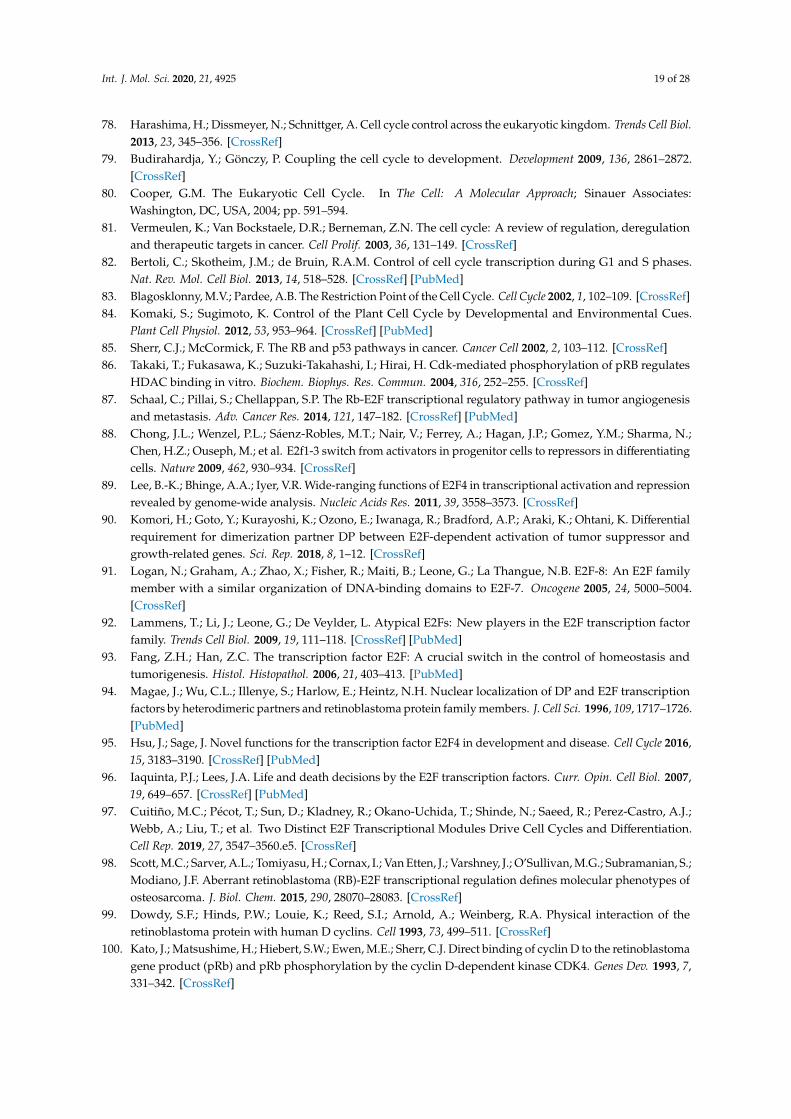
Int. J. Mol. Sci. 2020, 21, 4925 19 of 28
78. Harashima, H.; Dissmeyer, N.; Schnittger, A. Cell cycle control across the eukaryotic kingdom. Trends Cell Biol.2013, 23, 345–356. [CrossRef]
79. Budirahardja, Y.; Gönczy, P. Coupling the cell cycle to development. Development 2009, 136, 2861–2872.[CrossRef]
80. Cooper, G.M. The Eukaryotic Cell Cycle. In The Cell: A Molecular Approach; Sinauer Associates:Washington, DC, USA, 2004; pp. 591–594.
81. Vermeulen, K.; Van Bockstaele, D.R.; Berneman, Z.N. The cell cycle: A review of regulation, deregulationand therapeutic targets in cancer. Cell Prolif. 2003, 36, 131–149. [CrossRef]
82. Bertoli, C.; Skotheim, J.M.; de Bruin, R.A.M. Control of cell cycle transcription during G1 and S phases.Nat. Rev. Mol. Cell Biol. 2013, 14, 518–528. [CrossRef] [PubMed]
83. Blagosklonny, M.V.; Pardee, A.B. The Restriction Point of the Cell Cycle. Cell Cycle 2002, 1, 102–109. [CrossRef]84. Komaki, S.; Sugimoto, K. Control of the Plant Cell Cycle by Developmental and Environmental Cues.
Plant Cell Physiol. 2012, 53, 953–964. [CrossRef] [PubMed]85. Sherr, C.J.; McCormick, F. The RB and p53 pathways in cancer. Cancer Cell 2002, 2, 103–112. [CrossRef]86. Takaki, T.; Fukasawa, K.; Suzuki-Takahashi, I.; Hirai, H. Cdk-mediated phosphorylation of pRB regulates
HDAC binding in vitro. Biochem. Biophys. Res. Commun. 2004, 316, 252–255. [CrossRef]87. Schaal, C.; Pillai, S.; Chellappan, S.P. The Rb-E2F transcriptional regulatory pathway in tumor angiogenesis
and metastasis. Adv. Cancer Res. 2014, 121, 147–182. [CrossRef] [PubMed]88. Chong, J.L.; Wenzel, P.L.; Sáenz-Robles, M.T.; Nair, V.; Ferrey, A.; Hagan, J.P.; Gomez, Y.M.; Sharma, N.;
Chen, H.Z.; Ouseph, M.; et al. E2f1-3 switch from activators in progenitor cells to repressors in differentiatingcells. Nature 2009, 462, 930–934. [CrossRef]
89. Lee, B.-K.; Bhinge, A.A.; Iyer, V.R. Wide-ranging functions of E2F4 in transcriptional activation and repressionrevealed by genome-wide analysis. Nucleic Acids Res. 2011, 39, 3558–3573. [CrossRef]
90. Komori, H.; Goto, Y.; Kurayoshi, K.; Ozono, E.; Iwanaga, R.; Bradford, A.P.; Araki, K.; Ohtani, K. Differentialrequirement for dimerization partner DP between E2F-dependent activation of tumor suppressor andgrowth-related genes. Sci. Rep. 2018, 8, 1–12. [CrossRef]
91. Logan, N.; Graham, A.; Zhao, X.; Fisher, R.; Maiti, B.; Leone, G.; La Thangue, N.B. E2F-8: An E2F familymember with a similar organization of DNA-binding domains to E2F-7. Oncogene 2005, 24, 5000–5004.[CrossRef]
92. Lammens, T.; Li, J.; Leone, G.; De Veylder, L. Atypical E2Fs: New players in the E2F transcription factorfamily. Trends Cell Biol. 2009, 19, 111–118. [CrossRef] [PubMed]
93. Fang, Z.H.; Han, Z.C. The transcription factor E2F: A crucial switch in the control of homeostasis andtumorigenesis. Histol. Histopathol. 2006, 21, 403–413. [PubMed]
94. Magae, J.; Wu, C.L.; Illenye, S.; Harlow, E.; Heintz, N.H. Nuclear localization of DP and E2F transcriptionfactors by heterodimeric partners and retinoblastoma protein family members. J. Cell Sci. 1996, 109, 1717–1726.[PubMed]
95. Hsu, J.; Sage, J. Novel functions for the transcription factor E2F4 in development and disease. Cell Cycle 2016,15, 3183–3190. [CrossRef] [PubMed]
96. Iaquinta, P.J.; Lees, J.A. Life and death decisions by the E2F transcription factors. Curr. Opin. Cell Biol. 2007,19, 649–657. [CrossRef] [PubMed]
97. Cuitiño, M.C.; Pécot, T.; Sun, D.; Kladney, R.; Okano-Uchida, T.; Shinde, N.; Saeed, R.; Perez-Castro, A.J.;Webb, A.; Liu, T.; et al. Two Distinct E2F Transcriptional Modules Drive Cell Cycles and Differentiation.Cell Rep. 2019, 27, 3547–3560.e5. [CrossRef]
98. Scott, M.C.; Sarver, A.L.; Tomiyasu, H.; Cornax, I.; Van Etten, J.; Varshney, J.; O’Sullivan, M.G.; Subramanian, S.;Modiano, J.F. Aberrant retinoblastoma (RB)-E2F transcriptional regulation defines molecular phenotypes ofosteosarcoma. J. Biol. Chem. 2015, 290, 28070–28083. [CrossRef]
99. Dowdy, S.F.; Hinds, P.W.; Louie, K.; Reed, S.I.; Arnold, A.; Weinberg, R.A. Physical interaction of theretinoblastoma protein with human D cyclins. Cell 1993, 73, 499–511. [CrossRef]
100. Kato, J.; Matsushime, H.; Hiebert, S.W.; Ewen, M.E.; Sherr, C.J. Direct binding of cyclin D to the retinoblastomagene product (pRb) and pRb phosphorylation by the cyclin D-dependent kinase CDK4. Genes Dev. 1993, 7,331–342. [CrossRef]
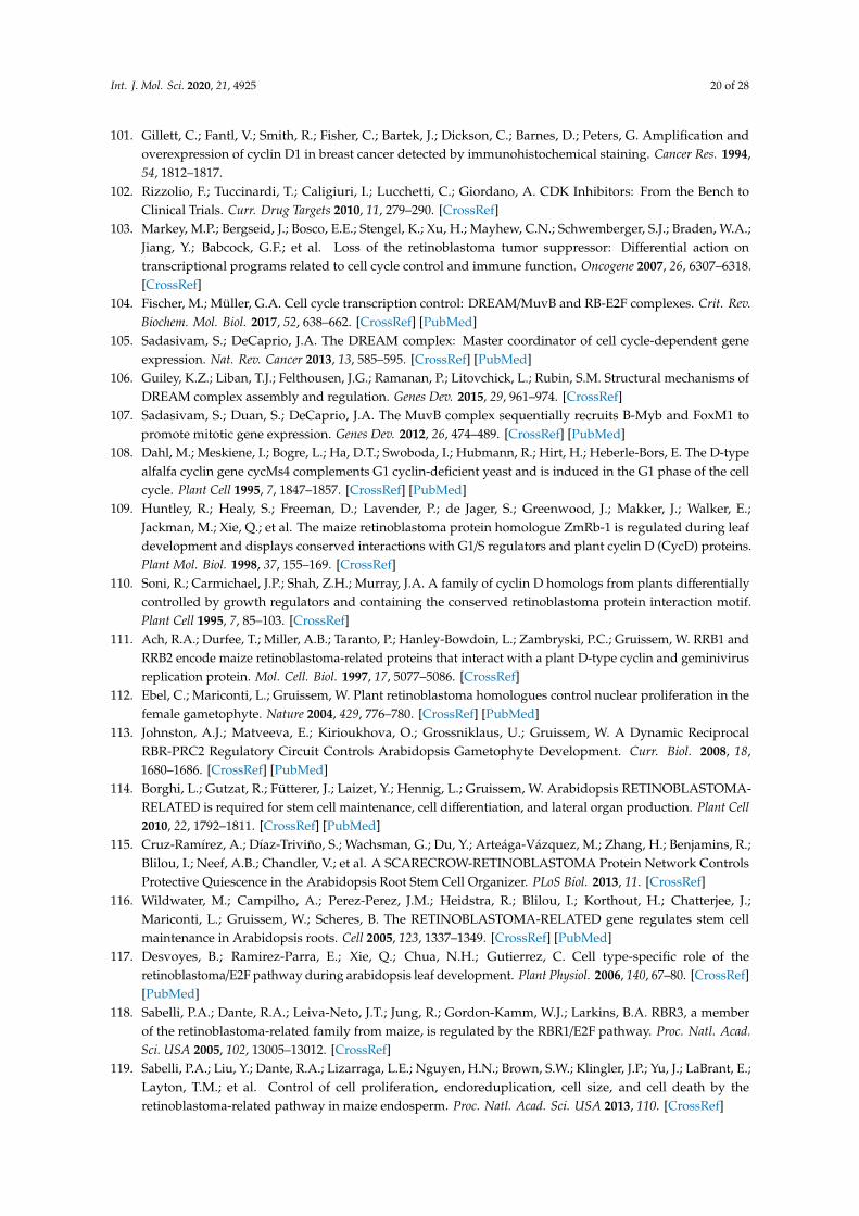
Int. J. Mol. Sci. 2020, 21, 4925 20 of 28
101. Gillett, C.; Fantl, V.; Smith, R.; Fisher, C.; Bartek, J.; Dickson, C.; Barnes, D.; Peters, G. Amplification andoverexpression of cyclin D1 in breast cancer detected by immunohistochemical staining. Cancer Res. 1994,54, 1812–1817.
102. Rizzolio, F.; Tuccinardi, T.; Caligiuri, I.; Lucchetti, C.; Giordano, A. CDK Inhibitors: From the Bench toClinical Trials. Curr. Drug Targets 2010, 11, 279–290. [CrossRef]
103. Markey, M.P.; Bergseid, J.; Bosco, E.E.; Stengel, K.; Xu, H.; Mayhew, C.N.; Schwemberger, S.J.; Braden, W.A.;Jiang, Y.; Babcock, G.F.; et al. Loss of the retinoblastoma tumor suppressor: Differential action ontranscriptional programs related to cell cycle control and immune function. Oncogene 2007, 26, 6307–6318.[CrossRef]
104. Fischer, M.; Müller, G.A. Cell cycle transcription control: DREAM/MuvB and RB-E2F complexes. Crit. Rev.Biochem. Mol. Biol. 2017, 52, 638–662. [CrossRef] [PubMed]
105. Sadasivam, S.; DeCaprio, J.A. The DREAM complex: Master coordinator of cell cycle-dependent geneexpression. Nat. Rev. Cancer 2013, 13, 585–595. [CrossRef] [PubMed]
106. Guiley, K.Z.; Liban, T.J.; Felthousen, J.G.; Ramanan, P.; Litovchick, L.; Rubin, S.M. Structural mechanisms ofDREAM complex assembly and regulation. Genes Dev. 2015, 29, 961–974. [CrossRef]
107. Sadasivam, S.; Duan, S.; DeCaprio, J.A. The MuvB complex sequentially recruits B-Myb and FoxM1 topromote mitotic gene expression. Genes Dev. 2012, 26, 474–489. [CrossRef] [PubMed]
108. Dahl, M.; Meskiene, I.; Bogre, L.; Ha, D.T.; Swoboda, I.; Hubmann, R.; Hirt, H.; Heberle-Bors, E. The D-typealfalfa cyclin gene cycMs4 complements G1 cyclin-deficient yeast and is induced in the G1 phase of the cellcycle. Plant Cell 1995, 7, 1847–1857. [CrossRef] [PubMed]
109. Huntley, R.; Healy, S.; Freeman, D.; Lavender, P.; de Jager, S.; Greenwood, J.; Makker, J.; Walker, E.;Jackman, M.; Xie, Q.; et al. The maize retinoblastoma protein homologue ZmRb-1 is regulated during leafdevelopment and displays conserved interactions with G1/S regulators and plant cyclin D (CycD) proteins.Plant Mol. Biol. 1998, 37, 155–169. [CrossRef]
110. Soni, R.; Carmichael, J.P.; Shah, Z.H.; Murray, J.A. A family of cyclin D homologs from plants differentiallycontrolled by growth regulators and containing the conserved retinoblastoma protein interaction motif.Plant Cell 1995, 7, 85–103. [CrossRef]
111. Ach, R.A.; Durfee, T.; Miller, A.B.; Taranto, P.; Hanley-Bowdoin, L.; Zambryski, P.C.; Gruissem, W. RRB1 andRRB2 encode maize retinoblastoma-related proteins that interact with a plant D-type cyclin and geminivirusreplication protein. Mol. Cell. Biol. 1997, 17, 5077–5086. [CrossRef]
112. Ebel, C.; Mariconti, L.; Gruissem, W. Plant retinoblastoma homologues control nuclear proliferation in thefemale gametophyte. Nature 2004, 429, 776–780. [CrossRef] [PubMed]
113. Johnston, A.J.; Matveeva, E.; Kirioukhova, O.; Grossniklaus, U.; Gruissem, W. A Dynamic ReciprocalRBR-PRC2 Regulatory Circuit Controls Arabidopsis Gametophyte Development. Curr. Biol. 2008, 18,1680–1686. [CrossRef] [PubMed]
114. Borghi, L.; Gutzat, R.; Fütterer, J.; Laizet, Y.; Hennig, L.; Gruissem, W. Arabidopsis RETINOBLASTOMA-RELATED is required for stem cell maintenance, cell differentiation, and lateral organ production. Plant Cell2010, 22, 1792–1811. [CrossRef] [PubMed]
115. Cruz-Ramírez, A.; Díaz-Triviño, S.; Wachsman, G.; Du, Y.; Arteága-Vázquez, M.; Zhang, H.; Benjamins, R.;Blilou, I.; Neef, A.B.; Chandler, V.; et al. A SCARECROW-RETINOBLASTOMA Protein Network ControlsProtective Quiescence in the Arabidopsis Root Stem Cell Organizer. PLoS Biol. 2013, 11. [CrossRef]
116. Wildwater, M.; Campilho, A.; Perez-Perez, J.M.; Heidstra, R.; Blilou, I.; Korthout, H.; Chatterjee, J.;Mariconti, L.; Gruissem, W.; Scheres, B. The RETINOBLASTOMA-RELATED gene regulates stem cellmaintenance in Arabidopsis roots. Cell 2005, 123, 1337–1349. [CrossRef] [PubMed]
117. Desvoyes, B.; Ramirez-Parra, E.; Xie, Q.; Chua, N.H.; Gutierrez, C. Cell type-specific role of theretinoblastoma/E2F pathway during arabidopsis leaf development. Plant Physiol. 2006, 140, 67–80. [CrossRef][PubMed]
118. Sabelli, P.A.; Dante, R.A.; Leiva-Neto, J.T.; Jung, R.; Gordon-Kamm, W.J.; Larkins, B.A. RBR3, a memberof the retinoblastoma-related family from maize, is regulated by the RBR1/E2F pathway. Proc. Natl. Acad.Sci. USA 2005, 102, 13005–13012. [CrossRef]
119. Sabelli, P.A.; Liu, Y.; Dante, R.A.; Lizarraga, L.E.; Nguyen, H.N.; Brown, S.W.; Klingler, J.P.; Yu, J.; LaBrant, E.;Layton, T.M.; et al. Control of cell proliferation, endoreduplication, cell size, and cell death by theretinoblastoma-related pathway in maize endosperm. Proc. Natl. Acad. Sci. USA 2013, 110. [CrossRef]

Int. J. Mol. Sci. 2020, 21, 4925 21 of 28
120. Grafi, G.; Burnett, R.J.; Helentjaris, T.; Larkins, B.A.; Decaprio, J.A.; Sellers, W.R.; Kaelin, W.G. A maize cDNAencoding a member of the retinoblastoma protein family: Involvement in endoreduplication. Proc. Natl.Acad. Sci. USA 1996, 93, 8962–8967. [CrossRef]
121. Rossi, V.; Locatelli, S.; Lanzanova, C.; Boniotti, M.B.; Varotto, S.; Pipal, A.; Goralik-Schramel, M.; Lusser, A.;Gatz, C.; Gutierrez, C.; et al. A maize histone deacetylase and retinoblastoma-related protein physicallyinteract and cooperate in repressing gene transcription. Plant Mol. Biol. 2003, 51, 401–413. [CrossRef]
122. Sabelli, P.A.; Larkins, B.A. Grasses Like Mammals? Redundancy and Compensatory Regulation within theRetinoblastoma Protein Family. Cell Cycle 2006, 5, 352–355. [CrossRef] [PubMed]
123. Villajuana-Bonequi, M.; Matei, A.; Ernst, C.; Hallab, A.; Usadel, B.; Doehlemann, G. Cell type specifictranscriptional reprogramming of maize leaves during Ustilago maydis induced tumor formation. Sci. Rep.2019, 9, 1–15. [CrossRef]
124. Sabelli, P.A.; Hoerster, G.; Lizarraga, L.E.; Brown, S.W.; Gordon-Kamm, W.J.; Larkins, B.A. Positive regulationof minichromosome maintenance gene expression, DNA replication, and cell transformation by a plantretinoblastoma gene. Proc. Natl. Acad. Sci. USA 2009, 106, 4042–4047. [CrossRef] [PubMed]
125. Gordon-Kamm, W.; Dilkes, B.P.; Lowe, K.; Hoerster, G.; Sun, X.; Ross, M.; Church, L.; Bunde, C.; Farrell, J.;Hill, P.; et al. Stimulation of the cell cycle and maize transformation by disruption of the plant retinoblastomapathway. Proc. Natl. Acad. Sci. USA 2002, 99, 11975–11980. [CrossRef] [PubMed]
126. Sage, J.; Miller, A.L.; Pérez-Mancera, P.A.; Wysocki, J.M.; Jacks, T. Acute mutation of retinoblastoma genefunction is sufficient for cell cycle re-entry. Nature 2003, 424, 223–228. [CrossRef]
127. Gutierrez, C. The Arabidopsis Cell Division Cycle. Arab. Book 2009, 7, e0120. [CrossRef]128. Wang, G.; Kong, H.; Sun, Y.; Zhang, X.; Zhang, W.; Altman, N.; DePamphilis, C.W.; Ma, H. Genome-wide
analysis of the cyclin family in arabidopsis and comparative phylogenetic analysis of plant cyclin-likeproteins. Plant Physiol. 2004, 135, 1084–1099. [CrossRef]
129. Quandt, E.; Ribeiro, M.P.C.; Clotet, J. Atypical cyclins: The extended family portrait. Cell. Mol. Life Sci. 2020,77, 231–242. [CrossRef]
130. Magyar, Z.; De Veylder, L.; Atanassova, A.; Bakó, L.; Inzé, D.; Bögre, L. The role of the Arabidopsis E2FBtranscription factor in regulating auxin-dependent cell division. Plant Cell 2005, 17, 2527–2541. [CrossRef]
131. Ramirez-Parra, E.; López-Matas, M.A.; Fründt, C.; Gutierrez, C. Role of an atypical E2F transcription factorin the control of Arabidopsis cell growth and differentiation. Plant Cell 2004, 16, 2350–2363. [CrossRef]
132. Vandepoele, K.; Vlieghe, K.; Florquin, K.; Hennig, L.; Beemster, G.T.S.; Gruissem, W.; Van De Peer, Y.; Inzé, D.;De Veylder, L. Genome-wide identification of potential plant E2F target genes. Plant Physiol. 2005, 139,316–328. [CrossRef] [PubMed]
133. Oszi, E.; Papdi, C.; Mohammed, B.; Petkó-Szandtner, A.; Leviczky, T.; Molnár, E.; Galvan-Ampudia, C.;Khan, S.; Juez, E.L.; Horváth, B.; et al. E2FB interacts with RETINOBLASTOMA RELATED and regulates cellproliferation during leaf development. Plant Physiol. 2020, 182, 518–533. [CrossRef] [PubMed]
134. Boudolf, V.; Barrôco, R.; De Engler, J.A.; Verkest, A.; Beeckman, T.; Naudts, M.; Inzé, D.; De Veylder, L.B1-type cyclin-dependent kinases are essential for the formation of stomatal complexes in Arabidopsisthaliana. Plant Cell 2004, 16, 945–955. [CrossRef] [PubMed]
135. Scofield, S.; Jones, A.; Murray, J.A.H. The plant cell cycle in context. J. Exp. Bot. 2014, 65, 2557–2562.[CrossRef]
136. Caldon, C.E.; Musgrove, E.A. Distinct and redundant functions of cyclin E1 and cyclin E2 in developmentand cancer. Cell Div. 2010, 5, 2. [CrossRef]
137. Teixeira, L.K.; Reed, S.I. Cyclin E deregulation and genomic instability. Adv. Exp. Med. Biol. 2017, 1042,527–547. [CrossRef]
138. Fischer, M.; DeCaprio, J.A. Does Arabidopsis thaliana DREAM of cell cycle control? EMBO J. 2015, 34,1987–1989. [CrossRef]
139. Magyar, Z.; Bögre, L.; Ito, M. DREAMs make plant cells to cycle or to become quiescent. Curr. Opin. Plant Biol.2016, 34, 100–106. [CrossRef] [PubMed]
140. Kobayashi, K.; Suzuki, T.; Iwata, E.; Magyar, Z.; Bögre, L.; Ito, M. MYB3Rs, plant homologs of Myboncoproteins, control cell cycle-regulated transcription and form DREAM-like complexes. Transcription 2015,6, 106–111. [CrossRef]

Int. J. Mol. Sci. 2020, 21, 4925 22 of 28
141. Wang, W.; Sijacic, P.; Xu, P.; Lian, H.; Liu, Z. Arabidopsis TSO1 and MYB3R1 form a regulatory module tocoordinate cell proliferation with differentiation in shoot and root. Proc. Natl. Acad. Sci. USA 2018, 115,E3045–E3054. [CrossRef]
142. Bouyer, D.; Heese, M.; Chen, P.; Harashima, H.; Roudier, F.; Grüttner, C.; Schnittger, A. Genome-wideidentification of RETINOBLASTOMA RELATED 1 binding sites in Arabidopsis reveals novel DNA damageregulators. PLoS Genet. 2018, 14, 1–35. [CrossRef]
143. Jiang, C.; Gu, J.; Chopra, S.; Gu, X.; Peterson, T. Ordered origin of the typical two- and three-repeat Mybgenes. Gene 2004, 326, 13–22. [CrossRef] [PubMed]
144. Kobayashi, K.; Suzuki, T.; Iwata, E.; Nakamichi, N.; Suzuki, T.; Chen, P.; Ohtani, M.; Ishida, T.; Hosoya, H.;Müller, S.; et al. Transcriptional repression by MYB 3R proteins regulates plant organ growth. EMBO J. 2015,34, 1992–2007. [CrossRef] [PubMed]
145. Araki, S.; Ito, M.; Soyano, T.; Nishihama, R.; Machida, Y. Mitotic cyclins stimulate the activity of c-Myb-likefactors for transactivation of G2/M phase-specific genes in tobacco. J. Biol. Chem. 2004, 279, 32979–32988.[CrossRef]
146. Del Pozo, J.C.; Boniotti, M.B.; Gutierrez, C. Arabidopsis E2Fc functions in cell division and is degraded bythe ubiquitin-SCFAtSKP2 pathway in response to light. Plant Cell 2002, 14, 3057–3071. [CrossRef] [PubMed]
147. Newman, S.A. Cell differentiation: What have we learned in 50 years? J. Theor. Biol. 2020, 485, 110031.[CrossRef] [PubMed]
148. Sánchez Alvarado, A.; Yamanaka, S. Rethinking Differentiation: Stem Cells, Regeneration, and Plasticity.Cell 2014, 157, 110–119. [CrossRef] [PubMed]
149. De Falco, G.; Comes, F.; Simone, C. pRb: Master of differentiation. Coupling irreversible cell cycle withdrawalwith induction of muscle-specific transcription. Oncogene 2006, 25, 5244–5249. [CrossRef]
150. Goodrich, D.W. The retinoblastoma tumor-suppressor gene, the exception that proves the rule. Oncogene2006, 25, 5233–5243. [CrossRef]
151. Hatzistergos, K.E.; Williams, A.R.; Dykxhoorn, D.; Bellio, M.A.; Yu, W.; Hare, J.M. Tumor Suppressors RB1and CDKN2a Cooperatively Regulate Cell-Cycle Progression and Differentiation During CardiomyocyteDevelopment and Repair. Circ. Res. 2019, 124, 1184–1197. [CrossRef]
152. Khidr, L.; Chen, P.-L. RB, the conductor that orchestrates life, death and differentiation. Oncogene 2006, 25,5210–5219. [CrossRef] [PubMed]
153. Li, J.; Narayanan, C.; Bian, J.; Sambo, D.; Brickler, T.; Zhang, W.; Chetty, S. A transient DMSO treatmentincreases the differentiation potential of human pluripotent stem cells through the Rb family. PLoS ONE2018, 13, e0208110. [CrossRef] [PubMed]
154. Lipinski, M.M.; Jacks, T. The retinoblastoma gene family in differentiation and development. Oncogene 1999,18, 7873–7882. [CrossRef] [PubMed]
155. Busch, M.; Philippeit, C.; Weise, A.; Dünker, N. Re-characterization of established human retinoblastoma celllines. Histochem. Cell Biol. 2014, 143, 325–338. [CrossRef] [PubMed]
156. Dyer, M.A.; Bremner, R. The search for the retinoblastoma cell of origin. Nat. Rev. Cancer 2005, 5, 91–101.[CrossRef]
157. Nair, R.M.; Vemuganti, G.K. Transgenic Models in Retinoblastoma Research. Ocul. Oncol. Pathol. 2015, 1,207–213. [CrossRef]
158. Clarke, A.R.; Maandag, E.R.; Van Roon, M.; Van Der Lugt, N.M.T.; Van Der Valk, M.; Hooper, M.L.; Berns, A.;Te Rielef, H. Requirement for a functional Rb-1 gene in murine development. Nature 1992, 359, 328–330.[CrossRef]
159. Jacks, T.; Fazeli, A.; Schmitt, E.M.; Bronson, R.T.; Goodell, M.A.; Weinberg, R.A. Effects of an Rb mutation inthe mouse. Nature 1992, 359, 295–300. [CrossRef]
160. Lee, E.Y.H.P.; Chang, C.Y.; Hu, N.; Wang, Y.C.J.; Lai, C.C.; Herrup, K.; Lee, W.H.; Bradley, A. Mice deficientfor Rb are nonviable and show defects in neurogenesis and haematopoiesis. Nature 1992, 359, 288–294.[CrossRef]
161. Morgenbesser, S.D.; Williams, B.O.; Jacks, T.; Depinho, R.A. P53-dependent apoptosis produced byRb-deficiency in the developing mouse lens. Nature 1994, 371, 72–74. [CrossRef]
162. Zhang, J.; Gray, J.; Wu, L.; Leone, G.; Rowan, S.; Cepko, C.L.; Zhu, X.; Craft, C.M.; Dyer, M.A. Rb regulatesproliferation and rod photoreceptor development in the mouse retina. Nat. Genet. 2004, 36, 351–360.[CrossRef] [PubMed]

Int. J. Mol. Sci. 2020, 21, 4925 23 of 28
163. Robanus-Maandag, E.; Dekker, M.; Van Der Valk, M.; Carrozza, M.L.; Jeanny, J.C.; Dannenberg, J.H.; Berns, A.;Riele, H. Te p107 is a suppressor of retinoblastoma development in pRB-deficient mice. Genes Dev. 1998, 12,1599–1609. [CrossRef] [PubMed]
164. Fajas, L.; Landsberg, R.L.; Huss-Garcia, Y.; Sardet, C.; Lees, J.A.; Auwerx, J. E2Fs regulate adipocytedifferentiation. Dev. Cell 2002, 3, 39–49. [CrossRef]
165. Flowers, S.; Xu, F.; Moran, E. Cooperative activation of tissue-specific genes by pRB and E2F1. Cancer Res.2013, 73, 2150–2158. [CrossRef]
166. Julian, L.M.; Liu, Y.; Pakenham, C.A.; Dugal-Tessier, D.; Ruzhynsky, V.; Bae, S.; Tsai, S.Y.; Leone, G.; Slack, R.S.;Blais, A. Tissue-specific targeting of cell fate regulatory genes by E2f factors. Cell Death Differ. 2016, 23,565–575. [CrossRef]
167. Müller, H.; Bracken, A.P.; Vernell, R.; Moroni, M.C.; Christians, F.; Grassilli, E.; Prosperini, E.; Vigo, E.;Oliner, J.D.; Helin, K. E2Fs regulate the expression of genes involved in differentiation, development,proliferation, and apoptosis. Genes Dev. 2001, 15, 267–285. [CrossRef]
168. Sellers, W.R.; Novitch, B.G.; Miyake, S.; Heith, A.; Otterson, G.A.; Kaye, F.J.; Lassar, A.B.; Kaelin, W.G. Stablebinding to E2F is not required for the retinoblastoma protein to activate transcription, promote differentiation,and suppress tumor cell growth. Genes Dev. 1998, 12, 95–106. [CrossRef]
169. Cecchini, M.J.; Thwaites, M.J.; Talluri, S.; MacDonald, J.I.; Passos, D.T.; Chong, J.-L.; Cantalupo, P.;Stafford, P.M.; Saenz-Robles, M.T.; Francis, S.M.; et al. A Retinoblastoma Allele That Is Mutated at ItsCommon E2F Interaction Site Inhibits Cell Proliferation in Gene-Targeted Mice. Mol. Cell. Biol. 2014, 34,2029–2045. [CrossRef]
170. Wyrzykowska, J.; Schorderet, M.; Pien, S.; Gruissem, W.; Fleming, A.J. Induction of differentiation in theshoot apical meristem by transient overexpression of a retinoblastoma-related protein. Plant Physiol. 2006,141, 1338–1348. [CrossRef]
171. Zhao, X.; Bramsiepe, J.; Van Durme, M.; Komaki, S.; Prusicki, M.A.; Maruyama, D.; Forner, J.;Medzihradszky, A.; Wijnker, E.; Harashima, H.; et al. RETINOBLASTOMA RELATED1 mediates germlineentry in Arabidopsis. Science 2017, 356. [CrossRef]
172. Perilli, S.; Perez-Perez, J.M.; Di Mambro, R.; Peris, C.L.; Díaz-Triviño, S.; Del Bianco, M.; Pierdonati, E.;Moubayidin, L.; Cruz-Ramírez, A.; Costantino, P.; et al. RETINOBLASTOMA-RELATED protein stimulatescell differentiation in the Arabidopsis root meristem by interacting with cytokinin signaling. Plant Cell 2013,25, 4469–4478. [CrossRef] [PubMed]
173. Moubayidin, L.; Perilli, S.; Dello Ioio, R.; Di Mambro, R.; Costantino, P.; Sabatini, S. The rate of celldifferentiation controls the arabidopsis root meristem growth phase. Curr. Biol. 2010, 20, 1138–1143.[CrossRef] [PubMed]
174. Fambrini, M.; Pugliesi, C. The Dynamic Genetic-Hormonal Regulatory Network Controlling the TrichomeDevelopment in Leaves. Plants 2019, 8, 253. [CrossRef] [PubMed]
175. Morohashi, K.; Grotewold, E. A systems approach reveals regulatory circuitry for Arabidopsis trichomeinitiation by the GL3 and GL1 selectors. PLoS Genet. 2009, 5, e1000396. [CrossRef]
176. Zhao, C.; Avci, U.; Grant, E.H.; Haigler, C.H.; Beers, E.P. XND1, a member of the NAC domain family inArabidopsis thaliana, negatively regulates lignocellulose synthesis and programmed cell death in xylem.Plant J. 2008, 53, 425–436. [CrossRef]
177. Zhao, C.; Lasses, T.; Bako, L.; Kong, D.; Zhao, B.; Chanda, B.; Bombarely, A.; Cruz-Ramírez, A.; Scheres, B.;Brunner, A.M.; et al. XYLEM NAC DOMAIN1, an angiosperm NAC transcription factor, inhibits xylemdifferentiation through conserved motifs that interact with RETINOBLASTOMA-RELATED. New Phytol.2017, 1. [CrossRef]
178. Putarjunan, A.; Torii, K.U. Stomagenesis versus myogenesis: Parallels in intrinsic and extrinsic regulation oftranscription factor mediated specialized cell-type differentiation in plants and animals. Dev. Growth Differ.2016, 58, 341–354. [CrossRef]
179. Matos, J.L.; Bergmann, D.C. Convergence of stem cell behaviors and genetic regulation between animals andplants: Insights from the Arabidopsis thaliana stomatal lineage. F1000Prime Rep. 2014, 6, 1–8. [CrossRef]
180. Le Grand, F.; Rudnicki, M.A. Skeletal muscle satellite cells and adult myogenesis. Curr. Opin. Cell Biol. 2007,19, 628–633. [CrossRef]

Int. J. Mol. Sci. 2020, 21, 4925 24 of 28
181. Novitch, B.G.; Mulligan, G.J.; Jacks, T.; Lassar, A.B. Skeletal muscle cells lacking the retinoblastoma proteindisplay defects in muscle gene expression and accumulate in S and G2 phases of the cell cycle. J. Cell Biol.1996, 135, 441–456. [CrossRef]
182. Zammit, P.S. Function of the myogenic regulatory factors Myf5, MyoD, Myogenin and MRF4 in skeletalmuscle, satellite cells and regenerative myogenesis. Semin. Cell Dev. Biol. 2017, 72, 19–32. [CrossRef][PubMed]
183. Buckingham, M.; Rigby, P.W.J. Gene Regulatory Networks and Transcriptional Mechanisms that ControlMyogenesis. Dev. Cell 2014, 28, 225–238. [CrossRef] [PubMed]
184. Dumont, N.A.; Wang, Y.X.; Rudnicki, M.A. Intrinsic and extrinsic mechanisms regulating satellite cellfunction. Development 2015, 142, 1572–1581. [CrossRef] [PubMed]
185. Cornelison, D.D.W.; Olwin, B.B.; Rudnicki, M.A.; Wold, B.J. MyoD(-/-) satellite cells in single-fiber culture aredifferentiation defective and MRF4 deficient. Dev. Biol. 2000, 224, 122–137. [CrossRef]
186. Endo, T.; Goto, S. Retinoblastoma gene product Rb accumulates during myogenic differentiation and isdeinduced by the expression of SV40 large T antigen. J. Biochem. 1992, 112, 427–430. [CrossRef] [PubMed]
187. Huh, M.S.; Parker, M.H.; Scimè, A.; Parks, R.; Rudnicki, M.A. Rb is required for progression throughmyogenic differentiation but not maintenance of terminal differentiation. J. Cell Biol. 2004, 166, 865–876.[CrossRef] [PubMed]
188. Tapscott, S.J. The circuitry of a master switch: Myod and the regulation of skeletal muscle gene transcription.Development 2005, 132, 2685–2695. [CrossRef] [PubMed]
189. Gu, W.; Schneider, J.W.; Condorelli, G.; Kaushal, S.; Mahdavi, V.; Nadal-Ginard, B. Interaction of myogenicfactors and the retinoblastoma protein mediates muscle cell commitment and differentiation. Cell 1993, 72,309–324. [CrossRef]
190. Smialowski, P.; Singh, M.; Mikolajka, A.; Majumdar, S.; Joy, J.K.; Nalabothula, N.; Krajewski, M.;Degenkolbe, R.; Bernard, H.-U.; Holak, T.A. NMR and mass spectrometry studies of putative interactions ofcell cycle proteins pRb and CDK6 with cell differentiation proteins MyoD and ID-2. Biochim. Biophys. Acta2005, 1750, 48–60. [CrossRef]
191. Magenta, A.; Cenciarelli, C.; De Santa, F.; Fuschi, P.; Martelli, F.; Caruso, M.; Felsani, A. MyoD Stimulates RBPromoter Activity via the CREB/p300 Nuclear Transduction Pathway. Mol. Cell. Biol. 2003, 23, 2893–2906.[CrossRef]
192. Martelli, F.; Cenciarelli, C.; Santarelli, G.; Polikar, B.; Felsani, A.; Caruso, M. MyoD induces retinoblastomagene expression during myogenic differentiation. Oncogene 1994, 9, 3579–3590.
193. Puri, P.L.; Sartorelli, V. Regulation of muscle regulatory factors by DNA-binding, interacting proteins,and post-transcriptional modifications. J. Cell. Physiol. 2000, 185, 155–173. [CrossRef]
194. Novitch, B.G.; Spicer, D.B.; Kim, P.S.; Cheung, W.L.; Lassar, A.B. pRb is required for MEF2-dependent geneexpression as well as cell-cycle arrest during skeletal muscle differentiation. Curr. Biol. 1999, 9, 449–459.[CrossRef]
195. Knight, J.D.R.; Kothary, R. The myogenic kinome: Protein kinases critical to mammalian skeletal myogenesis.Skelet. Muscle 2011, 1, 29. [CrossRef] [PubMed]
196. Rajabi, H.N.; Takahashi, C.; Ewen, M.E. Retinoblastoma protein and MyoD function together to effect therepression of Fra-1 and in turn cyclin D1 during terminal cell cycle arrest associated with myogenesis.J. Biol. Chem. 2014, 289, 23417–23427. [CrossRef] [PubMed]
197. Skapek, S.X.; Rhee, J.; Kim, P.S.; Novitch, B.G.; Lassar, A.B. Cyclin-mediated inhibition of muscle geneexpression via a mechanism that is independent of pRB hyperphosphorylation. Mol. Cell. Biol. 1996, 16,7043–7053. [CrossRef]
198. Ruijtenberg, S.; van den Heuvel, S. Coordinating cell proliferation and differentiation: Antagonism betweencell cycle regulators and cell type-specific gene expression. Cell Cycle 2016, 15, 196–212. [CrossRef] [PubMed]
199. Song, A.; Wang, Q.; Goebl, M.G.; Harrington, M.A. Phosphorylation of Nuclear MyoD Is Required for ItsRapid Degradation. Mol. Cell. Biol. 1998, 18, 4994–4999. [CrossRef] [PubMed]
200. Zhang, J.M.; Wei, Q.; Zhao, X.; Paterson, B.M. Coupling of the cell cycle and myogenesis through the cyclinD1-dependent interaction of MyoD with cdk4. EMBO J. 1999, 18, 926–933. [CrossRef] [PubMed]
201. Puri, P.L.; Iezzi, S.; Stiegler, P.; Chen, T.T.; Schiltz, R.L.; Muscat, G.E.O.; Giordano, A.; Kedes, L.; Wang, J.Y.J.;Sartorelli, V. Class I histone deacetylases sequentially interact with MyoD and pRb during skeletal myogenesis.Mol. Cell 2001, 8, 885–897. [CrossRef]
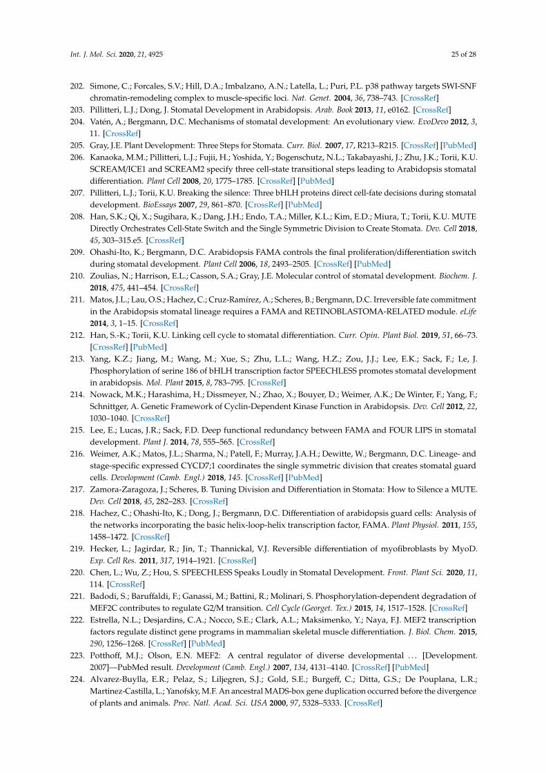
Int. J. Mol. Sci. 2020, 21, 4925 25 of 28
202. Simone, C.; Forcales, S.V.; Hill, D.A.; Imbalzano, A.N.; Latella, L.; Puri, P.L. p38 pathway targets SWI-SNFchromatin-remodeling complex to muscle-specific loci. Nat. Genet. 2004, 36, 738–743. [CrossRef]
203. Pillitteri, L.J.; Dong, J. Stomatal Development in Arabidopsis. Arab. Book 2013, 11, e0162. [CrossRef]204. Vatén, A.; Bergmann, D.C. Mechanisms of stomatal development: An evolutionary view. EvoDevo 2012, 3,
11. [CrossRef]205. Gray, J.E. Plant Development: Three Steps for Stomata. Curr. Biol. 2007, 17, R213–R215. [CrossRef] [PubMed]206. Kanaoka, M.M.; Pillitteri, L.J.; Fujii, H.; Yoshida, Y.; Bogenschutz, N.L.; Takabayashi, J.; Zhu, J.K.; Torii, K.U.
SCREAM/ICE1 and SCREAM2 specify three cell-state transitional steps leading to Arabidopsis stomataldifferentiation. Plant Cell 2008, 20, 1775–1785. [CrossRef] [PubMed]
207. Pillitteri, L.J.; Torii, K.U. Breaking the silence: Three bHLH proteins direct cell-fate decisions during stomataldevelopment. BioEssays 2007, 29, 861–870. [CrossRef] [PubMed]
208. Han, S.K.; Qi, X.; Sugihara, K.; Dang, J.H.; Endo, T.A.; Miller, K.L.; Kim, E.D.; Miura, T.; Torii, K.U. MUTEDirectly Orchestrates Cell-State Switch and the Single Symmetric Division to Create Stomata. Dev. Cell 2018,45, 303–315.e5. [CrossRef]
209. Ohashi-Ito, K.; Bergmann, D.C. Arabidopsis FAMA controls the final proliferation/differentiation switchduring stomatal development. Plant Cell 2006, 18, 2493–2505. [CrossRef] [PubMed]
210. Zoulias, N.; Harrison, E.L.; Casson, S.A.; Gray, J.E. Molecular control of stomatal development. Biochem. J.2018, 475, 441–454. [CrossRef]
211. Matos, J.L.; Lau, O.S.; Hachez, C.; Cruz-Ramírez, A.; Scheres, B.; Bergmann, D.C. Irreversible fate commitmentin the Arabidopsis stomatal lineage requires a FAMA and RETINOBLASTOMA-RELATED module. eLife2014, 3, 1–15. [CrossRef]
212. Han, S.-K.; Torii, K.U. Linking cell cycle to stomatal differentiation. Curr. Opin. Plant Biol. 2019, 51, 66–73.[CrossRef] [PubMed]
213. Yang, K.Z.; Jiang, M.; Wang, M.; Xue, S.; Zhu, L.L.; Wang, H.Z.; Zou, J.J.; Lee, E.K.; Sack, F.; Le, J.Phosphorylation of serine 186 of bHLH transcription factor SPEECHLESS promotes stomatal developmentin arabidopsis. Mol. Plant 2015, 8, 783–795. [CrossRef]
214. Nowack, M.K.; Harashima, H.; Dissmeyer, N.; Zhao, X.; Bouyer, D.; Weimer, A.K.; De Winter, F.; Yang, F.;Schnittger, A. Genetic Framework of Cyclin-Dependent Kinase Function in Arabidopsis. Dev. Cell 2012, 22,1030–1040. [CrossRef]
215. Lee, E.; Lucas, J.R.; Sack, F.D. Deep functional redundancy between FAMA and FOUR LIPS in stomataldevelopment. Plant J. 2014, 78, 555–565. [CrossRef]
216. Weimer, A.K.; Matos, J.L.; Sharma, N.; Patell, F.; Murray, J.A.H.; Dewitte, W.; Bergmann, D.C. Lineage- andstage-specific expressed CYCD7;1 coordinates the single symmetric division that creates stomatal guardcells. Development (Camb. Engl.) 2018, 145. [CrossRef] [PubMed]
217. Zamora-Zaragoza, J.; Scheres, B. Tuning Division and Differentiation in Stomata: How to Silence a MUTE.Dev. Cell 2018, 45, 282–283. [CrossRef]
218. Hachez, C.; Ohashi-Ito, K.; Dong, J.; Bergmann, D.C. Differentiation of arabidopsis guard cells: Analysis ofthe networks incorporating the basic helix-loop-helix transcription factor, FAMA. Plant Physiol. 2011, 155,1458–1472. [CrossRef]
219. Hecker, L.; Jagirdar, R.; Jin, T.; Thannickal, V.J. Reversible differentiation of myofibroblasts by MyoD.Exp. Cell Res. 2011, 317, 1914–1921. [CrossRef]
220. Chen, L.; Wu, Z.; Hou, S. SPEECHLESS Speaks Loudly in Stomatal Development. Front. Plant Sci. 2020, 11,114. [CrossRef]
221. Badodi, S.; Baruffaldi, F.; Ganassi, M.; Battini, R.; Molinari, S. Phosphorylation-dependent degradation ofMEF2C contributes to regulate G2/M transition. Cell Cycle (Georget. Tex.) 2015, 14, 1517–1528. [CrossRef]
222. Estrella, N.L.; Desjardins, C.A.; Nocco, S.E.; Clark, A.L.; Maksimenko, Y.; Naya, F.J. MEF2 transcriptionfactors regulate distinct gene programs in mammalian skeletal muscle differentiation. J. Biol. Chem. 2015,290, 1256–1268. [CrossRef] [PubMed]
223. Potthoff, M.J.; Olson, E.N. MEF2: A central regulator of diverse developmental . . . [Development.2007]—PubMed result. Development (Camb. Engl.) 2007, 134, 4131–4140. [CrossRef] [PubMed]
224. Alvarez-Buylla, E.R.; Pelaz, S.; Liljegren, S.J.; Gold, S.E.; Burgeff, C.; Ditta, G.S.; De Pouplana, L.R.;Martinez-Castilla, L.; Yanofsky, M.F. An ancestral MADS-box gene duplication occurred before the divergenceof plants and animals. Proc. Natl. Acad. Sci. USA 2000, 97, 5328–5333. [CrossRef]

Int. J. Mol. Sci. 2020, 21, 4925 26 of 28
225. Zhao, P.-X.; Miao, Z.-Q.; Zhang, J.; Liu, Q.-Q.; Xiang, C.-B. MADS-box factor AGL16 negatively regulatesdrought resistance via stomatal density and stomatal movement. bioRxiv 2019, 723106. [CrossRef]
226. Kutter, C.; Schöb, H.; Stadler, M.; Meins, F.; Si-Ammour, A. MicroRNA-mediated regulation of stomataldevelopment in Arabidopsis. Plant Cell 2007, 19, 2417–2429. [CrossRef] [PubMed]
227. Mohammad, K.; Dakik, P.; Medkour, Y.; Mitrofanova, D.; Titorenko, V.I. Quiescence entry, maintenance,and exit in adult stem cells. Int. J. Mol. Sci. 2019, 20, 2158. [CrossRef]
228. Santoro, A.; Vlachou, T.; Carminati, M.; Pelicci, P.G.; Mapelli, M. Molecular mechanisms of asymmetricdivisions in mammary stem cells. EMBO Rep. 2016, 17, 1700–1720. [CrossRef]
229. Rumman, M.; Dhawan, J.; Kassem, M. Concise Review: Quiescence in Adult Stem Cells: BiologicalSignificance and Relevance to Tissue Regeneration. Stem Cells 2015, 33, 2903–2912. [CrossRef]
230. Mushtaq, M.; Gaza, H.V.; Kashuba, E.V. Role of the RB-Interacting Proteins in Stem Cell Biology.Adv. Cancer Res. 2016, 131, 133–157. [CrossRef]
231. Kohno, S.; Kitajima, S.; Sasaki, N.; Takahashi, C. Retinoblastoma tumor suppressor functions shared by stemcell and cancer cell strategies. World J. Stem Cells 2016, 8, 170. [CrossRef]
232. Sage, J. The retinoblastoma tumor suppressor and stem cell biology. Genes Dev. 2012, 26, 1409–1420.[CrossRef] [PubMed]
233. Chen, D.; Livne-bar, I.; Vanderluit, J.L.; Slack, R.S.; Agochiya, M.; Bremner, R. Cell-specific effects of RB orRB/p107 loss on retinal development implicate an intrinsically death-resistant cell-of-origin in retinoblastoma.Cancer Cell 2004, 5, 539–551. [CrossRef]
234. Gutierrez, G.M.; Kong, E.; Sabbagh, Y.; Brown, N.E.; Lee, J.S.; Demay, M.B.; Thomas, D.M.; Hinds, P.W.Impaired bone development and increased mesenchymal progenitor cells in calvaria of RB1-/- mice. Proc. Natl.Acad. Sci. USA 2008, 105, 18402–18407. [CrossRef] [PubMed]
235. Hosoyama, T.; Nishijo, K.; Prajapati, S.I.; Li, G.; Keller, C. Rb1 gene inactivation expands satellite cell andpostnatal myoblast pools. J. Biol. Chem. 2011, 286, 19556–19564. [CrossRef] [PubMed]
236. Ruiz, S.; Santos, M.; Segrelles, C.; Leis, H.; Jorcano, J.L.; Berns, A.; Paramio, J.M.; Vooijs, M. Unique andoverlapping functions of pRb and p107 in the control of proliferation and differentiation in epidermis.Development (Camb. Engl.) 2004, 131, 2737–2748. [CrossRef]
237. Vanderluit, J.L.; Wylie, C.A.; McClellan, K.A.; Ghanem, N.; Fortin, A.; Callaghan, S.; MacLaurin, J.G.;Park, D.S.; Slack, R.S. The Retinoblastoma family member p107 regulates the rate of progenitor commitmentto a neuronal fate. J. Cell Biol. 2007, 178, 129–139. [CrossRef]
238. Viatour, P.; Somervaille, T.C.; Venkatasubrahmanyam, S.; Kogan, S.; McLaughlin, M.E.; Weissman, I.L.;Butte, A.J.; Passegué, E.; Sage, J. Hematopoietic Stem Cell Quiescence Is Maintained by CompoundContributions of the Retinoblastoma Gene Family. Cell Stem Cell 2008, 3, 416–428. [CrossRef]
239. Viatour, P.; Ehmer, U.; Saddic, L.A.; Dorrell, C.; Andersen, J.B.; Lin, C.; Zmoos, A.F.; Mazur, P.K.; Schaffer, B.E.;Ostermeier, A.; et al. Notch signaling inhibits hepatocellular carcinoma following inactivation of the RBpathway. J. Exp. Med. 2011, 208, 1963–1976. [CrossRef]
240. Takahira, T.; Oda, Y.; Tamiya, S.; Yamamoto, H.; Kobayashi, C.; Izumi, T.; Ito, K.; Iwamoto, Y.; Tsuneyoshi, M.Alterations of the RB1 gene in dedifferentiated liposarcoma. Mod. Pathol. 2005, 18, 1461–1470. [CrossRef]
241. Jopling, C.; Boue, S.; Belmonte, J.C.I. Dedifferentiation, transdifferentiation and reprogramming: Three routesto regeneration. Nat. Rev. Mol. Cell Biol. 2011, 12, 79–89. [CrossRef]
242. Bracken, A.P.; Pasini, D.; Capra, M.; Prosperini, E.; Colli, E.; Helin, K. EZH2 is downstream of the pRB-E2Fpathway, essential for proliferation and amplified in cancer. EMBO J. 2003, 22, 5323–5335. [CrossRef][PubMed]
243. Kareta, M.S.; Gorges, L.L.; Hafeez, S.; Benayoun, B.A.; Marro, S.; Zmoos, A.F.; Cecchini, M.J.; Spacek, D.;Batista, L.F.Z.; O’Brien, M.; et al. Inhibition of pluripotency networks by the Rb tumor suppressor restrictsreprogramming and tumorigenesis. Cell Stem Cell 2015, 16, 39–50. [CrossRef] [PubMed]
244. Lee, Y.; Lee, W.S.; Kim, S.-H. Hormonal regulation of stem cell maintenance in roots. J. Exp. Bot. 2013, 64,1153–1165. [CrossRef] [PubMed]
245. Jiang, K.; Feldman, L.J. REGULATION OF ROOT APICAL MERISTEM DEVELOPMENT. Annu. Rev. CellDev. Biol. 2005, 21, 485–509. [CrossRef]
246. Timilsina, R.; Kim, J.H.; Nam, H.G.; Woo, H.R. Temporal changes in cell division rate and genotoxic stresstolerance in quiescent center cells of Arabidopsis primary root apical meristem. Sci. Rep. 2019, 9, 1–9.[CrossRef]

Int. J. Mol. Sci. 2020, 21, 4925 27 of 28
247. Wachsman, G.; Heidstra, R.; Scheres, B. Distinct cell-autonomous functions ofRETINOBLASTOMA-RELATED in Arabidopsis stem cells revealed by the brother of BrainbowClonal analysis system. Plant Cell 2011, 23, 2581–2591. [CrossRef]
248. Cruz-Ramírez, A.; Díaz-Triviño, S.; Blilou, I.; Grieneisen, V.A.; Sozzani, R.; Zamioudis, C.; Miskolczi, P.;Nieuwland, J.; Benjamins, R.; Dhonukshe, P.; et al. A bistable circuit involving SCARECROW-RETINOBLASTOMA integrates cues to inform asymmetric stem cell division. Cell 2012, 150, 1002–1015.[CrossRef]
249. Weimer, A.K.; Nowack, M.K.; Bouyer, D.; Zhao, X.; Harashima, H.; Naseer, S.; De Winter, F.; Dissmeyer, N.;Geldner, N.; Schnittger, A. RETINOBLASTOMA RELATED1 regulates asymmetric cell divisions inArabidopsis. Plant Cell 2012, 24, 4083–4095. [CrossRef]
250. Zhou, W.; Lozano-Torres, J.L.; Blilou, I.; Zhang, X.; Zhai, Q.; Smant, G.; Li, C.; Scheres, B. A JasmonateSignaling Network Activates Root Stem Cells and Promotes Regeneration. Cell 2019, 177, 942–956.e14.[CrossRef]
251. Durand-Dubief, M.; Persson, J.; Norman, U.; Hartsuiker, E.; Ekwall, K. Topoisomerase I regulates openchromatin and controls gene expression in vivo. EMBO J. 2010, 29, 2126–2134. [CrossRef]
252. Zhang, F.; Rothermund, K.; Gangadharan, S.B.; Pommier, Y.; Prochownik, E.V.; Lazo, J.S. Phenotypicscreening reveals topoisomerase I as a breast cancer stem cell therapeutic target. Oncotarget 2012, 3, 998–1010.[CrossRef] [PubMed]
253. Zhang, Y.; Zheng, L.; Hong, J.H.; Gong, X.; Zhou, C.; Pérez-Pérez, J.M.; Xu, J. TOPOISOMERASE1α Actsthrough Two Distinct Mechanisms to Regulate Stele and Columella Stem Cell Maintenance. Plant Physiol.2016, 171, 483–493. [CrossRef] [PubMed]
254. Liu, X.; Gao, L.; Dinh, T.T.; Shi, T.; Li, D.; Wang, R.; Gao, L.; Xiao, L.; Chen, X. DNA topoisomerase I affectspolycomb group protein-mediated epigenetic regulation and plant development by altering nucleosomedistribution in Arabidopsis. Plant Cell 2014, 26, 2803–2817. [CrossRef] [PubMed]
255. Atlasi, Y.; Stunnenberg, H.G. The interplay of epigenetic marks during stem cell differentiation anddevelopment. Nat. Rev. Genet. 2017, 18, 643–658. [CrossRef]
256. Fiorentino, F.P.; Marchesi, I.; Giordano, A. On the role of retinoblastoma family proteins in the establishmentand maintenance of the epigenetic landscape. J. Cell. Physiol. 2013, 228, 276–284. [CrossRef]
257. Kalozoumi, G.; Tzimas, C.; Sanoudou, D. The expanding role of epigenetics. Glob. Cardiol. Sci. Pract. 2012,2012, 7. [CrossRef]
258. Zhao, Z.; Shilatifard, A. Epigenetic modifications of histones in cancer. Genome Biol. 2019, 20, 1–16. [CrossRef]259. Uchida, C. Roles of pRB in the Regulation of Nucleosome and Chromatin Structures. Biomed Res. Int. 2016,
2016, 5959721. [CrossRef]260. Gonzalo, S.; Blasco, M.A. Role of Rb Family in the Epigenetic Definition of Chromatin. Cell Cycle 2005, 4,
752–755. [CrossRef]261. Vélez-Cruz, R.; Johnson, D. The Retinoblastoma (RB) Tumor Suppressor: Pushing Back against Genome
Instability on Multiple Fronts. Int. J. Mol. Sci. 2017, 18, 1776. [CrossRef]262. Aprelikova, O.N.; Fang, B.S.; Meissner, E.G.; Cotter, S.; Campbell, M.; Kuthiala, A.; Bessho, M.; Jensen, R.A.;
Liu, E.T. BRCA1-associated growth arrest is RB-dependent. Proc. Natl. Acad. Sci. USA 1999, 96, 11866–11871.[CrossRef] [PubMed]
263. Magnaghi-Jaulin, L.; Groisman, R.; Naguibneva, I.; Robin, P.; Lorain, S.; Le Villain, J.P.; Troalen, F.; Trouche, D.;Harel-Bellan, A. Retinoblastoma protein represses transcription by recruiting a histone deacetylase. Nature1998, 391, 601–605. [CrossRef]
264. Robertson, K.D.; Ait-Si-Ali, S.; Yokochi, T.; Wade, P.A.; Jones, P.L.; Wolffe, A.P. DNMT1 forms a complex withRB, E2F1 and HDAC1 and represses transcription from E2F-responsive promoters. Nat. Genet. 2000, 25,338–342. [CrossRef] [PubMed]
265. Schuettengruber, B.; Bourbon, H.-M.; Di Croce, L.; Cavalli, G. Genome Regulation by Polycomb and Trithorax:70 Years and Counting. Cell 2017, 171, 34–57. [CrossRef] [PubMed]
266. Wen, Y.; Cai, J.; Hou, Y.; Huang, Z.; Wang, Z. Role of EZH2 in cancer stem cells: From biological insight to atherapeutic target. Oncotarget 2017, 8, 37974–37990. [CrossRef] [PubMed]
267. Ishak, C.A.; Marshall, A.E.; Passos, D.T.; White, C.R.; Kim, S.J.; Cecchini, M.J.; Ferwati, S.; MacDonald, W.A.;Howlett, C.J.; Welch, I.D.; et al. An RB-EZH2 Complex Mediates Silencing of Repetitive DNA Sequences.Mol. Cell 2016, 64, 1074–1087. [CrossRef]

Int. J. Mol. Sci. 2020, 21, 4925 28 of 28
268. Vélez-Cruz, R.; Manickavinayaham, S.; Biswas, A.K.; Clary, R.W.; Premkumar, T.; Cole, F.; Johnson, D.G. RBlocalizes to DNA double-strand breaks and promotes DNA end resection and homologous recombinationthrough the recruitment of BRG1. Genes Dev. 2016, 30, 2500–2512. [CrossRef]
269. Sokka, M.; Parkkinen, S.; Pospiech, H.; Syväoja, J.E. Function of TopBP1 in genome stability. Sub-Cell. Biochem.2010, 50, 119–141. [CrossRef]
270. Jiang, Y.; Chu, W.K. Potential Roles of the Retinoblastoma Protein in Regulating Genome Editing. Front. CellDev. Biol. 2018, 6. [CrossRef]
271. Dunaief, J.L.; Strober, B.E.; Guha, S.; Khavari, P.A.; Ålin, K.; Luban, J.; Begemann, M.; Crabtree, G.R.; Goff, S.P.The retinoblastoma protein and BRG1 form a complex and cooperate to induce cell cycle arrest. Cell 1994, 79,119–130. [CrossRef]
272. Marquez-Vilendrer, S.B.; Rai, S.K.; Gramling, S.J.; Lu, L.; Reisman, D.N. BRG1 and BRM loss selectivelyimpacts RB and P53, respectively: BRG1 and BRM have differential functions in vivo. Oncoscience 2016, 3,337–350. [CrossRef]
273. Strobeck, M.W.; Knudsen, K.E.; Fribourg, A.F.; DeCristofaro, M.F.; Weissman, B.E.; Imbalzano, A.N.;Knudsen, E.S. BRG-1 is required for RB-mediated cell cycle arrest. Proc. Natl. Acad. Sci. USA 2000, 97,7748–7753. [CrossRef]
274. Xiao, H.; Goodrich, D.W. The retinoblastoma tumor suppressor protein is required for efficient processingand repair of trapped topoisomerase II-DNA-cleavable complexes. Oncogene 2005, 24, 8105–8113. [CrossRef][PubMed]
275. Yarden, R.I.; Brody, L.C. BRCA1 interacts with components of the histone deacetylase complex. Proc. Natl.Acad. Sci. USA 1999, 96, 4983–4988. [CrossRef] [PubMed]
276. Cook, R.; Zoumpoulidou, G.; Luczynski, M.T.; Rieger, S.; Moquet, J.; Spanswick, V.J.; Hartley, J.A.;Rothkamm, K.; Huang, P.H.; Mittnacht, S. Direct involvement of retinoblastoma family proteins in DNArepair by non-homologous end-joining. Cell Rep. 2015, 10, 2006–2018. [CrossRef] [PubMed]
277. Desvoyes, B.; Fernández-Marcos, M.; Sequeira-Mendes, J.; Otero, S.; Vergara, Z.; Gutierrez, C. Looking atplant cell cycle from the chromatin window. Front. Plant Sci. 2014, 5, 369. [CrossRef] [PubMed]
278. de la Paz Sanchez, M.; Aceves-García, P.; Petrone, E.; Steckenborn, S.; Vega-León, R.; Álvarez-Buylla, E.R.;Garay-Arroyo, A.; García-Ponce, B. The impact of Polycomb group (PcG) and Trithorax group (TrxG)epigenetic factors in plant plasticity. New Phytol. 2015, 208, 684–694. [CrossRef]
279. Jullien, P.E.; Mosquna, A.; Ingouff, M.; Sakata, T.; Ohad, N.; Berger, F. Retinoblastoma and its binding partnerMSI1 control imprinting in Arabidopsis. PLoS Biol. 2008, 6, 1693–1705. [CrossRef]
280. Feil, R.; Berger, F. Convergent evolution of genomic imprinting in plants and mammals. Trends Genet. 2007,23, 192–199. [CrossRef]
281. Gutzat, R.; Borghi, L.; Fütterer, J.; Bischof, S.; Laizet, Y.; Hennig, L.; Feil, R.; Lunn, J.; Gruissem, W.Retinoblastoma-related protein controls the transition to autotrophic plant development. Development 2011,138, 2977–2986. [CrossRef]
282. Lee, E.; Lucas, J.R.; Goodrich, J.; Sack, F.D. Arabidopsis guard cell integrity involves the epigenetic stabilizationof the FLP and FAMA transcription factor genes. Plant J. 2014, 78, 566–577. [CrossRef] [PubMed]
283. Biedermann, S.; Harashima, H.; Chen, P.; Heese, M.; Bouyer, D.; Sofroni, K.; Schnittger, A. The retinoblastomahomolog RBR 1 mediates localization of the repair protein RAD 51 to DNA lesions in Arabidopsis. EMBO J.2017, 36, 1279–1297. [CrossRef] [PubMed]
284. Clark, S.L.; Rodriguez, A.M.; Snyder, R.R.; Hankins, G.D.V.; Boehning, D. Structure-function of the tumorsuppressor BRCA1. Comput. Struct. Biotechnol. J. 2012, 1, e201204005. [CrossRef] [PubMed]
285. Horvath, B.M.; Kourova, H.; Nagy, S.; Nemeth, E.; Magyar, Z.; Papdi, C.; Ahmad, Z.; Sanchez-Perez, G.F.;Perilli, S.; Blilou, I.; et al. Arabidopsis RETINOBLASTOMA RELATED directly regulates DNA damageresponses through functions beyond cell cycle control. EMBO J. 2017, 36, 1261–1278. [CrossRef] [PubMed]
© 2020 by the authors. Licensee MDPI, Basel, Switzerland. This article is an open accessarticle distributed under the terms and conditions of the Creative Commons Attribution(CC BY) license (http://creativecommons.org/licenses/by/4.0/).