Biotin metabolism in plants
32
? Annu. Rev. Plant Physiol. Plant Mol. Biol. 2000. 51:17–47 Copyright c 2000 by Annual Reviews. All rights reserved BIOTIN METABOLISM IN PLANTS Claude Alban, Dominique Job, and Roland Douce Laboratoire Mixte CNRS/Aventis (UMR 1932), Aventis CropScience, Lyon, France; e-mail: [email protected] Key Words biotin, biotin-containing proteins, biotin biosynthesis, protein biotinylation, plants ■ Abstract Biotin is an essential cofactor for a small number of enzymes involved mainly in the transfer of CO 2 during HCO - 3 -dependent carboxylation reactions. This review highlights progress in plant biotin research by focusing on the four major areas of recent investigation: the structure, enzymology, and localization of two important biotinylated proteins (methylcrotonoyl-CoA carboxylase involved in the catabolism of leucine and noncyclic isoprenoids; acetyl-CoA carboxylase isoforms involved in a number of biosynthetic pathways); the biosynthesis of biotin; the biotinylation of biotin-dependent carboxylases, including the characterization of biotin holocarboxy- lase synthetase isoforms; and the detailed characterization of a novel, seed-specific biotinylated protein. A central challenge for plant biotin research is to determine in molecular terms how plant cells regulate the flow of biotin to sustain the biotinylation of biotin-dependent carboxylases during biosynthetic reactions. CONTENTS INTRODUCTION ................................................ 17 Methylcrotonoyl-CoA Carboxylase .................................. 19 Acetyl-CoA Carboxylase ......................................... 22 BIOTIN BIOSYNTHETIC PATHWAY ................................. 28 BIOTIN HOLOCARBOXYLASE SYNTHETASE ......................... 31 STRUCTURAL REQUIREMENTS FOR PROTEIN LIPOYLATION AND COMPARISON WITH PROTEIN BIOTINYLATION ................. 33 SEED-SPECIFIC BIOTINYLATED PROTEINS .......................... 35 CONCLUSIONS ................................................. 37 INTRODUCTION Biotin is a water-soluble vitamin called vitamin H or B 8 that is essential for life in all organisms. Its biosynthesis is limited to most bacteria, some fungi, and plants. Thus, many fungi and bacteria and all animals must obtain biotin from their envi- ronment. In all organisms, biotin serves as an essential cofactor for a small number of enzymes involved in the transfer of CO 2 during carboxylation, decarboxylation, 1040-2519/00/0601-0017$14.00 17 Annu. Rev. Plant. Physiol. Plant. Mol. Biol. 2000.51:17-47. Downloaded from arjournals.annualreviews.org by CNRS-multi-site on 03/15/05. For personal use only.
Transcript of Biotin metabolism in plants

P1: FQK
March 22, 2000 10:17 Annual Reviews CHAP-02
?Annu. Rev. Plant Physiol. Plant Mol. Biol. 2000. 51:17–47Copyright c© 2000 by Annual Reviews. All rights reserved
BIOTIN METABOLISM IN PLANTS
Claude Alban, Dominique Job, and Roland DouceLaboratoire Mixte CNRS/Aventis (UMR 1932), Aventis CropScience, Lyon, France;e-mail: [email protected]
Key Words biotin, biotin-containing proteins, biotin biosynthesis, proteinbiotinylation, plants
■ Abstract Biotin is an essential cofactor for a small number of enzymes involvedmainly in the transfer of CO2 during HCO−3 -dependent carboxylation reactions. Thisreview highlights progress in plant biotin research by focusing on the four major areasof recent investigation: the structure, enzymology, and localization of two importantbiotinylated proteins (methylcrotonoyl-CoA carboxylase involved in the catabolismof leucine and noncyclic isoprenoids; acetyl-CoA carboxylase isoforms involved ina number of biosynthetic pathways); the biosynthesis of biotin; the biotinylation ofbiotin-dependent carboxylases, including the characterization of biotin holocarboxy-lase synthetase isoforms; and the detailed characterization of a novel, seed-specificbiotinylated protein. A central challenge for plant biotin research is to determine inmolecular terms how plant cells regulate the flow of biotin to sustain the biotinylationof biotin-dependent carboxylases during biosynthetic reactions.
CONTENTS
INTRODUCTION . . . . . . . . . . . . . . . . . . . . . . . . . . . . . . . . . . . . . . . . . . . . . . . . 17Methylcrotonoyl-CoA Carboxylase. . . . . . . . . . . . . . . . . . . . . . . . . . . . . . . . . . 19Acetyl-CoA Carboxylase. . . . . . . . . . . . . . . . . . . . . . . . . . . . . . . . . . . . . . . . . 22
BIOTIN BIOSYNTHETIC PATHWAY . . . . . . . . . . . . . . . . . . . . . . . . . . . . . . . . . 28BIOTIN HOLOCARBOXYLASE SYNTHETASE. . . . . . . . . . . . . . . . . . . . . . . . . 31STRUCTURAL REQUIREMENTS FOR PROTEIN LIPOYLATION
AND COMPARISON WITH PROTEIN BIOTINYLATION . . . . . . . . . . . . . . . . . 33SEED-SPECIFIC BIOTINYLATED PROTEINS. . . . . . . . . . . . . . . . . . . . . . . . . . 35CONCLUSIONS. . . . . . . . . . . . . . . . . . . . . . . . . . . . . . . . . . . . . . . . . . . . . . . . . 37
INTRODUCTION
Biotin is a water-soluble vitamin called vitamin H or B8 that is essential for life inall organisms. Its biosynthesis is limited to most bacteria, some fungi, and plants.Thus, many fungi and bacteria and all animals must obtain biotin from their envi-ronment. In all organisms, biotin serves as an essential cofactor for a small numberof enzymes involved in the transfer of CO2 during carboxylation, decarboxylation,
1040-2519/00/0601-0017$14.00 17
Ann
u. R
ev. P
lant
. Phy
siol
. Pla
nt. M
ol. B
iol.
2000
.51:
17-4
7. D
ownl
oade
d fr
om a
rjou
rnal
s.an
nual
revi
ews.
org
by C
NR
S-m
ulti-
site
on
03/1
5/05
. For
per
sona
l use
onl
y.

P1: FQK
March 22, 2000 10:17 Annual Reviews CHAP-02
?18 ALBAN ■ JOB ■ DOUCE
and transcarboxylation reactions (41, 93). Biotinylated proteins are extremely rarein nature. For example, the only biotin-dependent carboxylase inEscherichia coliis acetyl-CoA carboxylase (ACCase; EC 6.4.1.2), a multisubunit enzyme in whichone polypeptide is biotinylated and corresponds to the biotin carboxyl carrierprotein (BCCP). Other bacteria contain one to three biotinylated proteins (60).Eukaryotic cells appear to contain a slightly greater number of biotinylated pro-teins. For example,Saccharomyces cerevisiaecontains four or five biotinylatedproteins depending on growth conditions (107), whereas mammals (30, 134) arereported to contain four biotinylated proteins. [All these enzymes have crucial cel-lular housekeeping functions. More specifically, ACCase that catalyzes the ATP-and HCO−3 -dependent carboxylation of acetyl-CoA is recognized as the regulatoryenzyme of lipogenesis; methylcrotonoyl-CoA carboxylase (MCCase; EC 6.4.1.4)catalyzes the conversion of methylcrotonoyl-CoA to methylglutaconyl-CoA, a keyreaction in the Leu degradation pathway; propionyl-CoA carboxylase (PCCase;EC 6.4.1.3) is a key enzyme in the catabolism of odd-numbered fatty acids and theamino acids Ile, Thr, Met, and Val; and pyruvate carboxylase (PyrCase; EC 6.4.1.1)has an anaplerotic role in the formation of oxaloacetate.] The common feature ofthese reactions is the transfer of a carboxyl group from bicarbonate to an acceptorsubstrate, utilizing biotin as a carboxyl carrier. The reactions catalyzed by these en-zymes take place in two discrete steps [1] and [2], resulting in overall catalysis [3]:
HCO−3 + enzyme-biotin+ATP-Mg→ enzyme-biotin-CO−2 +ADP-Mg+Pi [1]
Enzyme-biotin-CO−2 + acceptor→ acceptor-CO−2 + enzyme-biotin [2]
HCO−3 + acceptor+ATP-Mg→ acceptor-CO−2 +ADP-Mg+Pi [3]
The features distinguishing the reactions of each of these enzymes are the ac-ceptor substrates (115). The family of biotin enzymes also includes oxaloacetate,methylmalonyl-CoA, and glutaconyl-CoA decarboxylases, which are involved insodium transport in anaerobic prokaryotes (47), as well as transcarboxylase (EC2.13.1), which participates in propionic acid fermentation inPropionibacteriumshermanii(178). The latter two classes of enzymes do not require ATP as a sub-strate. In all biotin enzymes described to date, the biotin is covalently linked tothe ε-amino group of a specific Lys residue located within a highly conserved(Ala/Val)-Met-Lys-(Met/Leu) tetrapeptide motif.
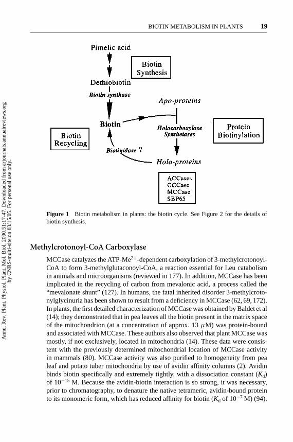
The presence of ACCase activity in plants was first documented in 1961 (79).The first direct evidence for the occurrence of biotin enzymes other than ACCasein plants was provided in 1990 by Wurtele & Nikolau (179). These researchersdetected MCCase, PCCase, and PyrCase activities, in addition to ACCase activity,in cell-free extracts of various monocot and dicot plant species. Thus, the fourbiotin-dependent activities found in mammals are also present in plants. Informa-tion on the structure, regulation, and function of plant biotin-dependent carboxy-lases, biotin biosynthesis, and protein biotinylation processes in higher plants hasburgeoned worldwide since 1990 (see below and Figure 1).
Ann
u. R
ev. P
lant
. Phy
siol
. Pla
nt. M
ol. B
iol.
2000
.51:
17-4
7. D
ownl
oade
d fr
om a
rjou
rnal
s.an
nual
revi
ews.
org
by C
NR
S-m
ulti-
site
on
03/1
5/05
. For
per
sona
l use
onl
y.
P1: FQK
March 22, 2000 10:17 Annual Reviews CHAP-02
?BIOTIN METABOLISM IN PLANTS 19
Figure 1 Biotin metabolism in plants: the biotin cycle. See Figure 2 for the details ofbiotin synthesis.
Methylcrotonoyl-CoA Carboxylase
MCCase catalyzes the ATP-Me2+-dependent carboxylation of 3-methylcrotonoyl-CoA to form 3-methylglutaconoyl-CoA, a reaction essential for Leu catabolismin animals and microorganisms (reviewed in 177). In addition, MCCase has beenimplicated in the recycling of carbon from mevalonic acid, a process called the“mevalonate shunt” (127). In humans, the fatal inherited disorder 3-methylcroto-nylglycinuria has been shown to result from a deficiency in MCCase (62, 69, 172).In plants, the first detailed characterization of MCCase was obtained by Baldet et al(14); they demonstrated that in pea leaves all the biotin present in the matrix spaceof the mitochondrion (at a concentration of approx. 13µM) was protein-boundand associated with MCCase. These authors also observed that plant MCCase wasmostly, if not exclusively, located in mitochondria (14). These data were consis-tent with the previously determined mitochondrial location of MCCase activityin mammals (80). MCCase activity was also purified to homogeneity from pealeaf and potato tuber mitochondria by use of avidin affinity columns (2). Avidinbinds biotin specifically and extremely tightly, with a dissociation constant (Kd)of 10−15 M. Because the avidin-biotin interaction is so strong, it was necessary,prior to chromatography, to denature the native tetrameric, avidin-bound proteinto its monomeric form, which has reduced affinity for biotin (Kd of 10−7 M) (94).
Ann
u. R
ev. P
lant
. Phy
siol
. Pla
nt. M
ol. B
iol.
2000
.51:
17-4
7. D
ownl
oade
d fr
om a
rjou
rnal
s.an
nual
revi
ews.
org
by C
NR
S-m
ulti-
site
on
03/1
5/05
. For
per
sona
l use
onl
y.

P1: FQK
March 22, 2000 10:17 Annual Reviews CHAP-02
?20 ALBAN ■ JOB ■ DOUCE
The use of purified mitochondria and affinity chromatography appeared to be themost convenient way to purify plant MCCase to homogeneity (2). Because plantMCCase is very unstable, attempts to purify the enzyme from crude extracts ofcarrot somatic embryos and maize leaves only yielded less pure and less activepreparations. Presumably, this was because other biotin proteins were present inthese extracts and the enzyme was not protected (by the mitochondrial membranes)from proteases released from the vacuolar space during tissue homogenization(32, 46). Structural and biochemical properties of MCCase from pea leaves andpotato tubers were determined (2) and found to be similar to those reported forbacterial and mammal MCCases (8, 61, 81, 93, 100, 138, 145, 146). Pea leaf andpotato tuber MCCases are composed of two nonidentical subunits. The largerα-subunit is biotinylated and has a molecular mass of 74–76 kDa, whereas thesmallerβ-subunit is biotin-free and has a molecular mass of 53–54 kDa. This het-eromeric structure was subsequently confirmed with the corresponding enzymesfrom carrot and maize (32, 46). The native enzyme behaves as a heterooctamercomposed of four large subunits and four small subunits (α4β4). Gel filtration ex-periments determined aMr in the range of 500,000 to 530,000 for both purifiedpea and potato MCCases. In addition, the biotin content of plant MCCase wasconsistent with a stoichiometry of four molecules perα4β4 octamer, presumablywith eachα-subunit containing one biotin prosthetic group (2). In this respect, peaand potato MCCases resemble bacterial MCCase (61), but differ from mammalian,carrot, and maize MCCases, which seem to adopt a dodecameric structure (α6β6)(32, 46, 100). Steady-state kinetic analyses demonstrated that the plant MCCase-catalyzed reaction proceeds by a double-displacement mechanism (Bi Bi Uni Uniping-pong), where ATP and bicarbonate bind to the enzyme in the first half-reactionand react to form the carboxybiotinyl enzyme derivative (see Reaction [1]). ADPand Pi produced by hydrolysis of ATP are then released. In the second partialreaction, 3-methylcrotonoyl-CoA binds to the carboxybiotinyl enzyme and be-comes carboxylated to form 3-methylglutaconoyl-CoA (2). The purified enzymeis substantially inhibited by the reaction end products, ADP and Pi (2), and byacetoacetyl-CoA, an end product of Leu degradation (32). Plant MCCase is alsoinactivated byN-ethylmaleimide, a sulfhydryl-modifying reagent, and phenylgly-oxal, a reagent that modifies Arg residues. Preincubation of MCCase with ATP and3-methylcrotonoyl-CoA prevented inactivation, which suggests that Cys and Argresidues are involved in catalysis or regulation of enzyme activity (2). In tomato,the MCCase activity found in leaves is lower than that in roots (169). However,the steady-state levels of the biotinyl subunit of MCCase (measured using spe-cific antibodies) and its mRNA are approximately equal in both tissues. Labelingexperiments with125I-streptavidin suggested that the lower activity of MCCasein leaves could be attributed to the reduced biotinylation of its biotin-containingsubunit. Consistent with this hypothesis, a pool of non-biotinylated enzyme wasfound in leaves (169). These data suggest, therefore, that the relative biotinylationof a biotin-containing enzyme can be a potential mechanism for regulating itsactivity. However, direct measurement of the biotin holo-carboxylase synthetase
Ann
u. R
ev. P
lant
. Phy
siol
. Pla
nt. M
ol. B
iol.
2000
.51:
17-4
7. D
ownl
oade
d fr
om a
rjou
rnal
s.an
nual
revi
ews.
org
by C
NR
S-m
ulti-
site
on
03/1
5/05
. For
per
sona
l use
onl
y.

P1: FQK
March 22, 2000 10:17 Annual Reviews CHAP-02
?BIOTIN METABOLISM IN PLANTS 21
(biotin ligase) activity showed no detectable biotin incorporation into protein frompea leaf extracts at any stage of the plant’s development, unless an exogenous apo-carboxylase substrate was added to the reaction medium (165), which indicatesthat the apo-carboxylases in pea leaves are at very low concentrations. On the otherhand, in seedling organs and developing cotyledons of soybean, differences in theabundance of the biotin prosthetic group during plant development closely paralleldifferences in the abundance of the biotinyl subunit of MCCase (7). Thus, MC-Case does not appear to be regulated by biotinylation in soybean. The isolation ofcDNAs and genes coding for the biotinylated subunit of MCCase revealed that, inaddition to the biotinylation domain, this subunit contains the functional domainsfor the first half-reaction catalyzed by all biotin-dependent carboxylases, namelythe carboxylation of biotin (158, 168, 170). These domains are arranged seriallyon the polypeptide, with the biotin carboxylase domain at the amino terminus andthe biotin-carboxyl carrier domain at the carboxyl terminus. Cloning of the cDNAand gene coding for the biotin-free subunit of this enzyme will be very useful instudying how the coordinate expression of the two MCCase subunits is regulated.
The metabolic functions of MCCase in plants are still not fully understood,notwithstanding these biochemical and molecular characterizations. As in mam-mals, MCCase may be involved in the mitochondrial catabolism of Leu and/orin the so-called “mevalonate shunt” by which mevalonate can be metabolized tononisoprenoid compounds. Previous radiotracer studies of plants grown in thepresence of either [14C]Leu (159) or [14C]mevalonate (117) indicated that Leucatabolism and the mevalonate shunt operate in vivo. Whether these two pathwaysinvolve MCCase remains to be demonstrated. The degradation of amino acids inplants in general and in particular the degradation pathway(s) of the branched-chainamino acids (Leu, Ile, and Val) are poorly understood. The first step in degradationleads, by transamination, to branched-chain 2-oxo acids (2-oxoisocaproate fromLeu), in which oxidative decarboxylation (to isovaleryl-CoA with Leu) and fur-ther oxidation (to 3-methylcrotonoyl-CoA in from Leu) take place in peroxisomes(66). Gerbling & Gerhardt (66) suggested that the subsequent steps could alsoinvolve peroxisomal enzymes. However, the fate of 3-methylcrotonoyl-CoA wasnot clearly identified. These authors (66) proposed that an extraperoxisomal path-way is involved in Leu catabolism since peroxisomes were unable to carboxylate3-methylcrotonoyl-CoA. Recent metabolic radiotracer studies in which extractsfrom isolated soybean seedling mitochondria were used have shown that all the en-zymes necessary for Leu degradation to acetoacetate, and presumably acetyl-CoA,are present in plant mitochondria and that this pathway likely involves MCCase (7).Despite the lack of direct evidence implicating plant MCCase in Leu catabolism,some circumstantial evidence indicates that the pathway may be similar to thatin animals and in bacteria. Plant MCCase is constitutively expressed, but its ac-tivity increases markedly during leaf senescence, when intense degradation ofproteins occurs (2). This accumulation of MCCase activity is correlated with adecline in Leu content (7). Leu is the second-most abundant amino acid in plantsafter Asn, and it transiently accumulates in plant cells during prolonged carbon
Ann
u. R
ev. P
lant
. Phy
siol
. Pla
nt. M
ol. B
iol.
2000
.51:
17-4
7. D
ownl
oade
d fr
om a
rjou
rnal
s.an
nual
revi
ews.
org
by C
NR
S-m
ulti-
site
on
03/1
5/05
. For
per
sona
l use
onl
y.

P1: FQK
March 22, 2000 10:17 Annual Reviews CHAP-02
?22 ALBAN ■ JOB ■ DOUCE
starvation when the induction of proteolytic activities leads to a massive break-down of proteins (27, 65). These amino acids, as well as the fatty acids derivedfrom lipid breakdown, are used in place of sugar to fuel the respiration of mito-chondria spared by autophagy (11). In addition, MCCase activity is induced in peacotyledons during the mobilization of storage proteins for seedling growth (51). Itis also noteworthy that MCCase activity is strongly induced in starved sycamorecells (up to a sixfold increase in specific activity after four days of sucrose starva-tion), which correlates with an accumulation of the enzyme in mitochondria (10).Taken together, these observations support the findings that plant MCCase, likeits animal counterpart, is involved in the degradation of Leu derived from proteinmobilization.
Plant MCCase could also be implicated in noncyclic isoprenoid catabolism. Insome bacterial species, noncyclic isoprenoids such as geranoyl-CoA are catabo-lized to acetyl-CoA via a set of reactions analogous to ß-oxidation; one reaction inthis pathway is the carboxylation of methylcrotonoyl-CoA (149). Little is knownabout the biochemistry of isoprenoid degradation in plants, although recent find-ings indicate that plants contain geranoyl-CoA carboxylase (GCCase; EC 6.4.1.5),a biotin-containing enzyme previously characterized inPseudomonasspecies (71).Thus, the catabolism of noncyclic isoprenoids in plants may be analogous to thatdescribed for bacterial species, i.e. after the carboxylation of theγ -methyl group ofgeranoyl-CoA, the carboxymethyl branch group of the product carboxygeranoyl-CoA is then eliminated. The resulting ß-keto thioester would then be amenableto ß-oxidation. Ultimately, such a process would generate methylcrotonoyl-CoA,which would require MCCase for its subsequent catabolism (71). Since GCCasewas reported to occur in chloroplasts (71), this catabolic pathway would necessitatecoordination of at least three intracellular compartments: plastids (the location ofphytol, carotenoids, and GCCase), microbodies (the major location of ß-oxidation),and mitochondria (the location of MCCase).
Acetyl-CoA Carboxylase
ACCase [acetyl-CoA: carbon dioxide ligase (ADP-forming)] catalyzes the ATP-Mg-dependent carboxylation of acetyl-CoA to form malonyl-CoA, with bicarbon-ate as the source of inorganic carbon. The reaction is the first committed step in thesynthesis of fatty acids. In plant cells, large amounts of malonyl-CoA are neededin the plastids to sustain de novo fatty acid synthesis (C16:0, C18:0, C18:1), butmalonyl-CoA is also needed in the cytosol for a variety of reactions including theelongation of very long-chain fatty acids, the synthesis of secondary metabolitessuch as flavonoids, anthocyanins, and stilbenoids, and the malonylation of someamino acids and secondary metabolites (reviewed in 75). The occurrence of cy-tosolic reactions requiring malonyl-CoA and the fact that the plastid envelope isnot permeable to malonyl-CoA led to the hypothesis that at least two isoenzymesof ACCase, a plastidic isoenzyme and a cytosolic isoenzyme, are present in plants(reviewed in 77).
Ann
u. R
ev. P
lant
. Phy
siol
. Pla
nt. M
ol. B
iol.
2000
.51:
17-4
7. D
ownl
oade
d fr
om a
rjou
rnal
s.an
nual
revi
ews.
org
by C
NR
S-m
ulti-
site
on
03/1
5/05
. For
per
sona
l use
onl
y.

P1: FQK
March 22, 2000 10:17 Annual Reviews CHAP-02
?BIOTIN METABOLISM IN PLANTS 23
Interest in plant ACCase has increased with the discovery that this enzyme isthe primary target site of several major classes of grass-specific herbicides thataffect fatty acid synthesis, principally the cyclohexanediones (CHD) and the ary-loxyphenoxypropionates (APP) (29, 76). These herbicides are reversible inhibitorsof grass ACCase, and there is kinetic evidence that these compounds inhibit thecarboxyltransferase partial reaction by acting as transition-state analogues of thecomplex formed at this site (175). This latter observation is interesting because itconcerns the partial reaction unique to individual biotin-dependent carboxylases(see Reaction [2]), and inhibition here is in keeping with the specificity of thesecompounds for plant ACCase. The other partial reaction, the carboxylation of bi-otin (see Reaction [1]), is common to all biotin enzymes, and inhibition at thisstep would render these other carboxylases sensitive, which is, in fact, not the case(2). Kinetic analyses also revealed that the CHD and APP are mutually exclusiveinhibitors of ACCase, i.e. the binding of a herbicide from one class prevents bind-ing of a herbicide from the other class (28, 131), either because the two bindingsites overlap, or through allosteric effects. Most grass species are susceptible toCHD and APP herbicides, whereas broad-leaved plants and monocotyledonousspecies other than grasses are resistant (77). With few exceptions, the selectivityof these compounds is expressed at the level of the target site. In contrast, herbicideselectivity is usually determined by the ability of plants to metabolize these pes-ticides, or by differences in uptake and/or translocation of the active compound.There are several examples where herbicide resistance in grasses correlates withthe presence of a tolerant form of ACCase, such as in natural populations of redfescue (Festuca rubra) and annual meadow grass (Poa annua) (83, 106). Moreover,widespread herbicide use has led to the selection of resistant biotypes of weedsthat were previously susceptible (reviewed in 45). Again, the main mechanism ofresistance appears to involve target site modification. Such modifications of thetarget ACCase resulting in a herbicide-resistant enzyme have been documented ina number of weed biotypes following selection in the field (for example, see 109),and in maize tissue culture following in vitro selection (111, 123). Resistance of abiotype ofLolium rigidum(resulting from 10 consecutive years of selection withdiclofop-methyl) to ACCase-inhibiting herbicides correlates with the possessionof a modified, resistant form of ACCase that is controlled by a single major gene(162). However, the structural and molecular basis for the difference in herbicidesensitivity of the enzymes from grasses and other plants, particularly dicotyledons,was unknown until the recent elucidation of the two different types of structuralorganization of plant ACCase, together with the discovery of ACCase isoforms indifferent cell and tissue compartments. These findings have improved our under-standing of the selectivity of ACCase-inhibiting herbicides.
The molecular organization of ACCase differs depending on the source of theenzyme. InE. coli, ACCase is composed of four distinct subunit types that read-ily dissociate into three components: a homodimer of 49-kDa subunits (biotincarboxylase module), a homodimer of 17-kDa subunits (BCCP), and a carboxyl-transferase heterotetramer containing two 33-kDa and two 35-kDa (α2β2) subunits.
Ann
u. R
ev. P
lant
. Phy
siol
. Pla
nt. M
ol. B
iol.
2000
.51:
17-4
7. D
ownl
oade
d fr
om a
rjou
rnal
s.an
nual
revi
ews.
org
by C
NR
S-m
ulti-
site
on
03/1
5/05
. For
per
sona
l use
onl
y.

P1: FQK
March 22, 2000 10:17 Annual Reviews CHAP-02
?24 ALBAN ■ JOB ■ DOUCE
These are encoded by four separate genes, which inE. coliare namedaccC,accB,accA, andaccD, respectively (6, 72, 95, 103, 104). In contrast, in animals, fungiand yeasts, these entities are located on a single, multifunctional polypeptide thathas a molecular mass exceeding 200 kDa (5, 12, 108). The functional ACCase en-zyme in these organisms is composed of multimers of this large polypeptide. Thestructure of plant ACCase has been the subject of considerable confusion in thepast. Early experiments indicated that ACCase in spinach and barley chloroplastsand avocado plastids had a multisubunit structure similar to that found in prokary-otes (90, 91, 113). Plants may also contain distinct isozymes of ACCase in separatespatial and temporal compartments to generate the malonyl-CoA needed for a va-riety of phytochemicals (160). However, the subsequent inclusion of proteinaseinhibitors in purification media and the development of avidin-affinity matrices,which allowed rapid purification of the enzyme, led to the isolation of a homo-meric ACCase of high molecular mass comparable to that found in other eukary-otes (31, 54, 55). Based on this finding, it was claimed and generally agreed thatthe occurrence of low-molecular-mass biotinyl polypeptides in purified ACCasepreparations was largely, if not exclusively, due to severe degradation of the high-molecular-mass polypeptide form during enzyme isolation (reviewed in 75, 77).Consequently, the concept of a prokaryotic form of ACCase in plants fell intodisfavor. However, because there was no evidence for compartmentation of AC-Case, the second hypothesis was rejected too. However, it is now evident that bothhypotheses are valid. The confusion about the exact nature of plant ACCase arosein large part because (a) plants contain structurally different forms of the enzyme,one of which rapidly loses activity during purification, not because of inactivationby proteases but rather because it readily dissociates into its component subunits,and (b) the enzymes from Gramineae were mainly studied. Most plants other thanGramineae are now known to have a multisubunit plastidial ACCase (frequentlyreferred to as the prokaryotic form) and a multifunctional, extraplastidial ACCase(also called the eukaryotic form), whereas members of the Gramineae have only themultifunctional-type ACCase in both plastids and cytosol (3, 56, 99, 141). To date,only two exceptions to this generalization have been reported: Chloroplasts fromBrassica napusappear to contain both multisubunit and multifunctional ACCases(59, 148); and chloroplasts from the dicotErodium moschatumlack the multisub-unit ACCase but instead contain the multifunctional-type enzyme (35). The firstmajor finding that revived the idea that plant ACCase can adopt a multisubunitstructure was the discovery of a putativeaccD homologue in the pea chloroplastgenome (105). It was subsequently shown that the product of this gene in pea bindsat least two other polypeptides, one of which was biotinylated (140). Shortly there-after, direct biochemical evidence established the presence of a prokaryotic formof ACCase in pea mesophyll chloroplasts (3, 98) composed of different-sized sub-units. One of these, apparently 38 kDa in size, is biotinylated, which gives a native600-kDa size. This form of the enzyme represents about 80% of the total ACCaseactivity in the whole leaf (3) and is totally insensitive to herbicides of the CHD andAPP classes (3, 43, 98). These enzyme subunits are organized into two functionaldomains that interact through ionic interactions and are readily dissociable and
Ann
u. R
ev. P
lant
. Phy
siol
. Pla
nt. M
ol. B
iol.
2000
.51:
17-4
7. D
ownl
oade
d fr
om a
rjou
rnal
s.an
nual
revi
ews.
org
by C
NR
S-m
ulti-
site
on
03/1
5/05
. For
per
sona
l use
onl
y.

P1: FQK
March 22, 2000 10:17 Annual Reviews CHAP-02
?BIOTIN METABOLISM IN PLANTS 25
reassociable according to the law of mass action (3, 157). A similar multisubunitorganization was recently reported for soybean chloroplastic ACCase (132). Thisdissociation/reassociation property of the multisubunit form of ACCase is thoughtto play a role in regulating ACCase activity in chloroplasts (132). The chloroplasticACCase is able to carboxylate free D-biotin as an alternate substrate in lieu of BCCPand in the absence of acetyl-CoA. This specific property was used to purify thebiotin carboxylase component of the enzyme (4). The minimal structure of the ac-tive biotin carboxylase domain corresponds to a complex of two, tightly interactingpolypeptides (the 38-kDa biotinylated subunit and a 32-kDa biotin-free polypep-tide) (4). This contrasts with the organization of bacterial ACCase in which allsubunit components are freely separable (72). Steady-state kinetic analyses of peabiotin carboxylase were compatible with an ordered mechanism in which MgATPbinds first, followed by free biotin and then bicarbonate. Consistent with this mech-anism, bicarbonate-dependent ATP hydrolysis by the enzyme could be observedonly in the presence of added D-biotin (4). These data suggest the existence offunctional and structural differences between the multisubunit isoform of ACCasefrom plants and bacteria (163). cDNAs and/or genes encoding BCCP and biotincarboxylase subunits from various plants have been cloned (19, 34, 59, 132, 156).These proteins are nuclear encoded and share substantial sequence similarity withthe corresponding bacterial ACCase subunits. Furthermore, their expression is co-ordinated during seed and leaf development (135). The second functional domainof ACCase found in pea or in soybean chloroplasts is composed of theα- andβ-subunits of carboxyltransferase (132, 157). Theα-subunit (73 to 91 kDa) is nu-clear encoded and possesses a structural motif similar to a prokaryotic membranelipoprotein lipid attachment site. As this polypeptide was previously identified asan inner-envelope membrane constituent of pea chloroplasts, this observation sug-gests that the prokaryotic form of ACCase in pea may be at least partly associatedwith chloroplast membranes (157). However, the insoluble nature of the carboxyl-transferase subunits of ACCase could be artifactual, resulting from a redistributionof the ACCase components among organellar subfractions following disruption ofthe complex during enzyme purification (132). In fact, the multisubunit ACCasecomplex remains soluble after chloroplast lysis if organelle fractionation is donerapidly (3). The gene encoding theβ-subunit (49 to 80 kDa) has been identifiedin the plastid genome by its similarity to one of the carboxyltransferase subunitsof E. coli ACCase (59, 105, 132, 140). This is the only component of plant lipidmetabolism known to be encoded by the plastid genome.
In addition to this major prokaryotic form, pea leaves also contain a minoreukaryotic form of ACCase that is mainly, if not exclusively, concentrated in theepidermis (3). By contrast, in mature pea seeds this ACCase comprises the majorportion of the enzyme activity (23, 43). This enzyme is a homodimer of a sin-gle biotinylated polypeptide of about 220 kDa that is sensitive to APP herbicides(3, 43). Several genes and cDNA clones have been isolated for this type of ACCasefrom other dicotyledonous plants. None of them seems to have typical organelle-targeting presequences (reviewed in 78, 118). All of these clones encode proteinswith the biotin carboxylase domain at the N-terminus, the biotin carboxyl carrier
Ann
u. R
ev. P
lant
. Phy
siol
. Pla
nt. M
ol. B
iol.
2000
.51:
17-4
7. D
ownl
oade
d fr
om a
rjou
rnal
s.an
nual
revi
ews.
org
by C
NR
S-m
ulti-
site
on
03/1
5/05
. For
per
sona
l use
onl
y.

P1: FQK
March 22, 2000 10:17 Annual Reviews CHAP-02
?26 ALBAN ■ JOB ■ DOUCE
domain in the middle, and the carboxyltransferase domain at the C-terminus. Asubstantial portion of the polypeptide that is located between the two latter do-mains has no correspondence withE. coli ACCase, and may have a structuralrole. Grasses such as maize and wheat also contain two ACCase isoforms, themajor one in the chloroplasts of mesophyll cells, the other in an extraplastidialcompartment (probably the cytosol) of another cell type(s) (56, 70, 84). However,in contrast to dicotyledonous plants, both isoforms are composed of a single type ofhigh-molecular-mass, biotin-containing polypeptide of 227 (ACCase 1, mesophyllchloroplast enzyme) or 219 kDa (ACCase 2, non-mesophyll-chloroplast enzyme)(9, 56). Antibodies raised against the 227-kDa form poorly recognized the minor219-kDa isoform. These ACCase isoforms are encoded by distinct nuclear genes(9). Finally, both enzymes are inhibited by CHD and APP herbicides, althoughACCase 1 is much more sensitive than ACCase 2 (56, 82, 84). Thus, it now ap-pears that all plants contain two ACCase isoforms in two different subcellular andtissue locations. However, the Gramineae family of plants is different in that boththe plastid and cytosolic ACCase isozymes are of the eukaryotic type. Coincidentwith this evolutionary difference, a recent finding noted that probably all plantsexcept the Gramineae contain the plastid-encodedaccD gene (99). Consistentwith this finding, both western and Southern blot analyses failed to detect subunitsof the prokaryotic form of ACCase in all grass species studied to date, whereasprokaryotic and eukaryotic forms of ACCase were detected in all the other plantsanalyzed, including monocotyledons and dicotyledons (99, 135). Thus, this differ-ence in ACCase molecular organization provides an explanation for the action ofgrass-specific herbicides, which inhibit only the eukaryotic and not the prokaryoticform of the enzyme (3, 98). The chloroplastic ACCase, thought to play a key rolein de novo fatty acid biosynthesis, is strongly inhibited by these compounds in theGramineae (eukaryotic form) but not in other plants (prokaryotic form). In con-trast, in all plant species the cytosolic ACCase (eukaryotic form) is only a minorform in leaves, probably involved in secondary metabolic pathways, and is muchless affected by CHD and APP herbicides. Interestingly,Erodium moschatum, theonly dicot known to be sensitive to APP herbicides, lacks the herbicide-insensitiveprokaryotic form of ACCase in its plastids (35).
In addition to these differences in sensitivity to herbicides, the two structurallydistinct ACCase isozymes exhibit important differences in their biochemical prop-erties. The multifunctional enzyme has a much lowerKm for acetyl-CoA than themultisubunit complex (3, 56). The multifunctional enzyme is able to carboxylatepropionyl-CoA at substantial rates, whereas the multisubunit complex is not activewith this substrate (43). Furthermore, no other PCCase activity, different from thatcatalyzed by the eukaryotic forms of ACCase, can be detected in either reproductiveor vegetative organs of pea plants or from maize leaves at any stage of development(43, 84). Steady-state kinetic analysis of the multifunctional form of ACCase, pu-rified from mature pea seeds, with respect to substrate specificity and inhibition byquizalofop, a member of the APP class of herbicides, demonstrated that both re-actions catalyzed by the enzyme (acetyl-CoA and propionyl-CoA carboxylations)proceed at separate sites on the enzyme and are inhibited by the herbicide. The two
Ann
u. R
ev. P
lant
. Phy
siol
. Pla
nt. M
ol. B
iol.
2000
.51:
17-4
7. D
ownl
oade
d fr
om a
rjou
rnal
s.an
nual
revi
ews.
org
by C
NR
S-m
ulti-
site
on
03/1
5/05
. For
per
sona
l use
onl
y.

P1: FQK
March 22, 2000 10:17 Annual Reviews CHAP-02
?BIOTIN METABOLISM IN PLANTS 27
sites, however, show different catalytic properties: One site binds either acetyl-CoAor propionyl-CoA and is inhibited by quizalofop, whereas the other is specific foracetyl-CoA and is much less affected by quizalofop. Owing to these two catalyti-cally distinct sites, the enzyme obeyed Michaelis-Menten kinetics with respect topropionyl-CoA, but exhibited kinetic cooperativity in the presence of acetyl-CoA.Also, the kinetics of PCCase activity exhibited hyperbolic inhibition in the pres-ence of quizalofop, but cooperative inhibition when measuring the ACCase activityof the enzyme. These results indicate that the higher the substrate specificity, thelower the quizalofop sensitivity of the active site, which suggests that the nature ofthe interactions between the two identical enzyme subunits may play a role in gov-erning the degree of herbicide sensitivity. Presumably, such interactions introducesome distortions in the structure of the individual active sites, for example, gener-ating kinetic cooperativity with substrates and inhibitors (43). Similar conclusionswere drawn from kinetic studies with maize ACCases (84). Also, this apparent cor-relation between substrate specificity and sensitivity of ACCase toward quizalofopwas confirmed by kinetic analysis of the prokaryotic form of ACCase from pealeaf chloroplasts. This enzyme, which is insensitive to quizalofop inhibition, isunable to carboxylate propionyl-CoA (43).
The discovery of the occurrence of ACCase isoforms in separate plant cell com-partments raises the question of their physiological significance. The main role ofACCase in plastids is to provide malonyl-CoA, the precursor for fatty acid biosyn-thesis. Indeed, plastids represent the major site for de novo fatty acid biosynthesisin plants (118, 119). In support of this conclusion, we note, for example, that theactivity level of the prokaryotic form of ACCase in pea shows considerable varia-tion during development; it is maximal in young pea leaves, presumably reflectinga high demand for fatty acids required for the biosynthesis of thylakoid membranes(43). Also, recent investigations on fatty acid biosynthesis in pea suggest that theenzymes of fatty acid synthesis are organized within the chloroplast into a multien-zyme assembly that channels acetate, through acetyl-CoA synthetase and ACCase,into long-chain fatty acids, glycerides, and CoA esters (137). Considerable in vivoand in vitro evidence suggests that chloroplastic ACCase is involved in the regula-tion of plant fatty acid synthesis (ARPP-1997). For example, analysis of substrateand product pool sizes implicated ACCase in the light/dark regulation of fatty acidsynthesis in spinach leaves and chloroplasts (128, 129). The biochemical environ-ment of the chloroplast stroma undergoes numerous reversible changes upon illu-mination, and there are increases particularly in pH, ATP, NADPH, and Mg2+ con-centrations and in the ATP/ADP ratio (75). These differences can largely accountfor the stimulation of ACCase in the light and therefore fatty acid synthesis (87).Also, it was recently shown that reduced thioredoxin activates multisubunit chloro-plastic ACCase from pea and spinach plants, which suggests that this mechanismis a link between light activation and fatty acid synthesis (87, 142). Herbicide inhi-bition of ACCase was used to determine flux control coefficients, which led to theconclusion that chloroplastic ACCase exerts a major control over fatty acid synthe-sis rates in barley and maize leaves (121). ACCase was also the apparent site of feed-back inhibition of fatty acid synthesis in tobacco suspension cells supplemented
Ann
u. R
ev. P
lant
. Phy
siol
. Pla
nt. M
ol. B
iol.
2000
.51:
17-4
7. D
ownl
oade
d fr
om a
rjou
rnal
s.an
nual
revi
ews.
org
by C
NR
S-m
ulti-
site
on
03/1
5/05
. For
per
sona
l use
onl
y.

P1: FQK
March 22, 2000 10:17 Annual Reviews CHAP-02
?28 ALBAN ■ JOB ■ DOUCE
with exogenous fatty acids (153). Finally, recent in vivo evidence for a regula-tory role of ACCase in oilseeds was obtained by genetic engineering approaches(136, 154). Now that clones are available for various chloroplastic ACCases, planttransformant experiments will provide tools to assess the role of this enzyme incontrolling flux through the fatty acid biosynthetic pathway. At present, there is noevidence that de novo fatty acid synthesis occurs in the cytosol of plant cells. How-ever, a recent finding indicated that plant mitochondria contain not only the acyl car-rier protein but also all the enzymes required for de novo fatty acid synthesis frommalonate, but not from acetate (167). Thus, in this case, ACCase is probably notrequired. Indeed, plant mitochondria do not have detectable ACCase activity (2).
Although malonyl-CoA is required, in addition to fatty acid synthesis, in anumber of biosynthetic pathways, the physiological role(s) of the multifunctional,cytosolic ACCase is not yet clearly established. There are indications that this en-zyme might be involved in the biosynthesis of very long-chain fatty acids requiredfor cuticular waxes, or the biosynthesis of flavonoids via chalcone synthase. In-terestingly, in pea leaves, both processes occur in the cytosol of epidermal cells,thus matching the tissue localization of the eukaryotic extraplastidial form of pealeaf ACCase (3). Cuticular wax and flavonoids are important in the interaction ofplants with their environment, for example, for protection against UV radiationand pathogens. Two recent findings indicate that this isozyme of ACCase probablyhelps to control the synthesis of such protective compounds: First, the transcriptfor the eukaryotic form of alfalfa ACCase is induced by yeast or fungal elicitorsof isoflavonoid phytoalexin synthesis (155); and second, the cytosolic eukaryoticform (but not the chloroplastic prokaryotic form) of ACCase is induced by UV-Birradiation of fully expanded pea leaves, in parallel to the induction of some ofthe enzymes involved in flavonoid synthesis, such as chalcone synthase (97). Theproducts of two different ACCase genes have been identified in the cytosol ofhuman and rat cells (1, 176). The presence of multiple cytosolic ACCase genesin mammals also reflects the need for differential expression of the enzyme inresponse to varying environmental or developmental cues. The mammalian andyeast ACCases are regulated by reversible phosphorylation through the action ofan AMP-activated protein kinase (92). Recent investigations demonstrated thatthe carboxyltransferaseβ-subunit of pea chloroplast ACCase is phosphorylated,which suggests that such a regulatory mechanism may also operate in plants (143).
BIOTIN BIOSYNTHETIC PATHWAY
Plants, like micro organisms, can synthesize biotin, whereas other multicellulareukaryotic organisms are biotin auxotrophs. Biotin biosynthesis has been well char-acterized inE. coliand, more recently, in other bacteria such asBacillus sphearicus,through combined biochemical and genetic studies. In these bacteria, thebio(ABFCD) locus contains the genes required for biotin biosynthesis (57, 67, 89).These genes encode the enzymes catalyzing the synthesis of biotin from pimeloyl-CoA and Ala: 7-keto-8-aminopelargonic acid synthase (KAPA synthase);
Ann
u. R
ev. P
lant
. Phy
siol
. Pla
nt. M
ol. B
iol.
2000
.51:
17-4
7. D
ownl
oade
d fr
om a
rjou
rnal
s.an
nual
revi
ews.
org
by C
NR
S-m
ulti-
site
on
03/1
5/05
. For
per
sona
l use
onl
y.

P1: FQK
March 22, 2000 10:17 Annual Reviews CHAP-02
?BIOTIN METABOLISM IN PLANTS 29
7,8-diaminopelargonic acid aminotransferase (DAPA aminotransferase); dethio-biotin (DTB) synthase; and biotin synthase, coded bybioF, bioA, bioD, andbioB,respectively (Figure 2). Until recently, little was known about biotin metabolismin plants. Initial information on biotin synthesis and transport in plants came fromanalysis of thebio1 biotin auxotroph ofA. thaliana, which was discovered by
Figure 2 Biotin biosynthetic pathway in plants and microorganisms. Positions wheremutations block biotin synthesis inE. coli (bioH, bioC, bioF, bioA, bioD,andbioB) andArabidopsis(bio1andbio2) are shown, as is inhibition by actithiazic acid (−). The immedi-ate precursor of pimeloyl-CoA appears to be pimelic acid inB. subtilis, B. sphaericus, andArabidopsisbut not inE. coli. SAM: S-adenosyl methionine; PLP: pyridoxal phosphate;FldX: flavodoxin; FldX reductase: flavodoxin reductase.
Ann
u. R
ev. P
lant
. Phy
siol
. Pla
nt. M
ol. B
iol.
2000
.51:
17-4
7. D
ownl
oade
d fr
om a
rjou
rnal
s.an
nual
revi
ews.
org
by C
NR
S-m
ulti-
site
on
03/1
5/05
. For
per
sona
l use
onl
y.

P1: FQK
March 22, 2000 10:17 Annual Reviews CHAP-02
?30 ALBAN ■ JOB ■ DOUCE
Schneider et al (147). Seeds homozygous for the mutation failed to develop unlessexogenous biotin, dethiobiotin, or DAPA, but not KAPA, was supplied to the plant(151). Recently, Patton et al (126) have shown that theE. coli bioAgene, whichcodes for DAPA aminotransferase, can genetically complement thebio1mutation,demonstrating thatbio1/bio1mutant plants are defective in this enzyme. A secondbiotin auxotroph ofA. thalianahas subsequently been identified. Arrested embryosfrom thisbio2mutant are defective in the final step of biotin synthesis, i.e. the con-version of dethiobiotin to biotin (125). Treatment of a biotin-overexpressing strainof lavender cells with [3H]pimelic acid showed that all the intermediates of biotinsynthesis established in bacteria, plus the novel metabolite 9-mercaptodethiobiotin(9-mDTB), accumulate in plants (17), which demonstrates that the pathway ofbiotin synthesis in bacteria is conserved in plants (Figure 2). Also, this study in-dicated that the reaction catalyzed by the plant biotin synthase proceeds in twodistinct steps involving 9-mDTB as an intermediate and that actithiazic acid, aninhibitor of biotin biosynthesis, specifically blocks the conversion of 9-mDTB tobiotin, i.e. the formation of the thiophene ring of biotin (17) (Figure 2). Subse-quent experimental evidence suggested that 9-mDTB may be an intermediate inthe bacterial biotin biosynthetic pathway as well (110). Inhibition of biotin synthe-sis by actithiazic acid is lethal to plants and can be prevented by supplementationwith nanomolar concentrations of biotin (13), findings that demonstrate both thespecificity of action of this inhibitor and the fact that plants, like all organisms,need only trace amounts of biotin for growth. The catalytic mechanism of the laststep of biotin biosynthesis is still unclear. It has been demonstrated inE. coli thatthe conversion of dethiobiotin to biotin is catalyzed by a complex involving 2 (or3) proteins in addition to thebioB gene product, which by itself is totally inac-tive (24, 89, 139). The term biotin synthase, previously used to designate thebioBgene product, should therefore be reserved for this multisubunit complex. A cDNAencoding anA. thalianahomologue of thebioB gene product has been isolatedby functional complementation of abioB biotin auxotroph mutant ofE.coli (18).This cDNA shows specific regions of similarity with the corresponding genes frombacteria and yeast. In particular, the predicted amino acid sequence of the plantprotein contains the consensus region (GXCXEDCXYCXQ) involved in bindinga [2Fe-2S] cluster (16). Interestingly, the plant sequence contains an N-terminalextension of about 40 amino acids that is not found in its bacterial counterparts,which suggests an organellar location for this enzyme (18). Computer analyses ofthe primary structure of theA. thaliana bioBgene product predict that this proteinmight be targeted to mitochondria (171). Indeed, western blot analyses with an-tibodies raised against the purified plant recombinant protein demonstrated sucha mitochondrial location (16). This subcellular location is intriguing since mostof the free biotin pool in plant mesophyll cells accumulates in the cytosol to aconcentration of about 11µM (15) (note that in bacteria free biotin never accu-mulates above a nM concentration range, see later). Thus, if biotin is synthesizedwithin mitochondria, it must be exported to accumulate in the cytosol. The preciserole of this pool of free biotin in the cytosol is not known. One hypothesis is that,
Ann
u. R
ev. P
lant
. Phy
siol
. Pla
nt. M
ol. B
iol.
2000
.51:
17-4
7. D
ownl
oade
d fr
om a
rjou
rnal
s.an
nual
revi
ews.
org
by C
NR
S-m
ulti-
site
on
03/1
5/05
. For
per
sona
l use
onl
y.

P1: FQK
March 22, 2000 10:17 Annual Reviews CHAP-02
?BIOTIN METABOLISM IN PLANTS 31
as in bacteria, the level of free biotin controls the expression of genes encodingthe biotin-containing enzymes and/or the enzymes involved in biotin synthesis. Insupport of this suggestion, it was demonstrated that the expression of the gene en-coding theA. thaliana bioBgene product is strongly induced under biotin-limitingconditions (124). Finally, the recombinant plant biotin synthase containing aniron-sulfur cluster was shown to catalyze the direct conversion of dethiobiotininto biotin (C Alban & R Douce, unpublished results). This reconstituted in vitrosystem requires an electron donor [NAD(P)H], S-adenosylmethionine (SAM) act-ing as a radical-forming molecule (74), and a soluble bacterial extract containingflavodoxin and flavodoxin reductase (89).
BIOTIN HOLOCARBOXYLASE SYNTHETASE
Biotinylation of biotin-dependent carboxylases permits the transformation of in-active apo-carboxylases into active holo-forms (138). InE. coli, the biotinylationof apo-ACCase (the unique biotin enzyme found in this organism) is carried out bybiotin ligase. The enzyme catalyzes the posttranslational attachment of D-biotin toa specific Lys residue of the apo-enzyme, via an amide linkage between the biotincarboxyl group and theε-amino group of Lys (138). This covalent attachmentoccurs in two discrete steps ([4] and [5]):
D-biotin+ ATP→ D-biotinyl 5′-AMP+ PPi [4]
D-biotinyl 5′-AMP+ apo-BCCP→ holo-BCCP+AMP+H2O [5]
The first step is the activation of D-biotin by ATP, which yields D-biotinyl 5′-AMP, followed by the covalent attachment of the biotinyl group to theε-amino-group of a specific Lys residue of the apo-BCCP, with release of AMP. Biotinligase has been purified fromE. coli and its gene cloned (58, 85). This enzyme,also called BirA, is a 33.5-kDa protein (85) that also acts as a repressor of thebiotin operon in the presence of D-biotinyl 5′-AMP as the co-repressor (39). Itsthree-dimensional structure has been determined at 2.3A resolution (174). Thecorresponding enzymes from various mammalian species have been purified andare referred to as biotin holo-carboxylase synthetase (HCS) (33, 181). Recently,clones encoding theSaccharomyces cerevisiaeHCS gene (40) and human HCScDNAs have been obtained (102, 161). These latter enzymes show some sequencesimilarity to the biotin ligases from bacteria, but are more than twice their size.The first direct evidence for the existence of HCS activity in plants came fromthe recent purification and characterization of HCS activity from pea leaves (165).The enzyme was able to use bacterial apo-BCCP as substrate, which demon-strates that plant HCS acts across species barriers. This cross-species activity re-vealed a molecular mechanism common to these enzymes. In contrast, plant HCSshowed a very high specificity for its biotin substrate, exhibiting an apparentKmvalue of 28 nM (165). It was subsequently shown that in plants, HCS activity is
Ann
u. R
ev. P
lant
. Phy
siol
. Pla
nt. M
ol. B
iol.
2000
.51:
17-4
7. D
ownl
oade
d fr
om a
rjou
rnal
s.an
nual
revi
ews.
org
by C
NR
S-m
ulti-
site
on
03/1
5/05
. For
per
sona
l use
onl
y.
P1: FQK
March 22, 2000 10:17 Annual Reviews CHAP-02
?32 ALBAN ■ JOB ■ DOUCE
associated with several subcellular compartments (164). Thus, three enzyme formscan be separated by anion-exchange chromatography of a pea leaf protein extract.The major form was found to be specific for the cytosolic compartment, whereasthe two minor forms were present in mitochondria and chloroplasts, respectively.The high purity and latency values of HCS activity measured in Percoll-purifiedchloroplasts and mitochondria, together with the observed protection of enzymeactivity in these organelles during thermolysin treatment, demonstrated that HCSis a genuine constituent of chloroplasts and mitochondria (164). The existenceof HCS isoforms in various plant cell compartments suggests that the differentbiotin-dependent carboxylases localized in chloroplasts, mitochondria, and cy-tosol are biotinylated in the cell compartment within which they are localized.One possible explanation for this feature is that the active site of HCS recognizes aparticular three-dimensional folded structure of apo-carboxylases rather than pri-mary amino acid structure alone (see below). By functional complementation ofthebirA 215 E. colimutant with anA. thalianacDNA expression library, a full-length HCS cDNA encoding a 41-kDa polypeptide was isolated (164). Althoughthis plant HCS shows specific regions of similarity with other known biotin ligasesof bacterial, yeast, and human origin, the similarities are restricted to the ATP- andbiotin-binding domains. These motifs, which are located in the central part of theprotein, are interconnected and thus reflect the requirement for ATP and biotinto be spatially close to allow the formation of the biotinyl-5′-AMP intermediate.Interestingly, the eight amino acid residues that have been shown to be in directcontact with biotin inE. coli BirA by X-ray crystallography (174) are strictlyconserved inA. thalianaHCS. In contrast, plant HCS does not contain the helix-turn-helix DNA binding domain of BirA fromE. coli (174) orB. subtilis(25). Inbacteria, this DNA-binding domain mediates the repressor function of the BirAprotein. Consistent with this observation, the cloned plant HCS proved unable tosubstitute for BirA as a repressor of the biotin operon in abirA derepressed mu-tant ofE. coli (C Alban & R Douce, in preparation). The N-terminal region ofA.thaliana HCS exhibits the characteristic features of an organelle transit peptide.Also, the occurrence of two Met residues close together in this region suggeststhe possible existence of cytosolic and “organelle-targeted” forms of HCS, syn-thesized from a single species of mRNA by alternative translational initiation.Such a possibility, together with an alternative splicing mechanism, has been sug-gested to account for the synthesis of human mitochondrial and cytosolic HCSisoforms (102). Two translation products of the expected sizes were obtained byin vitro transcription-translation experiments with the plant HCS cDNA, whichis consistent with this hypothesis (164). However, further studies were needed todefinitively assign the cellular localization of this clone to a specific compartment.To address this question, the clonedA. thalianaHCS cDNA was used to createan overproducingE. coli strain (166). Polyclonal antibodies raised against purerecombinant HCS were then produced to elucidate the subcellular localizationof this protein. Both immunodetection by western blotting of proteins from iso-lated pea leaf subcellular compartments, and immunocytology on tissue sections
Ann
u. R
ev. P
lant
. Phy
siol
. Pla
nt. M
ol. B
iol.
2000
.51:
17-4
7. D
ownl
oade
d fr
om a
rjou
rnal
s.an
nual
revi
ews.
org
by C
NR
S-m
ulti-
site
on
03/1
5/05
. For
per
sona
l use
onl
y.

P1: FQK
March 22, 2000 10:17 Annual Reviews CHAP-02
?BIOTIN METABOLISM IN PLANTS 33
of tobacco leaves expressing the complete coding sequence ofA. thalianaHCSdemonstrated that the enzyme encoded by this cDNA corresponds to the chloro-plastic isoform (166). Thus, this enzyme form is probably responsible for thebiotinylation of chloroplastic ACCase. Some physicochemical, biochemical, andkinetic properties of the pure recombinant HCS were also determined. The na-tive recombinant protein is a 37-kDa monomer. The enzyme is able to efficientlybiotinylate apo-BCCP fromE. coli and also apo-MCCase fromA. thaliana, butwith less efficiency (166). Such broad substrate specificity was previously notedfor HCS from various origins. However, neither apo-SBP65, the unbiotinylatedform of the 65-kDa pea seed-specific biotinyl protein [see below and (51)], nor asynthetic peptide produced as a maltose binding fusion protein inE. coli, whichwas previously shown to be efficiently biotinylated byE. coli biotin ligase in vivo(144), can serve as substrate for the recombinant chloroplasticA. thalianaHCS(166). Steady-state kinetic analyses of the HCS-catalyzed reaction were conductedto determine the reaction mechanism and inhibition by reaction products. Theseexperiments indicated that the reaction proceeds by a double-displacement “BiUni Uni Bi ping-pong Ter Ter” mechanism whereby ATP and D-biotin bind tothe enzyme in an ordered fashion during the first half-reaction and react to formbiotinyl-AMP. Then, the PPi produced by hydrolysis of ATP is released. In thesecond half-reaction, apo-BCCP binds to the biotinyl-AMP-enzyme complex andis biotinylated to form holo-BCCP. Finally, the holo-BCCP and AMP producedare released (166).
Owing to the presence of HCS activity in plant cell mitochondrial and cytosolicfractions as well as in chloroplasts, possible structurally distinct HCS isoformsneed investigation in these subcellular compartments if the mechanisms govern-ing the requirements and regulation of biotinylation in plants are to be understood.This information will also help elucidate why HCS activity has to be compartmen-talized in plant cells. Interestingly, a recent discovery revealed that human serumbiotinidase, an enzyme involved in biotin recycling in mammals, has biotinyl-transferase activity in addition to biotinidase hydrolase activity (88). Althoughbiotinidase activity is undetectable in plant extracts (13), the possible existence ofsuch biotinyl-transferase activity in plants merits exploration in the future.
STRUCTURAL REQUIREMENTS FOR PROTEINLIPOYLATION AND COMPARISON WITH PROTEINBIOTINYLATION
Lipoic acid (1,2-dithiolane-3-valeric acid) is the prosthetic group of the E2-proteincomponent (dihydrolipoylacyltransferase) of 2-oxoacid dehydrogenases, a familyof multienzyme complexes that catalyze the irreversible oxidative decarboxylationof 2-oxoacids (pyruvate, 2-oxoglutarate, and the three short-branched chain 2-oxoacids produced by transamination of Leu, Val, and Ile) (22, 112). The E2-protein
Ann
u. R
ev. P
lant
. Phy
siol
. Pla
nt. M
ol. B
iol.
2000
.51:
17-4
7. D
ownl
oade
d fr
om a
rjou
rnal
s.an
nual
revi
ews.
org
by C
NR
S-m
ulti-
site
on
03/1
5/05
. For
per
sona
l use
onl
y.

P1: FQK
March 22, 2000 10:17 Annual Reviews CHAP-02
?34 ALBAN ■ JOB ■ DOUCE
component forms the structural core of the complexes on the surface of whichoxoacid decarboxylase (E1-protein) and dihydrolipoamide dehydrogenase (E-3protein) are bound in a noncovalent manner (22, 112). Lipoic acid is also the pros-thetic group of the H-protein component of the glycine decarboxylase complexwhich catalyzes the oxidative decarboxylation and deamination of Gly with theformation of CO2, NH3, and methylene-tetrahydrofolate (48, 120). This complexconsists of four protein components (P-, H-, T-, and L-proteins). The H-proteincomponent forms the mechanistic “heart” of the Gly decarboxylase complex withthe P-, T-, and L-proteins (dihydrolipoamide dehydrogenase) interacting on its sur-face. The H-protein from pea leaf mitochondria has been crystallized and the X-raycrystal structure has been determined at 2A resolution (37, 122). The core of thisstructure consists of two antiparallelβ-sheets forming a hybrid barrel-sandwichstructure. One sheet has sixβ-strands and the other has three strands and two ad-jacent antiparallel strands joined by a loop (hairpinβ-motif) in which the lipoatecofactor is attached to a specific Lys residue. Despite minimal amino acid se-quence similarity, the structure of the lipoyl domain in the E2 protein componentof 2-oxo acid dehydrogenase multienzyme complexes and in H-protein closelyresembles that of the biotinyl domain of BCCP (26, 36, 133). How, therefore, dothe lipoate protein ligase and biotin holocarboxylase synthetase distinguish lipoyland biotinyl apo-domains as substrates for lipoylation and biotinylation, respec-tively, avoiding aberrant modifications? The correct positioning of the target Lysin the protrudingβ-turn is in fact essential. In the lipoyl-containing protein theconfiguration of the hairpin loop has the type I conformation, as defined by Wilmot& Thornton (173). In contrast, in BCCP, the configuration of the hairpin loop hasa type I′ conformation. Consequently, the superimposition of H-protein with thebiotin domain of BCCP revealed a mirror inversion between the hairpin loops inthe two structures (36). We may assume, therefore, that this structural differenceis important and plays a crucial role in the recognition by the lipoate and biotinligases. In support of this observation, mutagenesis studies of the hairpin regionaround the lipoylated or biotinylated Lys indicate that primary structure is notsufficient for the selective recognition process by the ligases (73, 101, 133, 152).Rather, these proximal amino acids, particularly define Met in BCCP, facilitatethe carboxylation reaction and have a critical role in the carboxyl transfer reaction(96). Consistent with this finding, Schatz (144) reported the isolation from a pep-tide library of synthetic peptides that did not contain the consensus sequence foundin natural biotinylated proteins but which were efficiently biotinylated inE. coli.This suggests that these peptides somehow mimic the folded structure formed bythe natural substrates. The specificity constant (kcat/Km) governing BirA-catalyzedbiotinylation of these peptides, including a 14-residue minimal substrate, is nearlyidentical to that measured for the natural substrate apo-BCCP (21). Likewise, Valand Ala residues surrounding the lipoyl-lysine (H-protein) play an important rolein the molecular events that govern the reaction between the P- and H-proteinsbut do not intervene in the recognition of the binding site of lipoic acid by lipoylligase (73).
Ann
u. R
ev. P
lant
. Phy
siol
. Pla
nt. M
ol. B
iol.
2000
.51:
17-4
7. D
ownl
oade
d fr
om a
rjou
rnal
s.an
nual
revi
ews.
org
by C
NR
S-m
ulti-
site
on
03/1
5/05
. For
per
sona
l use
onl
y.

P1: FQK
March 22, 2000 10:17 Annual Reviews CHAP-02
?BIOTIN METABOLISM IN PLANTS 35
SEED-SPECIFIC BIOTINYLATED PROTEINS
A salient trait typical of the plant kingdom is the existence of a unique, seed-specific, biotinylated protein that was first documented in pea and called SBP65(for Seed Biotinylated Protein of 65 kDa) (51). SBP65, which is the major bi-otinylated protein in mature pea seeds, is localized to the cytosol of embryoniccells. This protein behaves as a “sink” for free biotin during late stages of em-bryo development and is rapidly degraded during germination (51). In supportof a possible function for SBP65 is the fact that this protein is devoid of anybiotin-dependent carboxylase activity, presumably because, in breaking the rulefor protein biotinylation, covalent binding of biotin to the apoprotein does notoccur within the consensus tetrapeptide sequence of Val/Ala-Met-Lys-Met/Leu(50, 53). In particular, the two Met residues flanking the modified Lys in mostbiotin enzymes are absent in the pea seed protein. The existence of SBP65 is notrestricted to pea. Homologues of pea SBP65 have been identified in developingcastor seeds (135), carrot somatic embryos (180), and developing soybean seeds(63, 86, 150). A cDNA encoding the soybean protein has also been cloned (86).Analysis of its sequence revealed that the similarity between the soybean and peaproteins is about 72% and the identity 54%. As in pea embryos (51), little or noseed-specific biotinylated protein can be detected in young soybean seeds, andthe protein accumulates in the later stages of seed development. When immaturesoybean seeds are artificially dried, the seed-specific biotinylated protein increasesrapidly to a high level, similar to that found in mature seeds. Thus, in seeds but notin leaves, desiccation can elicit the accumulation of the seed-specific biotinylatedproteins, together with the expression of the biotinylation system responsible forbiotinylation of these proteins. Seed-specific biotinylated proteins have also re-cently been purified from mature seeds of soybean, lentil, peanut, cabbage, rape,tomato, and carrot and their biotinylation site biochemically characterized (C Job& D Job, unpublished results). For all of these proteins, the biotinylated Lys residueis within a conserved pentapeptide sequence of (M/V)GKF(E/Q/V).
SBP65 shares many properties in common with the Lea (Late embryogenesisabundant) proteins, a group of ubiquitous seed proteins that are very abundant inlate embryogenesis and disappear rapidly during early germination. Both typesof proteins are highly hydrophilic and their amino acid sequences have many re-peated motifs (49, 52, 53). The Lea proteins and their mRNAs can be induced toaccumulate in young excised embryos by pretreatment with the plant hormone ab-scisic acid (ABA), and in non-seed tissues upon ABA treatment and/or water stress(49, 64, 68, 116, 130). Thus, some Lea proteins are assumed to play a protectantrole during desiccation, which occurs in the later stages of embryo developmentand in vegetative tissues in response to water stress (49). However, SBP65 isnot an abundant protein, representing only about 10−4 of the total proteins in themature pea seeds (51), which raises questions about its role in a protectant mech-anism. Since SBP65 covalently binds biotin, which is an essential cofactor forbasic metabolic activity, its role, presumably, is different. Furthermore, owing to
Ann
u. R
ev. P
lant
. Phy
siol
. Pla
nt. M
ol. B
iol.
2000
.51:
17-4
7. D
ownl
oade
d fr
om a
rjou
rnal
s.an
nual
revi
ews.
org
by C
NR
S-m
ulti-
site
on
03/1
5/05
. For
per
sona
l use
onl
y.
P1: FQK
March 22, 2000 10:17 Annual Reviews CHAP-02
?36 ALBAN ■ JOB ■ DOUCE
its highly atypical biotinylation site (53), SBP65 defines a new class of biotinylatedprotein, with a role likely to be different from that of the well-characterized biotinenzymes from bacteria, animals, and plants. Taken together, these findings impli-cate a possible crucial role for biotin and protein biotinylation in plant embryodevelopment that might have no counterpart in other systems. As outlined above,the importance of biotin in seed development was first pointed out by Meinkeand coworkers, after they identified and characterized thebio1 biotin auxotrophmutant ofArabidopsis(151). They showed that virtually no biotin was detectablein the seeds of this embryo-lethal mutant, but that these embryos could be rescuedto produce phenotypically normal plants when cultured in the presence of eitherbiotin, dethiobiotin, or diaminopelargonic acid (151). Interestingly, Shellhammer& Meinke (151) also observed that maternal sources of biotin are insufficient torescue mutant embryos produced by heterozygous plants grown in the absenceof supplemental biotin. This means that embryo development cannot be achievedwithout some biotin synthesis occurring within the embryo itself.
The spatial and temporal expression ofsbp, the gene encoding SBP65, wascharacterized by northern blot analysis of mRNA from both in vivo and in vitrodeveloping pea embryos (44). As forlea genes, the expression of thesbpgene isinduced by ABA in immature embryos in culture. Accordingly, thesbppromoterregion contains several potentialcis-acting elements, some of which are analogousto those found in promoters of ABA-regulated plant genes. Note, in this context,that developing embryos from the viviparousvip-1 pea mutant, which containreduced ABA levels during mid-development compared with wild-type embryos,failed to accumulatesbpmRNA and the corresponding biotinylated polypeptide(44). Furthermore, mature seeds from the ABA-deficient “wilty” pea mutant, whichaccumulate five times less ABA than wild-type seeds during development (42),have a reduced SBP65 content, corresponding to only half the amount present inthe wild-type seeds (M Duval & D Job, unpublished results). These results stronglysupport the hypothesis that ABA is involved insbpgene expression during in vivoembryo development. However, in contrast to other ABA-regulated genes [e.g.someleagenes (49) and theEmgene (114)], ABA was unable to inducesbpmRNAaccumulation at stages other than embryo maturation, e.g. during germination ofmature seeds and seedling growth. Furthermore, during severe desiccation of pealeaves when ABA content increases 30-fold (20), thesbpmRNA level remainedundetectable in either the control or stressed leaves. Thus, in addition to ABA,expression of thesbpgene is dependent on some tissue-specific factors.
There are indications that a specific biotin ligase is involved in biotinylationof the apo-SBP65. Attempts to express the holo form of recombinant SBP65in bacteria transformed with thesbp cDNA have been unsuccessful; evidenceof the apoprotein form was found only in the transformed bacteria. This meansthat the bacterial biotin ligase, despite showing wide reactivity toward naturallyoccurring apoprotein substrates from eukaryotic cells (101) and synthetic peptides(144), does not accept apo-SBP65 as a substrate (44, 50). Similar results have beenobtained for the seed-specific biotinylated protein from soybean (86).
Ann
u. R
ev. P
lant
. Phy
siol
. Pla
nt. M
ol. B
iol.
2000
.51:
17-4
7. D
ownl
oade
d fr
om a
rjou
rnal
s.an
nual
revi
ews.
org
by C
NR
S-m
ulti-
site
on
03/1
5/05
. For
per
sona
l use
onl
y.
P1: FQK
March 22, 2000 10:17 Annual Reviews CHAP-02
?BIOTIN METABOLISM IN PLANTS 37
SBP65 might function in at least two different roles. A possible role for SBP65in the well-characterized storage systems (e.g. proteins, starch, triglycerides) thatare crucial for the long-term survival of plant species would be for the biotin boundto this protein to be used by the embryo during germination and initial seedlinggrowth. Although the resources in a seed and the requirements for germinationmay differ considerably in a natural environment from those in plants grown inthe laboratory, some data do not fully agree with this hypothesis. For example,immature pea embryos cultured in a medium lacking biotin can germinate preco-ciously and regenerate seedlings even though they contained undetectable amountsof sbpmRNA and holoprotein. Also,vip-1 mutant pea seeds, which are capableof germinating precociously on the mother plant, contain normal levels of freebiotin and are viable if removed from the pods and planted (44). Finally, for aprotein-bound biotin storage system to be efficient implies the participation ofbiotinidase, the only enzyme known to cleave the covalent bond between Lys andbiotin in biocytin (38). However, the existence of this enzyme in the plant king-dom has not yet been reported. Thus, these results implicate other mechanismsin which this protein somehow enables the developing embryonic cells to shiftfrom a metabolically active state to a resting state. One possibility would be thatduring late seed development this protein traps the essential cofactor biotin in aninactive form. Such a role would be detrimental to cells requiring biotin for basicmetabolism, especially for young tissues where the demand for biotin seems tobe higher because of the increased anabolism associated with cell division andexpansion (51).
CONCLUSIONS
Over the past several years, our understanding of the enzymes that impact uponbiotin metabolism in plants has advanced dramatically, including the characteriza-tion of biotin-containing carboxylases, biotin synthesizing enzymes, biotin ligases,and novel biotinylated proteins distinct from the well-characterized carboxylases.Most of these proteins have been, or soon will be, purified and/or their genescloned, which should further our understanding of the regulation and intercon-nection between these different pathways. This knowledge will allow for morerational and directed efforts at their manipulation by genetic engineering. Indeed,some of the processes involving biotin generate biochemicals that serve a broadrange of nutritional and industrial purposes. For example, plant storage oils, thebiosynthesis of which requires ACCase, are a major resource for both human andanimal nutrition, as well as nonfood uses including pharmaceuticals, cosmetics,detergents, and even fuels. We anticipate that the future elucidation of the structureof these enzymes will allow new inhibitor families to be designed with herbici-dal activities that affect plants in a specific manner and therefore with reducedenvironmental impact.
Ann
u. R
ev. P
lant
. Phy
siol
. Pla
nt. M
ol. B
iol.
2000
.51:
17-4
7. D
ownl
oade
d fr
om a
rjou
rnal
s.an
nual
revi
ews.
org
by C
NR
S-m
ulti-
site
on
03/1
5/05
. For
per
sona
l use
onl
y.

P1: FQK
March 22, 2000 10:17 Annual Reviews CHAP-02
?38 ALBAN ■ JOB ■ DOUCE
ACKNOWLEDGMENT
This article is dedicated to Professor Paul Stumpf, tireless champion of lipidmetabolism in plants.
Visit the Annual Reviews home page at www.AnnualReviews.org
LITERATURE CITED
1. Abu-Elheiga L, Jayakumar A, Baldini A,Chirala SS, Wakil SJ. 1995. Human acetyl-CoA carboxylase: characterization, molec-ular cloning, and evidence for two isoforms.Proc. Natl. Acad. Sci. USA92:4011–15
2. Alban C, Baldet P, Axiotis S, Douce R.1993. Purification and characterization of3-methylcrotonoyl-coenzyme A carboxy-lase from higher plant mitochondria.PlantPhysiol.102:957–65
3. Alban C, Baldet P, Douce R. 1994. Localiza-tion and characterization of two structurallydifferent forms of acetyl-CoA carboxylasein young pea leaves, of which one is sen-sitive to aryloxyphenoxypropionate herbi-cides.Biochem. J.300:557–65
4. Alban C, Jullien J, Job D, Douce R. 1995.Isolation and characterization of biotin car-boxylase from pea chloroplasts.Plant Phys-iol. 109:927–35
5. Al-Feel W, Chirala SS, Wakil SJ. 1992.Cloning of the yeastFAS3gene and primarystructure of yeast acetyl-CoA carboxylase.Proc. Natl. Acad Sci. USA89:4534–38
6. Alix JH. 1989. A rapid procedure for cloninggenes from lambda libraries by complemen-tation ofE. coli defective mutants. Applica-tion to the FabE region of theE. coli chro-mosome.DNA8:779–89
7. Anderson MD, Che P, Song J, NikolauBJ, Wurtele ES. 1998. 3-Methylcrotonyl-coenzyme A carboxylase is a component ofthe mitochondrial leucine catabolic pathwayin plants.Plant Physiol.118:1127–38
8. Apitz-Castro R, Rehn K, Lynen F. 1970.β-Methylcrotonyl-CoA carboxylase. Kris-tallisation und einige physikalische Eigen-schaften.Eur. J. Biochem.16:71–79
9. Ashton AR, Jenkins CLD, Whitfeld PR.1994. Molecular cloning of two differentcDNAs for maize acetyl-CoA carboxylase.Plant Mol. Biol.24:35–49
10. Aubert S, Alban C, Bligny R, DouceR. 1996. Induction ofβ-methylcrotonyl-coenzyme A carboxylase in higher plantcells during carbohydrate starvation: evi-dence for the role of MCCase in leucinecatabolism.FEBS Lett.383:175–80
11. Aubert S, Gout E, Bligny R, Marty-MazarsD, Barrieu F, et al. 1996. Ultrastructuraland biochemical characterization of auto-phagy in higher plant cells subjected to car-bon deprivation: control by the supply ofmitochondria with respiratory substrates.J. Cell Biol.133:1251–63
12. Bailey A, Keon J, Owen J, Hargreaves J.1995. TheACC1 gene, encoding acetyl-CoA carboxylase, is essential for growthin Ustilago maydis. Mol. Gen. Genet.249:191–201
13. Baldet P. 1993.Quelques observationssur le metabolisme de la biotine chezles plantes superieures.PhD thesis, Univ.Grenoble, France
14. Baldet P, Alban C, Axiotis S, Douce R.1992. Characterization of biotin and 3-methylcrotonyl-Coenzyme A carboxylasein higher plant mitochondria.Plant Phys-iol. 99:450–55
15. Baldet P, Alban C, Axiotis S, Douce R.1993. Localization of free and bound bi-otin in cells from green pea leaves.Arch.Biochem. Biophys.303:67–73
16. Baldet P, Alban C, Douce R. 1997. Bi-otin synthesis in higher plants: purifica-tion of bio B gene product equivalent from
Ann
u. R
ev. P
lant
. Phy
siol
. Pla
nt. M
ol. B
iol.
2000
.51:
17-4
7. D
ownl
oade
d fr
om a
rjou
rnal
s.an
nual
revi
ews.
org
by C
NR
S-m
ulti-
site
on
03/1
5/05
. For
per
sona
l use
onl
y.

P1: FQK
March 22, 2000 10:17 Annual Reviews CHAP-02
?BIOTIN METABOLISM IN PLANTS 39
Arabidopsis thalianaoverexpressed inEs-cherichia coli and its subcellular local-ization in pea leaf cells.FEBS Lett.419:206–10
17. Baldet P, Gerbling H, Axiotis S, Douce R.1993. Biotin biosynthesis in higher plantcells. Identification of intermediates.Eur.J. Biochem.217:479–85
18. Baldet P, Ruffet ML. 1996. Biotin synthesisin higher plants: isolation of a cDNAencodingArabidopsis thaliana bioB-geneproduct equivalent by functional comple-mentation of a biotin auxotroph mutantbioB105 of Escherichia coliK12. C. R.Acad. Sci. Paris309:99–106
19. Bao X, Shorrosh BS, Ohlrogge JB. 1997.Isolation and characterization of anAra-bidopsisbiotin carboxylase gene and itspromoter.Plant Mol. Biol.35:539–50
20. Barratt DHP, Clark JA. 1991. Proteins aris-ing during the late stages of embryogenesisin Pisum sativumL. Planta184:14–23
21. Beckett D, Kovaleva E, Schatz PJ. 1999. Aminimal peptide substrate in biotin holoen-zyme synthetase-catalyzed biotinylation.Protein Sci.8:921–29
22. Berg A, de Kok A. 1997. 2-Oxo acid de-hydrogenase multienzyme complexes. Thecentral role of the lipoyl domain.Biol.Chem.378:617–34
23. Bettey M, Ireland RJ, Smith AM. 1992.Purification and characterization of acetyl-CoA carboxylase from developing pea em-bryos.J. Plant Physiol.140:513–20
24. Birch OM, Fuhrmann M, Shaw NM. 1995.Biotin synthase fromEscherichia coli, aninvestigation of the low molecular weightand protein components required for ac-tivity in vitro. J. Biol. Chem.270:19158–65
25. Bower S, Perkins J, Yocum RR, Serror P,Sorokin A, et al. 1995. Cloning and charac-terization of theBacillus subtilis birAgeneencoding a repressor of the biotin operon.J. Bacteriol.177:2572–75
26. Brocklehurst SM, Perham RN. 1993. Pre-diction of the three-dimensional structures
of the biotinylated domain from yeast pyru-vate decarboxylase and of the lipoylatedH-protein from the pea leaf glycine cleav-age system: a new automated method forthe protection of protein tertiary structure.Protein Sci.2:626–39
27. Brouquisse R, James F, Pradet A, RaymondP. 1992. Asparagine metabolism and ni-trogen distribution during protein degrada-tion in sugar-starved maize root tips.Planta188:384–95
28. Burton JD, Gronwald JW, Keith RA,Somers DA, Gengenbach BG, Wyse BL.1991. Kinetics of inhibition of acetyl-coenzyme A carboxylase by Sethoxydimand Haloxyfop.Pestic. Biochem. Physiol.39:100–9
29. Burton JD, Gronwald JW, Somers DA,Connelly JA, Gengenbach BG, Wyse DL.1987. Inhibition of plant acetyl-CoA car-boxylase by the herbicides Sethoxydim andHaloxyfop.Biochem. Biophys. Res. Com-mun.148:1039–44
30. Chandler CS, Ballard FJ. 1988. Regulationof the breakdown rates of biotin-containingproteins in Swiss 3T3-L1 cells.Biochem. J.251:749–55
31. Charles DJ, Cherry JH. 1986. Purificationand characterization of acetyl-CoA car-boxylase from developing soybean seeds.Phytochemistry25:1067–71
32. Chen Y, Wurtele ES, Wang X, Nikolau BJ.1993. Purification and characterization of3-methylcrotonyl-CoA carboxylase fromsomatic embryos ofDaucus carota. Arch.Biochem. Biophys.305:103–9
33. Chiba Y, Suzuki Y, Aoki Y, Ishida Y, Nar-isawa K. 1994. Purification and propertiesof bovine liver holocarboxylase synthetase.Arch. Biochem. Biophys.313:8–14
34. Choi JK, Yu F, Wurtele ES, Nikolau BJ.1995. Molecular cloning and characteriza-tion of the cDNA coding for the biotin-containing subunit of the chloroplasticacetyl-CoA carboxylase.Plant Physiol.109:619–25
35. Christopher JT, Holtum JAM. 1998. The
Ann
u. R
ev. P
lant
. Phy
siol
. Pla
nt. M
ol. B
iol.
2000
.51:
17-4
7. D
ownl
oade
d fr
om a
rjou
rnal
s.an
nual
revi
ews.
org
by C
NR
S-m
ulti-
site
on
03/1
5/05
. For
per
sona
l use
onl
y.

P1: FQK
March 22, 2000 10:17 Annual Reviews CHAP-02
?40 ALBAN ■ JOB ■ DOUCE
dicotyledonous speciesErodium moscha-tum (L) L’H er. ex Aiton is sensitiveto haloxyfop herbicide due to herbicidesensitive acetyl-coenzyme A carboxylase.Planta207:275–79
36. Cohen-Addad C, Faure M, Neuburger M,Ober R, Sieker L, et al. 1997. Structuralstudies of the glycine decarboxylase com-plex from pea leaf mitochondria.Biochimie79:637–44
37. Cohen-Addad C, Pares S, Sieker L,Neuburger M, Douce R. 1995. Thelipoamide arm in the glycine decarboxy-lase complex is not freely swinging.NatureStruct. Biol.2:63–68
38. Craft DV, Goss NH, Chandramouli N,Wood HG. 1985. Purification of bio-tinidase from human plasma and its ac-tivity on biotinyl peptides.Biochemistry24:2471–76
39. Cronan JE Jr. 1989. TheE. coli biooperon:transcriptional repression by an essentialprotein modification enzyme.Cell58:427–29
40. Cronan JE Jr, Wallace JC. 1995. The geneencoding the biotin-apoprotein ligase ofSaccharomyces cerevisiae. FEMS Micro-biol. Lett.130:221–30
41. Dakshinamurti K, Cauhan J. 1989. Biotin.Vitam. Horm.45:337–84
42. de Bruijn SM, Buddendorf CJJ, Vreug-denhil D. 1993. Characterization of theABA-deficientPisum sativum‘wilty’ mu-tant.Acta Bot. Neerl.42:491–103
43. Dehaye L, Alban C, Job C, Douce R, JobD. 1994. Kinetics of the two formsof acetyl-CoA carboxylase fromPisumsativum.Correlation of the substrate speci-ficity of the enzymes and sensitivity to-wards aryloxyphenoxypropionate herbi-cides.Eur. J. Biochem.225:1113–23
44. Dehaye L, Duval M, Viguier D, YaxleyJ, Job D. 1997. Cloning and expressionof the pea gene encoding SBP65, a seed-specific biotinylated protein.Plant Mol.Biol. 35:605–21
45. Devine MD, Shimabukuro RH. 1994. Re-
sistance to acetyl coenzyme A carboxylaseinhibiting herbicides. InHerbicide Resis-tance in Plants: Biology and Biochemistry,ed. SB Powles, JAM Holtum, pp. 141–69.Boca Raton, FL: Lewis
46. Diez TA, Wurtele ES, Nikolau BJ. 1994.Purification and characterization of 3-methylcrotonyl-coenzyme A carboxylasefrom leaves ofZea mays. Arch. Biochem.Biophys.310:64–75
47. Dimroth P. 1985. Biotin-dependent decar-boxylases as energy transducing systems.Ann. NY Acad. Sci.447:72–85
48. Douce R, Neuburger M. 1989. The unique-ness of plant mitochondria.Annu. Rev.Plant Physiol. Plant Mol. Biol.40:371–414
49. Dure L III. 1993. The LEA proteins ofhigher plants. See Ref. 166a, pp. 325–35
50. Duval M, DeRose RT, Job C, Faucher D,Douce R, Job D. 1994. The major biotinylprotein from Pisum sativumseeds cova-lently binds biotin at a novel site.PlantMol. Biol. 26:265–73
51. Duval M, Job C, Alban C, Douce R, JobD. 1994. Developmental patterns of freeand protein-bound biotin during matura-tion and germination of seeds ofPisumsativum.Characterization of a novel seed-specific biotinylated protein.Biochem. J.299:141–50
52. Duval M, Loiseau J, Dehaye L, P´epin R,LeDeunff Y, et al. 1996. SBP65, a seed-specific biotinylated protein behaves as aLEA protein in developing pea embryos.C. R. Acad. Sci. Paris319:585–94
53. Duval M, Pepin R, Job C, Derpierre C,Douce R, Job D. 1995. Ultrastructural lo-calization of the major biotinylated pro-tein fromPisum sativumseeds.J. Exp. Bot.46:1783–86
54. Egin-Buhler B, Ebel J. 1983. Improvedpurification and further characterizationof acetyl-CoA carboxylase from culturedcells of parsley (Petroselinum hortense).Eur. J. Biochem.133:335–39
55. Egin-Buhler B, Loyal R, Ebel J. 1980.Comparison of acetyl-CoA carboxylases
Ann
u. R
ev. P
lant
. Phy
siol
. Pla
nt. M
ol. B
iol.
2000
.51:
17-4
7. D
ownl
oade
d fr
om a
rjou
rnal
s.an
nual
revi
ews.
org
by C
NR
S-m
ulti-
site
on
03/1
5/05
. For
per
sona
l use
onl
y.

P1: FQK
March 22, 2000 10:17 Annual Reviews CHAP-02
?BIOTIN METABOLISM IN PLANTS 41
from parsley cell cultures and wheat germ.Arch. Biochem. Biophys.203:90–100
56. Egli MA, Gengenbach BG, Gronwald JW,Somers DA, Wyse DL. 1993. Characteri-zation of maize acetyl-CoA carboxylase.Plant Physiol.101:499–506
57. Eisenberg MA. 1987. Biosynthesis of bi-otin and lipoic acid. InEscherichia coliand Salmonella typhimurium. In Cellularand Molecular Biology, ed. FC Neidhardt,JL Ingraham, KB Low, B Magasanik, MSchaechter, ME Umbarger, pp. 544–50.New York: Am. Soc. Microbiol.
58. Eisenberg MA, Prakash O, Hsiung S-C.1982. Purification and properties of the bi-otin repressor. A bifunctional protein.J.Biol. Chem.257:15167–73
59. Elborough KM, Winz R, Deka RK,Markham JE, White AJ, et al. 1996. Bi-otin carboxyl carrier protein and carboxyl-transferase subunit form of acetyl-CoA car-boxylase fromBrassica napus: cloning andanalysis of expression during oilseed rapeembryogenesis.Biochem. J.315:103–12
60. Fall RR 1979. Analysis of microbial biotinproteins.Methods Enzymol.62:390–98
61. Fall RR, Hector ML. 1977. Acyl-coenzyme A carboxylases. Homologous3-methylcrotonyl-CoA and geranyl-CoAcarboxylases fromPseudomonas citronel-lolis. Biochemistry16:4000–5
62. Finnie MDA, Cottrall K, Seakins JWT,Snedden W. 1976. Massive excretion of 2oxoglutaric acid and 3 hydroxy iso valeric-acid in a patient with a deficiency of 3-methylcrotonyl coenzyme A carboxylase.Clin. Chim. Acta73:513–19
63. Franca Neto JB, Shatters RG, West SH.1998. Developmental pattern of biotiny-lated proteins during embryogenesis andmaturation of soybean seed.Seed Sci. Res.7:377–84
64. Galau GA, Hughes DW, Dure L III. 1986.Abscisic acid induction of cloned lateembryogenesis-abundant (Lea) mRNAs.Plant Mol. Biol.7:155–70
65. Genix P, Bligny R, Martin JB, Douce
R. 1990. Transient accumulation of as-paragine in sycamore cells after a long pe-riod of sucrose starvation.Plant Physiol.94:717–22
66. Gerbling H, Gerhardt B. 1989. Peroxiso-mal degradation of branched-chain 2–oxoacids.Plant Physiol.91:1387–92
67. Gloecker R, Ohsawa I, Speck D, Ledoux C,Bernard S, et al. 1990. Cloning and charac-terization of theBacillus sphaericusgenescontrolling the bioconversion of pimelateinto dethiobiotin.Gene87:63–70
68. Gomez J, Sanchez-Martinez D, Stiefel V,Rigau J, Pages PM. 1988. A gene inducedby the plant hormone abscisic acid in re-sponse to water stress encodes a glycine-rich protein.Nature334:262–64
69. Gompertz D, Goodey PA, Barlett K. 1973.Evidence for the enzymatic defect inbeta-methylcrotonylglycinuria.FEBS Lett.32:13–14
70. Gornicki P, Haselkorn R. 1993. Wheatacetyl-CoA carboxylase.Plant Mol. Biol.22:547–52
71. Guan X, Diez T, Prasad TK, Nikolau BJ,Wurtele ES. 1999. Geranoyl-CoA carboxy-lase: a novel biotin-containing enzyme inplants.Arch. Biochem. Biophys.362:12–21
72. Guchhait RB, Polakis SE, Dimroth P, StollE, Moss J, Lane MD. 1974. Acetyl coen-zyme A carboxylase system ofEscherichiacoli. Purification and properties of the bi-otin carboxylase, carboxyltransferase, andcarboxyl carrier protein components.J.Biol. Chem.249:6633–45
73. Gueguen V, Macherel D, Neuburger M,Saint Pierre C, Jaquinod M, et al. 1999.Structural and functional characterizationof H protein of the glycine decarboxylasecomplex.J. Biol. Chem.274:26344–52
74. Guianvarc’h D, Florentin D, Tse Sum BuiB, Nunzi F, Marquet A. 1997. Biotin syn-thase, a new member of the family ofenzymes which useS-adenosylmethionineas a source of deoxyadenosyl radi-cal. Biochem. Biophys. Res. Commun.236:402–6
Ann
u. R
ev. P
lant
. Phy
siol
. Pla
nt. M
ol. B
iol.
2000
.51:
17-4
7. D
ownl
oade
d fr
om a
rjou
rnal
s.an
nual
revi
ews.
org
by C
NR
S-m
ulti-
site
on
03/1
5/05
. For
per
sona
l use
onl
y.

P1: FQK
March 22, 2000 10:17 Annual Reviews CHAP-02
?42 ALBAN ■ JOB ■ DOUCE
75. Harwood JL. 1988. Fatty acid metabolism.Annu. Rev. Plant Physiol. Plant Mol. Biol.39:101–38
76. Harwood JL. 1989. The properties and im-portance of acetyl-coenzyme A carboxy-lase in plants. InWeeds, Brighton CropProt. Conf., pp. 155–62. Br. Crop Prot.Counc., Farnham, UK
77. Harwood JL. 1991. Lipid synthesis. InTar-get Sites for Herbicide Action, ed. RCKirkwood, pp. 57–94. New York: Plenum
78. Harwood JL. 1996. Recent advances in thebiosynthesis of plant fatty acids.Biochim.Biophys. Acta1301:7–56
79. Hatch MD, Stumpf PK. 1961. Fatmetabolism in higher plants. XVI. Acetylcoenzyme A carboxylase and acyl coen-zyme A-malonyl coenzyme A transcar-boxylase from wheat germ.J. Biol. Chem.236:2879–85
80. Hector ML, Cochran BC, Logue EA, FallRR. 1980. Subcellular localization of 3-methylcrotonyl-coenzyme A carboxylasein bovine kidney.Arch. Biochem. Biophys.199:28–36
81. Hector ML, Fall RR. 1976. Evidencefor distinct 3-methylcrotonyl-CoA andgeranyl-CoA carboxylases in Pseu-domonas citronellolis. Biochem. Biophys.Res. Commun.71:746–53
82. Herbert D, Alban C, Cole DJ, Pallett KE,Harwood JL. 1994. Characteristics of twoforms of acetyl-CoA carboxylase frommaize leaves.Biochem. Soc. Trans.22:261
83. Herbert D, Cole DJ, Pallett KE, Har-wood JL. 1996. Susceptibilities of differ-ent test systems from maize (Zea mays),Poa annua, andFestuca rubrato herbicidesthat inhibit the enzyme acetyl-coenzymeA carboxylase.Pestic. Biochem. Physiol.55:129–39
84. Herbert D, Price LJ, Alban C, DehayeL, Job D, et al. 1996. Kinetic studies ontwo isoforms of acetyl-CoA carboxylasefrom maize leaves.Biochem. J.318:997–1006
85. Howard PK, Shaw J, Otsuka AJ. 1985.
Nucleotide sequence of thebirA gene en-coding the biotin operon repressor and bi-otin holoenzyme synthetase ofEscherichiacoli. Gene35:321–31
86. Hsing YC, Tsou CH, Hsu TF, Chen ZY,Hsieh KL, et al. 1998. Tissue- and stage-specific expression of a soybean (GlycinemaxL.) seed-maturation, biotinylated pro-tein.Plant Mol. Biol.38:481–90
87. Hunter SC, Ohlrogge JB. 1998. Reg-ulation of spinach chloroplast acetyl-CoA carboxylase.Arch. Biochem. Biophys.359:170–78
88. Hymes J, Fleischhauer K, Wolf B. 1995.Biotinylation of histones by human serumbiotinidase: assessment of biotinyl-trans-ferase activity in sera from normal indi-viduals and children with biotinidase defi-ciency.Biochem. Mol. Med.56:76–83
89. Ifuku O, Koga N, Haze S, Kishimoto J,Wachi Y. 1994. Flavodoxin is required forconversion of dethiobiotin to biotin inEs-cherichia coli. Eur. J. Biochem.224:173–78
90. Kannangara CG, Jensen CJ. 1975. Biotincarboxyl carrier protein in barley chloro-plast membranes.Eur. J. Biochem.54:25–30
91. Kannangara CG, Stumpf PK 1972. Fatmetabolism in higher plants. A prokaryotictype acetyl-CoA carboxylase in spinachchloroplasts.Arch. Biochem. Biophys.152:83–91
92. Kim KH, Lopez-Casillas F, Bai DH, LuoX, Pape ME. 1989. Role of reversible phos-phorylation of acetyl-CoA carboxylase inlong-chain fatty acid synthesis.FASEB J.89:2250–56
93. Knowles JR. 1989. The mechanism ofbiotin-dependent enzymes.Annu. Rev.Biochem.58:195–21
94. Kohansky RA, Lane DL. 1985. Ho-mogeneous functional insulin receptorfrom 3T3–L1 adipocytes. Purification us-ing NαB1-(biotinyl-ε-aminocaproyl) in-sulin and avidin-Sepharose.J. Biol. Chem.260:5014–25
Ann
u. R
ev. P
lant
. Phy
siol
. Pla
nt. M
ol. B
iol.
2000
.51:
17-4
7. D
ownl
oade
d fr
om a
rjou
rnal
s.an
nual
revi
ews.
org
by C
NR
S-m
ulti-
site
on
03/1
5/05
. For
per
sona
l use
onl
y.

P1: FQK
March 22, 2000 10:17 Annual Reviews CHAP-02
?BIOTIN METABOLISM IN PLANTS 43
95. Kondo H, Shiratsuchi K, Yoshimoto T,Masuda T, Kitazono A, et al. 1991.Acetyl-CoA carboxylase fromEscheri-chia coli: gene organization and nucleo-tide sequence of the biotin carboxylasesubunit.Proc. Natl. Acad. Sci. USA88:9730–33
96. Kondo H, Uno S, Komizo Y, Sunamoto J.1984. Importance of methionine residuesin the enzymatic carboxylation of biotin-containing peptides representing the lo-cal biotinyl site ofE. coliacetyl-CoA car-boxylase.Int. Pept. Protein Res.23:559–64
97. Konishi T, Kamoi T, Matsuno R, SasakiY. 1996. Induction of cytosolic acetyl-coenzyme A carboxylase in pea leaves byultraviolet-B irradiation.Plant Cell Phys-iol. 37:1197–200
98. Konishi T, Sasaki Y. 1994. Compartmen-talization of two forms of acetyl-CoA car-boxylase in plants and the origin of theirtolerance toward herbicides.Proc. Natl.Acad. Sci. USA91:3598–601
99. Konishi T, Shinohara K, Yamada K,Sasaki Y. 1996. Acetyl-CoA carboxy-lase in higher plants: most plants otherthan Gramineae have both the prokaryoticand the eukaryotic forms of this enzyme.Plant Cell Physiol.37:117–22
100. Lau EP, Cochran BC, Fall RR. 1980. Iso-lation of 3-methylcrotonyl-coenzyme Acarboxylase from bovine kidney.Arch.Biochem. Biophys.205:352–59
101. Leon-Del-Rio A, Gravel RA. 1994. Se-quence requirements for the biotinylationof carboxyl-terminal fragments of humanpropionyl-CoA carboxylaseα subunit ex-pressed inEscherichia coli.J. Biol. Chem.269:22964–68
102. Leon-Del-Rio A, Leclerc D, AkermanB, Wakamatsu N, Gravel RA. 1995.Isolation of a cDNA encoding humanholocarboxylase synthetase by functionalcomplementation of a biotin auxotroph ofEscherichia coli. Proc. Natl. Acad. Sci.USA92:4626–30
103. Li S, Cronan JE. 1992. The gene encod-ing the biotin carboxylase subunit ofEs-cherichia coliacetyl-CoA carboxylase.J.Biol. Chem.267:855–63
104. Li S, Cronan JE. 1992. The genes encod-ing the two carboxyltransferase subunitsof E. coliacetyl-CoA carboxylase.J. Biol.Chem.267:16841–47
105. Li S, Cronan JE. 1992. Putative zinc fingerprotein encoded by a conserved chloro-plast gene is very likely a subunit of abiotin-dependent carboxylase.Plant Mol.Biol. 20:759–61
106. Lichtenthaler HK, Focke M, Golz A,Hoffman S, Kobek K, Motel A. 1992.Investigation of the starting enzymes ofplant fatty acid biosynthesis and partic-ular inhibitors. InMetabolism Structureand Utilization of Plant Lipids. Proc. Int.Symp. Plant Lipids, 10th, ed. A Sherif, DBMiled-Daoul, B Marzouk, A Smaoui, MZarrouk, pp. 103–12. Tunisia: Cent. Natl.Pedagog.
107. Lim F, Rohde M, Morris CP, WallaceJC. 1987. Pyruvate carboxylase in theyeastpyc mutant. Arch. Biochem. Bio-phys.258:259–64
108. Lopez-Casillas F, Bai DH, Luo X, KongIS, Hermodson MA, Kim KH. 1988.Structure of the coding sequence and pri-mary amino acid sequence of acetyl-CoAcarboxylase.Proc. Natl. Acad. Sci. USA85:5784–88
109. Maneechote C, Holtum JAM, Preston C,Powles SB. 1994. Resistant acetyl-CoAcarboxylase is a mechanism of herbicideresistance in a biotype ofAvena ster-ilis ssp.ludoviciana. Plant Cell Physiol.35:627–35
110. Marquet A, Frappier F, Guillerm G, Azo-ulay M, Florentin D, Tabet JC. 1993. Bi-otin biosynthesis: synthesis and biologi-cal evaluation of the putative intermedi-ate thiols.J. Am. Chem. Soc.115:2139–45
111. Marshall LC, Somers DA, Dotray PD,Gengenbach BG, Wyse DL, Gronwald
Ann
u. R
ev. P
lant
. Phy
siol
. Pla
nt. M
ol. B
iol.
2000
.51:
17-4
7. D
ownl
oade
d fr
om a
rjou
rnal
s.an
nual
revi
ews.
org
by C
NR
S-m
ulti-
site
on
03/1
5/05
. For
per
sona
l use
onl
y.

P1: FQK
March 22, 2000 10:17 Annual Reviews CHAP-02
?44 ALBAN ■ JOB ■ DOUCE
JW. 1992. Allelic mutations in acetyl-coenzyme A carboxylase confer herbicidetolerance in maize.Theor. Appl. Genet.83:435–42
112. Mattevi A, de Kok A, Perham RN. 1992.The pyruvate dehydrogenase complex.Curr. Opin. Struct. Biol.2:277–87
113. Mohan SB, Kekwick RGO. 1980. Acetyl-coenzyme A carboxylase from avo-cado (Persea americana) plastids andspinach (Spinacia oleracea) chloroplasts.Biochem. J.187:667–76
114. Morris PC, Kumar A, Bowles DJ, Cum-ing AC. 1990. Osmotic stress and abscisicacid induce expression of the wheatEmgenes.Eur. J. Biochem.190: 625–30
115. Moss J, Lane MD. 1971. The biotin-dependent enzymes.Adv. Enzymol.35:321–42
116. Mundy J, Chua N-H. 1988. Abscisic acidand water stress induce the expression ofa novel rice gene.EMBO J.7:2279–86
117. Nes WD, Bach TJ. 1985. Evidence for amevalonate shunt in a tracheophyte.Proc.R. Soc. London Ser. B225:425–44
118. Ohlrogge JB, Browse J. 1995. Lipidbiosynthesis.Plant Cell7:957–70
119. Ohlrogge JB, Kuhn DN, Stumpf PK.1979. Subcellular localization of acyl car-rier protein in leaf protoplasts ofSpina-cia oleracea. Proc. Natl. Acad. Sci. USA76:1194–98
120. Oliver DJ. 1994. The glycine decarboxy-lase complex from plant mitochondria.Annu. Rev. Plant Physiol. Plant Mol. Biol.45:323–37
121. Page RA, Okada S, Harwood JL. 1994.Acetyl-CoA carboxylase exerts strongflux control over lipid synthesis in plants.Biochim. Biophys. Acta1210:369–72
122. Pares S, Cohen-Addad C, Sieker L,Neuburger M, Douce R. 1994. X-raystructure determination at 2.6A resolu-tion of a lipoate-containing protein: the H-protein of the glycine decarboxylase com-plex from pea leaves.Proc. Natl. Acad.Sci. USA91:4850–53
123. Parker WB, Marshall LC, Burton JD,Somers DA, Wyse DL, et al. 1990.Dominant mutations causing alterationsin acetyl-coenzyme A carboxylase con-fer tolerance to cyclohexanedione andaryloxyphenoxypropionate herbicides inmaize. Proc. Natl. Acad. Sci. USA87:7175–79
124. Patton DA, Johnson M, Ward ER.1996. Biotin synthase fromArabidopsisthaliana. cDNA isolation and characteri-zation of gene expression.Plant Physiol.112:371–78
125. Patton DA, Schetter AL, Franzmann LH,Nelson K, Ward ER, Meinke DW. 1998.An embryo-defective mutant ofAra-bidopsisdisrupted in the final step of bi-otin synthesis.Plant Physiol.116:935–46
126. Patton DA, Volrath S, Ward ER. 1996.Complementation of anArabidopsisthaliana biotin auxotroph with anEs-cherichia coli biotin biosynthetic gene.Mol. Gen. Genet.251:261–66
127. Popjak G. 1971. Specificity of enzymes ofsterol biosynthesis.Harvey Lect.65:127–56
128. Post-Beittenmiller D, Jaworski JG,Ohlrogge JB. 1991.In vivo pools of freeand acylated acyl carrier proteins inspinach: evidence for sites of regulationof fatty acid biosynthesis.J. Biol. Chem.266:1858–65
129. Post-Beittenmiller D, Roughan G,Ohlrogge JB. 1992. Regulation of plantfatty acid biosynthesis: analysis ofacyl-CoA and acyl-ACP substrate poolsin spinach and pea chloroplasts.PlantPhysiol.100:923–30
130. Quatrano RS, Guiltinan MJ, MarcotteWR. 1993. Regulation of gene expressionby abscisic acid. See Ref. 166a, pp. 69–90
131. Rendina AR, Craig-Kennard AC, Beau-doin JD, Breen MK. 1990. Inhibition ofacetyl-coenzyme A carboxylase by twoclasses of grass-selective herbicides.J.Agric. Food Chem.38:1282–87
132. Reverdatto S, Beilinson V, Nielsen N.
Ann
u. R
ev. P
lant
. Phy
siol
. Pla
nt. M
ol. B
iol.
2000
.51:
17-4
7. D
ownl
oade
d fr
om a
rjou
rnal
s.an
nual
revi
ews.
org
by C
NR
S-m
ulti-
site
on
03/1
5/05
. For
per
sona
l use
onl
y.

P1: FQK
March 22, 2000 10:17 Annual Reviews CHAP-02
?BIOTIN METABOLISM IN PLANTS 45
1999. A multisubunit acetyl coenzyme Acarboxylase from soybean.Plant Physiol.119:961–78
133. Roberts EL, Shu N, Howard MJ, Broad-hurst RW, Chapman-Smith A, et al.1999. Solution structures of apo andholo biotinyl domains from acetyl coen-zyme A carboxylase ofEscherichia colidetermined by triple-resonance nuclearmagnetic resonance spectroscopy.Bio-chemistry38:5045–53
134. Robinson BH, Oei J, Saunders M, GravelR. 1983. 3H-biotin-labeled proteins incultured human skin fibroblasts from pa-tients with pyruvate carboxylase defi-ciency.J. Biol. Chem.258:6660–64
135. Roesler KR, Savage LJ, Shintani DK,Shorrosh BS, Ohlrogge JB. 1996. Co-pu-rification, co-immuno-precipitation, andcoordinate expression of acetyl-coen-zyme A carboxylase activity, biotin car-boxylase, and biotin carboxyl carrier pro-tein of higher plants.Planta198: 517–25
136. Roesler K, Shintani D, Savage L, Boddu-palli S, Ohlrogge JB. 1997. Targeting ofthe Arabidopsishomomeric acetyl-CoAcarboxylase to plastid rapeseeds.PlantPhysiol.113:75–81
137. Roughan PG, Ohlrogge JB. 1996. Evi-dence that isolated chloroplasts containan integrated lipid-synthesizing assemblythat channels acetate into long-chain fattyacids.Plant Physiol.110:1239–47
138. Samols D, Thornton CG, Murtif VL,Kumar GK, Hasse FC, Wood HG. 1988.Evolutionary conservation among biotinenzymes.J. Biol. Chem.263:6461–64
139. Sanyal I, Cohen G, Flint DH. 1994. Bi-otin synthase: purification, characteriza-tion as a [2Fe-2S] cluster protein, andin vitro activity of the Escherichia colibioB gene product.Biochemistry 33:3625–31
140. Sasaki Y, Hakamada K, Suama Y,Nagano Y, Furusawa I, Matsuno R. 1993.Chloroplast-encoded protein as a subunitof acetyl-CoA carboxylase in pea plant.
J. Biol. Chem.268:25118–23141. Sasaki Y, Konishi T, Nagano Y. 1995. The
compartmentation of acetyl-coenzyme Acarboxylase in plants.Plant Physiol.108:445–49
142. Sasaki Y, Kozaki A, Hatano M. 1997.Link between light and fatty acid synthe-sis: thioredoxin-linked reductive activa-tion of plastidic acetyl-CoA carboxylase.Proc. Natl. Acad. Sci. USA94:11096–101
143. Savage LJ, Ohlrogge JB. 1999. Phospho-rylation of pea chloroplast acetyl-CoAcarboxylase.Plant J.18:521–27
144. Schatz PJ. 1993. Use of peptide librariesto map the substrate specificity of apeptide-modifying enzyme: a 13 residueconsensus peptide specifies biotinylationin Escherichia coli. BioTechnol.11:1138–43
145. Schiele U, Lynen F. 1981 3-Methyl-crotonyl-CoA carboxylase fromAchro-mobacter. Methods Enzymol.71:781–91
146. Schiele U, Niedermeier R, St¨urzer M, Ly-nen F. 1975. Investigations of structure of3-methylcrotonyl-CoA carboxylase fromAchromobacter.Eur. J. Biochem.60:259–66
147. Schneider T, Dinkins R, Robinson K,Shellhammer J, Meinke DW. 1989. Anembryo-lethal mutant ofArabidopsisthaliana is a biotin auxotroph.Dev. Biol.131:161–67
148. Schulte W, T¨opfer R, Stracke R, Schell J,Martini N. 1997. Multi-functional acetyl-CoA carboxylase fromBrassica napusisencoded by a multi-gene family: indica-tion for plastidic localization of at leastone isoform.Proc. Natl. Acad. Sci. USA94:3465–70
149. Seubert W, Remberger U. 1963. Unter-suchungen uber den bakteriellen Abbauvon Isoprenoiden. II. Die Rolle der Kohl-ensaure.Biochem. Z.338:245–63
150. Shatters RG, Boo SP, Fran¸ca Neto JB,West SH. 1997. Identification of biotiny-lated proteins in soybean [Glycine max
Ann
u. R
ev. P
lant
. Phy
siol
. Pla
nt. M
ol. B
iol.
2000
.51:
17-4
7. D
ownl
oade
d fr
om a
rjou
rnal
s.an
nual
revi
ews.
org
by C
NR
S-m
ulti-
site
on
03/1
5/05
. For
per
sona
l use
onl
y.

P1: FQK
March 22, 2000 10:17 Annual Reviews CHAP-02
?46 ALBAN ■ JOB ■ DOUCE
(L.) Merrill] seeds and their characteri-zation during germination and seedlinggrowth.Seed Sci. Res.7:373–76
151. Shellhammer J, Meinke D. 1990. Arrestedembryos from thebio1auxotroph ofAra-bidopsis thalianacontain reduced levelsof biotin. Plant Physiol.93:1162–67
152. Shenoy BC, Paranjape S, Murtif VL,Kumar GK, Samols D, Wood HG. 1988.Effect of mutation at Met-88 and Met-90on the biotinylation of Lys-89 of the apo1.3S subunit of transcarboxylase.FASEBJ. 2:2505–11
153. Shintani DK, Ohlrogge JB. 1995. Feed-back inhibition of fatty acid synthesis intobacco suspension cells.Plant J.7:577–87
154. Shintani D, Roesler K, Shorrosh B, Sav-age L, Ohlrogge J. 1997. Antisense ex-pression and overexpression of biotin car-boxylase in tobacco leaves.Plant Physiol.114:881–86
155. Shorrosh BS, Dixon RA, Ohlrogge JB.1994. Molecular cloning, characteriza-tion, and elicitation of acetyl-CoA car-boxylase from alfalfa.Proc. Natl. Acad.Sci. USA91:4323–27
156. Shorrosh BS, Roesler KR, Shintani D,van de Loo FJ, Ohlrogge JB. 1995. Struc-tural analysis, plastid localization, and ex-pression of the biotin carboxylase subunitof acetyl-coenzyme A carboxylase fromtobacco.Plant Physiol.108:805–12
157. Shorrosh BS, Savage LJ, Soll J, OhlroggeJB. 1996. The pea chloroplast membrane-associated protein, IEP96, is a subunit ofacetyl-CoA carboxylase.Plant J.10:261–68
158. Song J, Wurtele ES, Nikolau BJ. 1994.Molecular cloning and characterizationof the cDNA coding for the biotin-containing subunit of 3-methylcrotonyl-CoA carboxylase: identification of thebiotin carboxylase and biotin carrierdomains. Proc. Natl. Acad. Sci. USA91:5779–83
159. Stewart CR, Beevers H. 1967. Gluco-
neogenesis from amino acids in germi-nating castor bean endosperm and itsrole in transport to the embryo.PlantPhysiol.42:1587–95
160. Stumpf PK. 1980. Biosynthesis of satu-rated and unsaturated fatty acids, InTheBiochemistry of Plants, ed. PK Stumpf,10:177–204. New York: Academic Press
161. Suzuki Y, Aoki Y, Ishida Y, Chiba Y,Iwamatsu A, et al. 1994. Isolation andcharacterization of mutations in the hu-man holocarboxylase synthetase cDNA.Nature Genet.8:122–28
162. Tardif FJ, Preston C, Holtum JAM,Powles SB. 1996. Resistance to acetyl-Coenzyme A carboxylase-inhibitingherbicides endowed by a single majorgene encoding a resistant target site in abiotype ofLolium rigidum.Aust. J. PlantPhysiol.23:15–23
163. Tipton PA, Cleland WW. 1988. Cat-alytic mechanism of biotin carboxylase:steady-state kinetic investigations.Bio-chemistry27:4317–25
164. Tissot G, Douce R, Alban C. 1997. Evi-dence for multiple forms of biotin holo-carboxylase synthetase in pea (Pisumsativum) and in Arabidopsis thaliana:subcellular fractionation studies and iso-lation of a cDNA clone.Biochem. J.323:179–88
165. Tissot G, Job D, Douce R, Alban C.1996. Protein biotinylation in higherplants: characterization of biotin holo-carboxylase synthetase activity from pea(Pisum sativum) leaves. Biochem. J.314:391–95
166. Tissot G, P´epin R, Job D, Douce R,Alban C. 1998. Purification and prop-erties of the chloroplastic form of bi-otin holocarboxylase synthetase fromArabidopsis thalianaoverexpressed inEscherichia coli. Eur. J. Biochem.258:586–96
166a. Verma DPS, ed. 1993.Control of PlantGene Expression.Boca Raton, FL: CRCPress
Ann
u. R
ev. P
lant
. Phy
siol
. Pla
nt. M
ol. B
iol.
2000
.51:
17-4
7. D
ownl
oade
d fr
om a
rjou
rnal
s.an
nual
revi
ews.
org
by C
NR
S-m
ulti-
site
on
03/1
5/05
. For
per
sona
l use
onl
y.

P1: FQK
March 22, 2000 10:17 Annual Reviews CHAP-02
?BIOTIN METABOLISM IN PLANTS 47
167. Wada H, Shintani D, Ohlrogge JB. 1997.Why do mitochondria synthesize fattyacids? Evidence for involvement in lipoicacid production.Proc. Natl. Acad. Sci.USA94:1591–96
168. Wang X, Wurtele ES, Keller G, McK-ean AL, Nikolau BJ. 1994. Molecularcloning of cDNAs and genes codingof ß-methylcrotonyl-CoA carboxylase oftomato.J. Biol. Chem.269:11760–69
169. Wang X, Wurtele ES, Nikolau BJ.1995. Regulation ofβ-methylcrotonyl-coenzyme A carboxylase activity by bi-otinylation of the apoenzyme.Plant Phys-iol. 108:1133–39
170. Weaver LM, Lebrun L, Franklin A, HuangL, Hoffman N, et al. 1994. Molec-ular cloning of the biotinylated sub-unit of 3-methylcrotonyl-CoA carboxy-lase ofArabidopsis thaliana. Plant Phys-iol. 107:113–14
171. Weaver LM, Yu F, Wurtele ES, NikolauBJ. 1996. Characterization of the cDNAand gene coding for the biotin synthaseof Arabidopsis thaliana. Plant Physiol.110:1021–28
172. Weyler W, Sweetman L, Maggio DC,Nyhan WL. 1977. Deficiency ofpropionyl coenzyme A carboxylase andmethylcrotonyl coenzyme A carboxylasein a patient with methylcrotonylglycin-uria.Clin. Chim. Acta76:321–28
173. Wilmot CM, Thornton 1990.β-Turns andtheir distorsions: a proposed new nomen-clature.Protein Eng.3:479–93
174. Wilson KP, Shewchuk LM, BrennanRG, Otsuka AJ, Matthews BW. 1992.
Escherichia coli biotin holoenzymesynthetase-bio repressor crystal structuredelineates the biotin and DNA-bindingdomains. Proc. Natl. Acad. Sci. USA89:9257–61
175. Winkler DA, Liepa AJ, Anderson-McKayJE, Hart NK. 1989. A molecular graphicsstudy of factors influencing herbicidal ac-tivity of oximes of 3-acyl-tetrahydro-2H-pyran-2,4,-diones.Pestic. Sci.27, 45–63
176. Winz R, Hess D, Aebersold R, BrownseyRW. 1994. Unique structural features anddifferential phosphorylation of the 280-kDa component (isozyme) of rat liveracetyl-CoA carboxylase.J. Biol. Chem.,269:14438–45
177. Wood HG, Barden RE. 1977. Biotin en-zymes.Annu. Rev. Biochem.46:385–13
178. Wood HG, Kumar GK. 1985. Transcar-boxylase: its quaternary structure and therole of biotinyl subunit in the assemblyof the enzyme and in catalysis.Ann. NYAcad. Sci.447:1–22
179. Wurtele ES, Nikolau BJ. 1990. Plantscontains multiple biotin enzymes: discov-ery of 3-methylcrotonyl-CoA carboxy-lase, propionyl-CoA carboxylase and py-ruvate carboxylase in the plant kingdom.Arch. Biochem. Biophys.278:179–86
180. Wurtele ES, Nikolau BJ. 1992. Differen-tial accumulation of biotin enzymes dur-ing carrot somatic embryogenesis.PlantPhysiol.99:1699–703
181. Xia W-L, Zhang J, Ahmad F. 1994. Biotinholocarboxylase synthetase: purificationfrom rat liver cytosol and some proper-ties.Biochem. Mol. Biol. Int.34:225–32
Ann
u. R
ev. P
lant
. Phy
siol
. Pla
nt. M
ol. B
iol.
2000
.51:
17-4
7. D
ownl
oade
d fr
om a
rjou
rnal
s.an
nual
revi
ews.
org
by C
NR
S-m
ulti-
site
on
03/1
5/05
. For
per
sona
l use
onl
y.

Annual Review of Plant Physiology and Plant Molecular Biology Volume 51, 2000
CONTENTSFIFTY YEARS OF PLANT SCIENCE: Was There Really No Place for A Woman, Ann Oaks 1BIOTIN METABOLISM IN PLANTS, Claude Alban, Dominique Job, Roland Douce 17SUGAR-INDUCED SIGNAL TRANSDUCTION IN PLANTS, Sjef Smeekens 49THE CHLOROPLAST ATP SYNTHASE: A Rotary Enzeme?, R. E. McCarty, Y. Evron, E. A. Johnson 83NONPHOTOSYNTHETIC METABOLISM IN PLASTIDS, H. E. Neuhaus, M. J. Emes 111
PATHWAYS AND REGULATION OF SULFUR METABOLISM REVEALED THROUGH MOLECULAR AND GENETIC STUDIES, Thomas Leustek, Melinda N. Martin, Julie-Ann Bick, John P. Davies 141(TRANS)GENE SILENCING IN PLANTS: How Many Mechanisms?, M. Fagard, H. Vaucheret 167CEREAL CHROMOSOME STRUCTURE, EVOLUTION, AND PAIRING, Graham Moore 195AGROBACTERIUM AND PLANT GENES INVOLVED IN T-DNA TRANSFER AND INTEGRATION, Stanton B. Gelvin 223SIGNALING TO THE ACTIN CYTOSKELETON IN PLANTS, Chris J. Staiger 257CYTOSKELETAL PERSPECTIVES ON ROOT GROWTH AND MORPHOGENESIS, Peter W. Barlow, Frantisek Baluska 289THE GREAT ESCAPE: Phloem Transport and Unloading of Macromolecules, Karl J. Oparka, Simon Santa Cruz 323DEVELOPMENT OF SYMMETRY IN PLANTS, A. Hudson 349PLANT THIOREDOXIN SYSTEMS REVISITED, P. Schürmann, J.-P. Jacquot 371SELENIUM IN HIGHER PLANTS, N. Terry, A. M. Zayed, M. P. de Souza, A. S. Tarun 401DIVERSITY AND REGULATION OF PLANT Ca2+ PUMPS: Insights from Expression in Yeast, Heven Sze, Feng Liang, Ildoo Hwang, Amy C. Curran, Jeffrey F. Harper 433PLANT CELLULAR AND MOLECULAR RESPONSES TO HIGH SALINITY, Paul M. Hasegawa, Ray A. Bressan, Jian-Kang Zhu, Hans J. Bohnert 463GROWTH RETARDANTS: Effects on Gibberellin Biosynthesis and Other Metabolic Pathways, Wilhelm Rademacher 501
Ann
u. R
ev. P
lant
. Phy
siol
. Pla
nt. M
ol. B
iol.
2000
.51:
17-4
7. D
ownl
oade
d fr
om a
rjou
rnal
s.an
nual
revi
ews.
org
by C
NR
S-m
ulti-
site
on
03/1
5/05
. For
per
sona
l use
onl
y.