Yeast nuclear pore complexes have a cytoplasmic ring and internal filaments
35
Durham Research Online Deposited in DRO: 12 February 2009 Version of attached file: Accepted Version Peer-review status of attached file: Peer-reviewed Citation for published item: Kiseleva, E. and Allen, T. D. and Rutherford, S. and Bucci, M. and Wente, S. R. and Goldberg, M. W. (2004) ’Yeast nuclear pore complexes have a cytoplasmic ring and internal filaments.’, Journal of structural biology., 145 (3). pp. 272-288. Further information on publisher’s website: http://dx.doi.org/10.1016/j.jsb.2003.11.010 Use policy The full-text may be used and/or reproduced, and given to third parties in any format or medium, without prior permission or charge, for personal research or study, educational, or not-for-profit purposes provided that: • a full bibliographic reference is made to the original source • a link is made to the metadata record in DRO • the full-text is not changed in any way The full-text must not be sold in any format or medium without the formal permission of the copyright holders. Please consult the full DRO policy for further details.
-
Upload
independent -
Category
Documents
-
view
3 -
download
0
Transcript of Yeast nuclear pore complexes have a cytoplasmic ring and internal filaments
Durham Research Online
Deposited in DRO:
12 February 2009
Version of attached file:
Accepted Version
Peer-review status of attached file:
Peer-reviewed
Citation for published item:
Kiseleva, E. and Allen, T. D. and Rutherford, S. and Bucci, M. and Wente, S. R. and Goldberg, M. W.(2004) ’Yeast nuclear pore complexes have a cytoplasmic ring and internal filaments.’, Journal of structuralbiology., 145 (3). pp. 272-288.
Further information on publisher’s website:
http://dx.doi.org/10.1016/j.jsb.2003.11.010
Use policy
The full-text may be used and/or reproduced, and given to third parties in any format or medium, without prior permission orcharge, for personal research or study, educational, or not-for-profit purposes provided that:
• a full bibliographic reference is made to the original source
• a link is made to the metadata record in DRO
• the full-text is not changed in any way
The full-text must not be sold in any format or medium without the formal permission of the copyright holders.
Please consult the full DRO policy for further details.
Durham University Library, Stockton Road, Durham DH1 3LY, United KingdomTel : +44 (0)191 334 3042 — Fax : +44 (0)191 334 2971
http://dro.dur.ac.uk
2

Use policy The full-text may be used and/or reproduced, and given to third parties in any format or medium, without prior permission or charge, for personal research or study, educational, or not-for-profit purposes provided that :
a full bibliographic reference is made to the original source a link is made to the metadata record in DRO the full-text is not changed in any way
The full-text must not be sold in any format or medium without the formal permission of the copyright holders. Please consult the full DRO policy for further details.
Durham University Library, Stockton Road, Durham DH1 3LY, United Kingdom Tel : +44 (0)191 334 2975 | Fax : +44 (0)191 334 2971
http://dro.dur.ac.uk
Durham Research Online Deposited in DRO:12 February 2009
Version of attached file:Accepted
Peer-review status of attached file:Peer-reviewed
Citation for published item:Kiseleva, E. and Allen, T. D. and Rutherford, S. and Bucci, M. and Wente, S. R. and Goldberg, M. W. (2004)'Yeast nuclear pore complexes have a cytoplasmic ring and internal filaments.', Journal of structural biology.,145 (3), pp.�272-288.
Further information on publishers website:http://dx.doi.org/10.1016/j.jsb.2003.11.010
1
Yeast Nuclear Pore Complexes Have a Cytoplasmic Ring and
Internal Filaments
Elena Kiseleva1,2
, Terence D. Allen1, Sandra Rutherford
1, Mirella Bucci
3*, Susan
R. Wente4 and Martin W. Goldberg
5#
1. Structural Cell Biology
Paterson Institute for Cancer Research
Christie Hospital NHS Trust
Manchester
M20 4BX
U.K.
2.Institute of Cytology and Genetics
Russian Academy of Sciences
Novosibirsk-90
630090
Russia
3. Department of Biological Sciences
Stanford University
Stanford, CA 94305-5020
USA
4. Department of Cell and Developmental Biology
Vanderbilt University
3120A Medical Research Building III
Nashville, TN 37212
USA
5. University of Durham
Dept. of Biological and
Biomedical Sciences,
Science Laboratories, South Road
Durham
Co. Durham
DH1 3LE
UK
* current address: Nature, Nature Publishing Group, The Macmillan Building, 4
Crinan, St London, N1 9XW, UK
# corresponding author,
email address: m.w.goldberg@durham .ac.uk
Tel: +44 (0) 191 334 1250
Fax: +44 (0) 191 334 1201
2
Key words: yeast, nuclear pore complex, field emission scanning electron
microscopy, structure
Running title: yeast nuclear pore complex structure
Abbreviations: NPC, nuclear pore complex; feSEM, field emission scanning electron
microscopy; TEM, transmission electron microscopy: NE, nuclear envelope; FG,
phenylalanine-glycine
3
Abstract
The nuclear pore complex (NPC) controls transport of macromolecules across the
nuclear envelope. It is large and complex but appears to consist of only ~30 different
proteins despite its mass of >60 MDa. Vertebrate NPC structure has been analyzed by
several methods giving a comprehensive architectural model. Despite our knowledge
of yeast nucleoporins, structural data is more limited and suggests the basic
organization is similar to vertebrates, but may lack some peripheral and other
components. Using field emission scanning electron microscopy to probe NPC
structure we found that the yeast, like higher eukaryotic, NPCs contain similar
peripheral components. We can detect cytoplasmic rings and evidence of
nucleoplasmic rings in yeasts. A filamentous basket is present on the nucleoplasmic
face and evidence for cytoplasmic filaments is shown. We observe a central structure,
possibly the transporter, that which may be linked to the cytoplasmic ring by internal
filaments. Immuno-gold labeling suggests that Nup159p may be attached to the
cytoplasmic ring, whereas Nup116p may be associated, partly, with the cytoplasmic
filaments. Analysis of a Nup57p mutant suggested a role in maintaining the stability
of cytoplasmic components of the NPC. We conclude that peripheral NPC
components appear similar in yeasts compared to higher organisms and present a
revised model for yeast NPC structural composition.
4
Introduction
Nuclear pore complexes (NPCs) mediate the exchange of macromolecules across the
nuclear envelope (NE). Various soluble transport factors carry substrates to the NPC,
where they interact with nucleoporins and mediate translocation through the NPC in
specific directions (Mattaj and Englmeier, 1998; Adam, 1999; Talcott and Moore,
1999; Rout and Aitchison, 1999; Allen et al., 2000; Vasu and Forbes, 2001)
The NPCs are located in pores created by the fusion of the inner and outer nuclear
membranes (Goldbeg et al ., 1999; Stoffler et al., 1999; Kiseleva et al., 2000). In
vertebrates (Hinshaw et al., 1992; Akey and Radermacher, 1992; Goldberg and Allen,
1992) and invertebrates (Kiseleva et al., 1998) the center of the pore consists of a disc,
termed the inner spoke ring, which has a central aperture in which is located a
cylindrical structure called the transporter. The inner spoke ring has eight extensions
outwards through the pore membrane into the NE lumen. The two known vertebrate
integral membrane nucleoporins, POM121 and gp210, may form part of this trans-
membrane domain. These trans-membrane domains join together in the lumen to form
the radial arms.
In vertebrates, on the cytoplasmic face, above the level of the membrane, is an eight
subunit ring (Hinshaw et al., 1992; Akey and Radermacher, 1992; Goldberg and
Allen, 1992). The ring consists of at least three sequential components: (1) the star
ring which lies on and is embedded in the membrane; (2) the thin ring which lies on
top of the star ring and then (3) eight bipartite subunits that are molded onto the thin
ring (Goldberg and Allen, 1992).The star ring has only been detected by field
emission scanning electron microscopy (feSEM) (Goldberg and Allen, 1996) and may
form part of the cytoplasmic ring and transmembrane vertical domains seen in 3D
reconstructions (Akey and Radermacher, 1993), as the tips of its triangular shaped
subunits appear to be embedded in the membrane. Filaments extend from the
cytoplasmic ring into the cytoplasm and contain the Ran binding protein Nup358 (Wu
et al., 1995; Delphin et al., 1997; Walther and Pickersgill et al., 2002). They appear to
have controlled dynamics (Goldberg et al., 2000), but are not essential in at least some
nuclear transport pathways (Walther and Pickersgill et al., 2002).
Filaments extending from the cytoplasmic ring into the center of the NPC have also
been observed by feSEM (Kiseleva et al., 1998; Goldberg and Allen, 1996).These
“internal filaments” could link the cytoplasmic ring to the central transporter. Nup214
and Nup62 and their interacting nucleoporins localize to this central region Guan et
al., 1995; Grote at al., 1995). These proteins contain FG repeat domains, which
interact with transport factors and may play a direct role in guiding the substrates
through the NPC (Rout and Aitchison, 2001; Ribbeck and Gorlich., 2001).
The central transporter, which is a cylinder with a central aperture (Akey and
Radermacher, 1993) traverses the NPC and is therefore a major channel between the
nuclear and cytoplasmic compartments. The central aperture appears to open and
close which offers a possible control mechanism for transport (Kiseleva et al., 1998;
Akey, 1990; Feldherr et al., 2001). However, the transporter remains a controversial
component, believed to be an artifact by some, and there is some evidence that it is
not always present (Stoffler et al., 2003).

5
On the nucleoplasmic side there is a peripheral ring structure similar to the star ring.
Attached to this is a fishtrap- (Ris, 1991) or basket-like structure (Goldberg and Allen,
1992), consisting of eight filaments which join together at the distal basket ring.
The 3D structure of unfixed isolated yeast NPCs (Yang et al., 1998), rapidly frozen
and embedded in amorphous ice showed that the core of the yeast structure (the inner
spoke ring, transmembrane domain and transporter) was very similar to the vertebrate
structure. However, the cytoplasmic ring and filaments and the nucleoplasmic ring
and basket were not evident, although weak densities of some structures were
detected in these regions. Thin section TEM has been used to demonstrate that
filaments are attached to both sides of the NPC in yeast, and that there is probably a
nucleoplasmic basket (Fahrenkrog et al., 1998; Rout and Blobel, 1992). However, the
extent of conservation of NPC structure between yeast and higher eukaryotes remains
uncertain. The 2D projection maps and 3D structures have been calculated from
transmitted images of isolated yeast NPCs (Yang et al., 1998). Although this strategy
has provided a wealth of information concerning the 3D architecture particularly of
the core of the structure, the structures were calculated from many individual NPCs to
give a reasonable signal to noise ratio. Therefore, the methods could be biased against
structural components that are variable between NPCs or which are flexible and hence
do not occupy a clearly defined position. Such structural components would be
predicted from the protein composition of the NPC, which includes several proteins
which appear to be “mobile” in vertebrates (Nakielny et al., 1999; Griffis et al., 2002)
and in yeast (Dilworth et al., 2001; Denning et al., 2001). It is also possible that
isolation of NPCs, necessary for these structural studies could disrupt particularly
peripheral components.
Because of the experimental advantages of yeast, it is important to determine in finer
detail the yeast NPC architecture. FeSEM can be used to image individual NPCs at a
resolution comparable to 3D TEM reconstructions (Allen et al., 1997). FeSEM
therefore complements the 3D structure of the core obtained by TEM and provides
information on the more variable flexible components found at the periphery of the
NPC. Although cryo-TEM showed weak peripheral densities, that were consistent
with peripheral structures (Yang et al., 1998), which are also seen in thin sections,
here we directly demonstrate that these peripheral structures, such as the basket and
internal filaments, appear to be very similar to those in vertebrates and other
organisms.
The yeast NPC is formed from a series of distinct nucleoporin complexes, each of
which may contribute substructure to the NPC. Several nucleoporin subcomplexes
have been identified (Grandi et al., 1993; Grandi et al., 1995a; Grandi et al., 1995b;
Nehrbass et al., 1996; Zabel et al., 1996; Siniossoglou et al., 1996; Iovine et al., 1997;
Bailer et al., 1998; Belgareh et al., 1998; Marelli et al., 1998; Lutzmann et al., 2002).
Different nucleoporins have been localised to distinct substructures of both the
vertebrate and yeast NPCs (reviewed in Stoffler et al., 1999; Doye and Hurt, 1997;
Rout et al., 2000).
In this report we further resolve the structure of NPC substructures in the
evolutionarily diverse yeasts Saccharomyces cerevisiae and Schizosaccharomyces
pombe by combining new methods for isolation of nuclei with feSEM. We
demonstrate that in both yeast types, cytoplasmic and nucleoplasmic NPC
6
compartments are comprised of similar individual components that are inherent to
vertebrate NPCs. Immuno-feSEM suggests that Nup159p maybe part of the
cytoplasmic ring whereas Nup116p could be partly associated with the cytoplasmic
filaments. Finally we found that the loss of Nup57p in a temperature sensitive mutant
correlates with striking perturbations of the NPC structure at the cytoplasmic face of
the NE. This further refinement of both wild type and mutant yeast NPC structure is
necessary to couple yeast genetic and biochemical analysis of NPC function with the
structural architecture.
RESULTS
NPC numbers
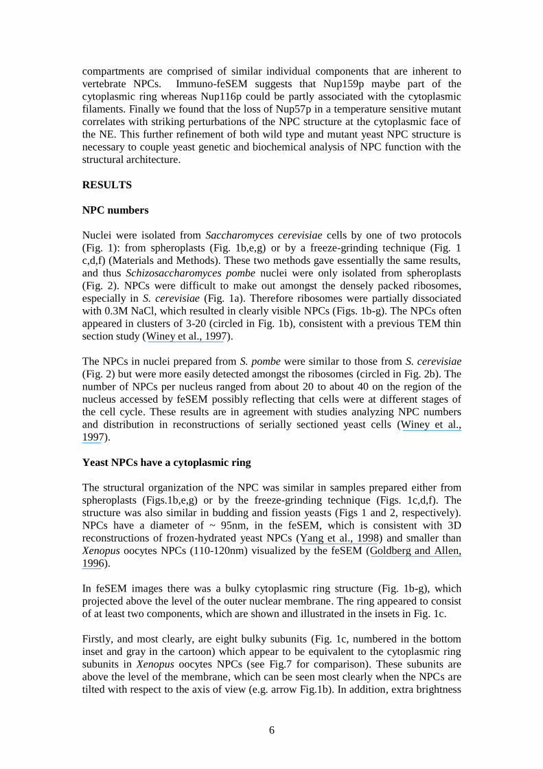
Nuclei were isolated from Saccharomyces cerevisiae cells by one of two protocols
(Fig. 1): from spheroplasts (Fig. 1b,e,g) or by a freeze-grinding technique (Fig. 1
c,d,f) (Materials and Methods). These two methods gave essentially the same results,
and thus Schizosaccharomyces pombe nuclei were only isolated from spheroplasts
(Fig. 2). NPCs were difficult to make out amongst the densely packed ribosomes,
especially in S. cerevisiae (Fig. 1a). Therefore ribosomes were partially dissociated
with 0.3M NaCl, which resulted in clearly visible NPCs (Figs. 1b-g). The NPCs often
appeared in clusters of 3-20 (circled in Fig. 1b), consistent with a previous TEM thin
section study (Winey et al., 1997).
The NPCs in nuclei prepared from S. pombe were similar to those from S. cerevisiae
(Fig. 2) but were more easily detected amongst the ribosomes (circled in Fig. 2b). The
number of NPCs per nucleus ranged from about 20 to about 40 on the region of the
nucleus accessed by feSEM possibly reflecting that cells were at different stages of
the cell cycle. These results are in agreement with studies analyzing NPC numbers
and distribution in reconstructions of serially sectioned yeast cells (Winey et al.,
1997).
Yeast NPCs have a cytoplasmic ring
The structural organization of the NPC was similar in samples prepared either from
spheroplasts (Figs.1b,e,g) or by the freeze-grinding technique (Figs. 1c,d,f). The
structure was also similar in budding and fission yeasts (Figs 1 and 2, respectively).
NPCs have a diameter of ~ 95nm, in the feSEM, which is consistent with 3D
reconstructions of frozen-hydrated yeast NPCs (Yang et al., 1998) and smaller than
Xenopus oocytes NPCs (110-120nm) visualized by the feSEM (Goldberg and Allen,
1996).
In feSEM images there was a bulky cytoplasmic ring structure (Fig. 1b-g), which
projected above the level of the outer nuclear membrane. The ring appeared to consist
of at least two components, which are shown and illustrated in the insets in Fig. 1c.
Firstly, and most clearly, are eight bulky subunits (Fig. 1c, numbered in the bottom
inset and gray in the cartoon) which appear to be equivalent to the cytoplasmic ring
subunits in Xenopus oocytes NPCs (see Fig.7 for comparison). These subunits are
above the level of the membrane, which can be seen most clearly when the NPCs are
tilted with respect to the axis of view (e.g. arrow Fig.1b). In addition, extra brightness
7
around the edge of these particles shows that they have topographical contrast, and
therefore are not at the level on the membrane. This is important because it shows that
this structure cannot be part of the spoke complex which only extends to the level of
the outer nuclear membrane in yeast (Yang et al., 1998) and in Xenopus (Akey and
Radermacher, 1993; Goldberg and Allen, 1993). Therefore we believe that they are
subunits of the cytoplasmic ring. The finding that yeast NPCs have a cytoplasmic ring
is significant because although the previous 3D reconstruction data (Yang et al., 1998)
showed some density in this region, it did not show a definitive ring structure
consisting of subunits. The peripheral density could therefore have been interpreted as
other peripheral structures such as filaments being attached directly to the spoke ring
complex (accounting for the extra peripheral density). FeSEM data, in contrast,
suggest that the cytoplasmic filaments or particles are, like in Xenopus, attached to
bulky cytoplasmic ring subunits. Additional cytoplasmic filaments/particles are
observed attached to these ring subunits (Fig.1c and d, small arrows and Fig.7).
The reason for this discrepancy is not clear but is likely to be related to one or more of
the following, which vary between the current and the previous (Yang et al., 1998)
studies:
1) isolation of the NPCs (here NPCs are left within the intact isolated nucleus,
whereas previously NPCs were isolated);
2) processing for imaging (here they are fixed and critical point dried, whereas
previously they were viewed unfixed, frozen hydrated);
3) the imaging itself (surface imaging by feSEM compared to projection imaging).
Because cytoplasmic rings were previously observed in TEM reconstructions in
Xenopus (Hinshaw et al., 1992; Akey and Radermacher, 1993) it is unlikely that the
imaging method could cause this discrepancy. It is possible that the fixation and
drying used here could cause collapse and aggregation of peripheral structures to form
subunit like structures similar to the Xenopus cytoplasmic ring. We do not favor this
interpretation because of the apparent similarity in the structure observed here by
feSEM compared to that observed in Xenopus by both feSEM and cryo-TEM. We
favor the interpretation that when NPCs are isolated (Yang et al., 1998) there may be
some disruption or disordering of the ring structure, which may preclude the detection
of a defined ring and the resolution of individual subunits. It may be that the density
detected in this region in 3D reconstructions (Yang et al., 1998) can be attributed to
disordered rings and/or cytoplasmic filaments that have collapsed onto them to
differing degrees. However, confirmation of the cytoplasmic ring in yeast will require
its observation by an alternative method, particularly of sample preparation. To this
end we are currently developing methods to examine frozen samples by feSEM.
Thin section TEM studies also suggest a structural similarity between yeast and
vertebrates in this region, particularly the presence of cytoplasmic filaments, but
could not demonstrate the presence of rings (Fahrenkrog et al., 1998; Rout and
Blobel, 1993; Allen and Douglas, 1989).
The size of these putative yeast cytoplasmic ring subunits is also not strikingly
different from Xenopus oocyte cytoplasmic ring subunits at about 25nm across (Fig.7
and Goldberg and Allen, 1993).
8
The second component of the cytoplasmic ring that was observed is the “thin ring”.
The thin ring underlies the cytoplasmic ring subunits and is observed in Xenopus in
3D reconstructions (Akey and Radermacher, 1993), by feSEM after partial proteolysis
(Goldberg and Allen, 1996) and as a putative assembly intermediate in Xenopus egg
extracts and Drosophila early embryos (Goldberg and Wiese et al., 1997; Kiseleva et
al., 2001). As with other specimens, the thin ring in yeast is difficult to detect in intact
NPCs. However, sometimes glimpses of a structure consistent with the thin ring can
be observed between two cytoplasmic ring subunits (arrows in the bottom inset in Fig.
1c and colored black in the cartoon in the upper inset). A similar structure is also seen
in S. pombe (Fig.2d, arrow). We have also observed a consistent structure in putative
incomplete NPCs (see below), which could be the thin ring without the cytoplasmic
ring subunits (Fig.3f and 5b), although it remains to be shown for certain that these
are NPC-related substructures. We suggest therefore that, like Xenopus, yeasts too
have a thin ring, which is an integral part of the cytoplasmic ring.
Yeast NPCs appear to have cytoplasmic particles or filaments
In S. cerevisae a variable number of globular particles were found attached to the
cytoplasmic ring subunits (Fig. 1d and e, small arrows). It was possible that these
were the cytoplasmic filaments. In nuclei isolated from S. pombe without 0.3M NaCl,
these putative cytoplasmic filaments were more consistently present and had a more
extended conformation, appearing more like a rod or filament than a particle (Fig. 2f).
The filaments could be quite extended (Fig. 2f and g, marked “1”) or more compact
(marked “2” and “3”), which was reminiscent of the apparent conformational
flexibility observed in vertebrates (Goldberg et al., 2000; Pante and Aebi, 1996;
Rutherford et al., 1997).
Such filaments emanating from the cytoplasmic face of the yeast NPC have
previously been suggested from structures observed in TEM thin sections (Fahrenkrog
et al., 1998; Rout and Blobel, 1993). The thin section data together with our
observation of globular or extended particles attached to the cytoplasmic ring suggests
that both yeasts have equivalent cytoplasmic filaments to vertebrates, although it
remains to be shown directly whether there are extensive filaments that attach to the
cytoplasmic ring and extend some distance into the cytoplasm.
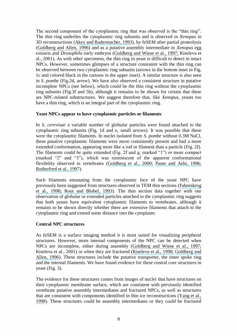
Central NPC structures
As feSEM is a surface imaging method it is most suited for visualizing peripheral
structures. However, more internal components of the NPC can be detected when
NPCs are incomplete, either during assembly (Goldberg and Wiese et al., 1997;
Kiseleva et al., 2001) or when they are fractured (Kiseleva et al., 1998; Goldberg and
Allen, 1996). These structures include the putative transporter, the inner spoke ring
and the internal filaments. We have found evidence for these central core structures in
yeast (Fig. 3).
The evidence for these structures comes from images of nuclei that have structures on
their cytoplasmic membrane surface, which are consistent with previously identified
vertebrate putative assembly intermediates and fractured NPCs, as well as structures
that are consistent with components identified in thin ice reconstructions (Yang et al.,
1998). These structures could be assembly intermediates or they could be fractured

9
NPCs, that have lost certain peripheral components to reveal underlying structure, and
we shall refer to them as “intermediates”. Because the intermediates are significantly
different in appearance to NPCs it is possible that they are not NPC-related structures.
The reason we think they are is because they are about the right diameter and have the
characteristic eight fold rotational symmetry of NPCs, and their components are
consistent with previously identified NPC substructures. We know of no other similar
structure that might be found on the nuclear envelope.
We observed, in regions of some nuclei, a striking structure (Fig. 3a, circles) which,
consisted of eight filaments radiating out from the center to give a wagon wheel like
structure. Such structures were also observed in Chironomus tentans salivary gland
NEs after NPCs were fractured by “rolling” the nucleus (Fig 3e). The filaments are
consistent with the internal filaments identified in Xenopus and other organisms
(Goldberg and Allen, 1996: Kiseleva et al., 1998; Goldberg et al., 1997; Kiseleva et
al., 2001). Again, this may not be an NPC-related structure, but the diameter and the
fact that there are eight radiating filaments is suggestive of a NPC-related structure
and not suggestive of any other known different structure.
Internal filaments were difficult to identify in complete NPCs, although we did find
evidence of them (Fig. 3b smaller arrows). It may be that they cannot be detected
easily in complete NPCs because they are obscured by the overlying cytoplasmic ring
subunits. The internal filaments are also clear in putative “intermediates” where the
thin ring appears to be present but the cytoplasmic ring subunits do not (Fig. 3f). It
should be noted that internal filaments have not been observed by any method other
than feSEM and it could be argued that they may be artifacts of fixation or critical
point drying. It is difficult to avoid chemical fixation in feSEM preparation, but we
have observed internal filaments, by feSEM, in frozen hydrated specimens, showing
that this potential artifact is not caused by critical point drying (unpublished result).
In Xenopus the internal filaments appear to attach to the “top” of the transporter,
which is a controversial cylindrical structure found in the central channel of the NPC
(Akey and Radermacher, 1993; Kiseleva et al., 1993; Goldberg and Allen, 1996; see
also Stoffler at al., 2003). In yeast a central mass is also observed (Fig. 1g large
arrows) and could represents the top part of the central cylinder or transporter. In
some NPCs the central globule protrudes from the central channel (Fig. 1f and g small
arrows). Such a structure was also reported in Chironomus and was suggested to be a
peripheral component of the transporter (Kiseleva et al., 1993), but alternatively it
could be material, such as mRNP, in transit.
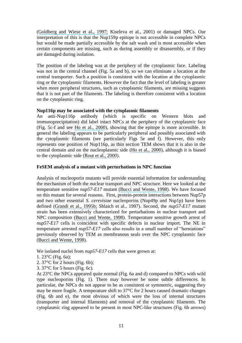
The nucleoplasmic face of the NPC
To reveal the nucleoplasmic face of the yeast NPC, nuclei were fractured by
centrifugation at 10,000 X g to disrupt the NE. This revealed NEs where the
nucleoplasmic face of NPCs were either mostly obscured by intranuclear material
(Fig. 4a) or NEs where some NPCs were visible and some were damaged (Fig. 4b).
However some NPCs retained a morphology that was similar to that in Xenopus and
other previously studied organisms. Yeast NPC baskets were however mostly at least
partially damaged compared to Xenopus. This is mostly likely because manual
isolation of Xenopus NEs is very mild compared to exposure of the nucleoplasmic
face of the yeast NE.

10
As with all other organisms studied so far, we found filaments attached to the outer
periphery of the NPC and extending into the nucleus (Fig. 4c white arrows).
Sometimes these filaments joined together to form the distinctive basket or fish-trap
structure (Goldberg and Allen, 1992; 1996; Ris, 1991). Yeast baskets have previously
been suggested from analysis of TEM thin sections (Stoffler et al., 1999; Fahrenkrog
et al., 1998; Rout and Blobel, 1993), but we can show here that the yeast basket
appears to be very similar to that of vertebrates and other organisms. There are up to
eight basket filaments, which are about 10nm in diameter and up to 40nm long, which
is similar to Xenopus when viewed by feSEM (Goldberg and Allen, 1996).
Evidence for the nucleoplasmic ring could also be observed (Fig. 4c black arrows).
The nucleoplasmic ring is less massive than the cytoplasmic ring (Akey and
Radermacher, 1993; Reichelt et al., 1990) and is not always so clearly observed, even
in Xenopus oocyte NEs. In S. cereviseae we can detect an electron emission from
some topographical structure between the filaments of the basket, where they appose
the membrane (black arrows in Fig.4c), which is consistent with the nucleoplasmic
ring. The ring is not always clear and requires careful inspection but can sometimes
be seen, even at low magnification (Fig. 4b, small arrows). As the structure is not
clearly visualized we cannot say whether its morphology is similar to other
organisms. What we can see, suggests that it may be a simpler smooth ring, rather
than having the eight clear subunits observed in Xenopus. The putative nucleoplasmic
ring diameter is 90-100nm which is larger than that previously reported for the
nuclear membrane pore (Yang et al., 1998). This suggests that, in our view, it is
unlikely to be part of the spoke ring complex, which lies within the pore.
Occasionally, thick filaments of about 20nm in diameter were attached to the top of
some baskets (Fig. 4b large arrows). These could correspond to filamentous structures
stretching from the nucleoplasmic face of the NE into the nucleoplasm as described
previously in budding yeast (Strambio-de-Castillia etal., 1999) and could represent a
structure similar to the NE lattice and “cables” observed in Xenopus (Goldberg and
Allen, 1996;Ris and Malecki, 1993). Alternatively they could be remnants of
chromatin.
Role of specific nucleoporins in the NPC
Having shown that yeast NPCs do have cytoplasmic rings and filaments we wanted to
determine what proteins were involved in these structures. Immuno-gold labeling can
help to determine the location of a protein, whereas the structural analysis of NPCs
containing mutant proteins gives clues as to the structural role of a given protein or
subcomplex.
Nup159p may be associated with the cytoplasmic ring
Anti-Nup159p labeling was observed at a low level on most nuclei (not shown), and
was increased slightly by a 0.3M NaCl wash . The highest level of labeling was found
on NPCs that appeared incomplete which was about 10 fold more gold particles per
NPC (1-4 particles/NPC or 30-35 particles/half nucleus compared to 0-3
particles/nucleus in the control where the primary antibody was omitted) than in
complete NPCs. We speculate that incomplete NPCs could be assembly intermediates
11
(Goldberg and Wiese et al., 1997; Kiseleva et al., 2001) or damaged NPCs. Our
interpretation of this is that the Nup159p epitope is not accessible in complete NPCs
but would be made partially accessible by the salt wash and is most accessible when
certain components are missing, such as during assembly or disassembly, or if they
are damaged during isolation.
The position of the labeling was at the periphery of the cytoplasmic face. Labeling
was not in the central channel (Fig. 5a and b), so we can eliminate a location at the
central transporter. Such a position is consistent with the location at the cytoplasmic
ring or the cytoplasmic filaments. However the fact that the level of labeling is greater
when more peripheral structures, such as cytoplasmic filaments, are missing suggests
that it is not part of the filaments. The labeling is therefore consistent with a location
on the cytoplasmic ring.
Nup116p may be associated with the cytoplasmic filaments
An anti-Nup116p antibody (which is specific on Western blots and
immunoprecipitation) did label intact NPCs at the periphery of the cytoplasmic face
(Fig. 5c-f and see Ho et al., 2000), showing that the epitope is more accessible. In
general the labeling appears to be particularly peripheral and possibly associated with
the cytoplasmic filaments (see particularly Figs 5e and f). However, this only
represents one position of Nup116p, as thin section TEM shows that it is also in the
central domain and on the nucleoplasmic side (Ho et al., 2000), although it is biased
to the cytoplasmic side (Rout et al., 2000).
FeSEM analysis of a mutant with perturbations in NPC function
Analysis of nucleoporin mutants will provide essential information for understanding
the mechanism of both the nuclear transport and NPC structure. Here we looked at the
temperature sensitive nup57-E17 mutant (Bucci and Wente, 1998). We have focused
on this mutant for several reasons. First, protein-protein interactions between Nup57p
and two other essential S. cerevisiae nucleoporins (Nup49p and Nsp1p) have been
defined (Grandi et al., 1995b; Shlaich et al., 1997). Second, the nup57-E17 mutant
strain has been extensively characterized for perturbations in nuclear transport and
NPC composition (Bucci and Wente, 1998). Temperature sensitive growth arrest of
nup57-E17 cells is coincident with specific defects in nuclear import. The NE in
temperature arrested nup57-E17 cells also results in a small number of “herniations”
previously observed by TEM as membranous seals over the NPC cytoplasmic face
(Bucci and Wente, 1998).
We isolated nuclei from nup57-E17 cells that were grown at:
1. 23°C (Fig. 6a);
2. 37°C for 2 hours (Fig. 6b);
3. 37°C for 5 hours (Fig. 6c).
At 23°C the NPCs appeared quite normal (Fig. 6a and d) compared to NPCs with wild
type nucleoporins (Fig. 1). There may however be some subtle differences. In
particular, the NPCs do not appear to be as consistent or symmetric, suggesting they
may be more fragile. A temperature shift to 37°C for 2 hours caused dramatic changes
(Fig. 6b and e), the most obvious of which were the loss of internal structures
(transporter and internal filaments) and removal of the cytoplasmic filaments. The
cytoplasmic ring appeared to be present in most NPC-like structures (Fig. 6b arrows)
12
but sometimes was partially fragmented. The ring appeared less massive as it had less
topography and emitted less signal, possibly because the cytoplasmic filaments were
missing. The channel through the pore appeared to be stabilized at a fairly consistent
size, suggesting that components of the spoke ring complex were still intact.
However, after 5 hours at 37°C (Fig. 6c and f) most of the remaining recognizable
structures were gone and the “hole” apparently closed up (Fig 6c arrows), leaving
structures that resembled the “dimples” previously suggested to be early assembly
intermediates (Goldberg and Wiese et al., 1997). NPCs of nuclei isolated from wild
type S. cerevisiae, that were incubated at 37°C were identical to those incubated at
23°C, showing that the structural alterations were due to the mutation.
These results show that disruption of Nup57p function initially causes a disruption of
the internal structures, as well as parts of the cytoplasmic ring (after 2 hours), but
ultimately the whole cytoplasmic side of the NPC is destabilized (after 5 hours). This
final step could represent an intermediate in the herniation of the NE observed in
TEM thin sections (Wente and Blobel, 1993). As all NPCs are affected in this way
these effects are most likely due to disruption of pre-existing NPCs rather than mis-
assembly of new ones, where only a proportion would be affected.
Discussion
Yeast has proved an invaluable tool for identifying nucleoporins and obtaining
information about their functional roles. However, until recently little was known
about the structure of the yeast NPC and how it related to the well defined structure of
the vertebrate NPC. TEM reconstructions provided the first 3D information for the
yeast NPC (Yang et al., 1998) and it appeared to be significantly smaller and simpler
than in vertebrates (Akey and Radermacher, 1993). This provided a puzzle because
recent proteomic analysis of both yeast (Rout et al., 2000) and rat (Cronshaw et al.,
2002) NPCs showed that they both contained a similar complexity of constituent
proteins. One explanation for this is that the 3D reconstructions (Yang et al., 1998)
contained data for part of the structure (the spoke ring complex and transporter) but
not for the more peripheral components (cytoplasmic ring and filaments and
nucleoplasmic ring and basket). This could be because they do not occupy a defined
position and are therefore not clearly resolved, or that they are disrupted, disordered
or lost during isolation or distorted upon interaction with carbon grid during freezing.
On the other hand, biochemical data (Rout et al., 2000) does suggest that there is not
an overall loss of nucleoporins upon isolation. It is difficult, however, to be sure that
there is no loss of unknown nucleoporins or partial loss of others, which could result
in the loss of a structural component without loosing all its constituent proteins. Thin
section data (Fahrenkrog et al., 1998) showed the attachment of filaments to both
sides of the yeast NPC, but components such as the cytoplasmic and nucleoplasmic
rings, the transporter and internal filaments are not readily visible in thin sections.
Yeast and Vertebrate NPCs are structurally similar
Here we have used feSEM to visualize the cytoplasmic face of individual NPCs in
situ in intact nuclei from fission and budding yeast. This has allowed us to look at the
surface of relatively intact NPCs. We believe, from this data, that the yeast NPC, like
that of other organisms so far studied, does have a cytoplasmic ring (consisting of a
thin ring and subunits) as well as cytoplasmic filaments and internal filaments. By
13
disrupting the nucleus and exposing the nucleoplasmic face we also found evidence
that the yeast NPC has a basket and possibly a nucleoplasmic ring, but these were
difficult to preserve and image. A comparison of yeast and Xenopus oocyte NPCs is
shown in Fig.7.
The data presented here show, by feSEM, that the peripheral components of yeast
NPCs are morphologically similar to other organisms so far studied. This view is
supported by thin section data (Fahrenkrog et al., 1998; Rout and Blobel, 1993).
However 3D reconstructions (Yang et al., 1998) did not detect cytoplasmic or
nucleoplasmic rings or baskets, but rather a weak density in these regions, which were
attributed to collapsed peripheral filaments. Here we show evidence, at least viewed
by feSEM, that there are cytoplasmic rings and filaments, which are very similar to
those observed in Xenopus. We favour the view that these structures are lost or
disordered when the yeast NPCs are isolated from the nucleus, which is avoided in
work presented here. Alternatively, it is possible that the ring, which we observe, is an
artifactual structure formed by chemical fixation and drying. We do not favour this
view because the structure is so similar to the Xenopus cytoplasmic ring seen by
feSEM. This in turn is the same as the ring clearly shown in unfixed frozen hydrated
Xenopus oocyte NEs (Akey and Radermacher, 1993) and negatively stained NEs
(Hinshaw et al., 1992) in TEM reconstructions.
The nucleoplasmic face of the NPC appears to be more difficult to preserve,
essentially because we have to use harsher conditions to disrupt the NE and remove
the associated nuclear contents in order to expose the nucleoplasmic face of the NPC.
For instance it was recently shown that Mlp1p and Mlp2p, which are the yeast Tpr
homologues, organise telomeres to the NPC (Galy et al., 2000). Therefore removal of
the NE from the chromosomes is likely to disrupt components of the NPC that contain
these proteins, such as the basket. Despite this, by looking at NEs that have been
differently disrupted, we have found structures that appear to be baskets. This was no
great surprise as fairly convincing thin section data showing possible baskets has
previously been published (Fahrenkrog et al., 1998; Rout and Blobel, 1993). We also
show evidence for nucleoplasmic rings. These appear to be thin and faintly contrasted
in the feSEM. This could be because they, too, are disrupted or extracted in the
isolation of the NE. Alternatively they could in fact be significantly different to
Xenopus nucleoplasmic rings. In particular they could be much less massive.
FeSEM Immunolocalisation
Having established the structural organisation of the yeast NPC, our long-term
objective is to determine which nucleoporins were involved in which structural
components. This is important because although immuno-TEM has been used to
locate yeast nucleoporins, the structural components of the NPC cannot be directly
visualized in thin sections. The immuno-feSEM presented here also has its
limitations, chiefly that the nuclear surface has to be exposed, providing the
opportunity for loss or rearrangement of epitopes. With further work it should also be
possible to increase the resolution of the labeling, by direct labeling and the use of
smaller probes.
First we looked at Nup159p, which is part of the Nup82p-Nup159p-Nsp1p complex
(Belgareh et al., 1998) and is thought to be the yeast homologue of vertebrate
Nup214/CAN. In yeast, Nup159p localises to the cytoplasmic face (Rout et al., 2000),
14
as does Nup214 in vertebrates (Kraemer et al., 1994). Nup214 is localised quite
centrally (Walther and Pickersgill et al., 2002), possibly on the inner rim of the
cytoplasmic ring. Here we confirm that in budding yeast Nup159p may also be on the
cytoplasmic ring. Removal of Nup214 from Xenopus nuclear assembly extracts
resulted in the assembly of NPCs with apparently normal cytoplasmic rings and
filaments (Walther and Pickersgill et al., 2002). Likewise TEM thin sections (Gorsch
et al., 1995) could not detect any structural effect of a Nup159p mutation. The
mutation did however inhibit mRNA export, but not import, which is also consistent
with Nup214 (Walther and Pickersgill et al., 2002). The results presented here,
therefore corroborate the view that yeast Nup159p is the functional homologue of
vertebrate Nup214/CAN.
Temperative sensitive mutant of Nup57
Like Nup159p, Nup57p also forms a complex with Nsp1, which is distinct from the
Nup159p complex above and contains Nup49p. A temperature sensitive mutation in
Nup57p resulted in NPCs that lack internal structures after 2 hours at the non-
permissive temperature, but retained elements of the cytoplasmic ring. It was
previously shown that this mutation perturbed the incorporation of not only the
Nup57-Nsp1-Nup49 complex but also of Nup116p (Bucci and Wente, 1998). The
lack of Nup116p is consistent with the lack of cytoplasmic filaments in this nup57-
E17 mutant at the non-permissive temperature. The mutation however permitted the
incorporation of the Nup159-Nup82-Nsp1 complex, which is consistent with our
finding that the cytoplasmic ring is at least partially present after 2 hours at 37 C.
Nup62 is the proposed vertebrate orthologue of Nsp1. In two similar studies, immuno-
depletion of Nup62 from Xenopus egg nuclear assembly extracts resulted either in an
apparent inhibition of NPC assembly (Dabauvalle et al., 1990) or assembly of non-
functional NPCs that were missing central structures (Finlay et al., 1991). Although
apparently inconsistent with each other, these results are both similar to certain
aspects to our findings. After 2 hours at the non-permissive temperature our mutant
had lost its central structures, presumably as the Nup57-Nsp1-Nup49 complex
disassociated. Then after 5 hours the cytoplasmic ring and at least parts of the spoke
ring complex dismantled, presumably because, although these structures are unlikely
to contain the Nsp1 complex, they must rely on it for their continued stability. The
position of Nup57p labelling (Fahrenkrog et al., 1998; Rout et al., 2000) is consistent
with the internal filaments that link the transporter to the cytoplasmic ring. Likewise
Nup62 is in a similar position (Guan et al., 1995) and certainly in the region of the
internal filaments. Therefore it is possible that the internal filaments consist of the
Nup57-Nsp1-Nup82 complex. So when this linkage is broken, in the nup57-E17
temperature sensitive mutant at 37ºC (because Nup57p holds together the Nsp1
complex - Schlaich et al., 1997), the structures that it may link together (the
transporter and the cytoplasmic ring) are no longer stably associated. Indeed the
transporter and the internal filaments, are the first structures to disappear at 37ºC. We
therefore speculate that the Nup57-Nsp1-Nup49 complex could be a component of the
internal filaments. The predicted coiled-coil nature of all the components of this
complex would be consistent with them forming a filament.
Cytoplasmic filaments
One surprising result is that yeast NPCs contain cytoplasmic filaments, which appear
to be very similar to those of vertebrates, although such filaments have been indicated

15
in thin sections (Fahrenkrog et al., 1998). Firstly, they are not detected in 3D
reconstructions (Yang et al., 1998). Secondly, it has been shown that the Ran binding
nucleoporin, Nup358, is a major constituent of the filaments (Wu et al., 1995; Delphin
et al., 1997; Walther and Pickersgill et al., 2002), possibly even the filament itself,
and there is apparently no similar protein in yeast. Despite the fact that Nup358
appears to be the major component of cytoplasmic filaments in the vertebrate NPC
(Walther and Pickersgill et al., 2002) in yeast a similar filament structure could be
constructed from one or more different proteins. As Nup358 is a multidomain,
multifunctional giant protein, it is entirely possible that several smaller proteins could
execute the same function.
In summary, we have shown that NPC structure, particularly with respect to the
peripheral components, is very similar in both budding and fission yeast compared to
vertebrates and invertebrates. We have shown evidence for yeast cytoplasmic and
possibly nucleoplasmic rings and internal filaments. We have confirmed evidence that
the basket exists in yeast and probably has the same structure as in other organisms.
Likewise we have confirmed that there are cytoplasmic filaments and shown that they
have a similar conformation as in other organisms. Nup159p, like its putative
vertebrate homologue Nup214, appears to be associated with the cytoplasmic ring.
Nup116p appears to be associated with the cytoplasmic filaments, although it also
occupies other positions. We have also shown that without Nup57p, which is required
to stabilise the Nup57-Nsp1-Nup49 complex, the internal filaments and the
transporter are absent. With the tools available to yeast cell biologists, such as
antibodies, tagged nucleoporins, temperature sensitive, deletion, and other
nucleoporin mutants, the approach shown here could be a useful way to determine the
roles of nucleoporins in the structural organisation and function of the NPC.
MATERIALS AND METHODS
Strain growth, media and reagents
Saccharomyces cerevisiae wild type strains AB1380 (Mata ade2-1 ura3-1 trp1
lys2 his5 can1-100) and J 1003-10 (Mata ade2-1 ura3-1 trp1-1 leu2-3,112 can1-100)
and Schizosaccharomyces pombe wild type strain HM123 h- leu1-32 (lab stock) were
analyzed. In addition, the yeast strain SWY1587 (Mata ura3-1 his3-11,15 trp1-1
leu2-3,112 can1-100 ade2-1:ADE2 nup57-E17 ) was analyzed at the permissive
(23°C) and restrictive (37°C) growth temperatures as described (Bucci and Wente,
1998). The cells were grown in liquid YPD medium (1% yeast extract, 2%
bactopeptone, 2% glucose). A stationary yeast culture was diluted at 1:2000 in YPD
medium and grown for ~10 hours at 30° C with moderate shaking (130 rpm) to
logarithmic phase and cells were used when about 80% of cells were dividing.
Reagents were obtained as follows: Zymolase 20T (Sigma), dithiothreitol (Sigma),
Novozyme (Sigma); Sucrose ultrapure (Agar), sorbitol (Serva), Poly-L-lysine and
Hoechst 33258 (Sigma).
Isolation of nuclei
Two methods of nuclear isolation were used: from spheroplasts (method A) and from
whole frozen cells (method B). The spheroplast methods for budding and fission yeast
were modified from previously described studies (Rozijn and Tonino, 1964; Hurt,
1988). About 3 g weight of wet yeast cells were harvested by centrifugation in a

16
Sorvall GS3-rotor, washed once in 100 ml water and incubated in 30 ml 0.1M Tris-
HCl, pH 9.4, 10 mM dithiothreitol for 25 min at 30°C with moderate shaking. Cells
were pelleted, washed in 50 ml spheroplast buffer (1.2M sorbitol, 20 mM potassium
phosphate, pH 7.4) and resuspended in 25 ml spheroplast buffer containing 1 mg
zymolase 20T per gram of wet cells for budding yeast and 10mg/ml Novozym for
fision yeast, then incubated for 20- 40 min at 30°C with gentle shaking. When about
70% of the cells were converted to spheroplasts, they were pelleted for 3 min at 3500
rpm in a Sorvall SS34 rotor and washed twice in spheroplast buffer. The final pellet
was resuspended at room temperature in 10 ml spheroplast buffer with 0.5 mM MgCl2
pH 6.5. All subsequent steps were at 4°C. 200 l of spheroplast homogenate were
transferred into a glass tube and diluted with 200 l distilled water. This was
incubated for 2 min to allow the spheroplasts to swell and was then vortexed for 2
seconds. The specimens were then checked using light and fluorescence microscopy
by staining with 5 g/ml Hoechst 33258. Before or after additional incubation of the samples for 10 min with 0.3 M NaCl in 20 mM potassium phosphate pH 6.5 to
remove ribosomes from the NE surface, they were immediately prepared for the
feSEM (below).
Nuclear isolation from whole cells involved manual grinding of frozen yeast cells,
and was modified from a previously described method (den Hollander et al., 1986).
Yeast cells were suspended in 1.2 M sorbitol, 2 M sucrose, 20 mM potassium
phosphate, 0.5 mM MgCl2 pH 6.5 and frozen as small drops in liquid nitrogen.
Frozen drops covered by liquid nitrogen were ground into powder with a pestle and
fixed immediately (see fixative solution below) after thawing to 4° C.
Preparation of samples for feSEM
For feSEM, 5mm x 5mm silicon chips were treated with 1 mg/ml poly-L-lysine in
distilled water for 30 min, then rinsed in water and transferred to buffer containing 20
mM potassium phosphate, 0.5 mM MgCl2, pH 6.5. 10 l of the samples prepared by
method A or by method B were layered on top of 40 l fixative (20 mM potassium phosphate, 0.5 mM MgCl2, 0.2M sucrose, 4% paraformaldehyde, pH 6.5) and spun
onto a silicon chip (Agar) at 4000 X g for 3 minutes in a swing-out rotor. During this
centrifugation, spheroplast membranes in samples from methods A were broken due
to impact with the chip surface, and their contents were released and fixed
immediately on the chips. To visualize the nucleoplasmic side of the NPC, the
spheroplast samples were pelleted for 3 min at 10,000 X g to disrupt the NE. After
centrifugation, the chips were further fixed in 2% glutaraldehyde, 0.2% tannic acid,
20 mM potassium phosphate, 0.5 mM MgCl2, pH 7 for 10 minutes at room
temperature, postfixed with 1% (w/v) osmium tetroxide for 10 minutes, then stained
with 1% (w/v) uranyl acetate for 10 minutes (both solutions in distilled water),
dehydrated in ethanol and critical-point dried from CO2 via Arklone (ICI, Runcorn,
Cheshire, UK). Specimens were sputter coated with 2-4 nm of tantalum in an
Edwards 306 cryo-pumped vacuum system with a magnetron head (Edwards High
Vacuum International, Crawley, West Sussex, UK). Samples were examined at 30kV
in the top stage of a Topcon (ABT) ISI DS-130F field emission scanning electron
microscope (Topcon Corporation, Tokyo, Japan). Measurements of individual NPC
components were done on micrographs of 20 different samples prepared by
spheroplast or freeze/grind methods.
17
Preparation of samples for immuno-feSEM
Yeast nuclei before or after removal of the ribosomes were isolated from spheroplasts
by method A and centrifuged onto silicon chips for 3 min at 4000 g through buffer A
(20mM Tris-HCl pH 7.5, 0.5mM MgCl2 , 0.2 M Sucrose). Samples were then
transferred to buffer A without sucrose containing 3.7% formaldehyde and fixed for
15 min, and then given two washes in buffer A without sucrose for 5 min. Samples
were blocked for 20 min in 1% BSA, 20 mM Tris-HCl, then incubated (with shaking)
for 60 min with either the rabbit anti-Nup116-C antibody (1:10 dilution) (raised
against the C-terminal 60kD domain and specific for Nup116p by Western blots and
immunoprecipitation and affinity purified against the cognate epitope) or mouse Mab
against Nup159p (Mab 165C10; 1:50 - Kraemer et al ., 1995), or with buffer as a
control, and washed twice for 5 min with 0.001% (wt/vol) Tween 20, 20 mM Tris-
HCl. The samples were incubated for 30 min with secondary gold-conjugated goat
anti-rabbit or anti-mouse antibody (AuroProb TM EM GAR IgG G10; 10 nm in
diameter; Amersham) in 0.2% BSA-20 mM Tris-HCl (1:20). The samples were
washed twice with 20mM Tris-HCl and fixed for 10 min in 2% (wt/vol)
glutaraldehyde, 0.2% (wt/vol) tannic acid in 20 mM Tris-HCl, rinsed with buffer, and
incubated for 10 min in 1% OsO4 in water. Further processing and feSEM analysis
were performed as described above, except that after critical point drying, the samples
were coated with 4 nm of chromium. The backscattered electron imaging mode was
used to resolve the gold particles by feSEM, using a solid-state retractable backscatter
electron detector in the top stage of the TOPCON (ISI) DS 130F SEM. To determine
the position of the gold particles, backscatter electron images were precisely
overlayed onto the secondary image in Adobe Photoshop, the position of the colloidal
gold signal marked with a black dot, then the backscatter image layer (which obscures
some of the secondary electron detail) removed.
Preparation of Chironomus nuclei
Isolated salivary gland cells were kept in cold TKM buffer (100mM KCl, 1mM
MgCl2, and 10mM trietahanolamine-HCl, pH 7.0), supplemented with 2% NP-40 for
10-30 seconds and then transferred into 0.025% NP-40. The nuclei were isolated from
glands by pipetting. To remove the cytoplasmic rings from the NPC, the nuclei were
allowed to adhere to the silicon chip and then gently rolled over the surface by
pushing with a glass needle as described previously (Kiseleva et al., 1998). The
samples were fixed and processed as described for the yeast nuclei.
Acknowledgements
We thank Dr. M. Rout for advice on the use of the freeze/grind method
for nuclear isolation from yeast cells. We also thank our colleagues in the Wente
laboratory (A. Ho, E. Ives, Y. Lee, K. Ryan, and L. Strawn) for critical
discussions and comments on the manuscript. The research was supported
by the Wellcome Trust Foundation (E.K., M.W.G. and T.D.A.), Cancer Research
UK (T. D. A. and M.W.G.), the Russian Foundation for Basic Research (E.K.), a
Beckman Young Investigator Award from the Arnold and Mabel Beckman
Foundation (S.R.W.), and a grant from the National Institutes of Health
(S.R.W.). Artwork (Fig. 7) was by P. Chantry.
18
References
Adam S.A. (1999). Transport pathways of macromolecules between the nucleus and
the cytoplasm. Curr. Opin. Cell Biol. 11, 402-6.
Akey C.W. (1990). Visualization of transport-related configurations of the nuclear
pore transporter. Biophys J. 58,. 341-55.
Akey, C.W. and. Radermacher, M. (1993). Architecture of the Xenopus nuclear pore
complex revealed by three-dimensional cryo-electron microscopy. J. Cell Biol., 122,
1-19.
Allen, J.L. and Douglas, M.G.. (1989). Organization of the nuclear pore complex in
Saccharomyces cerevisiae. J. Ultrastruct. Mol. Struct. Res. 102, 95-108.
Allen, T.D., Bennion, G.R., Rutherford, S.A., Reipert, S., Ramalho, A., Kiseleva, E.,
Goldberg, M.W. (1997). Macromolecular substructure in nuclear pore complexes by
in-lens field-emission scanning electron microscopy.
Scanning 19, 403-10.
Allen, T.D., Cronshaw, J.M., Bagley, S., Kiseleva, E. and Goldberg, M.W. (2000).
The nuclear pore complex: mediator of translocation between nucleus and cytoplasm.
J. Cell Sci. 113, 1651-9.
Bailer, S,M., Siniossoglou, S., Podtelejnikov, A., Hellwig, A., Mann, M. and Hurt, E.
(1998). Nup116p and nup100p are interchangeable through a conserved motif which
constitutes a docking site for the mRNA transport factor gle2p. EMBO J. 17, 1107-19.
Belgareh, N., Snay_Hodge, C., Pasteau, F., Dagher, S., Cole. C.N. and Doye, V.
(1998). Functional characterization of a Nup159p-containing nuclear pore
subcomplex. Mol. Biol. Cell 9, 3475-92.
Bucci, M. and Wente, S.R. (1998) A novel fluorescence-based genetic strategy
identifies mutants of Saccharomyces cereviseae defective for nuclear pore complex
assembly. Mol Biol Cell 9 22439-2461.
Cronshaw, J.M., Krutchinsky, A.N., Zhang, W., Chait, B.T. and Matunis, M.J. (2002).
Proteomic analysis of the mammalian nuclear pore complex. J Cell Biol 158 915-27.
Dabauvalle, M.C., Loos, K. and Scheer, U. (1990). Identification of a soluble
precursor complex essential for nuclear pore assembly in vitro. Chromosoma 100 56-
66.
Delphin, C., Guan, T., Melchior, F. and Gerace, L. (1997). RanGTP targets p97 to
RanBP2, a filamentous protein localized at the cytoplasmic periphery of the nuclear
pore complex. Mol. Biol. Cell 8, 2379-90.
den Hollander, J.A., Ugurbil, K. and Schulman, R.G. (1986). 31P and 13C NMR
studies of intermediates of aerobic and anerobic glycolysis Saccharomyces cerevisiae.
Biochem. 25 212-219.
19
Denning, D., Mykytka, B., Allen, N.P., Huang, L., Burlingame, A.I. and Rexach, M.
(2001). The nucleoporin Nup60p functions as a Gsp1p-GTP-sensitive tether for
Nup2p at the nuclear pore complex. J. Cell Biol. 154, 937-50.
Dilworth, D.J., Suprapto, D.J., Padovan, J.C., Chait, B.T., Wozniak, R.W., Rout, M.P.
and Aitchison, J.D. (2001). Nup2p dynamically associates with the distal regions of
the yeast nuclear pore complex. J. Cell Biol. 153, 1465-78
Doye, V. and Hurt, E. (1997). From nucleoporins to nuclear pore complexes. Curr.
Opin. Cell Biol. 9, 401-11.
Fahrenkrog, B., Hurt, E.C., Aebi, U. and Pante, N. (1998). Molecular architecture of
the yeast nuclear pore complex: localization of Nsp1p subcomplexes. J. Cell Biol.
143, 577-88.
Feldherr, C.M., Akin, D. and Cohen, R.J. (2001). Regulation of functional nuclear
pore size in fibroblasts. J. Cell Sci. 114, 4621-7.
Finlay, D.R., Meier, E., Bradley, P., Horecka, J. and Forbes, D.J. (1991). A complex
of nuclear pore proteins required for pore function. J Cell Biol 114 169-83.
Galy, V., Olivo-Marin, J.C., Scherthan, H., Doye, V., Rascalou, N. and Nehrbass, U.
(2000). Nuclear pore complexes in the organisation of silent telomeric chromatin.
Nature 403 108-112.
Goldberg, M.W. and Allen, T.D. (1992). High resolution scanning electron
microscopy of the nuclear envelope: demonstration of a new, regular, fibrous lattice
attached to the baskets of the nucleoplasmic face of the nuclear pores. J. Cell Biol.
119, 1429-40.
Goldberg and Allen (1993) The nuclear pore complex: three-dimensional surface
structure revealed by field emission, in-lens scanning electron microscopy, with
underlying structure uncovered by proteolysis. J. Cell Sci. 106, 261-274.
Goldberg, M.W. and Allen, T.D. (1996). The nuclear pore complex and lamina: three-
dimensional structures and interactions determined by field emission in-lens scanning
electron microscopy. J. Mol Biol. 257, 848-65.
Goldberg, M.W., Cronshaw, J.M., Kiseleva, E. and Allen, T.D. (1999). Nuclear pore
complex dynamics and transport in higher eukaryotes. Protoplasma 209, 144-156.
Goldberg, M.W., Wiese, C., Allen, T.D. and Wilson, K.L. (1997). Dimples, pores,
star-rings, and thin rings on growing nuclear envelopes: evidence for structural
intermediates in nuclear pore complex assembly. J. Cell Sci. 110, 409-20.
Goldberg MW, Solovei I, Allen TD. (1997) Nuclear pore complex structure in birds. J
Struct Biol. 119:284-94.
20
Goldberg, M.W., Rutherford, S.A., Hughes, M., Cotter, L.A., Bagley, S., Kiseleva, E.,
Allen, T.D. and Clarke, P.R. (2000). Ran alters nuclear pore complex conformation. J.
Mol. Biol. 300, 519-29.
Gorsch, L.C., Dockendorff, T.C., and Cole C.N., (1995). A conditional allele of the
novel repeat-containing yeast nucleoporin RAT7/NUP159 causes both rapid cessation
of mRNA export and reversible clustering of nuclear pore complexes. J Cell Biol 29
939-55.
Grandi, P., Doye, V. and Hurt, E.C. (1993). Purification of NSP1 reveals complex
formation with 'GLFG' nucleoporins and a novel nuclear pore protein NIC96. EMBO
J. 12, 3061-71.
Grandi, P., Emig, S., Weies, C., Hucho, F., Pohl, T. and Hurt, E.C. (1995a). A novel
nuclear pore protein Nup82p which specifically binds to a fraction of Nsp1p. J. Cell
Biol. 130, 1263-73.
Grandi, P., Schlaich, N., Tekotte, H. and Hurt, E.C. (1995b). Functional interaction of
Nic96p with a core nucleoporin complex consisting of Nsp1p, Nup49p and a novel
protein Nup57p. EMBO J. 14, 76-87.
Grote, M., Kubitscheck, U., Reichelt, R. and Peters, R. (1995). Mapping of
nucleoporins to the center of the nuclear pore complex by post-embedding
immunogold electron microscopy. J. Cell Sci. 108, 2963-72.
Griffis, E.R., Altan, N., Lippincott_Schwartz, J. and Powers, M.A. (2002). Nup98 is a
mobile nucleoporin with transcription-dependent dynamics. Mol. Biol. Cell 13, 1282-
97.
Guan, T., Muller, S., Klier, G., Pante, N., Blevitt, J.M., Haner, M., Paschal, B., Aebi,
U. and Gerace, L. (1995). Structural analysis of the p62 complex, an assembly of O-
linked glycoproteins that localizes near the central gated channel of the nuclear pore
complex. Mol. Biol. Cell 6, 1591-603.
Hinshaw, J.E., Carragher, B.O. and Milligan, R.A. (1992). Architecture and design of
the nuclear pore complex. Cell 69, 1133-41.
Ho, A.K., Shen, T.X., Ryan, K.J., Kiseleva, E., Levy, M.A., Allen, T.D. and Wente,
S.R. (2000). Assembly and preferential localization of Nup116p on the cytoplasmic
face of the nuclear pore complex by interaction with Nup82p. Mol Cell Biol 20 5736-
48.
Hurt, E.C. (1988). A novel nucleoskeletal-like protein located at the nuclear periphery
is required for the life cycle of Saccharomyces cerevisiae. EMBO J 7 4323-34.
Iovine M.K. and Wente, S.R. (1997). A nuclear export signal in Kap95p is required
for both recycling the import factor and interaction with the nucleoporin GLFG repeat
regions of Nup116p and Nup100p. J. Cell Biol. 137, 797-811.
21
Kiseleva, E., Goldberg, M.W., Allen, T.D. and Akey, C.W. (1998). Active nuclear
pore complexes in Chironomus: visualization of transporter configurations related to
mRNP export. J. Cell Sci. 111, 223-36.
Kiseleva, E., Goldberg, M.W., Cronshaw, J. and Allen, T.D. (2000). The nuclear pore
complex: structure, function, and dynamics. Crit. Rev. Eukaryot. Gene Expr. 10, 101-
12.
Kiseleva, E., Rutherford, S., Cotter, L.M., Allen, T.D. and Goldberg, M.W. (2001).
Steps of nuclear pore complex disassembly and reassembly during mitosis in early
Drosophila embryos. J. Cell Sci. 114, 3607-18.
Kraemer, D.M., Strambio,_de_Castillia C., Blobel, G. and Rout, M.P. (1995). The
essential yeast nucleoporin NUP159 is located on the cytoplasmic side of the nuclear
pore complex and serves in karyopherin-mediated binding of transport substrate. J
Biol Chem 270 19017-21.
Kraemer, D., Wozniak, R.W., Blobel, G. and Radu, A. (1994). The human CAN
protein, a putative oncogene product associated with myeloid leukemogenesis, is a
nuclear pore complex protein that faces the cytoplasm. Proc Natl Acad Sci U S A 91
1519-23.
Lutzmann, M., Kunze, R., Buerer, A., Aebi, U. and Hurt, E. (2002). Modular self-
assembly of a Y-shaped multiprotein complex from seven nucleoporins. EMBO J. 21,
387-97.
Marelli, M., Aitchison, J.D. and Wozniak, R.W. (1998). Specific binding of the
karyopherin Kap121p to a subunit of the nuclear pore complex containing Nup53p,
Nup59p, and Nup170p. J. Cell Biol. 143, 1813-30.
Mattaj, I.W. and Englmeier, L. (1998). Nucleocytoplasmic transport: the soluble
phase. Annu. Rev. Biochem. 67, 265-306.
Nehrbass, U., Rout, M.P., Maquire, S., Blobel, G. and Wozniak, R.W. (1996). The
yeast nucleoporin Nup188p interacts genetically and physically with the core
structures of the nuclear pore complex. J.Cell Biol. 133,1153-1162.
Nakielny, S., Shaikh, S., Burke, B. and Dreyfuss, G. (1999). Nup153 is an M9-
containing mobile nucleoporin with a novel Ran-binding domain. EMBO J. 18, 1982-
95.
Pante, N. and Aebi, U. (1996). Sequential binding of import ligands to distinct
nucleopore regions during their nuclear import. Science 273, 1729-32.
Pante, N. and Kann, M. (2002). Nuclear pore complex is able to transport
macromolecules with diameters of about 39 nm. Mol. Biol. Cell 13, 425-34.
Reichelt, R.A., Holzenberg, A., Buhle, E.L., Jarnik, M., Engel, E. and Aebi, U. (1990)
Correlation between structure and mass distribution of the nuclear pore complex and
distinct nuclear pore complex components. J Cell Biol 110 883-894.
22
Ribbeck, K. and Gorlich, D. (2001). Kinetic analysis of translocation through nuclear
pore complexes. EMBO J. 20, 1320-30.
Ris, H. (1991). The 3D structure of the nuclear pore complex as seen by high voltage
electron microscopy and high resolution low voltage scanning electron microscopy.
EMSA Bull. 21, 54-56.
Ris, H. and Malecki, M. (1993). High-resolution field emission scanning electron
microscope imaging of internal cell structures after Epon extraction from sections: a
new approach to correlative ultrastructural and immunocytochemical studies. J Struct
Biol 111 148-57.
Rozijn, T. H. and Tonino, G.J.M. (1964). Studies on the yeast nucleus. I. The isolation
of nuclei. Biochim. Biophys. Acta 91 105-112.
Rout, M.P. and Blobel, G. (1993). Isolation of the yeast nuclear pore complex.
J. Cell Biol. 123, 771-83.
Rout, M.P., Aitchison, J.D., Suprapto. A., Hjertaas, K., Zhao, Y. and Chait, B.T.
(2000). The yeast nuclear pore complex: composition, architecture, and transport
mechanism. J. Cell Biol. 148, 635-51.
Rout, M.P. and Aitchison, J.D. (2001). The nuclear pore complex as a transport
machine. J. Biol. Chem. 276, 16593-6.
Rutherford, S.A., Goldberg, M.W. and Allen, T.D. (1997). Three-dimensional
visualization of the route of protein import: the role of nuclear pore complex
substructures. Exp. Cell Res. 232, 146-60.
Schlaich, N.L., Haner, M., Lustig, A., Aebi, U. and Hurt, E.C. (1997). In vitro
reconstitution of a heterotrimeric nucleoporin complex consisting of recombinant
Nsp1p, Nup49p, and Nup57p. Mol Biol Cell 8 33-46.
Strambio_de_Castillia, C., Blobel, G. and Rout, M.P. (1999). Proteins connecting the
nuclear pore complex with the nuclear interior. J Cell Biol 144 839-55.
Siniossoglou, S., Wimmer, C., Rieger, M., Doye, V., Tekotte, H., Weise, C., Emig, S.,
Segref, A. and Hurt, E.C. (1996). A novel complex of nucleoporins, which includes
Sec13p and a Sec13p homolog, is essential for normal nuclear pores. Cell 84, 265-75.
Stoffler, D., Fahrenkrog, B. and Aebi, U. (1999). The nuclear pore complex: from
molecular architecture to functional dynamics. Curr. Opin. Cell Biol. 11, 391-401.
Stoffler D, Feja B, Fahrenkrog B, Walz J, Typke D, Aebi U. (2003) Cryo-electron
tomography provides novel insights into nuclear pore architecture: implications for
nucleocytoplasmic transport. J Mol Biol. 328:119-30.
23
Talcott, B. and Moore, M.S. (1999) Getting across the nuclear pore complex.
Trends Cell Biol. 9, 312-8.
Walther, T.C., Pickersgill, H.S., Cordes, V.C., Goldberg, M.W., Allen, T.D., Mattaj,
I.W. and Fornerod, M. (2002). The cytoplasmic filaments of the nuclear pore complex
are dispensable for selective nuclear protein import. J. Cell Biol. 158, 63-77.
Wente, S.R. and Blobel, G. (1993) A temperature sensitive NUP116 null mutant
forms a nuclear envelope seal over the yeast nuclear pore complex thereby blocking
nucleocytoplasmic traffic. J Cell Biol 123 275-284.
Winey, M., Yarar, D., Giddings, T.H., and Mastronarde, D.N. (1997). Nuclear pore
complex number and distribution throughout the Saccharomyces cerevisiae cell cycle
by three-dimensional reconstruction from electron micrographs of nuclear envelopes.
Mol. Biol. Cell 8, 2119-32.
Wu, J., Matunis, M.J., Kraemer, D., Blobel, G. and Coutavas, E. (1995). Nup358, a
cytoplasmically exposed nucleoporin with peptide repeats, Ran-GTP binding sites,
zinc fingers, a cyclophilin A homologous domain, and a leucine-rich region. J. Biol.
Chem. 270, 14209-13.
Vasu, S.K. and Forbes, D.J. (2001). Nuclear pores and nuclear assembly.
Curr. Opin. Cell Biol. 13, 363-75.
Yang, Q., Rout, M.P. and Akey, C.W. (1998). Three-dimensional architecture of the
isolated yeast nuclear pore complex: functional and evolutionary implications. Mol.
Cell 1, 223-34.
Zabel, U., Doye, V., Tekotte, H., Wepf, R., Grandi, P. and Hurt, E.C. (1996). Nic96p
is required for nuclear pore formation and functionally interacts with a novel
nucleoporin, Nup188p. J. Cell Biol. 133, 1141-52.
24
Figure legends
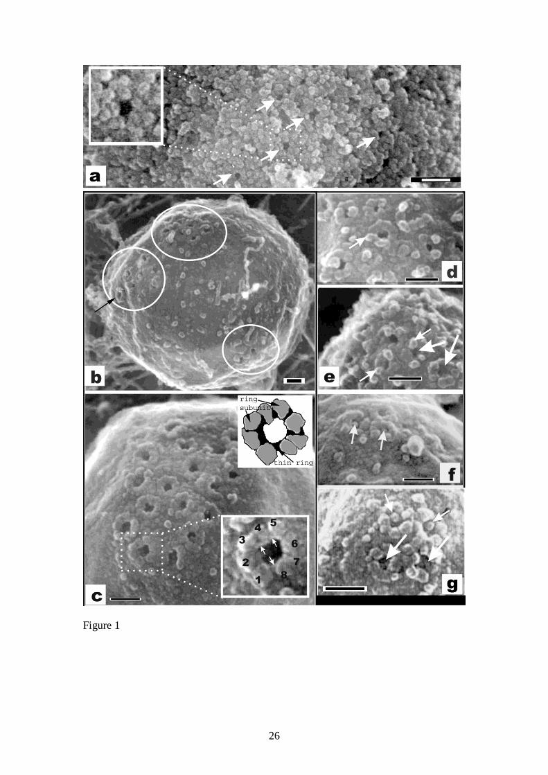
Figure 1. S. cerevisiae nuclei isolated with ribosomes still attached (a) where the
NPCs (arrows and inset) are difficult to make out and (b-g) with the ribosomes
removed with 0.3M NaCl. Nuclei were isolated either from spheroplasts (a,b,e,g) or
by a freeze-grinding technique (c,d,f) with similar results. NPCs were sometimes
clustered (b – circles). Previously recognised peripheral NPC components are
observed (c-g). Components include the eight subunit cytoplasmic ring (c - numbered
in bottom inset and grey in top inset, which is a cartoon of the image in the bottom
inset); the thin ring (c – white arrows in bottom inset and black in top inset);
cytoplasmic particles/filaments (smaller arrows in d and e); a central mass (large
arrows in e and g) and a central globule (small arrows f and g). Bars = 100nm
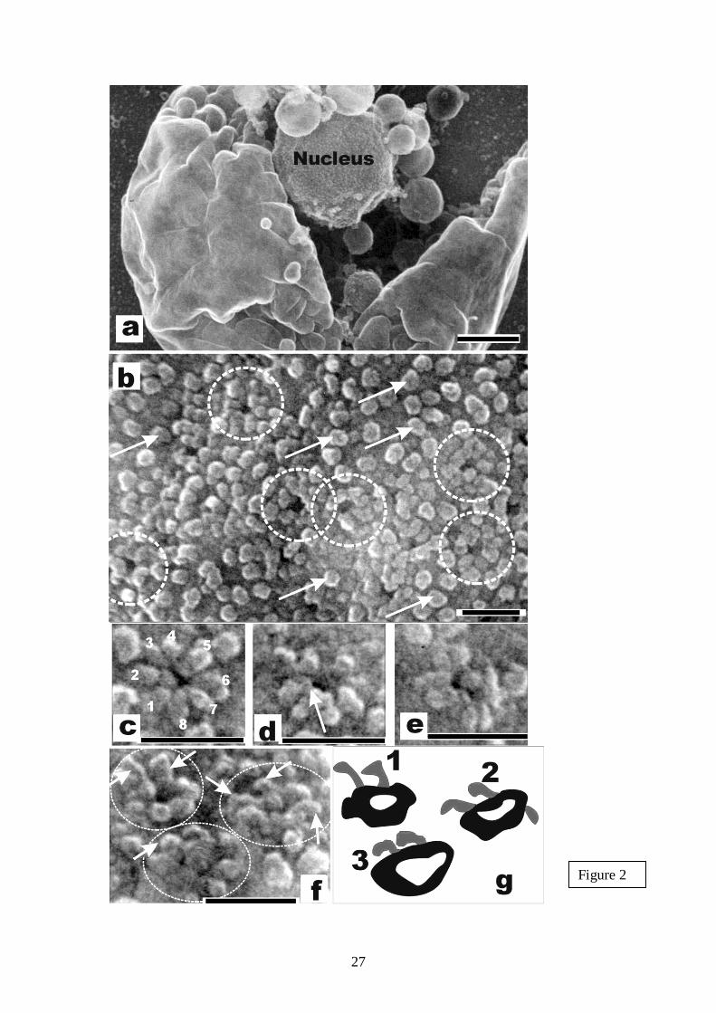
Figure 2. S. pombe nuclei isolated from spheroplasts. (a) Nucleus protruding from
broken cell. (b) NPCs (circled) are seen amongst the ribosomes (arrows). (c-e)
Examples of NPCs showing the eight subunits (numbered) and the thin ring (arrow).
Sometimes the filamentous nature of the cytoplasmic particles is evident (f) and can
be compact (illustrated in g “3”) or more extended (illustrated in g “1”). Bar in (a) =
500nm; Other Bars = 100nm.
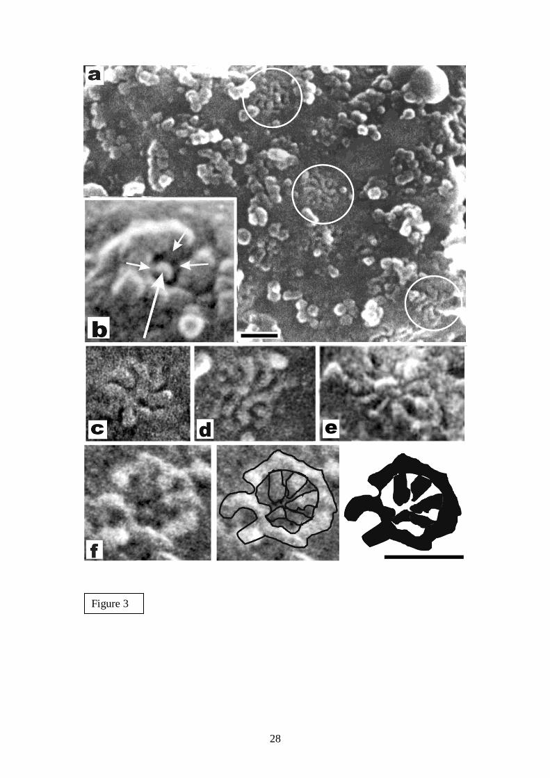
Figure 3. Evidence for internal filaments can be seen in intact NPCs (b small arrows).
Structures were also found that could be internal filaments of incomplete NPCs (a -
circled) where the cytoplasmic ring has not yet formed or detached. These structures
consist of eight radiating filaments and have a diameter of about 90nm, which is
suggestive of an NPC structure. Examples of these structures are shown at high
magnification (c and d) and an equivalent structure that was found in Chironomus
tentans salivary glands is shown in (e). Structures were also observed that were
similar but with an apparent peripheral ring, which could be the thin ring, as
illustrated (f). Bars = 100nm. Bar in (f) refers to (b-f).
Figure 4. Nucleoplasmic face of S. cerevisiae NE from ruptured nuclei. Often NPCs
are obscured by overlying material (a), but baskets (b - circled) and the nucleoplasmic
ring (b - small arrows) can be observed as well as filaments that interconnect the
distal parts of the baskets (b - large arrows). At high magnification (c) the basket
filaments (white arrows) and nucleoplasmic ring (black arrows) were evident. Bars =
100nm
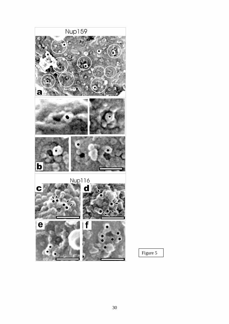
Figure 5. Immuno-feSEM of Nup159p (a and b) and Nup116p (c -f). (a) Some NPCs
(circled) are labelled by indirect immuno-gold labelling (black dots). The position of
the label appears peripheral and NPCs are most often labelled when they appear
incomplete (b). Nup116p labelling however could be more peripheral and labels
complete NPCs more frequently (c-f). Bars = 100nm
Figure 6. The nup57-E17 temperature sensitive mutant maintained at 23ºC (a and d),
37ºC for 2 hours (b and e) and 37ºC for 5 hours (c and f). At 23ºC, the NPCs appeared
quite normal. After 2 hours of cell growth at non-permissive temperature the NPCs
lost central structures (transporter and internal filaments). Only small holes or dimples
of about 20nm in diameter instead of NPCs were observed in the nuclear envelope
after 5 hours of cell growth at 37ºC. Bar = 100nm
25
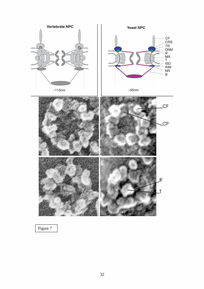
Figure 7. Model (adapted from Yang et al., 1998) and images of the cytoplasmic face
of the NPC comparing yeast and Xenopus. CF = cytoplasmic filaments; CRS =
cytoplasmic ring subunits; TR = thin ring; ONM = outer nuclear membrane; IF =
internal filaments; MR = membrane ring; T = transporter; ISD = inner spoke domain;
INM = inner nuclear membrane; NR = nucleoplasmic ring; B = basket. Coloured
components are those observed by feSEM, although the basket had previously been
indicated and the nucleoplasmic internal filaments remain a speculation. Bar = 50nm
26
Figure 1
27
Figure 2
28
Figure 3

29
Figure 4
30
Figure 5

31
Figure 6
32
Figure 7