Two-pore domain potassium channels in the adrenal cortex
16
INVITED REVIEW Two-pore domain potassium channels in the adrenal cortex Sascha Bandulik & Philipp Tauber & Enzo Lalli & Jacques Barhanin & Richard Warth Received: 5 September 2014 /Revised: 2 October 2014 /Accepted: 3 October 2014 /Published online: 23 October 2014 # The Author(s) 2014. This article is published with open access at Springerlink.com Abstract The physiological control of steroid hormone se- cretion from the adrenal cortex depends on the function of potassium channels. The “two-pore domain K + channels” (K2P) TWIK-related acid sensitive K + channel 1 (TASK1), TASK3, and TWIK-related K + channel 1 (TREK1) are strong- ly expressed in adrenocortical cells. They confer a background K + conductance to these cells which is important for the K + sensitivity as well as for angiotensin II and adrenocorticotro- pic hormone-dependent stimulation of aldosterone and corti- sol synthesis. Mice with single deletions of the Task1 or Task3 gene as well as Task1/Task3 double knockout mice display partially autonomous aldosterone synthesis. It appears that TASK1 and TASK3 serve different functions: TASK1 affects cell differentiation and prevents expression of aldosterone synthase in the zona fasciculata, while TASK3 controls aldo- sterone secretion in glomerulosa cells. TREK1 is involved in the regulation of cortisol secretion in fasciculata cells. These data suggest that a disturbed function of K2P channels could contribute to adrenocortical pathologies in humans. Keywords KCNK2 . KCNK3 . KCNK9 . TASK . TREK . Aldosterone Introduction The distinct zones of the adrenal cortex produce different steroid hormones, which regulate several important physio- logical functions. The mineralocorticoid aldosterone is syn- thesized by the outermost cell layer (zona glomerulosa) be- neath the capsule of the adrenal gland. Glucocorticoids are produced in the zona fasciculata that consists of column-like organized cells below zona glomerulosa. In humans, but not in rodents, the innermost zona reticularis cells produce andro- genic steroid hormones. In cells of all three zones of the adrenal cortex, the function of K + channels is an important determinant for controlling hormone secretion, cell differenti- ation, proliferation, and possibly apoptosis. This review aims to discuss the physiology and pathophysiology of “two-pore domain K + channels” (K2P) in the adrenal cortex, especially in aldosterone-producing glomerulosa cells. Aldosterone controls the extracellular fluid and salt balance by stimulation of sodium reabsorption and potassium secre- tion in the distal nephron of the kidney, in the distal colon, and in sweat glands. By controlling water and salt balance and by direct effects on the cardiovascular system, aldosterone has a major impact on blood pressure control. So-called primary aldosteronism is characterized by inappropriately high plasma aldosterone levels due to autonomous aldosterone synthesis. Inadequately high aldosterone secretion is believed to be causal for about 3 % of the cases of arterial hypertension [97]. Additionally, aldosterone contributes to cardiac fibrosis, cardiovascular dysfunction, and progressive kidney disease [59, 111]. The relevance of aldosterone as clinical risk factor has been stressed by clinical trials (Aldosterone Evaluation Study (RALES); EPlerenone HEart failure and SUrvival S. Bandulik (*) : P. Tauber : R. Warth Medical Cell Biology, University of Regensburg, Universitaetsstrasse 31, 93053 Regensburg, Germany e-mail: [email protected] E. Lalli Institut de Pharmacologie Moléculaire et Cellulaire, CNRS, Université de Nice Sophia Antipolis, 660 Route des Lucioles, 06560 Valbonne, France J. Barhanin LP2M-CNRS-UNS UMR 7370, Faculté de Médecine, Université de Nice Sophia Antipolis, 28 Avenue de Valombrose, 06107 Nice Cedex 2, France J. Barhanin Laboratories of Excellence, Ion Channel Science and Therapeutics, Nizza, France Pflugers Arch - Eur J Physiol (2015) 467:1027–1042 DOI 10.1007/s00424-014-1628-6
-
Upload
independent -
Category
Documents
-
view
2 -
download
0
Transcript of Two-pore domain potassium channels in the adrenal cortex
INVITED REVIEW
Two-pore domain potassium channels in the adrenal cortex
Sascha Bandulik & Philipp Tauber & Enzo Lalli &Jacques Barhanin & Richard Warth
Received: 5 September 2014 /Revised: 2 October 2014 /Accepted: 3 October 2014 /Published online: 23 October 2014# The Author(s) 2014. This article is published with open access at Springerlink.com
Abstract The physiological control of steroid hormone se-cretion from the adrenal cortex depends on the function ofpotassium channels. The “two-pore domain K+ channels”(K2P) TWIK-related acid sensitive K+ channel 1 (TASK1),TASK3, and TWIK-related K+ channel 1 (TREK1) are strong-ly expressed in adrenocortical cells. They confer a backgroundK+ conductance to these cells which is important for the K+
sensitivity as well as for angiotensin II and adrenocorticotro-pic hormone-dependent stimulation of aldosterone and corti-sol synthesis. Mice with single deletions of the Task1 or Task3gene as well as Task1/Task3 double knockout mice displaypartially autonomous aldosterone synthesis. It appears thatTASK1 and TASK3 serve different functions: TASK1 affectscell differentiation and prevents expression of aldosteronesynthase in the zona fasciculata, while TASK3 controls aldo-sterone secretion in glomerulosa cells. TREK1 is involved inthe regulation of cortisol secretion in fasciculata cells. Thesedata suggest that a disturbed function of K2P channels couldcontribute to adrenocortical pathologies in humans.
Keywords KCNK2 . KCNK3 . KCNK9 . TASK . TREK .
Aldosterone
Introduction
The distinct zones of the adrenal cortex produce differentsteroid hormones, which regulate several important physio-logical functions. The mineralocorticoid aldosterone is syn-thesized by the outermost cell layer (zona glomerulosa) be-neath the capsule of the adrenal gland. Glucocorticoids areproduced in the zona fasciculata that consists of column-likeorganized cells below zona glomerulosa. In humans, but not inrodents, the innermost zona reticularis cells produce andro-genic steroid hormones. In cells of all three zones of theadrenal cortex, the function of K+ channels is an importantdeterminant for controlling hormone secretion, cell differenti-ation, proliferation, and possibly apoptosis. This review aimsto discuss the physiology and pathophysiology of “two-poredomain K+ channels” (K2P) in the adrenal cortex, especiallyin aldosterone-producing glomerulosa cells.
Aldosterone controls the extracellular fluid and salt balanceby stimulation of sodium reabsorption and potassium secre-tion in the distal nephron of the kidney, in the distal colon, andin sweat glands. By controlling water and salt balance and bydirect effects on the cardiovascular system, aldosterone has amajor impact on blood pressure control. So-called primaryaldosteronism is characterized by inappropriately high plasmaaldosterone levels due to autonomous aldosterone synthesis.Inadequately high aldosterone secretion is believed to becausal for about 3 % of the cases of arterial hypertension[97]. Additionally, aldosterone contributes to cardiac fibrosis,cardiovascular dysfunction, and progressive kidney disease[59, 111]. The relevance of aldosterone as clinical risk factorhas been stressed by clinical trials (Aldosterone EvaluationStudy (RALES); EPlerenone HEart failure and SUrvival
S. Bandulik (*) : P. Tauber :R. WarthMedical Cell Biology, University of Regensburg,Universitaetsstrasse 31, 93053 Regensburg, Germanye-mail: [email protected]
E. LalliInstitut de Pharmacologie Moléculaire et Cellulaire, CNRS,Université de Nice Sophia Antipolis, 660 Route des Lucioles,06560 Valbonne, France
J. BarhaninLP2M-CNRS-UNS UMR 7370, Faculté de Médecine, Université deNice Sophia Antipolis, 28 Avenue de Valombrose, 06107 NiceCedex 2, France
J. BarhaninLaboratories of Excellence, Ion Channel Science and Therapeutics,Nizza, France
Pflugers Arch - Eur J Physiol (2015) 467:1027–1042DOI 10.1007/s00424-014-1628-6

Study (EPHESUS)) [16, 148]. Therefore, understanding thephysiology and pathophysiology of aldosterone synthesis is ofgreat relevance for the diagnosis and treatment of arterialhypertension and cardiovascular disease.
It is known for a long time that the regulation of aldoste-rone synthesis strongly depends on the modulation of themembrane potential of glomerulosa cells. Also cortisol syn-thesis appears to be stimulated by depolarization of the plasmamembrane. The membrane potential of resting adrenocorticalcells is mainly determined by the function of K+ channels.Accordingly, the disturbed function of adrenal K+ channelshas pathological consequences for the regulation of steroidhormone production, and it may lead to excessive prolifera-tion of adrenocortical cells.
Adrenocortical K+ channels
The resting membrane potential of human glomerulosa cells isset by a number of K+ channels, particularly of the K2Pfamily, which are highly expressed among species (Table 1).In rodents, two members of the TWIK-related acid sensitiveK+ (Task) family (Task1 and Task3) were shown to play animportant role in the regulation of aldosterone secretion andadrenocortical cell differentiation [7, 28, 34, 51, 55, 103].TWIK-related K+ channel 1 (Trek1) is important for thenormal function of the bovine adrenal cortex [38, 41]. Therole of K2P channels in the human adrenal gland is still underinvestigation. Several studies indicate that K2P channels con-tribute to the physiological control of aldosterone synthesis inhuman adrenocortical cells. TASK1 is strongly expressed inthe human adrenal cortex [22] and in the human adreno-cortical NCI-H295R cell line [96]. Silencing of TASK1expression stimulates aldosterone secretion in NCI-H295R cells [96]. TREK1 and TASK3 K+ channels arealso expressed in NCI-H295R cells, albeit on a muchlower level than TASK1 [96]. Inactivation of TREK1and TASK3 depolarizes the membrane potential ofNCI-H295R cells. However, the amount of this depolar-ization is likely small, because aldosterone production isnot significantly increased [14]. Decreased expression ofTASK2 was found in adrenal adenomas [5, 72], andsuppression of TASK2 activity in NCI-H295R cells in-creased aldosterone synthesis [72]. TREK1 was shown todominate the K+ conductance of human fasciculata cells[39]. Most of the knowledge about the functional role ofTask K+ channels has been obtained by phenotypingdifferent knockout mouse models. The following para-graphs aim at providing a comprehensive overview ofthe specific role and relevance of K2P channels for theregulation of steroid hormone synthesis and zonal differ-entiation of the adrenal gland.
Stimulation of aldosterone secretion
Aldosterone synthesis in adrenal zona glomerulosa cells ismainly stimulated by angiotensin II (Ang-II), by high plasmaK+ concentrations, and, to a minor extent, by the adrenocor-ticotropic hormone (ACTH). For the stimulation of aldoste-rone synthesis by Ang-II or hyperkalemia, modulation of themembrane potential is an early and critical early event in thecellular signaling cascade (Fig. 1). Therefore, precise controlof the membrane voltage is very important. A large proportionof the K+ channels that determine the resting membranevoltage of glomerulosa cells are constitutively open, e.g.,“background” or “leak” K+ channels of the K2P family. Dueto the high K+ conductance, the resting membrane potential ofglomerulosa cells is hyperpolarized (−80 mV), close to the K+
equilibrium potential. An increase of the extracellular K+
concentration, according to Nernst’s equation, leads to a pos-itive shift of the K+ equilibrium potential and to a depolariza-tion. By this mechanism, glomerulosa cells are able to sensechanges of plasma K+ concentration, reminiscent of K+-selec-tive electrodes. Upon depolarization of the membrane,voltage-dependent T-type and L-type Ca2+ channels are acti-vated, thereby translating the membrane depolarization into arise of the intracellular Ca2+ activity. High intracellular Ca2+
activity, via binding to calmodulin and activation ofcalmodulin-dependent kinases, induces transcription of par-ticular enzymes needed for aldosterone synthesis, e.g., aldo-sterone synthase (CYP11B2), and steroidogenic acute regula-tory protein (StAR) [23]. Aldosterone synthase catalyzes thefinal three-step reaction from 11-deoxycorticosterone to aldo-sterone, and it is considered to be the rate-limiting enzyme ofaldosterone synthesis. StAR is a transport protein facilitatingthe shuttling of cholesterol from the outer to the inner mito-chondrial membrane where cholesterol is converted to preg-nenolone, a precursor of steroid hormones.
The mechanism by which Ang-II depolarizes the mem-brane is different from the one of high extracellular K+.Ang-II depolarizes the plasma membrane by inhibiting back-ground K2P K+ channels. The molecular mechanism of theAng-II-mediated K+ channel inhibition was a matter of debatefor a long time [19, 79, 87, 121] but was solved only recently.Binding of Ang-II to the AT1 receptor activatesphospholipase-C via Gαq-proteins. By cleavage of phos-phatidylinositol 4,5-bisphosphate (PIP2), phospholipase-Cgenerates diacylglycerol (DAG) and inositol-triphosphate(IP3). Interestingly, it appears that DAG acts as a K2Pchannel-inhibiting factor leading to a strong decrease of thefractional K+ conductance and depolarization of the mem-brane [145]. Similar to the high K+-induced depolarization,the Ang-II-induced depolarization activates voltage-gatedCa2+ channels and leads to Ca2+ influx [1, 58, 137]. In addi-tion, Ang-II facilitates the opening of Ca2+ channels by low-ering the voltage threshold for activation [84], and it induces a
1028 Pflugers Arch - Eur J Physiol (2015) 467:1027–1042

Tab
le1
AdrenalK+channels
Channel
Expression
Functio
nPathology
Reference
TASK
1(K
CNK3)
Mouse:Z
G>ZF>
inneradrenalcortex
Maintenance
andregulatio
nof
mem
branepotential
ofadrenocorticalandmedullary
cells;Inhibition
byAng-IIandendothelin-1;p
reventionof
Cyp11b2
expression
inZFof
♀mice
Task1−
/−mouse:sex-dependent
hyperaldosteronism
dueto
ectopic
Cyp11b2
expression
inZF
[22,27,28,55,
62,63,86,96,121]
Rat:Z
G,m
edulla
Hum
an:p
ulmonaryhypertension
Guineapig:
medulla
Hum
an:Z
G>ZF>
ZR(unpublisheddata),
aldosterone-producingadenom
a,adrenocorticalcelllin
e(N
CI-H295R
cells)
TASK
3(K
CNK9)
Mouse:♂
ZG,Z
F;♀
ZG
Maintenance
andregulatio
nof
mem
branepotential
ofadrenalcortex;
inhibitio
nby
Ang-II;probably
heterodimerswith
Task1
Mild
hyperaldosteronism
inadultTask3
−/−
mice;severe
hyperaldosteronism
inneonatalTask3−
/−mice
[6,9,14,22,25,
26,28,51,83,103]
Rat:Z
G
Hum
an:low
adrenalexpressioncompared
toTA
SK1andTA
SK2;adrenocorticalcell
line(N
CI-H295R
cells)
Hum
an:hypotension
andmentalretardatio
n
TASK
2(K
CNK5)
Mouse:inner
adrenalcortex(unpublished
data)
Probablymaintenance
andregulatio
nof
mem
brane
potentialo
fadrenalcortex;
expression
ofa
dominantnegativeTA
SK2mutantinNCI-H295R
cells
stim
ulated
aldosteronesynthesis
Decreased
expression
inaldosterone-
producingadenom
as[5,22,26,72]
Hum
an:adrenalcortex
TREK1(K
CNK2)
Bovine:ZG,Z
FMaintenance
andregulatio
nof
mem
branepotential
ofadrenalcortex;
inhibitio
nby
Ang-II,ACTH
andvasopressin;
expression
inducedby
ACTHandcA
MP
[22,36–39,41,
80–82]
Hum
an:Z
F
Mouse:u
nknown
KCNJ5
(Kir3.4/GIRK4)
Hum
an:Z
G>ZF,aldosterone-producing
adenom
a,adrenocorticalcelllin
e(N
CI-H295R
cells)
Functio
ninZGstill
unknow
n;Gßγactiv
ated;A
ng-II
reducedKCNJ5
expression
Som
aticmutations
in30–40%
ofaldosterone-producingadenom
as;
germ
linemutations
inpatientswith
familialhyperaldosteronism
type
III
[5,22,69,98]
Pig:
ZG(unpublisheddata)
KCNQ1/KCNE1
Mouse:adrenalcortex
Repolarizationof
mem
branepotential;KCNE1
asregulatory
subunit;voltage
activ
ated
Kcnq1
−/−mouse:h
ypoaldosteronism
[4,22,116,138]
Hum
an:adrenalcortex
(K+channelw
iththehighestlevelof
expression),
adrenocorticalcelllin
e(N
CI-H295R
cells)
Kcne1
−/−mouse:h
yperaldosteronism
underhyperkalem
ia
MaxiK
(KCNMA1/KCNMB1)
Mouse:Z
Gandmedulla
Repolarizationof
mem
branepotential;Ca2
+
andvoltage
activ
ated;channelactiv
ationby
ANPinhibitsaldosteroneproductio
n;KCNMB1
asregulatory
subunit
Kcnma1
−/−mouse:h
yperaldosteronism
[22,46,48,118,
143,146,147]
Hum
an:adrenalcortex
Kcnmb1
−/−mouse:h
yperaldosteronism
ZGzona
glom
erulosa,ZFzona
fasciculata
Pflugers Arch - Eur J Physiol (2015) 467:1027–1042 1029
release of Ca2+ from IP3-sensitive intracellular Ca2+ stores[129]. The cytosolic rise of Ca2+ is further amplified bystore-operated Ca2+ entry [95, 106, 112, 139].
Besides its effects on gene transcription of steroidogenicenzymes, intracellular Ca2+ induces a very fast non-transcriptional stimulation of aldosterone secretion. In mice,a strong peak of aldosterone secretion is observed within10 min after injection of Ang-II [130], although aldosterone
is not believed to be stored within the cells. Probably, a varietyof non-genomic effects of Ca2+ underlies this fast response,e.g., an increase of intracellular Ca2+ stimulates the activity ofStAR [21, 76] and is paralleled by an influx of Ca2+ into themitochondria [144]. A rise in mitochondrial Ca2+ enhancesthe availability of NAPDH, a cofactor of several steroidogenicenzymes [107, 113]. As a negative feedback, an increase ofintracellular Ca2+ activates Ca2+-regulated K+ channels,
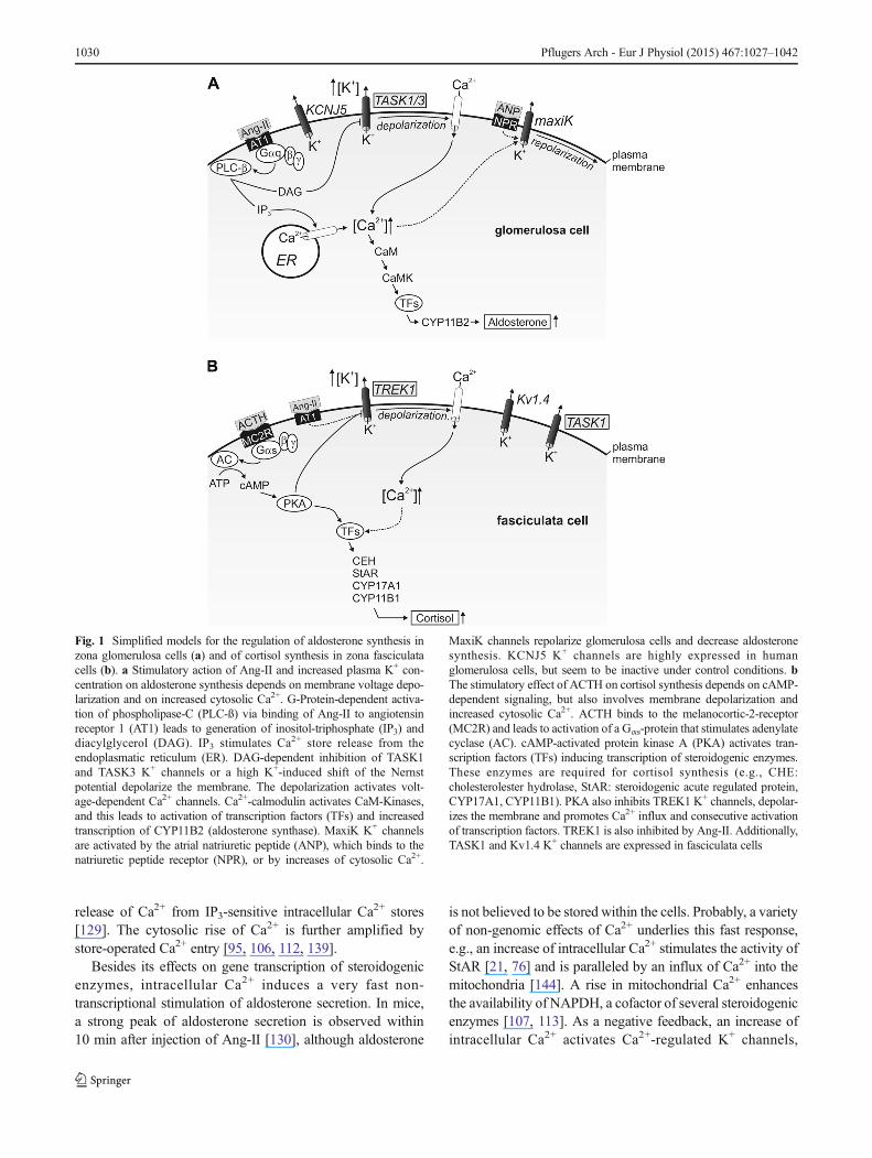
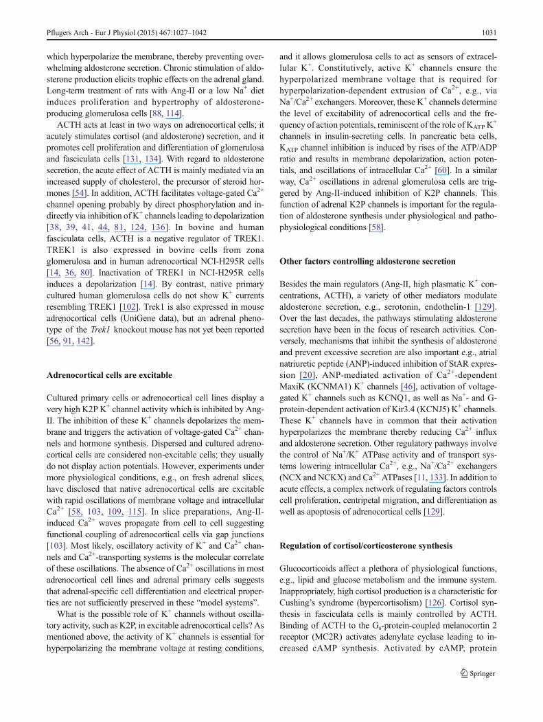
Fig. 1 Simplified models for the regulation of aldosterone synthesis inzona glomerulosa cells (a) and of cortisol synthesis in zona fasciculatacells (b). a Stimulatory action of Ang-II and increased plasma K+ con-centration on aldosterone synthesis depends on membrane voltage depo-larization and on increased cytosolic Ca2+. G-Protein-dependent activa-tion of phospholipase-C (PLC-ß) via binding of Ang-II to angiotensinreceptor 1 (AT1) leads to generation of inositol-triphosphate (IP3) anddiacylglycerol (DAG). IP3 stimulates Ca2+ store release from theendoplasmatic reticulum (ER). DAG-dependent inhibition of TASK1and TASK3 K+ channels or a high K+-induced shift of the Nernstpotential depolarize the membrane. The depolarization activates volt-age-dependent Ca2+ channels. Ca2+-calmodulin activates CaM-Kinases,and this leads to activation of transcription factors (TFs) and increasedtranscription of CYP11B2 (aldosterone synthase). MaxiK K+ channelsare activated by the atrial natriuretic peptide (ANP), which binds to thenatriuretic peptide receptor (NPR), or by increases of cytosolic Ca2+.
MaxiK channels repolarize glomerulosa cells and decrease aldosteronesynthesis. KCNJ5 K+ channels are highly expressed in humanglomerulosa cells, but seem to be inactive under control conditions. bThe stimulatory effect of ACTH on cortisol synthesis depends on cAMP-dependent signaling, but also involves membrane depolarization andincreased cytosolic Ca2+. ACTH binds to the melanocortic-2-receptor(MC2R) and leads to activation of a Gαs-protein that stimulates adenylatecyclase (AC). cAMP-activated protein kinase A (PKA) activates tran-scription factors (TFs) inducing transcription of steroidogenic enzymes.These enzymes are required for cortisol synthesis (e.g., CHE:cholesterolester hydrolase, StAR: steroidogenic acute regulated protein,CYP17A1, CYP11B1). PKA also inhibits TREK1 K+ channels, depolar-izes the membrane and promotes Ca2+ influx and consecutive activationof transcription factors. TREK1 is also inhibited by Ang-II. Additionally,TASK1 and Kv1.4 K+ channels are expressed in fasciculata cells
1030 Pflugers Arch - Eur J Physiol (2015) 467:1027–1042
which hyperpolarize the membrane, thereby preventing over-whelming aldosterone secretion. Chronic stimulation of aldo-sterone production elicits trophic effects on the adrenal gland.Long-term treatment of rats with Ang-II or a low Na+ dietinduces proliferation and hypertrophy of aldosterone-producing glomerulosa cells [88, 114].
ACTH acts at least in two ways on adrenocortical cells; itacutely stimulates cortisol (and aldosterone) secretion, and itpromotes cell proliferation and differentiation of glomerulosaand fasciculata cells [131, 134]. With regard to aldosteronesecretion, the acute effect of ACTH is mainly mediated via anincreased supply of cholesterol, the precursor of steroid hor-mones [54]. In addition, ACTH facilitates voltage-gated Ca2+
channel opening probably by direct phosphorylation and in-directly via inhibition of K+ channels leading to depolarization[38, 39, 41, 44, 81, 124, 136]. In bovine and humanfasciculata cells, ACTH is a negative regulator of TREK1.TREK1 is also expressed in bovine cells from zonaglomerulosa and in human adrenocortical NCI-H295R cells[14, 36, 80]. Inactivation of TREK1 in NCI-H295R cellsinduces a depolarization [14]. By contrast, native primarycultured human glomerulosa cells do not show K+ currentsresembling TREK1 [102]. Trek1 is also expressed in mouseadrenocortical cells (UniGene data), but an adrenal pheno-type of the Trek1 knockout mouse has not yet been reported[56, 91, 142].
Adrenocortical cells are excitable
Cultured primary cells or adrenocortical cell lines display avery high K2P K+ channel activity which is inhibited by Ang-II. The inhibition of these K+ channels depolarizes the mem-brane and triggers the activation of voltage-gated Ca2+ chan-nels and hormone synthesis. Dispersed and cultured adreno-cortical cells are considered non-excitable cells; they usuallydo not display action potentials. However, experiments undermore physiological conditions, e.g., on fresh adrenal slices,have disclosed that native adrenocortical cells are excitablewith rapid oscillations of membrane voltage and intracellularCa2+ [58, 103, 109, 115]. In slice preparations, Ang-II-induced Ca2+ waves propagate from cell to cell suggestingfunctional coupling of adrenocortical cells via gap junctions[103]. Most likely, oscillatory activity of K+ and Ca2+ chan-nels and Ca2+-transporting systems is the molecular correlateof these oscillations. The absence of Ca2+ oscillations in mostadrenocortical cell lines and adrenal primary cells suggeststhat adrenal-specific cell differentiation and electrical proper-ties are not sufficiently preserved in these “model systems”.
What is the possible role of K+ channels without oscilla-tory activity, such as K2P, in excitable adrenocortical cells? Asmentioned above, the activity of K+ channels is essential forhyperpolarizing the membrane voltage at resting conditions,
and it allows glomerulosa cells to act as sensors of extracel-lular K+. Constitutively, active K+ channels ensure thehyperpolarized membrane voltage that is required forhyperpolarization-dependent extrusion of Ca2+, e.g., viaNa+/Ca2+ exchangers. Moreover, these K+ channels determinethe level of excitability of adrenocortical cells and the fre-quency of action potentials, reminiscent of the role of KATP K
+
channels in insulin-secreting cells. In pancreatic beta cells,KATP channel inhibition is induced by rises of the ATP/ADPratio and results in membrane depolarization, action poten-tials, and oscillations of intracellular Ca2+ [60]. In a similarway, Ca2+ oscillations in adrenal glomerulosa cells are trig-gered by Ang-II-induced inhibition of K2P channels. Thisfunction of adrenal K2P channels is important for the regula-tion of aldosterone synthesis under physiological and patho-physiological conditions [58].
Other factors controlling aldosterone secretion
Besides the main regulators (Ang-II, high plasmatic K+ con-centrations, ACTH), a variety of other mediators modulatealdosterone secretion, e.g., serotonin, endothelin-1 [129].Over the last decades, the pathways stimulating aldosteronesecretion have been in the focus of research activities. Con-versely, mechanisms that inhibit the synthesis of aldosteroneand prevent excessive secretion are also important e.g., atrialnatriuretic peptide (ANP)-induced inhibition of StAR expres-sion [20], ANP-mediated activation of Ca2+-dependentMaxiK (KCNMA1) K+ channels [46], activation of voltage-gated K+ channels such as KCNQ1, as well as Na+- and G-protein-dependent activation of Kir3.4 (KCNJ5) K+ channels.These K+ channels have in common that their activationhyperpolarizes the membrane thereby reducing Ca2+ influxand aldosterone secretion. Other regulatory pathways involvethe control of Na+/K+ ATPase activity and of transport sys-tems lowering intracellular Ca2+, e.g., Na+/Ca2+ exchangers(NCX and NCKX) and Ca2+ ATPases [11, 133]. In addition toacute effects, a complex network of regulating factors controlscell proliferation, centripetal migration, and differentiation aswell as apoptosis of adrenocortical cells [129].
Regulation of cortisol/corticosterone synthesis
Glucocorticoids affect a plethora of physiological functions,e.g., lipid and glucose metabolism and the immune system.Inappropriately, high cortisol production is a characteristic forCushing’s syndrome (hypercortisolism) [126]. Cortisol syn-thesis in fasciculata cells is mainly controlled by ACTH.Binding of ACTH to the Gs-protein-coupled melanocortin 2receptor (MC2R) activates adenylate cyclase leading to in-creased cAMP synthesis. Activated by cAMP, protein
Pflugers Arch - Eur J Physiol (2015) 467:1027–1042 1031

kinase A (PKA) phosphorylates several target proteins. Twoof these target proteins improve the supply with cholesterol:cholesterol ester hydrolase (CEH) releases cholesterol fromesterified cholesterol in intracellular lipid droplets [125], andStAR shuttles cholesterol into the inner mitochondrial mem-brane. Moreover, PKA phosphorylates transcription factorsthat stimulate expression of the 11ß-hydroxylase (CYP11B1),the enzyme catalyzing the final step of glucocorticoid synthe-sis [47, 127]. The central role of PKA signaling for cortisolsynthesis was recently underlined by the observation that asomatic gain-of-function mutation of PKA is present in about50 % of patients with cortisol-producing adrenal adenomas[12, 15, 47, 117]. Another target of PKA is the K2P channelTREK1 that is inhibited by phosphorylation [39]. Due toinhibition of TREK1, ACTH stimulation leads to depolariza-tion of fasciculata cells and activation of voltage-dependentCa2+ channels [39]. One might speculate that chronic TREK1inhibition contributes to the pathophysiology of cortisol-producing adenoma cells carrying the gain-of-function muta-tion of PKA. Interestingly, the Trek1 knockout mouse does notpresent with obvious signs of Cushing’s syndrome suggestingthat Trek1 inhibition increases glucocorticoid secretion onlyin the presence of other stimulating factors.
Why are suchmutations enhancing cAMP-dependent path-ways not detected in aldosterone-producing adenomas? Ad-renocortical cells undergo a centripetal migration and differ-entiation starting from capsular and subcapsular stem cells andending up by apoptosis at the border between the adrenalcortex and medulla. The subcapsular-medullar migration isaccompanied by a shift of differentiation from theglomerulosa to the fasciculata cell type [43]. ACTH influencesthese important processes: ACTH stimulates proliferation andaccelerates centripetal differentiation of adrenocortical cells[131]. On the other hand, suppression of ACTH by dexameth-asone decreases proliferation and differentiation [131]. Thus,alterations of the cAMP/PKA pathway will most likely effectproliferation, migration, and differentiation of adrenocorticalcells. One might speculate that in glomerulosa cells, mutation-al activation of PKA will probably cause an accelerated dif-ferentiation. Thus, adenomas originating from glomerulosacells with constitutively active PKA might be phenotypicalclassified as cortisol secreting tumors. Further studies areneeded to test this hypothesis.
Besides activating aldosterone secretion, Ang-II also stim-ulates synthesis of cortisol in bovine and human fasciculatacells [39, 40]. Fasciculata cells display similar oscillations ofthe membrane potential as it was described for glomerulosacells [8, 58, 94, 108]. Via inhibition of K2P channels, Ang-IIdepolarizes fasciculata cells resulting in activation of voltage-dependent Ca2+ channels. These synergistic effects of ACTHand Ang-II on cortisol secretion might be of particularimportance under stress conditions when a strong increaseof cortisol secretion is needed.
Mutations of ion-transporting membrane proteins areassociated with aldosterone-producing adenomas
The regulation of membrane voltage and cytosolic Ca2+ ac-tivity is central for the physiological control of aldosteronesecretion. Disturbed function of proteins controlling mem-brane voltage and Ca2+ homeostasis can cause adrenal dis-eases. The importance of the control of membrane voltage andintracellular ion composition is exemplified by somatic muta-tions of K+ channels, Ca2+ channels, the Na+/K+-ATPase, anda plasma membrane Ca2+-ATPase found in aldosterone-producing adenomas [3]. The most frequently mutated geneis the inwardly rectifying K+ channel Kir3.4 (KCNJ5). About40 % of adrenal adenomas show somatic mutations of Kir3.4[13, 42, 122]. These mutations confer a pathological Na+
conductance (“gain-of-function”) to the channel that depolar-izes the cells and activates autonomous aldosterone synthesisand proliferation [17, 22, 70, 92, 93, 99, 123, 133]. Althoughthese results clearly established the link between Kir3.4 mu-tations and hyperaldosteronism, the physiological role of non-mutated Kir3.4 in the adrenal cortex is still largely elusive.The wild-type Kir3.4 channel seems to be inactive in humanadrenal cells under resting conditions [66, 70, 133].Probably, Kir3.4 modulates the membrane potential ofhuman glomerulosa cells after stimulation of aldosteronesynthesis and prevents excess secretion of aldosterone[98]. Unfortunately, Kir3.4 function cannot be studied inmice because the channel is not expressed in the mouseadrenal gland (unpublished data).
Adrenal phenotype of Task channel knockout mice
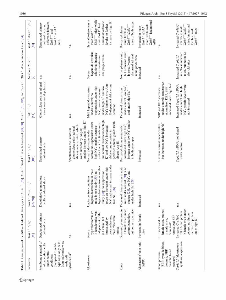
Different knockout mouse models were used to investigate therelevance of Task1 and Task3 K+ channels for the adrenalgland [7, 28, 34, 51, 55, 103]. Mice with single deletions ofthe Task1 [55] or Task3 gene [7, 51, 103] as well as Task1/Task3 double knockout mice displayed disturbances of thesteroid hormone homeostasis [28]. A common feature of allthese models was the partially autonomous aldosterone syn-thesis. The severity of the phenotype of the mice was depen-dent on the inactivated gene. Detailed analysis of the differentphenotypes revealed that both K+ channels, Task1 and Task3,are necessary for normal control of aldosterone synthesis.Moreover, each Task K+ channel seems to play a specific rolefor the sex- and age-dependent regulation of adrenocorticalcell function.
Adrenal phenotype of Task1−/− mice
In adrenal glands of rats and mice, Task1 is expressed in cellsof zona glomerulosa and zona fasciculata [27, 28, 55]. Besides
1032 Pflugers Arch - Eur J Physiol (2015) 467:1027–1042
this adrenal localization, Task1 is also expressed in the brain,the heart, and vascular tissue. The neurological phenotype ofTask1−/− mice is rather mild [2, 32, 77, 78, 90]. In addition,Task1 is expressed in the carotid bodies where it is involved inthe chemosensory control of breathing [135]. The characteri-zation of the cardiac phenotype of Task1−/− mice [29, 31] aswell as genetic studies identifying genetic TASK1 variationsassociated with arrhythmia [73] reveals the regulatory role ofTASK1 channels in the cardiac conduction system. Inhumans, several mutations of TASK1 are associated withautosomal dominant pulmonary hypertension [85].
In mice, aldosterone secretion is strongly altered by dele-tion of the Task1 gene [55]. Interestingly, the adrenal pheno-type of adult mice is restricted to females, which present asevere primary hyperaldosteronismwith low plasma renin andhypokalemia. Female Task1−/−mice are not able to adapt theirremarkably high aldosterone levels to different salt diets,which normally increase (lowNa+ or high K+ diet) or decrease(high Na+ diet) plasma aldosterone. Similar to patients withhyperaldosteronism, female Task1−/− mice develop arterialhypertension. Treatment with the mineralocorticoid receptorblocker canrenoate leads to normalization of the blood pres-sure corroborating the link between hyperaldosteronism andhypertension in these animals.
According to the model for the regulation of aldoste-rone synthesis, the deletion of Task1 leads to cell mem-brane depolarization, increased cytosolic Ca2+ activity,and increased transcription of aldosterone synthase(Cyp11b2). Indeed, female Task1−/− mice show increasedmessenger RNA (mRNA) and protein expression of aldo-sterone synthase. The histomorphological basis for thesex-specific hyperaldosteronism, however, is surprising.Glomerulosa cells of female Task1−/− mice are devoid ofaldosterone synthase (as measured by immunofluores-cence). Instead, female Task1−/− show a strong expressionof aldosterone synthase in zona fasciculata cells. Thepathological localization of aldosterone synthase suggestsa profoundly disturbed zonation of the adrenal cortex. Butsurprisingly, the localization of the glomerulosa markerDab2 is preserved, and corticosterone synthesis is alsonormal in Task1−/− mice. Apparently, the “dezonation” isrestricted to specific cellular properties such as the ectopicexpression of the aldosterone synthase and does not re-flect a totally disturbed adrenocortical zonal architecture.Moreover, treatment of female Task1−/− mice with thesynthetic glucocorticoid dexamethasone strongly sup-pressed the hyperaldosteronism. This suppression of al-dosterone secretion might be caused by direct effects ofdexamethasone on fasciculata cells [52, 67] or via sup-pression of ACTH. In Task1−/− fasciculata cells, ACTHmight act as a permissive factor for the abnormal ex-pression of the aldosterone synthase. This is reminiscentof a glucocorticoid-remediable form of familial
hyperaldosteronism (FH-I), which is caused by aCYP11B1/CYP11B2 chimeric gene expressed under thecontrol of ACTH [75]. In which way could ACTHmodulate the phenotype of female Task1−/− mice? Viainhibition of Trek1, ACTH probably depolarizes theplasma membrane [38, 39, 41, 81]. In mice lackingTask1, the ACTH-induced depolarization could be morepronounced and sufficient to elicit ectopic expression ofaldosterone synthase. In addition, effects of ACTH oncell proliferation and differentiation could influence theseverity of the phenotype in female Task1−/− mice.
Age-dependent phenotype of Task1−/− mice
Before puberty, the mislocalization of aldosterone synthase inthe zona fasciculata was observed in Task1−/− mice of bothsexes. After puberty, male Task1−/− mice restored the normalglomerulosa-specific localization of aldosterone synthase andnormal plasma aldosterone levels, while female Task1−/−micemaintained the ectopic expression of the aldosterone synthaseand the hyperaldosteronism phenotype. The compensation ofthe Task1 invalidation in male mice after puberty was proba-bly driven by androgen-dependent mechanisms. Castration ofyoung male Task1−/− mice prevented restoration ofglomerulosa-specific localization of aldosterone synthase asseen in adult male Task1−/− mice. Accordingly, treatment offemale Task1−/− mice with testosterone led to the disappear-ance of aldosterone synthase from fasciculata cells and to anormal expression in glomerulosa cells [55]. Different factorspossibly contribute to the compensation of the Task1 deletionin male mice or testosterone treated female mice. Adult malemice exhibit a higher expression of Task3 [55], Trek1, andKcnq1 (unpublished data) K+ channels than female mice. Inaddition, Task3 protein expression in male mice was found inzona glomerulosa and zona fasciculata, while it seems to belargely restricted to zona glomerulosa in female mice [103].
Adrenal phenotype of Task1−/−/Task3−/− double knockoutmice
The possible role of Task3 as a compensatory factor for Task1deletion in male mice was tested by analysis of the adrenalphenotype of mice with a double knockout of the Task1 andTask3 genes [28]. However, male Task1−/−/Task3−/− mice donot develop ectopic expression of aldosterone synthase asobserved in female Task1−/− mice. Although those male dou-ble knockout mice display hyperaldosteronism, while maleTask1−/− mice do not, the normal glomerulosa-specific local-ization of the aldosterone synthase is retained. Apparently,Task3 is not the sole androgen-dependent factor that
Pflugers Arch - Eur J Physiol (2015) 467:1027–1042 1033
establishes a normal distribution of aldosterone synthase in theadrenal cortex of mice lacking Task1.
In patch-clamp measurements, native glomerulosa cellsfrom male Task1−/−/Task3−/− mice have no Task-like currentsand are severely depolarized. As a consequence, plasma aldo-sterone levels in male Task1−/−/Task3−/− mice are increased.Normally, high Na+ diet suppresses plasma renin levels and,thereby, aldosterone secretion. In Task1−/−/Task3−/−mice, highNa+ diet does not lead to the physiological suppression ofaldosterone synthesis. In these mice, plasma renin levels arealready suppressed under normal diet, and a further suppres-sion by high Na+ intake is not possible. These results indicatethat Task3 K+ channels are needed for a normal control ofaldosterone synthesis in glomerulosa cells, but they arenot essential for the suppression of aldosterone synthaseexpression in zona fasciculata.
Effect of acidosis on aldosterone secretion of Taskknockout mice
Besides Ang-II and high plasma K+, acidosis is known tostimulate aldosterone synthesis [53, 110, 119, 120]. Task1and Task3 K+ channels are inhibited by extracellular acidifi-cation [26, 27, 33]. Therefore, Guagliardo et al. hypothesizedthat Task1−/−/Task3−/− mice exhibit an altered response ofaldosterone secretion upon NH4Cl-induced acidosis [50]. In-terestingly, stimulation of aldosterone production by mildacidosis, as observed in wild-type mice, was not completelyabrogated in Task1−/−/Task3−/− mice. Apparently, Task1 andTask3 channels are not essential for the stimulatory effect ofacidosis on aldosterone secretion. Most likely, acidosis has adual effect; it stimulates the renin/Ang-II system, and it has adirect effect on adrenal K+ channels.
Expression of Dkk3 modulates the adrenal phenotypeof male Task1−/− mice
The androgen-dependent compensatory mechanism in maleTask1−/− mice is presumably complex and probably involvesseveral factors on different levels of the signaling cascade. Inorder to identify those factors, ElWakil et al. performed a genechip analysis [34] to investigate potential changes of adrenalmRNA expression in Task1−/− mice with ectopic aldosteroneexpression. The most appealing differentially regulated factorwas dickkopf-3 (Dkk3). Dkk3 is a member of the dickkopffamily and modulates the Wnt/ß-catenin pathway, which isinvolved in the control of glomerulosa cell function anddifferentiation in mouse adrenal glands [35]. Dkk3 isexpressed in the zona glomerulosa of humans and mice[132], and its expression is stimulated by cytosolic Ca2+
[34]. The function of Dkk3 is to inhibit aldosterone synthesis
[18], and therefore, it could be a factor counterbalancing thehyperaldosteronism of Task1−/−mice. A possible role of Dkk3for the compensation of the Task1 deletion in male mice wasverified by phenotyping Task1−/−/Dkk3−/− double knockoutmice [34]. Similar to female Task1−/− mice [55], maleTask1−/−/Dkk3−/− mice showed increased plasma aldosteronelevels, which were not further stimulated by a K+-rich diet.The expression of Cyp11b2 mRNA was increased, but thelocalization of the aldosterone synthase was still restricted tothe zona glomerulosa. Obviously, Dkk3 functions as a repres-sor of Cyp11b2 expression in glomerulosa cells, but it is notessential for suppression of aldosterone synthase expression inzona fasciculata in male Task1−/− mice.
The adrenal phenotype of Task3−/− mice
The specific role of Task3 K+ channels for adrenocorticalfunction was investigated using two different Task3−/− mousemodels [51, 103]. Under high Na+ diet, adult Task3−/− animalsdo not show the physiological suppression of aldosteronesecretion and develop salt-sensitive arterial hypertension [51,103].What is the explanation for the lack of adaptation to highdietary Na+ intake? Normally, high Na+ intake leads to adecrease of the renin and Ang-II levels and to a suppressionof aldosterone. In Task3−/− mice, aldosterone secretion ispartially autonomous and does not require stimulation byrenin/Ang-II. Under normal diet, the autonomous componentof aldosterone secretion is masked by a compensatory sup-pression of renin/Ang-II and a reduction of Ang-II-drivenaldosterone secretion. At high Na+ diet, a further suppressionof renin/Ang-II is not possible. With regard to the high Na+
intake, aldosterone stays inappropriately high, Na+ is retainedin a pathological way, and arterial hypertension develops.Accordingly, the aldosterone/renin ratio, a clinical indicatorfor autonomous aldosterone production, is strongly increasedunder a control diet and a high Na+ diet in Task3−/− mice. Incontrast to the ectopic expression of aldosterone synthase infemale Task1−/− mice, Task3−/− mice of both sexes displaynormal localization of aldosterone synthase in zonaglomerulosa [103]. Obviously, invalidation of the Task3 geneaffects the physiological control of aldosterone production,but functional differentiation and zonation of the adrenalcortex are maintained.
Interestingly, Guagliardo et al. observed a hyperpolarizedmembrane voltage in glomerulosa cells of fresh adrenal slicesof Task3−/− mice, although Task3 is believed to be an impor-tant K+ channel of these cells [51]. How can this surprisingobservation be explained? In contrast to Guagliardo et al., wefound primary cultured adrenocortical cells of Task3−/− micedepolarized to −50 mV compared to −80 mV in wild-typecells. However, after stimulation with Ang-II, primary cells ofTask3−/− mice did not show the expected depolarization; they
1034 Pflugers Arch - Eur J Physiol (2015) 467:1027–1042
hyperpolarized transiently, probably due to enhanced activityof Ca2+-activated K+ channels (unpublished data). Most like-ly, the increased activity of Ca2+-activated K+ channels inglomerulosa cells of Task3−/− mice compensates for the lossof Task3 and masks the electrical phenotype under certainexperimental conditions.
To gain further insights into the role of Task3 for adrenalsignaling, intracellular Ca2+ was measured in freshly preparedadrenal slices [103]. Slices of wild-type mice showed a spon-taneous Ca2+ oscillation only in a small number of cells, mostof the cells were silent. After stimulation with Ang-II or highextracellular K+, most of the wild-type cells showedhigh frequency Ca2+ oscillations. By contrast, in slicesof Task3−/− mice, glomerulosa cells often showed spon-taneous Ca2+ oscillations under control conditions, butthe stimulatory effects of Ang-II and high extracellularK+ were attenuated [103].
From these Ca2+ measurements on adrenal slices, we ex-pected impaired aldosterone response of Task3−/−mice in vivoat high K+ diet and low Na+ diet (the latter increases renin andAng-II). Surprisingly, glomerulosa cell of Task3-/- mice stillshowed a normal increase of aldosterone production underlow Na+ and high K+ diets [51, 103]. Despite the impairedeffects of Ang-II and high K+ in the slice preparation, theadrenal responsiveness towards major stimulatory pathwaysappears to be preserved in Task3−/− mice, allowing aldoste-rone to increase normally in response to these strong stimuli.Probably, several compensatory mechanisms act in concert tocounterbalance the impaired membrane and Ca2+ signaling.For instance, Ca2+-independent signaling pathways (e.g., vialipoxygenase and activation of p38-MAPK [49, 100]) couldcontribute to the preserved Ang-II effect on aldosterone pro-duction. Moreover, an increase of the plasma K+ concentra-tion could activate K+-sensitive adrenomedullary cells whichstimulate glomerulosa cells via paracrine factors [10] or vianerve fibers projecting into the adrenal cortex [24].
Severe hyperaldosteronism in newborn Task3−/− mice
The adrenal phenotype of Task3−/− mice is age-dependent [7].Newbo r n Ta s k 3 − / − mi c e h ave a mo r e s e v e r ehyperaldosteronism than adult mice. In addition, plasma con-centrations of other steroid hormones such as corticosteroneand progesterone are increased and transcription of steroido-genic enzymes, e.g., aldosterone synthase and hydroxy-β-5-steroiddehydrogenase, 3 β-and steroid β-isomerase 6(Hsd3b6, an enzyme needed for glomerulosa-specificprogesterone synthesis), is enhanced.
A gene chip analysis was performed to identify transcrip-tionally regulated potential factors and pathways underlyingthe transient hyperaldosteronism of neonatal Task3−/− mice.This analysis revealed a strong but transient upregulation of
renin mRNA in adrenal glands of 1-day-old Task3−/− mice; in12-day-old animals, renin expression was back to controlvalues. The renin expressing cells were localized in zonafasciculata. Local renin expression in the adrenal gland andin other extra-renal tissues (e.g., in the heart and the eye) isknown for a long time [101]. The exact function of the localadrenal renin/Ang-II system is not well understood. It wassuggested that local renin has a role for the regulation of tissuefunction independently of or synergistically with the systemic(renal) renin signaling [104]. In mice, adrenal renin is normal-ly detected during fetal development, but it disappears at thetime of birth [64, 68]. Interestingly, adrenal renin expressioncan be activated under several conditions. Aldosterone syn-thase knockout mice show abnormal renin expression in theadrenal gland [71]. In adult rats, renin can be found inglomerulosa cells and appears to be involved in the regulationof aldosterone synthesis. Local renin is upregulated afternephrectomy and after stimulation with Ang-II, high K+, andACTH [30, 61, 105, 141]. The cellular signaling mechanismstranslating these conditions and stimuli into increased reninexpression, however, are not known. Moreover, it is not clearwhich pathways are involved to link the loss of Task3 chan-nels to the abnormal renin expression in fasciculata cells andhow local renin stimulates steroid hormone secretion.
Why is the hyperaldosteronism phenotype of neonatalTask3−/− mice transient in nature? To address this question,gene chip analyses of 1- and 12-day-old Task3−/− mice wereused. The comparison of the results at the two time pointsrevealed several age-dependently expressed genes that areknown modulators of adrenal function [7]. For instance, theexpression of the nicotinamide nucleotide transhydrogenase,which produces NAPDH as a cofactor for Cyp-enzymes,decreased over time [89, 128]. Similarly, the expression ofthe store-operated Ca2+ channel Trpc5, which is possiblyinvolved in the generation of the Ang-II dependent Ca2+
signal [57, 74], was decreased in 12-day-old Task3−/− mice.Other factors such as galanin, a neuropeptide stimulatingglucocorticoid secretion, showed enhanced expression withage. Most likely, a complex network of factors and pathwayscounterbalance the cellular deficit induced by the inactivationof Task3, and apparently, this compensatory mechanisms taketime to fully develop.
Task1 and Task3 channels serve distinct functions
Task1 and Task3 are related K+ channels and probably assem-ble to form Task1-Task3 heterodimers [25]. One might as-sume that the two channels serve a similar cellular function.However, the tissue distribution of mRNA expression is notidentical and genetic inactivation of each of these channels inmice led to different adrenal phenotypes (Table 2). It appearsthat an important function of Task1 is to prevent the
Pflugers Arch - Eur J Physiol (2015) 467:1027–1042 1035
Tab
le2
Com
parisonof
thedifferentadrenalphenotypes
ofTask1−
/−[55],Task1
−/− /Task3−
/−doubleknockout
[28,50],Task3−
/−[51,103],and
Task1−
/−/Dkk3−
/−doubleknockout
mice[34]
Param
eter
Task1−
/−♀+♂
[55]
Task1−
/−/Task3
−/−♂
[28,50]}
Task3−
/−♀+♂
[103]
Task3−
/−♂
[51]
New
born
Task3−
/−
[7]
Task1−
/−/Dkk3−
/−♀+♂
[34]
Mem
branepotentialo
fadrenocorticalcells
undercontrol
conditions
(com
paredto
wild
-type
cells;o
nlycells
from
malemicewere
analyzed)
Depolarized
prim
ary
cultu
redcells
Depolarized
glom
erulosa
cells
inadrenalslices
Depolarized
prim
ary
cultu
redcells
Glomerulosacells
inadrenal
slices
werenotdepolarized
n.a.
Depolarized
prim
ary
cultu
redcells;n
odifference
between
Task1−
/−and
Task1−
/−/Dkk3−
/−
cells
CytosolicCa2
+n.a.
n.a.
Spontaneousoscillatio
nsin
glom
erulosacells
(adrenal
slices)undercontrol,which
weresilenced
byAng-II;
smallerincrease
underhigh
K+
n.a.
n.a.
n.a.
Aldosterone
Severe
hyperaldosteronism
infemalemicewas
independento
fthe
saltintake,but
norm
alized
bydexamethasone;
malemicewere
norm
al
Under
controlconditio
nshyperaldosteronism
inmale
micein
onestudy[50];n
ohyperaldosteronism
inanother
study[28];n
osuppression
(evenan
increase)underhigh
Na+
andnorm
alincrease
under
lowNa+
[28]
Normalaldosteroneunder
control;no
suppressionunder
high
Na+,smallerdecrease
underlowK+in
femalemice,
andnorm
alincrease
underh
igh
K+andlowNa+;increased
aldosteronesecretionin
perifusedadrenalg
lands(w
ithabsent
Ang-II)
Mild
hyperaldosteronism
undercontrol;no
suppressionunderhigh
Na+,normalincrease
underhigh
K+andlow
Na+;h
igherin
vivo
Ang-
II-dependent
aldosterone
secretion
Severe
hyperaldosteronism
;additio
nalincrease
ofcorticosterone
andprogesterone
Hyperaldosteronism
inmaleTask1−
/−/
Dkk3−
/−mice,while
maleTask1-/-had
norm
alaldosterone
levels;n
ofurther
increase
byhigh
K+
Renin
Decreased
plasmarenin
infemalemiceunder
controlconditio
n,butn
otin
malemice
Decreased
plasmareninin
male
miceundercontrol[50]or
nochange
[28],low
Na+,and
underhigh
Na+
[28]
Decreased
plasmareninunder
controland
underhigh
Na+;
increase
underlowNa+
similar
inboth
genotypes
Decreased
plasmarenin
undercontrol,lowNa+,
andunderhigh
Na+
Normalplasmarenin,
butd
ecreased
renin
inrenallysates;
abnorm
aladrenal
reninproductio
n
Decreased
plasma
reninlevelsin
Task1−
/−/Dkk3−
/−
miceof
both
sexes
Aldosterone/renin
ratio
(ARR)
Increasedin
female
mice
Increased
Increased
Increased
Increased
Increasedin
male
Task1−
/−/Dkk3−
/−
mice,whilemale
Task1−
/−hadnorm
alARR
Blood
pressure
(SBP:systolic
blood
pressure,D
BP:
diastolic
blood
pressure)
SBPincreasedin
femalemice,butn
otin
malemice;
canrenoate
norm
alized
SBP
n.a.
SBPwas
norm
alundercontrol
butincreased
underhigh
Na+
SBPandDBPincreased
undercontrol;candesartan
norm
alized
DBP;S
BP
increasedunderhigh
Na+
n.a.
n.a.
Cyp11b2
(aldosterone
synthase)expression
IncreasedCyp11b2
mRNAandprotein
infemalemiceunder
control;no
further
increase
ofprotein
underhigh
K+
n.a.
Cyp11b2
mRNAnotaltered
undercontrol
IncreasedCyp11b2
mRNA
undercontroland
high
Na+,protein
levelswere
notincreased
IncreasedCyp11b2
mRNAin
newborn
mice,butn
otin
12-
day-oldmice
IncreasedCyp11b2
mRNAin
male
Task1−
/−/Dkk3−
/−
mice,butn
ormal
levelsin
male
Task1−
/−mice
1036 Pflugers Arch - Eur J Physiol (2015) 467:1027–1042
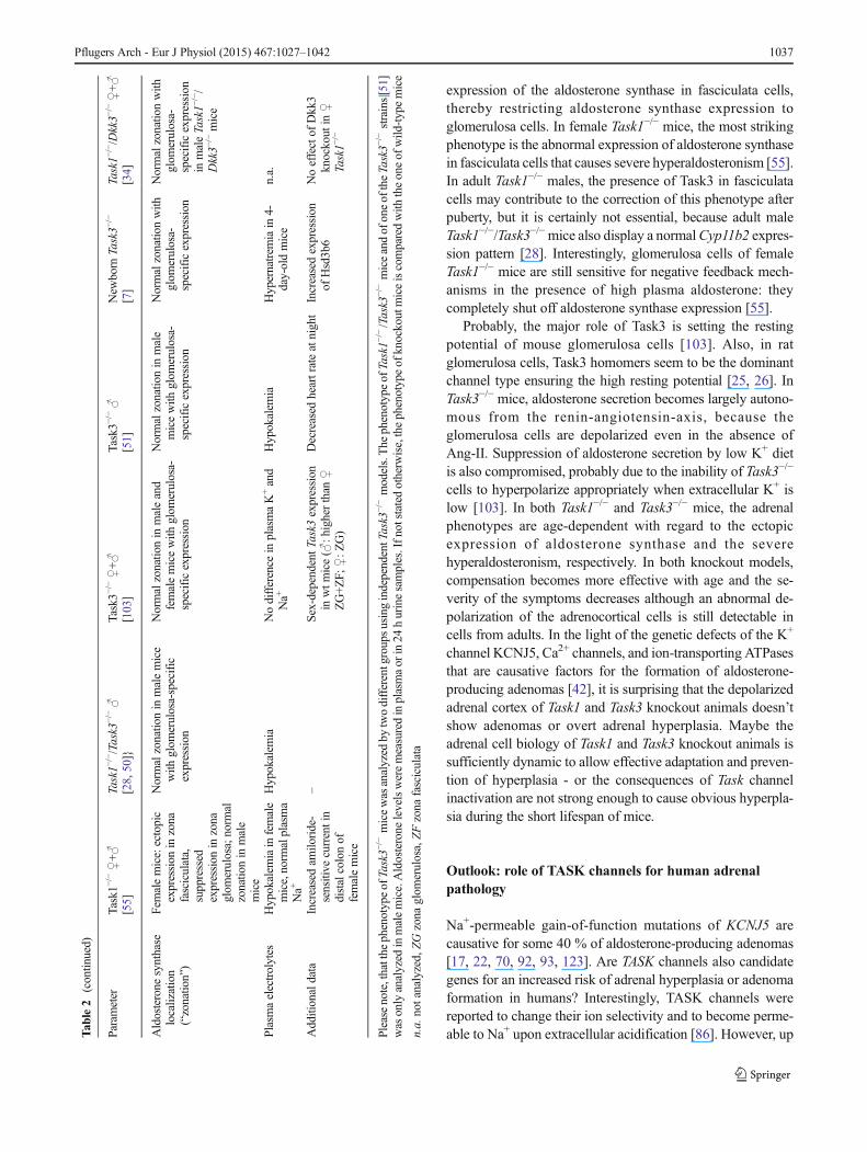
expression of the aldosterone synthase in fasciculata cells,thereby restricting aldosterone synthase expression toglomerulosa cells. In female Task1−/− mice, the most strikingphenotype is the abnormal expression of aldosterone synthasein fasciculata cells that causes severe hyperaldosteronism [55].In adult Task1−/− males, the presence of Task3 in fasciculatacells may contribute to the correction of this phenotype afterpuberty, but it is certainly not essential, because adult maleTask1−/−/Task3−/−mice also display a normalCyp11b2 expres-sion pattern [28]. Interestingly, glomerulosa cells of femaleTask1−/− mice are still sensitive for negative feedback mech-anisms in the presence of high plasma aldosterone: theycompletely shut off aldosterone synthase expression [55].
Probably, the major role of Task3 is setting the restingpotential of mouse glomerulosa cells [103]. Also, in ratglomerulosa cells, Task3 homomers seem to be the dominantchannel type ensuring the high resting potential [25, 26]. InTask3−/− mice, aldosterone secretion becomes largely autono-mous from the renin-angiotensin-axis, because theglomerulosa cells are depolarized even in the absence ofAng-II. Suppression of aldosterone secretion by low K+ dietis also compromised, probably due to the inability of Task3−/−
cells to hyperpolarize appropriately when extracellular K+ islow [103]. In both Task1−/− and Task3−/− mice, the adrenalphenotypes are age-dependent with regard to the ectopicexpression of aldosterone synthase and the severehyperaldosteronism, respectively. In both knockout models,compensation becomes more effective with age and the se-verity of the symptoms decreases although an abnormal de-polarization of the adrenocortical cells is still detectable incells from adults. In the light of the genetic defects of the K+
channel KCNJ5, Ca2+ channels, and ion-transporting ATPasesthat are causative factors for the formation of aldosterone-producing adenomas [42], it is surprising that the depolarizedadrenal cortex of Task1 and Task3 knockout animals doesn’tshow adenomas or overt adrenal hyperplasia. Maybe theadrenal cell biology of Task1 and Task3 knockout animals issufficiently dynamic to allow effective adaptation and preven-tion of hyperplasia - or the consequences of Task channelinactivation are not strong enough to cause obvious hyperpla-sia during the short lifespan of mice.
Outlook: role of TASK channels for human adrenalpathology
Na+-permeable gain-of-function mutations of KCNJ5 arecausative for some 40 % of aldosterone-producing adenomas[17, 22, 70, 92, 93, 123]. Are TASK channels also candidategenes for an increased risk of adrenal hyperplasia or adenomaformation in humans? Interestingly, TASK channels werereported to change their ion selectivity and to become perme-able to Na+ upon extracellular acidification [86]. However, upT
able2
(contin
ued)
Parameter
Task1−
/−♀+♂
[55]
Task1−
/−/Task3
−/−♂
[28,50]}
Task3−
/−♀+♂
[103]
Task3−
/−♂
[51]
New
born
Task3−
/−
[7]
Task1−
/−/Dkk3−
/−♀+♂
[34]
Aldosterone
synthase
localization
(“zonatio
n”)
Femalemice:ectopic
expression
inzona
fasciculata,
suppressed
expression
inzona
glom
erulosa;norm
alzonatio
nin
male
mice
Normalzonatio
nin
malemice
with
glom
erulosa-specific
expression
Normalzonatio
nin
maleand
femalemicewith
glom
erulosa-
specificexpression
Normalzonatio
nin
male
micewith
glom
erulosa-
specificexpression
Normalzonatio
nwith
glom
erulosa-
specificexpression
Normalzonatio
nwith
glom
erulosa-
specificexpression
inmaleTask1−
/−/
Dkk3−
/−mice
Plasmaelectrolytes
Hypokalem
iainfemale
mice,norm
alplasma
Na+
Hypokalem
iaNodifference
inplasmaK+and
Na+
Hypokalem
iaHypernatrem
iain
4-day-oldmice
n.a.
Additionaldata
Increasedam
iloride-
sensitive
currentin
distalcolonof
femalemice
–Sex-dependentTask3
expression
inwtm
ice(♂
:higherthan
♀ZG+ZF;
♀:Z
G)
Decreased
heartrateatnight
Increasedexpression
ofHsd3b6
Noeffectof
Dkk3
knockout
in♀
Task1−
/−
Pleasenote,thatthe
phenotypeofTask3−
/−micewasanalyzed
bytwodifferentgroupsusingindependentTask3
−/−models.The
phenotypeofTask1−
/−/Task3
−/−miceandofoneoftheTask3−
/−strains[51]
was
onlyanalyzed
inmalemice.Aldosterone
levelsweremeasuredinplasmaor
in24
hurinesamples.Ifn
otstated
otherw
ise,thephenotypeof
knockoutmiceiscomparedwith
theoneof
wild
-typemice
n.a.notanalyzed,ZGzona
glom
erulosa,ZFzona
fasciculata
Pflugers Arch - Eur J Physiol (2015) 467:1027–1042 1037

to now, no mutations of TASK channels have been found thatincrease the Na+ permeability and cause aldosterone-producingadenomas. Perhaps, such permeability-changing TASK channelmutations, even if they occur, do not induce proliferation that isa prerequisite for adenoma formation. Loss-of-function muta-tions of TASK3 have been linked to Birk Barel mental retarda-tion dysmorphism syndrome [9, 140]. It is, however, not knownif these patients have an adrenal phenotype. Interestingly,TASK3is a genomically imprinted gene showing paternal silencing [9].Therefore, a mutation in the maternal copy of TASK3 can leadto a disease, while a mutation in the paternal allele will have noeffect. In a genome-wide association study, a correlation ofSNPs in the TASK3 gene with aldosterone levels and the riskfor hypertension was found, but no difference between malesand femaleswas reported [65]. Also forTASK1, a human diseasewas linked to gene mutations. Loss-of-function mutations cancause pulmonary hypertension [85]. In addition, a single nucle-otide polymorphism nearby the TASK1 gene was found to beassociated with blood pressure [45], but no adrenal phenotypewas reported so far. It is possible that mutations of TASK genescan be compensated and do not cause phenotypes strong enoughto provoke monogenetic human adrenal diseases. Further stud-ies are required to investigate a potential role of TASK channelsas modifier genes of adrenocortical disorders.
Disclosure statement The authors have nothing to disclose.
Fund ing The s t udy was suppo r t ed by the Deu t s ch eForschungsgemeinschaft (FOR1086 to R.W. and S.B.), by the FrenchAgence Nationale pour la Recherche (ANR) BeyondTASKs grant (JBand EL), and LabEx Ion Channel Science and Therapeutics grant (ANR-11-LABX-0015-01, to JB).
Open Access This article is distributed under the terms of the CreativeCommons Attribution License which permits any use, distribution, andreproduction in any medium, provided the original author(s) and thesource are credited.
References
1. Akizuki O, Inayoshi A, Kitayama T, Yao K, Shirakura S, Sasaki K,Kusaka H, Matsubara M (2008) Blockade of T-type voltage-depen-dent Ca2+ channels by benidipine, a dihydropyridine calcium chan-nel blocker, inhibits aldosterone production in human adrenocorticalcell line NCI-H295R. Eur J Pharmacol 584:424–434
2. Aller MI, Veale EL, Linden AM, Sandu C, Schwaninger M, Evans LJ,Korpi ER, Mathie A, Wisden W, Brickley SG (2005) Modifying thesubunit composition of TASK channels alters the modulation of a leakconductance in cerebellar granule neurons. J Neurosci 25:11455–11467
3. Al-SalamehA, CohenR, DesailloudR (2014) Overview of the geneticdeterminants of primary aldosteronism. Appl Clin Genet 7:67–79
4. Arrighi I, Bloch-Faure M, Grahammer F, Bleich M, Warth R,Mengual R, Drici MD, Barhanin J, Meneton P (2001) Alteredpotassium balance and aldosterone secretion in a mouse model ofhuman congenital longQTsyndrome. Proc Natl Acad Sci U SA 98:8792–8797
5. Azizan EA, Lam BY, Newhouse SJ, Zhou J, Kuc RE, Clarke J,Happerfield L, Marker A, Hoffman GJ, Brown MJ (2012)Microarray, qPCR, and KCNJ5 sequencing of aldosterone-producing adenomas reveal differences in genotype and phenotypebetween zona glomerulosa- and zona fasciculata-like tumors. J ClinEndocrinol Metab 97:E819–E829
6. Bandulik S, Penton D, Barhanin J, Warth R (2010) TASK1and TASK3 potassium channels: determinants of aldosteronesecretion and adrenocortical zonation. Horm Metab Res 42:450–457
7. Bandulik S, Tauber P, Penton D, Schweda F, Tegtmeier I, Sterner C,Lalli E, Lesage F, Hartmann M, Barhanin J, Warth R (2013) Severehyperaldosteronism in neonatal Task3 potassium channel knockoutmice is associated with activation of the intraadrenal renin-angiotensin system. Endocrinology 154:2712–2722
8. Barbara JG, Takeda K (1995) Voltage-dependent currents and mod-ulation of calcium channel expression in zona fasciculata cells fromrat adrenal gland. J Physiol 488(Pt 3):609–622
9. Barel O, Shalev SA, Ofir R, Cohen A, Zlotogora J, Shorer Z, MazorG, Finer G, Khateeb S, Zilberberg N, Birk OS (2008) Maternallyinherited Birk Barel mental retardation dysmorphism syndromecaused by a mutation in the genomically imprinted potassiumchannel KCNK9. Am J Hum Genet 83:193–199
10. Belloni AS,Malendowicz LK, Rucinski M, Guidolin D, NussdorferGG (2007) Galanin stimulates cortisol secretion from human adre-nocortical cells through the activation of galanin receptor subtype 1coupled to the adenylate cyclase-dependent signaling cascade. Int JMol Med 20:859–864
11. Beuschlein F, Boulkroun S, Osswald A, Wieland T, Nielsen HN,Lichtenauer UD, Penton D, Schack VR, Amar L, Fischer E,WaltherA et al (2013) Somatic mutations in ATP1A1 and ATP2B3 lead toaldosterone-producing adenomas and secondary hypertension. NatGenet 45:440–444
12. Beuschlein F, Fassnacht M, Assie G, Calebiro D, Stratakis CA,Osswald A, Ronchi CL, Wieland T, Sbiera S, Faucz FR, SchaakK et al (2014) Constitutive activation of PKA catalytic subunit inadrenal Cushing’s syndrome. N Engl J Med 370:1019–1028
13. Boulkroun S, Beuschlein F, Rossi GP, Golib-Dzib JF, Fischer E,Amar L, Mulatero P, Samson-Couterie B, Hahner S, QuinklerM, Fallo F et al (2012) Prevalence, clinical, and molecularcorrelates of KCNJ5 mutations in primary aldosteronism.Hypertension 59:592–598
14. Brenner T, O’Shaughnessy KM (2008) Both TASK-3 and TREK-1two-pore loop K channels are expressed in H295R cells and mod-ulate their membrane potential and aldosterone secretion. Am JPhysiol Endocrinol Metab 295:E1480–E1486
15. Cao Y, He M, Gao Z, Peng Y, Li Y, Li L, Zhou W, Li X, Zhong X,Lei Y, Su T et al (2014) Activating hotspot L205R mutation inPRKACA and adrenal Cushing’s syndrome. Science 344:913–917
16. Chai W, Danser AH (2006) Why are mineralocorticoid receptorantagonists cardioprotective? Naunyn Schmiedebergs ArchPharmacol 374:153–162
17. Charmandari E, Sertedaki A, Kino T, Merakou C, Hoffman DA,Hatch MM, Hurt DE, Lin L, Xekouki P, Stratakis CA, Chrousos GP(2012) A novel point mutation in the KCNJ5 gene causing primaryhyperaldosteronism and early-onset autosomal dominant hyperten-sion. J Clin Endocrinol Metab 97:E1532–E1539
18. Chen M, Hornsby PJ (2006) Adenovirus-delivered DKK3/WNT4and steroidogenesis in primary cultures of adrenocortical cells.Horm Metab Res 38:549–555
19. Chen X, Talley EM, Patel N, Gomis A, McIntire WE, Dong B,Viana F, Garrison JC, Bayliss DA (2006) Inhibition of a backgroundpotassium channel by Gq protein alpha-subunits. Proc Natl AcadSci U S A 103:3422–3427
20. Cherradi N, Brandenburger Y, Rossier MF, Vallotton MB, StoccoDM, Capponi AM (1998)Atrial natriuretic peptide inhibits calcium-
1038 Pflugers Arch - Eur J Physiol (2015) 467:1027–1042

induced steroidogenic acute regulatory protein gene transcription inadrenal glomerulosa cells. Mol Endocrinol 12:962–972
21. Cherradi N, Brandenburger Y, Capponi AM (1998) Mitochondrialregulation of mineralocorticoid biosynthesis by calcium and theStAR protein. Eur J Endocrinol 139:249–256
22. ChoiM, Scholl UI, Yue P, Bjorklund P, Zhao B, Nelson-Williams C,Ji W, Cho Y, Patel A, Men CJ, Lolis E et al (2011) K+ channelmutations in adrenal aldosterone-producing adenomas and heredi-tary hypertension. Science 331:768–772
23. Clark BJ, Pezzi V, Stocco DM, Rainey WE (1995) The steroido-genic acute regulatory protein is induced by angiotensin II and K+in H295R adrenocortical cells. Mol Cell Endocrinol 115:215–219
24. Costa JJ, Averil l S, Ching YP, Priest ley JV (1994)Immunocytochemical localization of a growth-associated protein(GAP-43) in rat adrenal gland. Cell Tissue Res 275:555–566
25. Czirjak G, Enyedi P (2002) Formation of functional heterodimersbetween the TASK-1 and TASK-3 two-pore domain potassiumchannel subunits. J Biol Chem 277:5426–5432
26. Czirjak G, Enyedi P (2002) TASK-3 dominates the backgroundpotassium conductance in rat adrenal glomerulosa cells. MolEndocrinol 16:621–629
27. Czirjak G, Fischer T, Spat A, Lesage F, Enyedi P (2000) TASK(TWIK-related acid-sensitive K+ channel) is expressed inglomerulosa cells of rat adrenal cortex and inhibited by angiotensinII. Mol Endocrinol 14:863–874
28. Davies LA, Hu C, Guagliardo NA, Sen N, Chen X, Talley EM,Carey RM, Bayliss DA, Barrett PQ (2008) TASK channel deletionin mice causes primary hyperaldosteronism. Proc Natl Acad Sci U SA 105:2203–2208
29. Decher N, Wemhoner K, Rinne S, Netter MF, Zuzarte M, Aller MI,Kaufmann SG, Li XT, Meuth SG, Daut J, Sachse FB et al (2011)Knock-out of the potassium channel TASK-1 leads to a prolongedQT interval and a disturbed QRS complex. Cell Physiol Biochem28:77–86
30. Doi Y, Atarashi K, Franco-Saenz R, Mulrow PJ (1984) Effect ofchanges in sodium or potassium balance, and nephrectomy, onadrenal renin and aldosterone concentrations. Hypertension 6:I124–I129
31. Donner BC, Schullenberg M, Geduldig N, Huning A, Mersmann J,Zacharowski K, Kovacevic A, Decking U, Aller MI, Schmidt KG(2011) Functional role of TASK-1 in the heart: studies in TASK-1-deficient mice show prolonged cardiac repolarization and reducedheart rate variability. Basic Res Cardiol 106:75–87
32. Du G, Chen X, Todorovic MS, Shu S, Kapur J, Bayliss DA (2011)TASK channel deletion reduces sensitivity to local anesthetic-induced seizures. Anesthesiology 115:1003–1011
33. Duprat F, Lesage F, Fink M, Reyes R, Heurteaux C, Lazdunski M(1997) TASK, a human background K+ channel to sense externalpH variations near physiological pH. EMBO J 16:5464–5471
34. ElWakil A, Bandulik S, GuyN, Bendahhou S, ZennaroMC,NiehrsC, Mari B, Warth R, Barhanin J, Lalli E (2012) Dkk3 is a compo-nent of the genetic circuitry regulating aldosterone biosynthesis inthe adrenal cortex. Hum Mol Genet 21:4922–4929
35. El Wakil A, Lalli E (2011) The Wnt/beta-catenin pathway in adre-nocortical development and cancer. Mol Cell Endocrinol 332:32–37
36. Enyeart JJ, Danthi SJ, Liu H, Enyeart JA (2005) Angiotensin IIinhibits bTREK-1 K+ channels in adrenocortical cells by separateCa2+- and ATP hydrolysis-dependent mechanisms. J Biol Chem280:30814–30828
37. Enyeart JA, Danthi S, Enyeart JJ (2003) Corticotropin induces theexpression of TREK-1 mRNA and K+ current in adrenocorticalcells. Mol Pharmacol 64:132–142
38. Enyeart JA, Danthi SJ, Enyeart JJ (2004) TREK-1 K+ channelscouple angiotensin II receptors to membrane depolarization andaldosterone secretion in bovine adrenal glomerulosa cells. Am JPhysiol Endocrinol Metab 287:E1154–E1165
39. Enyeart JJ, Enyeart JA (2013) Ca2+ and K+ channels of normalhuman adrenal zona fasciculata cells: properties and modulation byACTH and AngII. J Gen Physiol 142:137–155
40. Enyeart JA, Liu H, Enyeart JJ (2009) Curcumin inhibits ACTH- andangiotensin II-stimulated cortisol secretion and Ca(v)3.2 current. JNat Prod 72:1533–1537
41. Enyeart JJ, Xu L, Danthi S, Enyeart JA (2002)AnACTH- and ATP-regulated background K+ channel in adrenocortical cells is TREK-1. J Biol Chem 277:49186–49199
42. Fernandes-Rosa FL, Williams TA, Riester A, Steichen O,Beuschlein F, Boulkroun S, Strom TM, Monticone S, Amar L,Meatchi T, Mantero F et al (2014) Genetic spectrum and clinicalcorrelates of somatic mutations in aldosterone-producing adenoma.Hypertension 64:354–361
43. Freedman BD, Kempna PB, Carlone DL, Shah MS, GuagliardoNA, Barrett PQ, Gomez-Sanchez CE, Majzoub JA, Breault DT(2013) Adrenocortical zonation results from lineage conversion ofdifferentiated zona glomerulosa cells. Dev Cell 26:666–673
44. Gallo-Payet N, Grazzini E, Cote M, Chouinard L, Chorvatova A,Bilodeau L, PayetMD, Guillon G (1996) Role of Ca2+ in the actionof adrenocorticotropin in cultured human adrenal glomerulosa cells.J Clin Invest 98:460–466
45. Ganesh SK, Chasman DI, Larson MG, Guo X, Verwoert G, Bis JC,GuX, Smith AV, YangML, Zhang Y, Ehret G et al (2014) Effects oflong-term averaging of quantitative blood pressure traits on thedetection of genetic associations. Am J Hum Genet 95:49–65
46. Ganz MB, Nee JJ, Isales CM, Barrett PQ (1994) Atrial natriureticpeptide enhances activity of potassium conductance in adrenalglomerulosa cells. Am J Physiol 266:C1357–C1365
47. Goh G, Scholl UI, Healy JM, ChoiM, PrasadML, Nelson-WilliamsC, Kuntsman JW, Korah R, Suttorp AC, Dietrich D, Haase M et al(2014) Recurrent activating mutation in PRKACA in cortisol-producing adrenal tumors. Nat Genet 46:613–617
48. Grimm PR, Irsik DL, Settles DC, Holtzclaw JD, Sansom SC (2009)Hypertension of Kcnmb1−/− is linked to deficient K secretion andaldosteronism. Proc Natl Acad Sci U S A 106:11800–11805
49. Gu J, Wen Y, Mison A, Nadler JL (2003) 12-lipoxygenasepathway increases aldosterone production, 3′,5′-cyclic adeno-sine monophosphate response element-binding protein phos-phorylation, and p38 mitogen-activated protein kinase activa-tion in H295R human adrenocortical cells. Endocrinology144:534–543
50. Guagliardo NA, Yao J, Bayliss DA, Barrett PQ (2011) TASKchannels are not required to mount an aldosterone secretory re-sponse to metabolic acidosis in mice. Mol Cell Endocrinol 336:47–52
51. Guagliardo NA, Yao J, Hu C, Schertz EM, Tyson DA, Carey RM,Bayliss DA, Barrett PQ (2012) TASK-3 channel deletion in micerecapitulates low-renin essential hypertension. Hypertension 59:999–1005
52. Gummow BM, Scheys JO, Cancelli VR, Hammer GD (2006)Reciprocal regulation of a glucocorticoid receptor-steroidogenicfactor-1 transcription complex on the Dax-1 promoter by glucocor-ticoids and adrenocorticotropic hormone in the adrenal cortex. MolEndocrinol 20:2711–2723
53. Gyorke ZS, Sulyok E, Guignard JP (1991) Ammonium chloridemetabolic acidosis and the activity of renin-angiotensin-aldosteronesystem in children. Eur J Pediatr 150:547–549
54. Hattangady NG, Olala LO, Bollag WB, Rainey WE (2012) Acuteand chronic regulation of aldosterone production. Mol CellEndocrinol 350:151–162
55. Heitzmann D, Derand R, Jungbauer S, Bandulik S, Sterner C,Schweda F, El Wakil A, Lalli E, Guy N, Mengual R, Reichold Met al (2008) Invalidation of TASK1 potassium channels disruptsadrenal gland zonation and mineralocorticoid homeostasis. EMBOJ 27:179–187
Pflugers Arch - Eur J Physiol (2015) 467:1027–1042 1039
56. Heurteaux C, Guy N, Laigle C, Blondeau N, Duprat F, Mazzuca M,Lang-Lazdunski L, Widmann C, Zanzouri M, Romey G, LazdunskiM (2004) TREK-1, a K+ channel involved in neuroprotection andgeneral anesthesia. EMBO J 23:2684–2695
57. Hong C, Kim J, Jeon JP, Wie J, Kwak M, Ha K, Kim H, Myeong J,Kim SY, Jeon JH, So I (2012) Gs cascade regulates canonicaltransient receptor potential 5 (TRPC5) through cAMP mediatedintracellular Ca2+ release and ion channel trafficking. BiochemBiophys Res Commun 421:105–111
58. Hu C, Rusin CG, Tan Z, Guagliardo NA, Barrett PQ (2012) Zonaglomerulosa cells of the mouse adrenal cortex are intrinsic electricaloscillators. J Clin Invest 122:2046–2053
59. IbrahimHN,Hostetter TH (2003) Aldosterone in renal disease. CurrOpin Nephrol Hypertens 12:159–164
60. Inagaki N, Gonoi T, Clement JP, Namba N, Inazawa J, Gonzalez G,Aguilar-Bryan L, Seino S, Bryan J (1995) Reconstitution of IKATP:an inward rectifier subunit plus the sulfonylurea receptor. Science270:1166–1170
61. Inagami T,Mizuno K, NaruseM, NakamaruM,Naruse K, HoffmanLH, McKenzie JC (1989) Active and inactive renin in the adrenal.Am J Hypertens 2:311–319
62. Inoue M, Harada K, Matsuoka H, Naakamura J and Warashina A(2012)Mechanisms and roles of muscarinic activation in guinea-pigadrenal medullary cells. Am J Physiol Cell Physiol
63. Inoue M, Harada K, Matsuoka H, Sata T, Warashina A (2008)Inhibition of TASK1-like channels by muscarinic receptor stimula-tion in rat adrenal medullary cells. J Neurochem 106:1804–1814
64. Jones CA, Sigmund CD, McGowan RA, Kane-Haas CM, GrossKW (1990) Expression of murine renin genes during fetal develop-ment. Mol Endocrinol 4:375–383
65. Jung J, Barrett PQ, Eckert GJ, Edenberg HJ, Xuei X, TuW, Pratt JH(2012) Variations in the potassium channel genes KCNK3 andKCNK9 in relation to blood pressure and aldosterone production:an exploratory study. J Clin Endocrinol Metab 97:E2160–E2167
66. Kienitz MC, Mergia E, Pott L (2014) The NCI-H295R cell line asin vitro model of hyperaldosteronism lacks funktional KCNJ5(GIRK4; Kir3.4) channels. Acta Physiol 210:213–213, Abstract
67. Kim AC, Hammer GD (2007) Adrenocortical cells withstem/progenitor cell properties: recent advances. Mol CellEndocrinol 265–266:10–16
68. Kon Y, Hashimoto Y, Kitagawa H, Sugimura M, Murakami K(1990) Renin immunohistochemistry in the adrenal gland of themouse fetus and neonate. Anat Rec 227:124–131
69. Krapivinsky G, Gordon EA, Wickman K, Velimirovic B,Krapivinsky L, Clapham DE (1995) The G-protein-gated atrialK+ channel IKACh is a heteromultimer of two inwardly rectifyingK(+)-channel proteins. Nature 374:135–141
70. Kuppusamy M, Caroccia B, Stindl J, Bandulik S, Lenzini L, GiocoF, Fishman V, Zanotti G, Gomez-Sanchez C, Bader M, Warth Ret al. (2014) A novel KCNJ5-insT149 somatic mutation close to, butoutside, the selectivity filter causes resistant hypertension by loss ofselectivity for potassium. J Clin Endocrinol Metab 20141927:
71. Lee G, Makhanova N, Caron K, Lopez ML, Gomez RA, SmithiesO, Kim HS (2005) Homeostatic responses in the adrenal cortex tothe absence of aldosterone in mice. Endocrinology 146:2650–2656
72. Lenzini L, Caroccia B, Campos AG, Fassina A, Belloni AS, SecciaTM, Kuppusamy M, Ferraro S, Skander G, Bader M, Rainey WEet al (2014) Lower expression of the TWIK-related acid-sensitiveK+ channel 2 (TASK-2) gene is a hallmark of aldosterone-producing adenoma causing human primary aldosteronism. J ClinEndocrinol Metab 99:E674–E682
73. Liang B, Soka M, Christensen AH, Olesen MS, Larsen AP, KnopFK, Wang F, Nielsen JB, Andersen MN, Humphreys D, Mann SAet al (2014) Genetic variation in the two-pore domain potassiumchannel, TASK-1, may contribute to an atrial substrate forarrhythmogenesis. J Mol Cell Cardiol 67:69–76
74. Liao Y, Plummer NW, George MD, Abramowitz J, Zhu MX,Birnbaumer L (2009) A role for Orai in TRPC-mediated Ca2+ entrysuggests that a TRPC: Orai complexmaymediate store and receptoroperated Ca2+ entry. Proc Natl Acad Sci U S A 106:3202–3206
75. Lifton RP, Dluhy RG, Powers M, Rich GM, Gutkin M, Fallo F, GillJR Jr, Feld L, Ganguly A, Laidlaw JC (1992) Hereditary hyperten-sion caused by chimaeric gene duplications and ectopic expressionof aldosterone synthase. Nat Genet 2:66–74
76. Lin D, Sugawara T, Strauss JF III, Clark BJ, Stocco DM, Saenger P,Rogol A, Miller WL (1995) Role of steroidogenic acute regulatoryprotein in adrenal and gonadal steroidogenesis. Science 267:1828–1831
77. Linden AM, Aller MI, Leppa E, Rosenberg PH, Wisden W, KorpiER (2008) K+ channel TASK-1 knockout mice show enhancedsensitivities to ataxic and hypnotic effects of GABA(A) receptorligands. J Pharmacol Exp Ther 327:277–286
78. Linden AM, Aller MI, Leppa E, Vekovischeva O, Aitta-Aho T,Veale EL, Mathie A, Rosenberg P, Wisden W, Korpi ER (2006)The in vivo contributions of TASK-1-containing channels to theactions of inhalation anesthetics, the alpha(2) adrenergic sedativedexmedetomidine, and cannabinoid agonists. J Pharmacol Exp Ther317:615–626
79. Lindner M, Leitner MG, Halaszovich CR, Hammond GR, Oliver D(2011) Probing the regulation of TASK potassium channels by PI4,5P(2) with switchable phosphoinositide phosphatases. J Physiol589:3149–3162
80. Liu H, Enyeart JA, Enyeart JJ (2007) Angiotensin II inhibits nativebTREK-1 K+ channels through a PLC-, kinase C-, and PIP2-independent pathway requiring ATP hydrolysis. Am J PhysiolCell Physiol 293:C682–C695
81. Liu H, Enyeart JA, Enyeart JJ (2008) ACTH inhibits bTREK-1 K+channels through multiple cAMP-dependent signaling pathways. JGen Physiol 132:279–294
82. Liu H, Enyeart JA and Enyeart JJ (2009) N6-substituted cAMPAnalogs Inhibit bTREK-1 K+ channels and stimulate cortisol se-cretion by a PKA-Independent mechanism. Mol Pharmacol
83. Lotshaw DP (2006) Biophysical and pharmacological characteris-tics of native two-pore domain TASK channels in rat adrenalglomerulosa cells. J Membr Biol 210:51–70
84. Lu HK, Fern RJ, Luthin D, Linden J, Liu LP, Cohen CJ, Barrett PQ(1996) Angiotensin II stimulates T-type Ca2+ channel currents viaactivation of a G protein, Gi. Am J Physiol 271:C1340–C1349
85. Ma L, Roman-Campos D, Austin ED, Eyries M, Sampson KS,Soubrier F, Germain M, Tregouet DA, Borczuk A, RosenzweigEB, Girerd B et al (2013) A novel channelopathy in pulmonaryarterial hypertension. N Engl J Med 369:351–361
86. Ma L, Zhang X, Zhou M, Chen H (2012) Acid-sensitive TWIK andTASK two-pore domain potassium channels change ion selectivityand become permeable to sodium in extracellular acidification. JBiol Chem 287:37145–37153
87. Mathie A (2007) Neuronal two-pore-domain potassium channels andtheir regulation by G protein-coupled receptors. J Physiol 578:377–385
88. McEwan PE, Vinson GP, Kenyon CJ (1999) Control of adrenal cellproliferation byAT1 receptors in response to angiotensin II and low-sodium diet. Am J Physiol 276:E303–E309
89. Meimaridou E, Kowalczyk J, Guasti L, Hughes CR, Wagner F,Frommolt P, Nurnberg P, Mann NP, Banerjee R, Saka HN,Chapple JP et al (2012) Mutations in NNT encoding nicotinamidenucleotide transhydrogenase cause familial glucocorticoid deficien-cy. Nat Genet 44:740–742
90. Meuth SG, Aller MI, Munsch T, Schuhmacher T, Seidenbecher T,Meuth P, Kleinschnitz C, Pape HC, Wiendl H, Wisden W, Budde T(2006) The contribution of TWIK-related acid-sensitive K+-con-taining channels to the function of dorsal lateral geniculatethalamocortical relay neurons. Mol Pharmacol 69:1468–1476
1040 Pflugers Arch - Eur J Physiol (2015) 467:1027–1042

91. Mirkovic K, Palmersheim J, Lesage F, Wickman K (2012)Behavioral characterization of mice lacking Trek channels. FrontBehav Neurosci 6:60
92. Monticone S, Hattangady NG, Penton D, Isales C, Edwards MA,Williams TA, Sterner C, Warth R, Mulatero P and Rainey WE(2013) A novel Y152C KCNJ5 mutation responsible for familialhyperaldosteronism type III. J Clin Endocrinol Metab Epub aheadof print:
93. Murthy M, Azizan EA, Brown MJ, O′Shaughnessy KM (2012)Characterization of a novel somatic KCNJ5 mutation delI157 inan aldosterone-producing adenoma. J Hypertens 30:1827–1833
94. Natke E Jr, Kabela E (1979) Electrical responses in cat adrenalcortex: possible relation to aldosterone secretion. Am J Physiol237:E158–E162
95. Nishi H, Arai H, Momiyama T (2013) NCI-H295R, a humanadrenal cortex-derived cell line, expresses purinergic receptorslinked to Ca(2)(+)-mobilization/influx and cortisol secretion.PLoS One 8:e71022
96. Nogueira EF, Gerry D, Mantero F, Mariniello B, RaineyWE (2010)The role of TASK1 in aldosterone production and its expression innormal adrenal and aldosterone-producing adenomas. ClinEndocrinol (Oxf) 73:22–29
97. Nussberger J (2003) Investigatingmineralocorticoid hypertension. JHypertens Suppl 21:S25–S30
98. Oki K, Plonczynski MW, Lam ML, Gomez-Sanchez EP, Gomez-Sanchez CE (2012) The potassium channel, Kir3.4 participates inangiotensin II-stimulated aldosterone production by a human adre-nocortical cell line. Endocrinology 153:4328–4335
99. Oki K, Plonczynski MW, Luis LM, Gomez-Sanchez EP, Gomez-Sanchez CE (2012) Potassium channel mutant KCNJ5 T158Aexpression in HAC-15 cells increases aldosterone synthesis.Endocrinology 153:1774–1782
100. Otis M, Gallo-Payet N (2007) Role of MAPKs in angiotensin II-induced steroidogenesis in rat glomerulosa cells. Mol CellEndocrinol 265–266:126–130
101. Paul M, Poyan MA, Kreutz R (2006) Physiology of local renin-angiotensin systems. Physiol Rev 86:747–803
102. PayetMD,Durroux T, Bilodeau L, GuillonG,Gallo-Payet N (1994)Characterization of K+ and Ca2+ ionic currents in glomerulosa cellsfrom human adrenal glands. Endocrinology 134:2589–2598
103. Penton D, Bandulik S, Schweda F, Haubs S, Tauber P, Reichold M,Cong LD, El WA, Budde T, Lesage F, Lalli E et al (2012) Task3potassium channel gene invalidation causes low renin and salt-sensitive arterial hypertension. Endocrinology 153:4740–4748
104. Peters J (2012) Local renin-angiotensin systems in the adrenalgland. Peptides 34:427–432
105. Peters J, Obermuller N, Woyth A, Peters B, Maser-Gluth C,Kranzlin B, Gretz N (1999) Losartan and angiotensin II inhibitaldosterone production in anephric rats via different actions onthe intraadrenal renin-angiotensin system. Endocrinology 140:675–682
106. Philipp S, Trost C, Warnat J, Rautmann J, Himmerkus N,Schroth G, Kretz O, Nastainczyk W, Cavalie A, Hoth M,Flockerzi V (2000) TRP4 (CCE1) protein is part of nativecalcium release-activated Ca2+-like channels in adrenal cells.J Biol Chem 275:23965–23972
107. Pralong WF, Hunyady L, Varnai P, Wollheim CB, Spat A (1992)Pyridine nucleotide redox state parallels production of aldosteronein potassium-stimulated adrenal glomerulosa cells. Proc Natl AcadSci U S A 89:132–136
108. Quinn SJ, Cornwall MC, Williams GH (1987) Electrical propertiesof isolated rat adrenal glomerulosa and fasciculata cells.Endocrinology 120:903–914
109. Quinn SJ, Williams GH, Tillotson DL (1988) Calcium oscillationsin single adrenal glomerulosa cells stimulated by angiotensin II.Proc Natl Acad Sci U S A 85:5754–5758
110. Radke KJ, Schneider EG, Taylor RE Jr, Kramer RE (1986) Effect ofhydrogen ion concentration on corticosteroid secretion. Am JPhysiol 250:E259–E264
111. Remuzzi G, Perico N, Macia M and Ruggenenti P (2005) The roleof renin-angiotensin-aldosterone system in the progression of chron-ic kidney disease. Kidney Int SupplS57–S65
112. Rohacs T, Bago A, Deak F, Hunyady L, Spat A (1994) CapacitativeCa2+ influx in adrenal glomerulosa cells: possible role in angioten-sin II response. Am J Physiol 267:C1246–C1252
113. Rohacs T, Nagy G, Spat A (1997) Cytoplasmic Ca2+ signalling andreduction of mitochondrial pyridine nucleotides in adrenalglomerulosa cells in response to K+, angiotensin II and vasopressin.Biochem J 322(Pt 3):785–792
114. Romero DG, Yanes LL, de Rodriguez AF, Plonczynski MW, WelshBL, Reckelhoff JF, Gomez-Sanchez EP, Gomez-Sanchez CE (2007)Disabled-2 is expressed in adrenal zona glomerulosa and is involvedin aldosterone secretion. Endocrinology 148:2644–2652
115. Rossig L, Zolyomi A, Catt KJ, Balla T (1996) Regulation ofangiotensin II-stimulated Ca2+ oscillations by Ca2+ influxmechanisms in adrenal glomerulosa cells. J Biol Chem 271:22063–22069
116. Sarzani R, Pietrucci F, Francioni M, Salvi F, Letizia C, D’Erasmo E,Dessi FP, Rappelli A (2006) Expression of potassium channelisoforms mRNA in normal human adrenals and aldosterone-secreting adenomas. J Endocrinol Invest 29:147–153
117. Sato Y, Maekawa S, Ishii R, Sanada M, Morikawa T, Shiraishi Y,Yoshida K, Nagata Y, Sato-Otsubo A, Yoshizato T, Suzuki H et al(2014) Recurrent somatic mutations underlie corticotropin-independent Cushing’s syndrome. Science 344:917–920
118. Sausbier M, Arntz C, Bucurenciu I, Zhao H, Zhou XB, Sausbier U,Feil S, Kamm S, Essin K, Sailer CA, Abdullah U et al (2005)Elevated blood pressure linked to primary hyperaldosteronism andimpaired vasodilation in BK channel-deficient mice. Circulation112:60–68
119. Scandling JD, Ornt DB (1987) Mechanism of potassium depletionduring chronic metabolic acidosis in the rat. Am J Physiol 252:F122–F130
120. Schambelan M, Sebastian A, Katuna BA, Arteaga E (1987)Adrenocortical hormone secretory response to chronic NH4Cl-induced metabolic acidosis. Am J Physiol 252:E454–E460
121. Schiekel J, Lindner M, Hetzel A, Wemhoner K, Renigunta V,Schlichthorl G, Decher N, Oliver D, Daut J (2013) The inhibitionof the potassium channel TASK-1 in rat cardiac muscle byendothelin-1 is mediated by phospholipase C. Cardiovasc Res 97:97–105
122. Scholl UI, Lifton RP (2013) New insights into aldosterone-producing adenomas and hereditary aldosteronism: mutationsin the K+ channel KCNJ5. Curr Opin Nephrol Hypertens 22:141–147
123. Scholl UI, Nelson-Williams C, Yue P, Grekin R, Wyatt RJ, DillonMJ, Couch R, Hammer LK, Harley FL, Farhi A, Wang WH et al(2012) Hypertension with or without adrenal hyperplasia due todifferent inherited mutations in the potassium channel KCNJ5. ProcNatl Acad Sci U S A 109:2533–2538
124. Sculptoreanu A, Scheuer T, Catterall WA (1993) Voltage-dependent potentiation of L-type Ca2+ channels due to phos-phorylation by cAMP-dependent protein kinase. Nature 364:240–243
125. Sewer MB, Waterman MR (2003) ACTH modulation of transcrip-tion factors responsible for steroid hydroxylase gene expression inthe adrenal cortex. Microsc Res Tech 61:300–307
126. Silver S (1940) The Cushing syndrome; neoplasms of the adrenalgland. Bull N YAcad Med 16:368–380
127. Simpson ER, Waterman MR (1988) Regulation of the synthesis ofsteroidogenic enzymes in adrenal cortical cells by ACTH. AnnuRev Physiol 50:427–440
Pflugers Arch - Eur J Physiol (2015) 467:1027–1042 1041
128. Spat A, Fulop L, Szanda G (2012) The role of mitochondrial Ca(2+)and NAD(P)H in the control of aldosterone secretion. Cell Calcium52:64–72
129. Spat A, Hunyady L (2004) Control of aldosterone secretion: amodel for convergence in cellular signaling pathways. PhysiolRev 84:489–539
130. Spyroglou A,Manolopoulou J,Wagner S, BidlingmaierM, ReinckeM, Beuschlein F (2009) Short term regulation of aldosterone secre-tion after stimulation and suppression experiments in mice. J MolEndocrinol 42:407–413
131. Stachowiak A, Nussdorfer GG, Malendowicz LK (1990)Proliferation and distribution of adrenocortical cells in the glandof ACTH- or dexamethasone-treated rats. Histol Histopathol 5:25–29
132. Suwa T, Chen M, Hawks CL, Hornsby PJ (2003) Zonal expressionof dickkopf-3 and components of the Wnt signalling pathways inthe human adrenal cortex. J Endocrinol 178:149–158
133. Tauber P, Penton D, Stindl J, Humberg E, Tegtmeier I, Sterner C,Beuschlein F, Reincke M, Barhanin J, Bandulik S, Warth R (2014)Pharmacology and pathophysiology of mutated KCNJ5 found inadrenal aldosterone-producing adenomas. Endocrinology 155:1353–1362
134. Thomas M, Keramidas M, Monchaux E, Feige JJ (2004) Dualhormonal regulation of endocrine tissue mass and vasculature byadrenocorticotropin in the adrenal cortex. Endocrinology 145:4320–4329
135. Trapp S, Aller MI,WisdenW, Gourine AV (2008) A role for TASK-1 (KCNK3) channels in the chemosensory control of breathing. JNeurosci 28:8844–8850
136. Tremblay E, Payet MD, Gallo-Payet N (1991) Effects of ACTH andangiotensin II on cytosolic calcium in cultured adrenal glomerulosacells. Role of cAMP production in the ACTH effect. Cell Calcium12:655–673
137. Uebele VN, Nuss CE, Renger JJ, Connolly TM (2004) Role ofvoltage-gated calcium channels in potassium-stimulated aldosteronesecretion from rat adrenal zona glomerulosa cells. J SteroidBiochem Mol Biol 92:209–218
138. Vallon V, Grahammer F, Volkl H, Sandu CD, Richter K, RexhepajR, Gerlach U, Rong Q, Pfeifer K, Lang F (2005) KCNQ1-
dependent transport in renal and gastrointestinal epithelia. ProcNatl Acad Sci U S A 102:17864–17869
139. Varnai P, Hunyady L, Balla T (2009) STIM and Orai: the long-awaited constituents of store-operated calcium entry. TrendsPharmacol Sci 30:118–128
140. Veale EL, Hassan M, Walsh Y, Al-Moubarak E, Mathie A (2014)Recovery of current through mutated TASK3 potassium channelsunderlying Birk Barel syndrome. Mol Pharmacol 85:397–407
141. Wang Y, Yamaguchi T, Franco-Saenz R, Mulrow PJ (1992)Regulation of renin gene expression in rat adrenal zona glomerulosacells. Hypertension 20:776–781
142. Westphalen RI, Krivitski M, Amarosa A, Guy N, Hemmings HC Jr(2007) Reduced inhibition of cortical glutamate and GABA releaseby halothane in mice lacking the K+ channel, TREK-1. Br JPharmacol 152:939–945
143. White RE, Lee AB, Shcherbatko AD, Lincoln TM, Schonbrunn A,Armstrong DL (1993) Potassium channel stimulation by natriureticpeptides through cGMP-dependent dephosphorylation. Nature 361:263–266
144. Wiederkehr A, Szanda G, Akhmedov D, Mataki C, Heizmann CW,Schoonjans K, Pozzan T, Spat A, Wollheim CB (2011)Mitochondrial matrix calcium is an activating signal for hormonesecretion. Cell Metab 13:601–611
145. Wilke BU, Lindner M, Greifenberg L, Albus A, Leitner MG,Oliver D (2014) TASK1/3 inhibition by Gq-protein coupledreceptors is mediated by diacylglycerol. Acta Physiol 210:81–81, Abstract
146. Williams DLJ, Katz GM, Roy-Contancin L, Reuben JP (1988)Guanosine 5′-monophosphate modulates gating of high-conductance Ca2+-activated K+ channels in vascular smooth mus-cle cells. Proc Natl Acad Sci U S A 85:9360–9364
147. Xu H, Garver H, Galligan JJ, Fink GD (2011) Large-conductance Ca2+-activated K+ channel beta1-subunit knock-out mice are not hypertensive. Am J Physiol Heart Circ Physiol300:H476–H485
148. Young MJ, Rickard AJ (2012) Mechanisms of mineralocorticoidsalt-induced hypertension and cardiac fibrosis. Mol Cell Endocrinol350:248–255
1042 Pflugers Arch - Eur J Physiol (2015) 467:1027–1042